mastectomy with immediate latissimus dorsi flap ... - thaijo
TRANSCRIPT

The THAI Journal of SURGERY 2020;41(2):52-64.Official Publication of The Royal College of Surgeons of Thailand
52
Mastectomy with Immediate Latissimus Dorsi Flap Reconstruction Chayanoot Rattadilok, MD* Prakasit Chirappapha, MD† Prapa Rattadilok, MD‡ *Department of Surgery, Nopparatrajathanee Hospital, Bangkok, Thailand †Department of Surgery, Faculty of Medicine, Ramathibodi Hospital, Mahidol University Bangkok, Thailand ‡School of Computer Science, University of Nottingham Ningbo China
Abstract Latissimus dorsi (LD) muscle is often used for breast reconstruction in cancer patients, i.e. a breast conserv-ing therapy (BCT) or a mastectomy, often in combination with a breast implant or a tissue expander. The shape of the reconstructed breasts using LD flap with breast implant may look like natural breasts, but may slightly impact shoulder movements, particularly in the extension and the adduction of the shoulder. With the appropriate choice of surgical methods, postoperative complications are minimal and a fast recovery can be expected. In the present article we review the techniques of breast reconstruction with the LD flap in patients with breast cancer undergo-ing mastectomy, using illustrative cases, as well as detailing the indications, technical pitfalls, and avoidance of complications.
Keywords: Extended latissimus dorsi flap, Latissimus dorsi flap with prosthesis, Flap complication, Breast reconstruction, Donor site seroma
Surgical Technique
Correspondence address: Chayanoot Rattadilok, Department of Surgery, Nopparatrajathanee Hospital, Bangkok, Thailand; Telephone: +66 81
899 8003, E-mail: [email protected]
IntroductIon
The latissimus dorsi (LD) muscle is often used for breast reconstruction in cancer patients, i.e. after breast conserving therapy (BCT) or mastectomy. An LD flap includes LD muscle, the fatty layer, as well as the covering skin and is used to replace an area of defective breast tissue. Tansini, et al.1,2 initiated the use of LD flap in breast reconstruction for cancer patients in 1906, where the mastectomy defects were closed us-ing the LD flap. Later, this method of surgery became common practice in Europe. In 1977, Schneider et al.3 discussed the anatomy of the LD flap and the surgical methods for breast reconstruction. In 1979, Maxwell et al.4 developed a new breast reconstruction technique for cancer patients, which
uses the transverse rectus abdominis musculocutaneous flap (TRAM flap), and gained popularity over the use of LD flap. However, when breast implants and tissue expanders became widely available, the use of LD flaps in breast reconstruction for breast cancer once again became popular. This is due to the fact that there are fewer complications associated with using the LD flap and the shape of the reconstructed breasts using LD flap with implant is closer to the natural breasts than those reconstructed using the TRAM flap.
Anatomy The latissimus dorsi muscle is a large triangular muscle of the back. It is approximately 25 x 35 cm2 and covers the postero-inferior half of the trunk.
Received for publication 3 March 2020; Accepted 26 April 2020
Mastectomy with Immediate Latissimus Dorsi Flap ReconstructionVol. 41 No. 2 53
The supero-medial border of the LD muscle is adjacent to the trapezius muscle. The LD muscle rests on the serratus anterior and serratus posterior muscles, a portion of the external oblique and erector spinae muscles. The origin of the LD muscle is from the iliac crest, the posterior layer of the thoracolumbar fascia, the 6th to 12th thoracic spine and 9th to 12th ribs. From there, the LD muscle passes towards the axilla, crosses over by the tip of scapula and inserts at the Intertubercular groove of the humerus. The vascular supply of the LD muscle is by the thoracodorsal trunk, which is composed of the thora-codosal artery, two thoracodorsal veins and the thora-codorsal nerve. The thoracodorsal artery branches from the subscapular artery, and supplies the serratus anterior before supplying the LD muscle at the posterior axilla approximately 10 cm inferior to the LD’s insertion. The thoracodorsal artery is approximately 2.5 mm in diam-eter and 8 cm in length. The vessel’s size and location are predictable, with minimal anatomic variation. The thoracodosal artery may be injured in patients who have undergone any axillary surgery. Parts of the LD muscle also receives its blood supply from the perforator branch of the intercostal muscle. This perforator artery branches from the lumbar artery and supplies the LD muscle at 5 cm from the middle of the back. The LD muscle supports shoulder movements i.e. adduction, extension and internal rotation, which is mainly performed by the rotator cuff i.e. the supraspina-tor, infraspinator, subscapularis and teres minor muscles. Therefore, a breast reconstruction using LD flap may have a slight impact on shoulder movements5,6, particu-larly in the extension and the adduction of the shoulder.Preoperative Planning To ensure an optimal surgical plan, relevant in-formation should be obtained from a careful physical examination and history taking. The information should include the stage of the cancer, the cancer treatment plan, the breast’s shape and size, the patient’s needs and the proficiency of the surgical team. Breast reconstruction can be divided into two main types: immediate recon-struction and delayed breast reconstruction. The main advantage of immediate reconstruction is that the surgi-cal incisions for both the breast cancer surgery and the reconstruction can be pre-planned to reduce scarring and provide optimal appearance of the reconstructed breast. This can also minimize the physiological impact to the patient. A delayed breast reconstruction is usually carried
out following a complete pathological examination and often a long time after the breast cancer surgery. Thus, necessary adjuvant treatments including chemotherapy and radiation, are known and therefore the reconstruction can be planned accordingly. Additionally, the chance of infection and hyperemic tissue necrosis is lower when compare to an immediate reconstruction. Prior to any breast reconstruction using the LD flap, the following points should be considered. 1. The amount of the skin and the breast tissue that will be removed. 2. The amount of the skin, the tissue and the LD muscle that will be used in the reconstruction. This can be evaluated by examining the thickness of the adipose layer i.e. the amount of fat in the supra-iliac area or the so-called “love handle”. This evaluation can help in estimating the size of the reconstructed breast (Figure 1). 3. The LD’s muscle function especially in patients who had undergone axillary surgery, and an angiography to confirm if the thoracodorsal artery has been injured. A breast implant or a tissue expander can be used in patients with small-volume LD flap, by placing the LD flap on top of a breast implant or a tissue expander. In patients with large breasts, breast implants may be used in combination with an extended LD flap. The LD flap can also be used in breast reconstruction for partial mastectomy.
Surgical Indications and Contraindications Surgical indications 1. Patients who are not suitable for a breast reconstruction using TRAM flap7 This includes patients with insufficient skin and tissue at the lower abdomen, e.g. abdominoplasty patients or high-risk patients including those who smoke, has diabetes, or are obese. 2. Patients who have previously received an irradiation during BCT This includes patients with a deformed breast, an insufficient blood supply of the skin, skin discoloration, or with infection. The LD flap has a good blood supply and would not have been damaged by the radiation. 3. Patients who have undergone partial mas-tectomy8
Partial mastectomy can sometimes create an undesirable defect. The LD flap can be used to rectify this.

Rattadilok C, et al. Thai J Surg Apr. - Jun. 202054
4. Patients with thin skin tissue. The LD flap can be used to cover the breast implant or tissue expander9,10. The skin on top of the LD flap can address this issue of thin skin overlying the chest wall. 5. Patients who have undergone a mastopexy or a breast reduction following a skin-sparing mastectomy In this group of patients, the skin is likely to have insufficient blood supply and therefore increases the likelihood of breast implant extrusion. The LD flap can be used on top of the breast implant to prevent this complication. 6. Patients who have undergone augmented breast surgery following a skin-sparing mastectomy The LD flap can be used to adjust the similarity of the appearance of both breasts11. 7. Patients with large or ptotic breasts The LD flap can be used with breast implant to
correct the differences in size or appearances of the 2 breasts, during both breast augmentation and the breast reduction. 8. Patients who have undergone a prophylactic mastectomy The LD flap can be used for aesthetic purposes in young patients who have undergone prophylactic mastectomy.
Surgical Contraindications 1. Patients who have undergone a posterolateral thoracotomy, where the LD muscle has already been divided. 2. Patients with atrophied LD muscle caused by injured thoracodorsal nerve following an axillary lymph node surgery. An atrophied LD muscle may affect the amount of tissue that is available for breast reconstruc-tion.
Figure 1 The assessment of the size of the LD flap prior to surgery

Mastectomy with Immediate Latissimus Dorsi Flap ReconstructionVol. 41 No. 2 55
SurgIcal technIqueS
Before surgery, patients should be carefully evalu-ated and the surgical and incision planning should be drawn on the patient in the standing position. The surgical plan includes a consideration regarding the appearance of the non-effected breast i.e. the size, the position, the position of the nipple, the size of the nipple, the areola, the inframammary fold, and the mid-sternal line; the location of the biopsy; the surgical incisions for both the breast surgery and the axillary lymph nodes surgery; the size of the nipples and the size of the areola that will be surgically removed; the surgical incision of the LD flap; the location and the size of the LD flap that will be used in breast reconstruction; the location of the tip of the scapula, the iliac crest, the posterior axillary line and spinal axis (Figures 2A-B). The alignments of the surgical incision to harvest the LD flap includes transverse, vertical and oblique. In a report of 250 patients12, 54% of the surgical incisions was a low transverse incision below level the inframam-mary fold, 21% was a middle transverse incision at level the inframammary fold, 13% was an upper transverse incision above level the inframammary fold, 9% was
an oblique incision, and 3% was a vertical incision. The decision on how to align the surgical incision depends on age, body mass index, patient’s clothing, patient’s needs, skin tension line, and the location of the blood vessels. In addition to this, the amount of skin tissue that is required for breast reconstruction should also be considered, e.g. an oblique incision may be used for a large reconstruction, or a transverse incision may be used for aesthetic purpose. Different alignments of the surgical incision have different advantages and disadvantages. In the case of an upper transverse incision above level the inframammary fold, the removal of the tissue over the tip of the scapula is simple, but a deformation of the shape of the back may result from too much of the tissue being removed. In the case of a middle transverse incision at level the inframa-mmary fold, the surgical scar will be hidden under the bra. In the case of a low transverse incision below level the inframammary fold, although the surgical scar will be hidden inside the bra but the harvesting of LD flap is more difficult than in other alignments. The oblique and vertical incisions are easier for harvesting the LD flap but there will be more surgical scar-tension.
2A 2B Figure 2 A: The surgical plan on the chest area B: The surgical plan on the back area

Rattadilok C, et al. Thai J Surg Apr. - Jun. 202056
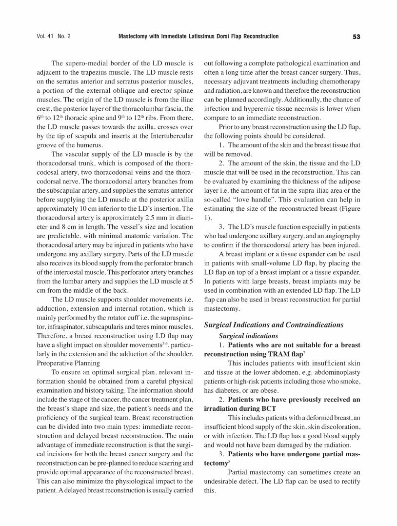
Details of the surgical procedure (Figures 3A-3L, 4A-4F) 1. Lay the patient on the lateral decubitus position and incise according to the surgical plan (Figure 3A-3C). 2. Dissect the fatty tissue above the LD muscle, by separating between the deep fat layer and superficial fat layer at the superficial fascia, from tip of the scapula to the pelvis iliac crest (Figure 3D-3F). 3. Separate the LD muscle from the serratus anterior muscle, the lumbosacral fascia and the para-spinous muscle fascia. This area has a large blood vessel and therefore extra care is required to prevent bleeding and the bleeding under the surgical incision (Figure 3G-3H). 4. Dissect the LD muscle tissue upward and sepa-rate the LD muscle from the trapezius muscle (Figure 3I). 5. Continue to separate the LD muscle from the teres major muscle. The separation continues and eventu-ally stops at the axilla i.e. close to the humerus. 6. Identify the thoracodorsal artery and veins that supply the blood to the LD muscle. Thin the LD flap under the axilla, to reduce the chance of bulging in the axilla following the surgery. Extra care should be given to avoid causing any injury to the blood vessels (Figure 3J). 7. Create a continuous cavity on the side of the body (Figure 3K). 8. Insert a drainage tube and move the harvested LD flap for the breast reconstruction through the created continuous cavity (Figure 3L). 9. Suture the superficial fascia by using vicryl 2-0, and suture the skin by using vicryl 3-0. 10. Lay the patient on the supine position. 11. Thin the LD flap under the axilla further to reduce the chance of bulging in the axilla following the surgery (Figure 4A) as the supine position allows further assessment once the patient is facing upwards. 12. Place the harvested LD flap in the desired posi-tion (Figure 4B). 13. For skin-sparing mastectomy, de-epithelize the skin on the LD flap according to the size of the re-moved nipple-areola. For nipple-sparing mastectomy, de-epithelize all of the skin on top of the LD flap. In the case of breast reconstruction using breast implant or tissue expander with LD flap, place the harvested LD flap over the breast implant or tissue expander, or on top of the pectoralis muscle if the breast implant or tissue
expander is placed below the pectoralis muscle. 14. Close the connecting cavity at the anterior axil-lary line to reduce the chance of a dislocated LD flap or the breast implant (Figure 4C). 15. In the case of breast surgery without the axillary lymph nodes surgery, inserts a silicone-drainage through the breast cavity. In the case of breast and axillary lymph nodes surgery, insert 2 silicone drainage tubes. 16. Position the patient in the sitting pose and adjust the reconstructed breast to ensure a similar appearance and size to the opposite side of the breast (Figure 4D-4E). 17. Suture the breast skin using Vicryl 4-0 (Figure 4F). An extended LD flap can be used for a full re-construction if the patient does not want to use breast implants. The volume of the extended LD flap is typi- cally 300-400 ml, but can atrophy up to 20-25% over-time. This needs to be taken int account, so that the resulting long-term size will be appropriate. According to Kim H. et al.23, more complications, such as donor site seroma and flap necrosis, can be expected when extended LD flap is used when compared with the original method. A muscle sparing Latissimus dorsi flap (MSLD flap) removes only some part of the LD muscle, which has its blood supply through the descending branch of the thoracodorsal artery13. On average, the location of the artery is about 5.1 cm from the armpit (2.1 to 7.5 cm), or on average about 2.2 cm from the side of the LD (1.3 to 3.1 cm) using only a part of the LD, e.g., 3 to 4 cm in width14. According to Saint-Cyr M. et al.13, with 20 patients undergoing breast reconstruction using the MSLD flap, there were no cases of donor site seroma.
Patient care following surgery Following the surgery, a few important points need to be considered. 1. Wound dressing changes at the breast and the back can be done. 2. Avoid direct pressure on the blood vessels at the axilla. 3. Patients should be hospitalized on average for about 3 days. 4. Patients could return to work within 3 to 6 weeks.
Mastectomy with Immediate Latissimus Dorsi Flap ReconstructionVol. 41 No. 2 57
Figure 3 A: The patient’s preoperative position i.e. on the lateral decubitus position B: The surgical plan for the transverse incision C: The surgical incisions according to the surgical plan D: The separation of the deep fat and superficial fat at the superficial fascia E: The dissection of the fat tissue along the superficial fascia up to the tip of the scapula F: The dissection of the fat
tissue down to the iliac crest
Figure 3A Figure 3B
Figure 3C Figure 3D
Figure 3E Figure 3F

Rattadilok C, et al. Thai J Surg Apr. - Jun. 202058
Figure 3 G: The separation of the LD muscle from the serratus anterior muscle at the side of the body H: The separation of the LD muscle down to the iliac crest I: The separation of the LD muscle from the trapezius muscle J: The thinning of the harvested LD flap to reduce the bulging of the axilla by avoiding the injury to the thoracodorsal
artery K: The harvested LD flap and the connecting cavity on the side of the body L: The connecting cavity on the side of
the body, the inserted silicone drainage, and the moving of the harvested LD flap through the cavity
Figure 3G Figure 3H
Figure 3I Figure 3J
Figure 3K Figure 3L
Mastectomy with Immediate Latissimus Dorsi Flap ReconstructionVol. 41 No. 2 59
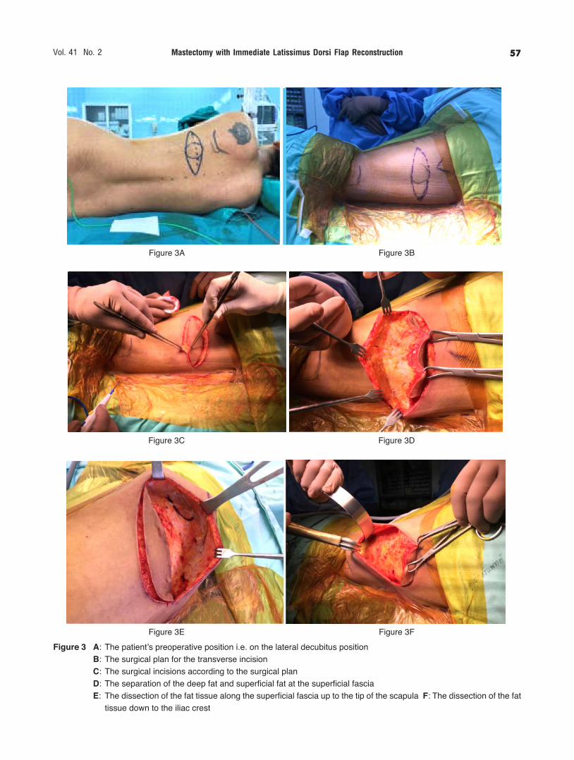
Figure 4 A: The thinning of the harvested LD flap to reduce the bulging of axilla B: The harvested LD flap in the desired position C: The suturing of the side of the body in order to prevent the dislocation of the harvested LD flap and breast implant
to the side of the body D: The harvested LD flap over the breast implant E: The suturing of the harvested LD flap and the chest wall tissue according to the surgical plan F: The breasts immediately following the breast reconstruction (on the left side) using LD flap with a breast implant
Figure 4A Figure 4B
Figure 4C Figure 4D
Figure 4E Figure 4F

Rattadilok C, et al. Thai J Surg Apr. - Jun. 202060
Figure 5 A: The breast of a breast cancer patient following a left skin sparing mastectomy and a breast reconstruction using an extended LD flap
B: The surgical scar on the back of the patient in Figure 5A; following a breast reconstruction using the LD flap i.e. a transverse incision
C: The breast of a breast cancer patient following a right partial mastectomy and a breast reconstruction using the LD flap
D: The surgical scar on the back of the patient in Figure 5C; following a breast reconstruction using the LD flap i.e. a transverse incision
E: The breast of a Granulomatous Mastitis patient following a right partial mastectomy and a breast reconstruction using the LD flap
F: The surgical scar on the back of the patient in Figure 5E; following a breast reconstruction using the LD flap i.e. an oblique incision
Figure 5A Figure 5B
Figure 5C Figure 5D
Figure 5E Figure 5F

Mastectomy with Immediate Latissimus Dorsi Flap ReconstructionVol. 41 No. 2 61
Figure 5 G: The breasts of a breast cancer patient following a right skin sparing mastectomy and a breast reconstruction using the LD flap with a breast implant
H: The surgical scar on the back of the patient in Figure 5G; following a breast reconstruction using the LD flap i.e. a transverse incision
I: The breasts of a breast cancer patient following a left skin sparing mastectomy and a breast reconstruction using the LD flap with a breast implant
J: The breasts of a breast cancer patient following a right skin sparing mastectomy and a breast reconstruction using the LD flap with a breast implant
Figure 5G Figure 5H
Figure 5I Figure 5J
Rattadilok C, et al. Thai J Surg Apr. - Jun. 202062
5. The drainage tubes can be removed when drain-age content is less than 30 ml per day. 6. Normal exercise of the arm and shoulder can be performed to reduce post-operative frozen shoulder or limitation of shoulder movements. Figures 5A-5J show results of breast reconstruc-tion using LD flaps in patients with various breast diseases.
Postoperative Complications Complications following a breast reconstruction using the LD flap include LD flap complication and the donor site complication. According to Chang DW et al.15, The LD flap complications are found approximately 28%. The donor site complications are found approxi-mately 39%. Commonly occurred complications can be summarized as follows. 1. Donor site seroma Donor site seroma is the most common complica-tion following LD flap reconstruction15-21. This complica-tion is more common after the use of the extended LD flap. Research shows that donor site seroma is found in approximately 20 to 34% of the patients15,17-18,22-23, although some studies show donor site seroma in ap-proximately 62% of patients with extended LD flap23
and 69% in patients with non-extended LD flap24. Risk factors for donor site seroma included a high body mass index (BMI > 23 kg/m2), LD flap heavier than 450 grams, and older patients (age > 45yrs)17, 24. In our recent study25 regarding donor site seroma in 58 patients, we found no significant difference in the incidence of donor site seroma between two groups of patients undergoing reconstruction with non-extend-ed LD or extended LD flaps. Significant factors asso-ciated with this complication included the amount of drainage during the 1st to 3rd day after surgery exceeding 340 ml, and the duration of drainage tube placement of less than 17 days. Age and body mass index were not significantly associated with donor site seroma. Additionally, the duration of donor site seroma was significantly associated with operative time longer than 250 minutes, nipple sparing mastectomy, and the use of chemotherapy. 2. LD Flap necrosis LD flap necrosis is rare. LD flap necrosis can result from direct thoracodorsal artery injury, twisted
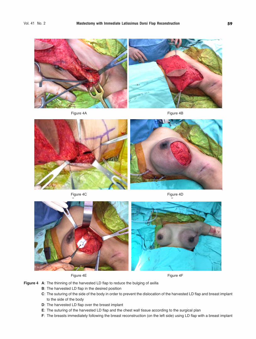
Figure 6 An example of a donor skin flap necrosis
thoracodorsal artery, or blood clot in this vessel from excessive skin tension. Partial necrosis of the LD flap can be found up to 7% of patients15-16,22,24, particularly when the extended LD flap is used. 3. Donor skin flap necrosis Donor skin flap necrosis is common when a large piece of skin is removed from the donor area. This complication is caused by skin tension and the damage to the blood supply of the skin (Figure 6). 4. Limitation of shoulder movement Limitation of shoulder movement is uncommon. Kim H. et al.23 found that shoulder movement is reduced by 76% and 25% in patients having undergone a breast reconstruction using the extended LD flap and an MSLD flap with breast implants respectively. 5. Dislocation of the LD flap or breast implant The LD flap or the breast implant can be dislocated towards the axilla or to the lateral side of the body. This complication can be reduced if a suture is placed so as to close off the cavity at the side of the body. 6. Infection Surgical site Infection is found in 3 to 4%15,18,22-23 and 1 to 6%18,22-23 of patients who has undergone a breast

Mastectomy with Immediate Latissimus Dorsi Flap ReconstructionVol. 41 No. 2 63
REFERENCES
1. Maxwell GP. Iginio Tansini and the origin of the latissimus dorsi musculocutaneous flap. Plast Reconstr Surg 1980;65:686-92.
2. Lassen M, Krag C, Nielsen IM. The latissimus dorsi flap: an overview. Scand J Plast Surg 1985;19:41-51.
3. Schneider WJ, Hill HL, Brown RG. Lastissimus dorsi myocutane-ous flap for breast reconstruction. Br J Plast Surg 1977;30:277- 81.
4. Maxwell GP, McGibbon BM, Hoopes JE. Vascular consider-ation in the use of a latissimus dorsi myocutaneous flap after a mastectomy with an axillary dissection. Plast Reconstr Surg 1979;64:771-80.
5. Laitung JKG, Peck F. Shoulder function following the loss of the latissimus dorsi muscle. Br J Plast Surg 1985;38:375-9.
6. Russell RC, Pribaz J, Zook EG, et al. Functional evaluation of latissimus dorsi donor site. Plast Reconstr Surg 1986;78:336-44.
7. Chang DW, Kroll SS, Dackiw A, et al. Reconstruction manage-ment of contralateral breast cancer in patients who previously underwent unilateral breast reconstruction. Plast Reconstr Surg 2001;108:352-8.
8. Clough KB, Kroll SS, Audretsch W. An approach to the repair of partial mastectomy defects. Plast Reconstr Surg 1999;104:409-20.
9. Lejour M, Alemanno P, De mey A, et al. Analysis of fifty-six breast reconstruction using the latissimus dorsi flap. Ann Chir Plast Esthet 1985;30:7-16.
10. Pendergrast WJ, Bostwick JB III, Jurkiewicz MJ. The subcu-taneous mastectomy cripple: surgical rehabilitation with the latissimus dorsi flap. Plast Reconstr Surg 1980;66:554-9.
11. Spear SL, Slack C, Howard MA. Postmastectomy reconstruc- tion of the previously augmented breast: diagnosis, staging, methodology, and outcome. Plast Reconstr Surg 2001;107: 1167-76.
12. Bailey S, Saint-Cyr M, Zhang K, et al. Breast reconstruction with latissimus dorsi flap: Woman’s preference for scar location. Plast Reconstr Surg 2010;126:358-65.
13. Saint-Cyr M, Nagarkar P, Schaverien M, et al. The pedicled descending branch muscle-sparing latissimus dorsi flap for breast reconstruction. Plast Reconstr Surg 2009;123:13-24.
14. Schwabegger AH, Harpf C, Rainer C. Muscle-sparing latissimus dorsi myocutaneous flap with maintenance of muscle innerva-tion, function, and aesthetic appearance of the donor site. Plast Reconstr Surg 2003;111:1407-11.
15. Chang DW, Youssef A, Cha S, et al. Autologous breast recon-struction with the extended latissimus dorsi flap. Plast Reconstr Surg 2002;110:751-9; discussion 60-1.
16. Lee JW, Chang TW. Extended latissimus dorsi musculocutaneous flap for breast reconstruction: experience in Oriental patients. Br J Plast Surg 1999;52:365-72.
17. Tomita K, Yano K, Masuoka T, et al. Postoperative seroma formation in breast reconstruction with latissimus dorsi flaps: a retrospective study of 174 consecutive cases. Ann Plast Surg 2007;59:149-51.
18. Pinsolle V, Grinfeder C, Mathoulin-Pelissier S, et al. Complica-tions analysis of 266 immediate breast reconstructions. JPRAS. 2006;59:1017-24.
19. Clough KB, Louis-Sylvestre C, Fitoussi A, et al. Donor site sequelae after autologous breast reconstruction with an extended latissimus dorsi flap. Plast Reconstr Surg 2002;109:1904-11.
20. Mendelson BC. Latissimus dorsi breast reconstruction--refine-ment and results. Br J Surg 1983;70:145-9.
21. Moore TS, Farrell LD. Latissimus dorsi myocutaneous flap for breast reconstruction: long-term results. Plast Reconstr Surg 1992;89:666-72; discussion 73-4.
22. Venus MR, Prinsloo DJ. Immediate breast reconstruction with latissimus dorsi flap and implant: audit of outcomes and patient satisfaction survey. JPRAS 2010;63:101-5.
reconstruction using the LD flap and those who has undergone a breast reconstruction using LD flap with breast implant, respectively. 7. Hematoma Hematoma is found up to 3% of patients who has undergone a breast reconstruction using LD flap15,18,22-24. 8. Capsular contracture Capsular contracture is found in 6 to 7% of patients who has undergone a breast reconstruction using LD flap with breast implant18,22.
concluSIonS
Breast reconstruction using LD flap is widely used following breast conserving surgery, mastectomy, in combination with a breast implant or a tissue expander in patients with breast cancer. Appropriate choice of surgi-cal procedures will contribute towards the similarity of the size and the appearance between the reconstructed and the opposite breasts. In addition to this, breast re-construction using the LD flap is associated with fewer postoperative complications and faster recovery.
acknowledgmentS We wish to acknowledge Dr. Christopher Roadk-night for English revision of the text.
FInancIal dIScloSureS
None of the authors has a financial interest in any of the products, devices, or drugs mentioned in this manuscript. The Article Processing Charge was paid for by the authors.
conFlIct oF IntereSt
All authors have no any financial and personal re-lationships with other people or organization that could inappropriately influence (bias) their work.

Rattadilok C, et al. Thai J Surg Apr. - Jun. 202064
23. Kim H, Wiraatmadja ES, Lim SY, et al. Comparison of morbidity of donor site following pedicled muscle-sparing latissimus dorsi flap versus extended latissimus dorsi flap breast reconstruction. JPRAS 2013;66:640-6.
24. Jeon BJ, Lee TS, Lim SY, et al. Risk factors for donor-site seroma formation after immediate breast reconstruction with
the extended latissimus dorsi flap: a statistical analysis of 120 consecutive cases. Ann Plast Surg 2012;69:145-7.
25. Chirappapha P, Rattadilok C, Lertsithichai P, et al. Predictor of donor site seroma in latissimus dorsi flap. J Med Assoc Thai 2017;100(Suppl 9):S103-S114.