does the latissimus dorsi tendon transfer for massive rotator cuff tears remain active...
TRANSCRIPT

The study (P07.1
(Leiden Univers
*Reprint req
box 9600, 2300
E-mail addre
J Shoulder Elbow Surg (2014) 23, 553-560
1058-2746/$ - s
http://dx.doi.org
www.elsevier.com/locate/ymse
Does the latissimus dorsi tendon transfer for massiverotator cuff tears remain active postoperatively andrestore active external rotation?
Jan Ferdinand Henseler, MDa,b,*, Jochem Nagels, MDa,Rob G.H.H. Nelissen, MD, PhDa, Jurriaan H. de Groot, PhDb,c
aDepartment of Orthopaedics, Leiden University Medical Center, Leiden, The NetherlandsbLaboratory for Kinematics and Neuromechanics, Leiden University Medical Center, Leiden, The NetherlandscDepartment of Rehabilitation, Leiden University Medical Center, Leiden, The Netherlands
Hypothesis: The purpose of this study is to evaluate the muscle activity with surface electromyography(EMG) and the clinical outcome of the latissimus dorsi transfer. It remains unclear whether the clinicalresults of the latissimus dorsi transfer for massive posterosuperior rotator cuff tears are achieved eitherby active muscle contractions or by a passive tenodesis effect of the transfer.Methods: Eight patients were evaluated preoperatively and at 1 year (SD, 0.1) after the latissimus dorsitransfer. Clinical evaluation of outcomes included active range of motion, Constant score, and visualanalog scale (VAS) for pain and activities of daily living (ADL). Muscle activity was recorded withEMG during directional isometric abduction and adduction tasks.Results: The external rotation in adduction improved from 23� to 51� (P ¼ .03). The external rotation inabduction improved from 10� to 70� (P ¼ .02). The mean Constant score improved from 39 to 62 postop-eratively (P ¼ .01). The VAS for pain at rest improved from 3.3 preoperatively to 0.1 (P ¼ .02). The VASfor ADL improved from 4.9 to 2.3 (P ¼ .05). The transferred latissimus dorsi remained active in all cases,as reflected by increased latissimus dorsi EMG activity during abduction tasks. In addition, the latissimusdorsi EMG activity shifted from preoperative antagonistic co-activation in adduction to synergistic activa-tion in abduction.Conclusion: The latissimus dorsi has synergistic muscle activity after transfer. Apart from a tenodesiseffect, directional muscle activity seems relevant for improved clinical outcome and pain relief. A specificgain was observed for external rotation in elevated arm positions, a motion essential for ADL tasks.Level of evidence: Level IV, Case Series, Treatment Study.� 2014 Journal of Shoulder and Elbow Surgery Board of Trustees.
Keywords: Rotator cuff tear; irreparable; tendon transfer; transplantation; latissimus dorsi; electromyography
23) was approved by the institution’s ethical review board
ity Medical Center Commisie Medische Ethiek).
uests: Jan Ferdinand Henseler, MD, Postzone J-11-R, PO
RC Leiden, The Netherlands.
ss: [email protected] (J.F. Henseler).
ee front matter � 2014 Journal of Shoulder and Elbow Surgery
/10.1016/j.jse.2013.07.055
Massive rotator cuff (RC) tears restrict patients in theirdaily activities because of severe pain and limitations inarm elevation and external rotation. Repair of poster-osuperior RC tears affecting the supraspinatus and infra-spinatus yields a poor outcome compared with isolated
Board of Trustees.
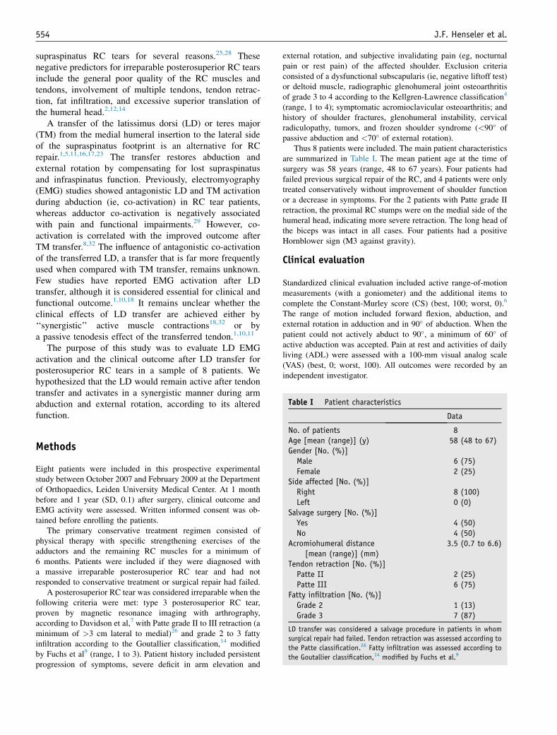
Table I Patient characteristics
Data
554 J.F. Henseler et al.
supraspinatus RC tears for several reasons.25,28 Thesenegative predictors for irreparable posterosuperior RC tearsinclude the general poor quality of the RC muscles andtendons, involvement of multiple tendons, tendon retrac-tion, fat infiltration, and excessive superior translation ofthe humeral head.2,12,14
A transfer of the latissimus dorsi (LD) or teres major(TM) from the medial humeral insertion to the lateral sideof the supraspinatus footprint is an alternative for RCrepair.1,5,11,16,17,23 The transfer restores abduction andexternal rotation by compensating for lost supraspinatusand infraspinatus function. Previously, electromyography(EMG) studies showed antagonistic LD and TM activationduring abduction (ie, co-activation) in RC tear patients,whereas adductor co-activation is negatively associatedwith pain and functional impairments.29 However, co-activation is correlated with the improved outcome afterTM transfer.8,32 The influence of antagonistic co-activationof the transferred LD, a transfer that is far more frequentlyused when compared with TM transfer, remains unknown.Few studies have reported EMG activation after LDtransfer, although it is considered essential for clinical andfunctional outcome.1,10,18 It remains unclear whether theclinical effects of LD transfer are achieved either by‘‘synergistic’’ active muscle contractions18,32 or bya passive tenodesis effect of the transferred tendon.1,10,11
The purpose of this study was to evaluate LD EMGactivation and the clinical outcome after LD transfer forposterosuperior RC tears in a sample of 8 patients. Wehypothesized that the LD would remain active after tendontransfer and activates in a synergistic manner during armabduction and external rotation, according to its alteredfunction.
No. of patients 8Age [mean (range)] (y) 58 (48 to 67)Gender [No. (%)]Male 6 (75)Female 2 (25)
Side affected [No. (%)]Right 8 (100)Left 0 (0)
Salvage surgery [No. (%)]Yes 4 (50)No 4 (50)
Acromiohumeral distance[mean (range)] (mm)
3.5 (0.7 to 6.6)
Tendon retraction [No. (%)]Patte II 2 (25)Patte III 6 (75)
Fatty infiltration [No. (%)]Grade 2 1 (13)Grade 3 7 (87)
LD transfer was considered a salvage procedure in patients in whom
surgical repair had failed. Tendon retraction was assessed according to
the Patte classification.26 Fatty infiltration was assessed according to
the Goutallier classification,14 modified by Fuchs et al.9
Methods
Eight patients were included in this prospective experimentalstudy between October 2007 and February 2009 at the Departmentof Orthopaedics, Leiden University Medical Center. At 1 monthbefore and 1 year (SD, 0.1) after surgery, clinical outcome andEMG activity were assessed. Written informed consent was ob-tained before enrolling the patients.
The primary conservative treatment regimen consisted ofphysical therapy with specific strengthening exercises of theadductors and the remaining RC muscles for a minimum of6 months. Patients were included if they were diagnosed witha massive irreparable posterosuperior RC tear and had notresponded to conservative treatment or surgical repair had failed.
A posterosuperior RC tear was considered irreparable when thefollowing criteria were met: type 3 posterosuperior RC tear,proven by magnetic resonance imaging with arthrography,according to Davidson et al,7 with Patte grade II to III retraction (aminimum of >3 cm lateral to medial)26 and grade 2 to 3 fattyinfiltration according to the Goutallier classification,14 modifiedby Fuchs et al9 (range, 1 to 3). Patient history included persistentprogression of symptoms, severe deficit in arm elevation and
external rotation, and subjective invalidating pain (eg, nocturnalpain or rest pain) of the affected shoulder. Exclusion criteriaconsisted of a dysfunctional subscapularis (ie, negative liftoff test)or deltoid muscle, radiographic glenohumeral joint osteoarthritisof grade 3 to 4 according to the Kellgren-Lawrence classification4
(range, 1 to 4); symptomatic acromioclavicular osteoarthritis; andhistory of shoulder fractures, glenohumeral instability, cervicalradiculopathy, tumors, and frozen shoulder syndrome (<90� ofpassive abduction and <70� of external rotation).
Thus 8 patients were included. The main patient characteristicsare summarized in Table I. The mean patient age at the time ofsurgery was 58 years (range, 48 to 67 years). Four patients hadfailed previous surgical repair of the RC, and 4 patients were onlytreated conservatively without improvement of shoulder functionor a decrease in symptoms. For the 2 patients with Patte grade IIretraction, the proximal RC stumps were on the medial side of thehumeral head, indicating more severe retraction. The long head ofthe biceps was intact in all cases. Four patients had a positiveHornblower sign (M3 against gravity).
Clinical evaluation
Standardized clinical evaluation included active range-of-motionmeasurements (with a goniometer) and the additional items tocomplete the Constant-Murley score (CS) (best, 100; worst, 0).6
The range of motion included forward flexion, abduction, andexternal rotation in adduction and in 90� of abduction. When thepatient could not actively abduct to 90�, a minimum of 60� ofactive abduction was accepted. Pain at rest and activities of dailyliving (ADL) were assessed with a 100-mm visual analog scale(VAS) (best, 0; worst, 100). All outcomes were recorded by anindependent investigator.

Figure 1 Measurement setup. The deltoid electrode (A) isplaced 2 cm below the acromion; the TM electrode (B) is placedover the middle of the muscle belly; and the LD electrode (C) isplaced 5 cm below the angulus inferior of the scapula, directedtoward the insertion site. Patients were seated with the arm fullysupported in a splint in 45� of elevation in the scapular plane and30� of internal rotation from the parasagittal plane with the elbowin 90� of flexion. The splint was attached to a 3-dimensional forcetransducer measuring 7 equal isometric abduction and adductionforce tasks.
Latissimus dorsi remains active after transfer 555
Electromyography
Bipolar surface EMG was recorded for the deltoid, LD, and TMduring isometric force tasks (inter-electrode distance, 10 mm;bandwidth, 20 to 450 Hz) (Bagnoli-16; DelSys, Boston, MA,USA) by use of an experimental setup that has been previouslydescribed.8,22,32 The skin was abraded and cleaned with a gel(Skin Pure; Nihon Kohden, Tokyo, Japan). The surface electrodeswere applied over the respective muscles after palpation. The firstelectrode was placed over the medial deltoid, 2 cm below theacromion; the second electrode was placed over the middle of themuscle belly of the TM; and the third electrode was placed 5 cmbelow the angulus inferior of the scapula, directed toward the LDinsertion site (Fig. 1).
Patients were in a seated position with the affected arm fullysupported in a splint in 45� of elevation in the scapular plane and30� of internal rotation from the parasagittal plane with the elbowin 90� of flexion. The splint was attached to a 3-dimensional forcetransducer (AMTI-300; Advanced Mechanical Technology,Watertown, MA, USA), and the force magnitude was set at thehighest level at which the patient could comfortably fulfill 7 equalisometric abduction (215�, 230�, 245�, 0�, 15�, 30�, and 45�) andadduction (135�, 150�, 165�, 180�, 195�, 210�, and 225�) forcetasks based on the lowest maximal voluntary contraction (Fig. 1).The isometric force task was controlled for direction and magni-tude by visual feedback. The sample rate of analog-filtered EMGand force data was 1,000 Hz. The EMG quality (signal-to-noiseratio) was controlled for per task and muscle. A muscle was notconsidered active for a specific task if EMG activity was less than2 times the resting EMG activity. Two different averaged andrectified EMG levels were determined for every muscle, that is,one over the 7 abduction forces and one over the 7 adduction forcedirections.
Muscle activation was qualified as either synergistic orantagonistic (ie, co-activation). For the deltoids, synergistic acti-vation was defined as activation during abduction and externalrotation and antagonistic activation was defined as activationduring adduction and internal rotation. For the LD and TM,synergistic activation was defined as activation during adductionand internal rotation and antagonistic activation was defined asactivation during abduction and external rotation. Synergistic LDactivation after transfer occurs with external rotation and duringabduction21,30 (Fig. 2). The activation ratio (AR) is calculatedbased on the relative muscle activation according to its directionalmoment’s arm.8 The AR ranges from �1 to 1, where �1 indicatesextreme antagonistic muscle activation and 1 indicates optimalsynergistic muscle activation. For AR of 0, muscle activation isequal for abduction- and adduction-directed tasks.
Surgical technique
The LD transfers were performed by 2 senior orthopaedicshoulder surgeons (R.G.H.H.N. or J.N.) who are both experiencedin the operative technique as described by Gerber.10 Patients werepositioned in a lateral decubitus position. The shoulder wasprepared and draped in the usual sterile fashion. A small curvedincision of approximately 5 cm was made at the posterior axillaryfold toward the humerus to harvest the LD tendon. The LD wasidentified anterior to the TM. The axillary nerve and artery wereidentified in the quadrangular space cranial to the tendon insertion,
and the course of the radial nerve was identified caudal to thetendon. The LD was detached from the humerus at the periosteallevel with the arm in maximum internal rotation, and the neuro-vascular pedicle was mobilized. The tendon was transfixed witha nonabsorbable Mersilene suture (Ethicon, Somerville, NJ,USA). A second minor incision was made in the Langerhans linesat the posterosuperior part of the humerus immediately lateral tothe mid portion of the acromion. The lateral humeral approach (ie,deltoid split) allows for inspection of the supraspinatus footprint.The existence of an irreparable RC tear was confirmed. Thesubdeltoid bursa was released digitally, and a deep tunnel wascreated by blunt preparation to the interval between the triceps andposterior deltoid. The LD was tunneled underneath the posteriordeltoid. It was confirmed that the tendon and muscle could runfreely under the deltoid. The arm was held in slight abduction andmaximum external rotation while the LD was attached just caudalto the supraspinatus footprint on the major tubercle but just ventralto the infraspinatus insertion with 2 RC Mitek anchors (DePuyMitek, Warsaw, IN, USA) (Fig. 3). The Mersilene is passedosseous through the major tubercle. The arm was passivelyinternally rotated to test when tension at the transferred muscleoccurred and to rule out any connections to the overlying sub-deltoid bursa.
The arm was placed in a shoulder brace (Neutral WedgeShoulder Brace; Breg, Carlsbad, CA, USA) postoperatively,positioned in adduction and 0� to 20� of external rotation. Thebrace was worn continuously for 6 weeks, prohibiting accidentalinternal rotation to protect the transfer fixation. The patients wereencouraged to intermittently move the elbow, wrist, and fingers.Patients received anticoagulants for 6 weeks as antithrombotic

Figure 2 Moments (M, arrow) of the LD after transfer in adduction (A) and during abduction (B) around the geometric center of thehumeral head (C).21,30 In adduction, the LD contributes to adduction and external rotation. During abduction, the LD moment arm changesto an abductor and external rotator.
Figure 3 Relationship of LD after transfer from posterior view.The LD insertion site is just caudal to the supraspinatus footprinton the major tubercle but just ventral to the infraspinatus insertion.
556 J.F. Henseler et al.
prophylaxis. After 6 weeks, use of the shoulder brace was dis-continued. Patients underwent rehabilitation by a specializedphysical therapist following a standard protocol. Patients wereinstructed to perform the exercises only as tolerable and withintheir comfort zone (no force, stretch, or pain). This includedscapula-neutral, pendulum, standard assistive movement formobilization consisting of rolling, gliding (or sliding), and spin-ning. The intensity was increased in the third month with wall (eg,walk up) and band exercises focused on rotation and depressortraining. Rehabilitation was continued for 6 months.
Statistics
A nonparametric distribution was assumed because of thelimited sample size. Preoperative and postoperative EMG,active range of motion, VAS scores, and CS were compared by
use of the Wilcoxon signed rank test. P � .05 (asymptoticsignificance, 2 tailed) was considered statistically significant.Statistical analysis was performed with R, version 2.15.2 (http://www.r-project.org/).
Results
The results of LD transfer are presented in Table II. Themean observed active forward elevation and abductiondid improve clinically, but this was not statisticallysignificant. In all but 1 case, abduction and forwardflexion increased. Mean external rotation in adductionimproved from 23� preoperatively to 51� postoperatively(P ¼ .03), and mean external rotation in abductionimproved from 10� preoperatively to 70� postoperatively(P ¼ .02). In all 4 patients with a positive Hornblowersign, it resolved after transfer. The CS and VAS scores forpain at rest and with ADL all improved with statisticalsignificance.
EMG measures during arm activity were more than2 times higher than resting EMG activity in all cases,indicating muscle activation after the transfer. In 2patients, EMG evaluation for the AR was not fullycompleted postoperatively. In the first case, the patientcould not complete the full test because of recurrent pain,and in the second case, there was hardware failure withinthe experimental setup. The mean LD AR changedsignificantly from �0.39 preoperatively to 0.59 post-operatively (95% confidence interval, 0.28 to 1.57;P ¼ .03) (Fig. 4). The positive LD AR corresponded withthe new anatomic position of the LD. The transferred LDactivated synergistic to abduction, instead of the preop-erative antagonistic behavior comparable to the adductorand internal rotator TM. The mean AR of the deltoid was0.56 preoperatively and 0.63 postoperatively (P ¼ .12).The mean TM AR was �0.16 preoperatively and

Table II Clinical results of LD tendon transfer
Preoperative Postoperative 95% CI P value
Mean (SD) Range Mean (SD) Range
Active range of motion (�)Abduction 85 (62.7) 10–180 118 (52.9) 45–180 �5.0–80.0 .062Forward flexion 94 (60.5) 30–180 123 (64.3) 10–180 �50–100 .236External rotation 23 (20.9) 0–60 51 (11.7) 40–50 5.1–44.9 .027External rotation in 90�
of abduction10 (14.2) 0–35 70 (21.8) 40–90 40.0–84.9 .018
Clinical scoresCS 39 (21.1) 15–75 62 (17.0) 31–85 11.5–35.5 .012VAS for pain 33 (17.3) 0–60 1 (1.9) 0–5 26.9–47.5 .018VAS for ADL 50 (26.6) 0–85 24 (31.1) 0–95 2.9–57.5 .046
CI, confidence interval.
Preoperative and postoperative scores were compared by use of the Wilcoxon signed rank test under the assumption of a nonparametric distribution. The
estimated 95% confidence intervals were obtained for the differences between preoperative and postoperative findings.
Figure 4 Preoperative and postoperative EMG results. ARs are presented for the deltoid, LD, and TM. The ARs of the LD changedsignificantly from preoperatively to postoperatively, indicating synergistic activity during abduction and external rotation after transfer.Asterisks, P < .05.
Latissimus dorsi remains active after transfer 557
decreased to �0.36 postoperatively (95% confidenceinterval, �0.36 to �0.05; P ¼ .03), indicating morepathologic co-activation of the remaining TM aftertransfer of the LD.
During tendon transfer surgery, no attempt was made torepair the RC defect because the supraspinatus and infra-spinatus could not be mobilized or identified in any of thecases. There were no postoperative complications, and norevision surgery was undertaken.
Discussion
EMG evaluation showed that there were active musclecontractions of the transferred LD postoperatively. Thisindicated remaining activity and potential functionalityafter transfer. The directional activation of the transferredLD was synergistic during abduction, in contrast to a moreantagonistic function before LD transfer. Together withpain relief and improved external rotation, LD transfer

558 J.F. Henseler et al.
leads to an overall clinical improvement in patients witha posterosuperior RC tear.
Studies on active performance of transferred musclesaround the shoulder are few, although muscle activity isconsidered important for the treatment outcome.1,10,18 Theresults in previous studies are contradictory, and differencesin methodology make comparisons and interpretation ofthese results difficult.18 In this study, we found increasedEMG activity during isometric arm movements in all cases,indicating LD activation after the transfer. The EMG resultsfurther showed that the transferred LD activated accordingto its new altered function as an external rotator andabductor. This is reflected in a positive AR after surgery.These results are similar to the EMG results of TM trans-fer.8,32 The AR indicates that LD transfer is predominatelyactive during synergistic abduction and external rotationinstead of the anatomic antagonistic activation duringadduction and internal rotation. The LD also remainedactive during its original antagonistic adduction task, albeitto a far lesser extent. A complementary passive tenodesiseffect cannot be excluded. The predominately synergisticLD activity during abduction and external rotation aftertransfer strongly suggests that the clinical improvement isnot solely based on a passive tenodesis effect, whichconcurred in the preceding studies.18,32
We observed increased TM antagonistic co-activationafter LD transfer. The TM seems to compensate for the lossof adductor co-activating force after LD transfer, butwhether this is clinically significant remains unclear. Inearlier reports, no additional LD adductor co-activation wasobserved after TM transfer.29,32 Possibly, LD transfer doesnot provide sufficient additional humeral head depression,necessitating increased TM co-activation during abduction.Further biomechanical studies are needed to ascertain thecontribution of the transferred LD or TM to disruptedglenohumeral stability in massive RC tears.
The association between muscle force and EMG activityhas regularly been a subject of study.8,15,18,19,22,31 Quantifi-cation from absolute EMG signals is problematic because ofinterpatient and intrapatient incomparability. However,dynamic EMG analysis tasks are difficult to interpret becauseof unknown parameters such as muscle length–EMG rela-tionships. This is circumvented by using isometric tasks andcomparing with equal-moment opposing tasks, allowing intra-conditional normalization.8,32 This is contrary to a dynamicEMG setup, in which normalization is only possible by useof EMG parameters (eg, maximal voluntary contraction[MVC]) that can vary between conditions (eg, preoperativelyand postoperatively).15 To compare the LD activation beforeand after transfer, we applied the AR, a relative EMGmeasure, within the experimental condition within a repeated-measurement design.22,32 However, isometric force tasks donot necessary have the same pattern of muscle activity duringdynamic tasks (eg, abduction or elevation).15 Althoughcaution should be taken when one is extrapolating isometricEMG results to dynamic conditions, the postoperative
synergistic activation of the transferred LD appears duringisometric tasks noted with improved shoulder function witha specific gain in external rotation in abduction. Furthermore,static function is also part of ADL, leading to complaints, andisdin our opiniondalso representative of a range of clini-cally relevant tasks (eg, lifting).
Our clinical results are in agreement with previouspublished studies on LD transfer.1,3,17,23,33 In this study,a specific gain was observed in external rotation in eleva-tion after LD transfer. In our opinion, restoration of externalrotation at higher arm elevation levels is essential forimprovement of functionality in ADL. Surprisingly, fewauthors have reported on external rotation in arm elevationafter LD transfer. Iannotti et al17 reported a clinicallyimportant gain in external rotation in elevated arm posi-tions. Nove-Josserand et al24 reported a decline in theHornblower phenomenon postoperatively, an indication ofincreased external rotation during abduction. Similar resultshave been reported for TM transfer.16 The improvedexternal rotation in arm elevation is probably due to theadditional external rotation and humeral head–depressingforce that the LD transfer exerts on the humeral head,centering it onto the glenoid and creating a better fulcrumfor abduction. To provide this optimal force application forexternal rotation, a 2-incision technique in accordance withthe technique of Gerber10 was used in this study. The LDtransfer is located laterally on the major tubercle in itstransferred position; therefore, it has a biomechanicallymore effective point of force application for external rota-tion at elevated arm positions (<30�).21,30 Alternatively,both the L’Episcopo technique and the Herzberg techniqueare single-incision techniques.13,20,27 The posterior incisionruns along the posterior axillary fold toward the axilla. Forthe L’Episcopo technique, no tunnel is created between thetriceps and posterior deltoid, so this transfer gainspredominately external rotation in adduction.20 For theHerzberg technique, the transfer can be placed moreproximally onto the posterior humeral head toward theremaining infraspinatus insertion site, providing a gain inexternal rotation while remaining an adductor. Because ofthe overlying deltoid, the LD transfer insertion site can onlyapproximate the remaining infraspinatus insertion.27 TheLD tendon cannot be inserted at the base of the lateralsupraspinatus footprint compared with the 2-incision tech-nique. Which surgical technique (single or double incision)is better remains unknown.
There are several limitations concerning this study. First,the limited follow-up does not allow observation of whetherthe results of LD transfer will last or stabilize from 1 yearof follow-up onward, although previous reports suggeststhis.11,20 Second, the study group is small, allowing onlyevaluation of LD transfer as a possible surgical modality.Other reports describing the results of LD transfer havesimilar small study sizes. Third, although the LD musclebelly is relatively easily accessible by use of surface EMGbefore and after the tendon transfer, a number of factors

Latissimus dorsi remains active after transfer 559
influence the EMG measurements. Skin impedance, thesubcutaneous adipose tissue, and the geometry andmorphology of the different muscles can influence theobservation. Therefore, we used normalized isometricmeasurements to compensate for individual anatomic andmorphologic differences. Lastly, we did not check forradiologic integrity of the LD transfer at follow-up. Clini-cally, after muscle palpation for electrode placement, theLD tendon appeared attached postoperatively in allpatients.
Conclusion
The LD has synergistic muscle activity for abductionand external rotation after tendon transfer for massiveposterosuperior RC tears. The changes in the directionof activation of the transferred LD visualized withsurface EMG seem relevant, apart from the passivetenodesis effect, to improved clinical outcome and painrelief. A specific gain was observed for external rotationin elevated arm positions, a motion essential for per-forming ADL tasks.
Acknowledgments
We gratefully acknowledge the work of F. Steenbrink forstudy design and H. Fraterman in building the experi-mental setup. The illustrations were provided by the firstauthor (J.F.H.).
Disclaimer
The authors, their immediate families, and any researchfoundations with which they are affiliated have notreceived any financial payments or other benefits fromany commercial entity related to the subject of thisarticle.
References
1. Aoki M, Okamura K, Fukushima S, Takahashi T, Ogino T. Transfer of
latissimus dorsi for irreparable rotator-cuff tears. J Bone Joint Surg Br
1996;5:761-6.
2. Bedi A, Dines J, Warren RF, Dines DM. Massive tears of the rotator
cuff. J Bone Joint Surg Am 2010;9:1894-908. http://dx.doi.org/10.
2106/JBJS.I.01531
3. Birmingham PM, Neviaser RJ. Outcome of latissimus dorsi transfer as
a salvage procedure for failed rotator cuff repair with loss of elevation.
J Shoulder Elbow Surg 2008;6:871-4. http://dx.doi.org/10.1016/j.jse.
2008.04.007
4. Boini S, Guillemin F. Radiographic scoring methods as outcome
measures in rheumatoid arthritis: properties and advantages. Ann
Rheum Dis 2001;9:817-27.
5. Celli L, Rovesta C, Marongiu MC, Manzieri S. Transplantation of
teres major muscle for infraspinatus muscle in irreparable rotator cuff
tears. J Shoulder Elbow Surg 1998;5:485-90.
6. Constant CR, Gerber C, Emery RJ, Sojbjerg JO, Gohlke F, Boileau P.
A review of the Constant score: modifications and guidelines for its
use. J Shoulder Elbow Surg 2008;2:355-61. http://dx.doi.org/10.1016/
j.jse.2007.06.022
7. Davidson JF, Burkhart SS, Richards DP, Campbell SE. Use of
preoperative magnetic resonance imaging to predict rotator cuff tear
pattern and method of repair. Arthroscopy 2005;12:1428. http://dx.doi.
org/10.1016/j.arthro.2005.09.015
8. de Groot JH, van de Sande MA, Meskers CG, Rozing PM. Patho-
logical teres major activation in patients with massive rotator cuff tears
alters with pain relief and/or salvage surgery transfer. Clin Biomech
(Bristol, Avon) 2006;21(Suppl 1):S27-32. http://dx.doi.org/10.1016/j.
clinbiomech.2005.09.011
9. Fuchs B, Weishaupt D, Zanetti M, Hodler J, Gerber C. Fatty degen-
eration of the muscles of the rotator cuff: assessment by computed
tomography versus magnetic resonance imaging. J Shoulder Elbow
Surg 1999;6:599-605.
10. Gerber C. Latissimus dorsi transfer for the treatment of irreparable
tears of the rotator cuff. Clin Orthop Relat Res 1992;275:152-60.
11. Gerber C, Maquieira G, Espinosa N. Latissimus dorsi transfer for the
treatment of irreparable rotator cuff tears. J Bone Joint Surg Am 2006;
1:113-20. http://dx.doi.org/10.2106/JBJS.E.00282
12. Gerber C, Wirth SH, Farshad M. Treatment options for massive rotator
cuff tears. J Shoulder Elbow Surg 2011;2(Suppl):S20-9. http://dx.doi.
org/10.1016/j.jse.2010.11.028
13. Gerhardt C, Lehmann L, Lichtenberg S, Magosch P, Habermeyer P.
Modified L’Episcopo tendon transfers for irreparable rotator cuff tears:
5-year follow-up. Clin Orthop Relat Res 2010;468:1572-7. http://dx.
doi.org/10.1007/s11999-009-1030-4
14. Goutallier D, Postel JM, Gleyze P, Leguilloux P, Van Driessche S.
Influence of cuff muscle fatty degeneration on anatomic and functional
outcomes after simple suture of full-thickness tears. J Shoulder Elbow
Surg 2003;6:550-4. http://dx.doi.org/10.1016/S1058274603002118
15. Hawkes DH, Alizadehkhaiyat O, Kemp GJ, Fisher AC, Roebuck MM,
Frostick SP. Shoulder muscle activation and coordination in patients
with a massive rotator cuff tear: an electromyographic study. J Orthop
Res 2012;7:1140-6. http://dx.doi.org/10.1002/jor.22051
16. Henseler JF, Nagels J, van der Zwaal P, Nelissen RGHH. Teres major
tendon transfer for patients with massive irreparable posterosuperior
rotator cuff tears: short-term clinical results. Bone Joint J 2013;4:523-
9. http://dx.doi.org/10.1302/0301-620X.95B4.30390
17. Iannotti JP, Hennigan S, Herzog R, Kella S, Kelley M, Leggin B, et al.
Latissimus dorsi tendon transfer for irreparable posterosuperior rotator
cuff tears. Factors affecting outcome. J Bone Joint Surg Am 2006;2:
342-8. http://dx.doi.org/10.2106/JBJS.D.02996
18. Irlenbusch U, Bernsdorf M, Born S, Gansen HK, Lorenz U. Electro-
myographic analysis of muscle function after latissimus dorsi tendon
transfer. J Shoulder Elbow Surg 2008;3:492-9. http://dx.doi.org/10.
1016/j.jse.2007.11.012
19. Kelly BT, Williams RJ, Cordasco FA, Backus SI, Otis JC,
Weiland DE, et al. Differential patterns of muscle activation in
patients with symptomatic and asymptomatic rotator cuff tears. J
Shoulder Elbow Surg 2005;2:165-71. http://dx.doi.org/10.1016/j.
jse.2004.06.010
20. Lichtenberg S, Magosch P, Habermeyer P. Are there advantages of the
combined latissimus-dorsi transfer according to L’Episcopo compared
with the isolated latissimus-dorsi transfer according to Herzberg after
a mean follow-up of 6 years? A matched-pair analysis. J Shoulder
Elbow Surg 2012;11:1499-507. http://dx.doi.org/10.1016/j.jse.2012.
01.002

560 J.F. Henseler et al.
21. Magermans DJ, Chadwick EK, Veeger HE, van der Helm FC,
Rozing PM. Biomechanical analysis of tendon transfers for massive
rotator cuff tears. Clin Biomech (Bristol, Avon) 2004;4:350-7. http://
dx.doi.org/10.1016/j.clinbiomech.2003.11.013
22. Meskers CG, de Groot JH, Arwert HJ, Rozendaal LA, Rozing PM.
Reliability of force direction dependent EMG parameters of shoulder
muscles for clinical measurements. Clin Biomech (Bristol, Avon)
2004;9:913-20. http://dx.doi.org/10.1016/j.clinbiomech.2004.05.012
23. Miniaci A, MacLeod M. Transfer of the latissimus dorsi muscle after
failed repair of a massive tear of the rotator cuff. A two to five-year
review. J Bone Joint Surg Am 1999;8:1120-7.
24. Nove-Josserand L, Costa P, Liotard JP, Safar JF, Walch G, Zilber S.
Results of latissimus dorsi tendon transfer for irreparable cuff tears.
Orthop Traumatol Surg Res 2009;2:108-13. http://dx.doi.org/10.1016/
j.otsr.2008.10.002
25. Oh LS, Wolf BR, Hall MP, Levy BA, Marx RG. Indications for rotator
cuff repair: a systematic review. Clin Orthop Relat Res 2007;455:52-
63. http://dx.doi.org/10.1097/BLO.0b013e31802fc175
26. Patte D. Classification of rotator cuff lesions. Clin Orthop Relat Res
1990;254:81-6.
27. Schoierer O, Herzberg G, Berthonnaud E, Dimnet J, Aswad R,
Morin A. Anatomical basis of latissimus dorsi and teres major trans-
fers in rotator cuff tear surgery with particular reference to the neu-
rovascular pedicles. Surg Radiol Anat 2001;2:75-80.
28. Seida JC, LeBlanc C, Schouten JR, Mousavi SS, Hartling L,
Vandermeer B, et al. Systematic review: nonoperative and operative
treatments for rotator cuff tears. Ann Intern Med 2010;4:246-55.
http://dx.doi.org/10.1059/0003-4819-153-4-201008170-00263
29. Steenbrink F, de Groot JH, Veeger HE, Meskers CG, van de
Sande MA, Rozing PM. Pathological muscle activation patterns in
patients with massive rotator cuff tears, with and without subacromial
anaesthetics. Man Ther 2006;3:231-7. http://dx.doi.org/10.1016/j.
math.2006.07.004
30. Steenbrink F, de Groot JH, Veeger HE, van der Helm FC, Rozing PM.
Glenohumeral stability in simulated rotator cuff tears. J Biomech
2009;11:1740-5. http://dx.doi.org/10.1016/j.jbiomech.2009.04.011
31. Steenbrink F, Meskers CG, Nelissen RG, de Groot JH. The relation
between increased deltoid activation and adductor muscle activation
due to glenohumeral cuff tears. J Biomech 2010;11:2049-54. http://dx.
doi.org/10.1016/j.jbiomech.2010.04.012
32. Steenbrink F, Nelissen RG, Meskers CG, van de Sande MA,
Rozing PM, de Groot JH. Teres major muscle activation relates to
clinical outcome in tendon transfer surgery. Clin Biomech (Bristol,
Avon) 2010;3:187-93. http://dx.doi.org/10.1016/j.clinbiomech.2009.
11.001
33. Warner JJ, Parsons IM. Latissimus dorsi tendon transfer: a compara-
tive analysis of primary and salvage reconstruction of massive,
irreparable rotator cuff tears. J Shoulder Elbow Surg 2001;6:514-21.