mast cells are dispensable for normal and activin-promoted wound healing and skin carcinogenesis
TRANSCRIPT
of March 12, 2014.This information is current as
CarcinogenesisActivin-Promoted Wound Healing and Skin Mast Cells Are Dispensable for Normal and
Rodewald, Daniel Hohl and Sabine WernerB. Feyerabend, Anja Förster, Karin Hartmann, Hans-Reimer Maria Antsiferova, Caroline Martin, Marcel Huber, Thorsten
http://www.jimmunol.org/content/191/12/6147doi: 10.4049/jimmunol.1301350November 2013;
2013; 191:6147-6155; Prepublished online 13J Immunol
MaterialSupplementary
0.DC1.htmlhttp://www.jimmunol.org/content/suppl/2013/11/13/jimmunol.130135
Referenceshttp://www.jimmunol.org/content/191/12/6147.full#ref-list-1
, 11 of which you can access for free at: cites 39 articlesThis article
Subscriptionshttp://jimmunol.org/subscriptions
is online at: The Journal of ImmunologyInformation about subscribing to
Permissionshttp://www.aai.org/ji/copyright.htmlSubmit copyright permission requests at:
Email Alertshttp://jimmunol.org/cgi/alerts/etocReceive free email-alerts when new articles cite this article. Sign up at:
Print ISSN: 0022-1767 Online ISSN: 1550-6606. Immunologists, Inc. All rights reserved.Copyright © 2013 by The American Association of9650 Rockville Pike, Bethesda, MD 20814-3994.The American Association of Immunologists, Inc.,
is published twice each month byThe Journal of Immunology
at ET
H B
ibliothek Zuerich on M
arch 12, 2014http://w
ww
.jimm
unol.org/D
ownloaded from
at E
TH
Bibliothek Z
uerich on March 12, 2014
http://ww
w.jim
munol.org/
Dow
nloaded from
The Journal of Immunology
Mast Cells Are Dispensable for Normal and Activin-PromotedWound Healing and Skin Carcinogenesis
Maria Antsiferova,* Caroline Martin,* Marcel Huber,† Thorsten B. Feyerabend,‡
Anja Forster,x Karin Hartmann,x Hans-Reimer Rodewald,‡,1 Daniel Hohl,†,1 and
Sabine Werner*
The growth and differentiation factor activin A is a key regulator of tissue repair, inflammation, fibrosis, and tumorigenesis. How-
ever, the cellular targets, which mediate the different activin functions, are still largely unknown. In this study, we show that activin
increases the number of mature mast cells in mouse skin in vivo. To determine the relevance of this finding for wound healing and
skin carcinogenesis, we mated activin transgenic mice with CreMaster mice, which are characterized by Cre recombinase-mediated
mast cell eradication. Using single- and double-mutant mice, we show that loss of mast cells neither affected the stimulatory effect of
overexpressed activin on granulation tissue formation and reepithelialization of skin wounds nor its protumorigenic activity in
a model of chemically induced skin carcinogenesis. Furthermore, mast cell deficiency did not alter wounding-induced inflammation
and new tissue formation or chemically induced angiogenesis and tumorigenesis in mice with normal activin levels. These findings
reveal that mast cells are not major targets of activin during wound healing and skin cancer development and also argue against
nonredundant functions of mast cells in wound healing and skin carcinogenesis in general. The Journal of Immunology, 2013, 191:
6147–6155.
Mast cells are tissue-resident cells and well known fortheir role in mediating IgE-driven allergic reactions.Over the past years, many other functions of mast cells
have been described, including contributions to tissue remodeling,angiogenesis, and cancer development and progression (1). Inaddition, it was proposed that mast cells regulate all phases ofwound healing from the initial inflammatory response to reepi-thelialization and collagen remodeling (2). Experiments with mastcell–deficient KitW/KitWv mice, which have mutations in the geneencoding the receptor tyrosine kinase Kit (3), gave inconsistentresults. One study showed reduced neutrophil infiltration afterwounding in these mice, whereas new tissue formation, includingreepithelialization, angiogenesis, and collagen synthesis, was notaffected (4). Two other studies confirmed that wound closure wasnot affected by loss of mast cells (5, 6), whereas alterations incollagen remodeling were observed (5). A fourth study then
confirmed the previously observed impairment in recruitment ofneutrophils to the wounded areas of KitW/KitWv mice and reporteda delayed wound closure (7). However, these results may havebeen in part influenced by other abnormalities of KitW/KitWv
mice, which could affect the wound-healing response. Therefore,it is important to analyze mast cell function during wound healingusing mast cell–deficient mice without Kit mutation, and ideallywithout a mutation in another gene. Such animals were recentlygenerated by expression of Cre recombinase under control of themast cell–specific carboxypeptidase A gene promoter (CreMastermice) (8). This leads to efficient depletion of mast cells duringtheir development due to the genotoxicity of Cre. Thus, CreMastermice are constitutively devoid of mast cells, and these cells do notreappear during postnatal development. Interestingly, variouspreviously predicted mast cell functions could not be confirmed instudies with CreMaster mice (8).Activin, a pleiotropic growth and differentiation factor of the
TGF-b superfamily, is strongly upregulated upon skin injury inmice and humans [(9) and M. Antsiferova and S. Werner, un-published observations] as well as in human cutaneous squamouscell carcinomas (SCC) (10). Interestingly, it was shown to act onmast cell precursors by inducing their migration and maturationin vitro (11). However, it is as yet unclear whether this activity isimportant for the different in vivo functions of activin, includingits remarkable capacity to enhance new tissue formation duringwound healing and to promote skin carcinogenesis. Thus, we pre-viously showed that transgenic mice overexpressing activin A inkeratinocytes under control of the keratin 14 promoter (Act mice)are characterized by enhanced granulation tissue formation andaccelerated reepithelialization after wounding, and also by en-hanced skin carcinogenesis and malignant progression (10, 12).Interestingly, acceleration of wound repair by activin is at least inpart mediated via stromal cells (13). The role of the stroma in theprotumorigenic effect of activin was even more remarkable, be-cause inhibition of activin signaling in keratinocytes did not re-duce tumorigenesis. By contrast, the enhanced tumor formation
*Department of Biology, Institute of Molecular Health Sciences, Swiss Federal In-stitute of Technology Zurich, 8093 Zurich, Switzerland; †Department of Dermatol-ogy, University of Lausanne, 1011 Lausanne, Switzerland; ‡German Cancer ResearchCenter, 69120 Heidelberg, Germany; and xDepartment of Dermatology, University ofCologne, 50937 Cologne, Germany
1H.-R.R. and D.H. contributed equally to this work.
Received for publication May 21, 2013. Accepted for publication October 7, 2013.
This work was supported by Swiss Cancer League Grant KFS-2822-08-2011 (to S.W.),Swiss National Science Foundation Grants 310030_132884/1 (to S.W.) and 31003A-140767 (to D.H.), Wilhelm Sander-Stiftung (to S.W.), and European Research CouncilAdvanced Grant 233074 (to H.-R.R.).
Address correspondence and reprint requests to Prof. Sabine Werner, Institute ofMolecular Health Sciences, ETH Zurich, Schafmattstrasse 22, HPL F12, 8093 Zur-ich, Switzerland. E-mail address: [email protected]
The online version of this article contains supplemental material.
Abbreviations used in this article: BMMC, bone marrow–derived mast cell; DMBA,7,12-dimethylbenz(a)anthracene; HPV, human papilloma virus; MPO, myeloperox-idase; qRT-PCR, quantitative RT-PCR; SCC, squamous cell carcinoma; TPA, 12-O-tetradecanoylphorbol-13-acetate; wt, wild-type.
Copyright� 2013 by The American Association of Immunologists, Inc. 0022-1767/13/$16.00
www.jimmunol.org/cgi/doi/10.4049/jimmunol.1301350
at ET
H B
ibliothek Zuerich on M
arch 12, 2014http://w
ww
.jimm
unol.org/D
ownloaded from
and malignant progression resulted from the generation of a pro-tumorigenic microenvironment by activin, which included deple-tion of epidermal gd T cells and an increase in epidermal abT cells and Langerhans cells (10). However, a possible involvementof mast cells in the protumorigenic phenotype of activin or in itshealing-promoting activity had previously not been considered. Totest these possibilities, we first analyzed the effect of activin onmast cells in vivo. In addition, we crossed Act mice with Cre-Master mice and performed wound-healing and skin carcinogen-esis studies. These experiments also allowed us to address thegeneral role of mast cells in wound repair and carcinogenesis bycomparing these processes in wild-type (wt) and CreMaster mice.
Materials and MethodsAnimals
Act mice in CD1 genetic background (14) were mated with CreMaster micein C57BL/6 genetic background (8). All experiments were performed withthe F1 progeny. Genotyping of these mice was previously described (8, 10).
Mice were housed under optimal hygiene conditions and maintainedaccording to Swiss animal protection guidelines. All procedures with micewere approved by the local veterinary authorities of Zurich or Lausanne,Switzerland.
In vivo activin injection
Mice were anesthetized, and a 2 3 2-cm area of the back skin was shaved.A quantity amounting to 1 mg recombinant human activin A (provided byChiron, Emeryville, CA) in a volume of 100 ml 0.9% NaCl/0.5% BSAwasinjected intradermally into the center of the shaved area, and the site ofinjection was marked with a pen. Vehicle solution was injected in a groupof control mice. Six, 12, or 24 h later, mice were sacrificed and the markedarea of the back skin was excised and bisected. One half was fixed in 95%ethanol/1% acetic acid and processed for paraffin embedding; another halfwas snap frozen in liquid nitrogen for RNA isolation. Sections (7 mm)were analyzed for mast cells, as described below.
Mast cell staining
For visualization of mast cells, paraffin sections of 95% ethanol/1% aceticacid–fixed tissue samples were stained with 0.5% toluidine blue (Sigma-Aldrich, Munich, Germany) in 0.5 N HCl for 30 min. Alternatively,chloroacetate esterase histochemistry was performed to visualize chymase-like activity of mast cells (15).
Stained sections were photographed using an Imager.A1 microscopeequipped with an Axiocam Mrm camera and enhanced-contrast Plan-Neofluor objectives (103/0.3 NA, 203/0.5 NA; all from Carl Zeiss,Oberkochen, Germany). Axiovision 4.6 software (Carl Zeiss) was used fordata acquisition.
RNA isolation and quantitative RT-PCR
Total RNA was isolated using the RNeasy fibrous tissue mini kit (Qiagen,Hilden, Germany). Remaining DNA was removed by incubation with RQ1DNase (Promega, Madison, WI). cDNA was synthesized using the iScript kit(Bio-Rad Laboratories, Hercules, CA). Relative gene expression was deter-mined using theRoche LightCycler 480 SYBRGreen system (Roche, Rotkreuz,Switzerland). Alternatively, semiquantitative RT-PCR was performed, and theamplification products were analyzed by agarose gel electrophoresis.
Primers used for quantitative and semiquantitative RT-PCR
Primers are as follows: Scf (59-CATTTATCTTCAACTGCTCCTATTT-39;59-GGTCATCCACTATTTTCCCAAG-39); Ngf (59-GTGCCTCAAGC-CAGTGAAAT-39; 59-GCGGCCAGTATAGAAAGCTG-39); Ccl2 (59-TTCTGGGCCTGCTGTTCAC-39; 59-GAGCCAACACGTGGATGCT-39);Ccl5 (59-GCAGTCGTGTTTGTCACTCG-39; 59-ATTACTGAGTGGCAT-CCCCA-39); Acvr1b (59-CTCCAAAGACAAGACGCTCC-39; 59-AGCAG-CAATAAAGCCAAGGA-39); Acvr1c (59-TATCACACTGCACCTTCCCA-39; 59-ACCAAGAGAGGCAGACCAGA-39); Acvr2a (59-CGTTCGCCGT-CTTTCTTATC-39; 59-GCCCTCACAGCAACAAAAGT-39); Acvr2b (59-ACTACAACGCCAACTGGGAG-39; 59-TGGCTCGTACGTGACTTCTG-39); Cpa3 (59-TGGTCATGGACACAGGATCG-39; 59-GTGGATGCTATT-GGGCCGTA-39); Mcp4 (59-AGAAAAGATCGGCATACAAGGG-39; 59-TCTCCGCGTCCATAAGATACA-39); Mcp6 (59-CTGGCTAGTCTGGTG-TACTCA-39; 59-CAGGGCCACCTACTCTCAGAA-39); Rps29 (59-GGTC-ACCAGCAGCTCTACTG-39; 59-GTCCAACTTAATGAAGCCTATGTCC-
39); Gapdh (59-TCGTGGATCTGCCGTGCCGCCTG-39; 59-CACCACCCT-GTTGCTGTAGCCGTAT-39).
Isolation of mast cells from mouse skin and culture of bonemarrow–derived mast cell precursors
Skin tissue from mouse ears was pooled from 10 C57BL/6 mice, minced,and resuspended in IMDM containing 2 mg/ml collagenase type IV. Aftertwo rounds of digestion (30 min at 37˚C each), liberated cells were stainedusing anti–Kit-allophycocyanin and anti–CD45-PeCy7 Abs. RNA was iso-lated from 20,000 cells with RNAzol (Sigma-Aldrich), according to themanufacturer’s instructions, pretreated with heparinase (NEB, Beverly,MA) for 90 min, followed by reverse transcription (SuperScript first strandcDNA synthesis kit; Invitrogen, Carlsbad, CA) with oligodT priming.
Bone marrow–derived mast cells (BMMCs) were isolated from the fem-oral and tibial bone marrow of a C57BL/6 mouse and cultured in DMEM(high glucose; Invitrogen) supplemented with 20% FCS, 2 mM L-glutamine,100 U/ml penicillin, 100 mg/ml streptomycin, and 30% WEHI-3B–condi-tioned medium (as a source of IL-3). RNAwas isolated from ∼200,000 cellsusing the RNeasy Plus micro kit (Qiagen, Hilden, Germany). The completeRNA sample was used for cDNA synthesis using the iScript kit (Bio-Rad).
Semiquantitative RT-PCR was performed with 1/50 of the cDNA (1 ml)using Taq polymerase (Invitrogen).
Wounding and preparation of wound tissue
Mice (8–10 wk old) were anesthetized by i.p. injection of ketamine (75mg/kg)/xylazine (5 mg/kg). Four full-thickness excisional wounds of 5 mmdiameter were generated on the back of mice by excising the skin and therodent-specific s.c. muscle panniculus carnosus, as described previously(16). Wounds were left uncovered and photographs were taken with a digitalcamera at different time points after injury. For macroscopic analysis ofwound area, photographs were analyzed using ImageJ software. For his-tological analysis, wounds were excised, including 2–3 mm of the adjacentback skin, bisected, fixed in 95% ethanol/1% acetic acid, and embedded inparaffin. H&E staining of 7-mm sections from the middle of the woundswas performed. Images were acquired with Slide Scanner (3DHistech,Budapest, Hungary), and histomorphometrical measurements were performedusing Fiji software (17). Only mice of the same age and gender were usedfor direct comparison.
Ly6G immunostaining
Three-day wounds were harvested, bisected, fixed with 95% ethanol/1%acetic acid, and embedded in paraffin. The 7-mm sections were stainedwith a Ly6G Ab (BD Biosciences, San Diego, CA), followed by an anti–rat-biotin secondary Ab (Jackson ImmunoResearch Laboratories, West Grove,PA), and by ABC Vectastain solution and AEC Substrate kit. Images wereacquired with Slide Scanner (3DHistech), and Ly6G-positive cells werequantified in the newly formed granulation tissue and in the adjacentdermis at the wound edges using Fiji software (17).
Masson trichrome staining
Masson’s trichrome staining was performed with a Masson-Goldner Tri-chrome kit (Merck, Darmstadt, Germany), according to manufacturer’sinstructions.
Myeloperoxidase enzymatic activity assay
To estimate the tissue neutrophil content,myeloperoxidase (MPO) activity assaywas performed according to (18) with some modifications. MPO was extractedby homogenizing frozen tissue samples in hexadecyltrimethylammoniumbromide extraction buffer (0.5% hexadecyltrimethylammonium bromide, 50mM potassium phosphate [pH 6.0]) using an Ultra Turrax homogenizer (Janke& Kunkel, Staufen, Germany), followed by sonication for 10 s in an ice bath.Specimens were freeze thawed three times, after which sonication was re-peated. Suspensions were then centrifuged at 4000 rpm for 30 min at 4˚C, andthe supernatant was assayed. A total of 50 ml of the sample was mixed with150 ml reaction buffer (0.229 mg/ml o-dianisidine dihydrochloride, 0.0005%H2O2, 50 mM potassium phosphate buffer), and the change in absorbance at450 nm was immediately measured in kinetic mode at 25˚C for 100 s. Resultswere analyzed using Prism software. Nonlinear regression, straight line fit wasused to calculate the slope (OD/min). A total of 1 mU MPO activity wasassigned to the amount of enzyme that gives an absorbance increment of0.001 OD/min.
Chemical skin carcinogenesis experiment
A total of 25 mg 7,12-dimethylbenz(a)anthracene (DMBA; Sigma-Aldrich)dissolved in acetone was applied on the back skin of 8- to 10-wk-old female
6148 ACTIVIN, MAST CELLS, WOUND HEALING, AND SKIN CANCER
at ET
H B
ibliothek Zuerich on M
arch 12, 2014http://w
ww
.jimm
unol.org/D
ownloaded from
mice 2 d after shaving. One week later, 7.5 mg 12-O-tetradecanoylphorbol-13-acetate (TPA; Sigma-Aldrich) in acetone was applied weekly for 20 wk.Tumor number and size were documented biweekly. Tumors were groupedinto four categories, as follows: small papilloma (,2 mm diameter), me-dium papilloma (2–6 mm diameter), large papilloma (.6 mm diameter),and SCC (ulcerating/invasive tumor). Twenty or 32 wk after DMBAtreatment, biopsies of nontumorigenic back skin and of tumors with at least2 mm diameter were taken and used for histology, immunohistochemistry,or RNA isolation. All large/ulcerating tumors (classified as SCC macro-scopically) were analyzed histologically for signs of invasiveness to con-firm the malignancy.
Acute DMBA/TPA-induced inflammation and MECA-32immunostaining
Female 13-wk-old mice were treated with 25 mg DMBA and 1 wk laterwith 7.5 mg TPA to induce acute inflammation. Twenty-four hours later,they were sacrificed, and the treated back skin was fixed with 95% ethanol/1% acetic acid and embedded in paraffin. Sections were stained with a Panendothelial cell Ag Ab (clone MECA-32; BD Biosciences), followed by ananti–rat-Cy3 secondary Ab (Jackson ImmunoResearch Laboratories), andcounterstained with Hoechst.
Statistical analysis
Statistical analysis was performed using GraphPad Prism version 5.0a forMac OS X (GraphPad Software, San Diego, CA). Mann–Whitney U testwas used for comparing two groups of data; two-way ANOVA test wasused for comparing mice of four different genotypes. For statistical anal-ysis of tumor incidence, comparison of the curves showing the number ofmice with tumors was performed using log-rank x2 test. Tumor multi-plicity was analyzed using two-way repeated measures ANOVA. The pvalues were *p # 0.05, **p # 0.01, and ***p # 0.001.
ResultsActivin increases mast cell numbers in vivo
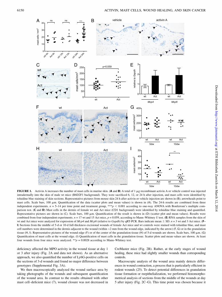
Activin had previously been shown to act as a chemoattractant andmaturation factor for cultured BMMCs (11). To test whether it canalso affect mast cells in vivo, we injected activin A intradermallyinto the skin of wt mice. Six and 12 h after injection, there wasa slight increase in the number of toluidine blue–positive mastcells at the site of activin injection (Fig. 1A), and the increase wasstatistically significant 24 h after injection (Fig. 1A, 1B). To testwhether the effect of activin might be mediated via upregulationof known mast cell survival or chemotactic factors, we analyzedthe mRNA levels of stem cell factor (Scf), nerve growth factor (Ngf),and the chemokines Ccl2 and Ccl5 in activin- or vehicle-injected skinusing quantitative RT-PCR (qRT-PCR). Ccl5 (RANTES), whichhad been shown to increase the number of mast cells in the skin4 h after s.c. injection (19), was slightly and transiently upregu-lated 6 h after activin injection, whereas expression of the otherfactors was not altered or even reduced in the presence of activin(Supplemental Fig. 1A). It was recently reported that activin in-duced Th9 differentiation in a model of allergic airway disease,resulting in recruitment and activation of mast cells in the lung(20). However, we were not able to detect mRNA for IL-9, neitherin normal skin of wt or Act mice, nor after activin injection (datanot shown), probably due to a generally low number of T cells inthe skin.Overall, these results suggest that the effect of activin on mast
cells could be mediated at least in part via Ccl5, but also througha direct attraction of mast cells from the bone marrow. Consistentwith the latter assumption, we found that BMMC precursors thathad been cultured with conditioned medium from WEHI3 cells,which is rich in IL-3, express all types of activin receptors (Sup-plemental Fig. 1B). However, none of the receptors was expressedin mast cells that had been isolated from ear skin by FACS (Sup-plemental Fig. 1B). Together, these findings suggest that mast cellprogenitors can be attracted to the skin by activin, but downregulateactivin receptors upon differentiation in this tissue.
We then analyzed mast cell numbers in the skin of Act mice underhomeostatic conditions. Toluidine blue staining revealed a slightincrease in mast cell number in the dermis of Act mice (Fig. 1C, 1D).Similar results were obtained with chloroacetate esterase histo-chemistry (data not shown). The generally higher number of mastcells in the dermis that we identified in this experiment (102 6 26cells/mm2 in wt mice) compared with the experiment shown in Fig.1A and 1B (15 6 2 cells/mm2 in vehicle-injected mice) most likelyresults from the use of a different mouse strain and the differentgender (male B6D2F1 mice [Fig. 1A, 1B] versus female CD1 mice[Fig. 1C, 1D]). Thus, male mice were shown to have lower hista-mine levels in dorsal and ventral skin as compared with femalemice (21), and C57BL/6 mice have a generally low mast cellcontent in the skin (22). Besides, male mice have a significantlythinner layer of hypodermis (s.c. fat), hardly distinguishable fromthe dermis on toluidine blue–stained sections. Therefore, mast cellswere quantified and related to the total area of dermis and hypo-dermis in male mice, but only to the area of dermis in female mice.As an independent approach, we analyzed the expression levels
of mast cell–specific enzymes in the skin of wt and Act mice usingqRT-PCR. Expression of mast cell proteases (Mcpt)-4 and -6, whichare expressed by mature mast cells (23), and which are upregulatedby activin in mast cell progenitors (Mcpt6) (24), was indeed higherin the skin of Act mice (Fig. 1E), suggesting an increase in maturemast cells. In summary, these results demonstrate that activin Aincreases the number of mature mast cells in the skin in vivo.We next analyzed mast cell numbers during wound healing of
control and Act mice. There was no significant increase in the numberof mast cells in Act mice compared with wt controls within 2 mmfrom the edge of full-thickness excisional wounds at day 5 or 10 (Fig.1F, 1G). Surprisingly, there were only very few mast cells in thegranulation tissue of wt and Act mice at days 5, 10, and 13 afterwounding, and their number was even lower in 10-d wounds of Actmice compared with wt controls (Fig. 1H, 1I, Supplemental Fig. 1C).Even 13 d after injury there were very few mast cells in the woundtissue (Supplemental Fig. 1C), and these were located at the peripheryof the granulation tissue (data not shown). However, their numberincreased in the vicinity of the wounds at this stage, indicating thatmast cell accumulation is a late event in wound healing. It seemslikely that mast cells are not efficiently attracted to the granulationtissue of healing wounds, even in the presence of high levels ofactivin. This may be due to the lack of additional factors required formast cell migration/survival in the newly formed granulation tissue orto the lack of expression of certain proteases that are required forinvasion into the dense granulation tissue. These deficiencies canobviously not be overcome by activin overexpression.
Wound closure is not impaired in mast cell–deficient mice
To determine whether loss of mast cells affects wound healing ina wt background and/or the accelerated healing seen in Act mice,we crossed Act mice with mast cell–deficient CreMaster mice (8)and performed wound-healing experiments with the F1 generation.Lack of expression of the mast cell–specific enzyme Cpa3 innormal skin of CreMaster or Act/CreMaster mice strongly sug-gested the complete lack of mast cells (Supplemental Fig. 2A).This was confirmed by toluidine blue staining of untreated orwounded back skin of CreMaster mice, as well as of Act/CreMasterdouble-transgenic animals. Mast cells could not be detected in thesemice at any stage of the healing process (Supplemental Fig. 2B–E).Because it was previously suggested that the ability of mast
cells to regulate vascular permeability and neutrophil infiltrationis important for wound closure (7), we first measured levels of theneutrophil-specific enzyme MPO in early wounds of mice of allgenotypes. However, neither activin overexpression nor mast cell
The Journal of Immunology 6149
at ET
H B
ibliothek Zuerich on M
arch 12, 2014http://w
ww
.jimm
unol.org/D
ownloaded from
deficiency affected the MPO activity in the wound tissue at day 1or 3 after injury (Fig. 2A and data not shown). As an alternativeapproach, we also quantified the number of Ly6G-positive cells onthe sections of 3-d wounds and found no major difference betweengenotypes (Supplemental Fig. 3A).We then macroscopically analyzed the wound surface area by
taking photographs of the wounds and subsequent quantificationof the wound area. In contrast to the results obtained with othermast cell–deficient mice (7), wound closure was not decreased in
CreMaster mice (Fig. 2B). Rather, at the early stages of woundhealing, these mice had slightly smaller wounds than correspondingcontrols.Macroscopic analysis of the wound area mainly detects differ-
ences in wound contraction, a process that is particularly efficient inrodent wounds (25). To detect potential differences in granulationtissue formation or reepithelialization, we performed histomorpho-metrical analysis of sections from the middle of the wounds at day5 after injury (Fig. 2C–G). This time point was chosen because it
FIGURE 1. Activin A increases the number of mast cells in murine skin. (A and B) A total of 1 mg recombinant activin A or vehicle control was injected
intradermally into the skin of male wt mice (B6D2F1 background). They were sacrificed 6, 12, or 24 h after injection, and mast cells were identified by
toluidine blue staining of skin sections. Representative pictures from mouse skin 24 h after activin or vehicle injection are shown in (B); arrowheads point to
mast cells. Scale bars, 100 mm. Quantification of the data (scatter plots and mean values) is shown in (A). The 24-h results are combined from three
independent experiments. n = 5–14 per time point and treatment group, ***p , 0.001 according to one-way ANOVA with Bonferroni’s multiple com-
parison test. (C and D) Mast cells in the dermis of female wt and Act mice (CD1 background) were identified by toluidine blue staining and quantified.
Representative pictures are shown in (C). Scale bars, 100 mm. Quantification of the result is shown in (D) (scatter plot and mean values). Results were
combined from four independent experiments. n = 17 wt and 15 Act mice, p = 0.059, according to Mann–Whitney U test. (E) RNA samples from the skin of
wt and Act mice were analyzed for expression ofMcp4 andMcp6 (relative to Gapdh) by qRT-PCR. Bars indicate mean6 SD. n = 3 wt and 3 Act mice. (F–
I) Sections from the middle of 5-d or 10-d full-thickness excisional wounds of female Act mice and wt controls were stained with toluidine blue, and mast
cell numbers were determined in the dermis adjacent to the wound (within ∼2 mm from the wound edge, indicated by the arrow) (F, G) or in the granulation
tissue (H, I). Representative pictures of the wound edge (F) or of the center of the granulation tissue (H) of 5-d wounds are shown. Scale bars, 100 mm. (G)
Quantification of mast cells at the wound edge. (I) Quantification of mast cells in the granulation tissue. Scatter plots and mean values are shown. At least
four wounds from four mice were analyzed. **p = 0.0028 according to Mann–Whitney test.
6150 ACTIVIN, MAST CELLS, WOUND HEALING, AND SKIN CANCER
at ET
H B
ibliothek Zuerich on M
arch 12, 2014http://w
ww
.jimm
unol.org/D
ownloaded from
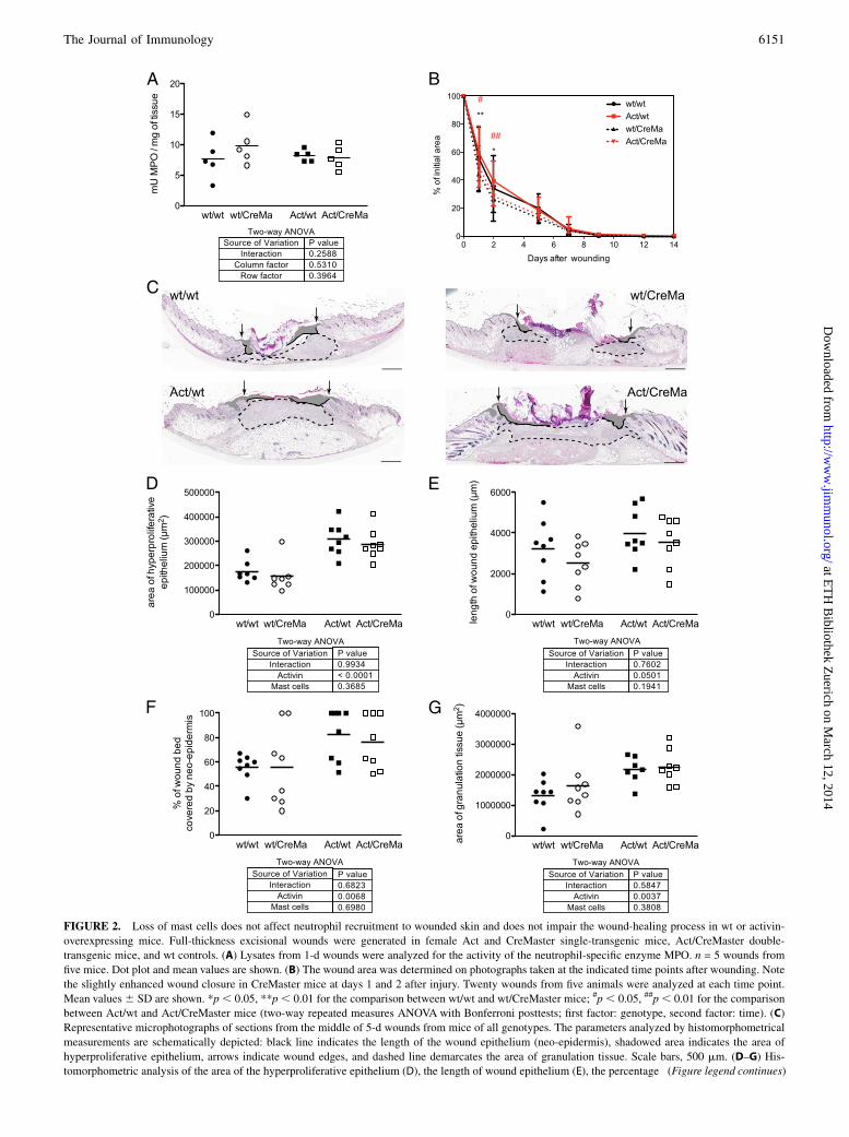
FIGURE 2. Loss of mast cells does not affect neutrophil recruitment to wounded skin and does not impair the wound-healing process in wt or activin-
overexpressing mice. Full-thickness excisional wounds were generated in female Act and CreMaster single-transgenic mice, Act/CreMaster double-
transgenic mice, and wt controls. (A) Lysates from 1-d wounds were analyzed for the activity of the neutrophil-specific enzyme MPO. n = 5 wounds from
five mice. Dot plot and mean values are shown. (B) The wound area was determined on photographs taken at the indicated time points after wounding. Note
the slightly enhanced wound closure in CreMaster mice at days 1 and 2 after injury. Twenty wounds from five animals were analyzed at each time point.
Mean values6 SD are shown. *p, 0.05, **p , 0.01 for the comparison between wt/wt and wt/CreMaster mice; #p , 0.05, ##p, 0.01 for the comparison
between Act/wt and Act/CreMaster mice (two-way repeated measures ANOVA with Bonferroni posttests; first factor: genotype, second factor: time). (C)
Representative microphotographs of sections from the middle of 5-d wounds from mice of all genotypes. The parameters analyzed by histomorphometrical
measurements are schematically depicted: black line indicates the length of the wound epithelium (neo-epidermis), shadowed area indicates the area of
hyperproliferative epithelium, arrows indicate wound edges, and dashed line demarcates the area of granulation tissue. Scale bars, 500 mm. (D–G) His-
tomorphometric analysis of the area of the hyperproliferative epithelium (D), the length of wound epithelium (E), the percentage (Figure legend continues)
The Journal of Immunology 6151
at ET
H B
ibliothek Zuerich on M
arch 12, 2014http://w
ww
.jimm
unol.org/D
ownloaded from
represents the peak of reepithelialization and granulation tissueformation in the wound model that we used (26). Consistent withour previous nonquantified observations (12), Act mice hada significantly larger area of hyperproliferative epithelium and ofgranulation tissue compared with control mice (Fig. 2D, 2G). Theincrease in both parameters in the absence of a difference inwound contraction also provides an explanation for the unalteredwound closure rate of these animals that was detected by mac-roscopic wound analysis. Furthermore, the length of the woundepithelium (neo-epidermis), which reflects keratinocyte migration,was also slightly increased in Act mice (Fig. 2E). However, noneof these parameters was affected by the absence of mast cells (Fig.2D–G). Loss of these cells also did not impair wound reepitheli-alization or granulation tissue formation in mice with normalactivin levels (Fig. 2D–G). Finally, loss of mast cells did not affectthe area and density of the late granulation tissue/early scar tissueat day 13 after injury in mice of a wt background, neither did itreduce the hyperthickened neo-epidermis and enlarged scar areaseen in Act mice at this time point (Supplemental Fig. 3B, 3C). Insummary, these results strongly suggest that mast cells are dis-pensable for the healing of full-thickness excisional wounds inmice and also do not obviously affect the scarring response.
Mast cell deficiency does not affect the activin-inducedacceleration of chemically induced skin carcinogenesis
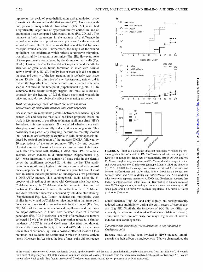
Because there are remarkable parallels between wound healing andcancer (27) and because mast cells had been proposed, based onwork in Kit mutants, to contribute to human papilloma virus (HPV)16-induced skin carcinogenesis (28), we asked whether these cellsalso play a role in chemically induced skin carcinogenesis. Thispossibility was particularly intriguing, because we recently showedthat Act mice are strongly susceptible to skin carcinogenesis in-duced by topical application of the mutagen DMBA followed by20 applications of the tumor promoter TPA (10), and becauseelevated numbers of mast cells were seen in the skin of Act mice24 h after treatment with DMBA, DMBA plus TPA, or vehicleacetone, which induces mild inflammation (Supplemental Fig.4A). Most importantly, the number of mast cells in the dermisbelow the papillomas collected 20 wk after the last TPA appli-cation was significantly higher in Act mice compared with controlmice (Supplemental Fig. 4B). To determine a potential role of mastcells in activin-induced promotion of tumorigenesis, we performeda DMBA/TPA-induced skin carcinogenesis study using the F1progeny of a breeding of Act mice with CreMaster mice (Act mice,CreMaster mice, Act/CreMaster double-transgenic mice, and wtcontrols). The absence of mast cells in the tumors of CreMasterand Act/CreMaster mice was confirmed by toluidine blue staining(Supplemental Fig. 4C). Tumor incidence and multiplicity weresimilar in wt/wt and wt/CreMaster mice, indicating that mast cellsdo not contribute to skin tumorigenesis in this model (Fig. 3A,3B). Most of the tumors were classical papillomas, and there wasno major difference in tumor size between mice of differentgenotypes (Fig. 3C). Histological analysis of large/invasive tumorscollected 12 wk after the last TPA application revealed a similarincidence of SCC in wt and CreMaster mice (data not shown).Because the tumor multiplicity in wt and wt/CreMaster mice waslow in this experiment (Fig. 3B), a possible effect of mast cell losson tumor load could not be determined in mice with normal activinlevels. However, in Act mice, the loss of mast cells did not reduce
tumor incidence (Fig. 3A) and only slightly, but nonsignificantly,reduced tumor multiplicity during the early stages of carcinogen-esis (Fig. 3B). Similarly, the incidence of SCC did not differ sig-nificantly between Act and Act/CreMaster mice (data not shown).Thus, mast cells are obviously not major regulators of activin-induced skin carcinogenesis.
Tumorigenesis-associated vascularization is not impaired inCreMaster mice
Because mast cells had been invoked in HPV8-induced tumori-genesis via their effects on angiogenesis (28), we characterized the
of the wound surface covered by neo-epidermis (wound epithelium) (F), and the area of granulation tissue (G) using sections from the middle of 5-d wounds
from mice of all genotypes. Dot plots and mean values are shown. At least eight wounds from four mice were analyzed. The results of two-way ANOVA are
shown below each graph (first factor: presence of CreMaster transgene, second factor: presence of activin transgene).
FIGURE 3. Mast cell deficiency does not significantly reduce the pro-
tumorigenic effect of activin in DMBA/TPA-induced skin carcinogenesis.
Kinetics of tumor incidence (A) or multiplicity (B) in Act/wt and wt/
CreMaster single-transgenic mice, Act/CreMaster double-transgenic mice,
and wt/wt controls. n = 17 mice per genotype. Mean 6 SEM are shown in
(B). ***p , 0.001 for the comparison between wt/wt and Act/wt mice and
between wt/CreMaster and Act/wt mice, ###p , 0.001 for the comparison
between wt/wt and Act/CreMaster and wt/CreMaster and Act/CreMaster
mice (two-way repeated measures ANOVA and Bonferroni posttest; first
factor: genotype, second factor: time). (C) Distribution of tumors, collected
after 20 TPA applications, according to tumor diameter and tumor type: SP,
small papilloma (,2 mm); MP, medium papilloma (2–6 mm); LP, large
papilloma (.6 mm).
6152 ACTIVIN, MAST CELLS, WOUND HEALING, AND SKIN CANCER
at ET
H B
ibliothek Zuerich on M
arch 12, 2014http://w
ww
.jimm
unol.org/D
ownloaded from
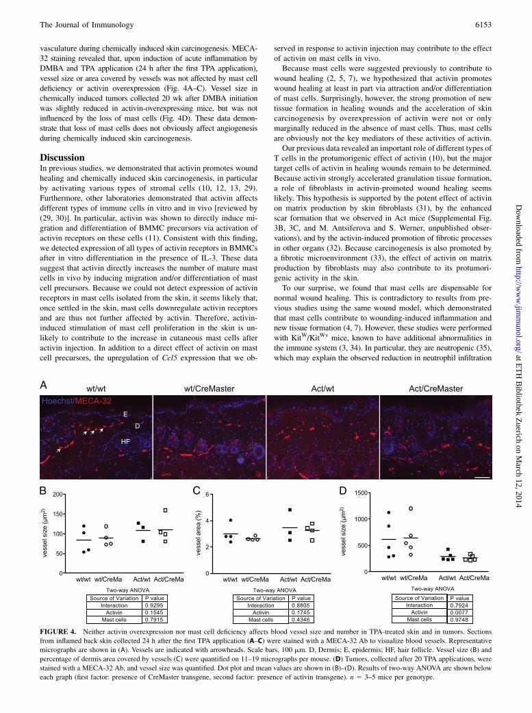
vasculature during chemically induced skin carcinogenesis. MECA-32 staining revealed that, upon induction of acute inflammation byDMBA and TPA application (24 h after the first TPA application),vessel size or area covered by vessels was not affected by mast celldeficiency or activin overexpression (Fig. 4A–C). Vessel size inchemically induced tumors collected 20 wk after DMBA initiationwas slightly reduced in activin-overexpressing mice, but was notinfluenced by the loss of mast cells (Fig. 4D). These data demon-strate that loss of mast cells does not obviously affect angiogenesisduring chemically induced skin carcinogenesis.
DiscussionIn previous studies, we demonstrated that activin promotes woundhealing and chemically induced skin carcinogenesis, in particularby activating various types of stromal cells (10, 12, 13, 29).Furthermore, other laboratories demonstrated that activin affectsdifferent types of immune cells in vitro and in vivo [reviewed by(29, 30)]. In particular, activin was shown to directly induce mi-gration and differentiation of BMMC precursors via activation ofactivin receptors on these cells (11). Consistent with this finding,we detected expression of all types of activin receptors in BMMCsafter in vitro differentiation in the presence of IL-3. These datasuggest that activin directly increases the number of mature mastcells in vivo by inducing migration and/or differentiation of mastcell precursors. Because we could not detect expression of activinreceptors in mast cells isolated from the skin, it seems likely that,once settled in the skin, mast cells downregulate activin receptorsand are thus not further affected by activin. Therefore, activin-induced stimulation of mast cell proliferation in the skin is un-likely to contribute to the increase in cutaneous mast cells afteractivin injection. In addition to a direct effect of activin on mastcell precursors, the upregulation of Ccl5 expression that we ob-
served in response to activin injection may contribute to the effectof activin on mast cells in vivo.Because mast cells were suggested previously to contribute to
wound healing (2, 5, 7), we hypothesized that activin promoteswound healing at least in part via attraction and/or differentiationof mast cells. Surprisingly, however, the strong promotion of newtissue formation in healing wounds and the acceleration of skincarcinogenesis by overexpression of activin were not or onlymarginally reduced in the absence of mast cells. Thus, mast cellsare obviously not the key mediators of these activities of activin.Our previous data revealed an important role of different types of
T cells in the protumorigenic effect of activin (10), but the majortarget cells of activin in healing wounds remain to be determined.Because activin strongly accelerated granulation tissue formation,a role of fibroblasts in activin-promoted wound healing seemslikely. This hypothesis is supported by the potent effect of activinon matrix production by skin fibroblasts (31), by the enhancedscar formation that we observed in Act mice (Supplemental Fig.3B, 3C, and M. Antsiferova and S. Werner, unpublished obser-vations), and by the activin-induced promotion of fibrotic processesin other organs (32). Because carcinogenesis is also promoted bya fibrotic microenvironment (33), the effect of activin on matrixproduction by fibroblasts may also contribute to its protumori-genic activity in the skin.To our surprise, we found that mast cells are dispensable for
normal wound healing. This is contradictory to results from pre-vious studies using the same wound model, which demonstratedthat mast cells contribute to wounding-induced inflammation andnew tissue formation (4, 7). However, these studies were performedwith KitW/KitWv mice, known to have additional abnormalities inthe immune system (3, 34). In particular, they are neutropenic (35),which may explain the observed reduction in neutrophil infiltration
FIGURE 4. Neither activin overexpression nor mast cell deficiency affects blood vessel size and number in TPA-treated skin and in tumors. Sections
from inflamed back skin collected 24 h after the first TPA application (A–C) were stained with a MECA-32 Ab to visualize blood vessels. Representative
micrographs are shown in (A). Vessels are indicated with arrowheads. Scale bars, 100 mm. D, Dermis; E, epidermis; HF, hair follicle. Vessel size (B) and
percentage of dermis area covered by vessels (C) were quantified on 11–19 micrographs per mouse. (D) Tumors, collected after 20 TPA applications, were
stained with a MECA-32 Ab, and vessel size was quantified. Dot plot and mean values are shown in (B)–(D). Results of two-way ANOVA are shown below
each graph (first factor: presence of CreMaster transgene, second factor: presence of activin transgene). n 5 3–5 mice per genotype.
The Journal of Immunology 6153
at ET
H B
ibliothek Zuerich on M
arch 12, 2014http://w
ww
.jimm
unol.org/D
ownloaded from
upon wounding (4, 7). Although the impaired healing of KitW/KitWv mice was rescued by reconstitution of the mice with mastcells (7), this finding does not provide a final proof for a role ofmast cells in wound healing. This conclusion is in agreement withseveral other in vivo experiments in which Kit mutant micereconstituted with mast cells had a different behavior than micewith wt Kit alleles with and without mast cells, depending on theexperimental setup (8). The normal wound closure in CreMastermice that we observed at the macroscopic and histological level isconsistent with recent data, which demonstrated that wound clo-sure as assessed macroscopically was not affected in splintedexcisional wounds of three different types of mast cell–deficientmice, including KitW/KitWv mice (6).Mast cells had previously been shown to contribute to collagen
remodeling after completion of healing (5) and to scar formationduring fetal wound healing (36). However, the analysis of 13-dwounds revealed no obvious difference in the area and density ofthe late granulation tissue/early scar tissue, although more subtledifferences in collagen remodeling after completion of woundingcannot be excluded. In the future, it will therefore be interesting toperform a detailed analysis of the scar tissue and to study otherfibrotic processes in CreMaster mice. This will reveal whethermast cells are indeed regulators of organ fibrosis in vivo.Finally, the results presented in this study strongly suggest that
mast cells are not key regulators of DMBA/TPA-induced skincarcinogenesis. This experiment should be repeated in the futurewithmice of another genetic background that are more susceptible to skincarcinogenesis than the CD1/C57BL/6 F1 mice used in this study.Furthermore, a potential role of mast cells in the progression frompapillomas to carcinomas remains to be determined. It was recentlyshown that expression of mast cell proteases correlates with an-giogenesis during progression of chemically induced tumors (37),but a functional role of mast cells in angiogenesis and tumor for-mation had not been addressed in this model. The results obtainedin our study, however, argue against a major role of mast cells in thecontrol of blood vessels in TPA-treated skin and in skin tumors.Interestingly, mast cells had previously been shown to contribute toearly neoplastic progression in a model of HPV16-induced carci-nogenesis, in which transgenic mice expressing the HPV16 onco-genes in keratinocytes were bred onto the KitW/KitWv background.Due to a high lethality of the double-mutant mice in FVB/N back-ground, however, only one mast cell–deficient HPV16-transgenicanimal was available for this analysis (28). Nevertheless, it maywell be that the reduced neoplastic progression in HPV16/KitW/KitWv mice is due to the Kit deficiency rather than to the loss ofmast cells. Alternatively, mast cells may have different roles inchemically versus virus-induced carcinogenesis. This possibility issupported by a previous study showing that DMBA/TPA-inducedskin carcinogenesis is not affected in Kit mutant mice (38).However, it needs to be considered that mast cells reappear in Kit-deficient mice upon induction of skin inflammation, includingTPA treatment (8, 39, 40). Therefore, it was unclear whether thereappearing mast cells were sufficient to drive carcinogenesis. Theuse of the CreMaster mouse line that is completely deficient inmast cells even under inflammatory conditions therefore clarifiedthis issue and revealed that DMBA/TPA-induced skin carcino-genesis is indeed not obviously affected by the loss of mast cells.Taken together, our results revealed that mast cells are not the
major targets of activin in wound healing and carcinogenesis andargue against the previously suggested nonredundant functions ofmast cells in these processes in general.
AcknowledgmentsWe thank Dr. Katharina Birkner for help with the mouse experiments.
DisclosuresThe authors have no financial conflicts of interest.
References1. Rodewald, H. R., and T. B. Feyerabend. 2012. Widespread immunological
functions of mast cells: fact or fiction? Immunity 37: 13–24.2. Noli, C., and A. Miolo. 2001. The mast cell in wound healing. Vet. Dermatol. 12:
303–313.3. Nocka, K., J. C. Tan, E. Chiu, T. Y. Chu, P. Ray, P. Traktman, and P. Besmer.
1990. Molecular bases of dominant negative and loss of function mutations at themurine c-kit/white spotting locus: W37, Wv, W41 and W. EMBO J. 9: 1805–1813.
4. Egozi, E. I., A. M. Ferreira, A. L. Burns, R. L. Gamelli, and L. A. Dipietro. 2003.Mast cells modulate the inflammatory but not the proliferative response inhealing wounds. Wound Repair Regen. 11: 46–54.
5. Iba, Y., A. Shibata, M. Kato, and T. Masukawa. 2004. Possible involvement ofmast cells in collagen remodeling in the late phase of cutaneous wound healingin mice. Int. Immunopharmacol. 4: 1873–1880.
6. Nauta, A. C., M. Grova, D. T. Montoro, A. Zimmermann, M. Tsai, G. C. Gurtner,S. J. Galli, and M. T. Longaker. 2013. Evidence that mast cells are not requiredfor healing of splinted cutaneous excisional wounds in mice. PLoS One 8:e59167.
7. Weller, K., K. Foitzik, R. Paus, W. Syska, and M. Maurer. 2006. Mast cells arerequired for normal healing of skin wounds in mice. FASEB J. 20: 2366–2368.
8. Feyerabend, T. B., A. Weiser, A. Tietz, M. Stassen, N. Harris, M. Kopf,P. Radermacher, P. Moller, C. Benoist, D. Mathis, et al. 2011. Cre-mediated cellablation contests mast cell contribution in models of antibody- and T cell-mediated autoimmunity. Immunity 35: 832–844.
9. Hubner, G., Q. Hu, H. Smola, and S. Werner. 1996. Strong induction of activinexpression after injury suggests an important role of activin in wound repair.Dev. Biol. 173: 490–498.
10. Antsiferova, M., M. Huber, M. Meyer, A. Piwko-Czuchra, T. Ramadan,A. S. MacLeod, W. L. Havran, R. Dummer, D. Hohl, and S. Werner. 2011.Activin enhances skin tumourigenesis and malignant progression by inducinga pro-tumourigenic immune cell response. Nat. Commun. 2: 576.
11. Funaba, M., T. Ikeda, K. Ogawa, M. Murakami, and M. Abe. 2003. Role ofactivin A in murine mast cells: modulation of cell growth, differentiation, andmigration. J. Leukoc. Biol. 73: 793–801.
12. Munz, B., H. Smola, F. Engelhardt, K. Bleuel, M. Brauchle, I. Lein, L. W. Evans,D. Huylebroeck, R. Balling, and S. Werner. 1999. Overexpression of activin A inthe skin of transgenic mice reveals new activities of activin in epidermal mor-phogenesis, dermal fibrosis and wound repair. EMBO J. 18: 5205–5215.
13. Bamberger, C., A. Scharer, M. Antsiferova, B. Tychsen, S. Pankow, M. Muller,T. Rulicke, R. Paus, and S. Werner. 2005. Activin controls skin morphogenesisand wound repair predominantly via stromal cells and in a concentration-dependent manner via keratinocytes. Am. J. Pathol. 167: 733–747.
14. Munz, B., G. Hubner, Y. Tretter, C. Alzheimer, and S. Werner. 1999. A novelrole of activin in inflammation and repair. J. Endocrinol. 161: 187–193.
15. Junankar, S. R., A. Eichten, A. Kramer, K. E. de Visser, and L. M. Coussens.2006. Analysis of immune cell infiltrates during squamous carcinoma develop-ment. J. Investig. Dermatol. Symp. Proc. 11: 36–43.
16. Werner, S., H. Smola, X. Liao, M. T. Longaker, T. Krieg, P. H. Hofschneider, andL. T. Williams. 1994. The function of KGF in morphogenesis of epithelium andreepithelialization of wounds. Science 266: 819–822.
17. Schindelin, J., I. Arganda-Carreras, E. Frise, V. Kaynig, M. Longair, T. Pietzsch,S. Preibisch, C. Rueden, S. Saalfeld, B. Schmid, et al. 2012. Fiji: an open-sourceplatform for biological-image analysis. Nat. Methods 9: 676–682.
18. Bradley, P. P., D. A. Priebat, R. D. Christensen, and G. Rothstein. 1982. Mea-surement of cutaneous inflammation: estimation of neutrophil content with anenzyme marker. J. Invest. Dermatol. 78: 206–209.
19. Conti, P., M. Reale, R. C. Barbacane, M. Felaco, A. Grilli, and T. C. Theoharides.1998. Mast cell recruitment after subcutaneous injection of RANTES in the soleof the rat paw. Br. J. Haematol. 103: 798–803.
20. Jones, C. P., L. G. Gregory, B. Causton, G. A. Campbell, and C. M. Lloyd. 2012.Activin A and TGF-beta promote T(H)9 cell-mediated pulmonary allergic pa-thology. J. Allergy Clin. Immunol. 129: 1000-1010.e3.
21. Ponvert, C., L. Galoppin, P. Scheinmann, P. Canu, and C. Burtin. 1985. Tissuehistamine levels in male and female mast cell deficient mice (W/Wv) and in theirlittermates (Wv/+, W/+ and +/+). Agents Actions 17: 1–4.
22. Koizumi, T., and J. Hayakawa. 1987. Single-locus control of the mast cellpopulation in mouse skin. Immunogenetics 26: 36–39.
23. Miller, H. R., and A. D. Pemberton. 2002. Tissue-specific expression of mast cellgranule serine proteinases and their role in inflammation in the lung and gut.Immunology 105: 375–390.
24. Funaba, M., T. Ikeda, M. Murakami, K. Ogawa, and M. Abe. 2005. Up-regulation of mouse mast cell protease-6 gene by transforming growth factor-beta and activin in mast cell progenitors. Cell. Signal. 17: 121–128.
25. Wang, X., J. Ge, E. E. Tredget, and Y. Wu. 2013. The mouse excisional woundsplinting model, including applications for stem cell transplantation. Nat. Protoc.8: 302–309.
26. Kumin, A., M. Schafer, N. Epp, P. Bugnon, C. Born-Berclaz, A. Oxenius,A. Klippel, W. Bloch, and S. Werner. 2007. Peroxiredoxin 6 is required for bloodvessel integrity in wounded skin. J. Cell Biol. 179: 747–760.
27. Schafer, M., and S. Werner. 2008. Cancer as an overhealing wound: an oldhypothesis revisited. Nat. Rev. Mol. Cell Biol. 9: 628–638.
6154 ACTIVIN, MAST CELLS, WOUND HEALING, AND SKIN CANCER
at ET
H B
ibliothek Zuerich on M
arch 12, 2014http://w
ww
.jimm
unol.org/D
ownloaded from
28. Coussens, L. M., W. W. Raymond, G. Bergers, M. Laig-Webster, O. Behrendtsen,Z. Werb, G. H. Caughey, and D. Hanahan. 1999. Inflammatory mast cells up-regulateangiogenesis during squamous epithelial carcinogenesis. Genes Dev. 13: 1382–1397.
29. Antsiferova, M., and S. Werner. 2012. The bright and the dark sides of activin inwound healing and cancer. J. Cell Sci. 125: 3929–3937.
30. de Kretser, D. M., R. E. O’Hehir, C. L. Hardy, and M. P. Hedger. 2012. The rolesof activin A and its binding protein, follistatin, in inflammation and tissue repair.Mol. Cell. Endocrinol. 359: 101–106.
31. Mukhopadhyay, A., S. Y. Chan, I. J. Lim, D. J. Phillips, and T. T. Phan. 2007.The role of the activin system in keloid pathogenesis. Am. J. Physiol. CellPhysiol. 292: C1331–C1338.
32. Werner, S., and C. Alzheimer. 2006. Roles of activin in tissue repair, fibrosis, andinflammatory disease. Cytokine Growth Factor Rev. 17: 157–171.
33. Otranto, M., V. Sarrazy, F. Bonte, B. Hinz, G. Gabbiani, and A. Desmouliere. 2012. Therole of the myofibroblast in tumor stroma remodeling. Cell Adhes. Migr. 6: 203–219.
34. Bernstein, A., B. Chabot, P. Dubreuil, A. Reith, K. Nocka, S. Majumder, P. Ray,and P. Besmer. 1990. The mouse W/c-kit locus. Ciba Found. Symp. 148: 158–166, discussion 166–172.
35. Zhou, J. S., W. Xing, D. S. Friend, K. F. Austen, and H. R. Katz. 2007. Mast celldeficiency in Kit(W-sh) mice does not impair antibody-mediated arthritis. J. Exp.Med. 204: 2797–2802.
36. Wulff, B. C., A. E. Parent, M. A. Meleski, L. A. DiPietro, M. E. Schrementi, andT. A. Wilgus. 2012. Mast cells contribute to scar formation during fetal woundhealing. J. Invest. Dermatol. 132: 458–465.
37. de Souza, D. A., Jr., V. D. Toso, M. R. Campos, V. S. Lara, C. Oliver, andM. C. Jamur. 2012. Expression of mast cell proteases correlates with mast cellmaturation and angiogenesis during tumor progression. PLoS One 7: e40790.
38. Muto, S., M. Katsuki, and S. Horie. 2007. Decreased c-kit function inhibitsenhanced skin carcinogenesis in c-Ha-ras protooncogene transgenic mice.Cancer Sci. 98: 1549–1556.
39. Gordon, J. R., and S. J. Galli. 1990. Phorbol 12-myristate 13-acetate-induceddevelopment of functionally active mast cells in W/Wv but not Sl/Sld geneticallymast cell-deficient mice. Blood 75: 1637–1645.
40. Waskow, C., S. Bartels, S. M. Schlenner, C. Costa, and H. R. Rodewald. 2007.Kit is essential for PMA-inflammation-induced mast-cell accumulation in theskin. Blood 109: 5363–5370.
The Journal of Immunology 6155
at ET
H B
ibliothek Zuerich on M
arch 12, 2014http://w
ww
.jimm
unol.org/D
ownloaded from