whole chromosome loss is promoted by telomere dysfunction in primary cells
TRANSCRIPT
GENES, CHROMOSOMES & CANCER 49:368–378 (2010)
Whole Chromosome Loss Is Promoted by TelomereDysfunction in Primary Cells
Judit Pampalona, David Soler, Anna Genesca, and Laura Tusell
Cell Biology Unit,Departmentof Cell Biology,Physiologyand Immunology,Universitat Auto' noma de Barcelona,Bellaterra,Spain
Errors in chromosome segregation during mitosis result in aneuploidy, which in humans may play a role in the onset of neo-
plasia by changing gene dosage. Nearly all solid tumors exhibit genomic instability at the chromosomal level, showing both
structural and numerical chromosome abnormalities. Chromosomal instability occurs early in the development of cancer and
may represent an important step in the initiation and/or progression of the disease. Telomere integrity appears to be a criti-
cal element in the genesis of structural chromosome imbalances, but it is still not clear whether it can also generate numeri-
cal chromosome aberrations. We investigated the possible relationship between telomere shortening and aneuploidy
formation in human mammary epithelial cells using the cytokinesis-block micronucleus assay combined with fluorescent DNA
probes. In this cell system, uncapped chromosomes fuse with each other resulting in dicentric chromosomes, which are
known to be a source of new structural chromosome rearrangements. Here, we show that in primary epithelial cells, the
chromosomes with short telomeres are more frequently involved in missegregation events than chromosomes of normal
telomere length. Whole chromosome aneuploidy occurs through both nondisjunction and anaphase lagging of dicentric chro-
matids, which suggests that pulling anaphase bridges toward opposite poles can generate the necessary force for detaching a
chromosome from the microtubules of one or both spindle poles. Therefore, telomere-driven instability can promote not
only the appearance of chromosomal rearrangements but also the appearance of numerical chromosome aberrations that
could favor cell immortalization and the acquisition of a tumor phenotype. VVC 2010 Wiley-Liss, Inc.
INTRODUCTION
Cancer is a complex disease in which cells
with altered gene expression grow abnormally,
invade other tissues, and disrupt their normal
functions. A crucial early event in carcinogenesis
is the induction of genomic instability, which
enables an initiated cell to evolve into a cancer
cell by achieving a greater proliferation capacity.
Genome instability can take different forms.
Inactivation of one or several genes in the DNA
mismatch repair pathway leads to microsatellite
instability (MIN). MIN cancers retain a normal
complement of chromosomes and do not develop
gross chromosomal aberrations. However, most
solid tumors are unable to maintain chromosomal
integrity and display a myriad of complex chro-
mosomal aberrations that are neither always
shared by cells of the same tumor nor linked to a
particular tumor type. This chromosomal instabil-
ity (CIN), which can be defined as the continu-
ous formation of new structural and numerical
chromosome aberrations, is one of the most fre-
quent forms of instability in human cancers (Len-
gauer et al., 1998).
It is well established that one of the mecha-
nisms for the genesis of structural chromosome
abnormalities is telomere exhaustion. Telomeres
are DNA–protein complexes that protect the
ends of chromosomes from end-to-end fusion.
They have become key players in maintaining
genomic integrity. Evidence is accumulating that
the loss of the telomere capping function due to
proliferation-dependent telomere erosion is a cru-
cial event in initiating cancer (Artandi et al.,
2000; Rudolph et al., 2001; Lo et al., 2002;
O’Hagan et al., 2002; Londono-Vallejo, 2008).
The covalent fusion of chromosomes with eroded
telomeres gives rise to dicentric or ring chromo-
somes that can initiate breakage-fusion-bridge
(BFB) cycles when they are simultaneously
pulled to opposite poles at anaphase. This may
lead to extensive genome remodeling and scram-
bling, which is characteristic of carcinomas, and
cause unbalanced translocations, which are the
*
Supported by: Consejo de Seguridad Nuclear; Fundacio la Mar-ato, Grant number: TV32005-050110; Ministerio de Educacion yCiencia, Grant number: SAF2006-01653; Instituto de Salud CarlosIII, Grant number: RD06/0020/1020; Generalitat de Catalunya,Grant number: 2009SGR-282; Generalitat de Catalunya, Grantnumber: 2009FIC00063.
*Correspondence to: Laura Tusell, Cell Biology Unit, Depart-ment of Cell Biology, Physiology and Immunology, School of Bio-sciences, Universitat Autonoma de Barcelona, 08193 Bellaterra,Spain. E-mail: [email protected]
Received 8 September 2009; Accepted 14 December 2009
DOI 10.1002/gcc.20749
Published online 19 January 2010 inWiley InterScience (www.interscience.wiley.com).
VVC 2010 Wiley-Liss, Inc.
most common type of structural chromosome ab-
normality in carcinoma cells (Gisselsson, 2003;
Gisselsson et al., 2001). Most tumors, as well as
exhibiting structural chromosomal rearrange-
ments, also show abnormal chromosome numbers.
Aneuploidy is the most frequently identified
genomic abnormality in cancer. It has been
shown to occur early in progression and is often
observed in premalignant lesions (Shih et al.,
2001). Because of the loss or gain of entire chro-
mosomes, aneuploidy could stimulate overexpres-
sion or the loss of specific loci relevant to cell
growth and thus provide the conditions for more
aggressively growing cells to be selected.
There are different theories on the mecha-
nisms underlying the genesis of aneuploidy in
relation to the cancer phenotype. Duesberg et al.
postulated that carcinogens generate aneuploidy
by chemically or physically altering one or more
of the chromosomes or the proteins of the spindle
apparatus (Duesberg et al., 2000; Li et al., 2000).
Then, the asymmetric segregation of chromo-
somes destabilizes the karyotype. This would ini-
tiate an autocatalytic karyotype evolution that
would generate the CIN that is characteristic of
preneoplastic and finally cancer cells. However,
in contrast to the opinion that aneuploidy is the
primary cause of CIN, other theories suggest that
telomere exhaustion is a possible origin not only
of structural (Artandi et al., 2000) but also of nu-
merical aberrations (Gisselsson and Hoglund,
2005) that could be at the basis of neoplasia. The
mechanical tension of bridged end-to-end chro-
mosome fusions has been suggested to result in
one or both chromatids detaching from the mi-
totic spindle, leading to whole chromosome aneu-
ploidy (Stewenius et al., 2005). Studies that link
aneuploidy to telomere dysfunction have been
carried out on solid tumors and tumor-derived
cell lines that have active telomerase, which
appears to protect against genomic instability by
stabilizing telomere ends. Under these circum-
stances, the telomere function can be restored
and whole-chromosome losses are not readily
associated with telomere dysfunction. Till now,
studies carried out on transformed nonimmortal
epithelial cells have failed to link telomere dys-
function with whole chromosome losses or gains
(Velicescu et al., 2003; Deng et al., 2007).
In this study, we sought to better understand
the relationship between telomere attrition and
the generation of aneuploidy, as this could pro-
vide important insights into the mechanisms
behind genetic instability in human cancers. We
took advantage of the fact that human mammary
epithelial cells (HMECs) represent a clean model
system in which mechanisms underlying telo-
mere-dependent genome instability can be dis-
sected. These cells do not show telomerase
activity, and telomere shortening occurs when
they are cultured in vitro. Continued proliferation
of HMECs due to spontaneous inactivation of
the CDKN2A gene leads cells to reach critically
short telomeres. By losing their telomere-capping
function, chromosomes with uncapped telomeres
tend to fuse with each other to originate dicentric
chromosomes. These unstable chromosome reor-
ganizations can enter BFB cycles and generate
massive CIN (Soler et al., 2005) before the cul-
ture terminates in a population growth plateau
(Romanov et al., 2001). To determine whether
there is a relationship between telomere shorten-
ing and aneuploidy, we applied the cytokinesis-
block micronucleus assay (CBMN) in combina-
tion with pancentromeric and pantelomeric
peptide nucleic acid (PNA) probes and DNA
probes specific for the centromeric region of chro-
mosomes with critically short telomeres. In sum-
mary, our results demonstrate that numerical
chromosomal alterations accumulate with telo-
mere erosion and that chromosomes with dys-
functional telomeres segregate erroneously more
often than other chromosomes in primary human
epithelial cells. These results provide evidence
that a telomere-dependent mechanism is behind
the development of aneuploidy during early
carcinogenesis.
MATERIALS AND METHODS
Cells and Culture Conditions
219-7 HMECs (BioWhittaker, Walkersville,
MD) were derived from normal breast tissue.
Cells obtained from the vendor at passage 6
(PD20) were cultured in serum-free MEGM me-
dium with supplements (BioWhittaker). The cells
were counted, plated at 2 � 105 cells per 75 cm2
flask, and grown at 37�C in a 5% CO2 atmos-
phere. The number of accumulated population
doublings (PDs) per passage was determined
using the equation PD ¼ initial PD þ log (no. of
viable cells harvested/no. of viable cells plated)/
log 2. The finite life span of primary HMECs
when cultured in MEGM medium is about 18–20
passages, which is equivalent to approximately 50
PDs.
TELOMERE-DRIVEN ANEUPLOIDY IN HMECS 369
Genes, Chromosomes & Cancer DOI 10.1002/gcc
Cytokinesis-Block Micronucleus Assay and
Metaphase Preparations
To obtain binucleated cells (BN-HMECs),
cytokinesis was blocked by adding cytochalasin B
(Cyt-B; Sigma, St. Louis, MO) at a final concen-
tration of 6 lg/ml (Ponsa et al., 2001). The Cyt-B
treatment was applied at PD25 and PD36, which
represent an early and an advanced PD, respec-
tively. As a control, Cyt-B nontreated mono-
nucleated HMECs (MoN-HMECs) from PD36
were also obtained. After tripsinization, BN- and
MoN-HMECs suspensions were centrifuged at
220g for 8 min, followed by hypotonic shock and
three rounds of methanol/acetic fixation. Before
the binucleated samples were dropped onto clean
slides, they were properly diluted to avoid cells
overlapping.
Metaphase spreads were also obtained at PD25
and PD36 by means of treatment with colcemid
0.02 lg/ml for 8 hr, followed by hypotonic shock
and methanol/acetic fixation. Cell suspensions
were dropped onto clean slides, which were
stored at �20�C.
Fluorescence In Situ Hybridization
PNA-FISH
To identify the specific chromosome arms car-
rying critically short telomeres, PNA-fluorescence
in situ hybridization (FISH) experiments were
performed on metaphase chromosome slides from
PD25 and PD36 using a Cy3-(CCCTAA)3 PNA-
probe for telomeres and an FITC-AAA-
CACTCTTTTTGTAGA probe for centromeres
(PE Biosystems, Foster City, CA; Martın et al.,
2003). After reverse DAPI staining (0.125 lg/ml
in antifade solution), which results in a reproduci-
ble G band-like pattern, each chromosome pair
was accurately identified and each signal-free
chromosome arm was recorded. In this analysis,
we pooled the results obtained in the two homo-
logs of each chromosome because they could not
be formally distinguished. Centromere and telo-
mere PNA-FISH probes were also applied to
BN-HMECs from PD25 and PD36 to distinguish
micronuclei that contain acentric fragments from
those with whole chromosomes and/or centric
chromosome fragments.
Chromosome-specific centromeric FISH
FISH was performed on BN-HMECs from
PD25 and PD36 as well as on MoN-HMECs
from PD36. A mixture of three commercial
probes (Abbott, Abbot Park, IL) that detects the
centromeric region of chromosomes 1 (CEP 1
Satellite II/III; Spectrum Orange) and 4 (CEP4;
Spectrum Aqua) and the breakpoint cluster region
(BCR) located close to the centromere of chromo-
some 22 (22q11.2) (LSI22; Spectrum Green) was
applied to the slides. The resulting signals were
red, light blue, and green for chromosomes 1, 4,
and 22, respectively. Before hybridization, the
slides were pretreated with pepsin (0.1 mg/ml;
Sigma) in 10 mM HCl and postfixed in 37%
formaldehyde in PBS/1 M MgCl2. All probes
were applied according to the manufacturer’s
instructions. Finally, slides were dehydrated and
mounted in antifade solution containing DAPI.
Fluorescence signals were visualized under an
Olympus BX microscope equipped with epifluor-
escent optics specific for each fluorochrome.
Images were captured and analyzed using Cytovi-
sion software (Applied Imaging, San Jose, CA).
Scoring Criteria
A binucleated cell was scored when the dis-
tance between the two sister nuclei was equal or
shorter than the radius of one nucleus. Moreover,
the main nuclei could touch each other, but cells
with overlapping nuclei were not considered.
Finally, both sister nuclei must be approximately
equal in size, with the same staining pattern and
staining intensity.
After FISH with the centromeric specific
probes, the distribution of fluorescent signals
within nuclei and micronuclei in each binucleate
was examined under a triple bandpass filter. The
hybridization patterns allowed us to differentiate
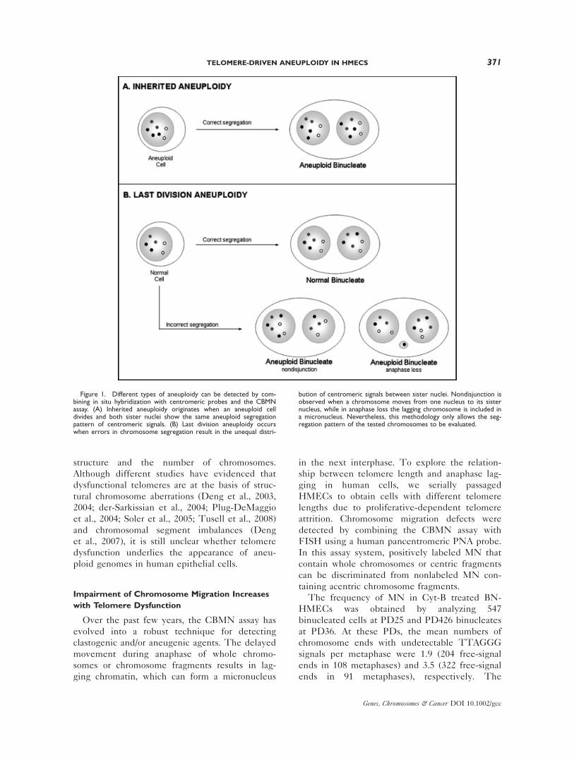
inherited aneuploidies (Fig. 1A) from those origi-
nated in the last nuclear division (Fig. 1B), and
within the latter, nondisjunction from anaphase
loss.
Statistical Analysis
All of the frequencies were calculated based on
events that occurred. The nonparametric Mann-
Whitney U-test was used to determine the differ-
ences between the frequencies observed in the
two PD analyzed. All of the statistical analyses
were performed using the SPSS14.0 (SPSS Inc.,
Chicago, IL) statistical package.
RESULTS
CIN in human cancers is manifested by the
presence of abnormalities that affect both the
370 PAMPALONA ET AL.
Genes, Chromosomes & Cancer DOI 10.1002/gcc
structure and the number of chromosomes.
Although different studies have evidenced that
dysfunctional telomeres are at the basis of struc-
tural chromosome aberrations (Deng et al., 2003,
2004; der-Sarkissian et al., 2004; Plug-DeMaggio
et al., 2004; Soler et al., 2005; Tusell et al., 2008)
and chromosomal segment imbalances (Deng
et al., 2007), it is still unclear whether telomere
dysfunction underlies the appearance of aneu-
ploid genomes in human epithelial cells.
Impairment of Chromosome Migration Increases
with Telomere Dysfunction
Over the past few years, the CBMN assay has
evolved into a robust technique for detecting
clastogenic and/or aneugenic agents. The delayed
movement during anaphase of whole chromo-
somes or chromosome fragments results in lag-
ging chromatin, which can form a micronucleus
in the next interphase. To explore the relation-
ship between telomere length and anaphase lag-
ging in human cells, we serially passaged
HMECs to obtain cells with different telomere
lengths due to proliferative-dependent telomere
attrition. Chromosome migration defects were
detected by combining the CBMN assay with
FISH using a human pancentromeric PNA probe.
In this assay system, positively labeled MN that
contain whole chromosomes or centric fragments
can be discriminated from nonlabeled MN con-
taining acentric chromosome fragments.
The frequency of MN in Cyt-B treated BN-
HMECs was obtained by analyzing 547
binucleated cells at PD25 and PD426 binucleates
at PD36. At these PDs, the mean numbers of
chromosome ends with undetectable TTAGGG
signals per metaphase were 1.9 (204 free-signal
ends in 108 metaphases) and 3.5 (322 free-signal
ends in 91 metaphases), respectively. The
Figure 1. Different types of aneuploidy can be detected by com-bining in situ hybridization with centromeric probes and the CBMNassay. (A) Inherited aneuploidy originates when an aneuploid celldivides and both sister nuclei show the same aneuploid segregationpattern of centromeric signals. (B) Last division aneuploidy occurswhen errors in chromosome segregation result in the unequal distri-
bution of centromeric signals between sister nuclei. Nondisjunction isobserved when a chromosome moves from one nucleus to its sisternucleus, while in anaphase loss the lagging chromosome is included ina micronucleus. Nevertheless, this methodology only allows the seg-regation pattern of the tested chromosomes to be evaluated.
TELOMERE-DRIVEN ANEUPLOIDY IN HMECS 371
Genes, Chromosomes & Cancer DOI 10.1002/gcc
presence of chromosome ends with undetectable
TTAGGG hybridization signals has been shown
to be a good indicator of critically short and prob-
ably dysfunctional telomeres (Hemann et al.,
2001; Espejel et al., 2002; Deng et al., 2004; Soler
et al., 2005). A moderate increase in the total fre-
quency of MN per binucleate was observed with
increasing telomere dysfunction: at PD25, 0.0146
MN per binucleate were observed, whereas at
PD36, the frequency increased to 0.0305 MN/
binucleate (Mann-Whitney U-test, P ¼ 0.091).
However, a significant increase was observed
when only the frequency of centromeric positive
MN was considered. In the early PD, MN-con-
taining centromeric signals accounted for 14.28%
of all MN. In contrast, at PD36, nearly half of
the MN showed hybridization with the pancen-
tromeric PNA probe (Fig. 2). Overall, the inci-
dence of anaphase lagging increased by a factor
of 7.8 from the initial to the advanced PD
(Mann-Whitney U-test, P ¼ 0.025). These results
indicate that chromosome loss due to defects in
their attachment to the spindle apparatus or in
their movement relative to the poles becomes
more frequent as telomere dysfunction increases
in HMECs.
Preferred Involvement of Chromosomes with
Eroded Telomeres in Aneuploid Segregations
Studies using FISH with telomeric probes have
demonstrated that individual telomere lengths in
normal somatic cells are heterogeneous and vary
between donors (Lansdorp et al., 1996; Soler
et al., 2005). To determine whether the telomere
length of an individual chromosome has any
influence on its probability of missegregating, the
distribution of specific chromosomes with crit-
ically short telomeres was analyzed in BN-
HMECs using centromeric-specific DNA probes.
Particular chromosomes with eroded telomeres
were selected based on telomeric PNA-FISH
metaphase analysis. It has been determined that
the resolution of PNA-FISH methodologies on
metaphase chromosomes is about 1,000 bp of
TTAGGG repeats (Poon et al., 1999) and that a
functional telomere should be greater than 76 bp
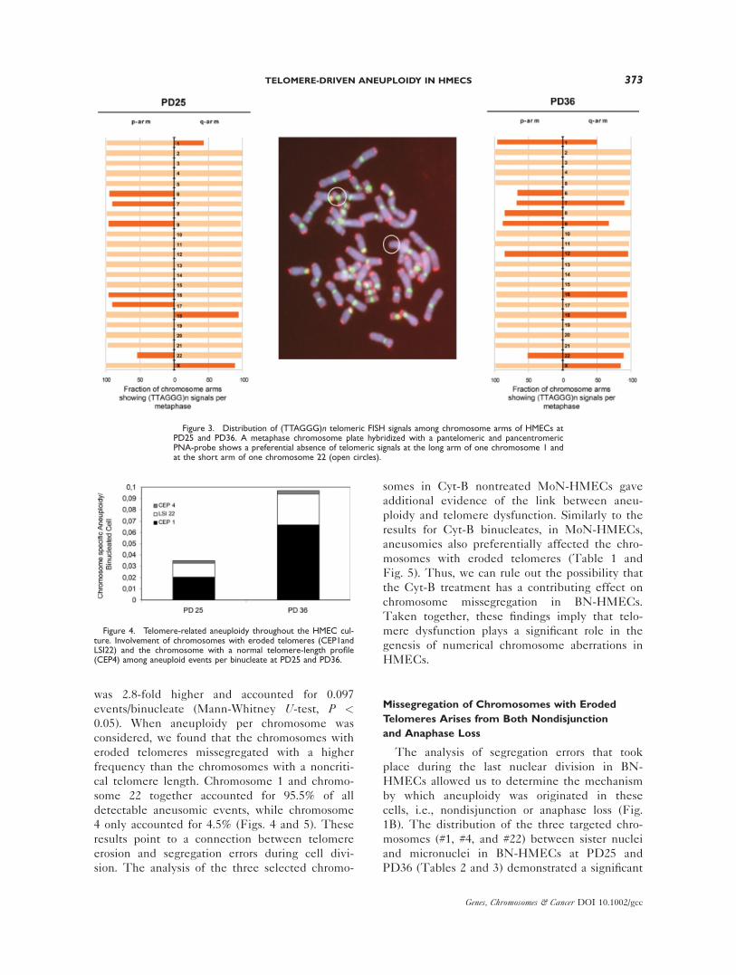
in length (Capper et al., 2007). Accordingly, chro-
mosome 4 was selected as having a noncritical
telomere length, as the p- and q-arms showed
PNA telomere signals in 96% of the metaphases
analyzed (Fig. 3) and thus reflected a telomere
repeat track greater than 1,000 bp. Chromosome
1 and chromosome 22 were chosen as chromo-
somes with critically short telomeres as one of
the 1q-arm and one of the 22p-arm homologs
lacked telomere signals in up to 54% and 48% of
the metaphases in this donor (Fig. 3).
Centromeric probes for chromosomes 1 and 22
(CEP1 and LSI22) with short telomeres and for
chromosome 4 (CEP4) with a noncritical telo-
mere length profile were applied to BN-HMECs
at PD25 and PD36. A total of 3,322 binucleates
was analyzed to detect whole chromosome aneu-
ploidy. At PD25, the total frequency of aneuploid
cells for the three chromosomes analyzed was
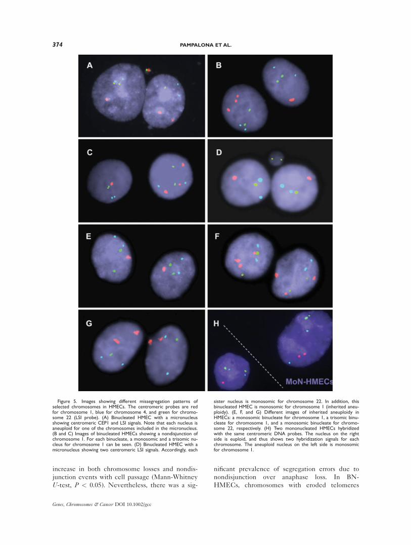
0.035 per binucleate (Fig. 4). The presence of
aneuploid segregations increased significantly
with cell passage. At PD36, with increased telo-
mere dysfunction, the incidence of aneuploidy
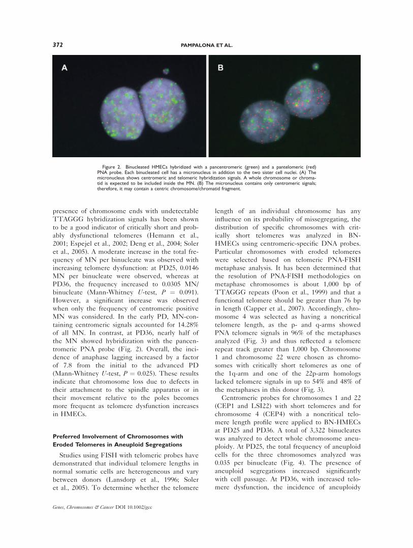
Figure 2. Binucleated HMECs hybridized with a pancentromeric (green) and a pantelomeric (red)PNA probe. Each binucleated cell has a micronucleus in addition to the two sister cell nuclei. (A) Themicronucleus shows centromeric and telomeric hybridization signals. A whole chromosome or chroma-tid is expected to be included inside the MN. (B) The micronucleus contains only centromeric signals;therefore, it may contain a centric chromosome/chromatid fragment.
372 PAMPALONA ET AL.
Genes, Chromosomes & Cancer DOI 10.1002/gcc
was 2.8-fold higher and accounted for 0.097
events/binucleate (Mann-Whitney U-test, P <0.05). When aneuploidy per chromosome was
considered, we found that the chromosomes with
eroded telomeres missegregated with a higher
frequency than the chromosomes with a noncriti-
cal telomere length. Chromosome 1 and chromo-
some 22 together accounted for 95.5% of all
detectable aneusomic events, while chromosome
4 only accounted for 4.5% (Figs. 4 and 5). These
results point to a connection between telomere
erosion and segregation errors during cell divi-
sion. The analysis of the three selected chromo-
somes in Cyt-B nontreated MoN-HMECs gave
additional evidence of the link between aneu-
ploidy and telomere dysfunction. Similarly to the
results for Cyt-B binucleates, in MoN-HMECs,
aneusomies also preferentially affected the chro-
mosomes with eroded telomeres (Table 1 and
Fig. 5). Thus, we can rule out the possibility that
the Cyt-B treatment has a contributing effect on
chromosome missegregation in BN-HMECs.
Taken together, these findings imply that telo-
mere dysfunction plays a significant role in the
genesis of numerical chromosome aberrations in
HMECs.
Missegregation of Chromosomes with Eroded
Telomeres Arises from Both Nondisjunction
and Anaphase Loss
The analysis of segregation errors that took
place during the last nuclear division in BN-
HMECs allowed us to determine the mechanism
by which aneuploidy was originated in these
cells, i.e., nondisjunction or anaphase loss (Fig.
1B). The distribution of the three targeted chro-
mosomes (#1, #4, and #22) between sister nuclei
and micronuclei in BN-HMECs at PD25 and
PD36 (Tables 2 and 3) demonstrated a significant
Figure 4. Telomere-related aneuploidy throughout the HMEC cul-ture. Involvement of chromosomes with eroded telomeres (CEP1andLSI22) and the chromosome with a normal telomere-length profile(CEP4) among aneuploid events per binucleate at PD25 and PD36.
Figure 3. Distribution of (TTAGGG)n telomeric FISH signals among chromosome arms of HMECs atPD25 and PD36. A metaphase chromosome plate hybridized with a pantelomeric and pancentromericPNA-probe shows a preferential absence of telomeric signals at the long arm of one chromosome 1 andat the short arm of one chromosome 22 (open circles).
TELOMERE-DRIVEN ANEUPLOIDY IN HMECS 373
Genes, Chromosomes & Cancer DOI 10.1002/gcc
increase in both chromosome losses and nondis-
junction events with cell passage (Mann-Whitney
U-test, P < 0.05). Nevertheless, there was a sig-
nificant prevalence of segregation errors due to
nondisjunction over anaphase loss. In BN-
HMECs, chromosomes with eroded telomeres
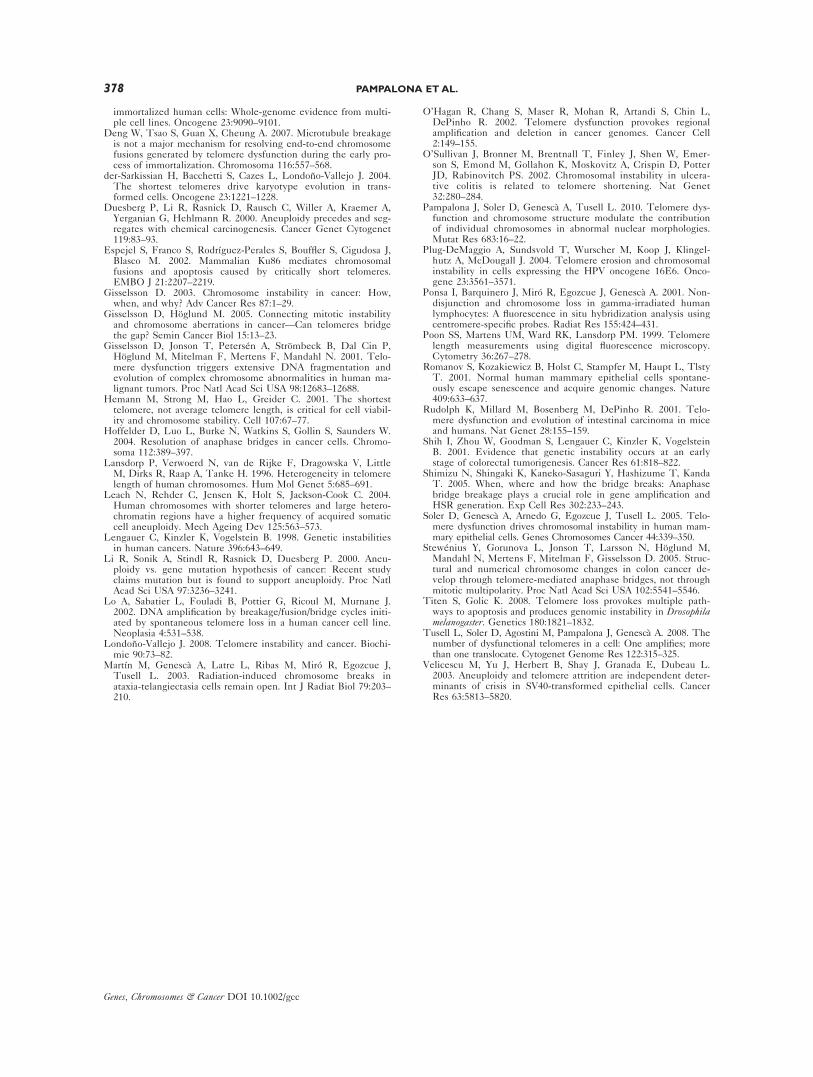
Figure 5. Images showing different missegregation patterns ofselected chromosomes in HMECs. The centromeric probes are redfor chromosome 1, blue for chromosome 4, and green for chromo-some 22 (LSI probe). (A) Binucleated HMEC with a micronucleusshowing centromeric CEP1 and LSI signals. Note that each nucleus isaneuploid for one of the chromosomes included in the micronucleus.(B and C) Images of binucleated HMECs showing a nondisjunction ofchromosome 1. For each binucleate, a monosomic and a trisomic nu-cleus for chromosome 1 can be seen. (D) Binucleated HMEC with amicronucleus showing two centromeric LSI signals. Accordingly, each
sister nucleus is monosomic for chromosome 22. In addition, thisbinucleated HMEC is monosomic for chromosome 1 (inherited aneu-ploidy). (E, F, and G) Different images of inherited aneuploidy inHMECs: a monosomic binucleate for chromosome 1, a trisomic binu-cleate for chromosome 1, and a monosomic binucleate for chromo-some 22, respectively. (H) Two mononucleated HMECs hybridizedwith the same centromeric DNA probes. The nucleus on the rightside is euploid, and thus shows two hybridization signals for eachchromosome. The aneuploid nucleus on the left side is monosomicfor chromosome 1.
374 PAMPALONA ET AL.
Genes, Chromosomes & Cancer DOI 10.1002/gcc

segregated erroneously between sister nuclei and
were included more frequently inside MN than
those with a noncritical telomere length (98.5%
and 94.1%, respectively; Fig. 5). Moreover, the
specific distribution of centromeric signals in
nuclei and micronuclei allowed the possible rear-
rangements that can lead to missegregation to be
identified. In BN-HMECs, most nondisjunction
events only affected one of the two tested chro-
mosomes, and only one binucleate showed non-
disjunction for both #1 and #22 (Table 2 and Fig.
5). This result implies that the two sister chroma-
tids of an interchromosome dicentric that bridges
at anaphase may have different fates: one dicen-
tric chromatid usually breaks while the other mis-
segregates between sister nuclei. However,
around 40% of MN contained two centromeric
signals either for the same chromosome or for dif-
ferent ones. This finding suggests that the forma-
tion of these MN is triggered by either lagging
chromosomes with fused chromatids or twisted
dicentric chromatids, respectively (Table 3).
Overall, our study demonstrates that telomere
shortening is linked to both chromosome lagging
at anaphase and to nondisjunctional events
between sister nuclei.
DISCUSSION
Alterations in the number of intact chromo-
somes, which is known as whole chromosome an-
euploidy, can be observed when errors in cell
division occur. Unlike point mutations, which
only affect a handful of genes, whole scale
changes in chromosome number dramatically al-
ter the landscape of gene expression. This can
potentially be involved in the onset and progres-
sion of neoplasia. Indeed, aneuploidy is the most
frequently identified genomic abnormality in can-
cer. It has been shown to occur early in cancer
development and is often observed in premalig-
nant lesions (Shih et al., 2001). A large body of
evidence from studies with mice supports the
notion that telomere shortening in proliferating
tissues together with cell cycle checkpoint aberra-
tions can harbor epithelial carcinogenesis. Differ-
ent studies in epithelial cells have shown that
chromosomes with eroded telomeres are those
TABLE 1. Aneuploidy for Chromosomes 1, 4, and 22 in Binucleated (BN) and Mononucleated (MoN) HMECs at PD25 and PD36
Type of cells PD N. Cells
Aneuploidy per chromosome
1 1 and 22 22 4 Total (frequency)
BN 25 1143 15 0 9 2 26 (0.002)BN 36 2179 94 0 41 5 140 (0.064)MoN 36 1956 98 4 27 12 141 (0.072)
TABLE 2. Last Division Aneuploidy: Nondisjunction Events of Selected Chromosomes in HMEC Binucleates (BN)
PD BN cells Aneuploid cells (%)
3/1 signal distribution
1 4 22
25 1,143 10a (0.87) 5 þ 1a 1 3 þ 1a
36 2,179 57 (2.62) 44 0 133,322 67 50 (1.50%) 1 (0.03%) 17 (0.51%)
aOne BN showed nondisjunction of both chromosomes 1 and 22.
TABLE 3. Last Division Aneuploidy: Distribution of Chromosomes 1 (Red), 4 (Blue), and 22 (Green) in Micronuclei (MN) ofBinucleated (BN) HMECs
PD BN cells N. MN (frequency)
Distribution of centromeric probes among MN
Total laggingevents (frequency)
25 1143 2 (0.0017) 1 0 1 0 0 0 3 (0.0026)36 2179 11 (0.0050) 4 1 1 3 1 1 14 (0.0064)
TELOMERE-DRIVEN ANEUPLOIDY IN HMECS 375
Genes, Chromosomes & Cancer DOI 10.1002/gcc

most frequently involved in structural chromo-
some aberrations (Deng et al., 2003, 2004; der-
Sarkissian et al., 2004; Plug-DeMaggio et al.,
2004; Soler et al., 2005) and in chromosomal seg-
ment imbalances in human cells (Deng et al.,
2007; Tusell et al., 2008). However, till now, it
has not been reported that losses or gains of
entire chromosomes with critically short telo-
meres are significantly more frequent or consist-
ent when compared with other chromosomes in
noncancerous epithelial cells (Deng et al., 2007).
Our results with HMECs clearly demonstrate a
connection between telomere dysfunction and
numerical chromosome aberrations. The underly-
ing causes behind aneuploidy acquired through
telomere dysfunctionality could be related to the
formation of end-to-end chromosome fusions. If a
twist in the intercentromeric region of a dicentric
chromosome occurs, the two kinetochores on
each dicentric chromatid will be directed toward
opposite poles and a chromatinic bridge will be
formed at anaphase. In HMECs, the frequency of
anaphase bridges statistically increased with cell
passage (Pampalona et al., 2010). Studies using
real-time imaging have revealed how these chro-
matin structures can be resolved during cell divi-
sion. Chromatin breakage has been observed as
the main mode of anaphase bridge resolution in
different cell types (Hoffelder et al., 2004; Shi-
mizu et al., 2005; Titen and Golic, 2008), and it
has also been found to be the main mechanism
of anaphase bridge resolution in HMECs (Pampa-
lona et al., 2010). This observation is consistent
with a prevalence of chromosomes with shortened
telomeres in structural chromosome aberrations
and segment imbalances (Fig. 6; Soler et al.,
2005). However, whole chromosome losses and
gains cannot result after bridge breakage at ana-
phase. Missegregation of chromosomes after the
resolution of chromatin bridging at anaphase can
Figure 6. Diagram showing possible segregation behaviors of end-to-end chromosome fusions. When the intercentromeric region of adicentric chromosome twists, the chromatinic bridge which is formedcan break (A) resulting in partial monosomies and trisomies in thedaughter cells, but there is a normal pattern of centromeric signals ininterphase binucleated nuclei. When the dicentric chromatid is onlyreleased from one pole, the tension exerted by the microtubules ofthe opposite pole engenders a nondisjunction (B). In this case, one of
the daughter cells will receive a whole chromosome and the otherwill lose one. However, the aneuploid event can be complete orincomplete depending on whether the second dicentric chromatidbreaks. (C) Finally, if the dicentric chromatid frees itself from themicrotubules of both spindle poles, it remains in the equator of thecell, while all other chromosomes move toward the poles. The ulti-mate decondensation of chromatin of the anaphase-lagged chromo-some will originate a micronucleus.
376 PAMPALONA ET AL.
Genes, Chromosomes & Cancer DOI 10.1002/gcc

only be explained if the dicentric chromatids
detach from the microtubules of only one spindle
pole, resulting in nondisjunction, or instead they
detach from both spindle poles, causing chromo-
some loss (Fig. 6). These assumptions have been
associated with the appearance of aneuploid
genomes in colon cancer cell lines (Stewenius
et al., 2005).
In HMECs, the enumeration of approximately
3,300 binucleates and nearly 2,000 mononucleates
with centromeric specific probes demonstrated
that chromosomes with shortened telomeres were
preferentially involved in aneuploid segregations.
These results clearly point to a connection
between mechanical problems of end-to-end
chromosome fusions at anaphase and their misse-
gregation. Moreover, the specific arrangement of
centromeric signals among sister nuclei and
micronuclei allowed us to determine the main
mechanism of aneuploidy formation in these
cells. Theoretically, the same number of hypo-
ploid and hyperploid nuclei is expected if nondis-
junction of dicentric chromatids is the sole
mechanism that contributes to the formation of
aneuploid nuclei; however, if anaphase lagging
also occurs, the equilibrium between losses and
gains originated by nondisjunction becomes
unbalanced, thus resulting in a higher number of
hypoploid cell nuclei. The analysis of BN-
HMECs revealed that the prevalent mechanism
leading to aneuploidy was nondisjunction (ratio
hyperploid:hypoploid around 1:1). However,
when MoN-HMECs were analyzed, more chro-
mosome losses than gains were observed (ratio
hyperploid:hypoploid of 1:2.3). These differences
could in part be related to the short distance
between the spindle poles in the Cyt-B-treated
BN-HMECs, which may increase the likelihood
of nondisjunction events in binucleates due to
lagging chromosomes being engulfed in one of
the main nuclei (Cimini et al., 1999). However,
more importantly, when nondisjunction of a
dicentric chromatid occurs, the addition of extra
chromosomes in one nucleus does not necessarily
result in the appearance of a trisomic clone of
cells because the extra chromosome is part of a
dicentric chromosome that can reorganize further.
Nevertheless, a real monosomy in the sister nu-
cleus is produced, which by division will originate
a hypoploid clone of cells. Therefore, as cells pro-
liferate there will be a higher proportion of hypo-
ploid when compared with hyperploid MoN-
HMECs. Consistent with these results, studies
on ulcerative colitis have found a strong correla-
tion between telomere shortening and losses of
both chromosome arms and centromeres, while
no correlation has been observed for gains (O’Sul-
livan et al., 2002). Similar results were observed
for cultured lymphocytes of healthy older women
(Leach et al., 2004), which provides evidence
that the loss of chromosomes at anaphase and
their subsequent inclusion in MN is the stem-
ming mechanism for telomere-related aneuploidy.
It is noteworthy that in HMECs, a gain in one
nucleus is balanced by the loss of the same chro-
mosome in the sister nucleus, which implies that
a whole chromosome moves and not a chromo-
some fragment. In summary, the emerging pic-
ture for HMECs is that the tension generated at
anaphase in twisted end-to-end fused chromo-
somes with eroded telomeres can lead to whole
chromosome missegregation.
In conclusion, the appearance of numerical
aberrations in this cellular model follows telomere
attrition, which implies that shortening of telo-
meres plays a significant role in the genesis of an-
euploidy. From our results, we can conclude that
telomere erosion destabilizes the cell karyotype
due to different events that occur sequentially:
the exhaustion of telomeric DNA repeats is fol-
lowed by the formation of end-to-end fusions,
and then by partial and/or whole chromosome
gains and losses. These results show a common
origin for regional chromosome number imbalan-
ces and whole chromosome missegregation
events, which are hallmarks of cancer cells.
ACKNOWLEDGMENTS
The authors thank the Language Advisory and
Translation Unit (SiMTRAD) at the UniversitatAutonoma de Barcelona Language Service for edit-
ing this manuscript.
REFERENCES
Artandi S, Chang S, Lee S, Alson S, Gottlieb G, Chin L, DePinhoR. 2000. Telomere dysfunction promotes non-reciprocal translo-cations and epithelial cancers in mice. Nature 406:641–645.
Capper R, Britt-Compton B, Tankimanova M, Rowson J, LetsoloB, Man S, Haughton M, Baird DM. 2007. The nature of telo-mere fusion and a definition of the critical telomere length inhuman cells. Genes Dev 21:2495–2508.
Cimini D, Tanzarella C, Degrassi F. 1999. Differences in malse-gregation rates obtained by scoring ana-telophases or binucleatecells. Mutagenesis 14:563–568.
Deng W, Tsao S, Guan X, Lucas J, Cheung A. 2003. Role of shorttelomeres in inducing preferential chromosomal aberrations inhuman ovarian surface epithelial cells: A combined telomerequantitative fluorescence in situ hybridization and whole-chro-mosome painting study. Genes Chromosomes Cancer 37:92–97.
Deng W, Tsao S, Guan X, Lucas J, Si H, Leung C, Mak P, WangL, Cheung A. 2004. Distinct profiles of critically short telomeresare a key determinant of different chromosome aberrations in
TELOMERE-DRIVEN ANEUPLOIDY IN HMECS 377
Genes, Chromosomes & Cancer DOI 10.1002/gcc
immortalized human cells: Whole-genome evidence from multi-ple cell lines. Oncogene 23:9090–9101.
Deng W, Tsao S, Guan X, Cheung A. 2007. Microtubule breakageis not a major mechanism for resolving end-to-end chromosomefusions generated by telomere dysfunction during the early pro-cess of immortalization. Chromosoma 116:557–568.
der-Sarkissian H, Bacchetti S, Cazes L, Londono-Vallejo J. 2004.The shortest telomeres drive karyotype evolution in trans-formed cells. Oncogene 23:1221–1228.
Duesberg P, Li R, Rasnick D, Rausch C, Willer A, Kraemer A,Yerganian G, Hehlmann R. 2000. Aneuploidy precedes and seg-regates with chemical carcinogenesis. Cancer Genet Cytogenet119:83–93.
Espejel S, Franco S, Rodrıguez-Perales S, Bouffler S, Cigudosa J,Blasco M. 2002. Mammalian Ku86 mediates chromosomalfusions and apoptosis caused by critically short telomeres.EMBO J 21:2207–2219.
Gisselsson D. 2003. Chromosome instability in cancer: How,when, and why? Adv Cancer Res 87:1–29.
Gisselsson D, Hoglund M. 2005. Connecting mitotic instabilityand chromosome aberrations in cancer—Can telomeres bridgethe gap? Semin Cancer Biol 15:13–23.
Gisselsson D, Jonson T, Petersen A, Strombeck B, Dal Cin P,Hoglund M, Mitelman F, Mertens F, Mandahl N. 2001. Telo-mere dysfunction triggers extensive DNA fragmentation andevolution of complex chromosome abnormalities in human ma-lignant tumors. Proc Natl Acad Sci USA 98:12683–12688.
Hemann M, Strong M, Hao L, Greider C. 2001. The shortesttelomere, not average telomere length, is critical for cell viabil-ity and chromosome stability. Cell 107:67–77.
Hoffelder D, Luo L, Burke N, Watkins S, Gollin S, Saunders W.2004. Resolution of anaphase bridges in cancer cells. Chromo-soma 112:389–397.
Lansdorp P, Verwoerd N, van de Rijke F, Dragowska V, LittleM, Dirks R, Raap A, Tanke H. 1996. Heterogeneity in telomerelength of human chromosomes. Hum Mol Genet 5:685–691.
Leach N, Rehder C, Jensen K, Holt S, Jackson-Cook C. 2004.Human chromosomes with shorter telomeres and large hetero-chromatin regions have a higher frequency of acquired somaticcell aneuploidy. Mech Ageing Dev 125:563–573.
Lengauer C, Kinzler K, Vogelstein B. 1998. Genetic instabilitiesin human cancers. Nature 396:643–649.
Li R, Sonik A, Stindl R, Rasnick D, Duesberg P. 2000. Aneu-ploidy vs. gene mutation hypothesis of cancer: Recent studyclaims mutation but is found to support aneuploidy. Proc NatlAcad Sci USA 97:3236–3241.
Lo A, Sabatier L, Fouladi B, Pottier G, Ricoul M, Murnane J.2002. DNA amplification by breakage/fusion/bridge cycles initi-ated by spontaneous telomere loss in a human cancer cell line.Neoplasia 4:531–538.
Londono-Vallejo J. 2008. Telomere instability and cancer. Biochi-mie 90:73–82.
Martın M, Genesca A, Latre L, Ribas M, Miro R, Egozcue J,Tusell L. 2003. Radiation-induced chromosome breaks inataxia-telangiectasia cells remain open. Int J Radiat Biol 79:203–210.
O’Hagan R, Chang S, Maser R, Mohan R, Artandi S, Chin L,DePinho R. 2002. Telomere dysfunction provokes regionalamplification and deletion in cancer genomes. Cancer Cell2:149–155.
O’Sullivan J, Bronner M, Brentnall T, Finley J, Shen W, Emer-son S, Emond M, Gollahon K, Moskovitz A, Crispin D, PotterJD, Rabinovitch PS. 2002. Chromosomal instability in ulcera-tive colitis is related to telomere shortening. Nat Genet32:280–284.
Pampalona J, Soler D, Genesca A, Tusell L. 2010. Telomere dys-function and chromosome structure modulate the contributionof individual chromosomes in abnormal nuclear morphologies.Mutat Res 683:16–22.
Plug-DeMaggio A, Sundsvold T, Wurscher M, Koop J, Klingel-hutz A, McDougall J. 2004. Telomere erosion and chromosomalinstability in cells expressing the HPV oncogene 16E6. Onco-gene 23:3561–3571.
Ponsa I, Barquinero J, Miro R, Egozcue J, Genesca A. 2001. Non-disjunction and chromosome loss in gamma-irradiated humanlymphocytes: A fluorescence in situ hybridization analysis usingcentromere-specific probes. Radiat Res 155:424–431.
Poon SS, Martens UM, Ward RK, Lansdorp PM. 1999. Telomerelength measurements using digital fluorescence microscopy.Cytometry 36:267–278.
Romanov S, Kozakiewicz B, Holst C, Stampfer M, Haupt L, TlstyT. 2001. Normal human mammary epithelial cells spontane-ously escape senescence and acquire genomic changes. Nature409:633–637.
Rudolph K, Millard M, Bosenberg M, DePinho R. 2001. Telo-mere dysfunction and evolution of intestinal carcinoma in miceand humans. Nat Genet 28:155–159.
Shih I, Zhou W, Goodman S, Lengauer C, Kinzler K, VogelsteinB. 2001. Evidence that genetic instability occurs at an earlystage of colorectal tumorigenesis. Cancer Res 61:818–822.
Shimizu N, Shingaki K, Kaneko-Sasaguri Y, Hashizume T, KandaT. 2005. When, where and how the bridge breaks: Anaphasebridge breakage plays a crucial role in gene amplification andHSR generation. Exp Cell Res 302:233–243.
Soler D, Genesca A, Arnedo G, Egozcue J, Tusell L. 2005. Telo-mere dysfunction drives chromosomal instability in human mam-mary epithelial cells. Genes Chromosomes Cancer 44:339–350.
Stewenius Y, Gorunova L, Jonson T, Larsson N, Hoglund M,Mandahl N, Mertens F, Mitelman F, Gisselsson D. 2005. Struc-tural and numerical chromosome changes in colon cancer de-velop through telomere-mediated anaphase bridges, not throughmitotic multipolarity. Proc Natl Acad Sci USA 102:5541–5546.
Titen S, Golic K. 2008. Telomere loss provokes multiple path-ways to apoptosis and produces genomic instability in Drosophilamelanogaster. Genetics 180:1821–1832.
Tusell L, Soler D, Agostini M, Pampalona J, Genesca A. 2008. Thenumber of dysfunctional telomeres in a cell: One amplifies; morethan one translocate. Cytogenet Genome Res 122:315–325.
Velicescu M, Yu J, Herbert B, Shay J, Granada E, Dubeau L.2003. Aneuploidy and telomere attrition are independent deter-minants of crisis in SV40-transformed epithelial cells. CancerRes 63:5813–5820.
378 PAMPALONA ET AL.
Genes, Chromosomes & Cancer DOI 10.1002/gcc