follistatin-related protein and follistatin differentially neutralize endogenous vs. exogenous...
TRANSCRIPT
Follistatin-Related Protein and Follistatin DifferentiallyNeutralize Endogenous vs. Exogenous Activin
YISRAEL SIDIS, DREW V. TORTORIELLO, WILLIAM E. HOLMES, YANG PAN,HENRY T. KEUTMANN, AND ALAN L. SCHNEYER
Reproductive Endocrine Unit and National Center for Infertility Research (Y.S., D.V.T., A.L.S.), Massachusetts GeneralHospital, Boston, Massachusetts 02144; Millennium Pharmaceuticals (W.E.H., Y.P.), Cambridge, Massachusetts 02144;and Endocrine Unit (H.T.K.), Massachusetts General Hospital, Boston, Massachusetts 02114
Follistatin-related protein (FSRP) is a new addition to theexpanding follistatin (FS)-related gene family whose mem-bers contain at least one conserved 10-cysteine follistatin do-main. In contrast to other members of this family, FSRP andfollistatin also share a common exon/intron domain structure,substantial primary sequence homology, and an ability to ir-reversibly bind activin. In this study, we further explored thehypothesis that FSRP is a functional as well as structuralhomologue of FS. N-terminal sequencing of recombinantFSRP revealed that signal peptide cleavage occurs withinexon 1, a significant structural difference from FS, in whichcleavage occurs at the exon/intron boundary. Solid-phase ra-dioligand competition assays revealed both FS and FSRP topreferentially bind activin with the next closest TGF-� super-family member, bone-morphogenic protein-7, being at least500-fold less potent. Consistent with their similar activin-binding affinities, FSRP and FS both prevented exogenous
(endocrine or paracrine) activin from accessing its receptorand inducing gene transcription in bioassays. However, FSwas at least 100-fold more potent than FSRP in inhibiting genetranscription and FSH release mediated by endogenously pro-duced (autocrine) activin-A or activin-B in multiple cell sys-tems. Finally, FSRP lacks the heparin-binding sequencefound in FS, and we found that it was also unable to bind cellsurface heparin sulfated proteoglycans. These findings sug-gest that structural differences between FSRP and FS mayunderlie their different neutralizating capabilities with re-spect to exogenous vs. endogenous activin. Taken togetherwith our previous studies showing that activin binding is es-sential for FS’s biological activity, the differential activitiesof FSRP and FS further indicate that activin binding isnecessary but not sufficient to account for all of FS’s actions.(Endocrinology 143: 1613–1624, 2002)
FOLLISTATIN (FS) (1) is a monomeric glycoprotein firstisolated from ovarian follicular fluid on the basis of its
ability to suppress FSH secretion by pituitary cells in vitro (1,2). FS has subsequently been shown to bind activin selec-tively, nearly irreversibly, and with high affinity (3, 4), ren-dering it biologically inactive (5) and prone to endocytoticdegradation (6).
The effects of activin, an important regulator of develop-mental and homeostatic mechanisms in multiple organ sys-tems (7, 8), are frequently modified in a paracrine/autocrinemanner by coordinate expression of FS (9). For example, FSis produced within the pituitary to locally modulate activin’sstimulatory effect on FSH release (10–12). In the ovary, FSmodulates such activin effects as oocyte germinal vesiclebreakdown and granulosa cell steroidogenesis (13–15). Dur-ing cartilage maturation and osteoclast formation in endo-chondral bone development, FS expression varies tempo-rally to precisely regulate activin’s signaling capacity (16–18). FS has also been implicated as a modulator of activin’seffects on mesoderm induction (19, 20), prostatic epithelialcell growth (21), pancreatic islet cell function (22), hepato-cellular regeneration (23), and placental function during par-
turition (24). Therefore, the local regulation of activin’s bio-availability has broad physiological significance.
A domain structure for FS has been proposed based on thediscovery that its exons encode highly conserved amino acidsequences enriched in cysteine residues (2, 25). The N-terminal domain of FS appears to be the site responsible forthe majority of its activin-binding ability, with tryptophanresidues at positions 4 and 36 being especially important inthis capacity (25, 26). This domain is followed by a series ofthree consecutive FS domains, which are encoded by sepa-rate exons and are distinguished by their conserved align-ment of 10 cysteine residues. Presumably arising from exonshuffling, these FS domains have also been identified inseveral extracellular matrix proteins collectively referred toas FS-related genes, including agrin, testican, and SPARC(secreted protein acidic and rich in cysteines) (27–29). Im-portantly, the first FS domain within FS contains a lysine-richheparin binding sequence that allows FS to associate with cellsurface heparin sulfated proteoglycans (30), thereby forminga barrier that can prevent activin from accessing its receptorand inducing specific gene transcription (31, 32).
Follistatin-related protein (FSRP) is a recently describedmember of the growing FS-related gene family that wasoriginally cloned from a B-cell leukemia line and was calledfollistatin-related gene based on primary sequence homol-ogy to FS (33). Further analysis of this protein revealed anextraordinary level of homology with FS, significantlygreater than that manifested by all previously identified
Abbreviations: ACTRII, Activin receptor type II; BMPs, bone-morphogenic proteins; FS, follistatin; FSRP, follistatin-related pro-tein; MIS, mullerian-inhibiting substance; pARE-GFP-lux, plasmidwith activin responsive element, green fluorescent protein, luciferase;pFAST-1, plasmid with forkhead activin stimulator.
0013-7227/02/$15.00/0 Endocrinology 143(5):1613–1624Printed in U.S.A. Copyright © 2002 by The Endocrine Society
1613

follistatin-related genes, which extended to exon/intron ar-rangement and overall domain structure (Fig. 1). To reduceconfusion within this family, we have referred to this geneproduct as FSRP (34–36). In addition to its structural ho-mology with FS, FSRP also preferentially binds and neutral-izes activin-A with greater potency, compared with bone-morphogenic protein (BMP)-2, -6, and -7 (35, 36). Like FS,FSRP’s activin binding is rapid and nearly irreversible, butthe affinity of FSRP for activin is 2.4-fold lower than thatobserved for FS (35). These observations suggest that, like FS,FSRP may also serve to effectively limit the bioavailability ofactivin, perhaps acting at different stages of development orwithin different tissues.
On the other hand, several intriguing differences betweenFS and FSRP suggest that these activin-binding proteins maynot be complete functional homologs. Although FSRP and FSare ubiquitously expressed, they are maximally expressed indifferent tissues (33, 35, 36). In addition, we have recentlydemonstrated that FSRP, unlike FS, is localized to the nucleusin many cell types and appears to be secreted only by thosecells that have the highest levels of FSRP transcription (35).
To further explore their functional differences, we used
highly purified preparations of recombinant human FSRPand FS288 to more precisely compare their ligand-bindingspecificity among different TGF-� superfamily members. Wethen examined FSRP and FS for inhibition of activin’s actionswhen activin was included in the treatments (exogenous)and activin was derived from the cells themselves (endog-enous). Despite their similar structure, ligand-binding spec-ificity, and ability to neutralize exogenous activin, FSRP wasnearly 100-fold less potent than FS in its ability to neutralizeendogenous activin. Taken together with our previous ob-servation that FSRP is both a secreted and nuclear protein,our results suggest that FSRP is not simply a functionalhomologue of FS and likely has unique biological actions.
Materials and MethodsMaterials
The FSRP cDNA was tagged at the C terminus with either FLAG orhuman Fc and was then expressed in 293 cells. Tagged FSRP was pu-rified by immunoaffinity chromatography using anti-FLAG M2 affinitygel or protein A resin, respectively. Eluted fractions were neutralized,pooled, and dialyzed against PBS, pH 7.4. By Coomassie-stained SDS-PAGE gels, the purities of the proteins were estimated to be more than
FIG. 1. Domain structures of FS and FSRP. Both FS and FSRP have a signal peptide, an N-terminal domain, follistatin domains, and aC-terminal domain. Each domain in FS is encoded by its own exon. The same is true for FSRP with the exception of its N-terminal domain.N-terminal sequencing identified the cleavage site of FSRP’s signal peptide nine residues before the exon/intron junction of exon 1. Therefore,FSRP’s N-domain has a nine-residue extension from exon 1 added to the beginning of exon 2.
1614 Endocrinology, May 2002, 143(5):1613–1624 Sidis et al. • FSRP and FS: Differential Activin Neutralization

90%. To determine the exact site of signal peptide cleavage, we per-formed 13 cycles of N-terminal sequencing of purified recombinantFSRP-Fc by automated Edman degradation in the MGH Protein CoreFacility.
The FSRP antibody was raised in rabbits to purified FSRP-Fc protein.High titer bleeds were pooled and purified by protein A affinity chro-matography. Recombinant FS288 and FSH RIA reagents were obtainedfrom the National Hormone and Pituitary Program, NIDDK, andNICHD, NIH. Activin-A, inhibin-A, BMP-4, BMP-6, BMP-7, and TGF-�1were obtained from R&D Systems (Minneapolis, MN). Mullerian-inhibiting substance (MIS) was a gift from Dr. David MacLaughlin(Pediatric Surgery, MGH). L�T2 cells were kindly provided by Dr.Pamela L. Mellon (Department of Reproductive Medicine, University ofCalifornia, San Diego, CA). The human activin receptor type II (ActRII)Avector was kindly provided by Dr. Lawrence Matthews (University ofMichigan, Ann Arbor, MI).
Competitive ligand-binding assay
Binding studies were conducted in 96-well microtiter plates that werecoated with 25 ng FSRP-Fc or FS288 in 100 �l 0.1 m carbonate buffer (pH9.6) and incubated overnight at 25 C on an orbital shaker. Unboundprotein was removed by washing three times for 5 min with 200 �l washbuffer (0.01% Tween in 10 mm PBS solution). Then 200 �l blockingsolution (3% BSA, 0.01% Tween in 10 mm PBS) was added to each well,and the trays were incubated at 25 C for 2 h on an orbital shaker. Thewells were then rinsed three times with wash buffer. After blocking,unlabeled activin-A standard or other competitors were preincubatedfor 1 h in 100 �l assay buffer (0.01 m PBS, 0.1% gelatin, 0.05% Tween).Radiolabeled activin-A (100,000 cpm), prepared by lactoperoxidasemethod as previously described (37), was then added in 50 �l and thereaction continued for an additional hour. This nonequilibrium assayformat was used to compensate for the heavily favored and nearlyirreversible activin binding that would have displaced competitors if theassay had continued for the usual 2 h. At the end of the 1-h incubation,the wells were washed and counted. The standard curves were analyzedusing the NIH RIA computer program that was also used to determinethe activity of the competitors.
Activin receptor-binding assay
HEK-293 cells or COS-7 cells were plated in DMEM/F12 media con-taining 10% FBS into 12-well trays. After 24 h, the cells were transfectedwith 3 �g hActRIIA cDNA using Effectene (QIAGEN, Valencia, CA). At36 h post transfection, the media was aspirated and replaced with freshmedia containing approximately 30,000 cpm of 125I-activin/ml that hadbeen allowed to form complexes with either FS288 or FSRP-Fc at varyingconcentrations (0.018 nm to 18 nm) for 16 h at 4 C. Nonspecific bindingwas determined by preincubating the radiolabeled activin-A with 100-fold excess unlabeled activin-A. In addition, as at least FS288 has theknown ability to adhere to cells by virtue of its heparin-binding sequence(30), all cell treatments were done in the presence of 5 �g/ml heparansulfate to eliminate background binding. The cells were incubated withthe activin complexes for 4 h at room temperature. The media were thenaspirated, the cells were gently washed with PBS, and the cells werelysed with 1 m NaOH. Lysates were then counted in a � counter.
HEK-293 cell activin-luciferase reporter system
The plasmid with activin responsive element, green fluorescent pro-tein (pARE-GL3) was constructed by inserting six repeats of the activinresponse element from the Xenopus mix2 gene upstream of the SV40basal promoter of pARE-GL3 (Promega Corp., Madison, WI) as previ-ously described in detail (32). The Xenopus transcription factor FAST-1(plasmid with forkhead activin stimulator, pFAST-1), which binds to thiselement, was cotransfected to obtain activin-stimulated reporter acti-vation and was kindly provided by Dr. Malcolm Whitman (HarvardMedical School). The inhibin/activin-�A and -�B cDNAs were kindlyprovided by Genentech, Inc. (South San Francisco, CA) and use a cy-tomegalovirus promoter to drive expression. The Renilla luciferase re-porter (Promega Corp.) was used as internal transfection control.
HEK 293 cells were maintained in RPMI 1640 medium containing 10%FBS (Life Technologies, Inc., Rockville, MD). To examine the effect of
FSRP or FS on endogenous activin, transfections were performed in24-well trays using Effectene with a total of 200 ng DNA (80 ng pARE-GL3, 100 ng pFAST-1, 19 ng pRL-TK, and 1 ng of either pINH�A orpINH�B cDNA). To analyze regulation of exogenous activin activity, the�A and �B cDNAs were replaced with empty pcDNA3.
Approximately 16 h after transfection, the media was replaced withRPMI supplemented with 1% FBS (and 0.3 nm activin-A for experimentsexamining exogenous activin) that had been preincubated for 1 h at 25C with FS288 (ranging from 0.006 nm to 3.9 nm) or FSRP-Fc (ranging from0.016 nm to 24.2 nm). After treating for 24 h, the cells were lysed andassayed for luciferase activity using the dual luciferase reporter assay kit(Promega Corp.). Interwell variations in transfection efficiency werecorrected by normalizing to Renilla luciferase.
Rat anterior pituitary bioassay
The anterior pituitary glands of adult male Sprague Dawley rats weremechanically and enzymatically dispersed with 0.4% trypsin and 0.25%DNase and then nylon mesh filtered to remove cellular debris. The cellswere then plated at 250,000 cells/well in 48-well tissue culture trays in�MEM containing 21 mm NaHCO3, 10% heat-inactivated FBS, and 10%penicillin/streptomycin solution, pH 7.4. Cells were incubated at 37 Cin 95% air, 5% CO2 for 72 h, after which time the monolayers werewashed with PBS and then reincubated in fresh media containing notreatment or various concentrations of pure recombinant human FS288,FSRP-FLAG, or FSRP-Fc. In addition, to examine the relative ability ofFS and FSRP-Fc to modulate exogenous vs. endogenous activin, theseassays were conducted in the presence or absence of exogenous ac-tivin-A. To prepare treatments, FSRP or FS was added to mediumcontaining no activin, or 0.35 nm activin-A, serially diluted in the samemedium and allowed to stand for 1 h before treating cells. After 72 h,the conditioned media were assayed for rat FSH. For this assay, purifiedrat FSH was iodinated using chloramine-T and purified from free iodineby PAGE (38). The assay consisted of 100 �l antibody, 50 �l radioligand(30,000 cpm), and 150 �l assay buffer (0.01 m PBS, 0.1% gelatin, and 0.05%Tween), sample or standard. The antirat FSH antibody (FSH S-11) wasdiluted in 1:400 normal rabbit serum to a final dilution of 1:125,000 thatbound 30% of radiolabeled rFSH (NIDDK I-9). The rat FSH RP-2 stan-dard was used to calibrate the assay.
L�T2 cell activin-luciferase reporter system
L�T2 cells, derived from mouse anterior pituitary tumors, have beenpreviously shown to produce abundant activin-�B message as well asFSH� subunit mRNA and FSH protein in response to exogenousactivin-A (39). Thus, these cells are an excellent in vitro model forgonadotroph function. Twenty-four hours before the transient transfec-tion experiments, L�T2 cells were plated in 24-well trays (1.25 � 105
cells/well) and grown in DMEM/F-12 media supplemented with 10%FBS. A total of 300 ng cDNA, consisting of 150 ng pARE-GLP-lux, 120ng pFAST-1, and 30 ng pRL-TK (Promega Corp.) were complexed to 3�l Effectene for each well as per manufacturer’s instructions. The cellswere then transfected with the DNA/lipid complexes for 16 h at 37 C.The media were then replaced with serum-free DMEM/F-12 containing0.1% BSA supplemented with FS288 (ranging from 2.5 ng/ml to 100ng/ml) or FSRP Fc (ranging from 5 ng/ml to 150 ng/ml). After treatingfor 24 h, the cells were lysed and assayed for luciferase activity. Interwellvariations in transfection efficiency were corrected by normalizing toRenilla using a dual-luciferase reporter assay (Promega Corp.).
Heparin-binding assays
To test for FSRP binding to heparin sulfate, we used a sulfate cellufineaffinity matrix (Amicon, Danvers, MA), which has been used extensivelyto characterize FS288 (31). FS288 or FSRP-Fc (30 ng), BSA (100 ng), ornothing was added to tubes containing RIA buffer (0.01 m PBS, 0.1%gelatin, 0.05% Tween-20) up to 200 �l total volume. Radiolabeled activin(20 �l, 200,000 cpm) was then added and the samples incubated at 20 Cfor 2 h. To precipitate activin complexed to heparin-binding proteins, 30�l of a 50% slurry of sulfate cellufine was added and the tubes incubatedfor another 2 h. At the end of this incubation, the tubes were centrifugedat 5000 � g for 5 min, the supernatant aspirated, RIA buffer added and
Sidis et al. • FSRP and FS: Differential Activin Neutralization Endocrinology, May 2002, 143(5):1613–1624 1615

the pellet resuspended. After centrifugation, the supernatant was aspi-rated and the pellet was counted in a � counter.
To examine FSRP binding to cell surface proteoglycans, HEK 293 cellswere plated in 6-well trays. When 50–70% confluent, the cells werewashed and medium replaced with 1 ml assay medium consisting ofDMEM � 0.1% BSA. FS288 or FSRP-Fc (30 ng) along with a controlcontaining medium alone were added to two wells each. A duplicate setof wells were treated with 10 �g/ml heparan sulfate (Sigma, St. Louis,MO) to remove FS complexes bound to cell surface proteoglycans. Aftera 2-h incubation, the medium was aspirated and the cells washed, freshassay medium containing 125,000 cpm radiolabeled activin was added,and the cells incubated at 20 C for an additional 4 h. At the end of theincubation, the cells were washed twice and solubilized in 1 n NaOH for15 min. The supernatant was transferred to RIA tubes and counted ina � counter.
ResultsN-terminal sequencing of recombinant human FSRP
Based on the high degree of structural and primary se-quence conservation between FSRP and FS, we first hypoth-esized that the cleavage site of FSRP’s signal peptide would,like FS, be located at the first exon/intron junction (35). Totest this, we sequenced purified, recombinant FSRP-Fc andfound that the first residue is actually methionine at position26 relative to the translational start site. Thus, signal peptidecleavage in FSRP actually precedes the exon/intron bound-ary for exon 1 by nine residues. When compared with theamino acid sequence of FS, the N-terminal domain in FSRPis extended by an additional nine residues (Fig. 1) so thatsignal peptide cleavage actually occurs within exon 1, a po-tentially important difference with FS.
Ligand specificity of FSRP and FS
To directly assess the relative specificity of FSRP and FS forbinding various members of the TGF-� superfamily, com-pared with activin-A, we used a previously developed solid-
phase radioligand-binding assay in a nonequilibrium format(4) because of the nonreversible nature of activin-FSRP (35)or FS (4) complexes. As shown in Fig. 2, increasing doses ofunlabeled activin inhibited binding of radiolabeled activinwith ED50 values of approximately 3.2 and 0.8 ng/well forFSRP and FS, respectively. In contrast, although BMP-7 wasable to compete with labeled activin for binding to both FSand FSRP, the relative potency of this closely related TGF-�family member was approximately 500- to 1000-fold lower.BMP-4, inhibin-A, MIS, and TGF-�1 had no detectable in-hibitory activity in this assay. Thus, FSRP and FS have asimilar preference for activin-A within the TGF-� superfam-ily, the difference in ED50 for unlabeled activin between FSRPand FS being consistent with our previously estimated 2.4-fold higher affinity of FS for activin (35).
FSRP and FS inhibit activin binding to its receptor
It has been previously shown that FS can prevent activinfrom binding its receptor, thereby accounting for at leastsome of its ability to inhibit activin’s actions (5). We thereforeexamined whether FSRP shared this activity using HEK 293cells that were transiently transfected with hActRII cDNA toenhance activin binding, a procedure that increased activinbinding almost 2.5-fold over basal, and more than 4-fold overnonspecific binding (Fig. 3). In this system, both FSRP and FSwere able to bind radiolabeled activin and inhibit its accessto hActRII, with FS being approximately 2-fold more potentthan FSRP-Fc, consistent with the 2.4-fold higher affinity ofFS for activin, compared with FSRP-Fc (34). These results areconsistent with a common mechanism for FS and FSRP in-hibition of activin in solution. A similar pattern was observedwhen COS 7 cells were tested under the same conditions(data not shown).
FIG. 2. Ligand specificity of FS and FSRP-nonequilibrium competition assay. Increasing doses of activin or related members of the TGF-�superfamily were added to FS (A) or FSRP (B) coated wells. After 1-h preincubation, radiolabeled activin was added for an additional hour.Of the ligands tested, only BMP-7 showed detectable competition with labeled activin for binding to either FSRP or FS but at 500- to 1000-foldlower potency than activin itself. Inhibin-A, MIS, TGF-�1, and BMP-4 were not active in this assay. Shown are results from one of threerepresentative assays.
1616 Endocrinology, May 2002, 143(5):1613–1624 Sidis et al. • FSRP and FS: Differential Activin Neutralization

FSRP and FS inhibition of endogenous activin activity
Follistatin’s biological activity was originally defined onthe basis of its ability to suppress FSH secretion from anteriorpituitary cell monolayer cultures, an effect mediated by itsability to neutralize endogenous activin produced by gona-dotropes (11). We therefore examined the potency of FSRPrelative to FS in this assay. As expected, FS was able to inhibitFSH secretion from rat gonadotropes at subnanomolardoses (Fig. 4). Surprisingly, neither FSRP-Fc nor FSRP-FLAG-suppressed activin-mediated FSH biosynthesis in this assayat concentrations nearly 50-fold greater than the minimallyeffective concentration of FS (�0.25 nm). The FSRP used forthis bioassay was confirmed to be biologically active becausealiquots from the identical vial used contemporaneouslywere able to prevent activin from binding to its type II re-ceptor (Fig. 3) and stimulating transcription (see Fig. 6).
To investigate this apparent paradox further, we tested therelative ability of FSRP-Fc and FS to inhibit activin-mediatedgene transcription in the L�T2 mouse pituitary cell line.These cells were previously shown to secrete FSH under theinfluence of exogenous activin, produce abundant activin-�Bmessage, and synthesize FSH� mRNA in response to endo-genously produced activin, an effect that can be inhibited byFS (39). As expected, the endogenous activin produced bythese cells resulted in relatively high basal reporter activity
that was inhibited dose dependently by FS288; the maximalinhibition being observed at 0.8 nm FS (Fig. 5). In contrast,FSRP-Fc was nearly 100-fold less potent at inhibiting reportergene activation by endogenous activin. Thus, in contrast totheir similar ability to inhibit activin binding to its receptor,these two binding proteins are quite different in their abilityto neutralize endogenous activin in primary gonadotropes ora gonadotrope cell line, an effect that cannot be accounted forby the 2.4-fold difference in activin-binding affinity.
Inhibition of exogenous vs. endogenous activin in an HEK293 cell model
To directly compare the differential neutralization activityof FSRP and FS on endogenously or exogenously producedactivin, we used our previously characterized activin tran-scriptional reporter system (32) in HEK 293 cells, which con-tain all of the necessary activin-signaling components. In theabsence of added activin, reporter activity is low but in-creased nearly 5-fold on stimulation with 0.2 nm activin-A(Fig. 6). In this assay system, both FS and FSRP were able tosuppress activin-stimulated reporter activation with approx-imately 3-fold difference in potency, consistent with the 2.4-fold higher affinity of FS for activin. Thus, both FS and FSRPcan inhibit the activity of exogenously added activin-A.
To examine the effects of FS and FSRP on endogenous
FIG. 3. Inhibition of activin’s access to its receptor. Activin complexed to either FS or FSRP was incubated with HEK 293 cells transientlytransfected with human ActRIIA cDNA. The ED50 for FSRP’s inhibition of activin binding to its receptor is approximately 2-fold higher thanthat of FS288, a difference consistent with the differential affinity of these two proteins for activin binding (35). Shown are results from oneof three representative experiments.
Sidis et al. • FSRP and FS: Differential Activin Neutralization Endocrinology, May 2002, 143(5):1613–1624 1617

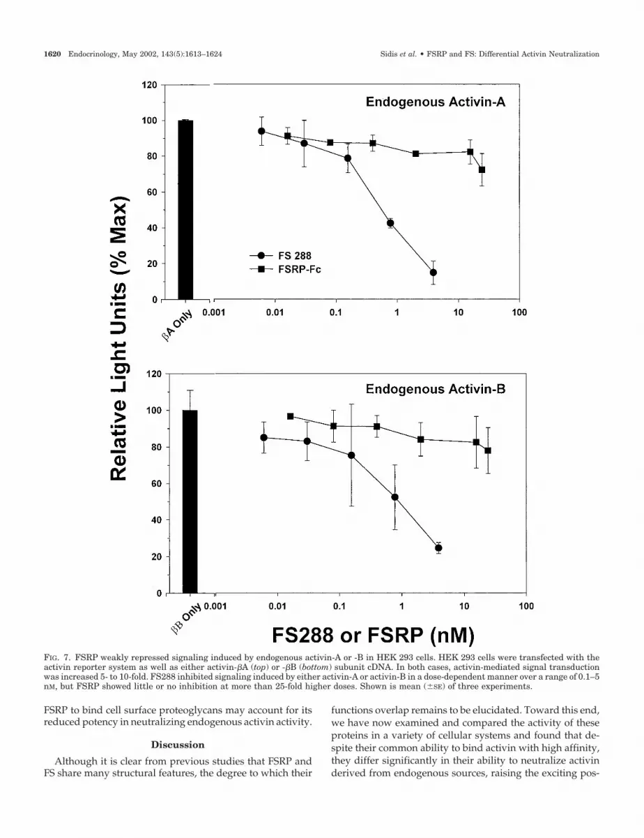
activin in this system, activin-�A or -�B subunit cDNA wascotransfected with the reporter, resulting in robust stimula-tion of reporter activity to levels comparable with thoseachieved by exogenous activin (Fig. 7). FS inhibited this basalactivin-A (Fig. 7, top) or activin-B (Fig. 7, bottom) activity ina dose-dependent manner as observed in the primary ratpituitary cells (Fig. 4) or L�T2 cell line (Fig. 5). In contrast,FSRP inhibited basal reporter activity by less than 20% atdoses at least 100-fold higher than those used for FS for thesame level of inhibition. Taken together, these results indi-cate that FSRP can inhibit exogenous activin-A with similarpotency to FS but is more than 100-fold less active than FSwhen the activin (A or B) is derived endogenously.
Inhibition of exogenous vs. endogenous activin in primarypituitary cells
To confirm these observations in a more physiologicalcontext, the neutralization activity of FS and FSRP werecompared using dispersed rat pituitary cultures in the pres-ence or absence of exogenous activin-A. As expected, in theabsence of exogenous activin, FS inhibited FSH release atsubnanomolar doses, and FSRP was inactive at more than
100-fold molar excess concentrations. However, in the pres-ence of 0.35 nm activin-A, a dose that maximally increasedFSH release by about 35%, both FS and FSRP inhibited theexogenous activin-mediated FSH secretion (Fig. 8). More-over, although higher doses of FS continued to inhibit FSHrelease mediated by endogenous activin, FSRP was unable tomodulate endogenous activin. These results therefore dem-onstrate that even in primary cells, FSRP, in stark contrast toFS, is able to neutralize activin from endocrine or paracrinesources but not autocrine-derived activin.
Binding to heparin sulfate or cell surface proteoglycans
FS288 can bind to cell surface heparin-sulfated proteogly-cans (30), under some conditions forming a barrier aroundcells such that exogenous activin is prevented from accessingits receptor (32). The absence of a consensus heparin bindingsequence in FSRP (Fig. 1) suggests that, unlike FS, FSRP willnot bind heparin or to cell surface heparin-sulfated proteo-glycans, perhaps accounting for its reduced ability to neu-tralize endogenous activin. As expected, FSRP was unable tobind to a negatively charged heparin-like matrix above thelevels attained by the buffer or BSA controls, whereas FS288
FIG. 4. FSRP did not repress the FSH secretion induced by endogenous activin in the rat pituitary bioassay. Rat anterior pituitaries weredispersed and cultured in the presence of either FS or FSRP. FS inhibited FSH secretion from rat anterior pituitary cells in a dose-dependentmanner over a range of 0.1–0.5 nM. However, neither highly purified recombinant FSRP-FLAG nor FSRP-Fc was active in this assay atconcentrations nearly 50-fold higher than the minimally effective concentration of FS. These same preparations of FSRP were active in receptorbinding and activin-binding assays. Shown are results from one of three representative experiments.
1618 Endocrinology, May 2002, 143(5):1613–1624 Sidis et al. • FSRP and FS: Differential Activin Neutralization

binding was 4-fold greater than controls (Fig. 9A). Similarly,FSRP was unable to bind to cell surface proteoglycans oncultured HEK 293 cells, whereas FS288 binding was 6-foldgreater than control wells (Fig. 9B). In both cases, FS288 or
FSRP binding was determined using labeled activin, and weconfirmed that the activin was bound to FS288 on the cellsurface by the addition of 10 �g/ml heparin sulfate, whichreleased the FS-activin complex (40). Thus, the inability of
FIG. 6. FSRP and FS repressed signaling induced by exogenous activin in HEK 293 cells. Activin-A treatment (0.3 nM) of HEK 293 cellstransiently transfected with an activin-responsive luciferase reporter system resulted in nearly 5-fold higher signal transduction over basal,and this effect was inhibited by increasing concentrations of FS288, reaching maximum at about 3 nM. FSRP-Fc was about approximately 2-foldless potent than FS288 in this assay, consistent with the 2.4-fold difference in their relative affinities for activin (26). Shown is the mean (�SE)for three experiments.
FIG. 5. FSRP weakly repressed sig-naling induced by endogenous activinin L�T2 cells. L�T2 cells transfectedwith an activin reporter system showa basal level of luciferase activity thatis potently inhibited by FS288 admin-istration. FSRP’s activity in this assayis approximately 100-fold lower thansimilar doses of FS, further demon-strating that FSRP is a relativelyweak inhibitor of endogenous activinactivity. Shown are results from one ofthree representative experiments.
Sidis et al. • FSRP and FS: Differential Activin Neutralization Endocrinology, May 2002, 143(5):1613–1624 1619
FSRP to bind cell surface proteoglycans may account for itsreduced potency in neutralizing endogenous activin activity.
Discussion
Although it is clear from previous studies that FSRP andFS share many structural features, the degree to which their
functions overlap remains to be elucidated. Toward this end,we have now examined and compared the activity of theseproteins in a variety of cellular systems and found that de-spite their common ability to bind activin with high affinity,they differ significantly in their ability to neutralize activinderived from endogenous sources, raising the exciting pos-
FIG. 7. FSRP weakly repressed signaling induced by endogenous activin-A or -B in HEK 293 cells. HEK 293 cells were transfected with theactivin reporter system as well as either activin-�A (top) or -�B (bottom) subunit cDNA. In both cases, activin-mediated signal transductionwas increased 5- to 10-fold. FS288 inhibited signaling induced by either activin-A or activin-B in a dose-dependent manner over a range of 0.1–5nM, but FSRP showed little or no inhibition at more than 25-fold higher doses. Shown is mean (�SE) of three experiments.
1620 Endocrinology, May 2002, 143(5):1613–1624 Sidis et al. • FSRP and FS: Differential Activin Neutralization

FIG. 9. FSRP does not associate with heparin-like matrix or cell surface proteoglycans. A, Binding to sulfate cellufine. Radiolabeled activin wasadded to tubes containing buffer, with or without BSA (nonspecific protein), FSRP, or FS. After 2 h, binding to sulfate cellufine, a heparinsulfate-like matrix, was assessed. FS288 bound to the matrix but FSRP binding was not different from controls, consistent with the lack of aheparin-binding domain in FSRP. B, Binding to HEK 293 cells. FSRP, FS, or BSA was added to culture HEK 293 cells for 2 h, the mediumchanged, and radiolabeled activin added for an additional 4 h. FS288-activin complexes bound well to cell surface proteoglycans, whereasFSRP-activin complexes did not bind above control levels. Heparin sulfate (10 �g/ml) inhibited FS288 binding to cell surface heparin-sulfatedproteoglycans as expected but had no effect on FSRP or control cells. Taken together, these studies confirm that the absence of a heparin-bindingmotif in FSRP likely precludes its binding to heparin or cell surface proteoglycans like FS. Shown are results from one of three representativeexperiments.
FIG. 8. Neutralization of activin by FS and FSRP in the primary rat pituitary assay. In the absence of added activin, FSRP had no activity,but FS inhibited endogenous activin at subnanomolar doses (compare open and closed circles). Addition of 0.35 nM activin-A, the maximallyeffective dose in this assay, increased FSH secretion by approximately 35% over basal. This stimulation by exogenous activin could be inhibitedby both FS and FSRP (compare open and closed triangles). However, FSH secretion because of endogenous activin was not blocked by FSRP,even at doses up to 100-fold greater than the minimally active FS dose (compare highest three open triangle doses to closed triangles), confirmingobservations in HEK 293 cells and indicating potential differential bioactivity for FSRP and FS. This figure shows one of three representativeexperiments.
Sidis et al. • FSRP and FS: Differential Activin Neutralization Endocrinology, May 2002, 143(5):1613–1624 1621

sibility that FSRP and FS have nonoverlapping cellularfunctions.
The striking homology in primary sequence, exon/intronarrangement, and domain structure previously identifiedbetween FSRP and FS supports the hypothesis that they haveoverlapping functions, perhaps predominating in differenttissues or at different developmental stages. Moreover, FSRP,like FS, binds activin with high affinity and nearly irrevers-ible kinetics and prefers activin over its close TGF-� super-family relatives BMP-6, BMP-7 (35), and BMP-2 (36). Finally,it was observed that FS plays a key role in Xenopus embryonicneuralization (19), yet the FS knockout mouse did not showsignificant neural defects (41). Because both FS and FSRPmRNA are expressed in the brain (35, 36), it is possible thatFSRP is able to functionally compensate for FS in the FSknockout mouse.
To explore further the degree of functional overlap of thesetwo proteins, we first employed a solid-phase competitivebinding assay previously used to characterize FS (4) to morecompletely characterize the relative specificity of FSRP andFS for activin and related ligands. Both FSRP and FS selec-tively bound activin, with the closely related BMP-7 (42)binding with approximately 500-fold lower potency. InhibinA, MIS, TGF-�1, and BMP-4 did not bind to either FS or FSRP.This agrees well with our previous demonstration of FSRPand FS directly binding radioiodinated activin, BMP-6, andBMP-7, with the latter ligands being at least 20- to 40-fold lessactive (35). Direct binding and inhibition of BMP-2 activityby FSRP and FS was also previously demonstrated (36),although the preparations of FSRP were only partially pu-rified so comparisons of relative affinity between the twobinding proteins could not be directly assessed. In addition,the tryptophan residues at positions 4 and 36 of FS, previ-ously shown to be critical for activin binding (26), are con-served in FSRP, suggesting that structural determinants ofactivin binding are preserved between the two molecules.Thus, the present data suggest that the determinants of li-gand specificity and binding are also preserved betweenFSRP and FS.
On the other hand, a number of differences between FSRPand FS have been identified that suggest the presence ofnonoverlapping roles in regulating activin action. We re-cently demonstrated that FSRP, unlike FS, can be found in thenucleus of many cell types; moreover, it appears to be se-creted only at expression levels higher than that required forits immunocytochemical detection within the nucleus (35).Although potential binding partners and nuclear actions ofFSRP remain to be defined, this difference in intracellulartrafficking suggests that FSRP may have biological roles thatare quite distinct from those attributable to FS. The obser-vations in the present study that FSRP signal peptide cleav-age results in an N-terminal extension of the critical activin-binding domain relative to FS and the greatly reduced abilityof FSRP to neutralize endogenously produced activin furthersupport this concept.
In fact, the nine-residue N-terminal extension of the Ndomain of FSRP is important from at least two standpoints.Because there appears to be no classic nuclear localizationsignal elsewhere within FSRP, this extension conceivably
could influence protein trafficking. Furthermore, our resultsshow that the addition of amino acids to the N-terminalregion of FS may not be as detrimental to activin binding aswas previously believed (43). Despite the nine additionalamino acids in its N-terminal domain, FSRP has an affinityfor activin quite comparable with that of FS (35).
In the present study, we used purified preparations of FSand FSRP to directly compare their activin-neutralizing ac-tivities in a variety of potentially physiological circum-stances. When we examined situations in which activinmight arrive from outside the responding cell, such as activinderived from paracrine or endocrine sources, the relativeinhibitory activities of FSRP and FS for activin binding to itsreceptor and the neutralization of activin-stimulated genetranscription were compatible with the 2.4-fold difference intheir affinities for activin (35). These results agree with therecent demonstration that relatively high doses (0.1–1 �g/ml) of a partially purified mouse FSRP preparation sup-pressed activin-A-stimulated signal transduction in K562and CHO cells. Unfortunately, this activity was not com-pared with FS so relative activities could not be directlyassessed (36).
On the other hand, when we compared the activin-neutralizing ability of FSRP and FS for activin derived fromthe responding cell, as with autocrine-derived activin, FSRPwas clearly much less potent. For example, in the primary ratpituitary cell bioassay in which FS was originally defined andactivin was produced by the gonadotrophs themselves, FSRPwas still inactive at concentrations nearly 100-fold higherthan the minimal effective concentration of FS. This obser-vation was confirmed using a mouse pituitary cell line(L�T2) in which endogenous activin-mediated reporter tran-scription is neutralized to a much greater degree by FS,compared with FSRP. Because our reporter system was max-imally stimulated in these cells, even in the absence of ex-ogenous activin, we could not use these cells to compare theeffects of FSRP and FS on activin derived from differentsources.
Thus, we used two approaches to investigate the differ-ential regulation of FSRP and FS for exogenous vs. endog-enous activin in the same in vitro assay. First, in a cellularsystem in which the dose of exogenous and endogenousactivin could be controlled, we employed HEK 293 cells,which contain a functional activin signaling system but donot produce detectable activin. When the cells were treatedwith exogenous activin, both FSRP and FS were able to neu-tralize activin-mediated gene transcription. However, if thecells were cotransfected with �A cDNA to produce endog-enous activin-A, FSRP was more than 50-fold less potent thanFS. Moreover, when �B cDNA was transfected to produceendogenous activin-B, FS had similar neutralizing activity tothat observed for activin-A, but FSRP was nearly inactiveeven at 100-fold higher concentrations. The second systemused primary rat pituitary monolayer cultures in the pres-ence or absence of exogenous activin. In this case, exogenousactivin was neutralized by both FS and FSRP with the ex-pected relative activity. However, higher doses of FS neu-tralized endogenous activin, whereas FSRP was unable to doso up to 100-fold molar excess over the minimally active FSdose. Taken together, these results confirm our observation
1622 Endocrinology, May 2002, 143(5):1613–1624 Sidis et al. • FSRP and FS: Differential Activin Neutralization

that FSRP and FS differentially neutralize exogenous vs. en-dogenous activin. Moreover, they demonstrate that FS iscapable of neutralizing both activin-A and activin-B withsimilar potency. Because many activin-responsive tissuesalso produce activin (12, 44–46), our results suggest thestrong possibility that the physiological activities of FSRPand FS do not entirely overlap.
At least one mechanism whereby FS inhibits activin actioninvolves binding to cell surface heparin-sulfated proteogly-cans (40), which is mediated by the heparin binding sequencelocated in FS domain 1 (Fig. 1). One consequence of the abilityof FS to bind cell surface proteoglycans is to form a barrier,at least at high concentrations, to the biological activity ofexogenous activin, as recently demonstrated in the FS288-secreting PA-1 teratocarcinoma cell line (32). Interestingly, inthese same cells, this FS barrier had no effect on endogenousactivin (32). In the present studies, however, exogenous FSwas able to modulate endogenous activin, both in the ratpituitary and in the L�T2 cell transcriptional assay. Thisdifference may be due to the level of activin productionrelative to the FS concentration outside the cell or may in-dicate that in some cells, activin can exert its biological ac-tion(s) before it is secreted and thus exposed to FS.
FSRP does not have a heparin-binding sequence, and ourstudies are a strong indication that its absence prevents cellsurface association of FSRP. Thus, it is possible that FSRP isunable to achieve the local concentration in the region of thecell surface activin receptor that FS can achieve, therebyleading to FSRP’s reduced ability to neutralize endogenousactivin signaling although it retains similar activity to FS inneutralizing exogenous activin. In any event, our resultssuggest that for exogenous activin, the relative ability ofextracellular FSRP and FS to neutralize its actions depend ontheir relative affinities for activin and their concentrationsrelative to activin. On the other hand, the ability to neutralizeendogenous activin appears to depend largely on the abilityof FS to associate with the cell surface and bind activin beforeit has an opportunity to access its type II receptor, an actionnot available to FSRP. Although mutational analysis of FSclearly correlates activin binding with biological activity (26),the distinctions between FS and FSRP described here suggestthat activin-binding activity is necessary but not sufficient toaccount for all of the actions of FS. Future investigation usingsite-directed mutagenesis and domain-swapping strategiesbetween FSRP and FS will likely shed additional light uponstructure-function relationships for these closely relatedproteins.
Acknowledgments
We are grateful to Dr. Al Parlow and the NIDDK’s National Hormoneand Pituitary Program for their provision of the rat FSH assay reagentsand FS288. We are also grateful to Pam Mellon for providing the L�T2cell line, Lawrence Matthews for providing the human ActRII construct,and Genentech, Inc. for providing the activin subunit cDNAs.
Received September 26, 2001. Accepted January 26, 2002.Address all correspondence and requests for reprints to: Alan L.
Schneyer, Ph.D., Reproductive Endocrine Unit BHX-5, MassachusettsGeneral Hospital, 55 Fruit Street, Boston, Massachusetts 02114. E-mail:[email protected].
Present address for D.V.T.: Columbia University College of Physi-cians and Surgeons, Department of Obstetrics and Gynecology, Pres-byterian Hospital, 16th Floor, New York, New York 10032.
This work was supported in part by NIH Grants HD-39777, DK-55838, HD-29164 (to A.L.S.), and DK-53828 (to H.T.K.).
References
1. Robertson DM, Klein R, de Vos FL, McLachlan RI, Wettenhall REH, HearnMTW, Burger HG, De Kretser DM 1987 The isolation of polypeptides withFSH suppressing activity from bovine follicular fluid which are structurallydifferent to inhibin. Biochem Biophys Res Commun 149:744–749
2. Shimasaki S, Koga M, Esch F, Cooksey K, Mercado M, Koba A, Ueno N, YingSY, Ling N, Guillemin R 1988 Primary structure of the human follistatinprecursor and its genomic organization. Proc Natl Acad Sci USA 85:4218–4222
3. Nakamura T, Takio K, Eto Y, Shibai H, Titani K, Sugino H 1990 Activin-binding protein from rat ovary is follistatin. Science 247:836–838
4. Schneyer AL, Rzucidlo DA, Sluss PM, Crowley Jr WF 1994 Characterizationof unique binding kinetics of follistatin and activin or inhibin in serum. En-docrinology 135:667–674
5. de Winter J, Ten Dijke P, de Vries CJM, van Achterberg TAE, Sugino H, DeWaele P, Huylebroeck D, Verschueren K, Eijnden-van Raaij AJM 1996 Fol-listatins neutralize activin bioactivity by inhibition of activin binding to its typeII receptors. Mol Cell Endocrinol 116:105–114
6. Hashimoto O, Nakamura T, Shoji H, Shimasaki S, Hayashi Y, Sugino S 1997A novel role of follistatin, an activin-binding protein, in the inhibition of activinaction in rat pituitary cells. J Biol Chem 272:13835–13842
7. Kim SK, Hebrok M, Li E, Oh SP, Schrewe H, Harmon EB, Lee JS, Melton DA2000 Activin receptor patterning of foregut organogenesis. Genes Dev 14:1866–1871
8. Asashima M, Ariizumi T, Malacinski GM 2000 In vitro control of organo-genesis and body patterning by activin during early amphibian development.Comp Biochem Physiol B Biochem Mol Biol 126:169–178
9. DePaolo LV, Bicsak TA, Erickson GF, Shimasaki S, Ling N 1991 Follistatinand activin: a potential intrinsic regulatory system within diverse tissues. ProcSoc Exp Med 198:500–512
10. Kogawa K, Nakamura T, Sugino K, Takio K, Titani K, Sugino H 1991Activin-binding protein is present in pituitary. Endocrinology 128:1434–1440
11. Corrigan AZ, Bilezikjian LM, Carroll RS, Bald LN, Schmelzer CH, FendlyBM, Mason AJ, Chin WW, Schwall RH, Vale W 1991 Evidence for an auto-crine role of activin B within anterior pituitary cultures. Endocrinology 128:1682–1684
12. Bilezikjian LM, Vaughan JM, Vale WW 1993 Characterization and the reg-ulation of inhibin/activin subunit proteins of cultured rat anterior pituitarycells. Endocrinology 133:2545–2553
13. Findlay JK 1993 An update on the roles of inhibin, activin, and follistatin aslocal regulators of folliculogenesis. Biol Reprod 48:15–23
14. Cataldo NA, Rabinovici J, Fujimoto VY, Jaffe RB 1994 Follistatin antagonizeseffects of activin-A on steroidogenesis in human luteinizing granulosa cells.J Clin Endocrinol Metab 79:272–277
15. Alak BM, Coskun S, Friedman CI, Kennard EA, Kim MH, Seifer DB 1998Activin A stimulates meiotic maturation of human oocytes and modulatesgranulosa cell steroidogenesis in vitro. Fertil Steril 70:1126–1130
16. Hashimoto M, Shoda A, Inoue S, Yamada R, Kondo T, Sakurai T, Ueno N,Maramatsu M 1992 Functional regulation of osteoblastic cells by the interac-tion of activin-A with follistatin. J Biol Chem 267:4999–5004
17. Funaba M, Ogawa K, Murata T, Fujimura H, Murata E, Abe M, TakahashiM, Torii K 1996 Follistatin and activin in bone: expression and localizationduring endochondral bone development. Endocrinology 137:4250–4259
18. Inoue S, Nomura S, Hosoi T, Ouchi Y, Orimo H, Muramatsu M 1994 Lo-calization of follistatin, an activin-binding protein, in bone tissues. CalcifTissue Int 55:395–397
19. Hemmati-Brivanlou A, Kelly OG, Melton DA 1994 Follistatin, an antagonistof activin, is expressed in the Spemann Organizer and displays direct neu-ralizing activity. Cell 77:283–295
20. Asashima M, Nakano H, Uchiyama H, Sugino H, Nakamura T, Eto Y, EjimaD, Nishimatsu S, Ueno N, Kinoshita K 1991 Presence of activin (erythroiddifferentiation factor) in unfertilized eggs and blastulae of Xenopus laevis. ProcNatl Acad Sci USA 88:6511–6514
21. Wang Q, Tabatabaei S, Planz B, Lin CW, Sluss PM 1999 Identification of anactivin-follistatin growth modulatory system in the human prostate: secretionand biological activity in primary cultures of prostatic epithelial cells. J Urol161:1378–1384
22. Miralles F, Czernichow P, Scharfmann R 1998 Follistatin regulates the relativeproportions of endocrine versus exocrine tissue during pancreatic develop-ment. Development 125:1017–1024
23. Kogure K, Zhang YQ, Kanzaki M, Omata W, Mine T, Kojima I 1996 Intra-venous administration of follistatin: delivery to the liver and effect on liverregeneration after partial hepatectomy. Hepatology 24:361–366
24. Qu J, Thomas K 1998 Advance in the study of inhibin, activin and follistatinproduction in pregnant women. Eur J Obstet Gynecol Reprod Biol 81:141–148
Sidis et al. • FSRP and FS: Differential Activin Neutralization Endocrinology, May 2002, 143(5):1613–1624 1623
25. Wang Q, Keutmann HT, Schneyer AL, Sluss PM 2000 Analysis of humanfollistatin structure: identification of two discontinuous N-terminal sequencescoding for activin A binding and structural consequences of activin bindingto native proteins. Endocrinology 141:3183–3193
26. Sidis Y, Schneyer AL, Sluss PM, Johnson LN, Keutmann HT 2001 Follistatin:essential role for the N-terminal domain in activin binding and neutralization.J Biol Chem 276:17718–17726
27. Patthy L, Nikolics K 1993 Functions of agrin and agrin-related proteins.Trends Neurosci 16:76–81
28. Lane TF, Sage EH 1994 The biology of SPARC, a protein that modulatescell-matrix interactions. FASEB J 8:163–173
29. Alliel PM, Perin JP, Jolles P, Bonnet FJ 1993 Testican, a multidomain testicularproteoglycan resembling modulators of cell social behaviour. Eur J Biochem214:347–350
30. Nakamura T, Sugino K, Titani K, Sugino H 1991 Follistatin, an activin-binding protein, associates with heparan sulfate chains of proteoglycans onfollicular granulosa cells. J Biol Chem 266:19432–19437
31. Schneyer AL, Wang QF, Weiss J, Boepple P, Hall J, Khoury R, Taylor A,Pralong F, Sluss P, Crowley WF 1997 Follistatin physiology and potentialmechanisms of action in the human. In: Aono T, Sugino H, Vale WW, eds.Inhibin, activin and follistatin: regulatory functions in system and cell biology.New York: Springer-Verlag; 28–38
32. Delbaere A, Sidis Y, Schneyer AL 1999 Differential response to exogenous andendogenous activin in a human ovarian teratocarcinoma-derived cell line(PA-1): regulation by cell surface follistatin. Endocrinology 140:2463–2470
33. Hayette S, Gadoux M, Martel S, Bertrand S, Tigaud I, Magaud J, Rimokh R1998 FLRG (follistatin-related gene), a new target of chromosomal rearrange-ment in malignant blood disorders. Oncogene 22:2949–2954
34. Schneyer A, Tortoriello D, Sidis Y, Keutmann H, Matsuzaki T, Holmes W2001 Follistatin-related protein (FSRP): a new member of the follistatin genefamily. Mol Cell Endocrinol 180:33–38
35. Tortoriello DV, Sidis Y, Holtzman DA, Holmes WE, Schneyer AL 2001
Human follistatin-related protein: a structural homologue of follistatin withnuclear localization. Endocrinology 142:3426–3434
36. Tsuchida K, Arai KY, Kuramoto Y, Yamakawa N, Hasegawa Y, Sugino H 2000Identification and characterization of a novel follistatin-like protein as a bind-ing protein for the TGF-beta family. J Biol Chem 275:40788–40796
37. Bernstein JR, Crowley Jr WF Schneyer AL 1990 An improved method ofpurifying inhibin radioligand for radioimmunoassay. Biol Reprod 43:492–496
38. Schneyer AL, Sluss PM, Bosukonda D, Reichert LE 1986 Electrophoreticpurification of radioiodinated follicle-stimulating hormone for radioligandreceptor assay and radioimmunoassay. Endocrinology 119:1446–1453
39. Pernasetti F, Vasilyev VV, Rosenberg SB, Bailey JS, Huang HJ, Miller WL,Mellon PL 2001 Cell-specific transcriptional regulation of follicle-stimulatinghormone-beta by activin and gonadotropin-releasing hormone in the LbetaT2pituitary gonadotrope cell model. Endocrinology 142:2284–2295
40. Nakamura T, Sugino S, Titani K, Sugino H 1991 Follistatin, an activin bindingprotein, associates with heparan sulfate chains of proteoglycans on folliculargranulosa cells. J Biol Chem 266:19432–19437
41. Matzuk MM, Lu N, Vogel HJ, Sellheyer K, Roop DR, Bradley A 1995 Multipledefects and perinatal death in mice deficient in follistatin. Nature 374:360–363
42. Yamashita H, Ten Dijke P, Huylebroeck D, Sampath TK, Andries M, SmithJC, Heldin CH, Miyazono K 1995 Osteogenic protein-1 binds to activin typeII receptors and induces certain activin-like effects. J Cell Biol 130:217–226
43. Inouye S, Guo Y, Ling N, Shimasaki S 1991 Site-specific mutagenesis ofhuman follistatin. Biochem Biophys Res Commun 179:352–358
44. Ni X, Luo S, Minegishi T, Peng C 2000 Activin A in JEG-3 cells: potential roleas an autocrine regulator of steroidogenesis in humans. Biol Reprod 62:1224–1230
45. Risbridger GP, Cancilla B 2000 Role of activins in the male reproductive tract.Rev Reprod 5:99–104
46. DePaolo LV 1997 Inhibins, activins, and follistatins: the saga continues. ProcSoc Exp Biol Med 214:328–339
The European Journal of Endocrinology Prize
The European Journal of Endocrinology Prize is awarded during the European Congress of Endocrinology toa candidate who has significantly contributed to the advancement of knowledge in the field of endocrinologythrough publication.
The prize consists of a certificate and Euro 7,250 plus traveling expenses and will be presented during the6th European Congress of Endocrinology to be held in Lyon, France from 26–30 April 2003. Nominationsshould be submitted to the Chief Editor of the European Journal of Endocrinology, Professor Paolo Beck-Peccoz,Istituto di Scienze Endocrine, Piano Terra, Padiglione Granelli, Ospedale Maggiore IRCCS, Via FrancescoSforza 35, 20122 Milan, Italy, by 31 October 2002. For more detailed information please check the websiteswww.eje.org and www.endocrinology2003.com or contact our office at the following e-mail address: [email protected].
1624 Endocrinology, May 2002, 143(5):1613–1624 Sidis et al. • FSRP and FS: Differential Activin Neutralization