mapping n-linked glycosylation sites in the secretome and whole cells of aspergillus niger using...
TRANSCRIPT
Published: November 07, 2011
r 2011 American Chemical Society 143 dx.doi.org/10.1021/pr200916k | J. Proteome Res. 2012, 11, 143–156
ARTICLE
pubs.acs.org/jpr
Mapping N-Linked Glycosylation Sites in the Secretome andWhole Cells of Aspergillus niger Using Hydrazide Chemistryand Mass SpectrometryLuWang,‡Uma K. Aryal,‡ Ziyu Dai,§ Alisa C. Mason,‡Matthew E. Monroe,‡ Zhi-Xin Tian,‡ Jian-Ying Zhou,‡
Dian Su,‡ Karl K. Weitz,‡ Tao Liu,‡ David G. Camp, II,‡ Richard D. Smith,‡ Scott E. Baker,§ andWei-Jun Qian*,‡
‡Biological Science Division and Environmental Molecular Sciences Laboratory, §Energy Process and Materials Division,Pacific Northwest National Laboratory, Richland, Washington 99352, United States
bS Supporting Information
’ INTRODUCTION
Filamentous fungi, such as Aspergillus niger, are well-known fortheir industrial applications. They are used to produce a diversevariety of products ranging from human therapeutics to specialtychemicals.1�6 Popularly known as an “industrial workhorse”,A. niger is capable of secreting large amounts of native proteinssuch as hydrolytic enzymes (e.g., glucoamylase) into the growthmedium. Many of those hydrolytic enzymes are widely usedfor biomass conversion into ethanol and other renewable chemi-cals and advanced biofuel precursors through fermentationprocesses. Similar to other microbial expression systems, A. nigerhas also been utilized as an expression host for heterologousproteins of industrial/pharmaceutical significance.7 Some fila-mentous fungi are also known as pathogens in both humans8,9
and plants.10 Because of their importance, an increasing numberof fungal genomes have recently been sequenced,11�15 layingthe foundation for functional genomics studies, for example,protein post-translational modifications (PTMs), in filamentousfungi.
Protein glycosylation is one of the most common and struc-turally diverse PTMs. N- and O-linked protein glycosylationrepresent two of the most common types of glycosylation thatoccur on asparagine (N) and serine/threonine (S/T) residues,respectively. N-linked glycosylation has been implicated in avariety of biochemical and cellular processes, including proteinsecretion, stability and translocation, maintenance of cell struc-ture, receptor�ligand interactions and cell signaling, cell�cellrecognition, pathogen infection, and host defense.16�18 Despitetheir importance, few studies have been performed on theglycoproteome in fungal systems. Therefore, the full range ofbiochemical and cellular functions of N-glycosylation in thesesystems is still waiting to be determined.16,19,20 In this work, wepresent the first global profiling of N-glycosylated sites in A. niger
Special Issue: Microbial and Plant Proteomics
Received: September 9, 2011
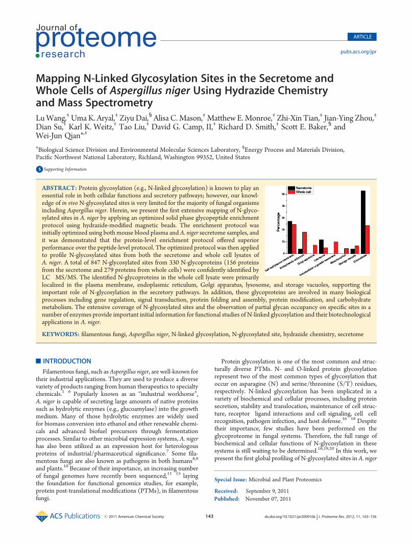
ABSTRACT: Protein glycosylation (e.g., N-linked glycosylation) is known to play anessential role in both cellular functions and secretory pathways; however, our knowl-edge of in vivo N-glycosylated sites is very limited for the majority of fungal organismsincluding Aspergillus niger. Herein, we present the first extensive mapping of N-glyco-sylated sites in A. niger by applying an optimized solid phase glycopeptide enrichmentprotocol using hydrazide-modified magnetic beads. The enrichment protocol wasinitially optimized using both mouse blood plasma and A. niger secretome samples, andit was demonstrated that the protein-level enrichment protocol offered superiorperformance over the peptide-level protocol. The optimized protocol was then appliedto profile N-glycosylated sites from both the secretome and whole cell lysates ofA. niger. A total of 847 N-glycosylated sites from 330 N-glycoproteins (156 proteinsfrom the secretome and 279 proteins from whole cells) were confidently identified byLC�MS/MS. The identified N-glycoproteins in the whole cell lysate were primarilylocalized in the plasma membrane, endoplasmic reticulum, Golgi apparatus, lysosome, and storage vacuoles, supporting theimportant role of N-glycosylation in the secretory pathways. In addition, these glycoproteins are involved in many biologicalprocesses including gene regulation, signal transduction, protein folding and assembly, protein modification, and carbohydratemetabolism. The extensive coverage of N-glycosylated sites and the observation of partial glycan occupancy on specific sites in anumber of enzymes provide important initial information for functional studies of N-linked glycosylation and their biotechnologicalapplications in A. niger.
KEYWORDS: filamentous fungi, Aspergillus niger, N-linked glycosylation, N-glycosylated site, hydrazide chemistry, secretome

144 dx.doi.org/10.1021/pr200916k |J. Proteome Res. 2012, 11, 143–156
Journal of Proteome Research ARTICLE
for gaining initial functional insight on the N-glycosylatedproteins.
With the recent advances in proteomic technologies, LC�MS/MS has become the key tool for analyzing PTMs, includingN-glycosylation.21,22 Similar to other PTMs, specific enrichmentof the N-glycopeptides is essential in order to comprehensivelycharacterize the often low abundance glycopeptides. A number ofenrichment methods, including lectin affinity,23�26 hydrophilicinteraction,27,28 and solid phase extraction using hydrazidechemistry,29�33 have been developed and applied for character-izing both N-glycoproteins and N-glycosylated sites.
For this study, we adapted the solid phase enrichment ofglycopeptide (SPEG) approach originally developed by Zhanget al.34 using hydrazide chemistry, where chemically oxidizedglycans are captured onto hydrazide-modified magnetic beadsand N-glycopeptides are released using the deglycosylatingenzyme PNGase F.29,35 The enrichment protocols were firstassessed with both mouse plasma and A. niger secretome bycomparing the peptide-level and protein-level enrichments. Thevalidated protocol was subsequently applied for N-glycopeptideenrichment in both the secretome and the whole cell lysatesamples of A. niger to gain an extensive coverage of N-glycosy-lated sites in the proteome. A total of 847 unique N-glycosylatedsites from 330 N-linked glycoproteins from both the secretomeand the whole cells were confidently identified in A. niger.Complex glycosylation patterns were observed in the aspectsof multiple glycosylation sites per protein as well as partial glycanoccupancies on specific sites. The extensive coverage of N-linkedglycoproteins and their glycosylated sites provides detailedinformation regarding cellular localization, molecular function,and biological processes for which the identified glycoproteinsare potentially involved.
’EXPERIMENTAL SECTION
MaterialsMouse blood plasma (protein concentration of 42 mg/mL)
was purchased from Equitech Bio, Inc. (Kerrville, TX). Hydrazide-modified magnetic beads with 1 and 5 μm diameters were obtainedfrom Bioclone, Inc. (San Diego, CA). The magnetic bead separatorwas from Invitrogen (Carlsbad, CA). The glycerol free PNGase Fwas from New England Biolabs, Inc. (Ipswich, MA). All otherreagents were from SigmaAldrich (St. Louis, MO) unless otherwisenoted.
Strain, Media, and Culture MethodsA. niger (NRRL3122), obtained from the American Type Cul-
ture Collection (Rockville, MD), was grown on potato dextroseagar (PDA) plates at 30 �C for culture maintenance and sporeproduction. Cultures were incubated for 4 days and the sporeswere harvested by washing with sterile 0.8% Tween 80 (polyoxy-ethylenesorbitan monooleate). Spores were enumerated with ahemacytometer. Aliquots of the resulting spore suspension (1�109 spores/mL) were used to inoculate baffled-flask liquidcultures. For preparation of both the secretome and whole celllysate, 2� 106 spores/mL were inoculated and grown in 200 mLof modified minimal medium in 1 L baffled flasks in an Innova4330 refrigerated incubator shaker (New Brunswick Scientific,NJ) at 30 �C and 200 rpm. The modified minimal medium perliter contained 18.2 g of D-sorbitol, 6 g of NaNO3, 0.52 g ofKCl, 0.52 g of MgSO4(H2O)7, 1.52 g of KH2PO4, 200 μL of1 M CaCl2, 1 g of yeast extract, and 2 mL of Tween 80 (100%).
The yeast extract is the water-soluble portion of autolyzed yeastby self-digestion of the cellular constituents of yeast cells using itsown enzymes, in which all proteins are broken down to smallpeptides (<1 kDa) and amino acids. The medium pH wasadjusted to 6.5 with NaOH before autoclaving. The mediumper liter also contained 50 mg of Na4EDTA, 0.512 mg ofZnSO4(H2O)7, 1.4 mg of MnCl2(H2O)4, 0.4 mg of CoCl2-(H2O)6, 0.4 mg of CuSO4(H2O)5, 0.034 mg of Na2MoO4-(H2O)2, 1.9 mg of H3BO3, 1 mg of FeSO4(H2O)7, 1 mg ofbiotin, 1 mg of pyridoxine, 1 mg of thiamine, 1 mg of riboflavin,1 mg of PABA (p-aminobenzoic acid), and 1mg of nicotinic acid.The spores were grown for the first 15 h in modified minimalmedium. Then, the cultures were grown another 2 h beforeharvesting following the addition of 10 mM of D-xylose for theinduction of glycosyl hydrolase production or water as thenoninduction condition.
Protein ExtractionTo prepare the secretome sample, the mycelia were separated
from the culture medium by filtration through two layers ofsterile miracloth (Calbiochem, San Diego, CA) and the super-natant was collected separately. Themycelia were washed 3 timeswith ice-cold 50 mM phosphate buffer (pH 7.0), immediatelyfrozen in liquid nitrogen for 10 min, and stored at �80 �C untilfurther use. The filtered medium (supernatant) was centrifugedat 15 000g at 4 �C for 10 min to remove any cell debris. Theproteins in the supernatant were concentrated to ∼40-fold at4 �C and 4000g in a swinging bucket rotor with an Amicon ultra-15 centrifugal filter (MWCO 10 kDa, Millipore, Billerica, MA).Residual yeast extract (molecular weight typically <1 kDa) notconsumed in cell growth was also removed by this filteringprocess. The concentrated secreted proteins were precipitatedovernight at �20 �C using 5 vol of acetone. After centrifugationat 4 �C and 15 000g for 15 min, the precipitated proteins werewashed twice with cold 80% acetone (�20 �C) and solubilizedin 8 M urea buffer for 2 h at room temperature. The samplewas centrifuged again at 15 000g to remove insoluble materialsand the protein concentration was measured using the BCA(bicinchoninic acid) assay (Pierce, Rockford, IL).
To extract proteins from whole cells, frozen mycelia wereground into a fine powder using a chilled mortar and pestle, andthen resuspended in the extraction buffer containing 100 mMTris-HCl, 2% sodium dodecyl sulfate (SDS), 1% dithiothreitol(DTT), 10% glycerol, and 0.5% protease inhibitor cocktail(P8215, Sigma, St. Louis, MO). After incubating at room tem-perature for 1 h with intermittent vortexing, insoluble materialwas removed by centrifugation (15 000g for 15 min). The sub-sequent preparation of the total proteins was the same as thepreparation for the secretome sample described above.
Protein Digestion for Global AnalysisThe secretome proteins were denatured in 8 M urea for 1 h
and reduced with 10 mMDTT for an additional 30 min at 37 �C.The free thiols were then alkylated with 40mM iodoacteamide at37 �C in the dark for 1 h. The proteins were diluted 10-fold with50 mMNH4HCO3 buffer (pH 7.8) and digested at 37 �C for 3 hwith sequencing grade modified porcine trypsin (Promega,Madison, WI) at a 1:50 trypsin to protein ratio (w/w). Thedigested samples were loaded on a 1-mL SPE C18 column(Supelco, Bellefonte, PA) and washed with 4 mL of 0.1%trifluoroacetic acid (TFA)/5% acetonitrile (ACN). Peptideswere eluted from the SPE column with 1 mL of 0.1% TFA/80% ACN and lyophilized.
145 dx.doi.org/10.1021/pr200916k |J. Proteome Res. 2012, 11, 143–156
Journal of Proteome Research ARTICLE
Protein-Level N-Glycopeptide EnrichmentThemodified protein-level enrichment protocol was similar to
those previously described30,31 in which hydrazide-modifiedmagnetic beads were used instead of the original agarose bead.Protein samples were diluted with coupling buffer (100 mMsodium acetate, 150 mM NaCl, pH 5.5) to a final concentrationof 1�5 mg/mL and then oxidized with 15 mM sodium periodate(NaIO4) at room temperature in the dark for 1 h. Eight molarurea was added to solubilize whole cell proteins during oxidation.The excess NaIO4 was removed after oxidation by performingbuffer exchange with coupling buffer containing 0.2% CHAPSusing an Amicon ultra-4 centrifugal filter (MWCO 30 kDa,Millipore, Billerica, MA). The concentrated proteins were thenincubated overnight at room temperature with an appropriateamount (10 mg beads for up to 2 mg proteins) of hydrazide-modified magnetic beads prewashed twice with 1 mL of couplingbuffer. Unbound proteins in the supernatant were removed usingthe magnetic bead separator. The beads were then washed
3 times with 2 mL of coupling buffer and 6 times with 2 mL ofurea solution (8 M urea, 0.4 M NH4HCO3, pH 8.3) to removenonspecifically bound proteins.
Glycoproteins bound on the beads were denatured andreduced by 8 M urea and 10 mM DTT for 1 h at 37 �C, andalkylated with 40 mM iodoacteamide for 1 h in the dark at 37 �C.The glycoproteins were then washed with 8 M urea and 50 mMNH4HCO3 three times each. The glycoproteins on the beadswere then digested overnight at 37 �C with trypsin with a 1:100enzyme to protein mass ratio. Nonglycopeptides were removedby washing 6 times with each of the following four solutions inthe order listed: (1) 80% ACN and 0.1% TFA, (2) 8 M urea in0.4 M NH4HCO3 with 0.1% SDS, (3) dimethyformamide(DMF), and (4) 0.1 M NH4HCO3 (pH 7.8). N-glycopeptideswere released by incubating the beads with 3 μL of PNGase F(500 units/ μL) in 1 mL of 0.1 M NH4HCO3 buffer in athermomixer (Cat # 022670107, Eppendorf, Westbury, NY) at37 �Cand 900 rpm for 5 h. Released glycopeptides were collected
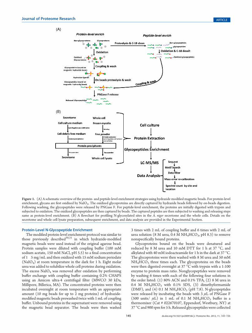
Figure 1. (A) A schematic overview of the protein- and peptide-level enrichment strategies using hydrazide modified magnetic beads. For protein-levelenrichment, glycans are first oxidized by NaIO4. The oxidized glycoproteins are directly captured by hydrazide beads followed by on-beads digestion.Following washing, the glycopeptides were released by PNGase F. For peptide-level enrichment, the proteins are initially digested with trypsin andsubjected to oxidation. The oxidized glycopeptides are then captured by beads. The captured peptides are then subjected to washing and releasing stepssame as protein-level enrichment. (B) A flowchart for profiling N-glycosylated sites in the A. niger secretome and the whole cells. Details on thesecretome and whole cell lysate preparation, subsequent enrichment, and data analysis are provided in the Experimental Section.
146 dx.doi.org/10.1021/pr200916k |J. Proteome Res. 2012, 11, 143–156
Journal of Proteome Research ARTICLE
from the supernatant and the magnetic beads were then washedtwice with 200 μL of 80% ACN and 0.1% TFA. The washsolutions were combined with the supernatant collected from thePNGase F incubation and dried down to a final volume of 100 μLusing a Speed-vac concentrator.
Peptide-Level N-Glycopeptide EnrichmentThe modified peptide-level enrichment protocol was similar
to the one previously described.35 Digested peptides were oxi-dized with 15mMNaIO4 for 1 h in the dark at room temperature.After oxidation, the excess NaIO4 was removed by SPE C-18cleanup. Oxidized peptides eluted in 80% ACN and 0.1% TFAwere incubated overnight with an appropriate amount (10 mgbeads for up to 2 mg proteins) of prewashed hydrazide magneticbeads in the thermomixer at room temperature. Nonglycopep-tides in the supernatant were removed by using the magneticseparator. Nonspecifically bound peptides were removed bywashing 6 times with the same four solutions in the same orderas described in the previous section. Glycopeptides on the beadswere released and collected with the same protocol as describedin the protein-level enrichment.
LC�MS/MS AnalysisEnriched glycopeptide samples were analyzed using a fully
automated, custom-built, four-column capillary LC systemcoupled online to an LTQ-Orbitrap mass spectrometer (ThermoFisher Scientific, San Jose, CA) via an electrospray ionization
(ESI) interfacemanufactured in-house.The75μm(innerdiameter)�65 cm fused silica capillary reversed phase column (PolymicroTechnologies, Phoenix, AZ) was prepared using the slurry-packing procedure with 3 μm diameter Jupiter C18 bondedparticles (Phenomenex, Torrence, CA) at 8000 psi. The mobilephases consisted of solution A (0.1% formic acid in water) andsolution B (0.1% formic acid in ACN). An exponential gradientstarting with 100% of mobile phase A to 60% of mobile phase Bover the course of 100 min was employed with a flow-rateof ∼500 nL/min over the separation column. The instrumentwas operated in data-dependent mode with an m/z range of400�2000 with a resolution of 100K for full MS scans. The10 most abundant ions from the MS analysis were selected forMS/MS analysis using a normalized collision energy setting of35% and a dynamic exclusion duration of 1min. The temperatureof the heated capillary and the ESI voltage were 200 �C and2.2 kV, respectively.
Data AnalysisLC�MS/MS raw data were converted into “.dta” files using
Extract_MSn (version 3.0) from Bioworks Cluster 3.2 (ThermoScientific), and the SEQUEST algorithm (version 27, revision 12)was used to independently search all the MS/MS spectra againstthe DOE Joint Genome Institute (JGI) A. niger v3.0 proteindatabase that had 11 200 entries. The false discovery rate (FDR)was estimated based on the decoy-database searching methodo-logy.36,37 The following dynamic modifications were included
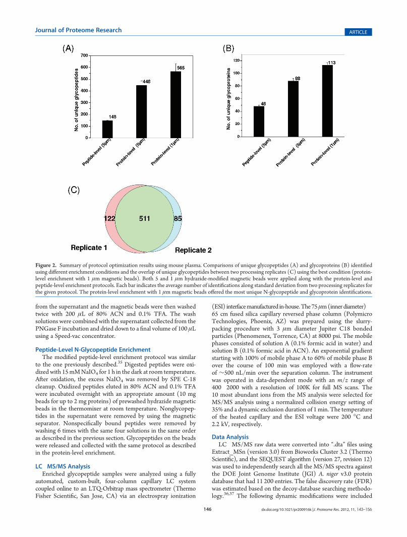
Figure 2. Summary of protocol optimization results using mouse plasma. Comparisons of unique glycopeptides (A) and glycoproteins (B) identifiedusing different enrichment conditions and the overlap of unique glycopeptides between two processing replicates (C) using the best condition (protein-level enrichment with 1 μm magnetic beads). Both 5 and 1 μm hydrazide-modified magnetic beads were applied along with the protein-level andpeptide-level enrichment protocols. Each bar indicates the average number of identifications along standard deviation from two processing replicates forthe given protocol. The protein-level enrichment with 1 μm magnetic beads offered the most unique N-glycopeptide and glycoprotein identifications.
147 dx.doi.org/10.1021/pr200916k |J. Proteome Res. 2012, 11, 143–156
Journal of Proteome Research ARTICLE
during the SEQUEST analysis: carbamidomethylation of cysteine,oxidation of methionine, and a PNGase F-catalyzed conversionof asparagine to aspartic acid at the site of glycan attachment(a mass increase of 0.9848 Da). MS Generating-Function(MSGF) scores were generated for each identified spectrum asdescribed previously by computing rigorous p-values (spectralprobabilities).38 The following filtering criteria were used tocontrol the FDR to be <0.01%: (1) MSGF scores were less than1 � 10�8 and 1 � 10�10 for fully and partially tryptic peptides,respectively, and mass measurement error less than 5 ppm; (2)for cases where nonmonoisotopic peaks were picked for frag-mentation with mass measurement errors of 1 Da or more, anMSGF score less than 1 � 10�11 for both fully and partiallytryptic peptides was used. The presence of at least one N-X-S/Tmotif, where X is any amino acids except proline, was required forall final N-glycopeptide identifications. For all glycopeptidesidentified with mass measurement errors greater than 1 Da,manual inspection of the raw MS spectra was performed andpeptide identifications were confirmed when the large errorswere found to be due to reporting the wrongmonoisotopic peaksand the actual mass errors were within 5 ppm. The glycosylatedsites were assigned based on the motif information withinpeptides. For peptides containing multiple N-X-S/T motifs,the sites of modification were also manually inspected basedon MS/MS fragment ions. Uniprot and gene ontology (GO)definitions, and other terms extracted from text-based annotationfiles downloaded from the JGI Web site (http://genome.jgi-psf.org/) were used to annotate the identified proteins.
’RESULTS
Analytical StrategyFigure 1 shows a schematic illustration of the overall enrich-
ment and analysis of N-glycopeptides using hydrazide chemistry.Both protein- and peptide-level enrichment protocols have beenapplied in proteomic studies within different organisms includingmammalian samples30,31,34,35 and zebrafish.39 In this method, thecis-diols of glycans are oxidized to aldehydes using NaIO4. Theoxidized glycans are captured by hydrazide-modified magneticbeads through formation of covalent hydrazone bonds betweenthe oxidized aldehyde and hydrazide groups on the beads. TheN-glycopeptides are finally released from magnetic beads withPNGase F. The enzyme converts the glycan attached asparagineresidues within the glycopeptides into aspartic acid, resulting in amass increment of 0.9848 Da for each N-glycosylated site.Recently, Berven et al. performed an assessment of glycopeptidecapture protocols and observed differences in performancebetween the protein-level and peptide-level captures usinghydrazide-modified magnetic beads.31 Building upon their ob-servations, we performed further evaluation of the enrichmentprotocols as shown in Figure 1A using mouse plasma and A. nigersecretome samples in order to determine the optimal protocolfor mapping the N-glycosylated sites in A. niger. Given theimportance of N-glycosylation in cellular regulation40�42 andprotein secretion,20 we analyzed the N-linked glycoproteins inboth the secretome and whole cell lysate of A. niger. Figure 1Billustrates the workflow for profiling N-glycosylated sites with
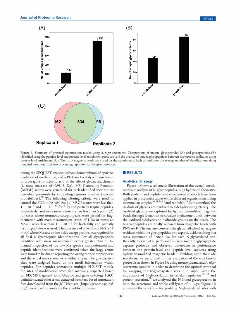
Figure 3. Summary of protocol optimization results using A. niger secretome. Comparisons of unique glycopeptides (A) and glycoproteins (B)identified using the peptide-level and protein-level enrichment protocols and the overlap of unique glycopeptides between two process replicates usingprotein-level enrichment (C). The 1 μmmagnetic beads were used for the experiments. Each bar indicates the average number of identifications alongstandard deviation from two processing replicates for the given protocol.
148 dx.doi.org/10.1021/pr200916k |J. Proteome Res. 2012, 11, 143–156
Journal of Proteome Research ARTICLE
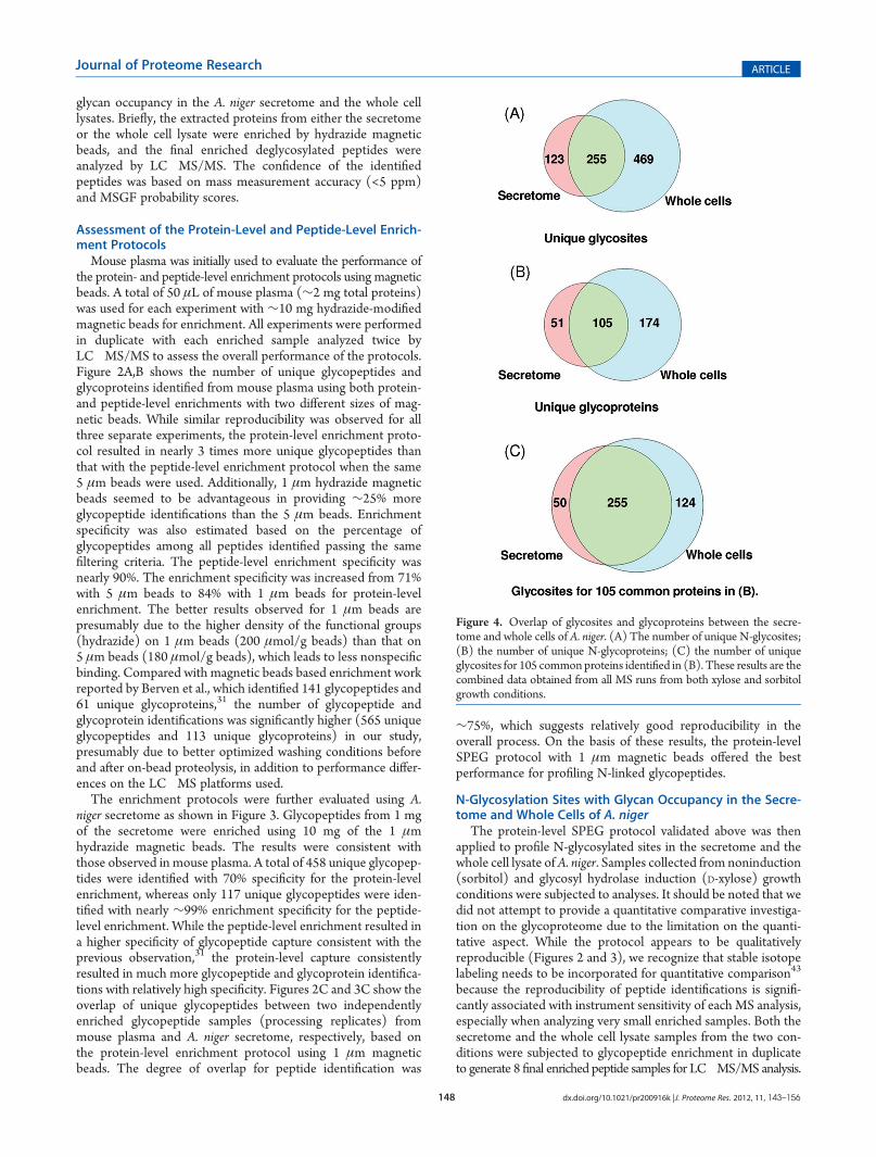
glycan occupancy in the A. niger secretome and the whole celllysates. Briefly, the extracted proteins from either the secretomeor the whole cell lysate were enriched by hydrazide magneticbeads, and the final enriched deglycosylated peptides wereanalyzed by LC�MS/MS. The confidence of the identifiedpeptides was based on mass measurement accuracy (<5 ppm)and MSGF probability scores.
Assessment of the Protein-Level and Peptide-Level Enrich-ment Protocols
Mouse plasma was initially used to evaluate the performance ofthe protein- and peptide-level enrichment protocols using magneticbeads. A total of 50 μL of mouse plasma (∼2 mg total proteins)was used for each experiment with ∼10 mg hydrazide-modifiedmagnetic beads for enrichment. All experiments were performedin duplicate with each enriched sample analyzed twice byLC�MS/MS to assess the overall performance of the protocols.Figure 2A,B shows the number of unique glycopeptides andglycoproteins identified from mouse plasma using both protein-and peptide-level enrichments with two different sizes of mag-netic beads. While similar reproducibility was observed for allthree separate experiments, the protein-level enrichment proto-col resulted in nearly 3 times more unique glycopeptides thanthat with the peptide-level enrichment protocol when the same5 μm beads were used. Additionally, 1 μm hydrazide magneticbeads seemed to be advantageous in providing ∼25% moreglycopeptide identifications than the 5 μm beads. Enrichmentspecificity was also estimated based on the percentage ofglycopeptides among all peptides identified passing the samefiltering criteria. The peptide-level enrichment specificity wasnearly 90%. The enrichment specificity was increased from 71%with 5 μm beads to 84% with 1 μm beads for protein-levelenrichment. The better results observed for 1 μm beads arepresumably due to the higher density of the functional groups(hydrazide) on 1 μm beads (200 μmol/g beads) than that on5 μm beads (180 μmol/g beads), which leads to less nonspecificbinding. Compared with magnetic beads based enrichment workreported by Berven et al., which identified 141 glycopeptides and61 unique glycoproteins,31 the number of glycopeptide andglycoprotein identifications was significantly higher (565 uniqueglycopeptides and 113 unique glycoproteins) in our study,presumably due to better optimized washing conditions beforeand after on-bead proteolysis, in addition to performance differ-ences on the LC�MS platforms used.
The enrichment protocols were further evaluated using A.niger secretome as shown in Figure 3. Glycopeptides from 1 mgof the secretome were enriched using 10 mg of the 1 μmhydrazide magnetic beads. The results were consistent withthose observed in mouse plasma. A total of 458 unique glycopep-tides were identified with 70% specificity for the protein-levelenrichment, whereas only 117 unique glycopeptides were iden-tified with nearly ∼99% enrichment specificity for the peptide-level enrichment. While the peptide-level enrichment resulted ina higher specificity of glycopeptide capture consistent with theprevious observation,31 the protein-level capture consistentlyresulted in much more glycopeptide and glycoprotein identifica-tions with relatively high specificity. Figures 2C and 3C show theoverlap of unique glycopeptides between two independentlyenriched glycopeptide samples (processing replicates) frommouse plasma and A. niger secretome, respectively, based onthe protein-level enrichment protocol using 1 μm magneticbeads. The degree of overlap for peptide identification was
∼75%, which suggests relatively good reproducibility in theoverall process. On the basis of these results, the protein-levelSPEG protocol with 1 μm magnetic beads offered the bestperformance for profiling N-linked glycopeptides.
N-Glycosylation Sites with Glycan Occupancy in the Secre-tome and Whole Cells of A. niger
The protein-level SPEG protocol validated above was thenapplied to profile N-glycosylated sites in the secretome and thewhole cell lysate ofA. niger. Samples collected from noninduction(sorbitol) and glycosyl hydrolase induction (D-xylose) growthconditions were subjected to analyses. It should be noted that wedid not attempt to provide a quantitative comparative investiga-tion on the glycoproteome due to the limitation on the quanti-tative aspect. While the protocol appears to be qualitativelyreproducible (Figures 2 and 3), we recognize that stable isotopelabeling needs to be incorporated for quantitative comparison43
because the reproducibility of peptide identifications is signifi-cantly associated with instrument sensitivity of each MS analysis,especially when analyzing very small enriched samples. Both thesecretome and the whole cell lysate samples from the two con-ditions were subjected to glycopeptide enrichment in duplicateto generate 8 final enriched peptide samples for LC�MS/MS analysis.
Figure 4. Overlap of glycosites and glycoproteins between the secre-tome and whole cells of A. niger. (A) The number of unique N-glycosites;(B) the number of unique N-glycoproteins; (C) the number of uniqueglycosites for 105 commonproteins identified in (B). These results are thecombined data obtained from all MS runs from both xylose and sorbitolgrowth conditions.

149 dx.doi.org/10.1021/pr200916k |J. Proteome Res. 2012, 11, 143–156
Journal of Proteome Research ARTICLE
Following stringent filtering (see details in Experimental Sections),a total of 7617 spectra were confidently identified as N-glyco-peptides from the combined data with the consensus N-X-S/Tmotif, corresponding to 1554 unique glycopeptides (Supplemen-tary Table 1) and 847 N-glycosylated sites (SupplementaryTable 2) in 330 N-glycoproteins (Supplementary Table 3).Figure 4 shows the overlap of the identified N-glycosylated sites
and N-glycoproteins between the secretome and whole cells.In this study, 724 and 378 N-glycosylated sites (Figure 4A) thatcorresponded to 279 and 156 N-glycoproteins (Figure 4B) wereidentified in the whole cells and the secretome, respectively.Among them, 105 N-glycoproteins were common to both com-partments, while the remaining N-glycoproteins were onlydetected in the whole cells or the secretome. Selected enzymes
Table 1. Selected Enzymes Identified As Glycoproteins in A. niger Secretome and Whole Cells
reference ID
protein
description
no. of
N-glycosylated
sites
spectral
count
(secretome)
spectral
count
(whole cells) site IDs*
Glycoside Hydrolases
43342 Alpha-glucosidase 2 3 3 10 279, 376, 655
52061 Acid trehalase 14 35 84 48, 138, 240, 310, 325, 621, 647, 670, 808,
832, 946, 969, 1005, 1047
53033 1,3-Beta-glucanosyltransferase gel3 5 32 151 38, 56,150, 168, 413
53702 Alpha-L-fucosidase 2 4 3 6 258, 280, 659, 703
55270 Beta-1,3-endoglucosidase 3 21 14 55, 375, 598
56782 Beta-glucosidase 1 9 15 33 211, 252, 322, 387, 442, 523, 542, 690, 712
138876 Beta-mannosidase 5 8 16 79, 483, 665, 673, 793
140567 Alpha-amylase A 3 83 6 45, 198, 218
194447 Endoglucanase/exoglucanase 2 13 1 113, 131
196122 Probable glycosidase crf2 2 81 7 61, 266
205517 Alpha-1,2-mannosidase 3 13 4 97, 117, 184
205670 Thermostable beta-glucosidase 13 56 269 124, 148, 242, 251, 357, 390, 413, 444, 573,
576, 665, 696, 718
207264 Alpha-galactosidase 3 52 0 156, 232, 282
208214 Beta-1,3-glucosidase 1 19 2 272
213597 Glucoamylase 3 783 83 195, 206, 419
Proteases
55493 Tripeptidyl-peptidase 1 9 255 193 44, 95, 202, 230, 257, 363, 440, 525, 611
55665 Tripeptidyl-peptidase 1 7 133 100 103, 128, 212, 230, 451, 462, 569
56689 Serine protease 8 28 41 55, 65, 74, 143, 260, 277, 377, 455
191956 Aspartic acid protease 5 14 31 64, 108, 123, 145, 295
211797 Acid protease 4 13 41 136, 143, 247, 163
Oxidases
198099 Chloroperoxidase 3 20 28 131, 225, 240
45804 6-hydroxy-D-nicotine oxidase 5 40 21 43, 57, 324, 361, 374
38973 Mitomycin radical oxidase 5 28 29 121, 261, 304, 448, 472
131352 Laccase (copper-containing oxidase) 6 37 24 23, 199, 316, 325, 413, 421
Phosphatases and Phospholipases
57215 Phosphate-repressible acid phosphatase 4 18 11 110, 161, 295, 578
56950 Lysophospholipase 13 8 97 51, 70, 80, 94, 110, 179, 233, 250, 296,
364, 474, 526, 554
210730 Mono- and diacylgerol lipase 2 13 0 59, 150
Miscellaneous Proteins
51794 Ribonuclease 2 45 16 97, 106
52629 Glycolipid-anchored surface protein 7 26 59 219, 255, 311,341, 359, 385,429
55604 Aldose 1-epimerase 6 16 44 88, 132, 149, 191, 290, 380
36604 Glutamyl-tRNA(Gln) amidotransferase
subunit A
8 36 36 23, 46, 209, 301, 321, 393, 416, 465
50333 3-phytase 3 4 14 162, 378, 387* Site ID indicates the amino acid residue site numbered starting from the N-terminus of protein sequence in the FASTA database.

150 dx.doi.org/10.1021/pr200916k |J. Proteome Res. 2012, 11, 143–156
Journal of Proteome Research ARTICLE
and proteins involved in various key biological processes werelisted in Table 1, which included a number of glycoside hydro-lases, peptidases and proteases, oxidases, phosphatases andphospholipases, and other miscellaneous proteins. These pro-teins included many secretory proteins44 reported previouslysuch as glucoamylase, 6-hydroxy-D-nicotine oxidase, aspartic acidprotease, and GPI-anchored protein. Several proteins that were
not previously reported in the secretome44,45 were also identifiedas glycoproteins, including acid trehalase and α-L-fucosidase.
More glycosylated sites were identified for the proteins fromthe whole cells than those from the secretome in the 105 com-mon glycoproteins (Figure 4C). Fifty sites were only observed insecretory glycoproteins from the secretome and 124 sites only inthe whole cell glycoproteins besides the 255 sites observed inglycoproteins from both the secretome and the whole cells.Figure 5 further illustrates the distribution of glycoproteins basedon the number of glycosylated sites observed per protein. Amongthe N-glycoproteins identified in the secretome, 43% (67 out of156) glycoproteins were identified with only one glycosylatedsite. The N-glycoproteins identified in the whole cells also hadsimilar distribution, where approximately 5% of them wereheavily glycosylated with more than 8 glycosylated sites. Theresults suggest that glycosylation patterns of N-glycoproteinsmay be different between secreted glycoproteins and the glyco-proteins expressed from the same genes inside the cells.
Partial Glycan Occupancy Observed on Specific N-Glycosy-lation Sites
By comparing the peptide identifications from the globaldigests and enriched N-glycopeptide samples, we observed theevidence that some glycosylation sites were only partially glyco-sylated because the same peptide was observed in enrichedsamples as a glycosylated form as well as in global digests as anonglycosylated form. Figure 6 shows a representative pair ofMS/MS spectra for peptide K.TPAGGYAQFLTNQTALK.G
Figure 5. Distribution of glycoproteins identified in both the secretomeand whole cells based on the number of glycosylated sites per protein.While ∼40% glycoproteins were identified with only one N-glycosite, amajority of glycoproteins have multiple glycosites.
Figure 6. MS/MS spectra indicative of partial glycosylation. Asterisk onN*QT indicates the N-glycosylated site with the consensus motif in bold. (A)Formerly N-glycosylated form, in which the asparagine was converted to aspartic acid at the site of glycan attachment due to PNGase F cleavage, leadingto a mass increase of 0.9848 Da; (B) nonmodified form of the peptide originating from protein 36604 (glutaminyl-tRNA synthase). The N-glycosylatedsite is indicated by an asterisk, and the consensus motif appears in bold. The y-fragment ions highlighted by circles in the top panel show∼1 Da increasecompared to those ions in the bottom panel, indicative of the modification.

151 dx.doi.org/10.1021/pr200916k |J. Proteome Res. 2012, 11, 143–156
Journal of Proteome Research ARTICLE
from protein 36604, a putative amidase, in both glycosylated andnonglycosylated forms. The MS/MS spectra of 2+ ions with m/z= 892.97 and m/z= 891.46 resulted in the confident identifica-tion of the fully tryptic, formerly N-glycosylated peptide (post-cleavage by PNGase F) in enriched sample (top) and nonglyco-sylated version in the global digest (bottom). The ∼1 Daincrease in the serial y-ions clearly indicates the modificationon the specific site with NQT motif. Table 2 listed 18 N-glyco-sylation sites with partial glycan occupancy from 16 proteinsobserved in the secretome, which included hydrolytic enzymes(glucosidase, protease, and peptidase) and several other enzymesas well as some unknown proteins.
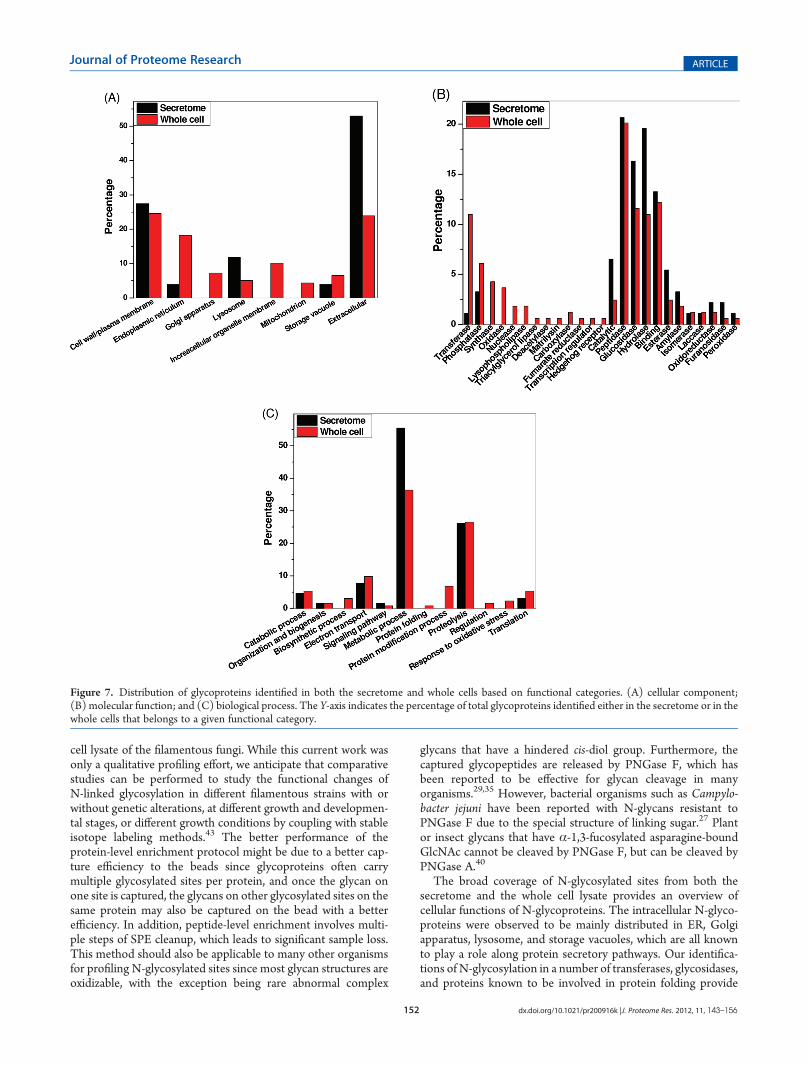
Functional Categories of N-Glycoproteins in A. nigerTo provide an overall picture of the general functions of these
identified N-linked glycoproteins, functional analyses were per-formed based on the Gene Ontology (GO) information in termsof cellular component, molecular function, and biological pro-cess. Figure 7A shows the distributions of glycoproteins in dif-ferent cellular components. As expected, the secreted proteinswere mainly annotated as in the extracellular, cell surface orplasma membrane, lysosome, and storage vacuole compart-ments. In addition to these components, the intracellular glyco-proteins were localized in endoplasmic reticulum (ER), Golgiapparatus, the membranes of other organelles, andmitochondria.
Figure 7B shows the distribution of molecular functions forproteins identified in both secretome and whole cells. Peptidase,glucosidase, hydrolase, and binding were the major molecularfunctions of N-glycoproteins observed in both the secretome andthe whole cells. Glycoproteins involved in a number of molecularfunctions were observed either uniquely in the cells or withsignificantly higher percentage in the cells than in the secretome.These categories included transferase, synthase, oxidase, nucle-ase, lipase, and several other categories (Figure 7B). The resultsindicate that many glycoproteins involved in these categories arenot secreted by the cells. Figure 7C displays the distribution of
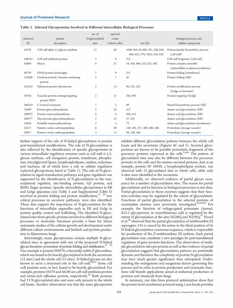
glycoproteins in terms of biological process. These glycoproteinswere involved in different metabolic processes, proteolysis, andcatabolic processes in both the secretome and the whole celllysate, which were consistent with the known functions ofA. nigeras a producer of hydrolytic enzymes. In addition to these bio-logical processes common to the secretome and the whole cells,the data also revealed glycoproteins that were involved in anumber of intracellular biological processes such as biosyntheticprocess, protein folding, protein modification, regulation, andresponse to oxidative stress. Table 3 lists some selected proteinsinvolved in different intracellular biological processes.
’DISCUSSION
The significance of filamentous fungi in biomass degradationand biofuel production, as well as other industrial applications,has been well recognized due to their ability in producingimportant enzymes and other products.5,46�52 It has been knownthat N-glycosylation exerts its effects in different biologicalfunctions53�55 and heterologous protein production.2 Thewell-known role of N-glycosylation is in the protein secretorypathway.20 Our work represents the first proteomics effort forprofiling the in vivo N-glycosylated sites in a filamentous fungus,A. niger, though enzymes involved in the glycosylation pathwayhave been globally predicted previously with the genome seq-uence database.56 By applying magnetic bead-based hydrazidechemistry and high mass accuracy LC�MS/MS, N-glycopep-tides along with specific glycosylated sites were identified withhigh confidence. The identified 847 N-glycosylated sites in 330glycoproteins from the secretome and the whole cells representan important resource for using A. niger to gain better under-standing of N-glycosylation in cellular functions, secretory path-ways, and protein production.
In this study, we demonstrated that the magnetic bead-basedhydrazide chemistry offers a robust strategy for enrichingN-linked glycopeptides from either the secretome or the whole
Table 2. List of Partially Glycosylated Sites Observed in the A. niger Secretome
reference
ID
protein
description
peptide
sequence
site
residue
spectral count
(unmodified)
spectral
count
(glycosylated)
170223 Endo-1,3(4)-beta-glucanase 1 K.KGIEN*VTTPIETNK.F 227 1 2
176050 Unknown protein R.QSAN*FTDTESQK.V 95 2 19
179912 Cholinesterase R.GFEHN*DTNVFLGIPFAETTAGENR.W 42 3 2
204301 Glycolipid-anchored surface protein 5 K.N*FTGYGLPLFLSEYGCNTNK.R 251 4 3
205670 Thermostable beta-glucosidase B K.HYAGYDIENWHN*HSR.L 242 1 4
205670 Thermostable beta-glucosidase B K.N*NTNVSALLWGGYPGQSGGFALR.D 573 1 2
208214 Glucan 1,3-beta-glucosidase K.GTDVFYFEAFDEVWKPN*STGDNGQSMDEK.H 272 1 19
211032 Tripeptidyl-peptidase 1 K.LN*NSDLPQVISTSYGEDEQTIPVPYAR.T 346 4 4
36048 Unknown protein T.N*ATTPSTFGIMSAR.S 26 12 31
36604 Cytokinin dehydrogenase 2 K.TPAGGYAQFLTN*QTALK.G 321 5 9
37529 Unknown protein K.SPGATTYDLCLYDVCGSGN*GSGR.V 121 2 11
37529 Unknown protein R.LFTPGCDPN*TTSFDLSIFHR.Q 63 3 1
38973 Mitomycin radical oxidase K.AHPDATISGAALEFTIAN*ITSDLFYEAVER.F 304 2 12
42759 Unknown protein K.IGN*YSFQNVVGVGHSAGSTLTQAITTQYPK.D 195 4 7
45021 Pectin lyase K.IGSN*TSIIGK.D 95 2 2
52703 Serine protease K.LQFDN*SSR.S 162 1 11
54860 Unknown protein K.VLTNGTGNYCASAQEDN*ATLEAMVR.A 269 2 6
56689 Serine protease K.SAFGGSATN*LSNQDFVSSLSYGLDSFQSR.N 277 7 1
152 dx.doi.org/10.1021/pr200916k |J. Proteome Res. 2012, 11, 143–156
Journal of Proteome Research ARTICLE
cell lysate of the filamentous fungi. While this current work wasonly a qualitative profiling effort, we anticipate that comparativestudies can be performed to study the functional changes ofN-linked glycosylation in different filamentous strains with orwithout genetic alterations, at different growth and developmen-tal stages, or different growth conditions by coupling with stableisotope labeling methods.43 The better performance of theprotein-level enrichment protocol might be due to a better cap-ture efficiency to the beads since glycoproteins often carrymultiple glycosylated sites per protein, and once the glycan onone site is captured, the glycans on other glycosylated sites on thesame protein may also be captured on the bead with a betterefficiency. In addition, peptide-level enrichment involves multi-ple steps of SPE cleanup, which leads to significant sample loss.This method should also be applicable to many other organismsfor profiling N-glycosylated sites since most glycan structures areoxidizable, with the exception being rare abnormal complex
glycans that have a hindered cis-diol group. Furthermore, thecaptured glycopeptides are released by PNGase F, which hasbeen reported to be effective for glycan cleavage in manyorganisms.29,35 However, bacterial organisms such as Campylo-bacter jejuni have been reported with N-glycans resistant toPNGase F due to the special structure of linking sugar.27 Plantor insect glycans that have α-1,3-fucosylated asparagine-boundGlcNAc cannot be cleaved by PNGase F, but can be cleaved byPNGase A.40
The broad coverage of N-glycosylated sites from both thesecretome and the whole cell lysate provides an overview ofcellular functions of N-glycoproteins. The intracellular N-glyco-proteins were observed to be mainly distributed in ER, Golgiapparatus, lysosome, and storage vacuoles, which are all knownto play a role along protein secretory pathways. Our identifica-tions of N-glycosylation in a number of transferases, glycosidases,and proteins known to be involved in protein folding provide
Figure 7. Distribution of glycoproteins identified in both the secretome and whole cells based on functional categories. (A) cellular component;(B) molecular function; and (C) biological process. The Y-axis indicates the percentage of total glycoproteins identified either in the secretome or in thewhole cells that belongs to a given functional category.
153 dx.doi.org/10.1021/pr200916k |J. Proteome Res. 2012, 11, 143–156
Journal of Proteome Research ARTICLE
further support of the role of N-linked glycosylation in proteinpost-translational modifications. The role of N-glycosylation isalso reflected by the identification of specific glycoproteins invarious intracellular regulatory enzymes such as cell wall α-1,3-glucan synthase, cell elongation protein, transferase, phospha-tase, triacylglycerol lipase, lysophospholipase, oxidase, reductase,and nuclease, all of which have a role in cellular regulation(selected glycoproteins listed in Table 1). The role of N-glyco-sylation in signal transduction pathways and gene regulation wassupported by the identification of N-glycosylation in the tran-scriptional regulator, zinc-binding protein, Gβ protein, andRING finger proteins. Specific intracellular glycoproteins in ERand Golgi apparatus (see Table 3 and Supplemental Table 3)involved in protein folding and protein modification,57�59 twocritical processes in secretory pathways, were also identified.These data support the importance of N-glycosylation for thefunctions of intracellular organelles such as ER and Golgi inprotein quality control and trafficking. The identified N-glyco-sylated sites from specific proteins involved in different biologicalprocesses or molecular functions provide a basis for futuresystematic studies of the cellular growth and development underdifferent culture environments and biofuels and protein produc-tion in filamentous fungi.
Interestingly, many glycoproteins contain multiple N-glyco-sylated sites, in agreement with one of the proposed N-linkedglycan functions: promotion of protein folding and stabilization.57�59
One example is protein 205670, a thermally stable β-glucosidase,which was found to be heavily glycosylated in both the secretome(12 sites) and the whole cell (11 sites). N-linked glycans are alsoknown to serve a structural role in the cell wall.60,61 Some cellwall proteins were, in fact, found to be heavily glycosylated. Forexample, proteins 54378 and 56240 are cell wall synthesis proteinand serine-rich adhesion protein, respectively.62 Both proteinshad 13 N-glycosylated sites and were only present in the wholecell lysate. Another observation was that the same glycoprotein
exhibits different glycosylation patterns between the whole celllysate and the secretome (Figures 4C and 5). Secreted glyco-proteins are known to be possible proteolytic fragments of theprecursor proteins expressed in the cells.63,64 The pattern ofglycosylated sites may also be different between the precursorproteins in the cells and the mature secreted proteins. Just as anexample, protein ID 56950, a lysophospholipase protein, wasobserved with 13 glycosylated sites in whole cells, while only4 sites were identified in the secretome.
Additionally, we observed evidence of partial glycan occu-pancy for a number of glycosylation sites. The reason for partialglycosylation and its function in biological processes is not clear.Partial glycosylation in those enzymes suggests that their func-tion/activities may be regulated by the extent of glycosylation.Functions of partial glycosylation in the selected proteins ofmammalian systems were previously investigated.24,65,66 Forexample, the function of voltage-gated potassium channel,Kv3.1 glycoprotein, in neuroblastoma cells is regulated by theextent of glycosylation at the sites N220Q and N229Q.67 Picardet al.66 observed that the partial glycosylation of antithrombin IIIasperagine-135 is caused by the serine in the third position of itsN-linked glycosylation consensus sequence, which is responsiblefor production of the β-antithrombine III isoform. Such partialglycosylation may constitute a new paradigm for post-translationalregulation of gene/protein functions. The observation of multi-ple glycosylation sites per protein as well as the evidence of partialglycosylation suggests that glycosylation patterns are potentiallydynamic and therefore the complexity of protein N-glycosylationmay have much greater significance than anticipated. Under-standing the endogenous and exogenous factors governing thisprocess and its roles in protein production and enzymatic func-tions will benefit applications aimed at industrial production ofproteins and chemicals from fungi.
In summary, our data from protocol optimization show thatthe protein-level enrichment protocol using 1 μmbeads provides
Table 3. Selected Glycoproteins Involved in Different Intracellular Biological Processes
reference
ID
protein
description
no. of
N-glycosylated
sites
spectral
count
(whole cells) site IDs
biological process and
cellular component
54378 Cell wall alpha-1,3-glucan synthase 13 82 1040, 825, 84, 602, 591, 220, 858,
869, 653, 378, 1022, 810, 832
Polysaccharide biosynthetic process
(cell wall)
180332 Cell wall synthesis protein 1 8 313 Cell wall biogenesis (cell wall)
42808 Mucin 6 31 54, 456, 468, 532, 627, 692 Protein complex assembly
(extracellular or plasmamembrane)
49729 DNAJ protein homologue 1 9 311 Protein folding (membrane)
124206 Uncharacterized J domain-containing
protein
1 7 42 Protein folding (ER)
212533 Palmitoyl-protein thioesterase 3 13 96, 231, 225 Protein modification process
(Golgi or lysosome)
54756 Vacuolar protein sorting/targeting
protein PEP1
2 11 326, 963 Protein targeting (Golgi)
206219 C-8 sterol isomerase 1 3 49 Steroid biosynthetic process (ER)
52489 Protein glycosyltransferase 1 18 317 Amino acid glycosylation (ER)
209072 Protein mannosyltransferase 2 4 394, 413 Amino acid glycosylation (ER)
209577 Glycoprotein glucosyltransferase 2 15 57, 325 Amino acid glycosylation (ER)
53826 Probable mannosyltransferase 1 5 72 Amino acid glycosylation (membrane)
52517 Putative serine carboxypeptidase 6 69 159, 185, 317, 409, 500, 586 Proteolysis (storage vacuole)
43917 Putative serine carboxypeptidase 3 13 48, 128, 180 Proteolysis (storage vacuole)
154 dx.doi.org/10.1021/pr200916k |J. Proteome Res. 2012, 11, 143–156
Journal of Proteome Research ARTICLE
improved coverage of N-linked glycopeptides, and a basis forfuture studies on the role of N-linked glycosylation in, forexample, cellular regulation in order to achieve improved in-dustrial production. The method is broadly applicable for globalprofiling of N-glycosylated sites in different organisms. In theapplication toA. niger, the coverage of 847 glycosylated sites from330 glycoproteins provided important insights into the functionsof N-linked glycoproteins involved in this industrially importantfungal model organism. The data also revealed complex glyco-sylation patterns in terms of multiple glycosylated sites perprotein as well as patterns of partial glycosylation for differentglycosylation sites. The biological implications of partial andcomplex glycosylation patterns warrant further investigations.
’ASSOCIATED CONTENT
bS Supporting InformationA complete list of identified glycopeptides, glycosylated sites,
and glycoproteins along with corresponding spectral count infor-mation is available in a Microsoft Excel worksheet. This materialis available free of charge via the Internet at http://pubs.acs.org.
’AUTHOR INFORMATION
Corresponding Author*Dr. Wei-Jun Qian, Biological Sciences Division, Pacific NorthwestNational Laboratory, P.O. Box 999, MSIN: K8-98, Richland, WA99352. Tel: 509-371-6572. Fax: 509- 371-6564. E-mail: [email protected].
’ACKNOWLEDGMENT
Portions of this work were supported by DOE Early CareerResearch Award and National Institutes of Health grant RR18522.Upstream cell culture was conducted in Fungal BiotechnologyLaboratory, which was supported by the DOE Office of theBiomass Program. Proteomics experiments were performed inthe Environmental Molecular Sciences Laboratory, a U.S. Depart-ment of Energy (DOE) Office of Biological and EnvironmentalResearch national scientific user facility on the Pacific NorthwestNational Laboratory (PNNL) campus. PNNL is multiprogramnational laboratory operated by Battelle for the DOE underContract No. DE-AC05-76RLO 1830.
’ABBREVIATIONS:
PTM, post-translational modification; SPEG, solid phase enrich-ment of glycopeptides; MSGF, MS generating function; GO,gene ontology; ER, endoplasmic reticulum
’REFERENCES
(1) Nevalainen, K. M.; Te’o, V. S.; Bergquist, P. L. Heterologousprotein expression in filamentous fungi. Trends Biotechnol. 2005, 23 (9),468–474.(2) Gerngross, T. U. Advances in the production of human therapeutic
proteins in yeasts and filamentous fungi. Nat. Biotechnol. 2004, 22 (11),1409–4414.(3) Punt, P. J.; van Biezen, N.; Conesa, A.; Albers, A.; Mangnus, J.;
van den Hondel, C. Filamentous fungi as cell factories for heterologousprotein production. Trends Biotechnol. 2002, 20 (5), 200–206.(4) Schuster, E.; Dunn-Coleman, N.; Frisvad, J. C.; Van Dijck, P. W.
On the safety of Aspergillus niger—A review. Appl. Microbiol. Biotechnol.2002, 59 (4�5), 426–435.
(5) Magnuson, J. K.; Lasure, L. L. Organic acid production byfilamentous fungi. Adv. Fungal Biotechnol. Ind., Agric., Med. 2004,307–340.
(6) Sauer, M.; Porro, D.; Mattanovich, D.; Branduardi, P. Microbialproduction of organic acids: Expanding the markets. Trends Biotechnol.2008, 26 (2), 100–108.
(7) Devchand, M.; Gwynne, D. I. Expression of heterologousproteins in Aspergillus. J. Biotechnol. 1991, 17 (1), 3–9.
(8) Segal, B. H.; Barnhart, L. A.; Anderson, V. L.; Walsh, T. J.;Malech, H. L.; Holland, S.M. Posaconazole as salvage therapy in patientswith chronic granulomatous disease and invasive filamentous fungalinfection. Clin. Infect. Dis. 2005, 40 (11), 1684.
(9) Cutler, S. J.; Fooks, A. R.; van der Poel, W. H. M. Public healththreat of new, reemerging, and neglected zoonoses in the industrializedworld. Emerging Infect. Dis. 2010, 16 (1), 1.
(10) Maor, R.; Shirasu, K. The arms race continues: battle strategiesbetween plants and fungal pathogens. Curr. Opin. Microbiol. 2005, 8 (4),399–404.
(11) Galagan, J. E.; Calvo, S. E.; Cuomo, C.; Ma, L. J.; Wortman,J. R.; Batzoglou, S.; Lee, S. I.; Basturkmen, M.; Spevak, C. C.; Clutterbuck,J.; Kapitonov, V.; Jurka, J.; Scazzocchio, C.; Farman, M.; Butler, J.; Purcell,S.; Harris, S.; Braus, G. H.; Draht, O.; Busch, S.; D’Enfert, C.; Bouchier, C.;Goldman, G. H.; Bell-Pedersen, D.; Griffiths-Jones, S.; Doonan, J. H.; Yu,J.; Vienken, K.; Pain, A.; Freitag, M.; Selker, E. U.; Archer, D. B.; Penalva,M. A.; Oakley, B. R.; Momany, M.; Tanaka, T.; Kumagai, T.; Asai, K.;Machida, M.; Nierman, W. C.; Denning, D. W.; Caddick, M.; Hynes,M.; Paoletti, M.; Fischer, R.; Miller, B.; Dyer, P.; Sachs, M. S.; Osmani,S. A.; Birren, B. W. Sequencing of Aspergillus nidulans and comparativeanalysis with A. fumigatus and A. oryzae. Nature 2005, 438 (7071),1105–1115.
(12) Machida, M.; Asai, K.; Sano, M.; Tanaka, T.; Kumagai, T.;Terai, G.; Kusumoto, K.; Arima, T.; Akita, O.; Kashiwagi, Y.; Abe, K.;Gomi, K.; Horiuchi, H.; Kitamoto, K.; Kobayashi, T.; Takeuchi, M.;Denning, D. W.; Galagan, J. E.; Nierman, W. C.; Yu, J.; Archer, D. B.;Bennett, J. W.; Bhatnagar, D.; Cleveland, T. E.; Fedorova, N. D.; Gotoh,O.; Horikawa, H.; Hosoyama, A.; Ichinomiya, M.; Igarashi, R.; Iwashita,K.; Juvvadi, P. R.; Kato, M.; Kato, Y.; Kin, T.; Kokubun, A.; Maeda, H.;Maeyama, N.; Maruyama, J.; Nagasaki, H.; Nakajima, T.; Oda, K.;Okada, K.; Paulsen, I.; Sakamoto, K.; Sawano, T.; Takahashi, M.; Takase,K.; Terabayashi, Y.;Wortman, J. R.; Yamada, O.; Yamagata, Y.; Anazawa,H.; Hata, Y.; Koide, Y.; Komori, T.; Koyama, Y.; Minetoki, T.; Suharnan,S.; Tanaka, A.; Isono, K.; Kuhara, S.; Ogasawara, N.; Kikuchi, H.Genome sequencing and analysis of Aspergillus oryzae. Nature 2005,438 (7071), 1157–1161.
(13) Dean, R. A.; Talbot, N. J.; Ebbole, D. J.; Farman, M. L.;Mitchell, T. K.; Orbach, M. J.; Thon, M.; Kulkarni, R.; Xu, J. R.;Pan, H.; Read, N. D.; Lee, Y. H.; Carbone, I.; Brown, D.; Oh, Y. Y.;Donofrio, N.; Jeong, J. S.; Soanes, D. M.; Djonovic, S.; Kolomiets, E.;Rehmeyer, C.; Li, W.; Harding, M.; Kim, S.; Lebrun, M. H.; Bohnert, H.;Coughlan, S.; Butler, J.; Calvo, S.; Ma, L. J.; Nicol, R.; Purcell, S.;Nusbaum, C.; Galagan, J. E.; Birren, B. W. The genome sequence ofthe rice blast fungus Magnaporthe grisea. Nature 2005, 434 (7036),980–986.
(14) Galagan, J. E.; Calvo, S. E.; Borkovich, K. A.; Selker, E. U.;Read, N. D.; Jaffe, D.; FitzHugh, W.; Ma, L. J.; Smirnov, S.; Purcell, S.;Rehman, B.; Elkins, T.; Engels, R.; Wang, S.; Nielsen, C. B.; Butler, J.;Endrizzi, M.; Qui, D.; Ianakiev, P.; Bell-Pedersen, D.; Nelson, M. A.;Werner-Washburne, M.; Selitrennikoff, C. P.; Kinsey, J. A.; Braun, E. L.;Zelter, A.; Schulte, U.; Kothe, G. O.; Jedd, G.; Mewes, W.; Staben, C.;Marcotte, E.; Greenberg, D.; Roy, A.; Foley, K.; Naylor, J.; Stange-Thomann, N.; Barrett, R.; Gnerre, S.; Kamal, M.; Kamvysselis, M.;Mauceli, E.; Bielke, C.; Rudd, S.; Frishman, D.; Krystofova, S.; Rasmussen,C.; Metzenberg, R. L.; Perkins, D. D.; Kroken, S.; Cogoni, C.; Macino, G.;Catcheside, D.; Li, W.; Pratt, R. J.; Osmani, S. A.; DeSouza, C. P.;Glass, L.; Orbach, M. J.; Berglund, J. A.; Voelker, R.; Yarden, O.;Plamann, M.; Seiler, S.; Dunlap, J.; Radford, A.; Aramayo, R.; Natvig,D. O.; Alex, L. A.; Mannhaupt, G.; Ebbole, D. J.; Freitag, M.; Paulsen,I.; Sachs, M. S.; Lander, E. S.; Nusbaum, C.; Birren, B. The genome

155 dx.doi.org/10.1021/pr200916k |J. Proteome Res. 2012, 11, 143–156
Journal of Proteome Research ARTICLE
sequence of the filamentous fungus Neurospora crassa. Nature 2003,422 (6934), 859–868.(15) Katinka, M. D.; Duprat, S.; Cornillot, E.; Metenier, G.;
Thomarat, F.; Prensier, G.; Barbe, V.; Peyretaillade, E.; Brottier, P.;Wincker, P.; Delbac, F.; El Alaoui, H.; Peyret, P.; Saurin, W.; Gouy, M.;Weissenbach, J.; Vivares, C. P. Genome sequence and gene compac-tion of the eukaryote parasite Encephalitozoon cuniculi. Nature 2001,414 (6862), 450–453.(16) De Groot, P. W.; Ram, A. F.; Klis, F. M. Features and functions
of covalently linked proteins in fungal cell walls. Fungal Genet. Biol. 2005,42 (8), 657–675.(17) Helenius, A.; Aebi, M. Intracellular functions of N-linked
glycans. Science 2001, 291 (5512), 2364–2369.(18) Meynial-Salles, I.; Combes, D. In vitro glycosylation of pro-
teins: an enzymatic approach. J. Biotechnol. 1996, 46 (1), 1–14.(19) Mechref, Y.; Novotny, M. V. Structural investigations of
glycoconjugates at high sensitivity. Chem. Rev. 2002, 102 (2), 321–369.(20) Roth, J. Protein N-glycosylation along the secretory pathway:
relationship to organelle topography and function, protein qualitycontrol, and cell interactions. Chem. Rev. 2002, 102 (2), 285–303.(21) Witze, E. S.; Old, W. M.; Resing, K. A.; Ahn, N. G. Mapping
protein post-translational modifications with mass spectrometry. Nat.Methods 2007, 4 (10), 798–806.(22) Pan, S.; Chen, R.; Aebersold, R.; Brentnall, T. A. Mass spectro-
metry based glycoproteomics—From a proteomics perspective. Mol.Cell. Proteomics 2011, 10 (1), R110 003251.(23) Zielinska, D. F.; Gnad, F.;Wisniewski, J. R.; Mann,M. Precision
mapping of an in vivo N-glycoproteome reveals rigid topological andsequence constraints. Cell 2010, 141 (5), 897–907.(24) Drake, P. M.; Schilling, B.; Niles, R. K.; Braten, M.; Johansen,
E.; Liu, H.; Lerch, M.; Sorensen, D. J.; Li, B.; Allen, S.; Hall, S. C.;Witkowska, H. E.; Regnier, F. E.; Gibson, B. W.; Fisher, S. J. A lectinaffinity workflow targeting glycosite-specific, cancer-related carbo-hydrate structures in trypsin-digested human plasma. Anal. Biochem.2011, 408 (1), 71–85.(25) Zeng, Z.; Hincapie, M.; Pitteri, S. J.; Hanash, S.; Schalkwijk, J.;
Hogan, J. M.; Wang, H.; Hancock, W. S.; Proteomics, A Platformcombining depletion, multi-lectin affinity chromatography (M-LAC),and isoelectric focusing to study the breast cancer proteome.Anal. Chem.2011, 83 (12), 4845–4854.(26) Kullolli, M.; Hancock, W. S.; Hincapie, M. Automated platform
for fractionation of human plasma glycoproteome in clinical proteomics.Anal. Chem. 2010, 82 (1), 115–120.(27) Scott, N. E.; Parker, B. L.; Connolly, A. M.; Paulech, J.;
Edwards, A. V.; Crossett, B.; Falconer, L.; Kolarich, D.; Djordjevic,S. P.; Hojrup, P.; Packer, N. H.; Larsen, M. R.; Cordwell, S. J.Simultaneous glycan-peptide characterization using hydrophilic interactionchromatography and parallel fragmentation by CID, higher energycollisional dissociation, and electron transfer dissociation MS appliedto the N-linked glycoproteome of Campylobacter jejuni. Mol. Cell.Proteomics 2011, 10 (2), M000031–MCP201.(28) Calvano, C. D.; Zambonin, C. G.; Jensen, O. N. Assessment of
lectin and HILIC based enrichment protocols for characterization ofserum glycoproteins by mass spectrometry. J. Proteomics 2008, 71 (3),304–317.(29) Zhang, H.; Li, X. J.; Martin, D. B.; Aebersold, R. Identification
and quantification of N-linked glycoproteins using hydrazide chemistry,stable isotope labeling and mass spectrometry. Nat. Biotechnol. 2003,21 (6), 660–666.(30) Liu, T.; Qian, W. J.; Gritsenko, M. A.; Camp, D. G., II; Monroe,
M. E.; Moore, R. J.; Smith, R. D. Human plasma N-glycoproteomeanalysis by immunoaffinity subtraction, hydrazide chemistry, and massspectrometry. J. Proteome Res. 2005, 4 (6), 2070–2080.(31) Berven, F. S.; Ahmad, R.; Clauser, K. R.; Carr, S. A. Optimizing
performance of glycopeptide capture for plasma proteomics. J. ProteomeRes. 2010, 9 (4), 1706–1715.(32) Stahl-Zeng, J.; Lange, V.; Ossola, R.; Eckhardt, K.; Krek, W.;
Aebersold, R.; Domon, B. High sensitivity detection of plasma proteins
by multiple reaction monitoring of N-glycosites. Mol. Cell. Proteomics2007, 6 (10), 1809–1817.
(33) Sun, B.; Ranish, J. A.; Utleg, A. G.; White, J. T.; Yan, X.; Lin, B.;Hood, L. Shotgun glycopeptide capture approach coupled with massspectrometry for comprehensive glycoproteomics. Mol. Cell. Proteomics2007, 6 (1), 141–149.
(34) Zhang, H.; Li, X.-j.; Martin, D. B.; Aerbersold, R. Identificationand quantification of N-linked glycoproteins using hydrazide chemistry,stable isotope labeling and mass spectrometry. Nat. Biotechnol. 2003,21 (6), 660–665.
(35) Tian, Y.; Zhou, Y.; Elliott, S.; Aebersold, R.; Zhang, H. Solid-phase extraction of N-linked glycopeptides. Nat. Protoc. 2007, 2 (2),334–339.
(36) Elias, J. E.; Gygi, S. P. Target-decoy search strategy for increasedconfidence in large-scale protein identifications by mass spectrometry.Nat. Methods 2007, 4 (3), 207–214.
(37) Qian, W. J.; Liu, T.; Monroe, M. E.; Strittmatter, E. F.; Jacobs,J. M.; Kangas, L. J.; Petritis, K.; Camp, D. G.; Smith, R. D. Probability-Based Evaluation of Peptide and Protein Identifications from TandemMass Spectrometry and SEQUEST Analysis: The Human Proteome.J. Proteome Res. 2005, 4, 53–62.
(38) Kim, S.; Gupta, N.; Pevzner, P. A. Spectral probabilities andgenerating functions of tandem mass spectra: A strike against decoydatabases. J. Proteome Res. 2008, 7 (8), 3354–3363.
(39) Baycin-Hizal, D.; Tian, Y.; Akan, I.; Jacobson, E.; Clark, D.;Wu, A.; Jampol, R.; Palter, K.; Betenbaugh, M.; Zhang, H. GlycoFish:a database of zebrafish N-linked glycoproteins identified usingSPEG method coupled with LC/MS. Anal. Chem. 2011, 83 (13), 5296–5303.
(40) Kukuruzinska, M. A.; Lennon, K. Growth-related coordinateregulation of the early N-glycosylation genes in yeast.Glycobiology 1994,4 (4), 437–443.
(41) Lennon, K.; Bird, A.; Kukuruzinska, M. A. Deregulation of thefirst N-glycosylation gene, ALG7, perturbs the expression of G1 cyclinsand cell cycle arrest in Saccharomyces cerevisiae. Biochem. Biophys. Res.Commun. 1997, 237 (3), 562–565.
(42) Motteram, J.; Lovegrove, A.; Pirie, E.; Marsh, J.; Devonshire, J.;van de Meene, A.; Hammond-Kosack, K.; Rudd, J. J. Aberrant proteinN-glycosylation impacts upon infection-related growth transitions ofthe haploid plant-pathogenic fungus Mycosphaerella graminicola. Mol.Microbiol. 2011, 81 (2), 415–33.
(43) Tian, Y.; Bova, G. S.; Zhang, H. Quantitative glycoproteomicanalysis of optimal cutting temperature-embedded frozen tissues identi-fying glycoproteins associated with aggressive prostate cancer. Anal.Chem. 2011, 83 (18), 7013–7019.
(44) Tsang, A.; Butler, G.; Powlowski, J.; Panisko, E. A.; Baker, S. E.Analytical and computational approaches to define the Aspergillus nigersecretome. Fungal Genet. Biol. 2009, 46 (Suppl. 1), S153–S160.
(45) Lu, X.; Sun, J.; Nimtz,M.;Wissing, J.; Zeng, A. P.; Rinas, U. Theintra- and extracellular proteome of Aspergillus niger growing on definedmedium with xylose or maltose as carbon substrate. Microb. Cell Fact.2010, 9, 23.
(46) S�anchez, C. Lignocellulosic residues: biodegradation and bio-conversion by fungi. Biotechnol. Adv. 2009, 27 (2), 185–194.
(47) Singh, P.; Sulaiman, O.; Hashim, R.; Rupani, P.; Peng, L. C.Biopulping of lignocellulosic material using different fungal species: Areview. Rev. Environ. Sci. Biotechnol. 2010, 9 (2), 141–151.
(48) Karaffa, L.; Kubicek, C. P. Aspergillus niger citric acid accumu-lation: do we understand this well working black box? Appl. Microbiol.Biotechnol. 2003, 61 (3), 189–196.
(49) Hoffmeister, D.; Keller, N. P. Natural products of filamentousfungi: enzymes, genes, and their regulation.Nat. Prod. Rep. 2007, 24 (2),393–416.
(50) Archer, D.; Connerton, I.; MacKenzie, D. Filamentous fungi forproduction of food additives and processing aids. Food Biotechnol.2008, 99–147.
(51) Yang, B.; Dai, Z.; Ding, S. Y.; Wyman, C. E. Enzymatichydrolysis of cellulosic biomass. Biofuels 2011, 2 (4), 421–450.

156 dx.doi.org/10.1021/pr200916k |J. Proteome Res. 2012, 11, 143–156
Journal of Proteome Research ARTICLE
(52) Manzoni, M.; Rollini, M. Biosynthesis and biotechnologicalproduction of statins by filamentous fungi and application of thesecholesterol-lowering drugs. Appl. Microbiol. Biotechnol. 2002, 58 (5),555–564.(53) Haltiwanger, R. S.; Lowe, J. B. Role of glycosylation in develop-
ment. Annu. Rev. Biochem. 2004, 73 (1), 491–537.(54) Cabral, C. M.; Liu, Y.; Sifers, R. N. Dissecting glycoprotein
quality control in the secretory pathway. Trends Biochem. Sci. 2001,26 (10), 619–624.(55) Helenius, A. Intracellular functions of N-linked glycans. Science
2001, 291 (5512), 2364.(56) Deshpande, N.; Wilkins, M. R.; Packer, N.; Nevalainen, H.
Protein glycosylation pathways in filamentous fungi. Glycobiology 2008,18 (8), 626–637.(57) Varki, A. Biological roles of oligosaccharides: all of the theories
are correct. Glycobiology 1993, 3 (2), 97–130.(58) Imperiali, B.; Ottesen, J. J. Uniquely foldedmini-protein motifs.
J. Pept. Res. 1999, 54 (3), 177–184.(59) Kern, G.; Kern, D.; Jaenicke, R.; Seckler, R. Kinetics of folding
and association of differently glycosylated variants of invertase fromSaccharomyces cerevisiae. Protein Sci. 1993, 2 (11), 1862–1868.(60) Schaffer, C.; Kahlig, H.; Christian, R.; Schulz, G.; Zayni, S.;
Messner, P. The diacetamidodideoxyuronic-acid-containing glycan chain ofBacillus stearothermophilus NRS 2004/3a represents the secondary cell-wall polymer of wild-type B. stearothermophilus strains.Microbiology 1999,145 (Pt 7), 1575–1583.(61) Schaffer, C.; Messner, P. The structure of secondary cell wall
polymers: how Gram-positive bacteria stick their cell walls together.Microbiology 2005, 151 (Pt 3), 643–651.(62) Siboo, I. R.; Chambers, H. F.; Sullam, P. M. Role of SraP, a
Serine-Rich Surface Protein of Staphylococcus aureus, in binding tohuman platelets. Infect. Immun. 2005, 73 (4), 2273–2280.(63) Shapiro, R. I.; Wen, D.; Levesque, M.; Hronowski, X.; Gill, A.;
Garber, E. A.; Galdes, A.; Strauch, K. L.; Taylor, F. R. Expression of Sonichedgehog-Fc fusion protein in Pichia pastoris. Identification and controlof post-translational, chemical, and proteolytic modifications. ProteinExpression Purif. 2003, 29 (2), 272–283.(64) Doucet, A.; Butler, G. S.; Rodriguez, D.; Prudova, A.; Overall,
C. M. Metadegradomics: Toward in vivo quantitative degradomics ofproteolytic post-translational modifications of the cancer proteome.Mol.Cell. Proteomics 2008, 7 (10), 1925–1951.(65) Nicolaes, G. A. F.; Villoutreix, B. O.; Dahlb€ack, B. Partial
glycosylation of Asn2181 in human factor V as a cause of molecularand functional heterogeneity. Modulation of glycosylation efficiency bymutagenesis of the consensus sequence for N-linked glycosylation.Biochemistry 1999, 38 (41), 13584–13591.(66) Picard, V.; Ersdal-Badju, E.; Bock, S. C. Partial glycosylation of
antithrombin III asparagine-135 is caused by the serine in the thirdposition of its N-glycosylation consensus sequence and is responsible forproduction of the β-antithrombin III isoform with enhanced heparinaffinity. Biochemistry 1995, 34 (26), 8433–8440.(67) Hall, M. K.; Cartwright, T. A.; Fleming, C. M.; Schwalbe, R. A.
Importance of glycosylation on function of a potassium channel inneuroblastoma cells. PLoS One 2011, 6 (4), e19317.