manganese-enhanced mri: an exceptional tool in translational neuroimaging
TRANSCRIPT

SCHIZOPHRENIA IN TRANSLATION
Manganese-Enhanced MRI: An Exceptional Tool in Translational Neuroimaging
Afonso C. Silva1,2 and Nicholas A. Bock2
2Cerebral Microcirculation Unit, Laboratory of Functional andMolecular Imaging, National Institute of Neurological Disordersand Stroke, National Institutes of Health, Bethesda, MD 20892,USA
The metal manganese is a potent magnetic resonance im-aging (MRI) contrast agent that is essential in cell biology.Manganese-enhancedmagnetic resonance imaging(MEMRI)is providing unique information in an ever-growing numberof applications aimed at understanding the anatomy, the inte-gration, and the functionof neural circuits both innormalbrainphysiology as well as in translational models of brain disease.A major drawback to the use of manganese as a contrastagent, however, is its cellular toxicity. Therefore, paramountto the successful applicationofMEMRI is the ability todeliverMn21 to the site of interest using as low a dose as possiblewhile preserving detectability by MRI. In the present work,the different approaches to MEMRI in translational neuroi-maging are reviewed and challenges for future identifiedfrom a practical standpoint.
Key words: manganese/magnetic resonance imaging/animal models/brain/contrast agents/brain function/neuronal tracts/brain disease
Introduction
The ability of magnetic resonance imaging (MRI) to pro-vide high spatial resolution images with exquisite soft tis-sue contrast noninvasively has led to great efforts in thedevelopment of contrast agents that add physiological,biochemical, and molecular information to the detailedanatomical information provided already available.1 Aparticularly useful contrast agent for imaging the brainis the metal manganese, in the form of its divalent ion,
Mn2þ, which is paramagnetic and causes strong reduc-tion of both T1 and T2 relaxation time constants oftissue water. Manganese is an essential heavy metalthat is an important cofactor in a number of keyenzymes, including manganese superoxide dismutase,2,3
pyruvate carboxylase,4 and glutamine synthetase.5
Indeed, complex mechanisms of nutritional absorption,transport in blood, and brain uptake are in place tomaintain levels of manganese at physiologically stablelevels,6–8 and many of those mechanisms are justbeginning to be understood at a molecular level.6,8,9 Inthe central nervous system (CNS), Mn2þ can enterexcitable cells via some of the transport mechanismsfor calcium (Ca2þ),6 including voltage-gated Ca2þ
channels, the sodium (Naþ)/Ca2þ exchanger, the Naþ/magnesium (Mg2þ) antiporter, and the active Ca2þ
uniporter in mitochondria.10 In addition, Mn2þ canbind intracellularly due to its affinity for Ca2þ andMg2þ binding sites on proteins and nucleic acids.Once inside cells, some Mn2þ is transported anterog-radely in axonal tracts.11,12 Upon reaching the presyn-aptic membrane, it is released into the synaptic cleftalong with the neurotransmitter glutamate,13 suggestingthat Mn2þ may influence synaptic transmission in thebrain.14
Taken together, the above mentioned properties ofmanganese make it a particularly attractive contrastagent for MRI of the brain, and 3 major classes of appli-cations of manganese-enhanced magnetic resonance im-aging (MEMRI) have materialized:15–18 First, systemicadministration of Mn2þ has opened up new MRI-basedstrategies for enhancement of the cerebral neuroarchitec-ture, leading to unique MRI enhancement in specificareas of the brain.19–26 Second, Mn2þ will move alongappropriate neuronal pathways in an anterograde fash-ion when injected to specific brain regions, allowingMEMRI to map neuronal tracts in the living brain.27–38
Third, due to the ability of Mn2þ to enter excitablecells through voltage-gated calcium channels, MEMRIcan be used to demarcate active regions of the brain,providing an attractive means to probe cerebral func-tion with a hemodynamic-independent contrast.39–47
Work in these 3 areas has lead to an emergence ofMEMRI to study translational models of cerebral diseasebased on the unique properties of Mn2þ as a contrastagent.
1To whom correspondence should be addressed: Cerebral?Microcirculation Unit, Laboratory of Functional andMolecular Imaging, National Institute of Neurological Disordersand Stroke, National Institutes of Health, 10 CenterDrive MSC1065, Building 10, Room B1D106, Bethesda,MD 20892-1065; tel: 301-402-9703, fax: 301-480-2558, e-mail:[email protected]
Schizophrenia Bulletin vol. 34 no. 4 pp. 595–604, 2008doi:10.1093/schbul/sbn056Advance Access publication on June 11, 2008
Published by Oxford University Press 2008.
595
by guest on April 15, 2016
http://schizophreniabulletin.oxfordjournals.org/D
ownloaded from

Administration of MnCl2 for MEMRI: How Much is TooMuch?
Key to the successful application ofMEMRI for the ma-jor classes of experiments mentioned above is the abilityto deliver Mn2þ to the site of interest at an appropriatedose and in a timely manner. However, while Mn2þ intrace amounts is an essential ion for normal develop-ment and cellular function, overexposure to the metalleads to a progressive and permanent neurodegenerativedisorder in humans and nonhuman primates calledmanganism, which resembles Parkinsonism in its symp-toms.48,49 Patients develop signs that include general-ized bradykinesia, widespread rigidity, and occasionalresting tremor that are related to damage to the basalganglia caused by substantial regional accumulationof manganese.48 In addition to the deleterious effectsof chronic overexposure, acute overexposure to Mn2þ
(as when a systemic dose of the contrast agent is admin-istered) can also lead to hepatic failure50 and cardiactoxicity.51 Nevertheless, because magnetic resonance re-laxation rates are proportional to the effective concen-tration of Mn2þ in tissue, significant amounts of Mn2þ
are required to produce robust and detectable MRI con-trast. This means that a compromise exists betweenavoiding toxicity and delivering adequate amounts ofmanganese for MEMRI.
For systemic injections targeting the whole brain, thesimplest way to deliver Mn2þ is to inject a solution of thesalt MnCl2 intravenously, intraperitoneally, or subcuta-neously. The solution can be made in distilled water butshould be buffered to a biological pH and corrected forosmolarity.52 There has been little work studying whetherdifferent salts of Mn2þ significantly alter the detectedcontrast, although any salt that readily produces freeMn2þ in biologically compatible solutions should be ad-equate. One study investigated the relativeMRI enhance-ment obtained with different routes of administrationand concluded that both the temporal and regionalchanges in cerebral T1 following MnCl2 infusion are rel-atively independent of the route of administration.53 Thematerial safety data sheet for MnCl2 shows that doses aslow as 93 mg/kg for rats or 38 mg/kg for mice have sig-nificant adverse effects and mortality rates.52 However,currentMEMRI experiments have been safely performedat similar doses, or even higher, with good results andmanageable adverse effects. When using systemic admin-istration, we have been able to reliably infuse as much as175 mg/kg intravenously in healthy rats up to 250 g bodyweight,22 and in mice up to 25 g body weight.54 On theother hand, it is unlikely that animal models of brain dis-eases will tolerate such a dose. In addition, such highdoses are not advisable for longitudinal studies in whichrepeated injections and imaging sessions are required.Furthermore, it is unlikely that the above doses scaleacross different species. Therefore, optimization of the
dose of manganese to be administered needs to be care-fully performed prior to carrying out fully fledged stud-ies.Recently, we devised a methodology that consists of
administering small fractionated injections of manga-nese over time to build detectable doses in the brainwhile lessening the effects of acute exposure to otherorgans.24 Our methodology is based on the long half-life of manganese in the brain (51–74 days)55 relativeto its short half-life in visceral organs like the heartand the liver. Based on a toxicology study that reporteda good tolerance in rats to daily systemic injections of 30mg/kg MnCl2,
56 we demonstrated significant enhance-ment followed by saturation of the MEMRI signal inmost regions of the brain, except the cortex, after 6 frac-tionated doses of 30 mg/kg administered every 48 h(figure 1).24 This fractionation method was well toler-ated by the rats even after 12 injections.24 We alsoshowed that signal enhancement increased with thedose of the individual fractions and concluded thatthe amount of manganese that ultimately accumulatesin the brain parenchyma depends on the concentrationgradient of manganese between the blood and the braintissue.24 Higher fractional doses deliver more manga-nese to tissue at each application, but the accumulationof manganese in the parenchyma with each dose reducesthe driving gradient, causing eventual saturation of sig-nal enhancement. Because systemic toxic effects becomeworse at higher fraction doses of manganese, researchersplanning MEMRI studies should first find the highestfraction dose that the animals will tolerate health-wiseand then determine the number of fractionated injec-tions needed to fully enhance the signal.The issue of toxicity is less severe in MEMRI experi-
ments that use local injections of MnCl2 in the brain be-cause there is little exposure to Mn2þ outside of theinjection site and immediately adjacent areas. A rangeof studies have used direct injections of small volumes(10–1000 nl) of MnCl2 in different concentrations (5mM–2.48 M) to trace neuronal connections in the brainof rodents,27–29 birds,30 monkeys,31,36 and the minipig.57
However, the local toxicity of the injections may still needto be considered, depending on the application. A recentstudy tracing sensorimotor connections in the rat brainreported that acute injections of 100 mM MnCl2 solu-tions in brain parenchyma cause glial reaction for man-ganese loads above 8 nmol and neuronal cell death forloads at or above 16 nmol.38 Notwithstanding, thesame study demonstrated that much larger amounts ofMn2þ could be delivered into brain tissue with negligibletoxic effects by continuous infusion of low concentra-tions of MnCl2, suggesting that under such conditionsMn2þ is continuously transported away from the injec-tion site by neurons and detoxified by glial cells, both fac-tors contributing to prevent the tissue concentration toreach the toxic threshold.38
A. C. Silva & N. A. Bock
596
by guest on April 15, 2016
http://schizophreniabulletin.oxfordjournals.org/D
ownloaded from

MEMRI of the Neuroarchitecture
Unlike chelated gadolinium contrast agents, which re-main in the cerebral vasculature, the MRI contrastobtained after a systemic administration of MnCl2 comesfrom the brain tissue itself. Since Mn2þ enhances brainregions according to their propensity to uptake the metal,it has been very useful for visualizing the brain architec-ture. Indeed, recent works in rodents,19–23,25,54, 58 non-human primates,26 and insects59 have demonstrated theusefulness of manganese to provide cytoarchitetoniccontrast of the brain. For example, figure 2A showsahor-izontal viewof a rat brain 24hafter a systemic intravenousinjectionof 180mg/kgMnCl2.A superb contrast enhance-ment of the brain cytoarchitecturewas obtaineddue to theincreased presence of Mn2þ in regions such as hippocam-pus (Hip), habenula (Hab), colliculi (Col), cerebellum(CEB), and olfactory bulb (OB). This pattern of contrastenhancement can be obtained over a wide range of sys-temic doses,54 with regions of the brain which lacka blood-brain barrier (BBB), such as the pituitary gland(Pit) and the pineal gland (Pi) showing strong enhance-ment at doses as small as 9 mg/kg, and regions of intactBBB, such as the Hip, CEB, and cortex showing a dose-
dependent increase in contrast enhancement. Indeed,there is even MEMRI contrast variation in the corticallaminae, the highest signal intensity coinciding with thehistological locations of mid-layer II and the transitionzone from mid-layers IV to V.25 Such detailed contrastcanevenbeused tocharacterizemutantmice; laminarcon-trast is not present in the reeler mutant mouse,25 which isknown to have a disorganized cortical structure, but is en-hanced in the bassoonmutantmouse,60 which is known tohave increased cortical volume and thickness. However,these finer details of the neuroarchitecture, as shown infigure 2A (arrows), can only be detected with high-resolu-tion MRI at higher doses such as 90 or 180 mg/kg.22,25,54
Thepattern of signal enhancement in the brain followingadministration of manganese originates in the ventriclesand moves to periventricular regions prior to reachingmore distant regions of the brain parenchyma,22,26,54 sug-gesting that, at thedosesused inMEMRI,Mn2þ crosses theblood-CSF barrier via the choroid plexus. This is in closeagreement with observations of a faster transport systemfor Mn2þ into brain via the choroid plexus, which is100 times faster than via the blood-brain barrier.61,62 Inaddition, the pattern of regional enhancement is also re-lated to the developmental age of the CNS.63,64
Fig. 1. Effect of increasing numbers of fractions of MnCl2 on the signal intensity in MEMRI of the rat brain. The top row shows typicalhorizontal slices from 3D T1-weighted images of a rat at increasing cumulative doses of 30 mg/kg MnCl2 delivered systemically at 48-hintervals between doses. The bottom graph shows the signal magnitude from each brain region normalized to the signal intensity in thetemporalismuscle (errorbars561SD,n54).Progressive signal enhancement isobserved inmost regionsof thebrain, except thecortex,withthe number of fractions, up to 6 fractions, after which saturation of the MEMRI signal intensity is reached. Open data points denotesignificantly higher signal intensity in manganese-injected rats compared with controls (P < .05).
597
MEMRI in Translational Neuroimaging
by guest on April 15, 2016
http://schizophreniabulletin.oxfordjournals.org/D
ownloaded from

Not all species will show the same pattern of regionalbrain enhancement due to manganese. We recently deter-mined that the pattern of enhancement in the brain ofa new-world nonhuman primate, the marmoset (Calli-thrix jacchus), is significantly different than the rodentpattern of enhancement.26 Figure 2B shows a typical hor-izontal view of the marmoset brain after 4 intravenousinjections of 30 mg/kg MnCl2. Not only is there greatersignal enhancement in the marmoset brain comparedwith enhancement in the rat brain after the same manga-nese administration regimen (see Figs. 1–3 and Table 1 inBock et al.),26 but also the pattern of enhancement is sig-nificantly different from the rat. This is in spite of bothspecies showing the same longitudinal relaxivity for man-ganese in the brain, which suggests marmosets havea higher brain uptake of manganese from the blood-stream or that the hepatobiliary clearance of manganesefrom the bloodstream is slower in marmosets so thatmore manganese is ultimately available for uptake. In ad-dition to showing contrast enhancement in the samebrain regions that showed significant enhancement inthe rat, the marmoset presents additional enhancementin the basal ganglia, such as the striatum (Str), andalso in the thalamus and the visual cortex (V1)—regionsthat do not enhance significantly in the rat with the samefractionation regimen. Interestingly, 3 of the marmoset
brain regions that showed significantly greater manga-nese enhancement relative to the rat are located nextto large volumes of CSF. The primate visual cortex is ad-jacent to the posterior horn of the lateral ventricle. In therat, the posterior horn does not extend to this structure.As well, the caudate of the striatum in the marmosetforms the lateral wall of the anterior horn and body ofthe lateral ventricle, while the thalamus is bathed anteri-orly by the third ventricle. While the same is true in therat, the CSF space is much smaller than in the marmoset,suggesting that the stronger manganese uptake in themarmoset brain is because of the geometry of the lateralventricles and third ventricle.The finding that the marmoset shows significant en-
hancement of the basal ganglia is consistent with similarpatterns of T1-weighted MRI enhancement in humanpatients suffering of chronic overexposure to manga-nese.65 The marmoset also presented strong enhancementin the DG and CA3 subregions of the hippocampus andin the visual cortex, which are regions that could also beinvolved in manganese-induced neurotoxicity, becausenot all the neurological symptoms of manganism inhumans are explained by damage to the basal ganglia.9
MEMRI of Neuronal Tracts
The fact that Mn2þ moves along established neuronalpathways is a very useful property for MEMRI applica-tions. The first MEMRI neuronal tracing studies were inthe mouse olfactory and visual pathway.27 In the olfac-tory pathway, MRI enhancement is seen following top-ical injection ofMnCl2 in the nostrils that moves from theturbinates to the olfactory bulb and out of the bulb to theprimary olfactory cortex, indicating that Mn2þ crossessynapses in addition to being transported along neurons.In the eye, after intravitreal injections of the manganesesolution, MRI enhancement moves down the optic nerveand then into the brain, where enhancement of the opticaltract and superior colliculus can be detected.27 Similarresults have been obtained in the rat28,66,67 and in mac-aques.36 While the earlier studies did not provide evi-dence of transsynaptic transport in the visualsystem,27,28 the more recent results indicate that thetransport of Mn2þ in the visual pathway is able to crosssynapses and reach the contralateral visual cortex.36,66,67
As well, MEMRI following direct injections of MnCl2 inseveral regions of the brain has been unanimously suc-cessful in detecting transport over multiple synapses inneuronal pathways in several species.30–33,38,68,69
One of the most exciting applications of MEMRI ofneuronal tracts has been in the study of the changes insize and connectivity of different brain regions due tocomplex brain processes associated with learning andplasticity. For example, in songbirds, manganese canbe used to trace the neuronal tracts connecting the differ-ent song control nuclei.30,70 Figure 3 shows a schematic
Fig. 2. Typical horizontal T1-weighted MEMRI of a rat (A) anda marmoset (B) obtained 48 h after systemic administration ofMnCl2. The rat received a single 180 mg/kg MnCl2 injection, whilethe marmoset received 4 fractionated injections of 30mg/kgMnCl2in 48-h intervals. Excellent cytoarchitectonic contrast due to thepresence of Mn2þ in regions such as the hippocampus (Hip),habenula (Hab), colliculus (Col), cerebellum (CEB), and olfactorybulb (OB).Thehighdoseof administration in the rat allowsMn2þ toreveal layers in theOBand in the cortex of the rat (panelA, arrows).In the marmoset (panel B), strong enhancement is observed in thestriatum (Str), which is consistent with similar patterns of T1-weighted MRI enhancement in human patients after chronicoverexposure to manganese. Strong enhancement occurs in theprimate visual cortex (panel B, V1) that is not observed in the rat.
598
A. C. Silva & N. A. Bock
by guest on April 15, 2016
http://schizophreniabulletin.oxfordjournals.org/D
ownloaded from

overview of the adult songbird brain showing the songcontrol nuclei and their connections in the telencephalon.Two major neuronal pathways exist, originating fromdistinct cell populations within the high vocal center(HVC), that project to area X or to the nucleus robustusarchistriatalis (RA). MEMRI obtained 6 h following in-jection of 200 nl of an 80-mM solution of MnCl2 into theHVC clearly shows theHVC, areaX, and areaRA (figure3).30 Using MEMRI, significant seasonal differences inthe volume, amount of Mn2þ uptake, and rate of trans-port in the song control nuclei betweenmales and femaleswere observed.30 In addition, changes in the song controlnuclei in response to exposure to conspecific songs (figure3),33 to steroid treatment,34 or to seasonal changes insong output71 shows that both functional and morpho-logical changes in the brain associated with behaviorcan be followed longitudinally with MEMRI.
MEMRI of Cerebral Function
A promising use of MEMRI is to map functional brainactivity. This is possible because one of the major meansof entry of Mn2þ into cells is through voltage-gated cal-
cium channels,72–74 meaning it can accumulate in excit-able cells such as neurons and astrocytes in an activity-dependent manner. Indeed, brain activity in rodentshas been successfully probed withMEMRI.39–42,45,46,75,76
In these studies,MnCl2 was infused into the bloodstream,while the brain was stimulated pharmacologically orwith a sensory stimulation paradigm. Local increasedbrain activity led to an increased Mn2þ influx and thusto increased contrast on T1-weighted MRI. Functionalmaps produced with MEMRI have the advantage ofbeing based on calcium influx, rather than on cerebralhemodynamics. Therefore, they show better spatiallocalization than functional maps produced by conven-tional fMRI methods.40 However, a significant problemfor detection of functional activation with MEMRI isthe fact that brain electrical activity is strongly dependenton the anesthetic depth. Deep anesthesia significantlysuppresses activity, making it difficult to detect signalchanges. Light anesthesia, on the other hand, mayinduce stimulus unrelated or spatially unspecific activa-tion. In order to control for the anesthetic depth, a dy-namic MEMRI technique was proposed to reducenonspecific signals associated with light anesthesia.41
Fig. 3. Schematic overviewof the adult songbird brain showing the song control nuclei (SCN) and their connections in the telencephalon (toprow, left panel). The black arrows represent the anterior forebrain pathway, which originates from the high vocal center (HVC) to area X,while the gray arrows represent the motor pathway, which originates in distinct cell populations within the HVC that project to the nucleusrobustus archistriatalis (RA). Sagittal in vivo MEMRI of a male starling brain obtained 6 h after MnCl2 injection into the HVC (top row,middlepanel).The injectionarea is indicatedby the grayarrow in the left panel andby the enhanced superficial areaon theMEMRI.The linesindicate the different planesof imaging labeled (1), (2), and (3),whichare displayedon the right panel in the top rowandon the left andmiddlepanels of thebottomrow, respectively.Theareas enhancedbyMn2þare indicated in theMEMRI images.AdaptedwithpermissionofA.VanderLinden et al.30 Bottom row, right panel: Changes in theMEMRI relative signal intensitywithin the 2 song control nuclei (RAand areaX)are plotted as a functionof time and fitted bynonlinear regression to a sigmoid curve todescribe the kinetics ofMn2þ accumulation in theRA(topgraph) and inareaX (bottomgraph) and reflects the activityof the respectiveHVCneuron type.A faster uptakeofMn2þwasobserved inboth areas when the canary was allowed to listen to conspecific canary songs (COSþ) while in the magnet relative to the control situation,without song stimulation (COS�). Adapted with permission of Tindemans et al.33
599
MEMRI in Translational Neuroimaging
by guest on April 15, 2016
http://schizophreniabulletin.oxfordjournals.org/D
ownloaded from
This technique relies on acquisition of a baseline ‘‘rest-ing’’ activity signal during infusion of Mn2þ, but beforepresentation of the stimulus, to probe for quiescent ratesof Mn2þ enhancement unrelated to the intended stimulusparadigm.41
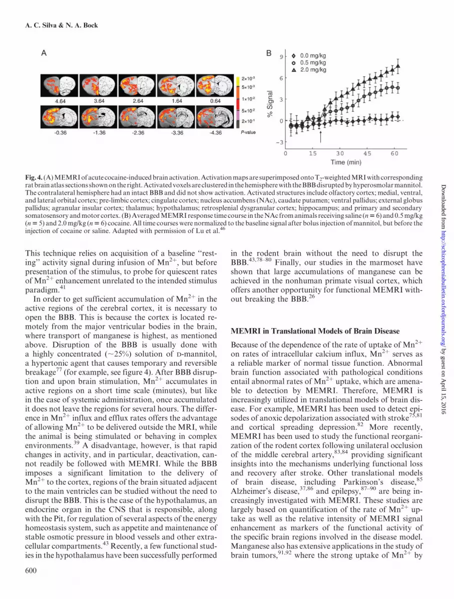
In order to get sufficient accumulation of Mn2þ in theactive regions of the cerebral cortex, it is necessary toopen the BBB. This is because the cortex is located re-motely from the major ventricular bodies in the brain,where transport of manganese is highest, as mentionedabove. Disruption of the BBB is usually done witha highly concentrated (;25%) solution of D-mannitol,a hypertonic agent that causes temporary and reversiblebreakage77 (for example, see figure 4). After BBB disrup-tion and upon brain stimulation, Mn2þ accumulates inactive regions on a short time scale (minutes), but likein the case of systemic administration, once accumulatedit does not leave the regions for several hours. The differ-ence in Mn2þ influx and efflux rates offers the advantageof allowing Mn2þ to be delivered outside the MRI, whilethe animal is being stimulated or behaving in complexenvironments.39 A disadvantage, however, is that rapidchanges in activity, and in particular, deactivation, can-not readily be followed with MEMRI. While the BBBimposes a significant limitation to the delivery ofMn2þ to the cortex, regions of the brain situated adjacentto the main ventricles can be studied without the need todisrupt the BBB. This is the case of the hypothalamus, anendocrine organ in the CNS that is responsible, alongwith the Pit, for regulation of several aspects of the energyhomeostasis system, such as appetite and maintenance ofstable osmotic pressure in blood vessels and other extra-cellular compartments.43 Recently, a few functional stud-ies in the hypothalamus have been successfully performed
in the rodent brain without the need to disrupt theBBB.43,78–80 Finally, our studies in the marmoset haveshown that large accumulations of manganese can beachieved in the nonhuman primate visual cortex, whichoffers another opportunity for functional MEMRI with-out breaking the BBB.26
MEMRI in Translational Models of Brain Disease
Because of the dependence of the rate of uptake of Mn2þ
on rates of intracellular calcium influx, Mn2þ serves asa reliable marker of normal tissue function. Abnormalbrain function associated with pathological conditionsentail abnormal rates of Mn2þ uptake, which are amena-ble to detection by MEMRI. Therefore, MEMRI isincreasingly utilized in translational models of brain dis-ease. For example, MEMRI has been used to detect epi-sodes of anoxic depolarization associated with stroke75,81
and cortical spreading depression.82 More recently,MEMRI has been used to study the functional reorgani-zation of the rodent cortex following unilateral occlusionof the middle cerebral artery,83,84 providing significantinsights into the mechanisms underlying functional lossand recovery after stroke. Other translational modelsof brain disease, including Parkinson’s disease,85
Alzheimer’s disease,37,86 and epilepsy,87–90 are being in-creasingly investigated with MEMRI. These studies arelargely based on quantification of the rate of Mn2þ up-take as well as the relative intensity of MEMRI signalenhancement as markers of the functional activity ofthe specific brain regions involved in the disease model.Manganese also has extensive applications in the study ofbrain tumors,91,92 where the strong uptake of Mn2þ by
Fig. 4. (A)MEMRIofacute cocaine-inducedbrainactivation.Activationmapsare superimposedontoT2-weightedMRIwithcorrespondingratbrainatlas sectionsshownontheright.Activatedvoxelsareclustered in thehemispherewiththeBBBdisruptedbyhyperosmolarmannitol.The contralateral hemisphere had an intact BBB and did not show activation. Activated structures include olfactory cortex; medial, ventral,and lateral orbital cortex; pre-limbic cortex; cingulate cortex; nucleus accumbens (NAc), caudate putamen; ventral pallidus; external globuspallidus; agranular insular cortex; thalamus; hypothalamus; retrosplenial dysgranular cortex; hippocampus; and primary and secondarysomatosensory andmotor cortex. (B)AveragedMEMRIresponse time course in theNAc fromanimals receiving saline (n56) and0.5mg/kg(n5 5) and 2.0mg/kg (n5 6) cocaine.All time courses were normalized to the baseline signal after bolus injection ofmannitol, but before theinjection of cocaine or saline. Adapted with permission of Lu et al.46
600
A. C. Silva & N. A. Bock
by guest on April 15, 2016
http://schizophreniabulletin.oxfordjournals.org/D
ownloaded from

the tumor is used to define tumor volume and aid in theevaluation of therapeutic efforts.Other applications of MEMRI in translational re-
search include its use to study spinal cord injury,93–95
in which the tract-tracing properties of manganese com-bine well with its functional properties to allow assess-ment of connectivity, direction and rate of transport,as well as the functional improvement associated withhealing and therapy strategies.96–98 The same appliesto the study of translational models of cerebralpalsy,99–101 in which a hypoxic-ischemic lesion of the cor-tex is created either in the pre- or the postnatal period.Finally, translational functional models of chemical de-pendency exist in which the functional brain responseto administration of cocaine46 (figure 4) or methamphet-amine,102 and MEMRI detects changes in the functionalbrain regions and in the neuronal pathways associatedwith the psychostimulants.All the above examples of the use of MEMRI in trans-
lational brain research point out to the usefulness andversatility of the technique in probing the anatomical,functional, and structural features of the most diversemodels of brain pathology, which can be used to defineand evaluate therapeutic strategies thatmay have a prom-ising impact in the improvement of human health care.
Challenges for MEMRI
A few challenges stand ahead of taking full advantage ofall the benefits MEMRI has to offer. First and foremost,in order for manganese to become a useful contrast agentin radiology, neurology, and psychiatry, a method ofminimizing its neurotoxicity in humans must be discov-ered. This requires using as low a dose as possible, whichlimits the detectability by MRI, an inherently insensitiveimaging modality. In addition, further development ofmanganese-based MRI contrast agents should be pur-sued. There is currently one agent, Mn-dipyridoxyl-di-phosphate (MnDPDP, or TeslascanTM, Mangafodipir,manufactured by GE Healthcare), which is currentlyused in human clinical imaging of the liver.103 Associatedwith the use of as small amounts of Mn2þ as possible isthe need to develop and further refine MRI pulse sequen-ces that are sensitive to very small changes in the waterrelaxation rates.104 While the doses of Mn2þ presentlyused in animals are too high for use in humans, subtlefeatures of the brain neuroarchitecture, such as corticallayers, need even higher doses to produce adequate imagecontrast for detection. Concomitant to improvements inthe sensitivity of MEMRI techniques, it is equally impor-tant to develop new ways to administer MnCl2 directly tothe target organ of interest without having to go to othervital organs, such as the heart, the liver, and the kidneysduring a systemic administration. For example, the use ofdirect, intrathechal,105 or intraventricular106 administra-tion ofMnCl2 is a feasible way to deliver large amounts of
MnCl2 to the brain without the complications of a largesystemic dose. As well, the use of a continuous infusionvia a cannula implanted directly in the region of interestaccomplishes efficient delivery of Mn2þ for tract-tracingpurposes.38
Second, further work is necessary to understand the bi-ology and the dynamics of Mn2þ at all levels of the body.It is essential to better understand the transport and life-time of manganese in the bloodstream to maximize itsavailability for uptake by the brain. The transport ofMn2þ across the BBB and across the choroid plexusneeds further investigation, as this determines the finalcontrast in the brain. It is equally important to under-stand exactly how Mn2þ enters the cells to turn MEMRIinto a quantitative index of calcium influx.Finally, as disruption in manganese homeostasis may
be itself related to a few specific brain disorders, such asParkinson’s disease107 or schizophrenia,108–111 carefulplanning of the experiments to use MEMRI in transla-tional models of such disorders are necessary to avoidpossible confounds that may complicate interpretationof the results and misguide future research.There is no doubt that the combination of the rich bi-
ology of Mn2þ and its properties as an MRI contrastagent allowMEMRI to be a uniquely useful and versatiletechnique in modern neuroimaging. Indeed, there arepresently 3 major ways to use MEMRI. First, simple sys-temic administration of Mn2þ leads to detection of ana-tomical structures that would otherwise be difficult toreveal. Second, Mn2þ is stored and transported in axonaltracts and is released in the synaptic clefts, becomingavailable for uptake by the next neuron in a way that ena-bles trans-synaptic tracing of neuronal pathways. Third,the dynamics of Mn2þ entry into excitable cells in theCNS is intrinsically linked to activity of voltage-gatedcalcium channels, allowing MEMRI to map functionalbrain regions. The combination of all the above proper-ties leads to ample opportunities to use MEMRI forstudying translational models of brain disease. It is dif-ficult to think of another neuroimaging technique withsuch a broad range of applications and versatility.
Funding
Intramural Research Program of the National Instituteof Neurological Disorders and Stroke, National Insti-tutes of Health (Alan P. Koretsky, Scientific Director).
References
1. Jasanoff A. MRI contrast agents for functional molecularimaging of brain activity. Curr Opin Neurobiol. 2007;17(5):593–600.
2. Li Y, Huang TT, Carlson EJ, et al. Dilated cardiomyopathyand neonatal lethality in mutant mice lacking manganese su-peroxide dismutase. Nat Genet. 1995;11(4):376–381.
601
MEMRI in Translational Neuroimaging
by guest on April 15, 2016
http://schizophreniabulletin.oxfordjournals.org/D
ownloaded from

3. Gunter TE, Gavin CE, Aschner M, Gunter KK. Speciationof manganese in cells and mitochondria: a search for theproximal cause of manganese neurotoxicity. Neurotoxicol-ogy. 2006;27(5):765–776.
4. Zwingmann C, Leibfritz D, Hazell AS. Brain energy metab-olism in a sub-acute rat model of manganese neurotoxicity:an ex vivo nuclear magnetic resonance study using [1-13C]glucose. Neurotoxicology. 2004;25(4):573–587.
5. Wedler FC, Denman RB. Glutamine synthetase: the majorMn(II) enzyme in mammalian brain. Curr Top Cell Regul.1984;24:153–169.
6. Crossgrove J, Zheng W. Manganese toxicity upon overexpo-sure. NMR Biomed. 2004;17(8):544–553.
7. Aschner JL, Aschner M. Nutritional aspects of manganesehomeostasis. Mol Aspects Med. 2005;26(4–5):353–362.
8. Aschner M, Dorman DC. Manganese: pharmacokineticsand molecular mechanisms of brain uptake. Toxicol Rev.2006;25(3):147–154.
9. Aschner M, Guilarte TR, Schneider JS, Zheng W. Manga-nese: recent advances in understanding its transport and neu-rotoxicity. Toxicol Appl Pharmacol. 2007;221(2):131–147.
10. Takeda A. Manganese action in brain function. Brain ResBrain Res Rev. 2003;41(1):79–87.
11. Sloot WN, Gramsbergen JB. Axonal transport of manga-nese and its relevance to selective neurotoxicity in the ratbasal ganglia. Brain Res. 1994;657(1–2):124–132.
12. Takeda A, Kodama Y, Ishiwatari S, Okada S. Manganesetransport in the neural circuit of rat CNS. Brain Res Bull.1998;45(2):149–152.
13. Takeda A, Ishiwatari S, Okada S. In vivo stimulation-induced release of manganese in rat amygdala. Brain Res.1998;811(1–2):147–151.
14. Takeda A, Sotogaku N, Oku N. Manganese influences thelevels of neurotransmitters in synapses in rat brain. Neuro-science. 2002;114(3):669–674.
15. Lee JH, Koretsky AP. Manganese enhanced magnetic reso-nance imaging. Curr Pharm Biotechnol. 2004;5(6):529–537.
16. Koretsky AP, Silva AC. Manganese-enhanced magnetic res-onance imaging (MEMRI). NMR Biomed. 2004;17(8):527–531.
17. Pautler RG. Biological applications of manganese-enhancedmagnetic resonance imaging. Methods Mol Med. 2006;124:365–386.
18. Bock NA, Silva AC. Manganese: a unique neuroimagingcontrast agent. Future Neurology. 2007;2(3):297–395.
19. Natt O, Watanabe T, Boretius S, Radulovic J, Frahm J,Michaelis T. High-resolution 3D MRI of mouse brainreveals small cerebral structures in vivo. J Neurosci Methods.2002;120(2):203–209.
20. Watanabe T, Natt O, Boretius S, Frahm J, Michaelis T. Invivo 3D MRI staining of mouse brain after subcutaneousapplication of MnCl2. Magn Reson Med. 2002;48(5):852–859.
21. Watanabe T, Radulovic J, Spiess J, et al. In vivo 3D MRIstaining of the mouse hippocampal system using intracere-bral injection of MnCl2. Neuroimage. 2004;22(2):860–867.
22. Aoki I, Wu YJ, Silva AC, Lynch RM, Koretsky AP. In vivodetection of neuroarchitecture in the rodent brain using man-ganese-enhanced MRI. Neuroimage. 2004;22(3):1046–1059.
23. Watanabe T, Radulovic J, Boretius S, Frahm J, Michaelis T.Mapping of the habenulo-interpeduncular pathway in livingmice using manganese-enhanced 3D MRI. Magn Reson Im-aging. 2006;24(3):209–215.
24. Bock NA, Paiva FF, Silva AC. Fractionated manganese-en-hanced MRI. NMR Biomed. October 14, 2007.
25. Silva AC, Lee JH, Wu CW, et al. Detection of cortical lam-inar architecture using manganese-enhanced MRI. J Neuro-sci Methods STAT- In-Data-Review. 2008;2:246–257.
26. Bock NA, Paiva FF, Nascimento GC, Newman JD, SilvaAC. Cerebrospinal fluid to brain transport of manganesein a non-human primate revealed by MRI. Brain Res.2008;1198:160–170.
27. Pautler RG, Silva AC, Koretsky AP. In vivo neuronal tracttracing using manganese-enhanced magnetic resonance im-aging. Magn Reson Med. 1998;40(5):740–748.
28. Watanabe T, Michaelis T, Frahm J. Mapping of retinal pro-jections in the living rat using high-resolution 3D gradient-echo MRI with Mn2+-induced contrast. Magn Reson Med.2001;46(3):424–429.
29. Pautler RG, Koretsky AP. Tracing odor-induced activation inthe olfactory bulbs of mice using manganese-enhanced mag-netic resonance imaging. Neuroimage. 2002;16(2):441–448.
30. Van der LA, Verhoye M, Van M, et al. In vivo manganese-enhanced magnetic resonance imaging reveals connectionsand functional properties of the songbird vocal control sys-tem. Neuroscience. 2002;112(2):467–474.
31. Saleem KS, Pauls JM, Augath M, et al. Magnetic resonanceimaging of neuronal connections in the macaque monkey.Neuron. 2002;34(5):685–700.
32. Pautler RG, Mongeau R, Jacobs RE. In vivo trans-synaptictract tracing from the murine striatum and amygdala utiliz-ing manganese enhanced MRI (MEMRI). Magn ResonMed. 2003;50(1):33–39.
33. Tindemans I, Verhoye M, Balthazart J, Van der LA. In vivodynamic ME-MRI reveals differential functional responsesof RA- and area X-projecting neurons in the HVC of canar-ies exposed to conspecific song. Eur J Neurosci. 2003;18(12):3352–3360.
34. Van Meir V, Verhoye M, Absil P, Eens M, Balthazart J, Vander LA. Differential effects of testosterone on neuronal pop-ulations and their connections in a sensorimotor brainnucleus controlling song production in songbirds: amanganese enhanced-magnetic resonance imaging study.Neuroimage. 2004;21(3):914–923.
35. Tindemans I, Boumans T, Verhoye M, Van der Linden A.IR-SE and IR-MEMRI allow in vivo visualization of oscineneuroarchitecture including the main forebrain regions ofthe song control system. NMR Biomed. 2006;19(1):18–29.
36. Murayama Y, Weber B, Saleem KS, Augath M, LogothetisNK. Tracing neural circuits in vivo with Mn-enhanced MRI.Magn Reson Imaging. 2006;24(4):349–358.
37. Smith KD, Kallhoff V, Zheng H, Pautler RG. In vivo axo-nal transport rates decrease in a mouse model of Alzheimer’sdisease. Neuroimage. 2007;35(4):1401–1408.
38. Canals S, Beyerlein M, Keller AL, Murayama Y, LogothetisNK. Magnetic resonance imaging of cortical connectivity invivo. Neuroimage. 2008;40(2):458–472.
39. Lin YJ. MRI of the rat and mouse brain after systemic ad-ministration of MnCl2 [dissertation]. Pittsburgh: CarnegieMellon University; 1997.
40. Duong TQ, Silva AC, Lee SP, Kim SG. Functional MRI ofcalcium-dependent synaptic activity: cross correlation withCBF and BOLD measurements. Magn Reson Med.2000;43(3):383–392.
41. Aoki I, Tanaka C, Takegami T, et al. Dynamic activity-induced manganese-dependent contrast magnetic resonance
602
A. C. Silva & N. A. Bock
by guest on April 15, 2016
http://schizophreniabulletin.oxfordjournals.org/D
ownloaded from

imaging (DAIM MRI). Magn Reson Med. 2002;48(6):927–933.
42. Yu X, Wadghiri YZ, Sanes DH, Turnbull DH. In vivo audi-tory brain mapping in mice with Mn-enhanced MRI. NatNeurosci. 2005;8(7):961–968.
43. Kuo YT, Herlihy AH, So PW, Bell JD. Manganese-enhancedmagnetic resonance imaging (MEMRI) without compromiseof the blood-brain barrier detects hypothalamic neuronal ac-tivity in vivo. NMR Biomed. 2006;19(8):1028–1034.
44. Yu X, Sanes DH, Aristizabal O, Wadghiri YZ, TurnbullDH. Large-scale reorganization of the tonotopic map inmouse auditory midbrain revealed by MRI. Proc NatlAcad Sci USA. 2007;104(29):12193–12198.
45. Weng JC, Chen JH, Yang PF, Tseng WY. Functional map-ping of rat barrel activation following whisker stimulationusing activity-induced manganese-dependent contrast. Neu-roimage. 2007;36(4):1179–1188.
46. Lu H, Xi ZX, Gitajn L, Rea W, Yang Y, Stein EA. Cocaine-induced brain activation detected by dynamic manganese-enhanced magnetic resonance imaging (MEMRI). ProcNatl Acad Sci USA. 2007;104(7):2489–2494.
47. Yu X, Zou J, Babb JS, Johnson G, Sanes DH, Turnbull DH.Statistical mapping of sound-evoked activity in the mouseauditory midbrain using Mn-enhanced MRI. Neuroimage.2008;39(1):223–230.
48. Dobson AW, Erikson KM, Aschner M. Manganese neuro-toxicity. Ann N Y Acad Sci. 2004;1012:115–128.
49. Racette BA, Antenor JA, McGee-Minnich L, et al.[18F]FDOPA PET and clinical features in parkinsonismdue to manganism. Mov Disord. 2005;20(4):492–496.
50. Chandra SV, Shukla GS. Role of iron deficiency in inducingsusceptibility to manganese toxicity. Arch Toxicol.1976;35(4):319–323.
51. Wolf GL, Baum L. Cardiovascular toxicity and tissue pro-ton T1 response to manganese injection in the dog and rab-bit. AJR Am J Roentgenol. 1983;141(1):193–197.
52. Silva AC, Lee JH, Aoki I, Koretsky AP. Manganese-en-hanced magnetic resonance imaging (MEMRI): methodo-logical and practical considerations. NMR Biomed. 2004;17(8):532–543.
53. Kuo YT, Herlihy AH, So PW, Bhakoo KK, Bell JD. In vivomeasurements of T1 relaxation times in mouse brain associ-ated with different modes of systemic administration of man-ganese chloride. J Magn Reson Imaging. 2005;21(4):334–339.
54. Lee JH, Silva AC, Merkle H, Koretsky AP. Manganese-enhanced magnetic resonance imaging of mouse brain aftersystemic administration of MnCl2: dose-dependent and tem-poral evolution of T1 contrast. Magn Reson Med. 2005;53(3):640–648.
55. Takeda A, Sawashita J, Okada S. Biological half-lives ofzinc and manganese in rat brain. Brain Res. 1995;695(1):53–58.
56. Zhang S, Zhou Z, Fu J. Effect of manganese chloride expo-sure on liver and brain mitochondria function in rats. Envi-ron Res. 2003;93(2):149–157.
57. Jelsing J, Hay-Schmidt A, Dyrby T, Hemmingsen R, UylingsHB, Pakkenberg B. The prefrontal cortex in the Gottingenminipig brain defined by neural projection criteria andcytoarchitecture. Brain Res Bull. 2006;70(4–6):322–336.
58. Watanabe T, Frahm J, Michaelis T. Functional mapping ofneural pathways in rodent brain in vivo using manganese-enhanced three-dimensional magnetic resonance imaging.NMR Biomed. 2004;17(8):554–568.
59. Watanabe T, Schachtner J, Krizan M, Boretius S, Frahm J,Michaelis T. Manganese-enhanced 3D MRI of establishedand disrupted synaptic activity in the developing insect brainin vivo. J Neurosci Methods. 2006;158(1):50–55.
60. Angenstein F, Niessen HG, Goldschmidt J, et al. Manga-nese-enhanced MRI reveals structural and functionalchanges in the cortex of Bassoon mutant mice. Cereb Cor-tex. 2007;17(1):28–36.
61. Murphy VA, Wadhwani KC, Smith QR, Rapoport SI. Sat-urable transport of manganese(II) across the rat blood-brainbarrier. J Neurochem. 1991;57(3):948–954.
62. Rabin O, Hegedus L, Bourre JM, Smith QR. Rapid brainuptake of manganese(II) across the blood-brain barrier. JNeurochem. 1993;61(2):509–517.
63. Wadghiri YZ, Blind JA, Duan X, et al. Manganese-enhanced magnetic resonance imaging (MEMRI) of mousebrain development. NMR Biomed. 2004;17(8):613–619.
64. de Sousa PL, de Souza SL, Silva AC, de Souza RE, de Cas-tro RM. Manganese-enhanced magnetic resonance imaging(MEMRI) of rat brain after systemic administration ofMnCl2: changes in T1 relaxation times during postnatal de-velopment. J Magn Reson Imaging. 2007;25(1):32–38.
65. Jiang Y, Zheng W, Long L, et al. Brain magnetic resonanceimaging and manganese concentrations in red blood cells ofsmelting workers: search for biomarkers of manganese expo-sure. Neurotoxicology. 2006;28(1):126–135.
66. Thuen M, Singstad TE, Pedersen TB, et al. Manganese-enhancedMRI of the optic visual pathway and optic nerve in-jury in adult rats. J Magn Reson Imaging. 2005;22(4):492–500.
67. Lindsey JD, Scadeng M, Dubowitz DJ, Crowston JG,Weinreb RN. Magnetic resonance imaging of the visual sys-tem in vivo: transsynaptic illumination of V1 and V2 visualcortex. Neuroimage. 2007;34(4):1619–1626.
68. Allegrini PR, Wiessner C. Three-dimensional MRI of cere-bral projections in rat brain in vivo after intracortical injec-tion of MnCl2. NMR Biomed. 2003;16(5):252–256.
69. Lee JW, Park JA, Lee JJ, et al. Manganese-enhanced audi-tory tract-tracing MRI with cochlear injection. Magn ResonImaging. 2007;25(5):652–656.
70. Van der LA, Van MV, Tindemans I, Verhoye M, BalthazartJ. Applications of manganese-enhanced magnetic resonanceimaging (MEMRI) to image brain plasticity in song birds.NMR Biomed. 2004;17(8):602–612.
71. Van Meir V, Pavlova D, Verhoye M, et al. In vivo MR im-aging of the seasonal volumetric and functional plasticity ofsong control nuclei in relation to song output in a femalesongbird. Neuroimage. 2006;31(3):981–992.
72. Drapeau P, Nachshen DA. Manganese fluxes and manga-nese-dependent neurotransmitter release in presynaptic nerveendings isolated from rat brain. J Physiol. 1984;348:493–510.
73. Narita K, Kawasaki F, Kita H. Mn and Mg influxesthrough Ca channels of motor nerve terminals are preventedby verapamil in frogs. Brain Res. 1990;510(2):289–295.
74. HunterDR,HaworthRA,BerkoffHA.Cellularmanganeseup-take by the isolated perfused rat heart: a probe for the sarco-lemmacalciumchannel.JMolCellCardiol. 1981;13(9):823–832.
75. Aoki I, Ebisu T, Tanaka C, et al. Detection of the anoxic de-polarization of focal ischemia using manganese-enhancedMRI. Magn Reson Med. 2003;50(1):7–12.
76. Brozoski TJ, Ciobanu L, Bauer CA. Central neural activityin rats with tinnitus evaluated with manganese-enhancedmagnetic resonance imaging (MEMRI). Hear Res. 2007;228(1–2):168–179.
603
MEMRI in Translational Neuroimaging
by guest on April 15, 2016
http://schizophreniabulletin.oxfordjournals.org/D
ownloaded from

77. Neuwelt EA,Frenkel EP,Diehl JT, et al.Osmotic blood-brainbarrier disruption: a newmeans of increasing chemotherapeu-tic agent delivery. Trans AmNeurol Assoc. 1979;104:256–260.
78. Chaudhri OB, Parkinson JR, Kuo YT, et al. Differential hy-pothalamic neuronal activation following peripheral injec-tion of GLP-1 and oxyntomodulin in mice detected bymanganese-enhanced magnetic resonance imaging. BiochemBiophys Res Commun. 2006;350(2):298–306.
79. Kuo YT, Parkinson JR, Chaudhri OB, et al. The temporalsequence of gut peptide CNS interactions tracked in vivoby magnetic resonance imaging. J Neurosci. 2007;27(45):12341–12348.
80. So PW, Yu WS, Kuo YT, et al. Impact of resistant starch onbody fat patterning and central appetite regulation. PLoSONE. 2007;2(12):e1309.
81. Aoki I, Naruse S, Tanaka C. Manganese-enhanced magneticresonance imaging (MEMRI) of brain activity and applica-tions to early detection of brain ischemia. NMR Biomed.2004;17(8):569–580.
82. Henning EC, Meng X, Fisher M, Sotak CH. Visualization ofcortical spreading depression using manganese-enhanced mag-netic resonance imaging.MagnResonMed. 2005;53(4):851–857.
83. van der Zijden JP, Wu O, van der TA, Roeling TP, BleysRL, Dijkhuizen RM. Changes in neuronal connectivity afterstroke in rats as studied by serial manganese-enhanced MRI.Neuroimage. 2007;34(4):1650–1657.
84. van der Zijden JP, Bouts MJ, Wu O, et al. Manganese-en-hanced MRI of brain plasticity in relation to functional re-covery after experimental stroke. J Cereb Blood FlowMetab. 2008;28(4):832–840.
85. Pelled G, Bergman H, Ben-Hur T, Goelman G. Manganese-enhanced MRI in a rat model of Parkinson’s disease.J Magn Reson Imaging. 2007;26(4):863–870.
86. Benveniste H, Ma Y, Dhawan J, et al. Anatomical and func-tional phenotyping of mice models of Alzheimer’s disease byMR microscopy. Ann N Y Acad Sci. 2007;1097:12–29.
87. Hsu YH, Lee WT, Chang C. Multiparametric MRI evalua-tion of kainic acid-induced neuronal activation in rat hippo-campus. Brain. 2007;130(Pt 12):3124–3134.
88. Alvestad S, Goa PE, Qu H, et al. In vivo mapping of tem-porospatial changes in manganese enhancement in rat brainduring epileptogenesis. Neuroimage. 2007;38(1):57–66.
89. Obenaus A, Jacobs RE. Magnetic resonance imaging offunctional anatomy: use for small animal epilepsy models.Epilepsia. 2007;48:Suppl. 411–17.
90. Immonen RJ, Kharatishvili I, Sierra A, Einula C, PitkanenA, Grohn OH. Manganese enhanced MRI detects mossy fi-ber sprouting rather than neurodegeneration, gliosis or sei-zure-activity in the epileptic rat hippocampus. Neuroimage.2008;40(4):1718–1730.
91. Cross DJ, Flexman JA, Anzai Y, et al. In vivo manganeseMR imaging of calcium influx in spontaneous rat pituitaryadenoma. AJNR Am J Neuroradiol. 2007;28(10):1865–1871.
92. Hegedus B, Banerjee D, Yeh TH, et al. Preclinical cancertherapy in a mouse model of neurofibromatosis-1 optic gli-oma. Cancer Res. 2008;68(5):1520–1528.
93. Bilgen M, Dancause N, Al-Hafez B, He YY, Malone TM.Manganese-enhanced MRI of rat spinal cord injury. MagnReson Imaging. 2005;23(7):829–832.
94. Bilgen M, Peng W, Al-Hafez B, Dancause N, He YY, CheneyPD. Electrical stimulation of cortex improves corticospinaltract tracing in rat spinal cord using manganese-enhancedMRI. J Neurosci Methods. 2006;156(1–2):17–22.
95. Bilgen M. Imaging corticospinal tract connectivity in injuredrat spinal cord using manganese-enhanced MRI. BMC MedImaging. 2006;6:15.
96. Stieltjes B, Klussmann S, BockM, et al. Manganese-enhancedmagnetic resonance imaging for in vivo assessment of damageand functional improvement following spinal cord injury inmice. Magn Reson Med. 2006;55(5):1124–1131.
97. Bonny JM, Mailly P, Renou JP, Orsal D, Benmoussa A,Stettler O. Analysis of laminar activity in normal and injuredrat spinal cord by manganese enhanced MRI. Neuroimage.2008;40(4):154–155.
98. Walder N, Petter-Puchner AH, Brejnikow M, Redl H, EssigM, Stieltjes B. Manganese enhanced magnetic resonanceimaging in a contusion model of spinal cord injury in rats:correlation with motor function. Invest Radiol. 2008;43(5):277–283.
99. Drobyshevsky A, Robinson AM, Derrick M, et al. Sensorydeficits and olfactory system injury detected by novel appli-cation of MEMRI in newborn rabbit after antenatal hyp-oxia-ischemia. Neuroimage. 2006;32(3):1106–1112.
100. Yang J, Wu EX. Manganese-enhanced MRI detected thegray matter lesions in the late phase of mild hypoxic-ische-mic injury in neonatal rat. Conf Proc IEEE Eng Med BiolSoc. 2007;2007:51–54.
101. Yang J, Wu EX. Detection of cortical gray matter lesion inthe late phase of mild hypoxic-ischemic injury by manga-nese-enhanced MRI. Neuroimage. 2008;39(2):669–679.
102. Hsu YH, Chen CC, Zechariah A, Yen CC, Yang LC, ChangC. Neuronal dysfunction of a long projecting multisynapticpathway in response to methamphetamine using manganese-enhanced MRI. Psychopharmacology (Berl). 2008;196(4):543–553.
103. Federle MP, Chezmar JL, Rubin DL, et al. Safety and efficacyof mangafodipir trisodium (MnDPDP) injection for hepaticMRI in adults: results of the U.S. multicenter phase III clinicaltrials (safety). J Magn Reson Imaging. 2000;12(1):186–197.
104. Chuang KH, Koretsky A. Improved neuronal tract tracing us-ing manganese enhanced magnetic resonance imaging withfast T(1) mapping. Magn Reson Med. 2006;55(3):604–611.
105. Liu CH, D’Arceuil HE, de Crespigny AJ. Direct CSF injec-tion of MnCl(2) for dynamic manganese-enhanced MRI.Magn Reson Med. 2004;51(5):978–987.
106. Morita H, Ogino T, Fujiki N, et al. Sequence of forebrainactivation induced by intraventricular injection of hyper-tonic NaCl detected by Mn2+ contrasted T1-weightedMRI. Auton Neurosci. 2004;113(1–2):43–54.
107. Olanow CW. Manganese-induced parkinsonism andParkinson’s disease. Ann N Y Acad Sci. 2004;1012:209–223.
108. Loven DP, James JF, Biggs L, Little KY. Increased manga-nese-superoxide dismutase activity in postmortem brainfrom neuroleptic-treated psychotic patients. Biol Psychiatry.1996;40(3):230–232.
109. Parikh V, Khan MM, Mahadik SP. Differential effects ofantipsychotics on expression of antioxidant enzymes andmembrane lipid peroxidation in rat brain. J Psychiatr Res.2003;37(1):43–51.
110. Yanik M, Vural H, Kocyigit A, et al. Is the arginine-nitricoxide pathway involved in the pathogenesis of schizophre-nia? Neuropsychobiology. 2003;47(2):61–65.
111. Yanik M, Kocyigit A, Tutkun H, Vural H, Herken H.Plasma manganese, selenium, zinc, copper, and iron concen-trations in patients with schizophrenia. Biol Trace Elem Res.2004;98(2):109–117.
604
A. C. Silva & N. A. Bock
by guest on April 15, 2016
http://schizophreniabulletin.oxfordjournals.org/D
ownloaded from