neuroimaging biomarkers in bipolar disorder
TRANSCRIPT
[Frontiers in Bioscience E4, 593-606, January 1, 2012]
593
Neuroimaging biomarkers in bipolar disorder
Josselin Houenou1,2,3,4, Marc-Antoine d'Albis1,2,3, Francois-Eric Vederine1,2, Chantal Henry1,2,3, Marion Leboyer1,2,3,Michele Wessa5
1AP-HP, University Paris-East, Department of Psychiatry, Henri Mondor-Albert Chenevier Hospitals, Creteil, F-94010, France,2INSERM, U955 Unit, IMRB, Department of Medical Genetics, Psychiatry Genetics, Creteil, F-94010, France, 3FondaMentalFoundation, Creteil, F-94010, France, 4Neurospin, CEA Saclay, LNAO, Gif-Sur-Yvette, France, 5Department of Cognitive andClinical Neuroscience, Central Institute of Mental Health, Mannheim, Germany
TABLE OF CONTENTS
1.Abstract2.Introduction3.Structural and functional biomarkers of bipolar disorder
3.1. Structural imaging biomarkers3.2. Functional imaging biomarkers3.3. Heterogeneity of results
4.Neurobiological models of bipolar disorder4.1. Models of normal emotional processing4.2. Models of emotional dysregulation in BD
5.Connectivity in bipolar disorder5.1. Anatomical connectivity (DTI)5.2. Functional connectivity (fMRI)5.3. Linking structural and functional connectivity: bipolar disorder as a connectivity disorder?
6.Perspective6.1. Individual biomarkers in bipolar disorder6.2. Imaging genetics of bipolar disorder
7. Acknowledgments8. References
1. ABSTRACT
There is an urgent need to identify objectivebiomarkers for the assessment of bipolar disorder, toimprove diagnosis and prognostic evaluation.Neuroimaging is a particularly promising approach. Wereview here the structural and functional neuroimagingstudies carried out on bipolar disorder. These studies haveled to the development of neurobiological models ofbipolar disorder assuming cortical-limbic dysregulation.Dorsal brain structures are thought to decrease in volumeand activity in bipolar disorder, reducing inhibition of theventral-limbic network and enhancing emotional responses.These models also assume abnormal prefrontal-subcorticallimbic connectivity. This abnormal connectivity has beenidentified by both diffusion tensor imaging studies(anatomical connectivity) and functional MRI (functionalconnectivity). However, studies are currently limited by theheterogeneity of the patients included. Future researchshould include studies to validate biomarkers for theassessment of bipolar disorder and studies of large and wellcharacterized samples of patients with bipolar disorder.
2. INTRODUCTION
Bipolar disorder (BD) is a severe chronic mentalillness that affects about 1% of the population. Thediagnosis of bipolar disorder is currently based entirely onclinical evaluation, without any possibility of confirmingthe diagnosis by laboratory tests. Recent neurobiologicalstudies have raised hopes that it may be possible to identifybiomarkers of BD (1). Such biomarkers would improveboth the diagnosis and assessment of BD.
Tremendous progress has recently been made inthe neuroimaging of bipolar disorder, for several reasons.First, new imaging techniques and analysis methods arebeing developed and existing techniques refined. Thesenew methods include high-strength magnetic fields (3 and 7Tesla), diffusion tensor imaging and tractographyalgorithms, event-related fMRI, analyses of functionalconnectivity, MRI spectroscopy and the combined use ofgenetics and neuroimaging — the so-called “imaginggenetics” approach. Second, sample sizes have greatlyincreased in MRI studies. Third, better clinical
Neuroimaging biomarkers in BD
594
characterization of patients has resulted in betterdelineation of their underlying neurobiology. Recentneuroimaging studies have therefore increased ourunderstanding of the physiopathology of BD, creating asolid basis for current neurobiological models of BD (2, 3).
Despite these advances, imaging biomarkers forBD have been described only for groups of patients. Thereare still no valid and reproducible individual biomarkers ofBD, the predictive value of the biomarkers describedremains low and several issues remain unexplored.
In this review, we will explore existing studiesexploring the anatomical and functional neuroimaging ofBD (section 3). We will then review the neurobiologicalmodels developed from these imaging studies (section 4).The exploration of brain networks connectivity is apromising new approach that may make it possible todistinguish between bipolar and unipolar depression(section 5). Finally, we will examine the potential ofneuroimaging approaches in BD and their potential clinicalapplications, particularly in terms of predictive biomarkers(section 6). This review will be essentially restricted toadult bipolar disorder, as pediatric bipolar disorder seemsto have different underlying mechanisms (4).
3. STRUCTURAL AND FUNCTIONALBIOMARKERS OF BIPOLAR DISORDER
3.1. Structural imaging biomarkersThe first neuroimaging studies in BD investigated
anatomical changes in the brains of patients with BD, bycomputed tomography (CT) and structural (T1 and T2)magnetic resonance imaging (MRI). No change in totalbrain volume was found in patients with BD, by contrast towhat has been reported for schizophrenia, although thisremains a matter of debate. However, meta-analysesrevealed an association between BD and lateral ventricleenlargement (5, 6). It further seems that total white mattervolume is altered in contrast to gray matter volume. Thiswhite matter volume decrease is present at the onset ofbipolar disorder (7).
White matter changes are the most consistent,reproducible structural abnormalities observed in theneuroimaging of BD. An increase in the frequency of whitematter hyperintensities (WMH) on T2 images hasrepeatedly been reported in patients with BD (6). TheseWMH are located in the deep white matter and in theperiventricular areas, and have been identified in BDpatients during their first episode (8). White matterhyperintensities have been proposed as a potentialendophenotype of bipolar disorder (9) (for more data onthis topic, see section 5.1). Such WMH do not seem to bepresent in unipolar depression, except in late-lifedepression (10).
Changes in the volumes of gray matter structuresinvolved in emotional processing (prefrontal cortex (PFC),cingulate cortex, amygdala, insula, thalamus) have beenidentified (5, 11). A recent meta-analysis (11) identifiedregions consistently reduced in BD, including the anterior
cingulate cortex and the insula. The amygdala, a key limbicregion, is also frequently affected. The amygdala is locatedin the medial and anterior part of the temporal lobe andplays an important role in the automatic regulation ofemotion processing. In BD, a recent meta-analysis revealedthat the amygdala is smaller in children with BD than inhealthy children, whereas it is larger in adults with BD thanin healthy controls (12). Furthermore, a meta-regressionanalysis revealed a positive correlation between age andamygdala volume in patients with BD (13). Severalexplanations of this phenomenon have been put forward,including an abnormal course of development of thisstructure in adolescence and early adulthood (14) ordifferences in amygdala volume as a function of age atonset of bipolar disorder (13). Such increase of volumewith age may be linked either to the pathophysiologicalcourse of BD or to concomitant factors such ascomorbidities, medication or repetition of episodes (13).Longitudinal studies are required to elucidate this issue.
A very recent meta-analysis investigated thespecificity of the gray-matter changes in BD (11). Theauthors included 42 schizophrenia studies and 14 studies onBD. Decreases in gray matter volume were limited inextent in patients with BD and were restricted to theanterior cingulate cortex (perigenual and subgenualcingulate cortex) and bilateral insula. In schizophrenia,gray matter volume decreased in a wider range of regions.The only region of gray matter reduction specific to bipolardisorder was located in the anterior cingulate cortex.
3.2. Functional imaging biomarkersRecent functional imaging studies in BD have
been based mostly on fMRI, as the spatial resolution ofPET is low and magnetoencephalography is not yet widelyused. The diagnostic criteria for BD suggest that thecapacity to regulate emotional state is impaired in thesepatients. Most fMRI studies have explored the neuralnetworks underlying emotional processing in patients withBD. The paradigms used in the different studies includeexplicit and implicit affect recognition tasks (15-20),emotional go/no go tasks (21, 22), emotional Stroop tasks(23, 24), emotional memory tasks (25) and emotional face-matching paradigms (26, 27). The results obtained are quitedisparate, but generally indicate that patients with BDdisplay hyperactivity of a ventral-limbic brain networkencompassing structures such as the amygdala, theparahippocampal gyrus, the subgenual cingulate cortex, theventrolateral prefrontal cortex, the orbitofrontal cortex(OFC), the caudate nucleus and the thalamus. By contrast,hypoactivity of dorsal brain structures, such as the inferiorand medial frontal gyrus, the dorsal and posterior cingulatecortex and the precuneus, has been reported in patients withBD (for a specific review of these studies see (3)). Suchemotional tasks have been used to explore unipolardepression, with much more complex results that in bipolardisorder (28) and in schizophrenia, reportingunderrecruitment of limbic regions (29).
Functional neuroimaging studies of BD havemade use of non emotional cognitive tasks, as impairedexecutive functioning has been described in BD, even when
Neuroimaging biomarkers in BD
595
the patients are in euthymic state (30). Such studies (31, 32)have provided evidence of an increase in activity in ventral-limbic brain structures during purely cognitive-attentionaltasks (sustained attention, working memory) in euthymicbipolar patients with preserved behavioral performances.This suggests that patients with BD attach an emotionalvalence to a task for which no processing of emotionalinformation is required, providing additional support for thehypothesis that emotional reactivity is generally heightenedin BD patients (33).
In contrast, patients with schizophrenia havereduced activation in dorsolateral prefrontal cortex andanterior cingulate cortex during executive tasks, butwithout any evidence of increased activity in limbic regions(34).
Even though we and others have tried tosummarize results of various fMRI studies in bipolardisorder, we must note that the discrepancies between thetasks used limit the range of the common findings betweenstudies. Furthermore, longitudinal studies and explorationsof first-episode patients are required in order to assess theevolution of the functional findings in the course of thedisease.
3.3. Heterogeneity of resultsHeterogeneous results have been obtained in the
structural and functional neuroimaging of BD. However,the main source of this heterogeneity in the results ofneuroimaging studies of BD is the heterogeneity of theclinical samples, which probably confounds the observedresults. The clinical characteristics of the patients studiedare diverse, with, for example, different subtypes of BD(e.g., types I and II, rapid cycling) and differences in age atonset (early, intermediate, late), thymic state, inclusion orexclusion of patients with comorbid psychiatric disorders,such as psychotic features, comorbid alcohol dependence,anxiety disorders or comorbid medical conditions. Nodifference in brain anatomy or function has yet beenassociated with some of these clinical features (e.g.(35)),but decreases in gray and white matter volumes have beenobserved in recovering alcoholic patients without BD (36)even after long periods of abstinence. The inclusion ofpatients with lifetime substance abuse or dependence maytherefore have a major impact on the results. Another majorconfounding variable is psychotropic medication. Severalauthors have observed a neurotrophic effect of lithium andother mood stabilizers (37, 38). Similarly, changes in brainvolume have been associated with the use of antipsychoticdrugs (39). Lithium and valproate are known to influencefMRI activation patterns in patients with BD (40). Theheterogeneity of results from fMRI studies may also resultfrom the use of different activation tasks, making it difficultto compare findings directly.
Another factor contributing to the observedheterogeneity is the limited sample sizes used in thesestudies. In a meta-analysis (6), type I error (false-positiveerror rate) for typical neuroimaging studies of bipolardisorder was estimated at 0.34. The type II error rate is alsohigh (e.g. 70% when measuring the lateral ventricular
volume with 25 patients and 33 controls). Typical studiesinclude groups of 20 to 30 patients and controls, but alarger number of subjects is required to obtain sufficientpower.
4. NEUROBIOLOGICAL MODELS OF BIPOLARDISORDER
Based on previous structural and functionalneuroimaging studies in BD, several authors havedeveloped putative neurobiological models of bipolardisorder. As bipolar disorder is primarily associated withemotional symptoms and disturbances, these models havefocused mostly on emotional processing and its cognitivecontrol. We review here models of the regulation ofemotional processing in healthy controls and in patientswith bipolar disorder.
4.1. Models of normal emotional processingInvestigations of emotional reactivity, its
regulation and its underlying pathophysiologicalmechanisms in bipolar disorder require the identification ofdifferent stages of emotional processing. The identificationof these stages is also important for the interpretation andcomparison of published results.
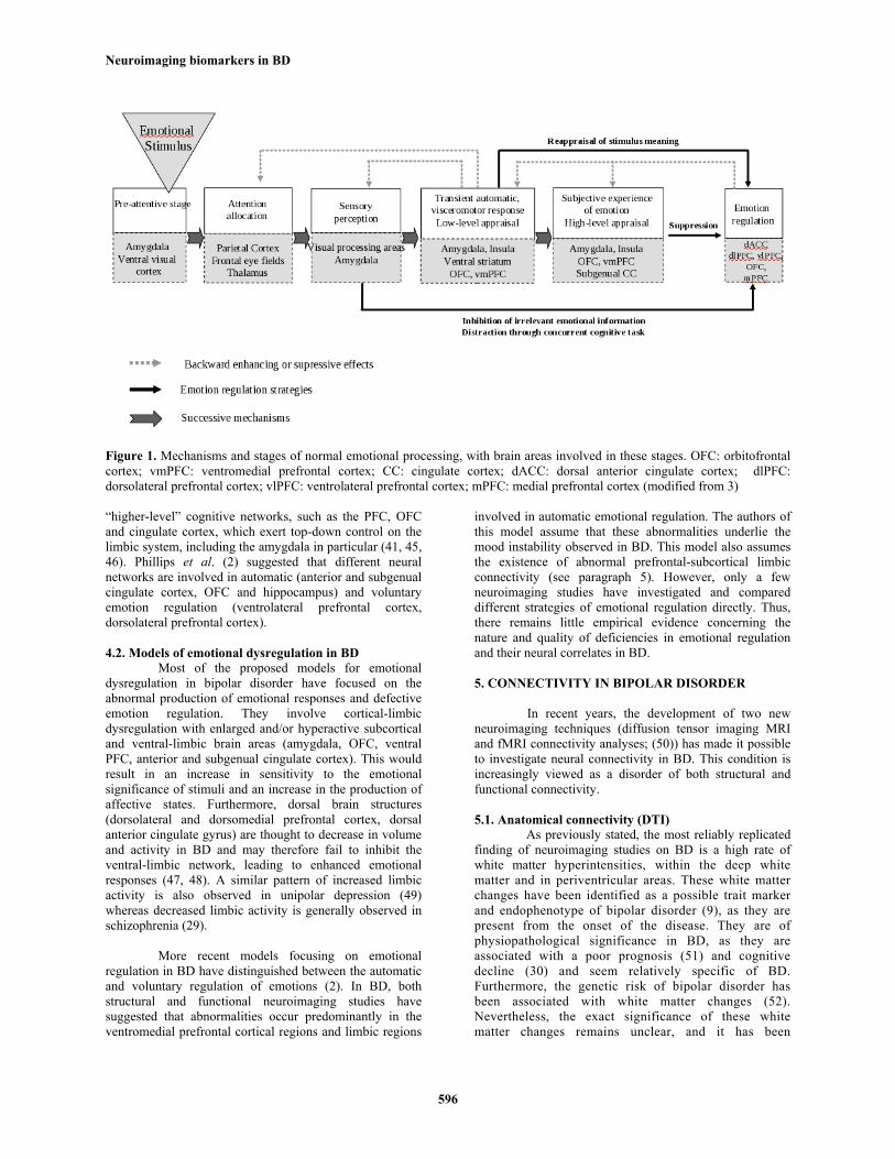
Emotional processing is not a uniform process,and has been divided into various stages (3): (1) earlyemotional processing, including the pre-attentive stage,attention allocation and sensory perception; (2) emotionalresponses with automatic emotional response, experienceand expression of emotion (3) emotional regulation,involving the initiation of new emotional responses or thealteration of existing responses (41) (see Figure 1). Thesethree stages involve both different and common neuralnetworks.
Early emotional processing depends principallyon the attentional resources allocated. It has consistentlybeen shown that emotional stimuli and mechanisms ofselective attention modulate each other (42). It has beenshown that the limbic system, including the amygdala inparticular, mediates this increase in attentional load onemotional stimuli (43). The amygdala exerts this effectthrough amygdala-cortical connections (sensory andassociative cortices; (44)). The prefrontal cortex, includingthe VMPFC in particular, also modulates responses toemotional stimuli by modifying the priority level ofstimulus processing (43).
The amygdala, along with other subcorticalstructures, such as the ventral striatum, is crucial for thegeneration and experience of emotion (3). Amygdalaactivation is more specifically associated with thegeneration of negative emotional responses, whereas othersubcortical regions, such as the ventral striatum, are morestrongly linked to positive emotions.
Emotional regulation involves severalmechanisms, such as reappraisal, suppression, inhibition,extinction, reversal and the maintenance of representations.Emotional appraisal networks can be modulated by
Neuroimaging biomarkers in BD
596
Figure 1. Mechanisms and stages of normal emotional processing, with brain areas involved in these stages. OFC: orbitofrontalcortex; vmPFC: ventromedial prefrontal cortex; CC: cingulate cortex; dACC: dorsal anterior cingulate cortex; dlPFC:dorsolateral prefrontal cortex; vlPFC: ventrolateral prefrontal cortex; mPFC: medial prefrontal cortex (modified from 3)
“higher-level” cognitive networks, such as the PFC, OFCand cingulate cortex, which exert top-down control on thelimbic system, including the amygdala in particular (41, 45,46). Phillips et al. (2) suggested that different neuralnetworks are involved in automatic (anterior and subgenualcingulate cortex, OFC and hippocampus) and voluntaryemotion regulation (ventrolateral prefrontal cortex,dorsolateral prefrontal cortex).
4.2. Models of emotional dysregulation in BDMost of the proposed models for emotional
dysregulation in bipolar disorder have focused on theabnormal production of emotional responses and defectiveemotion regulation. They involve cortical-limbicdysregulation with enlarged and/or hyperactive subcorticaland ventral-limbic brain areas (amygdala, OFC, ventralPFC, anterior and subgenual cingulate cortex). This wouldresult in an increase in sensitivity to the emotionalsignificance of stimuli and an increase in the production ofaffective states. Furthermore, dorsal brain structures(dorsolateral and dorsomedial prefrontal cortex, dorsalanterior cingulate gyrus) are thought to decrease in volumeand activity in BD and may therefore fail to inhibit theventral-limbic network, leading to enhanced emotionalresponses (47, 48). A similar pattern of increased limbicactivity is also observed in unipolar depression (49)whereas decreased limbic activity is generally observed inschizophrenia (29).
More recent models focusing on emotionalregulation in BD have distinguished between the automaticand voluntary regulation of emotions (2). In BD, bothstructural and functional neuroimaging studies havesuggested that abnormalities occur predominantly in theventromedial prefrontal cortical regions and limbic regions
involved in automatic emotional regulation. The authors ofthis model assume that these abnormalities underlie themood instability observed in BD. This model also assumesthe existence of abnormal prefrontal-subcortical limbicconnectivity (see paragraph 5). However, only a fewneuroimaging studies have investigated and compareddifferent strategies of emotional regulation directly. Thus,there remains little empirical evidence concerning thenature and quality of deficiencies in emotional regulationand their neural correlates in BD.
5. CONNECTIVITY IN BIPOLAR DISORDER
In recent years, the development of two newneuroimaging techniques (diffusion tensor imaging MRIand fMRI connectivity analyses; (50)) has made it possibleto investigate neural connectivity in BD. This condition isincreasingly viewed as a disorder of both structural andfunctional connectivity.
5.1. Anatomical connectivity (DTI)As previously stated, the most reliably replicated
finding of neuroimaging studies on BD is a high rate ofwhite matter hyperintensities, within the deep whitematter and in periventricular areas. These white matterchanges have been identified as a possible trait markerand endophenotype of bipolar disorder (9), as they arepresent from the onset of the disease. They are ofphysiopathological significance in BD, as they areassociated with a poor prognosis (51) and cognitivedecline (30) and seem relatively specific of BD.Furthermore, the genetic risk of bipolar disorder hasbeen associated with white matter changes (52).Nevertheless, the exact significance of these whitematter changes remains unclear, and it has been
Neuroimaging biomarkers in BD
597
suggested that they may reflect changes in anatomicalconnectivity.
A recently developed technique, diffusion tensorimaging (DTI), can be used to explore connectivity andwhite matter in vivo, providing information about bothmacrostructure (tracts) and microstructure (localorganization; (53)). DTI explores the features of waterdiffusion in the brain. Water diffusion in the brain is restrictedprincipally by axons and myelin sheaths. DTI thereforeexplores the integrity of white matter in voxels, giving afractional anisotropy (FA) value for each voxel. Fractionalanisotropy is correlated with the integrity and coherence ofwhite matter. Decreases in FA have been associated withedema, demyelination and brain inflammation. DTI alsoprovides information about mean diffusivity (generallyexpressed as ADC, or apparent diffusion coefficient) and theprincipal direction of diffusion in each voxel. This principaldirection of diffusion within a voxel is thought to be parallel tothe principal direction of the white matter tract in this voxel.These data allow a step-by-step reconstruction of white mattertracts in the whole brain and the comparison of these tractsbetween groups (54). In sum, DTI can be used to explore whitematter and brain connectivity, in terms of both macrostructure(with tractography) and microstructure (with FA and ADC).
The first DTI-based studies in patients with BDreported a decrease in FA in frontal regions of interest (ROIs)(55-57), interpreted by the authors as axonal disorganization, aloss of coherence and, thus, changes in structural connectivity.This decrease in frontal FA has been confirmed and extendedby larger whole-brain studies, which have identified diffusedecreases of FA in the corpus callosum, the fornix and in theprefrontal, limbic and striatal regions (58-61). These changesoccur in key intra- and interhemispheric tracts, such as thefronto-occipital fasciculus (FOF), the inferior longitudinalfasciculus (ILF), the superior longitudinal fasciculus (SLF), thecingulum, the corpus callosum and the uncinate fasciculus(UF). DTI can also be used to evaluate these tracts and toevaluate their characteristics. Using this technique, wereconstructed the UF in a group of 16 euthymic patients withbipolar disorder and 16 healthy controls. We found that therewas a significantly higher number of reconstructed fibers in theleft hemisphere in patients with BD, consistent with anincrease in anatomical connectivity in the left uncinatefasciculus (62). Two other DTI-based studies (63, 64)confirmed these changes to the uncinate fasciculus in BD.Connectivity changes in the UF are of particular interest, asthis structure connects critical limbic areas of the brain, such asthe amygdala, the subgenual cingulate cortex and theorbitofrontal cortex. It has been suggested that this brain circuitis misconnected or dysregulated in bipolar disorder (seesection 4), and that this probably underlies the observedhyperactivity of the corresponding brain areas and the moodinstability and inability to regulate emotional states in patientswith BD. Possible increases in anatomical connectivity foundwith DTI are of particular interest as they are likely to bespecific of BD as similar studies in schizophrenia or unipolardepression found no area of increased anatomical connectivity(65, 66).
DTI studies of BD have generated some convergingresults, but some inconsistencies have been found between the
empirical findings of such studies. Some studies have reportedan absence of change in FA (67), whereas others have reportedan increase in FA in the corpus callosum (68), UF or in morediffuse brain regions (69, 70). The heterogeneity observed inDTI studies of bipolar disorder may result from the clinicaldiversity of samples, as previously discussed, or the recentnature of DTI, for which methodologies have yet to bestandardized. Some DTI studies have used processing softwaredeveloped for classical anatomical MRI, yielding to somepotential issues (71). Specific techniques and software havethus been developed to process DTI data (71). Additionally,the exact nature of anatomical connectivity changes in first-episode patients still needs to be more extensively assessed.
It has been suggested that the expression ofsymptoms of bipolar disorder may result from dysfunctions ofdiscrete brain networks, such as the anterior limbic network (2,72). This model is supported by connectivity data and bygenetic studies of bipolar disorder. Some allelic variants ofneuregulin-1 have been repeatedly associated with bipolardisorder (73-78). Neuregulin-1 is crucial for neuronalmigration, synapse formation, oligodendrocyte differentiation,neuronal myelination and, thus, brain connectivity.Interestingly, several groups have reported that neuregulin-1variants are associated with differences in FA, as measured byDTI, in the frontal medial area (79) and anterior cingulatecortex(80).
5.2. Functional connectivity (fMRI)In addition to DTI-based studies of anatomical
connectivity, new analytical techniques for exploringfunctional connectivity by fMRI have recently been developed.Functional connectivity (FC) is the “temporal correlationsbetween spatially remote neurophysisological events” (81). Itprovides insight into the degree to which different parts ofbrain networks are functionally coupled together, during theperformance of a task or at rest (82). FC methods can be usedto identify the brain areas communicating with each otherduring visual, emotional, language and motor-related functions(82, 83). The use of FC methods to analyze brain connectivityat rest has led to identification of the so-called “default-modenetwork” (DMN) (83). The DMN includes theprecuneus/posterior cingulate cortex, medial prefrontal cortex,and medial, lateral and inferior parietal cortex (84). Thisnetwork is activated at rest and deactivated during goal-oriented behavior. Studies of functional connectivity withinthis network have already yielded interesting results in othermental disorders, such as schizophrenia, demonstratingincreases in connectivity within the DMN (85). By contrast,effective connectivity (EC) is defined as “the influence oneneural system exerts over another either directly or indirectly”(81). EC includes directional data concerning the relationshipsbetween two areas.
Only a few studies have investigated FC or EC,either at rest or during cognitive and emotional tasks, inpatients with BD. These studies have generated convergingevidence to suggest that the connectivity betweenamygdala/hippocampus and ventral prefrontal/perigenualcortex is altered in BD (86). During an emotion-labelingtask, patients with bipolar disorder displayed significantly
Neuroimaging biomarkers in BD
598
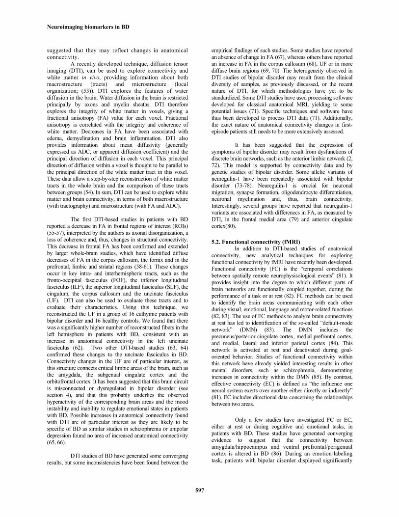
Figure 2. Proposed neurobiological model of BD on asagittal medial view of left hemisphere (modified from 2)In red: increased activity in ventral-limbic regions; in blue:decreased activity in dorsal regions; disrupted connectivitybetween both networks (green). VMPFC : ventromedialprefrontal cortex; CG : cingulate gyrus; OFC :Orbitofrontal Cortex.
higher levels of effective connectivity between the rightparahippocampal gyrus and the right subgenual cingulatecortex (87). During a male-female determination task withemotional interference, Wang et al. reported lowerfunctional connectivity between the amygdala and theperigenual anterior cingulate cortex in patients with BDthan in healthy controls (88). Almeida et al. reported thatpatients with bipolar and unipolar depression had differentpatterns of effective connectivity between the amygdalaand orbitomedial prefrontal cortex during emotionalintensity labeling tasks (89). Similarly, the same groupdescribed abnormal bilateral amygdala-orbitofrontalcortical functional connectivity in patients with BD: thesepatients had a significantly higher FC between the rightamygdala and orbitofrontal cortex, whereas depressedfemale patients with BD had a significantly higher FCbetween the left amygdala and orbitofrontal cortex, thesedifferences not being observed in patients in remission(90).
Even at rest, abnormalities in functionalconnectivity have been identified in patients with BD. Thefirst study to investigate resting state connectivityabnormalities in BD found that patients with this disorderhad significantly lower perigenual cingulate connectivity tothe amygdala, thalamus and pallidostriatum than unaffectedsubjects (91). Similarly, Chepenik et al. reported a lowerlevel of connectivity between the amygdala and ventralprefrontal cortex in patients with BD (92). One very recentstudy reported abnormalities within the DMN in manicpatients, with abnormal recruitment of the parietal cortex,whereas patients with schizophrenia displayed greaterrecruitment of the basal ganglia and frontopolar cortex(93).
These converging results showing changes inamygdala/prefrontal connectivity are consistent with theproposed neurobiological models of BD reviewed above,all of which assume that the ventro-limbic networkinvolved in the processing and regulation of emotions is
dysfunctional in BD patients (see Figure 2). Nevertheless,the exact nature of connectivity changes in first-episodepatients and their evolution still need to be more assessed.Furthermore, regarding both anatomical and functionalconnectivity techniques, we should keep in mind that theseare recent techniques, still under development. Theinterpretation of the results should thus be cautious.
5.3. Linking structural and functional connectivity:bipolar disorder as a connectivity disorder?
With the development of structural and functionalconnectivity exploration techniques, new avenues ofresearch and advanced theories have been developed basedon information from different types of imaging, which isused to construct unified models of aberrant large-scalebrain networks in mental disorders, such as BD (50, 94).Several studies have shown that resting-state functionalconnectivity (as measured with fMRI) is positivelycorrelated with structural connectivity (as measured withDTI) (94-97). Multimodal approaches of this type areparticularly useful in pathological conditions, as onetechnique alone may not be specific and sensitive enoughfor use as an individual biomarker (94), additionalinformation from other modes of imaging thereforeincreasing the accuracy of the test.
Only one study has used both structural (DTI)and functional (fMRI) connectivity measurements for thesame sample of patients with BD (88). This study reportedlower functional connectivity between the amygdala andthe perigenual anterior cingulate cortex (pACC) in patientswith BD than in healthy controls. Interestingly, it alsoreported a positive correlation between pACC-amygdalafunctional connectivity and FA in ventrofrontal whitematter, including the region of the UF. The authorssuggested that disruption of the structural integrity of whitematter bundles linking the pACC and amygdala mightcontribute to the pACC-amygdala functional couplingdeficit. This study highlights the importance of mergingdata from different sources to reveal the roles of particularstructures or brain networks in the pathophysiology ofmental disorders.
6. PERSPECTIVE
6.1. Individual biomarkers in bipolar disorderNeuroimaging studies of BD have yielded some
interesting and convergent results supporting currentneurobiological models of BD. However, the diagnosticbiomarkers identified in these studies cannot yet be used byclinicians, because they have been validated at group level,but not at individual level. Valid diagnostic biomarkersmust be sensitive and specific, with positive predictivevalue (PPV) and negative predictive value (NPV).
However, it will probably be possible to identifybiomarkers suitable for individual use in the diagnosis ofBD in the next few years, due to advances in bioinformaticsinvolving the development of machine learning algorithms.In such algorithms, the computer discovers (in a supervisedor unsupervised design) and learns from a “learningdataset” (group information supplied to the computer), the
Neuroimaging biomarkers in BD
599
rules for distinguishing the MRI scans of patients fromthose of healthy controls, based on various mathematicalmethods (e.g. support vector machine algorithms). Thecomputer then applies these rules to new datasets, for theautomatic classification of patients and healthy subjectswithin the sample.
Proof-of-concept for such approaches has alreadybeen demonstrated in schizophrenia and autism. In 2005,Davatzikos and colleagues (98) applied such an automatedclassification technique to T1 MRI scans from 69 patientswith schizophrenia and 79 healthy controls. They achieveda classification accuracy of 81%. They have also used asimilar method to predict disease transition: using T1 MRIscans from at-risk subjects, they were able to predicttransition to psychosis four years later, with an accuracy of82% (99). Similar computerized approaches have been usedin autism (100). Some groups are developing techniques forclassifying subjects on the basis of their fMRI data (101),cortical folding patterns (102) or DTI data. Very promisingresults have been obtained in schizophrenia, but replicationstudies in independent samples and cross-validation againstother clinically relevant diagnoses such as major depressionand schizophrenia, are required.
Such classification approaches have not beenused in BD. Previous neuroimaging studies have suggestedthat neuroanatomical changes in bipolar disorder are moresubtle than those in schizophrenia and autism, but theapplication of machine learning approaches nonethelessappears worthwhile in this patient group. Such approachesmay make it easier to distinguish between patients with BDand healthy controls, but may also facilitate differentiationbetween different subtypes of BD and between unipolarand bipolar depression. Differences between unipolar andbipolar depression have already been observed for fMRIand DTI (89, 103, 104). Finally, machine learningapproaches may help to predict the course of the illness orthe response to treatment (medication or psychotherapy),both of which are highly relevant in clinical research.
6.2. Imaging genetics of bipolar disorderWith the increasing power of genetics and the
new techniques used in neuroimaging, a new field, calledimaging genetics, has emerged in recent years (105, 106).Imaging genetics assesses the impact of allelic variation onbrain structure and function. It is based on the assumptionthat brain structure and function 1) are under strong geneticcontrol (106) and 2) represent an intermediate phenotype.In the future, such approaches and the results they generateshould facilitate elucidation of the functional role of riskgene variants and the identification of trait andvulnerability markers of mental illnesses. For example, 5-HTT promoter polymorphism has been associated withsusceptibility to mood disorders in stressful conditions(107). Brain imaging studies exploring the impact of the s-allele have repeatedly demonstrated that this risk alleleincreases the reactivity of the amygdala to negativeemotional stimuli (see (108) for a meta-analysis of thiseffect). This modulation of amygdala reactivity by thisgene may account for the greater reactivity to negativeevents in subjects carrying this risk allele.
A few genes, including those encodingCACNA1C and neuregulin-1, have recently beenrepeatedly associated with BD. CACNA1C is a subunit of avoltage-gated calcium channel. Variants of CACNA1Cwere found to be associated with BD in a collaborativegenome-wide analysis of 4387 patients and 6209 healthycontrols (109). The impact of CACNA1C allelic variationwas largely unknown until recent neuroimaging studiesrevealed that carriers of the CACNA1C risk allele in agroup of healthy controls displayed stronger limbic (rightamygdala) activation in response to reward (110). Thesame risk allele also seems to be associated with largercerebral gray matter volume (111, 112), although this resultrequires replication, as another group reported an effect onbrainstem gray matter, but not on cerebral gray matter(112).
Similarly, as reported above, neuregulin-1variation has been associated both with BD (73-78) andchanges in frontal and cingulum FA (79, 80).
The impact of susceptibility gene factors on brainstructure and function can also be assessed by studying thehealthy relatives of patients with BD. Such studies aredifficult to perform, due to recruitment constraints, but theygenerate unique information. They make it possible toidentify endophenotypes of BD — intermediate phenotypesassociated with the genetic risk factors of bipolar disorder.White matter changes have been identified as a potentialendophenotype of bipolar disorder (9). Recent studies haveshown that genetic predisposition to BD is associated withlow FA throughout the white matter regions (60). In graymatter, the reported deficits in healthy relatives of patientswith BD are located in caudate volumes (113) and includeincreases in left insula and left cerebellum (114) and leftparahippocampal gyrus (115) volumes. However, moststudies have yielded negative results (113, 116-122).Healthy first-degree relatives of patients with BD haveshown to be unable to suppress activation in theorbitofrontal cortex, superior parietal cortex, precuneus andinsula during cognitive tasks, whereas such suppression isobserved in healthy controls (123, 124). During a facialemotion processing task, first-degree relatives have beenshown to display strong medial prefrontal cortical andsubcortical (putamen and amygdala) activation, similarly tothat observed in patients with BD (125). All these patternsmay represent potential endophenotypes of bipolardisorder.
In conclusion, neuroimaging studies of BD haveled to the formulation of neurobiological models based ondysfunctional connectivity between prefrontal andsubcortical regions. Nevertheless, several issues remainunresolved, mostly due to the differences between thesamples used in different studies. Two types of clinicaldifferences may represent bias in existing studies: thoselinked to different pathophysiological processes (e.g. type Ivs type II, rapid cycling) and those associated with theevolution of the disease (medication, substance abuse,number of episodes). The existing transversal studiescannot solve this issue. We yet do not know whichabnormalities are vulnerability biomarkers, biomarkers

Neuroimaging biomarkers in BD
600
present before onset, after onset or associated with theevolution of the disease. No biomarker suitable for thediagnosis of BD in individual patients is yet available.
Based on the reviewed studies and theproblematic issues and technical advances in neuroimagingresearch in BD, we make the following suggestions forfuture research aiming to identify neuroimaging biomarkersof BD:
1) The inclusion of a large (N > 200) number of patients.This would make it possible to compare data betweensubgroups of patients with sufficient statistical power (e.g.bipolar I versus bipolar II, psychotic features versus non-psychotic, etc...). It would also make it possible to take intoaccount confounding variables, such as alcoholabuse/dependence. Multisite studies would also facilitatethe development of individually usable biomarkers.
2) The inclusion of longitudinal cohort of patients, whowould be scanned at the 1st episode (1st manic episode) andrescanned a few years later. This design would dramaticallyincrease statistical power (as subjects could act as their owncontrols), and would make it possible to carry out a preciseclinical evaluation of the confounding factors. Longitudinalapproaches might also be useful for predicting the onset ofdisease in high-risk subjects (126).
7. ACKNOWLEDGMENTS
Financial support: this work was supported by theFrench Agence Nationale pour la Recherche ANR MNP2008 (JH, MAD, FEV, ML), Fondation pour la RechercheMédicale (FEV), Deutsche Forschungsgemeinschaft(DFG): We3638/3-1, SFB636/C6 (MW) and theFondaMental foundation (a French Science Foundation).
8. REFERENCES
1. Singh, I. & N. Rose: Biomarkers in psychiatry. Nature,460, 202-7 (2009)
2. Phillips, M. L., C. D. Ladouceur & W. C. Drevets: Aneural model of voluntary and automatic emotionregulation: implications for understanding thepathophysiology and neurodevelopment of bipolar disorder.Mol Psychiatry, 13, 829, 833-57 (2008)
3. Wessa, M. & J. Linke: Emotional processing in bipolardisorder: behavioural and neuroimaging findings. Int RevPsychiatry, 21, 357-67 (2009)
4. Terry, J., M. Lopez-Larson & J. A. Frazier: Magneticresonance imaging studies in early onset bipolar disorder:an updated review. Child Adolesc Psychiatr Clin N Am, 18,421-39, ix-x (2009)
5. McDonald, C., J. Zanelli, S. Rabe-Hesketh, I. Ellison-Wright, P. Sham, S. Kalidindi, R. M. Murray & N.Kennedy: Meta-analysis of magnetic resonance imagingbrain morphometry studies in bipolar disorder. BiolPsychiatry, 56, 411-7 (2004)
6. Kempton, M. J., J. R. Geddes, U. Ettinger, S. C.Williams & P. M. Grasby: Meta-analysis, database, andmeta-regression of 98 structural imaging studies in bipolardisorder. Arch Gen Psychiatry, 65, 1017-32 (2008)
7. Vita, A., L. De Peri & E. Sacchetti: Gray matter, whitematter, brain, and intracranial volumes in first-episodebipolar disorder: a meta-analysis of magnetic resonanceimaging studies. Bipolar Disord, 11, 807-14 (2009)
8. Pillai, J. J., L. Friedman, T. A. Stuve, S. Trinidad, J. A.Jesberger, J. S. Lewin, R. L. Findling, T. P. Swales & S. C.Schulz: Increased presence of white matter hyperintensitiesin adolescent patients with bipolar disorder. Psychiatry Res,114, 51-6 (2002)
9. Hasler, G., W. C. Drevets, T. D. Gould, Gottesman, II &H. K. Manji: Toward constructing an endophenotypestrategy for bipolar disorders. Biol Psychiatry, 60, 93-105(2006)
10. Tham, M. W., P. S. Woon, M. Y. Sum, T. S. Lee & K.Sim: White matter abnormalities in major depression:Evidence from post-mortem, neuroimaging and geneticstudies. J Affect Disord (2011)
11. Ellison-Wright, I. & E. Bullmore: Anatomy of bipolardisorder and schizophrenia: a meta-analysis. Schizophr Res,117, 1-12 (2010)
12. Hajek, T., M. Kopecek, J. Kozeny, E. Gunde, M. Alda& C. Hoschl: Amygdala volumes in mood disorders--meta-analysis of magnetic resonance volumetry studies. J AffectDisord, 115, 395-410 (2009)
13. Usher, J., S. Leucht, P. Falkai & H. Scherk: Correlationbetween amygdala volume and age in bipolar disorder - asystematic review and meta-analysis of structural MRIstudies. Psychiatry Res, 182, 1-8 (2010)
14. Strakowski, S. M., M. P. Delbello & C. M. Adler: Thefunctional neuroanatomy of bipolar disorder: a review ofneuroimaging findings. Mol Psychiatry, 10, 105-16 (2005)
15. Chen, C. H., B. Lennox, R. Jacob, A. Calder, V.Lupson, R. Bisbrown-Chippendale, J. Suckling & E.Bullmore: Explicit and implicit facial affect recognition inmanic and depressed States of bipolar disorder: a functionalmagnetic resonance imaging study. Biol Psychiatry, 59, 31-9 (2006)
16. Lawrence, N. S., A. M. Williams, S. Surguladze, V.Giampietro, M. J. Brammer, C. Andrew, S. Frangou, C.Ecker & M. L. Phillips: Subcortical and ventral prefrontalcortical neural responses to facial expressions distinguishpatients with bipolar disorder and major depression. BiolPsychiatry, 55, 578-87 (2004)
17. Hassel, S., J. R. Almeida, N. Kerr, S. Nau, C. D.Ladouceur, K. Fissell, D. J. Kupfer & M. L. Phillips:Elevated striatal and decreased dorsolateral prefrontalcortical activity in response to emotional stimuli in
Neuroimaging biomarkers in BD
601
euthymic bipolar disorder: no associations withpsychotropic medication load. Bipolar Disord, 10, 916-27(2008)
18. Jogia, J., M. Haldane, A. Cobb, V. Kumari & S.Frangou: Pilot investigation of the changes in corticalactivation during facial affect recognition with lamotriginemonotherapy in bipolar disorder. Br J Psychiatry, 192, 197-201 (2008)
19. Lennox, B. R., R. Jacob, A. J. Calder, V. Lupson & E.T. Bullmore: Behavioural and neurocognitive responses tosad facial affect are attenuated in patients with mania.Psychol Med, 34, 795-802 (2004)
20. Malhi, G. S., J. Lagopoulos, P. S. Sachdev, B.Ivanovski, R. Shnier & T. Ketter: Is a lack of disgustsomething to fear? A functional magnetic resonanceimaging facial emotion recognition study in euthymicbipolar disorder patients. Bipolar Disord, 9, 345-57 (2007)
21. Elliott, R., A. Ogilvie, J. S. Rubinsztein, G. Calderon,R. J. Dolan & B. J. Sahakian: Abnormal ventral frontalresponse during performance of an affective go/no go taskin patients with mania. Biol Psychiatry, 55, 1163-70 (2004)
22. Wessa, M., J. Houenou, M. L. Paillere-Martinot, S.Berthoz, E. Artiges, M. Leboyer & J. L. Martinot: Fronto-striatal overactivation in euthymic bipolar patients duringan emotional go/nogo task. Am J Psychiatry, 164, 638-46(2007)
23. Lagopoulos, J. & G. S. Malhi: A functional magneticresonance imaging study of emotional Stroop in euthymicbipolar disorder. Neuroreport, 18, 1583-7 (2007)
24. Malhi, G. S., J. Lagopoulos, P. S. Sachdev, B.Ivanovski & R. Shnier: An emotional Stroop functionalMRI study of euthymic bipolar disorder. Bipolar Disord, 7Suppl 5, 58-69 (2005)
25. Malhi, G. S., J. Lagopoulos, A. M. Owen, B. Ivanovski,R. Shnier & P. Sachdev: Reduced activation to implicitaffect induction in euthymic bipolar patients: an fMRIstudy. J Affect Disord, 97, 109-22 (2007)
26. Altshuler, L., S. Bookheimer, J. Townsend, M. A.Proenza, F. Sabb, J. Mintz & M. S. Cohen: Regional brainchanges in bipolar I depression: a functional magneticresonance imaging study. Bipolar Disord, 10, 708-17(2008)
27. Foland, L. C., L. L. Altshuler, S. Y. Bookheimer, N.Eisenberger, J. Townsend & P. M. Thompson: Evidence fordeficient modulation of amygdala response by prefrontalcortex in bipolar mania. Psychiatry Res, 162, 27-37 (2008)
28. Fitzgerald, P. B., A. R. Laird, J. Maller & Z. J.Daskalakis: A meta-analytic study of changes in brainactivation in depression. Hum Brain Mapp, 29, 683-95(2008)
29. Li, H., R. C. Chan, G. M. McAlonan & Q. Y. Gong:Facial emotion processing in schizophrenia: a meta-analysis of functional neuroimaging data. SchizophrBull, 36, 1029-39 (2010)
30. Bearden, C. E., K. M. Hoffman & T. D. Cannon:The neuropsychology and neuroanatomy of bipolaraffective disorder: a critical review. Bipolar Disord, 3,106-50; discussion 151-3 (2001)
31. Strakowski, S. M., C. M. Adler, S. K. Holland, N.Mills & M. P. DelBello: A preliminary FMRI study ofsustained attention in euthymic, unmedicated bipolardisorder. Neuropsychopharmacology, 29, 1734-40(2004)
32. Adler, C. M., S. K. Holland, V. Schmithorst, M. J.Tuchfarber & S. M. Strakowski: Changes in neuronalactivation in patients with bipolar disorder duringperformance of a working memory task. Bipolar Disord, 6,540-9 (2004)
33. M'Bailara, K., J. Demotes-Mainard, J. Swendsen, F.Mathieu, M. Leboyer & C. Henry: Emotional hyper-reactivity in normothymic bipolar patients. Bipolar Disord,11, 63-9 (2009)
34. Minzenberg, M. J., A. R. Laird, S. Thelen, C. S. Carter& D. C. Glahn: Meta-analysis of 41 functional neuroimagingstudies of executive function in schizophrenia. Arch GenPsychiatry, 66, 811-22 (2009)
35. Ha, T. H., K. Ha, J. H. Kim & J. E. Choi: Regional braingray matter abnormalities in patients with bipolar II disorder: acomparison study with bipolar I patients and healthy controls.Neurosci Lett, 456, 44-8 (2009)
36. Chanraud, S., M. Reynaud, M. Wessa, J. Penttila, N.Kostogianni, A. Cachia, E. Artiges, F. Delain, M. Perrin, H. J.Aubin, Y. Cointepas, C. Martelli & J. L. Martinot: Diffusiontensor tractography in mesencephalic bundles: relation tomental flexibility in detoxified alcohol-dependent subjects.Neuropsychopharmacology, 34, 1223-32 (2009)
37. Germana, C., M. J. Kempton, A. Sarnicola, T.Christodoulou, M. Haldane, M. Hadjulis, P. Girardi, R.Tatarelli & S. Frangou: The effects of lithium andanticonvulsants on brain structure in bipolar disorder. ActaPsychiatr Scand (2010)
38. Yucel, K., M. C. McKinnon, V. H. Taylor, K. Macdonald,M. Alda, L. T. Young & G. M. MacQueen: Bilateralhippocampal volume increases after long-term lithiumtreatment in patients with bipolar disorder: a longitudinal MRIstudy. Psychopharmacology (Berl), 195, 357-67 (2007)
39. Moncrieff, J. & J. Leo: A systematic review of the effectsof antipsychotic drugs on brain volume. Psychol Med, 40,1409-22 (2010)
40. Bell, E. C., M. C. Willson, A. H. Wilman, S. Dave & P.H. Silverstone: Differential effects of chronic lithium and
Neuroimaging biomarkers in BD
602
valproate on brain activation in healthy volunteers. HumPsychopharmacol, 20, 415-24 (2005)
41. Ochsner, K. N. & J. J. Gross: The cognitive controlof emotion. Trends Cogn Sci, 9, 242-9 (2005)
42. Raymond, J.: Interactions of attention, emotion andmotivation. Prog Brain Res, 176, 293-308 (2009)
43. Taylor, J. G. & N. F. Fragopanagos: The interactionof attention and emotion. Neural Netw, 18, 353-69(2005)
44. Anderson, A. K.: Affective influences on theattentional dynamics supporting awareness. J ExpPsychol Gen, 134, 258-81 (2005)
45. Schaefer, A., F. Collette, P. Philippot, M. van derLinden, S. Laureys, G. Delfiore, C. Degueldre, P.Maquet, A. Luxen & E. Salmon: Neural correlates of"hot" and "cold" emotional processing: a multilevelapproach to the functional anatomy of emotion.Neuroimage, 18, 938-49 (2003)
46. Blair, K. S., B. W. Smith, D. G. Mitchell, J. Morton,M. Vythilingam, L. Pessoa, D. Fridberg, A. Zametkin,D. Sturman, E. E. Nelson, W. C. Drevets, D. S. Pine, A.Martin & R. J. Blair: Modulation of emotion bycognition and cognition by emotion. Neuroimage, 35,430-40 (2007)
47. Blumberg, H. P., D. S. Charney & J. H. Krystal:Frontotemporal neural systems in bipolar disorder.Semin Clin Neuropsychiatry, 7, 243-54 (2002)
48. Phillips, M. L., W. C. Drevets, S. L. Rauch & R.Lane: Neurobiology of emotion perception II:Implications for major psychiatric disorders. BiolPsychiatry, 54, 515-28 (2003)
49. Ressler, K. J. & H. S. Mayberg: Targeting abnormalneural circuits in mood and anxiety disorders: from thelaboratory to the clinic. Nat Neurosci, 10, 1116-24(2007)
50. Guye, M., F. Bartolomei & J. P. Ranjeva: Imagingstructural and functional connectivity: towards a unifieddefinition of human brain organization? Curr OpinNeurol, 21, 393-403 (2008)
51. Moore, P. B., D. J. Shepherd, D. Eccleston, I. C.Macmillan, U. Goswami, V. L. McAllister & I. N.Ferrier: Cerebral white matter lesions in bipolaraffective disorder: relationship to outcome. Br JPsychiatry, 178, 172-6 (2001)
52. McDonald, C., E. T. Bullmore, P. C. Sham, X.Chitnis, H. Wickham, E. Bramon & R. M. Murray:Association of genetic risks for schizophrenia andbipolar disorder with specific and generic brainstructural endophenotypes. Arch Gen Psychiatry, 61,974-84 (2004)
53. Le Bihan, D.: Looking into the functionalarchitecture of the brain with diffusion MRI. Nat RevNeurosci, 4, 469-80 (2003)
54. Mori, S. & P. C. van Zijl: Fiber tracking: principles andstrategies - a technical review. NMR Biomed, 15, 468-80(2002)
55. Adler, C. M., S. K. Holland, V. Schmithorst, M. Wilke,K. L. Weiss, H. Pan & S. M. Strakowski: Abnormal frontalwhite matter tracts in bipolar disorder: a diffusion tensorimaging study. Bipolar Disord, 6, 197-203 (2004)
56. Adler, C. M., J. Adams, M. P. DelBello, S. K. Holland,V. Schmithorst, A. Levine, K. Jarvis & S. M. Strakowski:Evidence of white matter pathology in bipolar disorderadolescents experiencing their first episode of mania: adiffusion tensor imaging study. Am J Psychiatry, 163, 322-4 (2006)
57. Haznedar, M. M., F. Roversi, S. Pallanti, N. Baldini-Rossi, D. B. Schnur, E. M. Licalzi, C. Tang, P. R. Hof, E.Hollander & M. S. Buchsbaum: Fronto-thalamo-striatalgray and white matter volumes and anisotropy of theirconnections in bipolar spectrum illnesses. Biol Psychiatry,57, 733-42 (2005)
58. Zanetti, M. V., M. P. Jackowski, A. Versace, J. R.Almeida, S. Hassel, F. L. Duran, G. F. Busatto, D. J.Kupfer & M. L. Phillips: State-dependent microstructuralwhite matter changes in bipolar I depression. Eur ArchPsychiatry Clin Neurosci, 259, 316-28 (2009)
59. Barnea-Goraly, N., K. D. Chang, A. Karchemskiy, M.E. Howe & A. L. Reiss: Limbic and corpus callosumaberrations in adolescents with bipolar disorder: a tract-based spatial statistics analysis. Biol Psychiatry, 66, 238-44(2009)
60. Chaddock, C. A., G. J. Barker, N. Marshall, K. Schulze,M. H. Hall, A. Fern, M. Walshe, E. Bramon, X. A. Chitnis,R. Murray & C. McDonald: White matter microstructuralimpairments and genetic liability to familial bipolar Idisorder. Br J Psychiatry, 194, 527-34 (2009)
61. Chan, W. Y., G. L. Yang, M. Y. Chia, P. S. Woon, J.Lee, R. Keefe, Y. Y. Sitoh, W. L. Nowinski & K. Sim:Cortical and subcortical white matter abnormalities inadults with remitted first-episode mania revealed by Tract-Based Spatial Statistics. Bipolar Disord, 12, 383-9 (2010)
62. Houenou, J., M. Wessa, G. Douaud, M. Leboyer, S.Chanraud, M. Perrin, C. Poupon, J. L. Martinot & M. L.Paillere-Martinot: Increased white matter connectivity ineuthymic bipolar patients: diffusion tensor tractographybetween the subgenual cingulate and the amygdalo-hippocampal complex. Mol Psychiatry, 12, 1001-10 (2007)
63. McIntosh, A. M., S. Munoz Maniega, G. K. Lymer, J.McKirdy, J. Hall, J. E. Sussmann, M. E. Bastin, J. D.Clayden, E. C. Johnstone & S. M. Lawrie: White matter
Neuroimaging biomarkers in BD
603
tractography in bipolar disorder and schizophrenia. BiolPsychiatry, 64, 1088-92 (2008)
64. Versace, A., J. R. Almeida, S. Hassel, N. D. Walsh, M.Novelli, C. R. Klein, D. J. Kupfer & M. L. Phillips:Elevated left and reduced right orbitomedial prefrontalfractional anisotropy in adults with bipolar disorderrevealed by tract-based spatial statistics. Arch GenPsychiatry, 65, 1041-52 (2008)
65. Ellison-Wright, I. & E. Bullmore: Meta-analysis ofdiffusion tensor imaging studies in schizophrenia.Schizophr Res, 108, 3-10 (2009)
66. Korgaonkar, M. S., S. M. Grieve, S. H. Koslow, J. D.Gabrieli, E. Gordon & L. M. Williams: Loss of white matterintegrity in major depressive disorder: Evidence using tract-based spatial statistical analysis of diffusion tensor imaging.Hum Brain Mapp (2010)
67. Beyer, J. L., W. D. Taylor, J. R. MacFall, M. Kuchibhatla,M. E. Payne, J. M. Provenzale, F. Cassidy & K. R. Krishnan:Cortical white matter microstructural abnormalities in bipolardisorder. Neuropsychopharmacology, 30, 2225-9 (2005)
68. Yurgelun-Todd, D. A., M. M. Silveri, S. A. Gruber, M. L.Rohan & P. J. Pimentel: White matter abnormalities observedin bipolar disorder: a diffusion tensor imaging study. BipolarDisord, 9, 504-12 (2007)
69. Wessa, M., J. Houenou, M. Leboyer, S. Chanraud, C.Poupon, J. L. Martinot & M. L. Paillere-Martinot:Microstructural white matter changes in euthymic bipolarpatients: a whole-brain diffusion tensor imaging study. BipolarDisord, 11, 504-14 (2009)
70. Mahon, K., J. Wu, A. K. Malhotra, K. E. Burdick, P.DeRosse, B. A. Ardekani & P. R. Szeszko: A voxel-baseddiffusion tensor imaging study of white matter in bipolardisorder. Neuropsychopharmacology, 34, 1590-600 (2009)
71. Smith, S. M., H. Johansen-Berg, M. Jenkinson, D.Rueckert, T. E. Nichols, K. L. Miller, M. D. Robson, D. K.Jones, J. C. Klein, A. J. Bartsch & T. E. Behrens: Acquisitionand voxelwise analysis of multi-subject diffusion data withtract-based spatial statistics. Nat Protoc, 2, 499-503 (2007)
72. Adler, C. M., M. P. DelBello & S. M. Strakowski: Brainnetwork dysfunction in bipolar disorder. CNS Spectr, 11, 312-20; quiz 323-4 (2006)
73. Green, E. K., R. Raybould, S. Macgregor, K. Gordon-Smith, J. Heron, S. Hyde, D. Grozeva, M. Hamshere, N.Williams, M. J. Owen, M. C. O'Donovan, L. Jones, I. Jones, G.Kirov & N. Craddock: Operation of the schizophreniasusceptibility gene, neuregulin 1, across traditional diagnosticboundaries to increase risk for bipolar disorder. Arch GenPsychiatry, 62, 642-8 (2005)
74. Thomson, P. A., A. Christoforou, S. W. Morris, E.Adie, B. S. Pickard, D. J. Porteous, W. J. Muir, D. H.Blackwood & K. L. Evans: Association of Neuregulin 1
with schizophrenia and bipolar disorder in a second cohortfrom the Scottish population. Mol Psychiatry, 12, 94-104(2007)
75. Walss-Bass, C., H. Raventos, A. P. Montero, R. Armas,A. Dassori, S. Contreras, W. Liu, R. Medina, D. F.Levinson, M. Pereira, R. J. Leach, L. Almasy & M. A.Escamilla: Association analyses of the neuregulin 1 genewith schizophrenia and manic psychosis in a Hispanicpopulation. Acta Psychiatr Scand, 113, 314-21 (2006)
76. Prata, D. P., G. Breen, S. Osborne, J. Munro, D. StClair & D. A. Collier: An association study of theneuregulin 1 gene, bipolar affective disorder and psychosis.Psychiatr Genet, 19, 113-6 (2009)
77. Goes, F. S., V. L. Willour, P. P. Zandi, P. L. Belmonte,D. F. MacKinnon, F. M. Mondimore, B. Schweizer, E. S.Gershon, F. J. McMahon & J. B. Potash: Family-basedassociation study of Neuregulin 1 with psychotic bipolardisorder. Am J Med Genet B Neuropsychiatr Genet, 150B,693-702 (2009)
78. Georgieva, L., A. Dimitrova, D. Ivanov, I. Nikolov, N.M. Williams, D. Grozeva, I. Zaharieva, D. Toncheva, M. J.Owen, G. Kirov & M. C. O'Donovan: Support forneuregulin 1 as a susceptibility gene for bipolar disorderand schizophrenia. Biol Psychiatry, 64, 419-27 (2008)
79. Winterer, G., A. Konrad, G. Vucurevic, F. Musso, P.Stoeter & N. Dahmen: Association of 5' end neuregulin-1(NRG1) gene variation with subcortical medial frontalmicrostructure in humans. Neuroimage, 40, 712-8 (2008)
80. Wang, F., T. Jiang, Z. Sun, S. L. Teng, X. Luo, Z. Zhu,Y. Zang, H. Zhang, W. Yue, M. Qu, T. Lu, N. Hong, H.Huang, H. P. Blumberg & D. Zhang: Neuregulin 1 geneticvariation and anterior cingulum integrity in patients withschizophrenia and healthy controls. J Psychiatry Neurosci,34, 181-6 (2009)
81. Friston, K. J., C. D. Frith, P. F. Liddle & R. S.Frackowiak: Functional connectivity: the principal-component analysis of large (PET) data sets. J Cereb BloodFlow Metab, 13, 5-14 (1993)
82. Rogers, B. P., V. L. Morgan, A. T. Newton & J. C.Gore: Assessing functional connectivity in the human brainby fMRI. Magn Reson Imaging, 25, 1347-57 (2007)
83. Raichle, M. E., A. M. MacLeod, A. Z. Snyder, W. J.Powers, D. A. Gusnard & G. L. Shulman: A default modeof brain function. Proc Natl Acad Sci U S A, 98, 676-82(2001)
84. Broyd, S. J., C. Demanuele, S. Debener, S. K. Helps, C.J. James & E. J. Sonuga-Barke: Default-mode braindysfunction in mental disorders: a systematic review.Neurosci Biobehav Rev, 33, 279-96 (2009)
85. Whitfield-Gabrieli, S., H. W. Thermenos, S. Milanovic,M. T. Tsuang, S. V. Faraone, R. W. McCarley, M. E.
Neuroimaging biomarkers in BD
604
Shenton, A. I. Green, A. Nieto-Castanon, P. LaViolette, J.Wojcik, J. D. Gabrieli & L. J. Seidman: Hyperactivity andhyperconnectivity of the default network in schizophreniaand in first-degree relatives of persons with schizophrenia.Proc Natl Acad Sci U S A, 106, 1279-84 (2009)
86. Womer, F. Y., J. H. Kalmar, F. Wang & H. P.Blumberg: A Ventral Prefrontal-Amygdala Neural Systemin Bipolar Disorder: A View from Neuroimaging Research.Acta Neuropsychiatr, 21, 228-238 (2009)
87. Almeida, J. R., A. Mechelli, S. Hassel, A. Versace, D.J. Kupfer & M. L. Phillips: Abnormally increased effectiveconnectivity between parahippocampal gyrus andventromedial prefrontal regions during emotion labeling inbipolar disorder. Psychiatry Res, 174, 195-201 (2009)
88. Wang, F., J. H. Kalmar, Y. He, M. Jackowski, L. G.Chepenik, E. E. Edmiston, K. Tie, G. Gong, M. P. Shah, M.Jones, J. Uderman, R. T. Constable & H. P. Blumberg:Functional and structural connectivity between theperigenual anterior cingulate and amygdala in bipolardisorder. Biol Psychiatry, 66, 516-21 (2009)
89. Almeida, J. R., A. Versace, A. Mechelli, S. Hassel, K.Quevedo, D. J. Kupfer & M. L. Phillips: Abnormalamygdala-prefrontal effective connectivity to happy facesdifferentiates bipolar from major depression. BiolPsychiatry, 66, 451-9 (2009)
90. Versace, A., W. K. Thompson, D. Zhou, J. R. Almeida,S. Hassel, C. R. Klein, D. J. Kupfer & M. L. Phillips:Abnormal left and right amygdala-orbitofrontal corticalfunctional connectivity to emotional faces: state versus traitvulnerability markers of depression in bipolar disorder. BiolPsychiatry, 67, 422-31 (2010)
91. Anand, A., Y. Li, Y. Wang, M. J. Lowe & M.Dzemidzic: Resting state corticolimbic connectivityabnormalities in unmedicated bipolar disorder and unipolardepression. Psychiatry Res, 171, 189-98 (2009)
92. Chepenik, L. G., M. Raffo, M. Hampson, C. Lacadie, F.Wang, M. M. Jones, B. Pittman, P. Skudlarski & H. P.Blumberg: Functional connectivity between ventralprefrontal cortex and amygdala at low frequency in theresting state in bipolar disorder. Psychiatry Res, 182, 207-10 (2010)
93. Ongur, D., M. Lundy, I. Greenhouse, A. K. Shinn, V.Menon, B. M. Cohen & P. F. Renshaw: Default modenetwork abnormalities in bipolar disorder andschizophrenia. Psychiatry Res, 183, 59-68 (2010)
94. Damoiseaux, J. S. & M. D. Greicius: Greater than thesum of its parts: a review of studies combining structuralconnectivity and resting-state functional connectivity. BrainStruct Funct, 213, 525-33 (2009)
95. van den Heuvel, M. P., R. C. Mandl, R. S. Kahn & H.E. Hulshoff Pol: Functionally linked resting-state networks
reflect the underlying structural connectivity architecture ofthe human brain. Hum Brain Mapp, 30, 3127-41 (2009)
96. Honey, C. J., O. Sporns, L. Cammoun, X. Gigandet, J.P. Thiran, R. Meuli & P. Hagmann: Predicting humanresting-state functional connectivity from structuralconnectivity. Proc Natl Acad Sci U S A, 106, 2035-40(2009)
97. Greicius, M. D., K. Supekar, V. Menon & R. F.Dougherty: Resting-state functional connectivity reflectsstructural connectivity in the default mode network. CerebCortex, 19, 72-8 (2009)
98. Davatzikos, C., D. Shen, R. C. Gur, X. Wu, D. Liu, Y.Fan, P. Hughett, B. I. Turetsky & R. E. Gur: Whole-brainmorphometric study of schizophrenia revealing a spatiallycomplex set of focal abnormalities. Arch Gen Psychiatry,62, 1218-27 (2005)
99. Koutsouleris, N., E. M. Meisenzahl, C. Davatzikos, R.Bottlender, T. Frodl, J. Scheuerecker, G. Schmitt, T.Zetzsche, P. Decker, M. Reiser, H. J. Moller & C. Gaser:Use of neuroanatomical pattern classification to identifysubjects in at-risk mental states of psychosis and predictdisease transition. Arch Gen Psychiatry, 66, 700-12 (2009)
100. Ecker, C., V. Rocha-Rego, P. Johnston, J. Mourao-Miranda, A. Marquand, E. M. Daly, M. J. Brammer, C.Murphy & D. G. Murphy: Investigating the predictivevalue of whole-brain structural MR scans in autism: apattern classification approach. Neuroimage, 49, 44-56(2010)
101. Fu, C. H., J. Mourao-Miranda, S. G. Costafreda, A.Khanna, A. F. Marquand, S. C. Williams & M. J. Brammer:Pattern classification of sad facial processing: toward thedevelopment of neurobiological markers in depression.Biol Psychiatry, 63, 656-62 (2008)
102. Duchesnay, E., A. Cachia, A. Roche, D. Riviere, Y.Cointepas, D. Papadopoulos-Orfanos, M. Zilbovicius, J. L.Martinot, J. Regis & J. F. Mangin: Classification based oncortical folding patterns. IEEE Trans Med Imaging, 26,553-65 (2007)
103. Versace, A., J. R. Almeida, K. Quevedo, W. K.Thompson, R. A. Terwilliger, S. Hassel, D. J. Kupfer & M.L. Phillips: Right orbitofrontal corticolimbic and leftcorticocortical white matter connectivity differentiatebipolar and unipolar depression. Biol Psychiatry, 68, 560-7(2010)
104. Almeida, J. R., A. Versace, S. Hassel, D. J. Kupfer &M. L. Phillips: Elevated amygdala activity to sad facialexpressions: a state marker of bipolar but not unipolardepression. Biol Psychiatry, 67, 414-21 (2010)
105. Meyer-Lindenberg, A. & D. R. Weinberger:Intermediate phenotypes and genetic mechanisms ofpsychiatric disorders. Nat Rev Neurosci, 7, 818-27 (2006)
Neuroimaging biomarkers in BD
605
106. Hariri, A. R., E. M. Drabant & D. R. Weinberger:Imaging genetics: perspectives from studies of geneticallydriven variation in serotonin function and corticolimbicaffective processing. Biol Psychiatry, 59, 888-97 (2006)
107. Caspi, A., K. Sugden, T. E. Moffitt, A. Taylor, I. W.Craig, H. Harrington, J. McClay, J. Mill, J. Martin, A.Braithwaite & R. Poulton: Influence of life stress ondepression: moderation by a polymorphism in the 5-HTTgene. Science, 301, 386-9 (2003)
108. Munafo, M. R., S. M. Brown & A. R. Hariri:Serotonin transporter (5-HTTLPR) genotype and amygdalaactivation: a meta-analysis. Biol Psychiatry, 63, 852-7(2008)
109. Ferreira, M. A., M. C. O'Donovan, Y. A. Meng, I. R.Jones, D. M. Ruderfer, L. Jones, J. Fan, G. Kirov, R. H.Perlis, E. K. Green, J. W. Smoller, D. Grozeva, J. Stone, I.Nikolov, K. Chambert, M. L. Hamshere, V. L.Nimgaonkar, V. Moskvina, M. E. Thase, S. Caesar, G. S.Sachs, J. Franklin, K. Gordon-Smith, K. G. Ardlie, S. B.Gabriel, C. Fraser, B. Blumenstiel, M. Defelice, G. Breen,M. Gill, D. W. Morris, A. Elkin, W. J. Muir, K. A.McGhee, R. Williamson, D. J. MacIntyre, A. W. MacLean,C. D. St, M. Robinson, M. Van Beck, A. C. Pereira, R.Kandaswamy, A. McQuillin, D. A. Collier, N. J. Bass, A.H. Young, J. Lawrence, I. N. Ferrier, A. Anjorin, A.Farmer, D. Curtis, E. M. Scolnick, P. McGuffin, M. J.Daly, A. P. Corvin, P. A. Holmans, D. H. Blackwood, H.M. Gurling, M. J. Owen, S. M. Purcell, P. Sklar & N.Craddock: Collaborative genome-wide association analysissupports a role for ANK3 and CACNA1C in bipolardisorder. Nat Genet, 40, 1056-8 (2008)
110. Wessa, M., J. Linke, S. H. Witt, V. Nieratschker, C.Esslinger, P. Kirsch, O. Grimm, M. G. Hennerici, A. Gass,A. V. King & M. Rietschel: The CACNA1C risk variantfor bipolar disorder influences limbic activity. MolPsychiatry, 15, 1126-7 (2010)
111. Kempton, M. J., G. Ruberto, E. Vassos, R. Tatarelli, P.Girardi, D. Collier & S. Frangou: Effects of the CACNA1Crisk allele for bipolar disorder on cerebral gray mattervolume in healthy individuals. Am J Psychiatry, 166, 1413-4 (2009)
112. Franke, B., A. A. Vasquez, J. A. Veltman, H. G.Brunner, M. Rijpkema & G. Fernandez: Genetic variationin CACNA1C, a gene associated with bipolar disorder,influences brainstem rather than gray matter volume inhealthy individuals. Biol Psychiatry, 68, 586-8 (2010)
113. Hajek, T., E. Gunde, C. Slaney, L. Propper, G.MacQueen, A. Duffy & M. Alda: Striatal volumes inaffected and unaffected relatives of bipolar patients--high-risk study. J Psychiatr Res, 43, 724-9 (2009)
114. Kempton, M. J., M. Haldane, J. Jogia, P. M. Grasby,D. Collier & S. Frangou: Dissociable brain structuralchanges associated with predisposition, resilience, and
disease expression in bipolar disorder. J Neurosci, 29,10863-8 (2009)
115. Ladouceur, C. D., J. R. Almeida, B. Birmaher, D. A.Axelson, S. Nau, C. Kalas, K. Monk, D. J. Kupfer & M. L.Phillips: Subcortical gray matter volume abnormalities inhealthy bipolar offspring: potential neuroanatomical riskmarker for bipolar disorder? J Am Acad Child AdolescPsychiatry, 47, 532-9 (2008)
116. Hajek, T., E. Gunde, D. Bernier, C. Slaney, L.Propper, P. Grof, G. Macqueen, A. Duffy & M. Alda:Subgenual cingulate volumes in affected and unaffectedoffspring of bipolar parents. J Affect Disord, 108, 263-9(2008)
117. Hajek, T., E. Gunde, D. Bernier, C. Slaney, L.Propper, G. Macqueen, A. Duffy & M. Alda: Pituitaryvolumes in relatives of bipolar patients: high-risk study.Eur Arch Psychiatry Clin Neurosci, 258, 357-62 (2008)
118. Hajek, T., D. Bernier, C. Slaney, L. Propper, M.Schmidt, N. Carrey, G. MacQueen, A. Duffy & M. Alda: Acomparison of affected and unaffected relatives of patientswith bipolar disorder using proton magnetic resonancespectroscopy. J Psychiatry Neurosci, 33, 531-40 (2008)
119. McDonald, C., N. Marshall, P. C. Sham, E. T.Bullmore, K. Schulze, B. Chapple, E. Bramon, F. Filbey, S.Quraishi, M. Walshe & R. M. Murray: Regional brainmorphometry in patients with schizophrenia or bipolardisorder and their unaffected relatives. Am J Psychiatry,163, 478-87 (2006)
120. Takahashi, T., M. Walterfang, S. J. Wood, M. J.Kempton, J. Jogia, V. Lorenzetti, B. Soulsby, M. Suzuki,D. Velakoulis, C. Pantelis & S. Frangou: Pituitary volumein patients with bipolar disorder and their first-degreerelatives. J Affect Disord, 124, 256-61 (2010)
121. Hajek, T., T. Novak, M. Kopecek, E. Gunde, M. Alda& C. Hoschl: Subgenual cingulate volumes in offspring ofbipolar parents and in sporadic bipolar patients. Eur ArchPsychiatry Clin Neurosci, 260, 297-304 (2010)
122. McIntosh, A. M., D. E. Job, W. J. Moorhead, L. K.Harrison, H. C. Whalley, E. C. Johnstone & S. M. Lawrie:Genetic liability to schizophrenia or bipolar disorder and itsrelationship to brain structure. Am J Med Genet BNeuropsychiatr Genet, 141B, 76-83 (2006)
123. Thermenos, H. W., J. M. Goldstein, S. M. Milanovic,S. Whitfield-Gabrieli, N. Makris, P. Laviolette, J. K. Koch,S. V. Faraone, M. T. Tsuang, S. L. Buka & L. J. Seidman:An fMRI study of working memory in persons with bipolardisorder or at genetic risk for bipolar disorder. Am J MedGenet B Neuropsychiatr Genet, 153B, 120-31 (2010)
124. Allin, M. P., N. Marshall, K. Schulze, M. Walshe, M.H. Hall, M. Picchioni, R. M. Murray & C. McDonald: Afunctional MRI study of verbal fluency in adults with

Neuroimaging biomarkers in BD
606
bipolar disorder and their unaffected relatives. PsycholMed, 40, 2025-35 (2010)
125. Surguladze, S. A., N. Marshall, K. Schulze, M. H.Hall, M. Walshe, E. Bramon, M. L. Phillips, R. M. Murray& C. McDonald: Exaggerated neural response to emotionalfaces in patients with bipolar disorder and their first-degreerelatives. Neuroimage, 53, 58-64 (2010)
126. Job, D. E., H. C. Whalley, A. M. McIntosh, D. G.Owens, E. C. Johnstone & S. M. Lawrie: Grey matterchanges can improve the prediction of schizophrenia insubjects at high risk. BMC Med, 4, 29 (2006)
Key Words: MRI, Bipolar Disorders, Biomarkers,Connectivity, Fmri, Diffusion Tensor Imaging, Review
Send correspondence to: Josselin Houenou, INSERM,U955, IMRB, Department of Medical Genetics, PsychiatryGenetics, Creteil, France, Tel: 33149813051, Fax:33149813059, E-mail: [email protected]
http://www.bioscience.org/current/vol4E.htm