management of allosensitized cardiac transplant candidates
TRANSCRIPT
MANAGEMENT OF ALLOSENSITIZED CARDIAC TRANSPLANTCANDIDATES
Mauricio Velez, M.D. and Maryl R. Johnson, M.D.Department of Medicine, University of Wisconsin School of Medicine and Public Health, Madison,WI 53792
AbstractCardiac transplantation remains the best treatment in advanced heart failure patients with a high riskof death. However, an inadequate supply of donor hearts decreases the likelihood of transplantationfor many patients. Ventricular assist devices (VAD) are being increasingly used as a bridge totransplant in patients who may not survive long enough to receive a heart. This expansion in VADuse has been associated with increasing rates of allosensitization in cardiac transplant candidates.Anti-HLA antibodies can be detected prior to transplantation using different techniques.Complement-dependent lymphocytotoxicity assays are widely used to measure the panel reactiveantibody (PRA), and for crossmatch purposes. Newer assays using solid phase flow techniquesfeature improved specificity and offer detailed information concerning antibody specificities, whichmay lead to improvements in donor-recipient matching. Allosensitization prolongs the wait time fortransplantation and increases the risk of post-transplant complications and death; therefore,decreasing anti-HLA antibodies in sensitized transplant candidates is of vital importance.Plasmapheresis, intravenous immunoglobulin (IVIG), and rituximab have been used to decrease thePRA prior to transplantation with varying degrees of success. The most significant post-transplantcomplications seen in allosensitized recipients are antibody-mediated rejection (AMR) and cardiacallograft vasculopathy (CAV). AMR often manifests with severe allograft dysfunction andhemodynamic compromise. The underlying pathophysiology is not fully understood, but appears toinvolve complement-mediated activation of endothelial cells resulting in ischemic injury. Thetreatment of AMR in cardiac recipients is largely empirical, and includes high-dose corticosteroids,plasmapheresis, IVIG and rituximab. Cardiac allograft vasculopathy (CAV) is characterized bydiffuse concentric stenosis of allograft coronary arteries due to intimal expansion. Itspathophysiology is unclear, but may involve chronic complement-mediated endothelial injury.Sirolimus and everolimus can delay the progression of CAV. In some non-sensitized cardiactransplant recipients, the de novo formation of anti-HLA antibodies after transplantation may increasethe likelihood of adverse clinical outcomes. Serial post-transplant PRAs may be advisable in patientsat high risk of de novo allosensitization.
Index wordsHistocompatibility; graft rejection/therapy; HLA antigens/immunology; isoantibodies/blood; hearttransplantation; heart-assist devices; adult
Corresponding author: Mauricio Velez, M.D. E5/582 Clinical Sciences Center, Mail Code 5710, 600 Highland Ave, Madison, WI 53792,Telephone (608)263-0080, Fax (608)265-1918, [email protected]'s Disclaimer: This is a PDF file of an unedited manuscript that has been accepted for publication. As a service to our customerswe are providing this early version of the manuscript. The manuscript will undergo copyediting, typesetting, and review of the resultingproof before it is published in its final citable form. Please note that during the production process errors may be discovered which couldaffect the content, and all legal disclaimers that apply to the journal pertain.
NIH Public AccessAuthor ManuscriptTransplant Rev (Orlando). Author manuscript; available in PMC 2010 October 1.
Published in final edited form as:Transplant Rev (Orlando). 2009 October ; 23(4): 235–247. doi:10.1016/j.trre.2009.07.001.
NIH
-PA Author Manuscript
NIH
-PA Author Manuscript
NIH
-PA Author Manuscript
1. BackgroundCardiac transplantation has evolved over the last several decades to become the best availabletherapy in select patients with advanced heart failure with a high probability of death. Theevolution in the field has been propelled by the development of newer, more effectiveimmunosuppressive agents that decrease the likelihood of acute cellular rejection and increasepost-transplant survival, while having modest effects on the incidence of infection andmalignancy after transplantation. However, in spite of encouraging progress, the availabilityof donor hearts remains rate-limiting in the provision of transplantation to those in need1. Aninadequate number of available hearts means longer wait list times for many transplantcandidates, with a potential for higher wait list mortality for the sickest patients.
Recognizing the limitations of the donor pool, pioneer cardiothoracic surgeons in the late 1960sushered in an alternative for cardiac transplant candidates who would not live long enough toobtain a new heart. This technology involved mechanical circulatory support with a totalartificial heart or ventricular assist devices (VADs). Mechanical circulatory support as a bridgeto transplantation was introduced in 1969 when the first total artificial heart was implanted asa bridge to transplantation. Initially, the technology had major disadvantages that limited itswidespread applicability but, over the last 40 years, tremendous progress has been achieved.In the mid-1990s wearable implantable VADs began to be used widely as a bridge totransplant2. By the end of the last decade, the mechanical performance and clinical benefits ofVADs had noticeably outweighed their drawbacks. With broader utilization of VADs, higherrates of allosensitization were increasingly recognized in supported transplant candidates3–5,complicating the ability to obtain an appropriate donor organ.
In view of the inadequate supply of donor hearts, and the growing prevalence of heart failurein developed countries, it is expected that the number of patients with advanced heart failurerequiring bridging to transplantation with VADs will increase. Recently published data showthat the mean survival of UNOS status 2 patients on the cardiac transplant waiting list hasimproved since 1990 and currently matches mean post-transplant survival at 1 year. Thisobservation suggests that the risk-benefit ratio may not favor transplantation in patients listedunder status 26. In the coming years, primarily those patients who are eligible for status 1 willbe likely to receive a heart transplant. Currently, the status 1 category on the heart transplantwait list is largely populated by VAD-supported patients, and this phenomenon is expected togrow in the future. Understanding this trend in cardiac “transplantability” is fundamental inrecognizing the increasing challenge that allosensitization represents for the ever-growingnumber of cardiac transplant candidates that are bridged to transplant with VADs. Pre- andpost-transplant allosensitization have been associated with outcomes that impact allograftsurvival negatively; therefore, effective strategies to prevent and decrease allosensitization inthis population are necessary.
This review will focus on the clinical aspects of allosensitized cardiac transplant recipients.We will discuss methods for determining allosensitization, risk factors for allosensitization,the impact of allosensitization pre- and post-cardiac transplant, and available strategies todecrease sensitization in patients awaiting heart transplantation and to treat antibody-mediatedrejection following transplantation.
2. Detection of anti-HLA antibodiesHistocompatibility testing identifies appropriate donor-recipient pairs to achieve successfultransplantation. Pre-transplant crossmatching identifies recipient serum antibodies that reactwith donor antigens, a condition that defines the concept of allosensitization. It is criticallyimportant to determine whether these antibodies may increase the risk of post-transplantadverse outcomes, as is the case with anti-HLA immunoglobulins7.
Velez and Johnson Page 2
Transplant Rev (Orlando). Author manuscript; available in PMC 2010 October 1.
NIH
-PA Author Manuscript
NIH
-PA Author Manuscript
NIH
-PA Author Manuscript
Screening for allosensitization through the detection of anti-HLA antibodies is at the core ofcompatible donor selection in solid organ transplantation. One of major the limitations of ourcurrent understanding of histocompatibility testing is the lack of complete knowledge regardingwhich antibody specificities are likely to increase the risk of post-transplant complications.The limited specificity of certain crossmatch techniques has confounded this issue further.
Anti-HLA antibodies are directed to donor major histocompatibility complex (MHC) class Iand II HLA antigens that are expressed on allograft endothelial cells. The risk of early graftfailure after transplant is higher in the presence of a positive crossmatch with donor HLAantigens, due to circulating recipient anti-donor antibodies. To detect allosensitization,transplant candidates undergo testing that exposes HLA antigens from random individuals tothe recipient’s serum through a variety of different techniques, collectively referred to as apanel-reactive antibody (PRA) test8, 9. The rationale for PRA testing in cardiac transplantcandidates comes from prior experience in kidney transplantation, showing an inverserelationship between PRA level and allograft survival10–12. Most early studies identified HLAclass I and II antibodies with complement-dependent lymphocytotoxic techniques, howeverprogress over the years has made other analytical methods available including flow cytometryand solid-phase flow methods. Over the last four decades, technological progress has led tothe introduction of different methods for antibody detection13. Table 1 includes a summary ofcharacteristics of currently used antibody detection techniques in recipients awaiting hearttransplantation.
2.1. Clinical relevance of detected antibodiesThe histocompatibility testing techniques used vary among centers. The complement-dependent lymphocytotoxicity assay continues to be broadly used in all cardiac transplantcandidates as an initial screening method to rule out an elevated PRA and as a rapid crossmatchtechnique. Considering the limited specificity of this technique, patients who have a PRA ≥10% undergo further testing with more specific methods, typically flow cytometry or antibodydetection using single antigen beads in a Luminex® template. These tests can identify highrisk Class I or Class II anti-HLA antibodies, and commonly offer perspective regarding thelikelihood that a particular candidate will receive a new heart within a reasonable timeframe.Unacceptable donor antigens can also be identified, making possible the use of virtualcrossmatching in some cases14.
Complement-dependent lymphocytotoxicity has been studied specifically in the context ofheart transplantation outcomes. In the early 1990s, Lavee and colleagues described how a CDCPRA ≥ 10% was associated with an increased incidence of acute cellular rejection and cardiacallograft vasculopathy in the early post-transplant period8. Smith and colleagues also showedthat heart transplant candidates transplanted against a positive crossmatch had drasticallyreduced allograft survival during the first year when compared with patients with a negativecrossmatch15. In a retrospective study of heart transplant recipients, Kobashigawa andcolleagues found that transplant candidates with a PRA ≥ 11% detected by CDC had higherpost-transplant mortality when compared to those with a lower PRA. Eighty-eight percent ofdeaths in allosensitized patients occurred within 3 months after transplant and were mostly dueto immune-related causes (allograft rejection and cardiac allograft vasculopathy)16.
While flow PRA results correlate well with post-transplant clinical outcomes17, data on theclinical use of newer solid-phase assays such as antibody detection using single antigen beadsin a Luminex® template to estimate the likelihood of antibody-mediated rejection (AMR),cardiac allograft vasculopathy (CAV), or allograft survival are lacking. In addition,contemporary solid-phase techniques based on the Luminex® template use recombinant HLAantigens which may not be identical in shape to the HLA antigens found on donor cell surfaces.This fact may raise questions concerning the validity of the information obtained. It is also
Velez and Johnson Page 3
Transplant Rev (Orlando). Author manuscript; available in PMC 2010 October 1.
NIH
-PA Author Manuscript
NIH
-PA Author Manuscript
NIH
-PA Author Manuscript
clear that besides offering information on known specificities, more specific techniques offera wealth of data concerning less known antibodies, which have uncertain clinical significance.While transplant centers that use contemporary antibody-detection technologies rely on theirresults to make clinical decisions, evidence-based data on the clinical utility and prognosticsignificance of newer methods for histocompatibility testing are limited.
Post-transplant outcomes in VAD recipients are also affected by allosensitization as evidencedby slightly higher rates of allograft rejection. However, overall allograft survival seems to beunaffected18, 19. Recent small studies of heart transplant candidates supported with VADs haveoffered limited evidence into the relevance of pre-transplant allosensitization in this specificgroup in the contemporary era. Schmid and colleagues followed 41 patients bridged with VADsand found that post-transplant survival and the incidence of allograft rejection was comparableto controls without VAD support20. Pamboukian and colleagues studied 98 patients with andwithout VAD support and reported on their rates of allosensitization and post-transplantoutcomes. Even though VAD patients had a higher likelihood of having a PRA ≥ 10% (19%vs. 2%), this was not associated with higher rates of allograft rejection or vasculopathy. Post-transplant survival was unaffected in VAD-supported patients21. Other studies have foundsimilar rates of allosensitization in VAD recipients, however this factor does not seem to affectthe likelihood of unfavorable immune outcomes or allograft survival22, 23.
The apparent lack of impact of allosensitization on post-transplant outcomes in VAD recipientsmay be related to more aggressive immunosuppression used in this group. Because of theirhigher PRAs, VAD-supported heart transplant candidates are more likely to receivedesensitization therapies prior to transplant, and allograft rejection episodes may be treatedmore aggressively. Post-transplant survival rate in patients supported with VADs is similar tothat of non-supported patients, yet the causes of death are different. Up to 75% of post-transplant mortality in VAD-supported heart recipients is related to infectious complications,perhaps related to the more aggressive immunosuppression they receive, or direct effects ofsome VADs on the immune system (see below), whereas rejection may account for 20%. Non-supported transplant recipients commonly die from rejection (38%), ischemic complications(31%), and respiratory failure (23%)24.
3. Mechanisms of allosensitization3.1. Allosensitization by exposure to foreign antigens
Commonly recognized risk factors for allosensitization in all transplant candidates includeprevious allografts, blood product transfusions, and pregnancy5. As explained above, the useof VADs as a bridge to transplantation has also been recognized as a risk factor forallosensitization resulting in an elevated PRA25, 26. The most frequent cause ofallosensitization before cardiac transplantation is previous blood transfusions. With theincreasing number of older patients who have undergone previous cardiac surgery being listedfor transplantation, the number of sensitized candidates is likely to increase. Although cardiacretransplantation represents less than 3% of all cardiac transplants1, patients with priorallografts are also more likely to be sensitized. Finally, women with a history of pregnancymay have become sensitized to paternal antigens27, 28.
3.2. Ventricular assist devices as risk factors for allosensitization in cardiac transplantcandidates
Ventricular assist devices consist of a combination of non-biological and bioprostheticmaterials, in continuous contact with circulating blood (Fig. 1). Prior to VAD introduction, itwas known that common inert materials trigger host responses that include inflammation,fibrosis, and coagulation, and, not unexpectedly, similar responses were seen in VAD
Velez and Johnson Page 4
Transplant Rev (Orlando). Author manuscript; available in PMC 2010 October 1.
NIH
-PA Author Manuscript
NIH
-PA Author Manuscript
NIH
-PA Author Manuscript
recipients9, 29. These responses contribute to the pathogenesis of complications seen in VADrecipients, such as thromboembolism and systemic infection. The incidence ofthromboembolism has decreased significantly in VADs that feature a textured interior surfacethat promotes the growth of a neo-intimal layer. However, the development of a neo-intima isassociated with the deposition of cytokine-releasing macrophages and activated helper T-lymphocytes on the VAD surface9. These helper T-lymphocytes show a heightened level ofactivation when compared with those of controls with advanced heart failure. Their activationprofile is marked by enhanced spontaneous proliferation after interleukin-2 (IL-2) stimulation,but contrasted by a susceptibility to premature cell death, as evidenced by surface expressionof CD95, a marker of activation-induced apoptosis. In addition to a susceptibility to early celldeath, T-lymphocytes of VAD recipients exhibit impaired proliferative responses to T-cellreceptor (TCR)-mediated activation30. The combination of these observations may result in animpairment of cellular immunity in VAD recipients, with vulnerability to systemic Candidainfections30. Activated T-lymphocytes in VAD patients selectively express Th2-typecytokines, such as IL-4 and transformation growth factor β(TGF-β). It has been postulated thatan excessive load of circulating apoptotic waste and the predominant expression of Th2-typecytokines is responsible for B-lymphocyte hyperreactivity, evolution into plasma cells, andauto-antibody production in VAD recipients. These patients show a three to four-foldfrequency of anti-HLA class I and II IgG levels (Fig. 2), and also significantly higher levelsof anti-phospholipid antibody, when compared to controls with advanced heart failure9. Whileanti-HLA IgM antibodies that may cause a positive crossmatch are also produced under thesecircumstances, there is no definite association between their presence and deleterious effectsafter cardiac transplantation15, 31 (Table 2).
Polyclonal expansion of B-lymphocytes and their subsequent hyperreactivity may also beassociated with elevated levels of CD40 ligand (CD40L) derived from inappropriatelyactivated T-lymphocytes. CD40 is a member of the TNF receptor superfamily that is expressedin B-lymphocytes and has an important role in stimulatory pathways resulting in B-cell survivaland proliferation. Its ligand, CD40L, is expressed by activated T-lymphocytes. Its circulatingform in serum has been found to be biologically active in terms of B-lymphocyte activationand is predictive of autoantibody formation and autoimmune disease activity32. In a study of111 patients supported with textured pulsatile LVADs as bridge to transplantation, Schusterand colleagues showed that increased serum levels of CD40L are associated with clinicalallosensitization detected with a complement-dependent lymphocytotoxicity assay in VADrecipients32(Fig. 3).
Perioperative platelet transfusions also result in the development of anti-HLA class I antibodiesin VAD recipients4, 33. Red blood cell transfusions appear to have a less significant impact onthe level of circulating anti-HLA antibodies, especially when leukocyte-reduced products areused34, 35. Anti-HLA class II antibody levels do not seem to be affected by blood producttransfusions. HLA-DR3 may be associated with a higher likelihood of developing anti-HLAclass II antibodies in VAD recipients9.
There is some evidence to suggest that the degree of sensitization may vary between differentVAD types, being lower for devices without a textured interior surface and axial flow devices,due to a smaller area of contact between the device and bloodstream, with a lesser degree ofimmune activation36, 37. Some early data with axial flow devices(MicroMed DeBakey®LVAD) showed elevated production of C5a and IL-6 during the first 12 weeks afterimplantation, when compared to patients that received a non-textured pulsatile device(Novacor® LVAD). This finding created concern for the possibility of increased B-cellactivation and subsequent sensitization mediated by IL-638. However, investigators in thatstudy did not look at anti-HLA antibodies or post-transplant outcomes in those patients.
Velez and Johnson Page 5
Transplant Rev (Orlando). Author manuscript; available in PMC 2010 October 1.
NIH
-PA Author Manuscript
NIH
-PA Author Manuscript
NIH
-PA Author Manuscript
More recent data have shown that the initial immune system abnormalities noted after theimplantation of axial flow pumps, including CD95-mediated T lymphocyte activation andapoptosis, seem to resolve after 7 weeks39. This improvement in immune activation is thoughtto account for the lower degree of sensitization more commonly seen in patients with axialflow devices. A small study in patients supported with an axial flow pump who had no anti-HLA antibodies prior to implantation, found that there were no detectable levels of anti-HLAClass I or Class II IgG after a mean follow up period of 87 days post-implantation. Furthermore,acute cellular rejection episodes and post-transplant survival within the first year werecomparable to published statistics in patients without mechanical circulatory support40.
4. Management of allosensitized cardiac transplant candidatesThe management of allosensitized cardiac transplant candidates presents steep challenges fortransplant cardiologists and surgeons. The differences in specificity of different antibody-detection techniques, the uncertainty about which antibody specificities are relevant, and theincomplete understanding of B-cell immunity in allotransplantation make solid progress in thisarea difficult. There is limited available literature on strategies to treat allosensitization incardiac transplant candidates, and much of the current therapeutic practices are derived fromexperience with the transplantation of other solid organs. It is clear, however, that allosensitizedpatients experience delays to transplantation due the relatively limited acceptable donor poolimposed by their anti-HLA antibody specificities9, 35 (Fig. 4).
The initial step in managing allosensitized transplant candidates is to avoid further exposureto foreign human antigens by minimizing the transfusion of blood products as much as possible.Once a cardiac transplant candidate has become sensitized, traditionally indicated by a PRA ≥10%, the time required to wait for a donor that is crossmatch-negative may be prohibitive.Therefore, measures to decrease the likelihood of having a positive prospective crossmatch,which could result in transplantation delays and a higher chance of dying on the wait list, shouldbe considered.
In patients ill enough to require VAD support, decreasing the levels of circulatingalloantibodies is of particular importance. Contemporary VADs have limited durability, andcontinuously expose patients to serious complications including systemic infection, hemolysis,and thromboembolism. Some of these complications can jeopardize the candidate’s eligibilityfor transplantation, and also can profoundly affect quality of life and survival. Until furtherimprovements are made to mechanical circulatory support devices, the goal at most centers isto transplant VAD recipients as expeditiously as possible, following an initial period ofpostoperative recovery, to allow improvement in end-organ function, and physicalrehabilitation. High degrees of allosensitization pose a great threat for this patient subset, andbroader regional sharing for status 1A and 1B heart recipients makes crossmatchingchallenging.
4.1. Methods to decrease levels of allosensitization4.1.1. Plasmapheresis—Mechanical removal of circulating antibodies withplasmapheresis has been used in highly allosensitized cardiac transplant candidates to decreasethe likelihood of allograft rejection. It can be implemented in patients with a high PRA, eitheren route to the operating room on the day they will receive their transplant or while on the waitlist to decrease their PRA and increase the likelihood of finding a negative crossmatch donor.It has been often combined with varying doses of intravenous immunoglobulin (IVIG). Thereis data that support the preoperative use of plasmapheresis and IVIG, showing post-transplantoutcomes similar to those of non-allosensitized patients. In a study by Pisani et al., 16 out of118 cardiac transplant candidates were found to have a PRA ≥ 10%, with a high rate of positivecrossmatch. All sensitized patients underwent plasmapheresis in combination with IVIG
Velez and Johnson Page 6
Transplant Rev (Orlando). Author manuscript; available in PMC 2010 October 1.
NIH
-PA Author Manuscript
NIH
-PA Author Manuscript
NIH
-PA Author Manuscript
immediately prior to transplantation. The frequency of rejection and allograft survival wassimilar between sensitized patients who underwent plasmapheresis and IVIG when comparedto non-sensitized controls41. Similar results have been reported by other investigators withsmall case series42, 43.
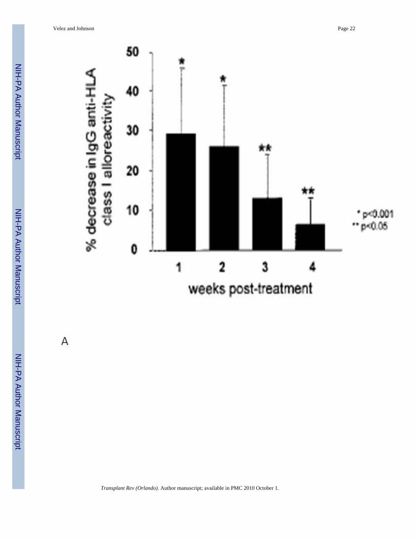
4.1.2. Intravenous immunoglobulin—Intravenous immunoglobulin has also been usedalone to decrease the level of allosensitization prior to heart transplantation, especially in VADrecipients. In a series of four VAD recipients who developed high levels of allosensitizationafter device implantation, treatment with IVIG resulted in decreases in their PRA to levelsbelow 10% within 4 months from administration44. John et al. published a head-to-headcomparison of IVIG and plasmapheresis in 55 allosensitized VAD recipients prior totransplantation. IVIG resulted in a mean reduction of 33% in anti-HLA Class I reactivity oneweek after treatment, with minimal side effects. Plasmapheresis achieved similar results, butrequired longer treatments and was associated with a higher rate of infectious complications(Fig. 5). In patients whose PRA did not respond to low-dose IVIG, higher doses achievedcomparable reductions in the degree of allosensitization, with an observed increased incidenceof acute renal failure. Notably in this series, treatment with IVIG significantly reduced waittime to transplantation45.
4.1.3. Rituximab—In recent years, rituximab, a chimeric monoclonal antibody to CD20, hasbeen used anecdotically to diminish the degree of allosensitization in cardiac transplantcandidates. Rituximab depletes B-lymphocytes through complement-dependent cytotoxicity,antibody-dependent cytotoxicity, and induction of apoptosis46. Animal studies in baboons haveshown that this agent effectively depletes CD20+ B-lymphocytes, resulting in significantblunting of IgM and IgG responses. Studies in humans have focused mostly on the use ofrituximab to treat B-cell lymphomas, however it has also been used to treat autoimmunedisorders and in organ transplantation, recognizing its immunomodulatory properties47. Astudy in allosensitized patients on dialysis awaiting kidney transplantation found that whilerituximab was a powerful B-cell ablational agent, some B-cell subpopulations such as CD19+/CD5+ B-cells recovered to baseline levels within 6 months. Other subsets, such as B memorycells may remain suppressed for up to 2 years after treatment48. These findings suggest thatalloreactivity may not be fully suppressed after rituximab administration.
Most of the available data in treating allosensitized transplant candidates with rituximab comefrom the renal transplant literature. Vieira et al. selected 9 highly sensitized kidney transplantcandidates to be treated with escalating single doses of rituximab. They achieved significantB-lymphocyte depletion with a moderate decrease in PRA in two patients and the suggestionof loss of antibody specificities in 5. Two patients showed no change in their PRA afterrituximab. These effects were associated with infectious complications in some patients49.
Among very few early reports in the heart transplant literature, Balfour et al. reported the useof rituximab in a pediatric transplant candidate who had failed previous treatments with IVIG,plasmapheresis and mycophenolate mofetil. The patient was successfully transplanted after acrossmatch-negative donor was found50.
Unfortunately, published experience with rituximab in allosensitized cardiac transplantcandidates is scarce at this time. Further data are needed to determine the usefulness ofrituximab in allosensitized transplant candidates.
5. Clinical outcomes in allosensitized cardiac transplant candidatesPre-transplant allosensitization increases the likelihood of AMR and CAV, and decreasesoverall allograft survival3, 15, 16, 51–53 (Fig. 6). The relationship between anti-HLA antibodies
Velez and Johnson Page 7
Transplant Rev (Orlando). Author manuscript; available in PMC 2010 October 1.
NIH
-PA Author Manuscript
NIH
-PA Author Manuscript
NIH
-PA Author Manuscript
prior to transplantation and the development of these conditions is well recognized,emphasizing the importance of proper PRA screening, and assignment of appropriate donor-recipient matches.
5.1. Antibody-mediated rejectionAntibody-mediated rejection is not an unusual occurrence54. Institutional experience suggeststhat the incidence of AMR may be approximately 15%5, however this figure varies dependingon the patients studied and the diagnostic methods used. In series where patients werecommonly given OKT3, AMR was evident in up to 52% of cases; whereas in series where C4ddeposition associated with allograft dysfunction is used as the diagnostic criterion, the reportedincidence has been as low as 3%53. Acute AMR typically occurs shortly after transplantation,usually within the first four months, but more commonly during the first 4 weeks aftertransplantation55. However, it can occasionally present several months or years after transplant.Up to 68% patients who develop AMR early on show evidence of significant allograftdysfunction, in contrast with those in whom AMR presents late, where the frequency ofallograft dysfunction is estimated at 13%5, 53.
The pathophysiology of AMR is not fully understood. It is likely the result of antibody-induced,complement-mediated activation of endothelial cells. The process continues with cytokinerelease and increased endothelial adherence of leukocytes, culminating in allograft ischemicinjury56. C4d, an inactive byproduct of complement activation, has been observed in thecapillaries of cardiac allografts with AMR, suggesting recent complement activity57.
The criteria to diagnose AMR have not been uniform. Clinically, it presents with hemodynamiccompromise in 29–47% of cases, especially when it occurs early post-transplant. The criteriafor hemodynamic compromise also vary between centers, but generally include a decrease inleft ventricular ejection fraction, unexplained elevation in cardiac filling pressures with asimultaneous decrease in cardiac output, and the need for inotropic support. In addition toclinical signs, pathologic markers of AMR have to be present.
In 2005, ISHLT proposed criteria for the immunopathologic diagnosis of AMR5. In thepresence of clinical evidence of allograft dysfunction, the following diagnostic criteria weresuggested:
• Histologic evidence of acute capillary injury (endothelial swelling or denudation withcongestion and macrophage infiltration, with possible neutrophil infiltration andinterstitial edema and/or hemorrhage in more severe cases).
• Immunopathologic evidence for antibody-mediated injury (tissueimmunofluorescence positive for IgM or IgG + complement deposition (C3d, C4d orC1q); or CD68-positive macrophages in endothelium; endovascular fibrin can be seenin more severe cases).
• Serologic evidence of donor-specific anti-HLA Class I or Class II antibodies, or otheranti-donor antibodies at the time of biopsy.
Even though immunoglobulin (Ig) deposits are part of the proposed diagnostic criteria forAMR, immunofluorescence for endothelial Ig deposits does not correlate well with clinicalAMR, or with circulating anti-HLA antibodies58. Recently, immunofluorescence for C4d hasshown a high degree of correlation with serum anti-HLA antibodies57. Rodriguez et al. studied665 consecutive endomyocardial biopsies from 165 cardiac transplant recipients withimmunofluorescence for the presence of Ig and complement deposits. The combined detectionof C4d and C3d correlated well with acute AMR and clinical evidence of heart failure. Theadditional detection of Ig and C1q did not improve diagnostic accuracy59. In addition, some
Velez and Johnson Page 8
Transplant Rev (Orlando). Author manuscript; available in PMC 2010 October 1.
NIH
-PA Author Manuscript
NIH
-PA Author Manuscript
NIH
-PA Author Manuscript
early evidence suggests that immunofluorescence for C4d may be helpful in assessing theresponse to treatment for AMR in cardiac transplant recipients57, 60.
AMR causes very severe episodes of rejection, often with hemodynamic compromise, that maynot respond to intensification of immunosuppressive therapy alone. In addition tohemodynamic support with inotropic agents, current therapeutic strategies center oninactivation of circulating antibodies.
5.1.1. Treatment of AMR—Recognizing the importance of antibody production andcomplement deposition in allograft endothelium as the underlying pathophysiology in AMR,mechanical removal of circulating antibodies with plasmapheresis was one of the first therapiestested on affected patients. In a series of 328 cardiac transplant recipients in the late 1980s,3.4% of patients were found to have a positive prospective IgG crossmatch. These patientsexperienced acute AMR much earlier than controls with a negative crossmatch. Moreover,AMR was associated with hemodynamic compromise in 73% of cases. In addition tointensification of immunosuppression, plasmapheresis was used in the patients affected withAMR, with a treatment success rate of 75%55. Other smaller series in Europe and Asia havedocumented similar experiences61, 62. High-dose intravenous corticosteroids, T-lymphocytedepleting agents such as rabbit anti-thymocyte globulin (r-ATG), and tacrolimus have beenused in addition to plasmapheresis, with varying degrees of success. Intravenousimmunoglobulin has also been used with to provide further immune modulation in somepatients with AMR. More than 90% of patients treated aggressively for AMR recover, butremain with a high risk of recurrence53.
A few case series and case reports have also documented successful treatment of AMR withrituximab-based therapy63–65. Garrett et al. treated 8 patients with pathologic evidence of AMRwith weekly doses of rituximab for 4 weeks. All patients recovered their baseline LVEF withoutassociated infectious or other drug-related complications66.
5.2. Cardiac allograft vasculopathyAllosensitization also contributes significantly to an increased risk of developing CAV52, 56,59, 67, 68. CAV is a major cause of cardiac allograft failure and decreased survival after thefirst post-transplantation year67. While it is possible that both T- and B-lymphocyte mediatedimmunity play a role in its pathogenesis, patients who demonstrate circulating donor-specificanti-HLA antibodies, or an immunohistologic pattern of antibody-mediated rejection earlyafter transplant, are more like to develop CAV52.
CAV causes diffuse concentric stenosis of the coronary arteries, as a result of intimal expansionand adventitial sclerosis. While detailed human studies on the pathogenesis of CAV are lacking,it is believed that the pathologic process is initiated by antibody and complement-mediatedinjury to endothelial cells, and may be accelerated by common coronary disease risk factorssuch as hypertension and hyperlipidemia. The antibodies most frequently associated with CAVare those against donor HLA, in particular to Class I antigens, which are richly expressed inhuman endothelial cells. HLA Class II antigens are constitutively expressed in humanendothelium, and their synthesis can be stimulated by pro-inflammatory molecules such asinterferon-γ59. Anti-HLA IgG can stimulate the proliferation of endothelial and smooth musclecells, causing intimal expansion. While lytic levels of complement cause hyperacute rejection,the role of complement in CAV is not well understood. Complement activation can result inthe release of tissue growth factors that cause endothelial proliferation, and migration offibroblasts and smooth muscle cells. In addition to this, sub-lytic doses of the membrane attackcomplex of complement can induce endothelial production of tissue factor. This phenomenonhas been deemed responsible for the pro-coagulant characteristics and intimal fibrin formation
Velez and Johnson Page 9
Transplant Rev (Orlando). Author manuscript; available in PMC 2010 October 1.
NIH
-PA Author Manuscript
NIH
-PA Author Manuscript
NIH
-PA Author Manuscript
seen in coronary arteries with CAV. The presence of fibrin has been independently associatedwith CAV, allograft failure, and death67, 69.
The diagnosis of CAV is challenging due to the often diffuse nature of the disease. Comparativecoronary angiography and intravascular ultrasound (IVUS)70 are widely used to demonstrateprogressive narrowing of allograft coronary arteries (Fig. 7). Serial dobutamine stressechocardiography is also used to detect clinically significant CAV, and some data suggest thatit may have prognostic value comparable to IVUS and coronary angiography in predicting theoccurrence of cardiac events71.
The therapeutic options for CAV are limited. In contrast with native coronary artery disease,percutaneous coronary interventions are not commonly possible due to the diffuse nature ofstenotic process. Stents have been used occasionally for palliative purposes in patients withsymptomatic ischemia. Coronary artery bypass grafts are of limited applicability for similarreasons. Retransplantation may be an option for select candidates with severe multivessel CAVwith evidence of allograft dysfunction. However, patients must be chosen carefully to allowacceptable outcomes of retransplantation to be achieved72.
Contemporary immunosuppressive agents such as sirolimus and everolimus havedemonstrated benefits in delaying the onset of clinically-evident CAV73, 74. When CAV isdetected with angiography, intensification of immunosuppression with these agents is anacceptable choice to slow the progression of CAV75.
6. The importance of de novo anti-HLA antibodies after cardiactransplantation
In spite of advances in immunosuppressive therapy, the incidence of acute AMR and CAVcontinues to limit long-term outcomes54, 76. A study by Rose et al. demonstrated that thepresence of anti-HLA antibodies post-cardiac transplant results in lower survival whencompared to patients who are antibody-free. At 5 years post-transplant, survival in theantibody-negative group was 90%, compared to 53% in the antibody-positive group. Acuterejection and infection, partly related to augmented immunosuppression to treat rejectionepisodes, were the leading causes of death in antibody-positive patients. Circulating anti-HLAantibodies were also associated with higher rates of CAV77. These findings have beencorroborated by subsequent studies12, 78, 79.
Anti-HLA antibodies can develop in patients who were not allosensitized beforetransplantation. This suggests that the transplanted heart may release alloantigens responsiblefor neo-sensitization of the recipient. Tambur et al. showed that up to 35% of non-allosensitizedcardiac allograft recipients can develop anti-HLA antibodies within the first year post-transplant. Antibodies against HLA Class I antigens were more commonly present than anti-HLA Class II antibodies, with PRAs ranging widely from 10% to almost 80% for both classes.HLA-A mismatch, female gender, and longer ischemic time were identified as risk factors forde novo sensitization after transplantation. Anti-HLA Class I and II antibodies showed strongassociations with the incidence of early acute cellular rejection. In addition, de novo Class IIantibodies were associated with more severe CAV and higher mortality due to allograft failure(Fig. 8). Of all anti-HLA Class I antibodies, 41% represented donor-specific antibodies. Donor-specific Class II antibodies were uncommon (<10%). The relative infrequency of donor-specific antibodies may suggest that most are bound to allograft antigens, therefore beingunderrepresented in the serum80.
While contemporary data in cardiac transplant recipients show that de novo anti-HLAantibodies may have a major impact on post-transplant outcomes, the available evidence is not
Velez and Johnson Page 10
Transplant Rev (Orlando). Author manuscript; available in PMC 2010 October 1.
NIH
-PA Author Manuscript
NIH
-PA Author Manuscript
NIH
-PA Author Manuscript
enough to recommend serial PRA screening in all cardiac allograft recipients. It may beadvisable to conduct serial PRAs during the first year post-transplant in patients with featuresthat will place them in a high risk category for post-transplant allosensitization. Serial PRAsin this subset could potentially signal the need for more aggressive immunosuppression toprevent acute rejection episodes and delay allograft failure, however the clinical utility andcost-effectiveness of such an approach remain in question.
7. ConclusionsCardiac transplantation has become the best available therapy in select patients with advancedheart failure with a high probability of death. However, an inadequate number of availablehearts remains rate-limiting in the provision of transplantation to those in need, leading tolonger wait list times for many transplant candidates, with a potential for higher wait listmortality.
In view of the limited donor pool, mechanical circulatory support with VADs was introducedas a bridge to transplant. Unfortunately, broad use of VADs has been associated with higherrates of allosensitization, causing delays to transplantation due to difficulty in finding acrossmatch-negative donor. In addition to VAD-related sensitization, cardiac transplantcandidates are frequently exposed to other sources of sensitization such as blood producttransfusions.
The consequences of pre-transplant allosensitization on clinical outcomes after transplantationcan be serious. Antibody-mediated rejection and cardiac allograft vasculopathy are particularlyprominent and can result in graft failure and decreased survival. The pathophysiology of theseconditions is not fully understood; therefore, available treatments are based on expert opinionand anecdotal clinical data. For this reason, significant emphasis is placed in preventing anddecreasing allosensitization prior to transplantation.
Current strategies to decrease allosensitization focus on the direct removal of circulatingantibodies with plasmapheresis, inactivation of antibodies with IVIG, and B-lymphocytedepletion with rituximab. These approaches have also been used to treat antibody-mediatedrejection after transplantation with encouraging results. However, most treatments are basedon theoretical assumptions and scarce clinical data, therefore their true efficacy is unknown.
In the future, current strategies to avoid allosensitization will be complemented byimprovements in VAD design that will decrease the likelihood of immune activation. Newergeneration devices may result in lower degrees of allosensitization by reducing the surface areain direct contact with the bloodstream, limiting the use of textured surfaces, and utilizing lessimmunogenic biocompatible materials.
References1. Taylor DO, Edwards LB, Boucek MM, Trulock EP, Aurora P, Christie J, et al. Registry of the
International Society for Heart and Lung Transplantation: twenty-fourth official adult heart transplantreport--2007. J Heart Lung Transplant 2007;26:769–81. [PubMed: 17692781]
2. Frazier, OH.; Kirklin, JK., editors. Mechanical circulatory support. Oxford, England: Elsevier; 2006.3. Gonzalez-Stawinski GV, Atik FA, McCarthy PM, Roselli EE, Hoercher K, Navia JL, et al. Early and
late rejection and HLA sensitization at the time of heart transplantation in patients bridged with leftventricular assist devices. Transplant Proc 2005;37:1349–51. [PubMed: 15848717]
4. Massad MG, Cook DJ, Schmitt SK, Smedira NG, McCarthy JF, Vargo RL, et al. Factors influencingHLA sensitization in implantable LVAD recipients. Ann Thorac Surg 1997;64:1120–5. [PubMed:9354538]
Velez and Johnson Page 11
Transplant Rev (Orlando). Author manuscript; available in PMC 2010 October 1.
NIH
-PA Author Manuscript
NIH
-PA Author Manuscript
NIH
-PA Author Manuscript
5. Reed EF, Demetris AJ, Hammond E, Itescu S, Kobashigawa JA, Reinsmoen NL, et al. Acute antibody-mediated rejection of cardiac transplants. J Heart Lung Transplant 2006;25:153–9. [PubMed:16446213]
6. Lietz K, Miller LW. Improved survival of patients with end-stage heart failure listed for hearttransplantation: analysis of organ procurement and transplantation network/U.S. United Network ofOrgan Sharing data, 1990 to 2005. J Am Coll Cardiol 2007;50:1282–90. [PubMed: 17888847]
7. Kerman RH. Understanding the sensitized patient. Heart Fail Clin 2007;3:1–9. [PubMed: 17545004]8. Lavee J, Kormos RL, Duquesnoy RJ, Zerbe TR, Armitage JM, Vanek M, et al. Influence of panel-
reactive antibody and lymphocytotoxic crossmatch on survival after heart transplantation. J Heart LungTransplant 1991;10:921–9. discussion 29–30. [PubMed: 1756157]
9. Itescu S, Schuster M, Burke E, Ankersmit J, Kocher A, Deng M, et al. Immunobiologic consequencesof assist devices. Cardiol Clin 2003;21:119–33. ix–x. [PubMed: 12790051]
10. Terasaki P, Mickey MR, Iwaki Y, Cicciarelli J, Cecka M, Cook D, et al. Long-term survival of kidneygrafts. Transplant Proc 1989;21:615–7. [PubMed: 2650206]
11. Opelz G. Correlation of HLA matching with kidney graft survival in patients with or withoutcyclosporine treatment. Transplantation 1985;40:240–3. [PubMed: 3898488]
12. Barr ML, Cohen DJ, Benvenisty AI, Hardy M, Reemtsma K, Rose EA, et al. Effect of anti-HLAantibodies on the long-term survival of heart and kidney allografts. Transplant Proc 1993;25:262–4.[PubMed: 8438294]
13. Saidman SL. Histocompatibility testing for highly sensitized transplant candidates. Transplant Proc2007;39:673–5. [PubMed: 17445570]
14. Appel JZ 3rd, Hartwig MG, Cantu E 3rd, Palmer SM, Reinsmoen NL, Davis RD. Role of flowcytometry to define unacceptable HLA antigens in lung transplant recipients with HLA-specificantibodies. Transplantation 2006;81:1049–57. [PubMed: 16612283]
15. Smith JD, Danskine AJ, Laylor RM, Rose ML, Yacoub MH. The effect of panel reactive antibodiesand the donor specific crossmatch on graft survival after heart and heart-lung transplantation. TransplImmunol 1993;1:60–5. [PubMed: 8081763]
16. Kobashigawa JA, Sabad A, Drinkwater D, Cogert GA, Moriguchi JD, Kawata N, et al. Pretransplantpanel reactive-antibody screens. Are they truly a marker for poor outcome after cardiactransplantation? Circulation 1996;94:II294–7. [PubMed: 8901763]
17. Tambur AR, Bray RA, Takemoto SK, Mancini M, Costanzo MR, Kobashigawa JA, et al. Flowcytometric detection of HLA-specific antibodies as a predictor of heart allograft rejection.Transplantation 2000;70:1055–9. [PubMed: 11045642]
18. DeNofrio D, Rho R, Morales FJ, Kamoun M, Kearns J, Dorozinsky C, et al. Detection of anti-HLAantibody by flow cytometry in patients with a left ventricular assist device is associated with earlyrejection following heart transplantation. Transplantation 2000;69:814–8. [PubMed: 10755532]
19. John R, Lietz K, Schuster M, Naka Y, Rao V, Mancini DM, et al. Immunologic sensitization inrecipients of left ventricular assist devices. J Thorac Cardiovasc Surg 2003;125:578–91. [PubMed:12658200]
20. Schmid C, Welp H, Klotz S, Baba HA, Wilhelm MJ, Scheld HH. Outcome of patients surviving toheart transplantation after being mechanically bridged for more than 100 days. J Heart LungTransplant 2003;22:1054–8. [PubMed: 12957616]
21. Pamboukian SV, Costanzo MR, Dunlap S, Rayburn B, Westfall AO, You ZY, et al. Relationshipbetween bridging with ventricular assist device on rejection after heart transplantation. J Heart LungTransplant 2005;24:310–5. [PubMed: 15737758]
22. Gonzalez-Stawinski GV, Cook DJ, Chang AS, Atik F, Navia JL, Banbury M, et al. Early and midtermrisk of coronary allograft vasculopathy in patients bridged to orthotopic heart transplantation withventricular assist devices. Transplantation 2005;79:1175–9. [PubMed: 15880065]
23. Pagani FD, Dyke DB, Wright S, Cody R, Aaronson KD. Development of anti-majorhistocompatibility complex class I or II antibodies following left ventricular assist deviceimplantation: effects on subsequent allograft rejection and survival. J Heart Lung Transplant2001;20:646–53. [PubMed: 11404170]
Velez and Johnson Page 12
Transplant Rev (Orlando). Author manuscript; available in PMC 2010 October 1.
NIH
-PA Author Manuscript
NIH
-PA Author Manuscript
NIH
-PA Author Manuscript

24. Gonzalez-Stawinski GV, Cook DJ, Chang AS, Banbury MK, Navia JL, Hoercher K, et al. Ventricularassist devices and aggressive immunosuppression: looking beyond overall survival. J Heart LungTransplant 2006;25:613–8. [PubMed: 16730565]
25. Erren M, Schluter B, Fobker M, Plenz G, Baba H, Willeke P, et al. Immunologic effects ofimplantation of left ventricular assist devices. Transplant Proc 2001;33:1965–8. [PubMed:11267590]
26. McKenna DH Jr, Eastlund T, Segall M, Noreen HJ, Park S. HLA alloimmunization in patientsrequiring ventricular assist device support. J Heart Lung Transplant 2002;21:1218–24. [PubMed:12431496]
27. Miller LW, Phelan DL, Noedel N, McBride LR, Pennington DG. Multiparous women: is routineantibody screening enough in cardiac transplantation? Transplant Proc 1991;23:1135–6. [PubMed:1989169]
28. Tait BD, Dandie WJ, Griffiths AP, Esmore DS, Snell GI, Bergin P, et al. Covert presensitization toHLA antigens in parous heart and lung transplant recipients may predispose to early allograftrejection. Transplant Proc 1995;27:2143–4. [PubMed: 7792912]
29. Spanier T, Oz M, Levin H, Weinberg A, Stamatis K, Stern D, et al. Activation of coagulation andfibrinolytic pathways in patients with left ventricular assist devices. J Thorac Cardiovasc Surg1996;112:1090–7. [PubMed: 8873737]
30. Ankersmit HJ, Tugulea S, Spanier T, Weinberg AD, Artrip JH, Burke EM, et al. Activation-inducedT-cell death and immune dysfunction after implantation of left-ventricular assist device. Lancet1999;354:550–5. [PubMed: 10470699]
31. Scheinin SA, Radovancevic B, Kimball P, Duncan JM, Van Buren CT, Frazier OH, et al. Effect ofIgM-positive crossmatches on survival in heart transplant recipients. Tex Heart Inst J 1995;22:67–71. [PubMed: 7787472]
32. Schuster M, Kocher A, John R, Hoffman M, Ankersmit J, Lietz K, et al. B-cell activation andallosensitization after left ventricular assist device implantation is due to T-cell activation and CD40ligand expression. Hum Immunol 2002;63:211–20. [PubMed: 11872239]
33. Moazami N, Itescu S, Williams MR, Argenziano M, Weinberg A, Oz MC. Platelet transfusions areassociated with the development of anti-major histocompatibility complex class I antibodies inpatients with left ventricular assist support. J Heart Lung Transplant 1998;17:876–80. [PubMed:9773859]
34. van Marwijk Kooy M, van Prooijen HC, Moes M, Bosma-Stants I, Akkerman JW. Use of leukocyte-depleted platelet concentrates for the prevention of refractoriness and primary HLAalloimmunization: a prospective, randomized trial. Blood 1991;77:201–5. [PubMed: 1984797]
35. Drakos SG, Stringham JC, Long JW, Gilbert EM, Fuller TC, Campbell BK, et al. Prevalence andrisks of allosensitization in HeartMate left ventricular assist device recipients: the impact ofleukofiltered cellular blood product transfusions. J Thorac Cardiovasc Surg 2007;133:1612–9.[PubMed: 17532964]
36. Joyce DL, Southard RE, Torre-Amione G, Noon GP, Land GA, Loebe M. Impact of left ventricularassist device (LVAD)-mediated humoral sensitization on post-transplant outcomes. J Heart LungTransplant 2005;24:2054–9. [PubMed: 16364849]
37. Baran DA, Gass AL, Galin ID, Zucker MJ, Arroyo LH, Goldstein DJ, et al. Lack of sensitization andequivalent post-transplant outcomes with the Novacor left ventricular assist device. J Heart LungTransplant 2005;24:1886–90. [PubMed: 16297796]
38. Loebe M, Koster A, Sanger S, Potapov EV, Kuppe H, Noon GP, et al. Inflammatory response afterimplantation of a left ventricular assist device: comparison between the axial flow MicroMedDeBakey VAD and the pulsatile Novacor device. Asaio J 2001;47:272–4. [PubMed: 11374772]
39. Ankersmit HJ, Wieselthaler G, Moser B, Gerlitz S, Roth G, Boltz-Nitulescu G, et al. Transitoryimmunologic response after implantation of the DeBakey VAD continuous-axial-flow pump. JThorac Cardiovasc Surg 2002;123:557–61. [PubMed: 11882831]
40. Grinda JM, Bricourt MO, Amrein C, Salvi S, Guillemain R, Francois A, et al. Human leukocyteantigen sensitization in ventricular assist device recipients: a lesser risk with the DeBakey axial pump.Ann Thorac Surg 2005;80:945–8. [PubMed: 16122460]
Velez and Johnson Page 13
Transplant Rev (Orlando). Author manuscript; available in PMC 2010 October 1.
NIH
-PA Author Manuscript
NIH
-PA Author Manuscript
NIH
-PA Author Manuscript
41. Pisani BA, Mullen GM, Malinowska K, Lawless CE, Mendez J, Silver MA, et al. Plasmapheresiswith intravenous immunoglobulin G is effective in patients with elevated panel reactive antibodyprior to cardiac transplantation. J Heart Lung Transplant 1999;18:701–6. [PubMed: 10452347]
42. Larson DF, Elkund DK, Arabia F, Copeland JG. Plasmapheresis during cardiopulmonary bypass: aproposed treatment for presensitized cardiac transplantation patients. J Extra Corpor Technol1999;31:177–83. [PubMed: 10915474]
43. Leech SH, Lopez-Cepero M, LeFor WM, DiChiara L, Weston M, Furukawa S, et al. Managementof the sensitized cardiac recipient: the use of plasmapheresis and intravenous immunoglobulin. ClinTransplant 2006;20:476–84. [PubMed: 16842525]
44. Dowling RD, Jones JW, Carroll MS, Gray LA Jr. Use of intravenous immunoglobulin in sensitizedLVAD recipients. Transplant Proc 1998;30:1110–1. [PubMed: 9636450]
45. John R, Lietz K, Burke E, Ankersmit J, Mancini D, Suciu-Foca N, et al. Intravenous immunoglobulinreduces anti-HLA alloreactivity and shortens waiting time to cardiac transplantation in highlysensitized left ventricular assist device recipients. Circulation 1999;100:II229–35. [PubMed:10567309]
46. Smith MR. Rituximab (monoclonal anti-CD20 antibody): mechanisms of action and resistance.Oncogene 2003;22:7359–68. [PubMed: 14576843]
47. Becker YT, Samaniego-Picota M, Sollinger HW. The emerging role of rituximab in organtransplantation. Transpl Int 2006;19:621–8. [PubMed: 16827678]
48. Sidner RA, Book BK, Agarwal A, Bearden CM, Vieira CA, Pescovitz MD. In vivo human B-cellsubset recovery after in vivo depletion with rituximab, anti-human CD20 monoclonal antibody. HumAntibodies 2004;13:55–62. [PubMed: 15598985]
49. Vieira CA, Agarwal A, Book BK, Sidner RA, Bearden CM, Gebel HM, et al. Rituximab for reductionof anti-HLA antibodies in patients awaiting renal transplantation: 1. Safety, pharmacodynamics, andpharmacokinetics Transplantation 2004;77:542–8.
50. Balfour IC, Fiore A, Graff RJ, Knutsen AP. Use of rituximab to decrease panel-reactive antibodies.J Heart Lung Transplant 2005;24:628–30. [PubMed: 15896765]
51. Loh E, Bergin JD, Couper GS, Mudge GH Jr. Role of panel-reactive antibody cross-reactivity inpredicting survival after orthotopic heart transplantation. J Heart Lung Transplant 1994;13:194–201.[PubMed: 8031799]
52. Taylor DO, Yowell RL, Kfoury AG, Hammond EH, Renlund DG. Allograft coronary artery disease:clinical correlations with circulating anti-HLA antibodies and the immunohistopathologic pattern ofvascular rejection. J Heart Lung Transplant 2000;19:518–21. [PubMed: 10867330]
53. Uber WE, Self SE, Van Bakel AB, Pereira NL. Acute antibody-mediated rejection following hearttransplantation. Am J Transplant 2007;7:2064–74. [PubMed: 17614978]
54. Lones MA, Czer LS, Trento A, Harasty D, Miller JM, Fishbein MC. Clinical-pathologic features ofhumoral rejection in cardiac allografts: a study in 81 consecutive patients. J Heart Lung Transplant1995;14:151–62. [PubMed: 7727464]
55. Ratkovec RM, Hammond EH, O’Connell JB, Bristow MR, DeWitt CW, Richenbacher WE, et al.Outcome of cardiac transplant recipients with a positive donor-specific crossmatch--preliminaryresults with plasmapheresis. Transplantation 1992;54:651–5. [PubMed: 1412756]
56. Michaels PJ, Espejo ML, Kobashigawa J, Alejos JC, Burch C, Takemoto S, et al. Humoral rejectionin cardiac transplantation: risk factors, hemodynamic consequences and relationship to transplantcoronary artery disease. J Heart Lung Transplant 2003;22:58–69. [PubMed: 12531414]
57. Smith RN, Brousaides N, Grazette L, Saidman S, Semigran M, Disalvo T, et al. C4d deposition incardiac allografts correlates with alloantibody. J Heart Lung Transplant 2005;24:1202–10. [PubMed:16143234]
58. Cherry R, Nielsen H, Reed E, Reemtsma K, Suciu-Foca N, Marboe CC. Vascular (humoral) rejectionin human cardiac allograft biopsies: relation to circulating anti-HLA antibodies. J Heart LungTransplant 1992;11:24–9. discussion 30. [PubMed: 1540608]
59. Rodriguez ER, Skojec DV, Tan CD, Zachary AA, Kasper EK, Conte JV, et al. Antibody-mediatedrejection in human cardiac allografts: evaluation of immunoglobulins and complement activationproducts C4d and C3d as markers. Am J Transplant 2005;5:2778–85. [PubMed: 16212640]
Velez and Johnson Page 14
Transplant Rev (Orlando). Author manuscript; available in PMC 2010 October 1.
NIH
-PA Author Manuscript
NIH
-PA Author Manuscript
NIH
-PA Author Manuscript
60. Crespo-Leiro MG, Veiga-Barreiro A, Domenech N, Paniagua MJ, Pinon P, Gonzalez-Cuesta M, etal. Humoral heart rejection (severe allograft dysfunction with no signs of cellular rejection orischemia): incidence, management, and the value of C4d for diagnosis. Am J Transplant2005;5:2560–4. [PubMed: 16162208]
61. Grauhan O, Knosalla C, Ewert R, Hummel M, Loebe M, Weng YG, et al. Plasmapheresis andcyclophosphamide in the treatment of humoral rejection after heart transplantation. J Heart LungTransplant 2001;20:316–21. [PubMed: 11257558]
62. Wang SS, Chou NK, Ko WJ, Chi NH, Hung SC, Hsu RB, et al. Effect of plasmapheresis for acutehumoral rejection after heart transplantation. Transplant Proc 2006;38:3692–4. [PubMed: 17175369]
63. Aranda JM Jr, Scornik JC, Normann SJ, Lottenberg R, Schofield RS, Pauly DF, et al. Anti-CD20monoclonal antibody (rituximab) therapy for acute cardiac humoral rejection: a case report.Transplantation 2002;73:907–10. [PubMed: 11923690]
64. Baran DA, Lubitz S, Alvi S, Fallon JT, Kaplan S, Galin I, et al. Refractory humoral cardiac allograftrejection successfully treated with a single dose of rituximab. Transplant Proc 2004;36:3164–6.[PubMed: 15686719]
65. Kaczmarek I, Deutsch MA, Sadoni S, Brenner P, Schmauss D, Daebritz SH, et al. Successfulmanagement of antibody-mediated cardiac allograft rejection with combined immunoadsorption andanti-CD20 monoclonal antibody treatment: case report and literature review. J Heart Lung Transplant2007;26:511–5. [PubMed: 17449422]
66. Garrett HE Jr, Duvall-Seaman D, Helsley B, Groshart K. Treatment of vascular rejection withrituximab in cardiac transplantation. J Heart Lung Transplant 2005;24:1337–42. [PubMed:16143254]
67. Soleimani B, Lechler RI, Hornick PI, George AJ. Role of alloantibodies in the pathogenesis of graftarteriosclerosis in cardiac transplantation. Am J Transplant 2006;6:1781–5. [PubMed: 16771817]
68. Wehner J, Morrell CN, Reynolds T, Rodriguez ER, Baldwin WM 3rd. Antibody and complement intransplant vasculopathy. Circ Res 2007;100:191–203. [PubMed: 17272820]
69. Labarrere CA, Pitts D, Halbrook H, Faulk WP. Tissue plasminogen activator, plasminogen activatorinhibitor-1, and fibrin as indexes of clinical course in cardiac allograft recipients. Animmunocytochemical study. Circulation 1994;89:1599–608. [PubMed: 8149527]
70. Mehra MR, Ventura HO, Stapleton DD, Smart FW, Collins TC, Ramee SR. Presence of severe intimalthickening by intravascular ultrasonography predicts cardiac events in cardiac allograft vasculopathy.J Heart Lung Transplant 1995;14:632–9. [PubMed: 7578168]
71. Spes CH, Klauss V, Mudra H, Schnaack SD, Tammen AR, Rieber J, et al. Diagnostic and prognosticvalue of serial dobutamine stress echocardiography for noninvasive assessment of cardiac allograftvasculopathy: a comparison with coronary angiography and intravascular ultrasound. Circulation1999;100:509–15. [PubMed: 10430765]
72. Johnson MR, Aaronson KD, Canter CE, Kirklin JK, Mancini D, Mehra MR, et al. Heartretransplantation. Am J Transplant 2007;7:2075–81. [PubMed: 17640316]
73. Eisen HJ, Tuzcu EM, Dorent R, Kobashigawa J, Mancini D, Valantine-von Kaeppler HA, et al.Everolimus for the prevention of allograft rejection and vasculopathy in cardiac-transplant recipients.N Engl J Med 2003;349:847–58. [PubMed: 12944570]
74. Keogh A, Richardson M, Ruygrok P, Spratt P, Galbraith A, O’Driscoll G, et al. Sirolimus in de novoheart transplant recipients reduces acute rejection and prevents coronary artery disease at 2 years: arandomized clinical trial. Circulation 2004;110:2694–700. [PubMed: 15262845]
75. Mancini D, Pinney S, Burkhoff D, LaManca J, Itescu S, Burke E, et al. Use of rapamycin slowsprogression of cardiac transplantation vasculopathy. Circulation 2003;108:48–53. [PubMed:12742978]
76. Leech SH, Mather PJ, Eisen HJ, Pina IL, Margulies KB, Bove AA, et al. Donor-specific HLAantibodies after transplantation are associated with deterioration in cardiac function. Clin Transplant1996;10:639–45. [PubMed: 8996758]
77. Rose EA, Smith CR, Petrossian GA, Barr ML, Reemtsma K. Humoral immune responses after cardiactransplantation: correlation with fatal rejection and graft atherosclerosis. Surgery 1989;106:203–7.discussion 07–8. [PubMed: 2669195]
Velez and Johnson Page 15
Transplant Rev (Orlando). Author manuscript; available in PMC 2010 October 1.
NIH
-PA Author Manuscript
NIH
-PA Author Manuscript
NIH
-PA Author Manuscript
78. Reed EF, Hong B, Ho E, Harris PE, Weinberger J, Suciu-Foca N. Monitoring of soluble HLAalloantigens and anti-HLA antibodies identifies heart allograft recipients at risk of transplant-associated coronary artery disease. Transplantation 1996;61:566–72. [PubMed: 8610382]
79. Leprince P, Fretz C, Dorent R, Boudifa A, Jourdan J, Youssoub JJ, et al. Posttransplantation cytotoxicimmunoglobulin G is associated with a high rate of acute allograft dysfunctions in heart transplantrecipients. Am Heart J 1999;138:586–92. [PubMed: 10467212]
80. Tambur AR, Pamboukian SV, Costanzo MR, Herrera ND, Dunlap S, Montpetit M, et al. The presenceof HLA-directed antibodies after heart transplantation is associated with poor allograft outcome.Transplantation 2005;80:1019–25. [PubMed: 16278580]
Velez and Johnson Page 16
Transplant Rev (Orlando). Author manuscript; available in PMC 2010 October 1.
NIH
-PA Author Manuscript
NIH
-PA Author Manuscript
NIH
-PA Author Manuscript
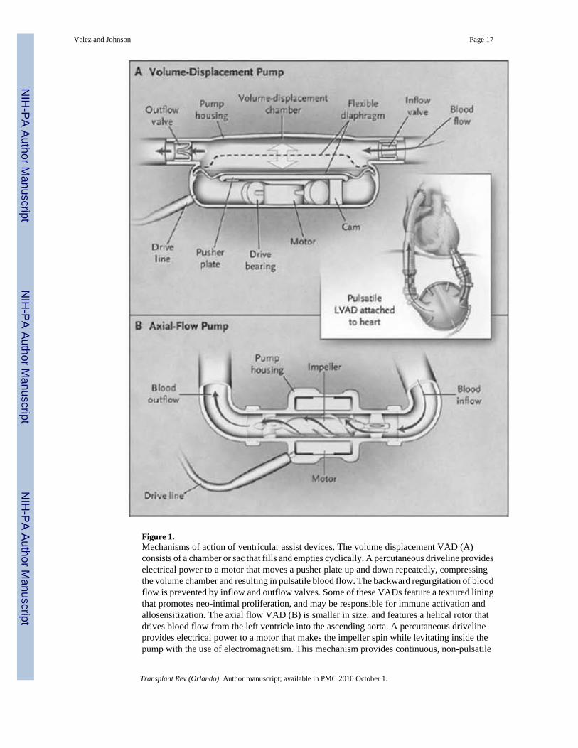
Figure 1.Mechanisms of action of ventricular assist devices. The volume displacement VAD (A)consists of a chamber or sac that fills and empties cyclically. A percutaneous driveline provideselectrical power to a motor that moves a pusher plate up and down repeatedly, compressingthe volume chamber and resulting in pulsatile blood flow. The backward regurgitation of bloodflow is prevented by inflow and outflow valves. Some of these VADs feature a textured liningthat promotes neo-intimal proliferation, and may be responsible for immune activation andallosensitization. The axial flow VAD (B) is smaller in size, and features a helical rotor thatdrives blood flow from the left ventricle into the ascending aorta. A percutaneous drivelineprovides electrical power to a motor that makes the impeller spin while levitating inside thepump with the use of electromagnetism. This mechanism provides continuous, non-pulsatile
Velez and Johnson Page 17
Transplant Rev (Orlando). Author manuscript; available in PMC 2010 October 1.
NIH
-PA Author Manuscript
NIH
-PA Author Manuscript
NIH
-PA Author Manuscript
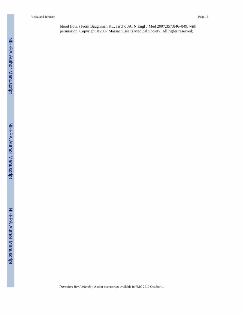
blood flow. (From Baughman KL, Jarcho JA. N Engl J Med 2007;357:846–849, withpermission. Copyright ©2007 Massachussetts Medical Society. All rights reserved).
Velez and Johnson Page 18
Transplant Rev (Orlando). Author manuscript; available in PMC 2010 October 1.
NIH
-PA Author Manuscript
NIH
-PA Author Manuscript
NIH
-PA Author Manuscript
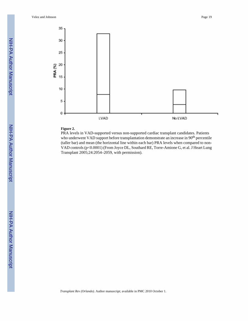
Figure 2.PRA levels in VAD-supported versus non-supported cardiac transplant candidates. Patientswho underwent VAD support before transplantation demonstrate an increase in 90th percentile(taller bar) and mean (the horizontal line within each bar) PRA levels when compared to non-VAD controls (p<0.0001) (From Joyce DL, Southard RE, Torre-Amione G, et al. J Heart LungTransplant 2005;24:2054–2059, with permission).
Velez and Johnson Page 19
Transplant Rev (Orlando). Author manuscript; available in PMC 2010 October 1.
NIH
-PA Author Manuscript
NIH
-PA Author Manuscript
NIH
-PA Author Manuscript
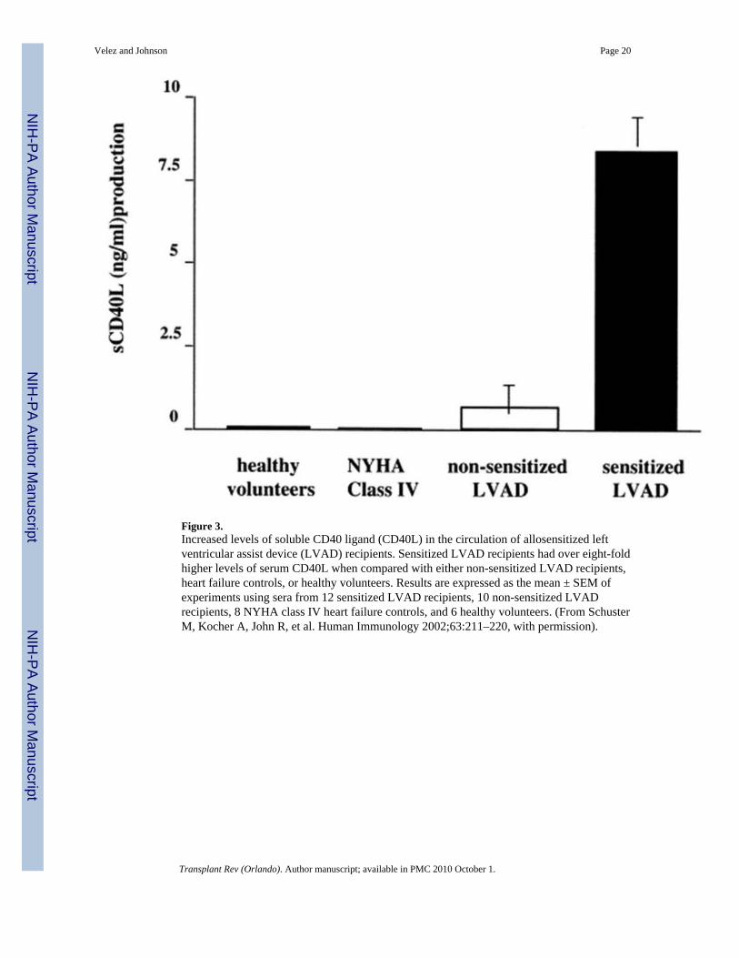
Figure 3.Increased levels of soluble CD40 ligand (CD40L) in the circulation of allosensitized leftventricular assist device (LVAD) recipients. Sensitized LVAD recipients had over eight-foldhigher levels of serum CD40L when compared with either non-sensitized LVAD recipients,heart failure controls, or healthy volunteers. Results are expressed as the mean ± SEM ofexperiments using sera from 12 sensitized LVAD recipients, 10 non-sensitized LVADrecipients, 8 NYHA class IV heart failure controls, and 6 healthy volunteers. (From SchusterM, Kocher A, John R, et al. Human Immunology 2002;63:211–220, with permission).
Velez and Johnson Page 20
Transplant Rev (Orlando). Author manuscript; available in PMC 2010 October 1.
NIH
-PA Author Manuscript
NIH
-PA Author Manuscript
NIH
-PA Author Manuscript
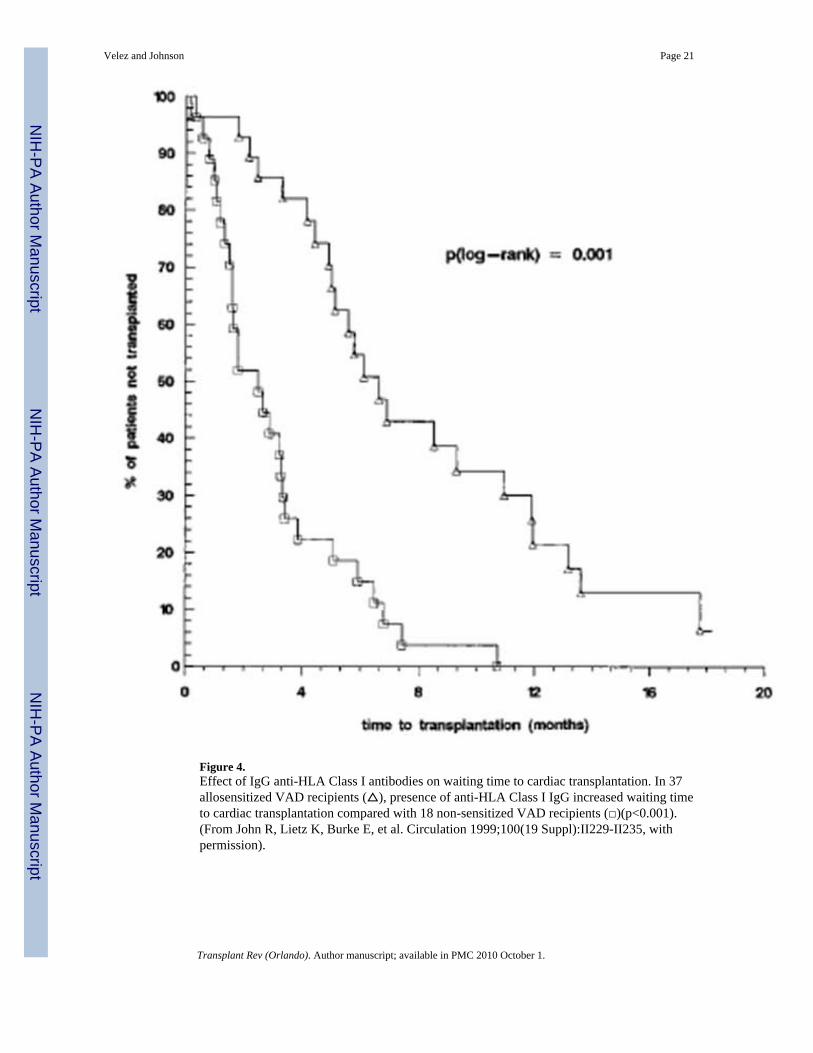
Figure 4.Effect of IgG anti-HLA Class I antibodies on waiting time to cardiac transplantation. In 37allosensitized VAD recipients (△), presence of anti-HLA Class I IgG increased waiting timeto cardiac transplantation compared with 18 non-sensitized VAD recipients (□)(p<0.001).(From John R, Lietz K, Burke E, et al. Circulation 1999;100(19 Suppl):II229-II235, withpermission).
Velez and Johnson Page 21
Transplant Rev (Orlando). Author manuscript; available in PMC 2010 October 1.
NIH
-PA Author Manuscript
NIH
-PA Author Manuscript
NIH
-PA Author Manuscript
Velez and Johnson Page 22
Transplant Rev (Orlando). Author manuscript; available in PMC 2010 October 1.
NIH
-PA Author Manuscript
NIH
-PA Author Manuscript
NIH
-PA Author Manuscript
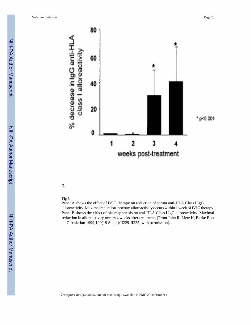
Fig 5.Panel A shows the effect of IVIG therapy on reduction of serum anti-HLA Class I IgGalloreactivity. Maximal reduction in serum alloreactivity occurs within 1 week of IVIG therapy.Panel B shows the effect of plasmapheresis on anti-HLA Class I IgG alloreactivity. Maximalreduction in alloreactivity occurs 4 weeks after treatment. (From John R, Lietz K, Burke E, etal. Circulation 1999;100(19 Suppl):II229-II235, with permission).
Velez and Johnson Page 23
Transplant Rev (Orlando). Author manuscript; available in PMC 2010 October 1.
NIH
-PA Author Manuscript
NIH
-PA Author Manuscript
NIH
-PA Author Manuscript
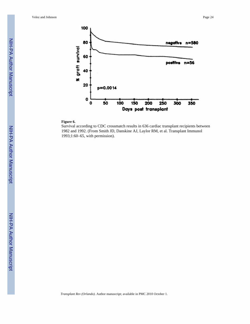
Figure 6.Survival according to CDC crossmatch results in 636 cardiac transplant recipients between1982 and 1992. (From Smith JD, Danskine AJ, Laylor RM, et al. Transplant Immunol1993;1:60–65, with permission).
Velez and Johnson Page 24
Transplant Rev (Orlando). Author manuscript; available in PMC 2010 October 1.
NIH
-PA Author Manuscript
NIH
-PA Author Manuscript
NIH
-PA Author Manuscript
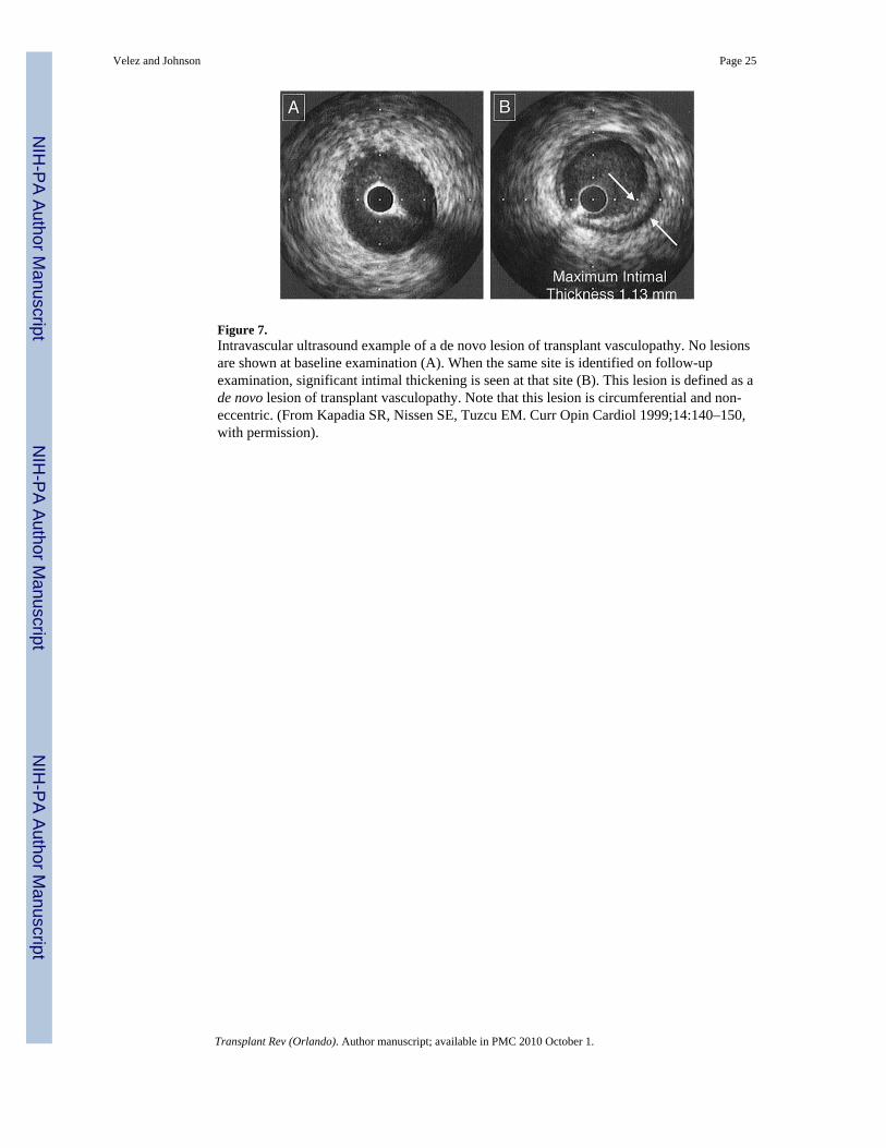
Figure 7.Intravascular ultrasound example of a de novo lesion of transplant vasculopathy. No lesionsare shown at baseline examination (A). When the same site is identified on follow-upexamination, significant intimal thickening is seen at that site (B). This lesion is defined as ade novo lesion of transplant vasculopathy. Note that this lesion is circumferential and non-eccentric. (From Kapadia SR, Nissen SE, Tuzcu EM. Curr Opin Cardiol 1999;14:140–150,with permission).
Velez and Johnson Page 25
Transplant Rev (Orlando). Author manuscript; available in PMC 2010 October 1.
NIH
-PA Author Manuscript
NIH
-PA Author Manuscript
NIH
-PA Author Manuscript
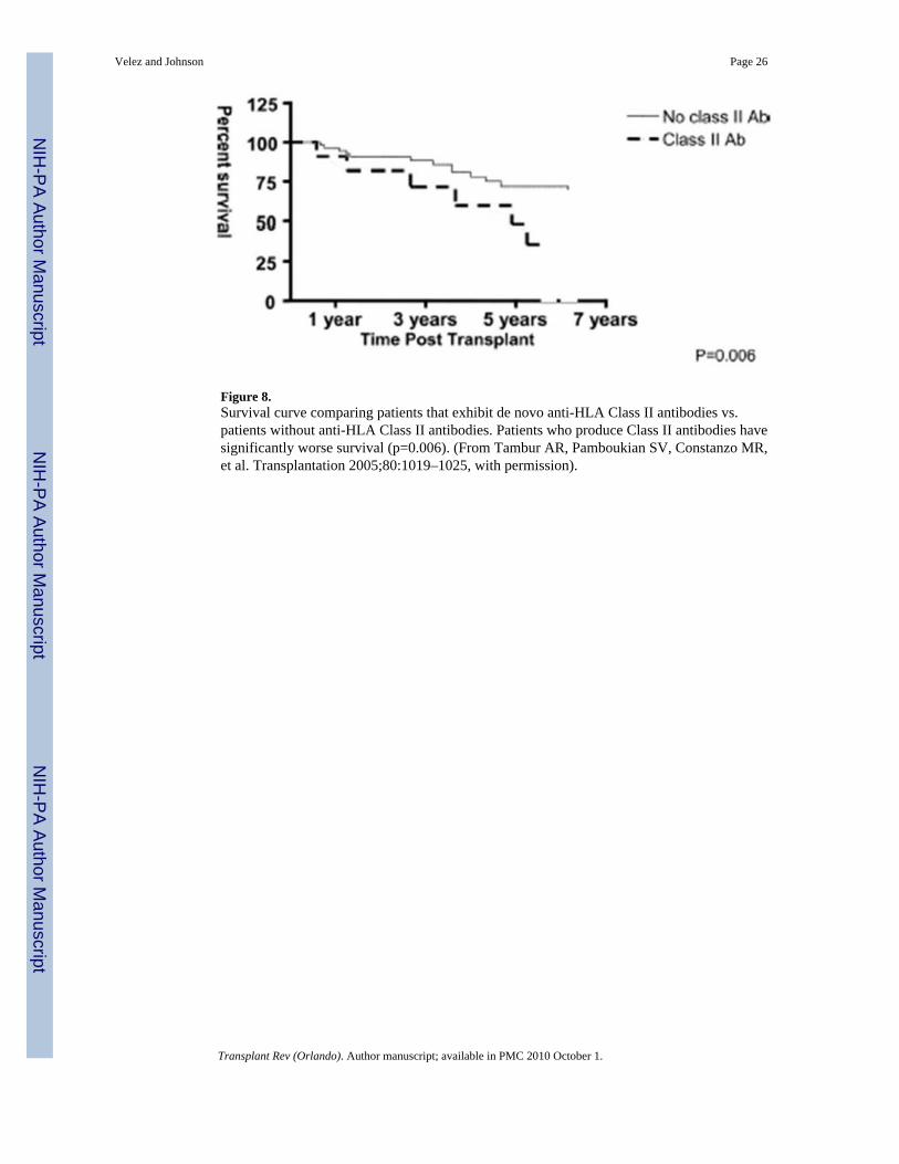
Figure 8.Survival curve comparing patients that exhibit de novo anti-HLA Class II antibodies vs.patients without anti-HLA Class II antibodies. Patients who produce Class II antibodies havesignificantly worse survival (p=0.006). (From Tambur AR, Pamboukian SV, Constanzo MR,et al. Transplantation 2005;80:1019–1025, with permission).
Velez and Johnson Page 26
Transplant Rev (Orlando). Author manuscript; available in PMC 2010 October 1.
NIH
-PA Author Manuscript
NIH
-PA Author Manuscript
NIH
-PA Author Manuscript
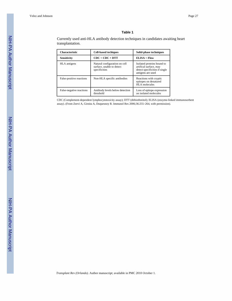
Table 1
Currently used anti-HLA antibody detection techniques in candidates awaiting hearttransplantation.
Characteristic Cell-based techiques Solid-phase techniques
Sensitivity CDC < CDC + DTT ELISA < Flow
HLA antigens Natural configuration on cellsurface, unable to detectspecificities
Isolated proteins bound toartifical surface, maydetect specificities if singleantigens are used
False-positive reactions Non-HLA specific antibodies Reactions with crypticepitopes on denaturedHLA molecules
False-negative reactions Antibody levels below detectionthreshold
Loss of epitope expressionon isolated molecules
CDC (Complement-dependent lymphocytotoxicity assay); DTT (dithiothreitol); ELISA (enzyme-linked immunosorbentassay). (From Zeevi A, Girnita A, Duquesnoy R. Immunol Res 2006;36:255–264, with permission).
Velez and Johnson Page 27
Transplant Rev (Orlando). Author manuscript; available in PMC 2010 October 1.
NIH
-PA Author Manuscript
NIH
-PA Author Manuscript
NIH
-PA Author Manuscript
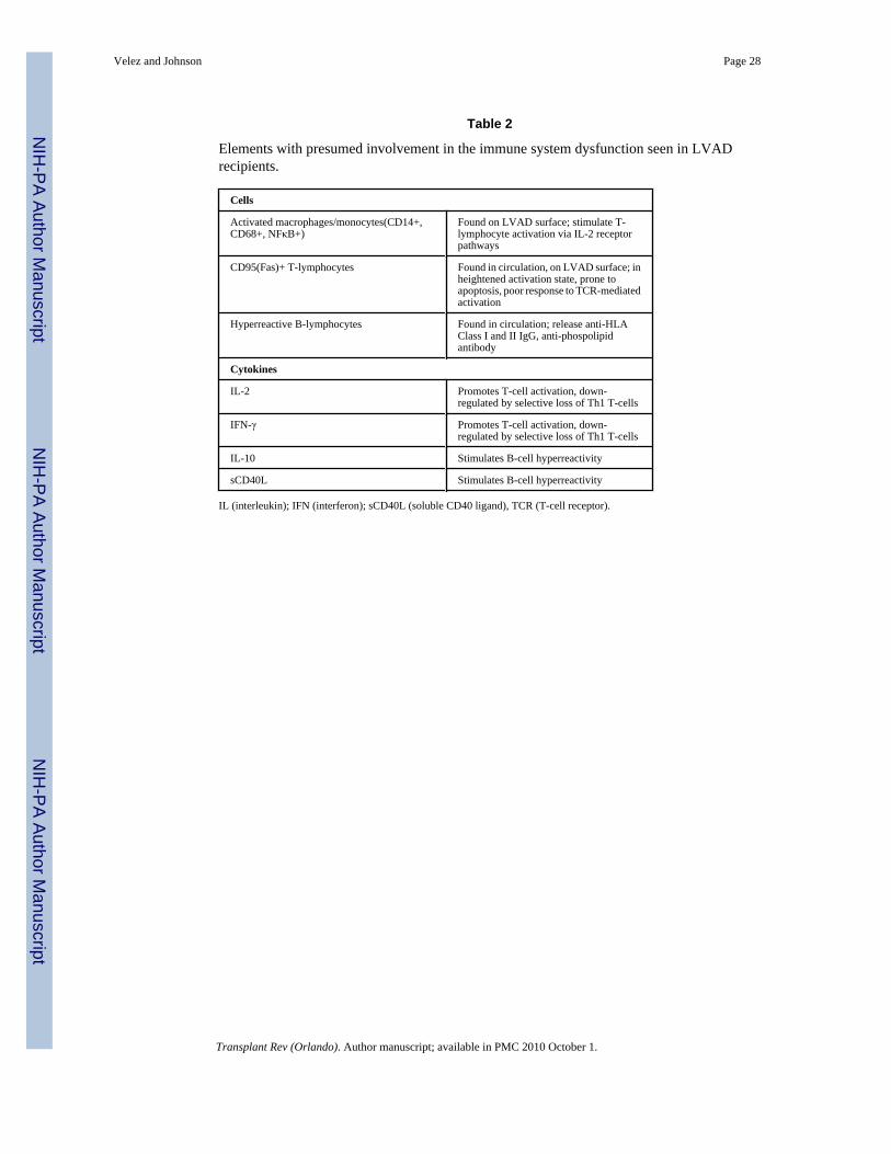
Table 2
Elements with presumed involvement in the immune system dysfunction seen in LVADrecipients.
Cells
Activated macrophages/monocytes(CD14+,CD68+, NFκB+)
Found on LVAD surface; stimulate T-lymphocyte activation via IL-2 receptorpathways
CD95(Fas)+ T-lymphocytes Found in circulation, on LVAD surface; inheightened activation state, prone toapoptosis, poor response to TCR-mediatedactivation
Hyperreactive B-lymphocytes Found in circulation; release anti-HLAClass I and II IgG, anti-phospolipidantibody
Cytokines
IL-2 Promotes T-cell activation, down-regulated by selective loss of Th1 T-cells
IFN-γ Promotes T-cell activation, down-regulated by selective loss of Th1 T-cells
IL-10 Stimulates B-cell hyperreactivity
sCD40L Stimulates B-cell hyperreactivity
IL (interleukin); IFN (interferon); sCD40L (soluble CD40 ligand), TCR (T-cell receptor).
Velez and Johnson Page 28
Transplant Rev (Orlando). Author manuscript; available in PMC 2010 October 1.
NIH
-PA Author Manuscript
NIH
-PA Author Manuscript
NIH
-PA Author Manuscript