magnetic nanoparticles with bulklike properties (invited)
TRANSCRIPT

1
Magnetic nanoparticles with bulk-like properties
Xavier Batllea)
, N. Pérez, P. Guardia, O. Iglesias and A. Labarta
Dep. Física Fonamental and Institut de Nanociència i Nanotecnologia (IN2UB), Universitat
de Barcelona, Martí i Franquès 1, 08028 Barcelona, Spain
F. Bartolomé, L.M. García and J. Bartolomé
Instituto de Ciencia de Materiales de Aragón (ICMA)-CSIC and Dep. Física Materia
Condensada, Universidad de Zaragoza, 50009 Zaragoza, Spain
A.G. Roca, M. P. Morales and C.J. Serna
Instituto de Ciencia de Materiales de Madrid (ICMM)-CSIC, 28049 Madrid, Spain
(Dated: November 5th, 2010)
a)
Invited speaker and author for correspondence: [email protected]

2
Abstract. The magnetic behavior of Fe3−xO4 nanoparticles synthesized either by high-
temperature decomposition of an organic iron precursor or low-temperature co-precipitation
in aqueous conditions, is compared. Transmission electron microscopy, X-ray absorption
spectroscopy, X-ray magnetic circular dichroism and magnetization measurements show that
nanoparticles synthesized by thermal decomposition display high crystal quality and bulk-like
magnetic and electronic properties, while nanoparticles synthesized by co-precipitation show
much poorer crystallinity and particle-like phenomenology, including reduced magnetization,
high closure fields and shifted hysteresis loops. The key role of the crystal quality is thus
suggested since particle-like behavior for particles larger than about 5 nm is only observed
when they are structurally defective. These conclusions are supported by Monte Carlo
simulations. It is also shown that thermal decomposition is capable of producing nanoparticles
that, after further stabilization in physiological conditions, are suitable for biomedical
applications such as magnetic resonance imaging or bio-distribution studies.
3
INTRODUCTION
Nanostructured magnetic materials comprise a very active research field due to the
new phenomena taking place at the nanoscale as a consequence of the interplay of quantum,
finite-size, surface and interfacial effects. Magnetic nanoparticles (NPs) [1] are an excellent
example of nanostructured materials, which provide the critical building blocks for the
booming of many applications of nanotechnology in fields such as biomedicine [2], high-
density magnetic recording [3] or magnetic resonance imaging (MRI) [4]. A key question in
these systems is how the nanostructure modifies their magnetic and electronic properties and
how one can take advantage of those new properties to improve applications. Consequently,
understanding and controlling the effects of the nanostructure on the properties of the particles
have become increasingly relevant issues for technological applications.
As the size of particles is reduced below about 100 nm, deviations from bulk behavior
have been widely reported and attributed to changes in the magnetic ordering at the surface
layer with respect to that of the particle core and also to finite-size effects, thus giving rise to a
significant decrease in the particle magnetization [1]. Typically, transition metal oxide NPs,
including iron oxide, show surface spin disorder due to symmetry breaking at the surface,
leading to [5-8]: (i) saturation magnetization smaller than in the bulk material, in some cases
by a factor of two or more; (ii) high-field differential susceptibility; (iii) extremely high
closure fields (even up to 120 kOe); (iv) shifted hysteresis loops after field cooling the sample
together with glassy behavior, which have been explained in terms of the existence of a
surface layer of disordered spins that freeze in a spin glass-like state due to magnetic
frustration, yielding both an exchange field acting on the ordered core and an increase in the
particle anisotropy; and (v) extremely high magnetic anisotropy associated with the fact that
very high magnetic fields are required to reach magnetic saturation.
4
However, that particle-like behavior is undesirable in many applications, where the
NPs should comply with a variety of requirements. In the case of biomedical applications,
those comprise [2]: (i) superparamagnetic behavior at room temperature in order to avoid
particle aggregation due to inter-particle interactions; (ii) high saturation magnetization to
achieve a high magnetic response under the application of moderate magnetic fields; (iii)
limiting size for in vivo applications within 1 and 50 nm; and (iv) bio-compatibility and
functionality to reach specific targets inside the body. Consequently, biomedical applications
require of magnetic NPs of a few nanometers in size showing magnetic properties close to the
bulk material. Research in this field is thus mainly focused on new synthesis methods
enabling the preparation of NPs with high crystal quality and reduced magnetic disorder. Iron
oxide NPs are suitable candidates due to their low toxicity and ease to be functionalized.
Magnetite Fe3O4 and maghemite γ-Fe2O3 are probably among the most widely studied
magnetic materials. However, they still arise much attention since several questions remain
open. For example, the orbital contribution to the magnetic moment in bulk magnetite is
under discussion [9].
In this paper, we report on the effect of the synthesis method and surface coating on
the structural quality and magnetic properties of two sets of samples of Fe3−xO4 NPs of about
5 nm, synthesized either by (i) thermal decomposition of an organic iron precursor in an
organic medium [10], with a fatty acid (e.g., oleic acid) used as surfactant covalently bonded
to the particle surface [11]; or (ii) co-precipitation of Fe[II] and Fe[III] salts in an alkaline
aqueous medium [12], with polyvinyl alcohol (PVA) used as a protective coating against
oxidation adsorbed at the NP surface but without any chemical bond. The crystal quality of
both sets of samples determines the occurrence of particle-like phenomenology, which cannot
be considered as intrinsic properties arising from finite-size and surface effects.

5
SAMPLE PREPARATION
Oleic acid-coated particles of 5 nm in mean diameter were synthesized by thermal
decomposition of an iron precursor [10,11]. One mmol of Fe(III)-acetylacetonate was mixed
with 3 mmol of oleic acid, 3 mmol of oleilamine, 5 mmol of 1,2-hexadecanediol and 10 ml of
phenyl ether. The degassed mixture was heated to 200 ºC and kept at that temperature for two
hours. Then, it was further heated up to the reflux temperature of phenyl ether (260~270 ºC)
and kept at that temperature for 30 minutes. After cooling down to room temperature, the
particles were extracted by solvent-non solvent techniques and finally re-suspended in
hexane. Interestingly enough, variations of this procedure enable to tune the size and shape of
the particles. For example, solvents with higher boiling point lead to larger particles [10] or
the use of decanoic acid instead of oleic acid as capping ligand yields particles in a wider
range of sizes [13]. By varying the molar ratio of the decanoic acid to the iron precursor and
adjusting the final synthesis temperature, NPs within 5 and 50 nm are obtained [13]. The
particle size range can be further expanded up to 180 nm by tuning the heating rate [14].
PVA-coated 5 nm in size NPs were prepared by the standard co-precipitation
procedure [12]. A stoichiometric mixture of FeCl3 and FeCl2 was dissolved in 10 ml de-
ionized water and added drop-wise to 300 ml of a sodium hydroxide aqueous solution,
containing PVA at room temperature and pH 10. The particles formed instantaneously, were
extracted by precipitation and were finally suspended in de-ionized water at pH 7.
STRUCTURAL CHARACTERIZATION
High resolution transmission electron microscopy (TEM) images of NPs from thermal
decomposition show regular shapes with very high crystal quality up to the NP surface (Figs.1
(a)-(b)). In contrast, NPs from co-precipitation show much more irregular shapes and lower
degree of crystallinity at the surface (Figs.1 (c)-(d)), together with in-volume defects. Many of

6
those particles are aggregates of individual crystallites randomly oriented. TEM also shows
that the particles prepared by thermal decomposition have a narrow size distribution and are
individually coated by the surfactant, while those by co-precipitation tend to agglomerate and
show a much broader size distribution.
Figure 2 shows an example of X-ray absorption spectroscopy (XAS) data at the Fe L2,3
absorption edges, for Fe3-xO4 NPs of about 5 nm in size. The spectrum for thermal
decomposition particles shows 30% more holes in the 3d band [15] as compared to the bulk
counterpart [9,16], indicating an electronic transfer associated with the chemical bonding of
the oleic acid at the particle surface [15]. In contrast, co-precipitation particles of similar size
show a XAS spectrum similar to the bulk counterpart, as expected for particles with just a
protective adsorbed coating. Both spectra can be superimposed almost perfectly, provided that
the spectrum for co-precipitation be multiplied by an appropriate scale factor (inset to Fig. 2),
which evidences that the stoichiometry is the same within the experimental error. The actual
stoichiometry of the 5 nm NPs from thermal decomposition was evaluated from Mössbauer
spectroscopy to be Fe2.93O4 [17], which is compatible with the presence of up to 21% of
maghemite, forming an overoxidized surface layer of about one unit cell.
PARTICLE-LIKE BEHAVIOR VERSUS STRUCTURAL QUALITY
Magnetization hysteresis loops and zero field cooling-field cooling (ZFC-FC) curves
were measured with a SQUID magnetometer. The ZFC-FC curves in Fig. 3 are compared for
particles of about 5 nm in size prepared by the two methods. For thermal decomposition, the
ZFC curve evidences a very narrow size distribution. The progressive increase of the FC
curve below the irreversibility onset indicates the absence of relevant dipolar interactions,
which is in agreement with the individual coating of the particles. The coincidence of the
onset of the irreversibility and the maximum of the ZFC also confirms that the surfactant
7
coating minimizes the formation of aggregates, avoiding inter-particle interactions. In
contrast, the ZFC-FC curves for the co-precipitation sample reflects a broader size distribution
and particle agglomeration, and the existence of significant inter-particle interactions. The
mean activation magnetic volume of the two samples was evaluated by fitting the ZFC curves
to a distribution of Langevin functions [18]. The diameter corresponding to the mean
activation magnetic volume is d 5.0 nm in the case of thermal decomposition, with a
standard deviation 4.4 nm, in very good agreement with the size distribution obtained
from TEM. For the co-precipitation sample, d 27 nm and 26 nm, which are much
larger than those deduced from the TEM size distribution due to particle aggregation favoring
inter-particle interactions.
Figure 4 shows the hysteresis loops at 5 K for particles of about 5 nm. Saturation
magnetization is significantly higher for the thermal decomposition sample. Additionally, a
higher value of the high-field differential susceptibility and a much rounded magnetization
curve are observed for the co-precipitation sample, facts which are indicative of the presence
of magnetic disorder throughout the particles. Moreover, the magnification of the hysteresis
curves (inset to Fig. 4) shows the occurrence of much higher closure fields (indicated by
arrows in Fig. 4) in the co-precipitation sample, which could be related to the existence of
very high energy barriers associated with magnetic frustration induced by structural disorder.
Magnetization values at 5 T and 5 K as a function of the particle size are shown in Fig.
5 [13,15,19,20]. The values corresponding to the thermal decomposition samples are always
significantly higher than those for co-precipitation, both sets of data displaying some size
dependence. Finite-size and surface effects are evident in the results from Monte Carlo
simulations, also shown in Fig. 5 (solid triangles) for a spherical particle with Ising spins on
an inverse spinel lattice [21]. These data have been scaled to the saturation magnetization of
bulk magnetite in order to be compared to the experimental results. Experimental values lay

8
clearly below the ones obtained by Monte Carlo simulation, even for thermal decomposition
samples. This could be due to the difficulty in obtaining pure, stoichiometric magnetite
without a significant amount of the maghemite phase for particle sizes below a few tens of
nm. In the case of co-precipitation, the lack of crystal quality yields a large additional
reduction in the magnetization due to magnetic disorder within the particle.
X-ray magnetic circular dicroism (XMCD) was measured at the Fe L2,3 absorption
edges for Fe3-xO4 NPs of about 5nm in size [15]. The measurements were carried out with
total electron yield detection and an applied field of 2 T parallel to the X-ray beam. The
application of magneto-optical sum rules on the XMCD spectra enables to obtain the
magnetic moment per formula unit, µFe, and the spin and orbital contributions [22] (Table I).
The total magnetic moment per formula unit for the thermal decomposition sample is about
42% larger than that for the co-precipitation sample, in excellent agreement with
magnetization results. Furthermore, µFe for the thermal decomposition sample is just about
16% smaller than that reported from previous XMCD measurements on a magnetite single-
crystal [9], also in excellent agreement with magnetization results. Besides, the calculated
orbital magnetic moments are 0.036(8) and 0.081(9) µB/f.u. for the thermal decomposition
and co-precipitation samples, respectively. Although both values are in qualitative agreement
with Reference 9, the co-precipitation sample shows about a 3 fold increase with respect to
the expected value of the ratio of the orbital-to-spin angular moments mL/mS ≈ 0.013,
obtained from LDA calculations for bulk magnetite [23]. Increased values of mL/mS are
usually found in low dimensional and particle-like systems [24]. In contrast, the thermal
decomposition sample fully recovers the value for the bulk orbital-to-spin moment, which
reinforces the idea that the high crystal quality of the sample is behind the observed bulk-like
magnetic and electronic behavior [15].

9
In order to elucidate the influence of the crystal quality and particle/surfactant
chemical bond of thermal decomposition samples on the magnetic disorder at the NP surface,
we evaluated the surface and volume anisotropies (SK and
vK , respectively) from the size
distribution obtained from TEM and 0ln( / )T t scaling of the thermo-remanence relaxation data
[7,8,25,26]. In the absence of sizeable inter-particle interactions, as is the case for the thermal
decomposition particles (see Fig. 3), the derivative of the0ln( / )T t scaling yields the energy
barrier distribution associated with the effective anisotropy for magnetization reversal.
Assuming the ad hoc expression for the effective, uniaxial anisotropy, 6 /eff v sK K DK , the
size distribution obtained from TEM can be superimposed onto the energy barrier distribution
by a fitting procedure that yields the volume and surface anisotropies [27]. The values
obtained are given in Table II, being in very good agreement with other experimental values
(see 28, 27 and references therein). Thus, intrinsic surface effects, rather than the lack of
crystallinity, dominate the surface anisotropy in high crystal quality particles nanometer in
size.
Another effect usually related to magnetic disordered systems is the occurrence of
shifted magnetization hysteresis loops, which in many cases is explained assuming the
existence of the exchange bias phenomenon [29,30]. Shifted loops can be observed after FC
processes, or even ZFC, for samples prepared by co-precipitation [19,31], as shown in Fig. 6
for 5 nm Fe3-xO4 PVA-coated NPs, having poor crystallinity. Additionally, maghemite hollow
particles of 8 nm in size composed of about 10 crystallites randomly oriented, show strongly
shifted loops and very high closure fields due to their highly defective crystallographic
structure and the large interface areas among single crystallites [32]. All those systems have in
common the occurrence of magnetic disorder associated with defective crystal structures,
giving rise to high energy barriers that can be only overcome at very high fields. On the
contrary, thermal decomposition samples with high crystal quality do not show this

10
phenomenology at all. For very defective particles showing high closure fields (inset to Fig.
4), the applied magnetic field of a FC process imprints an effective anisotropy that cannot be
erased at the maximum applied field of the hysteresis loop. Therefore, the system follows a
minor loop that, in some cases, has been attributed to an exchange coupling phenomenon
between a core-shell structure [29,31,33].
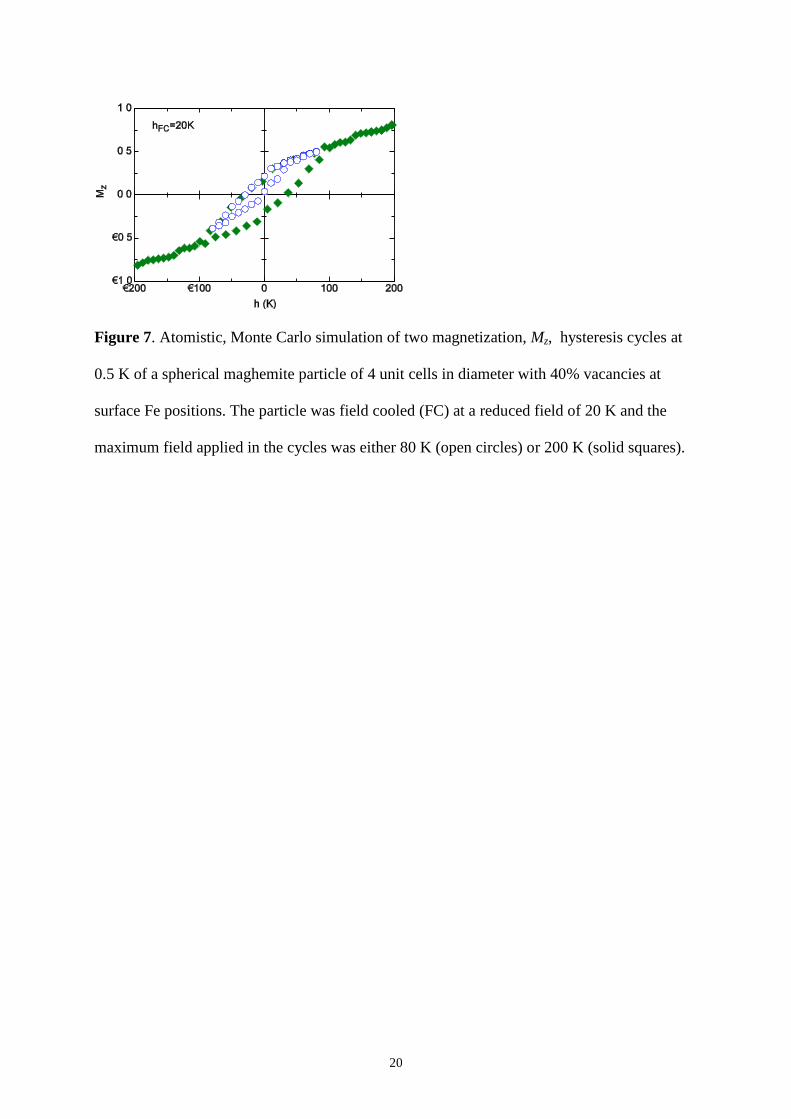
In order to reinforce this interpretation, Fig. 7 shows two hysteresis loops obtained by
atomistic Monte Carlo simulations of a single spherical maghemite particle of 4 unit cells in
diameter, with 40% of vacancies in the Fe positions at the surface [21]. Both magnetization
curves were obtained after FC down to 0.5 K in a field of hFC = 20 K (the field is given in
temperature units as / BH k ), while the maximum applied field was 80 K in one case and 200
K in the other. It is obvious that the apparent shifted loop in the case of h = 80 K is just a
minor loop of that corresponding to h = 200 K, because it was obtained for a maximum field
lower than the closure field (approximately 90 K). Similar apparent shifted loops, which in
fact are minor loops, have also been reported [32].
MAGNETITE NANOPARTICLES FOR BIOMEDICAL APPLICATIONS
As iron oxides are approved for biomedical use in humans due to their low toxicity,
magnetite is the material of choice when the highest signal is desired, since its saturation
magnetization per unit volume is about 14% higher than that of maghemite. We have shown
that thermal decomposition of an organic Fe precursor is an appropriate method to obtain
magnetite NPs with high structural and magnetic quality. However, this chemical route yields
hydrophobic particles that require of further functionalization and stabilization under
physiological conditions. This implies surface modification by attaching specific ligands
and/or coating with inorganic shells. As noted above, the use of decanoic acid as capping
ligand yields particles of regular shapes, with high crystal quality up to the surface in a wide

11
range of sizes [13]. The relatively short hydrocarbon chain of decanoic acid makes these NPs
dispersible in slightly polar organic media, such as alcohols. This is an advantage when
further coating of the particles is required, since usually that reactions take place in polar
solvents, as is the case of SiO2 coating by the Stöber method [34].
On the other hand, when oleic acid is used as capping ligand, it is possible to ligand
exchange the NPs to dimercaptosuccinic acid (DMSA), which renders the particle surface
hydrophilic [35]. Although the ligand exchange process reduces the saturation magnetization
in about 10% due to some surface oxidation and promotes the formation of a small amount of
aggregates constituted of a few particles, the magnetic quality is still much higher than that of
the NPs prepared by co-precipitation [36]. These DMSA-coated NPs display MRI relaxativity
values higher than those of commercially available contrast agents. Moreover, it also is
possible to obtain high-contrast MRI images of liver and brain of mice injecting the particles
intravenously and making them overcome the brain blood barrier by osmotic disruption [36].
The high magnetic signal of these particles enabled us to carry out a bio-distribution study by
means of magnetization measurements, which could be an excellent alternative to the usual
histograms obtained from microscopy. The magnetization for samples of lyophilized liver,
kidney and spleen of mice was measured, showing the presence of diamagnetic material
(tissue), paramagnetic natural ferritin, and superparamagnetic particles. The comparison of the
signal of the samples extracted from inoculated animals to those of control animals allowed
determining accurately the percentage of internalization of the NPs with respect to the total
dose, leading to 35% for liver, 1.5% for spleen, and 0.5% for kidney [36].
SUMARY AND CONCLUSIONS
We have shown that in magnetic NPs, bulk-like behavior is strongly tied to the crystal
quality of the samples. When particles are larger than about 5 nm in size, particle-like
12
behavior, including strongly reduced magnetization, high values of the closure fields and
shifted loops, are only observable in NPs with poor crystallinity. This is supported by Monte
Carlo simulation showing that intrinsic finite-size and surface effects [1] are relevant only for
sizes below about 5 nm. We have also shown that thermal decomposition is a very versatile
method to produce high quality nanoparticles with bulk-like properties within 5 and 50 nm in
size. These particles are also suitable for biomedical applications. All in all, these results
suggest the key role of the crystal quality on the magnetic and electronic properties of
ferrimagnetic NPs and, in particular, the fact that, in many cases, the magnetic disorder
phenomena observed in single-phase particles larger than a few nanometers in diameter
should not be considered as an intrinsic effect associated with the finite size.
ACKNOWLEDGEMENTS
The funding of the Spanish MICINN (MAT2009-08667, MAT2008-01077 and
CSD2006-00012), Catalan DIUE (2009SGR856) and Aragonese DGA (CAMRADS and
IMANA) is acknowledged.
13
REFERENCES
[1] X. Batlle and A. Labarta, J. Phys. D: Appl. Phys. 35, R15 (2002).
[2] P. Tartaj, M. P. Morales, S. Veintemillas-Verdaguer, T. Gonzalez Carreno and C. J. Serna,
J. Phys. D: Appl. Phys 36, R182 (2003).
[3] T. Hayashi, S. Hirono, M. Tomita and S. Umemura, Nature 381, 772 (1996).
[4] J. Kim et al., Angew. Chem. Int. Ed. 45, 7754 (2006).
[5] X. Batlle, X. Obradors, M. Medarde, J. Rodríguez-Carvajal, M. Pernet and M. Vallet, J.
Magn. Magn. Mater. 124, 228 (1993).
[6] X. Batlle, M. García del Muro, J. Tejada, H. Pfeiffer, P. Görnert and E. Sinn, J. Appl.
Phys. 74, 3333 (1993).
[7] X. Batlle, M. García del Muro and A. Labarta, Phys Rev. B. 55, 6440 (1997).
[8] M. García del Muro, X. Batlle and A. Labarta, Phys Rev. B. 59, 13584 (1999).
[9] E. Goering et al., Europhys. Lett. 73, 97 (2007); ibid Phys. Rev. Lett. 96, 039701 (2006).
[10] J. Park, K. An, Y. Hwang, J-G. Park, H-J. Noh, J-Y. Kim, J-H. Park, N-M. Hwang, and
T. Hyeon, Nat. Mater. 3, 891 (2004).
[11] A.G. Roca, M.P. Morales, K. O'Grady and C.J. Serna, Nanotechnology, 17, 2783 (2006).
[12] R. Massart, IEEE Transactions on Magnetics 17, 1247 (1981).
[13] P. Guardia, N. Pérez, A. Labarta and X. Batlle, Langmuir 26, 5843 (2010).
[14] P. Guardia, J. Pérez-Yuste. A. Labarta, X. Batlle and L. Liz-Marzán, Chem. Commun.
46, 6108 (2010).
[15] N. Pérez, F. Bartolomé, L. M. García, J. Bartolomé, M. P. Morales, C. J. Serna, A.
Labarta and X. Batlle, Appl. Phys. Lett. 94, 093108 (2009)
[16] Unpublished data courtesy by J. C. Cèzar (ESRF).
[17] A. G. Roca, J. F. Marco, M. P. Morales and C. J. Serna, J. Phys. Chem. C 111, 18577
(2007).

14
[18] J.L. Dormann, D. Fiorani and E. Tronc, Adv. Chem. Phys. 98, 283 (1997).
[19] G. F. Goya, T. S. Berquo, F. C. Fonseca and M. P. Morales, J. Appl. Phys. 94, 3520
(2003)
[20] P. Guardia, B. Batlle-Brugal, A. G. Roca, O. Iglesias, M. P. Morales, C. J. Serna, A.
Labarta and X. Batlle, J. Magn. Magn. Mat 316, e756 (2007)
[21] O. Iglesias and A. Labarta, Phys. Rev. B 63, 184416 (2001).
[22] B.T. Thole et al., Phys. Rev. Lett. 68, 1943 (1992).
[23] H-T. Jeng and G. Y. Guo, Phys. Rev. B 65, 094429 (2002).
[24] J. Bartolomé et al., Phys. Rev. B 81, 195405 (2010); ibid. 80, 014404 (2009).
[25] A. Labarta, O. Iglesias, Ll. Balcells and F. Badia, Phys. Rev. B 48, 10240(1993)
[26] O. Iglesias, F. Badia, A. Labarta and Ll. Balcells, Z. Phys. B 100, 173 (1996)
[27] N. Pérez, P. Guardia, A. G. Roca, M. P. Morales, C. J. Serna, O. Iglesias, F. Bartolome,
L. M. Garcia, X. Batlle and A. Labarta, Nanotechnology 19, 475704 (2008).
[28] R. A. Lefever, Magnetic and Other Properties of Oxides and Related Compounds:
Spinels, Fe oxides, and Fe–Me–O Compounds, in Landolt–Börnstein Numerical Data and
Functional Relationships in Science and Technology, New Series vol. III/12b; Eds. K H
Hellwege and A M Hellwege (Berlin: Springer); pp 61–2, 1980.
[29] B. Martinez, X. Obradors, L. Balcells, A. Rouanet and C. Monty, Phys. Rev. Lett. 80,
181 (1998).
[30] S.A. Makhlouf, H. Al-Attar and R.H. Kodama, Solid. Stat. Comm. 145, 1 (2008).
[31] K. Tao, H. Dou, and K. Sun, Chem. Mater. 18, 5273 (2006).
[32] A. Cabot, A.P. Alivisatos, V.F. Puntes, L. Balcells, O. Iglesias and A. Labarta, Phys.
Rev. B 79, 094419 (2009).
[33] O. Iglesias, A. Labarta and X. Batlle, J. Nanosci. Nanotechnol., 8, 2761 (2008).
[34] W. Stöber, A. Fink and A. Bohm., J. Colloid Interface Sci. 26, 62 (1968).

15
[35] A. G. Roca, S. Veintemillas-Verdaguer, M. Port, C. Robic, C. J. Serna and M. P.
Morales, J. Phys. Chem. B 113, 7033 (2009).
[36] R. Mejías et al., Nanomedicine 5, 397-408 (2010).

16
TABLES
Sample Nh µL (µB) µS (µB) µFe (µB) mL/mS (%)
Thermal decomposition 17.5 (3) 0.036 (8) 3.24(5) 3.27(6) 1.1(3)
Co-precipitation 13.7 (3) 0.081 (9) 2.25(3) 2.31(3) 3.6(4)
Table I. Total number of 3d holes, Nh, magnetic moment per formula unit, µL (orbital), µS
(spin) and µFe (total), and orbital-to-spin moment ratio, mL/mS, for oleic acid and PVA
samples.
Fitted Expected
vK 2.4 ×10⁵ erg cm
3 2.2 ×10
5 erg cm
3 (below the Verwey transition)
SK 0.029 erg cm-2
0.02-0.04 erg cm-2
Table II. Fitted values of the volume and surface anisotropy constants, Kv and Ks
respectively, for thermal decomposition samples.

17
FIGURE CAPTIONS
Figure 1. Oleic acid-coated Fe3-xO4 NPs prepared by thermal decomposition: [(a) and (b)]
high resolution TEM images, showing high crystal quality and individual coating by the
surfactant. PVA-protected Fe3-xO4 NPs prepared by co-precipitation: [(c) and (d)] high
resolution TEM images, showing poorer crystallinity as compared to the previous case and
particle aggregation.
Figure 2. X-ray absorption spectra in the Fe L2,3 edges for Fe3-xO4 NPs of about 5 nm in size,
synthesized by thermal decomposition (dashed curve) and co-precipitation (solid curve).

18
Inset: Superimposition of the spectra for the two samples, by multiplying the spectrum of the
co-precipitation sample by an appropriate scale factor.
Figure 3. ZFC-FC curves measured at 50 Oe for Fe3-xO4 NPs of about 5 nm in size, obtained
by thermal decomposition (open circles) and co-precipitation (solid circles). The fits to a
distribution of Langevin functions are plotted as solid lines. The diameter <d> corresponding
to the mean activation magnetic volume and standard deviation σ are indicated.
Figure 4. Magnetization curves at 5 K for Fe3-xO4 NPs of about 5 nm in size obtained by
thermal decomposition (dashed lines) and co-precipitation (solid lines). The closure of the
hysteresis loops are indicated by vertical arrows. Inset: detail of the curves for magnetic fields
below 6 kOe.

19
Figure 5. Magnetization as a function of the particle diameter for Fe3-xO4 NPs synthesized by
co-precipitation (solid circles) and thermal decomposition (solid squares), at 5 T and 5 K.
Spontaneous magnetization obtained from Monte Carlo simulations, scaled to the saturation
magnetization of bulk magnetite (solid triangles). Saturation magnetization Ms for bulk
magnetite and maghemite are indicated as dashed horizontal lines. Solid lines are guides for
the eye.
Figure 6. Detail of the hysteresis loop at 5 K for 5 nm Fe3-xO4 PVA-coated NPs synthesized
by co-precipitation, measured after field cooling the sample at 2 kOe, showing loop shift to
negative fields.
20
Figure 7. Atomistic, Monte Carlo simulation of two magnetization, Mz, hysteresis cycles at
0.5 K of a spherical maghemite particle of 4 unit cells in diameter with 40% vacancies at
surface Fe positions. The particle was field cooled (FC) at a reduced field of 20 K and the
maximum field applied in the cycles was either 80 K (open circles) or 200 K (solid squares).