multifunctional magnetic nanoparticles for magnetic resonance imaging and cancer therapy
TRANSCRIPT
Multi-functional Magnetic Nanoparticles for Magnetic ResonanceImaging and Cancer Therapy
Murali M. Yallapua, Shadi F. Othmanb, Evan T. Curtisb, Brij K. Guptaa, Meena Jaggia,c, andSubhash C. Chauhana,c,*aCancer Biology Research Center, Sanford Research/University of South Dakota, Sioux Falls, SD57104, USAbDepartment of Biological Systems Engineering, University of Nebraska – Lincoln, Lincoln, NE68583, USAcDepartment of OB/GYN and Basic Biomedical Science Division, Sanford School of Medicine,University of South Dakota, Sioux Falls, SD 57104, USA
AbstractWe have developed a multi-layer approach for the synthesis of water-dispersiblesuperparamagnetic iron oxide nanoparticles for hyperthermia, magnetic resonance imaging (MRI)and drug delivery applications. In this approach, iron oxide core nanoparticles were obtained byprecipitation of iron salts in the presence of ammonia and provided β-cyclodextrin and pluronicpolymer (F127) coatings. This formulation (F127250) was highly water dispersible which allowedencapsulation of the anti-cancer drug(s) in β-cyclodextrin and pluronic polymer for sustained drugrelease. The F127250 formulation has exhibited superior hyperthermia effects over time underalternating magnetic field compared to pure magnetic nanoparticles (MNP) and β-cyclodextrincoated nanoparticles (CD200). Additionally, the improved MRI characteristics were also observedfor the F127250 formulation in agar gel and in cisplatin resistant ovarian cancer cells (A12780CP)compared to MNP and CD200 formulations. Furthermore, the drug loaded formulation ofF127250 exhibited many folds of imaging contrast properties. Due to the internalization capacityof the F127250 formulation, its curcumin loaded formulation (F127250-CUR) exhibited almostequivalent inhibition effects on A2780CP (ovarian), MDA-MB-231 (breast), and PC3 (prostate)cancer cells even though curcumin release was only 40%. The improved therapeutic effects wereverified by examining molecular effects using Western blotting and transmission electronmicroscopic (TEM) studies. F127250-CUR also exhibited haemocompatibility, suggesting ananochemo-therapuetic agent for cancer therapy.
KeywordsMagnetic nanoparticles; multi-layer coating; MRI; drug delivery; hyperthermia
© 2010 Elsevier Ltd. All rights reserved*To whom correspondence should be addressed: Subhash C. Chauhan, PhD Cancer Biology Research Center Sanford Research/University of South Dakota 2301 E. 60th Street North Sioux Falls, SD-57104-0589, USA Tel.: 1-605-312-6106; fax: [email protected] (or) [email protected]'s Disclaimer: This is a PDF file of an unedited manuscript that has been accepted for publication. As a service to ourcustomers we are providing this early version of the manuscript. The manuscript will undergo copyediting, typesetting, and review ofthe resulting proof before it is published in its final citable form. Please note that during the production process errors may bediscovered which could affect the content, and all legal disclaimers that apply to the journal pertain.
NIH Public AccessAuthor ManuscriptBiomaterials. Author manuscript; available in PMC 2012 March 1.
Published in final edited form as:Biomaterials. 2011 March ; 32(7): 1890–1905. doi:10.1016/j.biomaterials.2010.11.028.
NIH
-PA Author Manuscript
NIH
-PA Author Manuscript
NIH
-PA Author Manuscript
1. IntroductionMagnetic nanoparticles (MNPs) are increasingly being considered for a number ofbiomedical applications due to their inherent ultra fine size, biocompatibility andsuperparamagnetic properties [1–3]. The functional properties of the MNPs can be tailoredfor specific biological functions, such as drug delivery [4], hyperthermia or magnetictargeting [5–7], magnetic resonance imaging (MRI) [8,9], cell labeling and sorting [10,11],and immunoassays [12]. Among the MNPs, iron oxide nanoparticles (magnetite γ-Fe2O3 ormagnetite Fe3O4) are the most popular formulations [4]. The applicability of iron oxidenanoparticles depends upon nanoparticles size, functionality, stability, dispensability, andinterfacial surfaces [4,13–15]. Because of high spatial resolution, polymer stabilizedmagnetic nanoparticles are being used for MRI, tracking cell migration and monitoring invivo status of cell differentiation [14,16,17]. However, conventional iron oxide nano-formulations stabilized by natural/synthetic polymer or encapsulated in micro/nanogels,colloidosome/liposome, micelles, microcapsules, or transfecting reagents (cationic lipids,polylysine, and protamine sulfate), etc. [4,8] have exhibited lower efficacy of drug loadingor rapid release of drug molecules, loss of magnetization properties and often increase theparticle size of the formulation. Some of these complexes are unstable and tend to aggregatein reaction tubes or even precipitate in the cell culture medium, resulting in cytotoxicity[18]. Such formulations eventually lead to rapid clearance from the body's circulation by thereticuloendothelial system (RES) and limit the efficacy of magnetic nanoparticle mediateddrug targeting ability.
Currently a number of MNP formulations have been developed to serve specific needs;however limited efforts have been made toward developing a universal combinedformulation [18] for cancer applications. Therefore, developing a multi-functional magneticnanoparticle formulation which does not compromise basic characteristics is highlydesirable. Recently, such formulations were developed to gain different biological functions[19]. These formulations can be utilized not only for drug delivery techniques but also forMRI visible targeting [20], magnetically targeted photodynamic therapy [21], targetedthermo-sensitive chemotherapy [22–24], and luminescence/near-IR/multi-model imaging[25–26] applications. In this regard, a formulation composed of iron oxide nano-corestabilized with a multi-layer coating could help to increase feasibility in drug delivery,imaging and hyperthermia properties. However, the higher hydrodynamic diameter (> 200nm) in aqueous medium limits its use in cancer therapeutic applications [27–29]. Therefore,in our current investigation, we have applied different coating approaches to improve theefficacy of such formulations for enhanced cancer therapeutics.
Our goal is to develop MNPs with multi-functional characteristics for drug delivery, MRI,and hyperthermia applications. These applications are complimentary to each other andprovide the unique ability to review the drug delivery efficiency at the tumor site [8,19,30].The MNPs loaded with anti-cancer drugs with detection capabilities promotes the clinicalimportance of this approach. Accordingly, we have developed a formula of magneticnanoparticles composed of iron oxide core that is subsequently coated with β-cyclodextrin(CD) and pluronic polymer (F-127). The advantages of our formulation include smallerparticle size, relatively lower protein binding, higher drug loading efficacy and enhancedparticles uptake in cancer cells without hampering inherent magnetization characteristics. Inthis investigation, we have formulated these magnetic nanoparticles which are optimizedand characterized for physico-chemical properties. The magnetic and nuclear magneticresonance (NMR) relaxometry properties were studied in detail. In addition, thenanoparticles loaded with curcumin demonstrated an enhanced uptake in cancer cells andexhibited improved therapeutic effect of curcumin in in vitro cell culture models.
Yallapu et al. Page 2
Biomaterials. Author manuscript; available in PMC 2012 March 1.
NIH
-PA Author Manuscript
NIH
-PA Author Manuscript
NIH
-PA Author Manuscript

2. Materials and Methods2.1. Materials
Fe(III) chloride hexahydrate (99%), Fe(II) chloride tetrahydrate (99%), ammoniumhydroxide (28% w/v in water), β-cyclodextrin (CD), pluronic polymer (F127), curcumin(≥95% purity, (E,E)-1,7-bis(4-Hydroxy-3-methoxyphenyl)-1,6-heptadiene-3,5-dione),acetone (≥99.5, ACS reagent grade), dimethyl sulphoxide (DMSO) (anhydrous grade),ammonium acetate, hydroxylamine hydrochloride (reagent grade, 99%), 1,10-phenontroline(99%), ammonium iron (II) persulphate hexahydrate (99%), bovine serum albumin (BSA)(96%) and hydrochloric acid (HCl) (34–37%) were purchased from Sigma Chemical Co. (StLouis, MO, USA). All the chemicals and reagents were used without further purification.Bacto nutrient agar dehydrated was purchased from Difco Laboratories (Detroit, MI, USA).Millipore Milli-Q® (Burlington, MA, USA) purified water was used to make all aqueoussolutions.
2.1.1. Cell culture—A2780CP ovarian cancer cells were generously provided by Dr.Stephen Howell (University of California, San Diego, USA). MDA-MB-231, MCF-7 breastcancer cells were generously provided by Dr. W. Keith Miskimins (Director, CancerBiology Research Center, Sanford Research/USD, Sioux Falls, SD, USA), and PC3 prostatecancer cells were generously provided by Dr. Meena Jaggi, (Associate Scientist, CancerBiology Research Center, Sanford Research/USD, Sioux Falls, SD, USA). These cells weremaintained as monolayer cultures in RPMI-1640 medium (A2780CP and PC3 cells) orDulbecco's Modified Eagle's Medium-High Glucose (DMEM-Hi) (MDA-MB-231 andMCF-7) (Hyclone Laboratories, Inc., Logan, UT, USA) supplemented with 10% fetalbovine serum (Atlanta Biologicals, Lawrenceville, GA, USA) and 1% penicillin-streptomycin (Gibco BRL, Grand Island, NY, USA) at 37 °C in a humidified atmosphere(5% CO2).
2.2. Synthesis of magnetic nanoparticles (MNPs) formulations2.2.1. Pure magnetic nanoparticles (MNPs)—Pure magnetic nanoparticles wereprepared by co-precipitating Fe2+ and Fe3+ ions in the presence of aqueous ammoniasolution [31] under nitrogen atmosphere. About 45 ml of water containing 810 mg of Fe3+
and 297 mg of Fe2+ ions (molar ratio 2:1) in a 100 ml beaker was stirred at 400 rpm undernitrogen atmosphere on a stir plate for 20 min. To this solution, 3 ml of ammoniumhydroxide was slowly added and the speed was increased to 900 rpm in order to uniformlyprecipitate magnetic nanoparticles. The resulting precipitate was stirred overnight toevaporate excess ammonia. After three washes with water, the nanoparticles wereresuspended in 25 ml water and centrifuged at 1000 rpm to remove larger aggregates. Thesupernatant stock solution was kept refrigerated until further use.
2.2.2. β-cyclodextrin modified magnetic nanoparticles—Similar to the previousmethod, the surface modified magnetic nanoparticles with β-cyclodextrin (CD) wereprepared using 45 ml of water containing 810 mg of Fe3+ and 297 mg of Fe2+ ions (molarratio 2:1), and varying amounts of CD (50–300 μg), that was stirred at 400 rpm in a 100 mlbeaker under nitrogen atmosphere on a stir plate for 20 min. To these solutions, 3 ml ofammonium hydroxide was slowly added and the speed was adjusted to 900 rpm. Theresulting precipitate was stirred overnight to evaporate excess ammonia. After washing, thenanoparticles were resuspended in water and centrifuged to remove larger aggregates asdescribed above. The stock solutions were designated as CD50, CD100, CD150, CD200,CD250 and CD300 formulations according to the amount of CD used.
Yallapu et al. Page 3
Biomaterials. Author manuscript; available in PMC 2012 March 1.
NIH
-PA Author Manuscript
NIH
-PA Author Manuscript
NIH
-PA Author Manuscript
2.2.3. β-cyclodextrin-pluronic modified magnetic nanoparticles—To preparethese formulations, 45 ml of water containing 810 mg of Fe3+ and 297 mg of Fe2+ ions(molar ratio 2:1), and 200 mg of β-cyclodextrin, was placed in a 100 ml beaker and stirredfor 20 min on a stir plate at 400 rpm under nitrogen atmosphere. To this solution, 3 ml ofammonium hydroxide was slowly added and speed was adjusted to 900 rpm. After 6 hrs, 50mg of pluronic polymer (F127) was added to the nanoparticles suspension while stirring toachieve a thin coating. The resulting precipitate was stirred overnight to evaporate excessammonia. After triple washes with water, the nanoparticles were resuspended in 25 ml waterand centrifuged at 1000 rpm to remove larger aggregates. This formulation was designatedas F12750. Similarly, various F127 coated formulations (F127100, F127150, F127200,F127250 and F127300) were also prepared using 100, 150, 200, 250, and 300 mg of F127polymer.
2.3. Characterization of magnetic nanoparticle formulations2.3.1. Particles size and zeta potential—The particles size, distribution and zetapotential of magnetic nanoparticle formulations were determined using Zetasizer (Nano ZS,Malvern Instruments, Malvern, UK) based on dynamic light scattering principle technique.For these measurements, 25 μl of 1 mg/ml nanoparticles suspension was added to 3 ml ofdistilled water and ultra sonication was applied for 30 seconds. To determine particles sizeand distribution, particles suspension was measured at 3 min at 25°C. An average diameterand distribution of particles size was reported from 3 runs of each formulation. The zetapotential of nanoparticles formulations was based on the average of 3 readings (each reading= 30 runs).
2.3.2. Particles size and morphology—Nanoparticles size and morphology wereevaluated using JEOL-1210 Transmission Electron Microscope (TEM) (JEOL, Tokyo,Japan) operating at 60 kV. For these measurements, 50–100 μl of nanoparticles suspension(500 μg/ml) in water was ultra sonicated for 30 seconds and carefully placed on 200 meshformvar-coated copper TEM grid (grid size: 97 μm) (Ted Pella, Inc., Redding, CA, USA).The excess suspension on the grid was removed using a piece of fine filter paper and thesamples were allowed to air dry for 10 hours prior to imaging the particles under themicroscope.
2.3.3. Physical characterization—For the physical characterization of Fouriertransform infrared (FTIR) spectra, X-ray diffraction (XRD) and thermo-gravimetric analyzer(TGA), the magnetic nanoparticle formulations were lyophilized to obtain dry solid particlesusing the Labconco Freeze Dry System (−48 °C, 133 × 10−3 mBar; Labconco, Kansas City,MO, USA). The FTIR of particles were recorded employing a Smiths Detection IlluminatIRFT-IR microscope (Danbury, CT, USA) with diamond ATR objective. FTIR spectra ofsamples were acquired by placing nanoparticles on the tip of the ATR objective. Data wasacquired between 4000–750 cm−1 at a scanning speed of 4 cm−1 for 32 scans. The averagedata of 32 scans was presented as FTIR spectra. X-ray diffraction (XRD) patterns ofnanoparticles were recorded employing a D/Max–B Rikagu diffractometer (RigakuAmericas Corporation, Woodlands, TX, USA) using Cu radiation at λ = 0.1546 nm andoperating at 40 kV and 40 mA. The samples were mounted on double sided silicone tape andmeasurements were performed at 2θ from 20 to 70°. Thermo-gravimetric analysis ofnanoparticles was accomplished on a TA Instruments Q50 TGA (TA Instruments, NewCastle, Delaware, USA) from 25 °C to 700 °C at a heating ramp of 10 °C, under a constantflow (100 ml/min) of nitrogen gas.
Yallapu et al. Page 4
Biomaterials. Author manuscript; available in PMC 2012 March 1.
NIH
-PA Author Manuscript
NIH
-PA Author Manuscript
NIH
-PA Author Manuscript
2.4. Curcumin loadingCurcumin (CUR) was used as a model cancer prevention and therapeutic drug. Diluted CURin acetone (200 μl, 10 mg/ml) was added drop-wise to an aqueous dispersion of magneticnanoparticles (10 mg of particles in 3 ml water) while stirring at 400 rpm on a magneticplate. The mixture was stirred overnight so that the CUR molecules would penetrate the CDor CD-F127 polymer layers surrounding the nanoparticles core. The CUR loadednanoparticles were separated from the free drug using magnetic separation [32]. The drug-loaded nanoparticles were washed three times by re-suspending them in water and thenseparated with the help of magnets. Finally, the drug-loaded nanoparticles were dispersed in2 ml sterile PBS solution in a refrigerator until further use. The curcumin loading estimationwas determined using UV spectrophotometer at 450 nm, following our previously reportedprocedure [33].
2.5. Magnetic propertiesThe hysteresis for solid iron oxide formulations (2–3 mg) was measured in a smallpolypropylene straw with a Lakeshore vibrating sample magnetometer using maximumfields of 150 Oe. Their long axis was oriented parallel to the external field. The saturationmagnetization (Ms) was determined from Ms versus plots and extrapolated to infinite fields.The heating effects of formulations (hyperthermia phenomenon) were evaluated with 1 mlof iron oxide formulations (200 μg to 5 mg of formulation/ml) at H = 150 Oe and f = 300kHz. The temperature rise in the formulation was measured with a thermocoupleimmediately after the magnetic field was turned off. Similarly, the heating efficacy of theseformulations was also evaluated with 1 ml of iron oxide formulations (200 μg to 5 mg offormulation/ml) containing 3% (w/v) agar solution phantom gels at H = 150 Oe and f = 300kHz.
2.6. In vitro magnetic resonance imagingFor MRI studies, 3% (w/v) agar solution containing different amounts of magneticnanoparticles formulations was prepared by heating agar solution at 80 °C for about 20 minand stirring thoroughly to obtain uniform solution, then allowed to cool down to roomtemperature. These phantom gels were employed to test the in vitro MRI properties. In vitroMRI properties were measured using a 9.4T (400MHz H1), 89 mm vertical bore MR system(Varian, Inc. Walnut Creek, CA, USA) equipped with triple axis gradients (100 G/cm) and a4 cm Millipede transmit/receive radiofrequency coil. T1 relaxation times of the sampleswere measured using a spectroscopic inversion-recovery sequence. The sequence wasapplied using 12 inversion times and a repetition time (TR) of 10s [34]. T2 relaxation timewas measured using a spin-echo NMR spectra analysis sequence with 32 echo times arrayedexponentially from 5 to 300 ms and TR of 8000 ms. The T1 and T2 relaxation times werecomputed using a nonlinear regression applied by the system software (VnmrJ 2.3A). Forfurther analysis, the T1 and T2 relaxation curves were exported so that relaxivities could beextracted by graphing the relaxation rates (1/T1 and 1/T2) versus concentration using Origin6.1 software. Additionally, images of each sample were acquired using a multiple-echomultiple-slice (MEMS) sequence with the following parameters: repetition time (TR), 1000ms; echo time (TE), 8 ms; number of echoes (NE), 8; number of excitations (NEX), 4;matrix size of 128 × 128; and field of view 15 mm × 15 mm. The concentration of ironoxide used in each formula was assessed to be within 10–40 μg Fe/ml.
2.7. Drug delivery2.7.1. Protein Binding—The protein binding interaction study with nanoparticlesillustrates the behavior of nanoparticles in circulation. To determine an effective formulationthat can be used for drug delivery application, we performed an in vitro bovine serum
Yallapu et al. Page 5
Biomaterials. Author manuscript; available in PMC 2012 March 1.
NIH
-PA Author Manuscript
NIH
-PA Author Manuscript
NIH
-PA Author Manuscript
albumin (BSA) interaction with magnetic nanoparticle suspensions in 1× PBS solution. Forthis experiment, 1 ml of BSA solution (330 μg/ml) was titrated against 1 mg/ml of magneticnanoparticle formulations. The intensity of interaction of protein molecules andnanoparticles was determined using an intrinsic fluorescence quenching in fluorescencespectrum [35]. The extent of decrease in the intensity of fluorescence due to interaction withnanoparticles was recorded between 300–500 nm at λex = 295 nm using a ShimadzuRF-5301PC Fluorimeter (Shimadzu Scientific Instruments, Columbia, MD, USA). TheChipman and Beaven methods [35,36] were employed [Equation (1)] to determine bindingconstant (kb) and number of binding sites or binding stoichiometry (n).
(1)
where F0 and Fs are relative fluorescence intensities of protein solution alone and proteinsolution saturated with MNPs, respectively. The relative fluorescence intensity (F) wasobtained from the area under the fluorescence curve and [MNP] is the concentration ofnanoparticles (mg/ml). Number of binding sites (n) was obtained from the slope of plot, log[(F0−F)/(F−Fs)] vs log [MNP]. Logarithm of dissociation constant (Kdiss) equals log[MNP] at log [(F0−F)/(F−Fs)] = 0. Binding constant (Kb) is reciprocal of Kdiss. Standarddeviations were obtained from 3 replicates.
2.7.2. Nanoparticles cellular uptake—To compare the cellular uptake of magneticnanoparticles in cancer cells (A2780CP, MDA-MB-231 and MCF-7), 5 × 10⁵ cells wereseeded in 6-well plates in 2 ml medium. After cells were attached, media was replaced with25–100 μg of medium containing nanoparticles. After 6 hrs, cells were washed twice with1× PBS, trypsinized, centrifuged and collected in 2 ml media. These cell suspensions (50 μl)were injected into an Acuri C6 Flow Cytometer (Accuri Cytometer, Inc., Ann Arbor, MI,USA) to determine the side scattering height fluorescence levels in FL1 channel [32].Standard deviations were calculated from 3 replicates.
The uptake (internalization) pattern of nanoparticles was monitored by transmission electronmicroscopy (TEM) to further validate the cellular uptake capability of magneticnanoparticles in cancer cells which was visually observed. For this experiment,aforementioned cells (1 × 10⁷ cells per 150 mm plate) were incubated with 1 mg of particlesin 20 ml media for 6 hrs. The treated cells were centrifuged and fixed with standardformaldehyde (4%)-glutaraldehyde (1%) fixative solution followed by OsO4 fixativesolution. Next, cells were dehydrated in a graded series of acetone and embedded in Spurrresin. These cell-containing resin blocks were sectioned using an ultramicrotome andultrathin sections (70–90 nm thickness) were transferred onto TEM grid (grid size: 97 μm)(Ted Pella Inc., Redding, CA, USA). The grids were processed with uranyl acetate and leadacetate solutions to visualize cellular ultra structures.
2.7.3. Quantitative internalization estimation of magnetic nanoparticleformulations in macrophages and A2780CP cancer cells—To determine whetherour formulations are useful for drug delivery, we have evaluated their uptake in macrophagecells and A2780CP metastatic ovarian cancer cells. For this, 5 × 10⁵ macrophage (RAW264.7) cells or A2780CP cells were seeded in 6-well plates in 2 ml medium. After cells wereattached, media was replaced with 50 or 100 μg of iron containing nanoparticles in medium.After 6 hrs, cells were washed twice with 1× PBS, trypsinized/scraped and centrifuged at1000 rpm for 5 min. Obtained cell pellet was lysed in 500 μl HCl and analyzed for ironlevels in cells using 1,10-phenonthroline colorimetric method [27]. Standard deviations werecalculated from 3 replicates. To further confirm this uptake phenomenon in macrophages
Yallapu et al. Page 6
Biomaterials. Author manuscript; available in PMC 2012 March 1.
NIH
-PA Author Manuscript
NIH
-PA Author Manuscript
NIH
-PA Author Manuscript
and A2780CP cells, a transmission electron microscopy (TEM) analysis was employed asexplained in the previous section.
2.7.4. In vitro cytotoxicity (MTT Assay)—In vitro cytotoxicity was assessed using astandard 3-(4,5-dimethylthiazol-2yl)-2,5-diphenyltetrazolium bromide (MTT) basedcolorimetric assay (CellTiter 96 AQeous, Promega, Madison, WI, USA). Ovarian(A2780CP), breast (MDA-MB-231), and prostate (PC3) cancer cells were used for thisexperiment. Cells (5000 cells/well in 100 μl media) were cultured in RPMI-1640 or DMEMmedium containing 10% FBS and 1% penicillin-streptomycin in 96-well plates and allowedto attach overnight. The media was replaced with fresh media containing differentconcentrations (2.5–40 μM) of CUR and CUR containing MNPs (F127250-CUR).Equivalent amounts of DMSO or F127250 MNPs without drug in PBS were used as control.These plates were incubated at 37 °C for 2 days. After day 2, the media was replaced with100 μl fresh media and the MTT reagent (25 μl/well) was added to each well and plates wereincubated for 3 hrs at 37 °C in an incubator. The color intensity was measured at 492 nmusing a microplate reader (BioMate 3 UV-Vis Spectrophotometer, Thermo ElectronCorporation, Hudson, NH, USA). The anti-proliferation potential of CUR and F127250-CUR treatments was calculated as a percentage of cell growth with respect to the DMSOand F127250 formulation in PBS controls. Standard deviations were obtained from 6replicates.
2.7.5. Colony Formation—Colony formation assay was performed to determine long-term anti-cancer potential of our formulations. For this assay, cancer cells (A2780CP, MDA-MB-231, and PC3) were seeded in 2 ml media in 6-well plates (1000 per well) and allowed2 days to initiate the colonies. Cells were then treated with different concentrations (2–10μM) of CUR or F127250-CUR over a period of 10 days. The plates were washed three timeswith 1× PBS, fixed in chilled methanol, stained with hematoxylin (Fisher Scientific, FairLawn, NJ, USA), washed with water and air dried. The number of colonies was countedusing MultimageTM Cabinet (Alpha Innotech Corporation, San Leandro, CA, USA) andAlphaEase Fc software. The percent colonies were calculated using the number of coloniesformed in treatment divided by the number of colonies formed in DMSO or F127250without drug in PBS. Standard deviations were obtained from 3 replicates.
2.7.6. Western blot analysis—The immunoblot analyses were performed to determineanti-cancer effects of our formulation at the molecular level. For these experiments, cancercells were collected after 2 days of treatment with 10 μM and 20 μM CUR and 10 μM and20 μM of F127250-CUR and processed for protein extraction and Western blotting usingstandard procedures [33,37]. The cell lysates were separated by gel electrophoresis onpolyacrylamide gels containing sodium dodecyl sulfate and then transferred to PVDFmembranes. The membranes were blocked with Tris-buffered saline (TBS) containing 5%(w/v) skimmed milk. After washing thrice with Tween 20, the membranes were incubatedovernight with primary antibody specific to Bcl-xL or PARP at 4 °C. After washing, themembranes were incubated for 1 hr with secondary antibody (Promega, Madison WI, USA).Protein bands were visualized using the Lumi-Light Detection Kit (Roche, Nutley, NJ,USA) and detected with a BioRad Gel Doc (BioRad, Hercules, CA, USA).
2.8. HaemocompatibilityFor this study, 8 ml healthy male human blood (Donor # 53554, Registration # 2577632,Biological Specialty Corp, Colmar, PA, USA) was centrifuged at 2000 rpm for 10 min andsupernatant was discarded and red blood cells (RBC) were collected. RBCs wereresuspended in 8 ml RPMI1640 growth media. A total 100 μl cell suspension containingRBCs was treated with CUR containing MNP, CD200 or F127250 formulations (10–100
Yallapu et al. Page 7
Biomaterials. Author manuscript; available in PMC 2012 March 1.
NIH
-PA Author Manuscript
NIH
-PA Author Manuscript
NIH
-PA Author Manuscript
μg). The cells were incubated for 2 hrs at 37 °C, centrifuged and supernatant was collectedto determine the degree of haemolysis at λmax 570 nm using absorbance spectrophotomer.The treated RBC pellets were redispersed in PBS and a drop of these solutions was placedand spread on a glass slide and images were taken under an Olympus BX 41 phase contrastmicroscope (Olympus, Center Valley, PA, USA)
2.9. Statistical analysisValues were processed using Microsoft Excel 2007 software and presented as mean ±standard error of the mean (S.E.M.). Statistical analyses were performed using an unpaired,two tailed student t-test. The level of significance was set at *p < 0.05. All the graphs wereplotted using Origin 6.1 software.
3. Results and discussionRecent literature reveals that there are many methods to produce stable water dispersiblesuperparamagnetic iron oxide nanoparticles [4,8]. Although most of the conventionalmethodologies provide a uniform magnetic nanoparticle formulation they often fail inachieving additional features such as (i) smaller particles size, (ii) good aqueous stabilityover a period of time, (iii) higher magnetization, (iv) surface functionality and antibodyconjugation capability, (v) higher drug/bio-macromolecular encapsulation, and (vi)bioavailability. We have employed a simple precipitation approach [28,31] to develop amagnetic nanoparticle formulation with multi-functional properties, in which iron (II) andiron (III) salts are reduced by ammonia in the presence of β-cyclodextrin (CD) and pluronicpolymer [(F-127, poly(ethylene-co-propylene glycol)]. Using nitrogen atmospherethroughout the preparation to prevent oxidation resulted in formulations in the form ofmagnetite. The developed formulations are schematically illustrated in Scheme 1. Theselection of CD for this formulation is based upon its combination of hydrophilic units(−OH) which can bind to iron oxide nanoparticle surface and the presence of a hydrophobiccavity to load anti-cancer drug(s) [28,31,38]. F127 consists of a hydrophobic(polypropylene, PPO) chain which can bind to the hydrophobic cavities of CD and thehydrophilic (polyethylene glycol, PEO) chain provides additional hydrophilicity andstability to overall formulation [39–42]. Therefore, this formulation is comprised of an ironoxide core with the presence of hydrophobic and hydrophilic layers.
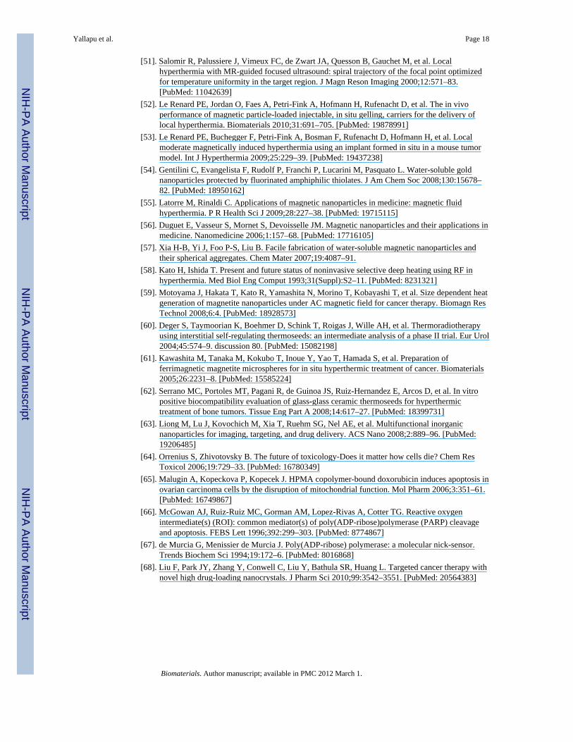
3.1. Characterization of magnetic nanoparticles3.1.1. Particles size and morphology—The initial focus of our investigation was toelucidate which formulation has smaller particles size and distribution in aqueous mediumafter surface engineering with CD or CD and F127 polymer. The particles size anddistribution measurements were obtained using a dynamic light scattering (DLS) instrument(Table 1). Pure magnetic nanoparticles (MNPs) have shown a larger particles size of 201.4nm with a polydispersity index (PI) of 0.22%. Coating of CD (50–150 mg) onto MNPsresulted in a slight increase in particles size (208.43 nm to 256.17 nm) due to randomcoating and/or the formed coating layers of CD molecules on the surface were not stable.But further increases in CD (200–300 mg) for coating onto MNPs produced more uniformformulations with average particles size ~175 nm. Therefore, we have selected 200 mg ofCD that is optimized to provide a good dispersion formulation. Further, it is shown thatF127 polymer layer coating (50–250 mg) improves its overall stability in aqueous dispersionby reducing the size of CD200 particles from ~175 nm to ~90 nm. Overall, the DLS datasuggest that CD200 and F127 coatings prevent aggregation of iron oxide formulations,unlike bare magnetic nanoparticles, conventional and multi-layer MNP formulations[4,28,29,43–45] (Fig. 1A). The average cluster formation of MNPs consistently decreasedwith the CD and F-127 coatings. This behavior can be seen visually in TEM studies. Pure
Yallapu et al. Page 8
Biomaterials. Author manuscript; available in PMC 2012 March 1.
NIH
-PA Author Manuscript
NIH
-PA Author Manuscript
NIH
-PA Author Manuscript
magnetic nanoparticles aggregate to have an average cluster size of > 300 nm (Fig. 1B (a)).When the particles were coated with 200 mg of CD, the cluster size is decreased to 170 nm(Fig. 1B (b)). F127 polymer coating further reduced particle size and provided uniformlysuspended particles with an average size of 90 nm (Fig. 1B (c)). These data suggest that CDand F127 layers on iron oxide nanoparticles not only coat particles but also attenuate theircluster behavior in aqueous media. Further, F127250 formulation particles size (90.72 ±0.23 nm) is considerably smaller compared to many double layered iron oxide formulationsprepared by co-precipitation approach [28,29]. However, an individual nanoparticle grainsize of this formulation is slightly increased after coating with CD and F127 polymers(Table 1). But, all the individual particle grain sizes ranged between 7–10 nm which iscommonly observed with many precipitation procedures [4,14,28,39–42] (Fig. 1C).
Good stability in aqueous medium of CD or CD and F127 polymer coated magneticnanoparticle formulations is due to their negative zeta potential values (Table 1). The CDcoated formulations (CD50 to CD250) have − 32 to + 0.59 mV while CD200 with F127coated formulations (F12750 to F127250) exhibited − 9.42 to − 10.79 mV. Such negativezeta potential formulations help repel each particle in the suspension, ensuring long-termstability and avoiding particles aggregation [46–48], whereas MNP formulations exhibitedpositive zeta potential, i.e., 6.17 mV indicates some degree of aggregation phenomenon.
3.1.2. Physical characterization—X-ray diffraction (XRD) patterns of differentformulations were analyzed to determine the crystal phase of the iron oxide nanoparticlesand surface engineered iron oxide nanoparticles (Fig. 2A). All the formulations (MNPs,CD200, and F127250) have shown diffraction peaks at 2θ = 30.1°, 36.2°, 42.4°, 52.5°, 57.5°and 62.2° due to face centered cubic lattice structures 220, 311, 400, 422, 440 and 511which are characteristic peaks of Fe3O4 crystal structure [31]. All of the diffraction peaks inFig. 2A can be indexed and assigned to the cubic structure of Fe3O4 which is consistent withthe theoretical values (JCPDS card no.: 01-088-0315). Additionally, there are no peaks at31° corresponding to γ-Fe2O3 and α-Fe2O3 for 210 and 213 in XRD patterns, supporting thepurity of synthesized iron oxide nanoparticles. This clearly suggests that iron oxideformulations are composed of magnetite (Fe3O4), not maghemite. Further, formulationswere stored under nitrogen atmosphere to prevent possible oxidation which is responsiblefor producing maghemite from magnetite.
To confirm the presence of β-cyclodextrin or F127 polymer layer(s) on magneticnanoparticles, FTIR analysis was taken into consideration (Fig. 2B). Pure magneticnanoparticles exhibited a broad peak between 3500–3000 cm−1 due to the presence ofhydroxyl/amino groups on the surface and a strong peak in the 550 cm−1 region due to –O–Fe of iron oxide skeleton [32] (Fig. 2B, black line). In addition, β-cyclodextrin coated MNPs(CD200) showed an intense band at 1010 cm−1 due to glycosidic (C-O-C) vibration and thecoupled (C-C/C-O) stretch vibrations. (Fig. 2B, red line) [31]. The F127 polymer coated onthe CD-MNP formulation (F127250) demonstrated the same peaks that appeared in CD200.The peak at 1010 cm−1 belongs to the CH2 rocking and C-O-C stretch vibrations of F127polymer (green line) [28].
We performed thermogravitic analyses to further confirm the presence of CD and F127layer(s) on magnetic nanoparticle formulations (Fig. 2C). Pure magnetic nanoparticles havea weight loss ~6.78 wt.% (iron oxide core content 93.22 wt.%), whereas CD coatedformulations lost ~11.38 wt.% (6.78 wt.% degradation of NPs + 4.6 wt.% is due to CDcoating) indicating 88.62 wt.% of iron oxide core in the formulation. In the case of F127250formulations it was noticed 87.76 wt.% iron oxide core (6.78 wt.% degradation of NPs +5.46 wt.% due to CD and F127 polymer layer coatings). Thus, we can confirm that
Yallapu et al. Page 9
Biomaterials. Author manuscript; available in PMC 2012 March 1.
NIH
-PA Author Manuscript
NIH
-PA Author Manuscript
NIH
-PA Author Manuscript
additional weight loss in the case of CD200 (4.6 wt.%) and F127250 (5.46 wt.%)formulations is due to coating of CD and F127 polymer layer(s).
3.1.3. Curcumin loading and release—The curcumin loading was estimated using aUV-vis spectrophotometer as described in our previous report [49]. It was confirmed that theloading capacity continuously increased as the amount of CD used for nanoparticles coatingincreased. This indicates that curcumin molecules were entering into the CD layer onnanoparticles via hydrophobic-hydrophobic interactions. Our recent report supports thisbehavior of curcumin encapsulation into the hydrophobic bucket structure of the CDmolecule [38]. In addition, F127 polymer promotes its loading to a greater extent due toPPO hydrophobic chains [28]. It was also confirmed that in pure magnetic nanoparticles,curcumin is primarily on the surface of the nanoparticles. Because curcumin molecules areloosely bound to surface of nanoparticles, curcumin release is much faster (Fig. 2D),whereas CD200 and F127250 magnetic nanoparticle formulations have shown a bi-phasicrelease characteristic. The initial burst of release was due to immediate dissociation ofsurface bound curcumin molecules that exist on the CD or F127 polymer matrix. Theremaining sustained drug release was due to the slow release of the drug entrapped insideCD and/or F127 polymer layers. The curcumin existence in nanoparticle layers wasconfirmed by FTIR analysis. Curcumin exhibited sharp absorption bands at 1605 cm−1
(stretching vibrations of benzene ring), 1502 cm−1 (C=O and C=C vibrations of benzene),1435 cm−1 (olefinic C-H bending vibration), 1285 cm−1 (aromatic C-O stretchingvibrations), and 1025 cm−1 (C-O-C stretching vibrations of CUR) (Fig. 2B, inset, blackline). The curcumin encapsulated formulation F127250-CUR also exhibited all these peaksin addition to the parent F127250 formulation (Fig. 2B, blue line), indicating the presence ofcurcumin in the formulation.
3.2. Hyperthermia ApplicationMagnetic nanoparticles are especially susceptible to induce localized hyperthermia in analternating magnetic field which may potentially shirk or destroy the tumors and sensitize toradiation or chemo-therapies [50–54]. Iron oxide based magnetic nanoparticle formulationsare often employed for this purpose because of fewer side effects [55,56]. For a givenmagnetic targeted application, an iron oxide formulation needs to meet superparamagneticproperties (high saturation magnetization, Ms) with greater heat effect. Therefore, ourformulations were evaluated for magnetic properties, i.e., magnetic saturation (Ms), using avibrating sample magnetometer at 300 K and at alternating ± 12000 Oe magnetic field (Fig.3A). The MNP, CD200 and F127250 formulations have saturation magnetization values of54.47, 47.51 and 52.58 emu/g, respectively. These saturation magnetization values correlatewith the reported values for many polymer stabilized iron oxide nanoparticle formulations[32,34,57]. This slight variation between the formulations is due to coating with F127polymer and/or β-cyclodextrin, and a small variation in overall particles size. Further, nocoercivity and remanence were observed for any of the formulations at lower magnetic fieldcurves, confirming superparamagnetic characteristics (Fig. 3A, inset).
The deficiency of hyperthermic cancer treatment is due to the difficulty of raising the tissuetemperature properly [58]. Hyperthermia using magnetic nanoparticles can raise thetemperature in the tumor locally up to 41–45 °C and is capable of damaging the tumor cellswithout damaging the healthy cells [59]. Our formulations were tested for these heatingeffects and results are presented in Fig. 3B. The temperature of formulations is plotted as afunction of time at the field of 150 Oe and a constant frequency of 300 kHz. The heatingeffects (hyperthermia) of formulations resulted from absorbing energy from the alternatingmagnetic field which was transformed into heat by means of hysteresis loss during reversalof magnetization. The order of heating effects of formulations was found to be F127250 >
Yallapu et al. Page 10
Biomaterials. Author manuscript; available in PMC 2012 March 1.
NIH
-PA Author Manuscript
NIH
-PA Author Manuscript
NIH
-PA Author Manuscript
CD200 > MNP. This can be explained since iron oxide core is available for excellent heateffects in the modified formulation of F127250 due to its freely dispersed stage, in contrastto other formulations. On the other hand, smaller clustered particles have a higher specificsurface area which generates higher heat [59]. Therefore, we speculate the F127250nanoparticles formulation would be highly useful as thermoseeds for localized hyperthermiatreatment of cancers [60–62]. Additionally, our formulations exhibited a continuous increasein temperature with respect to an increased concentration of F127250 formulationsemployed (Fig. 3C). This behavior is similar whether it is in solution form or in the gelform. This is further support that the heat buildup is uniform throughout the sample.
3.3. Magnetic Resonance ImagingNanomedicine platforms combine therapeutic function with imaging abilities which haveproven to be the next generation of medicine [41]. Unlike traditional contrast agents ordrugs, image visible nanomedicine has the ability for simultaneous diagnosis and therapy inone formulation [20,21,23]. We have evaluated our iron oxide formulations for in vitro MRIagent characteristics. These tailor-made cocktails can address the challenges of tumorheterogeneity and adaptive resistance which can ultimately help achieve the goal ofpersonalized medicine for cancer therapy [63].
Based upon the collected images (Fig. 4A), increased concentration of MNPs resulted in agreater reduction in signal. Relative to the control gel, the increase in iron oxideconcentration also resulted in shorter transverse relaxation times (Fig. 4B). For example, asthe concentration of iron oxide in the MNP increased from 10 μg Fe/ml to 40 μg Fe/ml, T2relaxation times diminished from 20.8ms to 6.3ms. Similar to T2 relaxation times,longitudinal relaxation T1 noted a reduction in relaxation time with iron concentration. Byplotting the transverse relaxation rate, R2, (1/T2) as a function of the concentration of Fe ineach sample, R2 increased linearly with the concentration of Fe (Fig. 4C) in all theformulations according to the equation: R2 = 1/T2 = 1/T°2 + r2*[Fe], where 1/T2 is therelaxation rate in the presence of iron oxide, 1/T°2 is the relaxation rate of pure water, r2 isthe transverse relaxivity, and [Fe] is the concentration of iron in each sample. The T2relaxivity (r2) was found to decrease in the following order for the tested compounds:F127250-CUR > F127250-CUR in cells > F127250 > F127250 in cells > MNP > CD200. Incomparison to T2 relaxation, application of F127250 to curcumin with and without cellsresulted in an increase in relaxivity. For example, the r1 for F127250-CUR was found to be28.6 × 10−3 s−1 μg−1 ml; whereas, F127250 had a relaxivity of 12.5 s−1 μg−1 ml. Theobserved difference suggests that curcumin increases local inhomogeneity in the magneticfield. For T2-weighted imaging procedures, the F127250 MNP can potentially improveobservation through contrast enhancement. It has been proven in previous reports that thesemulti-layer coated magnetic nanoparticles have superior imaging characteristics overFeridex IV® formulations [28].
3.4. Drug Delivery3.4.1. F127250 formulation lowers protein binding characteristics—One majorshortcoming of the magnetic nanoparticles is their destabilization following adsorption ofplasma proteins which leads to nonspecific uptake by the reticulum-endothelial system(RES). To evade clearance by RES and avoid agglomeration, and to improve the circulationtime of particles, iron oxide nanoparticles (MNP) were coated with CD (CD100) or CD andF127 polymers (F127250). This coating process led to composite heterogeneous particlescomposed of an iron oxide inner core with a modifying CD or CD-F127 outer coating.Agglomeration is prevented by the firm coating, emulsifying and adhesive properties ofF127 efficiently. To prove this concept, our formulations were tested for in vitro proteinadsorption (BSA) in 1× PBS solutions. The BSA adsorption was measured by the dimension
Yallapu et al. Page 11
Biomaterials. Author manuscript; available in PMC 2012 March 1.
NIH
-PA Author Manuscript
NIH
-PA Author Manuscript
NIH
-PA Author Manuscript
of fluorescence of tryptophan residue of BSA. It was noticed that fluorescence intensityreduced with increase of particle addition to BSA solution (Fig. 5A). This reduction isprobably dependent upon the formulation. The order of reduction was found to be MNP >CD200 > F127250 formulation. From this data, the number of binding sites (n) and bindingconstant (k) were calculated for each formulation presented in Fig. 5B. It was found thatF127250 has a low number of binding sites and binding constant (n = 1.57 and k = 0.025 μg/μg) while CD200 and MNP formulations have n = 1.74 and 2.08, k = 0.037 μg/μg and 0.038μg/μg, respectively. These analyses suggest that the F127250 formulation will have greatercirculation time than remaining formulations due to its number of binding sites and lowerbinding constant [28]. Therefore, in the next section we have evaluated the internalizationefficacy of F127250 formulation to determine its utility as a drug delivery carrier.
3.4.2. F127250 formulation intracellular uptake—Intracellular uptake ofnanoparticles improves therapeutic outcome because the internalized nanoparticlescontaining drug are retained in cancer cells. Higher internalization is an index for moreaccumulation of drug molecules, which release slowly and have sustained effects on cancercells. We have evaluated our formulations for cellular uptake in three different cancer cells(A2780CP, MDA-MB-231, and MCF-7) by using Flow Cytometeric (Fluorescence activatedcell shorter, FACS) analysis [32,38,49]. Particles uptake by cancer cells leads to shifting inthe side scattered (SSC) height in the Flow Cytometeric analysis. An increased uptake wasnoticed with increased amount of nanoparticles (0–100 μg) incubated for internalization(Fig. 6A, black to green). The qualitative estimation of particles uptake by various cancercells demonstrates higher uptake by cisplatin resistant ovarian cancer cells (A2780CP)compared to MCF-7 and MDA-MB-231 breast cancer cells at all the concentrations (Fig.6B). The order of particles uptake by cancer cells is A2780CP > MDA-MB-231 > MCF-7.This phenomenon is clearly observed in the transmission electron microscopy particleuptake experiments (Fig. 6C). A large portion of magnetic nanoparticles (F127250) areaccumulated in the epithelial membrane, endoplasmic reticulum, golgi and cytosolic ofA2780CP cancer cells. The particles aggregation is also observed in MDA-MB-231 at a fewspots. Compared to A2780 and MDA-MB-231 cells, less uptake/accumulation of particleswas observed in MCF-7 (non-metastatic) cells. This data demonstrate that our nanoparticlesare capable of internalizing within 6 hrs, even in resistant and metastatic cancer cells. Inaddition, we have also observed that the magnetic nanoparticles uptake in A2780CP cancercells is relatively higher compared to our recently fabricated PLGA nanoparticles [49].
3.4.3. Internalization of formulations in macrophage and A2780CP ovariancancer cells—A drug delivery carrier must be present in the blood stream for anappropriately long time to reach and accumulate at its therapeutic site. The opsonization/phagocytosis or removal of drug delivery vehicles from the body by a mononuclearphagocytic system which is also known as the reticuloendothelial system (RES) is a majorobstacle to achieving efficient drug delivery. Knowledge on how to design particles toescape phagocytosis could help to overcome this limitation. The macrophages of themononuclear phagocytic system have the capability to drain nanoparticles from thecirculation within seconds. In other words, lower uptake of particles by macrophagesdetermines their efficient use for the drug delivery applications. Therefore, we compared theuptake (indication of phagocytosis) efficiency of MNP, CD200 and F127250 formulations inRAW 264.7 (Mouse leukaemic monocyte macrophage cell line) as determined by Yallapu etal. [27]. Our MNP and CD200 formulations exhibited a greater level of phagocytosis(uptake) compared to the F127250 formulation (Fig. 7A) at two concentrations (50 and 100μg/ml). The reason for lower phagocytosis of the F127250 formulation is probably due tothe protective coating of F127 polymer (pluronic) which helps to form stable nanoparticlesin aqueous media [28,29]. Altogether, these observations reveal clear differences in the
Yallapu et al. Page 12
Biomaterials. Author manuscript; available in PMC 2012 March 1.
NIH
-PA Author Manuscript
NIH
-PA Author Manuscript
NIH
-PA Author Manuscript
phagocytosis pattern of different particles. The lower macrophage uptake of F127250formulation suggests that this would be a better choice for drug delivery application.
Next, we monitored the internalization of MNP, CD200, and F127250 formulations withrespect to 50 and 100 mg dosages (Fig. 7B) in A2789CP metastatic ovarian cancer cells. Theentry of MNP, CD200, and F127250 formulations into the cancer cells was determined [27]and it is varied in different formulations. Evidently, with increase of dose, their uptake isincreased. The cellular uptake of the nanocarriers is mainly dependent upon the route ofentry, i.e., endocytosis or phagocytosis. Phagocytosis is considered when the particles sizewas above 300 nm [8,14,19]. Whereas endocytic pathways for nanocarriers are subdividedinto four categories: namely, clathrin-mediated endocytosis, caveolae-mediated endocytosis,macropinocytosis, and clathrinand caveolae-independent endocytosis [8,14,19]. Takentogether, our studies suggest that the internalization of F127250 formulation is 5% inmacrophages and is > 75% in cancer cells, indicating uptake is based on endocytosis but notphagocytosis (Fig. 7A–B). This phenomenon is also evident in the TEM analysis of particlesuptake in macrophage and A2780CP cancer cells (Fig. 7C). A large number of F127250nanoparticles can be seen inside the A2780CP cancer cells but not in macrophages (RAW264.7).
3.4.4. CUR encapsulated F127250 formulation anticancer efficacy—Thetherapeutic efficacy of curcumin encapsulated F127250 was evaluated in three differentcancer cell lines (A2780CP: ovarian, MDA-MB-231: breast, and PC3: prostate) by MTS cellviability assay [38]. All the studied cell lines have shown typical dose dependant anti-proliferative effects (5–40 μM) by both native curcumin and curcumin encapsulatedF127250 formulation (Fig. 8). The control (DMSO or F127250) treatments did not show anyeffects on cell growth. The in vitro 50% cell growth inhibitory concentration (IC50) is thequantitative measure for the cell toxicity induced by chemotherapeutic drug. The IC50values were 12.6, 18.8 and 10.6 μM with curcumin and 12.1, 11.9, and 12.8 μM withcurcumin encapsulated F127250 formulation, for A2780CP, MDA-MB-231 and PC3 cancercells, respectively. This data demonstrates that F127250 containing curcumin is equallyefficient in suppressing cell growth even though the release is only 40 percent ofnanoformulations within the 48 hour treatment time. Therefore, we can consider thatinhibition of cell proliferation is more effective and sustained for long period of times.
Previous literature suggests that colony formation assay provides the ability to evaluatelong-term anti-cancer efficacy of the developed drug(s) or drug(s) formulations. To proveour MNP curcumin formulation has greater effects in long term anti-cancer efficacy assays,we have studied free CUR and F127250 curcumin formulation in A2780CP, MDA-MB-231and PC3 cancer cell lines at equivalent doses 4, 6, and 8 μM. Equivalent quantities ofDMSO or F127250 formulations were used as controls for CUR and F127250 CURformulation, respectively. A significant (p<0.05) decrease in the density of colonies wasobserved with F12725 curcumin formulation compared to free curcumin (Fig. 9). Forexample, at 4 μM, F127250 CUR formulation showed 38, 52, 21 and 56% colony densities,whereas free curcumin showed 59, 58, 30, and 100% colony densities after 10 day treatmentin A2780CP, MDA-MB-231, and PC3 cancer cells, respectively. The lower coloniesindicate an improved therapeutic efficacy of F127250 CUR formulation, resulting from anintracellular uptake effect and sustained release properties.
3.4.5. Molecular pathway—Bcl-xl is a transmembrane molecule in the mitochondria. Itis one of several anti-apoptotic proteins which are members of the Bcl-2 family of proteins.It has been implicated in the survival of cancer cells [33,64,65]. The expression levelanalysis suggests strong suppression of Bcl-xL expression after treatment with 10 or 20 μMF127250-CUR compared to equivalent amounts of CUR, DMSO and F127250. This data
Yallapu et al. Page 13
Biomaterials. Author manuscript; available in PMC 2012 March 1.
NIH
-PA Author Manuscript
NIH
-PA Author Manuscript
NIH
-PA Author Manuscript
demonstrate less cancer cell survival with the F127250-CUR formulation compared to CURtreatment (Fig. 10A). No change in Bcl-xL expression was observed with control (DMSOand F127250 formulation) treatments. From this study, it was clear that F127250-CUR at 10and 20 μM exhibited less cancer cell survival and implies induction of apoptosis or cancercell death. To study its apoptosis, we have investigated the pattern of Poly(ADP-ribose)polymerase (PARP) cleavage which is a protein involved in a number of cellular processesincluding DNA repair and cell death [66,67]. Cleavage of PARP is an indicator of DNAdamage and apoptosis in response to a diverse range of cytotoxic agents. Because of goodPARP expression and its cleavage in response to stress signal, we have chosen PC3 cells forPARP apoptosis assay. PC3 cells treated with 20 μM CUR or equivalent amounts ofF127250-CUR exhibited considerable cleavage of full length PARP (116 kDa) into cleavedPARP (86 kDa), which indicates the cancer cells are undergoing cell death via apoptosispathway (Fig. 10B). The PARP cleavage caused by F127250-CUR formulation is muchstronger compared to pure CUR treatment, which suggests an improved efficacy ofF127250-CUR formulation for cancer therapy.
The variation of ultrastructural changes in the cancer cells upon exposure to control (DMSOand F127250 formulation), 20 μM CUR or 20 μM F127250-CUR were observed bytransmission electron microscopy (TEM) analysis. The control treatments did not cause anyultrastructural changes in PC3 cancer cells (Fig. 10C (a–b)), but both F127250-CUR and 20μM CUR treated cells demonstrated the formation of endosomal-lysosomal-vacules, whichis an indication of cell death (Fig. 10C (c–d). However, this effect was more pronounced inF127250-CUR treated cells, suggesting its improved therapeutic efficacy (Fig. 10C (d)). Thevacuole formation is usually caused by the destabilization of subcellular organelle,mitochondrial swelling, opening of the permeability, dissipation of the mitochondrialpotential which is a hallmark of typical apoptosis. The internalization of the F127250-CURand sustained release of active curcumin in the cells probably enhanced the apoptosis incancer cells, which as a result, further improved therapeutic efficacy of our formulation.
3.5. Toxicity evaluationIn general, nanoparticles have extremely fast systemic translocation rates following in vivoadministration. Blood is one of the common translocation routes to organs for anynanoparticle-mediated therapy. Thus, we want to evaluate our CUR encapsulated F127250formulation for haemocompatibility. For this, different concentrations (10–100 μg) of CURcontaining MNP, CD200, and F127250 formulations were incubated in 100 μl of humanblood for 2 hrs. OD measurements recorded on a UV-vis spectrophotometer at λmax 570 nmindicate both CUR containing CD200 and F127250 formulations are haemocompatible butCUR containing MNP formulation is toxic after a concentration of 30 μg (Fig. 11A). Asimilar observation can be found with the red blood cells morphology studies (Fig 11B).Data in Fig. 11B(a–b) shows a clear morphology of red blood cells without any particles.After 2 hrs of MNP-CUR particles exposure to red blood cells, a rigorous change in themorphology and a higher amount of deposition of particles were noticed (Fig. 11B(c)).Other CUR containing formulations (CD200 and F127250) showed slight deposition ofparticle clumps; however, no change in the morphology of red blood cells was noted (Fig.11B(d–e)). In conclusion, F127250-CUR formulation is haemocompatible. The possiblereason for diminished haemolytic activity or haemocompatibility is low toxicity coatings ofcyclodextrin and/or pluronic polymer F127. These results are consistent with reportednanoparticles [68] coated with pluronic polymers which showed almost no toxicity.
4. ConclusionIt is essential to develop a tailor made magnetic nanoparticle formulation for multi-functional biomedical applications. A number of magnetic nanoparticle formulations have
Yallapu et al. Page 14
Biomaterials. Author manuscript; available in PMC 2012 March 1.
NIH
-PA Author Manuscript
NIH
-PA Author Manuscript
NIH
-PA Author Manuscript
been generated in the recent past. However, their effective utility has been limited due tohigher particle size, loss of magnetization, other inherent properties and lowerinternalization capacity into the cancer cells which ultimately results in poor therapeuticefficacy in cancer treatment. Our present study illustrates that multi-layer β-cyclodextrin andF127 polymer coated magnetic nanoparticles offer good stability, enhanced cellular uptake,sustained release characteristic of encapsulated anti-cancer drug (curcumin) with improvedanti-cancer therapeutic efficacy. The F127250-CUR formulation has shown almost similargrowth inhibitory effects as pure curcumin in the cancer cells. The F127250-CURformulation has also exhibited haemocompatibility, representing an excellent drug deliveryapproach. On the other hand, recent reports on these types of nanoformulations have shownsimilar anti-proliferative effects [28,29,38,49] but they do not provide MRI andhyperthermia properties. Additionally, our formulation has shown enhanced moleculareffects in cancer cells toward apoptosis.
AcknowledgmentsWe thank Cathy Christopherson (Sanford Research/USD, Sioux Falls) for editorial assistance, Robert Japs (SanfordHealth), Sara Basiaga (Department of Chemistry, UN-Lincoln), Shah R Valloppilly (Nebraska Center for Materialsand Nanoscience, Department of Physics and Astronomy, UN-Lincoln), and Crittenden J. Ohlemacher (AppliedPolymer Research Center, University of Akron) for their help in characterization of our samples. We also thank Dr.Omathanu P. Perumal (Department of Pharmaceutical Sciences, South Dakota State University, Brookings, SD) forproviding access to DLS instrument for particles size and zeta potential measurements. The authors also thank Dr.Diane Maher, Dr. Vasudha Sundram and Mara Ebeling (Sanford Research/USD) for their suggestions throughoutthis study. This work was supported by grants from Sanford Research/USD, Department of Defense (DOD)(PC073887), Governor's Cancer 2010, and NIH RO1 (CA142736) awarded to SCC and Department of Defense(DOD) (PC073643) and Governor's Cancer 2010 grants awarded to MJ.
References[1]. Lu AH, Salabas EL, Schuth F. Magnetic nanoparticles: synthesis, protection, functionalization,
and application. Angew Chem Int Ed Engl 2007;46:1222–44. [PubMed: 17278160][2]. Ito A, Shinkai M, Honda H, Kobayashi T. Medical application of functionalized magnetic
nanoparticles. J Biosci Bioeng 2005;100:1–11. [PubMed: 16233845][3]. Saiyed Z, Telang S, Ramchand C. Application of magnetic techniques in the field of drug
discovery and biomedicine. Biomagn Res Technol 2003;1:2. [PubMed: 14521720][4]. Namdeo M, Saxena S, Tankhiwale R, Bajpai M, Mohan YM, Bajpai SK. Magnetic nanoparticles
for drug delivery applications. J Nanosci Nanotechnol 2008;8:3247–71. [PubMed: 19051873][5]. Zhang L, Yu F, Cole AJ, Chertok B, David AE, Wang J, et al. Gum arabic-coated magnetic
nanoparticles for potential application in simultaneous magnetic targeting and tumor imaging.Aaps J 2009;11:693–9. [PubMed: 19842043]
[6]. Johannsen M, Gneveckow U, Eckelt L, Feussner A, Waldofner N, Scholz R, et al. Clinicalhyperthermia of prostate cancer using magnetic nanoparticles: presentation of a new interstitialtechnique. Int J Hyperthermia 2005;21:637–47. [PubMed: 16304715]
[7]. Wilhelm C, Fortin JP, Gazeau F. Tumour cell toxicity of intracellular hyperthermia mediated bymagnetic nanoparticles. J Nanosci Nanotechnol 2007;7:2933–7. [PubMed: 17685322]
[8]. Sun C, Lee JS, Zhang M. Magnetic nanoparticles in MR imaging and drug delivery. Adv DrugDeliv Rev 2008;60:1252–65. [PubMed: 18558452]
[9]. Kohler N, Sun C, Fichtenholtz A, Gunn J, Fang C, Zhang M. Methotrexate-immobilizedpoly(ethylene glycol) magnetic nanoparticles for MR imaging and drug delivery. Small2006;2:785–92. [PubMed: 17193123]
[10]. Bruce IJ, Sen T. Surface modification of magnetic nanoparticles with alkoxysilanes and theirapplication in magnetic bioseparations. Langmuir 2005;21:7029–35. [PubMed: 16008419]
[11]. Wilhelm C, Gazeau F. Universal cell labelling with anionic magnetic nanoparticles. Biomaterials2008;29:3161–74. [PubMed: 18455232]
Yallapu et al. Page 15
Biomaterials. Author manuscript; available in PMC 2012 March 1.
NIH
-PA Author Manuscript
NIH
-PA Author Manuscript
NIH
-PA Author Manuscript
[12]. Osaka T, Matsunaga T, Nakanishi T, Arakaki A, Niwa D, Iida H. Synthesis of magneticnanoparticles and their application to bioassays. Anal Bioanal Chem 2006;384:593–600.[PubMed: 16402174]
[13]. Dey T. Polymer-coated magnetic nanoparticles: surface modification and end-functionalization. JNanosci Nanotechnol 2006;6:2479–83. [PubMed: 17037859]
[14]. Gupta AK, Naregalkar RR, Vaidya VD, Gupta M. Recent advances on surface engineering ofmagnetic iron oxide nanoparticles and their biomedical applications. Nanomedicine 2007;2:23–39. [PubMed: 17716188]
[15]. Lee SY, Harris MT. Surface modification of magnetic nanoparticles capped by oleic acids:characterization and colloidal stability in polar solvents. J Colloid Interface Sci 2006;293:401–8.[PubMed: 16054635]
[16]. Flexman JA, Minoshima S, Kim Y, Cross DJ. Magneto-optical labeling of fetal neural stem cellsfor in vivo MRI tracking. Conf Proc IEEE Eng Med Biol Soc 2006;1:5631–4. [PubMed:17947156]
[17]. Hoehn M, Kustermann E, Blunk J, Wiedermann D, Trapp T, Wecker S, et al. Monitoring ofimplanted stem cell migration in vivo: a highly resolved in vivo magnetic resonance imaginginvestigation of experimental stroke in rat. Proc Natl Acad Sci U S A 2002;99:16267–72.[PubMed: 12444255]
[18]. Montet-Abou K, Montet X, Weissleder R, Josephson L. Cell internalization of magneticnanoparticles using transfection agents. Mol Imaging 2007;6:1–9. [PubMed: 17311760]
[19]. McCarthy JR, Weissleder R. Multifunctional magnetic nanoparticles for targeted imaging andtherapy. Adv Drug Deliv Rev 2008;60:1241–51. [PubMed: 18508157]
[20]. Guthi JS, Yang SG, Huang G, Li S, Khemtong C, Kessinger CW, et al. MRI-visible micellarnanomedicine for targeted drug delivery to lung cancer cells. Mol Pharm 2010;7:32–40.[PubMed: 19708690]
[21]. Cinteza LO, Ohulchanskyy TY, Sahoo Y, Bergey EJ, Pandey RK, Prasad PN. Diacyllipidmicelle-based nanocarrier for magnetically guided delivery of drugs in photodynamic therapy.Mol Pharm 2006;3:415–23. [PubMed: 16889435]
[22]. Pradhan P, Giri J, Rieken F, Koch C, Mykhaylyk O, Doblinger M, et al. Targeted temperaturesensitive magnetic liposomes for thermo-chemotherapy. J Control Release 142:108–21.[PubMed: 19819275]
[23]. Shubayev VI, Pisanic TR 2nd, Jin S. Magnetic nanoparticles for theragnostics. Adv Drug DelivRev 2009;61:467–77. [PubMed: 19389434]
[24]. Rubio-Retama J, Zafeiropoulos NE, Serafinelli C, Rojas-Reyna R, Voit B, Cabarcos EL, et al.Synthesis and characterization of thermosensitive PNIPAM microgels covered withsuperparamagnetic gamma-Fe2O3 nanoparticles. Langmuir 2007;23:10280–5. [PubMed:17718580]
[25]. Wang L, Yang Z, Zhang Y, Wang L. Biofunctional nanoparticles with magnetization andluminescence. J Phys Chem C 2009;113:3955–9.
[26]. Guo R, Zhang L, Qian H, Li R, Jiang X, Liu B. Multifunctional nanocarriers for cell imaging,drug delivery, and near-IR photothermal therapy. Langmuir 2010;26:5428–34. [PubMed:20095619]
[27]. Yallapu MM, Foy SP, Jain TK, Labhasetwar V. PEG-Functionalized Magnetic Nanoparticles forDrug Delivery and Magnetic Resonance Imaging Applications. Pham Res. 2010 DOI: 10.1007/s11095-010-0260-1.
[28]. Jain TK, Morales MA, Sahoo SK, Leslie-Pelecky DL, Labhasetwar V. Iron oxide nanoparticlesfor sustained delivery of anticancer agents. Mol Pharm 2005;2:194–205. [PubMed: 15934780]
[29]. Jain TK, Reddy MK, Morales MA, Leslie-Pelecky DL, Labhasetwar V. Biodistribution,clearance, and biocompatibility of iron oxide magnetic nanoparticles in rats. Mol Pharm2008;5:316–27. [PubMed: 18217714]
[30]. Gao J, Gu H, Xu B. Multifunctional magnetic nanoparticles: design, synthesis, and biomedicalapplications. Acc Chem Res 2009;42:1097–107. [PubMed: 19476332]
[31]. Banerjee SS, Chen D-H. Magnetic Nanoparticles Grafted with Cyclodextrin for HydrophobicDrug Delivery. Chem Mater 2007;19:6345–9.
Yallapu et al. Page 16
Biomaterials. Author manuscript; available in PMC 2012 March 1.
NIH
-PA Author Manuscript
NIH
-PA Author Manuscript
NIH
-PA Author Manuscript
[32]. Bhattarai SR, Kc RB, Kim SY, Sharma M, Khil MS, Hwang PH, et al. N-hexanoyl chitosanstabilized magnetic nanoparticles: Implication for cellular labeling and magnetic resonanceimaging. J Nanobiotechnology 2008;6:1. [PubMed: 18173857]
[33]. Yallapu MM, Maher DM, Sundram V, Bell MC, Jaggi M, Chauhan SC. Curcumin induceschemo/radio-sensitization in ovarian cancer cells and curcumin nanoparticles inhibit ovariancancer cell growth. J Ovarian Res 2010;3:11. [PubMed: 20429876]
[34]. Luo B, Song XJ, Zhang F, Xia A, Yang WL, Hu JH, et al. Multi-functional thermosensitivecomposite microspheres with high magnetic susceptibility based on magnetite colloidalnanoparticle clusters. Langmuir 2010;26:1674–9. [PubMed: 19754089]
[35]. Beaven GH, Chen S-H, D'albis A, Gratzer WB. A spectroscopic study of the haemin-human-serum-albumin system. Eur J Biochem 1974;41:539–46. [PubMed: 4817561]
[36]. Chipman DM, Grisaro V, NSharon N. The binding of oligosaccharides containing N-acetylglucosamin and N-acetylmuramic acid to lysozyme. J Biol Chem 1967;242:4388–94.[PubMed: 6070843]
[37]. Chauhan SC, Vannatta K, Ebeling MC, Vinayek N, Watanabe A, Pandey KK, et al. Expressionand functions of transmembrane mucin MUC13 in ovarian cancer. Cancer Res 2009;69:765–74.[PubMed: 19176398]
[38]. Yallapu MM, Jaggi M, Chauhan SC. beta-Cyclodextrin-curcumin self-assembly enhancescurcumin delivery in prostate cancer cells. Colloids Surf B Biointerfaces 2010;79:113–25.[PubMed: 20456930]
[39]. Bae KH, Ha YJ, Kim C, Lee KR, Park TG. Pluronic/chitosan shell cross-linked nanocapsulesencapsulating magnetic nanoparticles. J Biomater Sci Polym Ed 2008;19:1571–83. [PubMed:19017471]
[40]. Lim JK, Majetich SA, Tilton RD. Stabilization of superparamagnetic iron oxide core-gold shellnanoparticles in high ionic strength media. Langmuir 2009;25:13384–93. [PubMed: 19928938]
[41]. Lin JJ, Chen JS, Huang SJ, Ko JH, Wang YM, Chen TL, et al. Folic acid-Pluronic F127 magneticnanoparticle clusters for combined targeting, diagnosis, and therapy applications. Biomaterials2009;30:5114–24. [PubMed: 19560199]
[42]. Xiong XY, Tam KC, Gan LH. Release kinetics of hydrophobic and hydrophilic model drugsfrom pluronic F127/poly(lactic acid) nanoparticles. J Control Release 2005;103:73–82. [PubMed:15710501]
[43]. Dorris A, Rucareanu S, Reven L, Barrett CJ, Lennox RB. Preparation and characterization ofpolyelectrolyte-coated gold nanoparticles. Langmuir 2008;24:2532–8. [PubMed: 18229959]
[44]. Latham AH, Williams ME. Controlling transport and chemical functionality of magneticnanoparticles. Acc Chem Res 2008;41:411–20. [PubMed: 18251514]
[45]. Peracchia MT, Vauthier C, Puisieux F, Couvreur P. Development of sterically stabilizedpoly(isobutyl 2-cyanoacrylate) nanoparticles by chemical coupling of poly(ethylene glycol). JBiomed Mater Res 1997;34:317–26. [PubMed: 9086401]
[46]. Gupta AK, Gupta M. Synthesis and surface engineering of iron oxide nanoparticles forbiomedical applications. Biomaterials 2005;26:3995–4021. [PubMed: 15626447]
[47]. Billotey C, Wilhelm C, Devaud M, Bacri JC, Bittoun J, Gazeau F. Cell internalization of anionicmaghemite nanoparticles: quantitative effect on magnetic resonance imaging. Magn Reson Med2003;49:646–54. [PubMed: 12652535]
[48]. Smirnov P. Cellular magnetic resonance imaging using superparamagnetic anionic iron oxidenanoparticles: applications to in vivo trafficking of lymphocytes and cell-based anticancertherapy. Methods Mol Biol 2009;512:333–53. [PubMed: 19347287]
[49]. Yallapu MM, Gupta BK, Jaggi M, Chauhan SC. Fabrication of curcumin encapsulated PLGAnanoparticles for improved therapeutic effects in metastatic cancer cells. J Colloid Interface Sci.2010
[50]. Salomir R, Vimeux FC, de Zwart JA, Grenier N, Moonen CT. Hyperthermia by MR-guidedfocused ultrasound: accurate temperature control based on fast MRI and a physical model oflocal energy deposition and heat conduction. Magn Reson Med 2000;43:342–7. [PubMed:10725875]
Yallapu et al. Page 17
Biomaterials. Author manuscript; available in PMC 2012 March 1.
NIH
-PA Author Manuscript
NIH
-PA Author Manuscript
NIH
-PA Author Manuscript
[51]. Salomir R, Palussiere J, Vimeux FC, de Zwart JA, Quesson B, Gauchet M, et al. Localhyperthermia with MR-guided focused ultrasound: spiral trajectory of the focal point optimizedfor temperature uniformity in the target region. J Magn Reson Imaging 2000;12:571–83.[PubMed: 11042639]
[52]. Le Renard PE, Jordan O, Faes A, Petri-Fink A, Hofmann H, Rufenacht D, et al. The in vivoperformance of magnetic particle-loaded injectable, in situ gelling, carriers for the delivery oflocal hyperthermia. Biomaterials 2010;31:691–705. [PubMed: 19878991]
[53]. Le Renard PE, Buchegger F, Petri-Fink A, Bosman F, Rufenacht D, Hofmann H, et al. Localmoderate magnetically induced hyperthermia using an implant formed in situ in a mouse tumormodel. Int J Hyperthermia 2009;25:229–39. [PubMed: 19437238]
[54]. Gentilini C, Evangelista F, Rudolf P, Franchi P, Lucarini M, Pasquato L. Water-soluble goldnanoparticles protected by fluorinated amphiphilic thiolates. J Am Chem Soc 2008;130:15678–82. [PubMed: 18950162]
[55]. Latorre M, Rinaldi C. Applications of magnetic nanoparticles in medicine: magnetic fluidhyperthermia. P R Health Sci J 2009;28:227–38. [PubMed: 19715115]
[56]. Duguet E, Vasseur S, Mornet S, Devoisselle JM. Magnetic nanoparticles and their applications inmedicine. Nanomedicine 2006;1:157–68. [PubMed: 17716105]
[57]. Xia H-B, Yi J, Foo P-S, Liu B. Facile fabrication of water-soluble magnetic nanoparticles andtheir spherical aggregates. Chem Mater 2007;19:4087–91.
[58]. Kato H, Ishida T. Present and future status of noninvasive selective deep heating using RF inhyperthermia. Med Biol Eng Comput 1993;31(Suppl):S2–11. [PubMed: 8231321]
[59]. Motoyama J, Hakata T, Kato R, Yamashita N, Morino T, Kobayashi T, et al. Size dependent heatgeneration of magnetite nanoparticles under AC magnetic field for cancer therapy. Biomagn ResTechnol 2008;6:4. [PubMed: 18928573]
[60]. Deger S, Taymoorian K, Boehmer D, Schink T, Roigas J, Wille AH, et al. Thermoradiotherapyusing interstitial self-regulating thermoseeds: an intermediate analysis of a phase II trial. Eur Urol2004;45:574–9. discussion 80. [PubMed: 15082198]
[61]. Kawashita M, Tanaka M, Kokubo T, Inoue Y, Yao T, Hamada S, et al. Preparation offerrimagnetic magnetite microspheres for in situ hyperthermic treatment of cancer. Biomaterials2005;26:2231–8. [PubMed: 15585224]
[62]. Serrano MC, Portoles MT, Pagani R, de Guinoa JS, Ruiz-Hernandez E, Arcos D, et al. In vitropositive biocompatibility evaluation of glass-glass ceramic thermoseeds for hyperthermictreatment of bone tumors. Tissue Eng Part A 2008;14:617–27. [PubMed: 18399731]
[63]. Liong M, Lu J, Kovochich M, Xia T, Ruehm SG, Nel AE, et al. Multifunctional inorganicnanoparticles for imaging, targeting, and drug delivery. ACS Nano 2008;2:889–96. [PubMed:19206485]
[64]. Orrenius S, Zhivotovsky B. The future of toxicology-Does it matter how cells die? Chem ResToxicol 2006;19:729–33. [PubMed: 16780349]
[65]. Malugin A, Kopeckova P, Kopecek J. HPMA copolymer-bound doxorubicin induces apoptosis inovarian carcinoma cells by the disruption of mitochondrial function. Mol Pharm 2006;3:351–61.[PubMed: 16749867]
[66]. McGowan AJ, Ruiz-Ruiz MC, Gorman AM, Lopez-Rivas A, Cotter TG. Reactive oxygenintermediate(s) (ROI): common mediator(s) of poly(ADP-ribose)polymerase (PARP) cleavageand apoptosis. FEBS Lett 1996;392:299–303. [PubMed: 8774867]
[67]. de Murcia G, Menissier de Murcia J. Poly(ADP-ribose) polymerase: a molecular nick-sensor.Trends Biochem Sci 1994;19:172–6. [PubMed: 8016868]
[68]. Liu F, Park JY, Zhang Y, Conwell C, Liu Y, Bathula SR, Huang L. Targeted cancer therapy withnovel high drug-loading nanocrystals. J Pharm Sci 2010;99:3542–3551. [PubMed: 20564383]
Yallapu et al. Page 18
Biomaterials. Author manuscript; available in PMC 2012 March 1.
NIH
-PA Author Manuscript
NIH
-PA Author Manuscript
NIH
-PA Author Manuscript
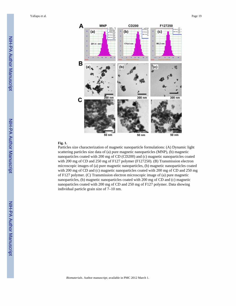
Fig. 1.Particles size characterization of magnetic nanoparticle formulations: (A) Dynamic lightscattering particles size data of (a) pure magnetic nanoparticles (MNP), (b) magneticnanoparticles coated with 200 mg of CD (CD200) and (c) magnetic nanoparticles coatedwith 200 mg of CD and 250 mg of F127 polymer (F127250). (B) Transmission electronmicroscopic images of (a) pure magnetic nanoparticles, (b) magnetic nanoparticles coatedwith 200 mg of CD and (c) magnetic nanoparticles coated with 200 mg of CD and 250 mgof F127 polymer. (C) Transmission electron microscopic image of (a) pure magneticnanoparticles, (b) magnetic nanoparticles coated with 200 mg of CD and (c) magneticnanoparticles coated with 200 mg of CD and 250 mg of F127 polymer. Data showingindividual particle grain size of 7–10 nm.
Yallapu et al. Page 19
Biomaterials. Author manuscript; available in PMC 2012 March 1.
NIH
-PA Author Manuscript
NIH
-PA Author Manuscript
NIH
-PA Author Manuscript
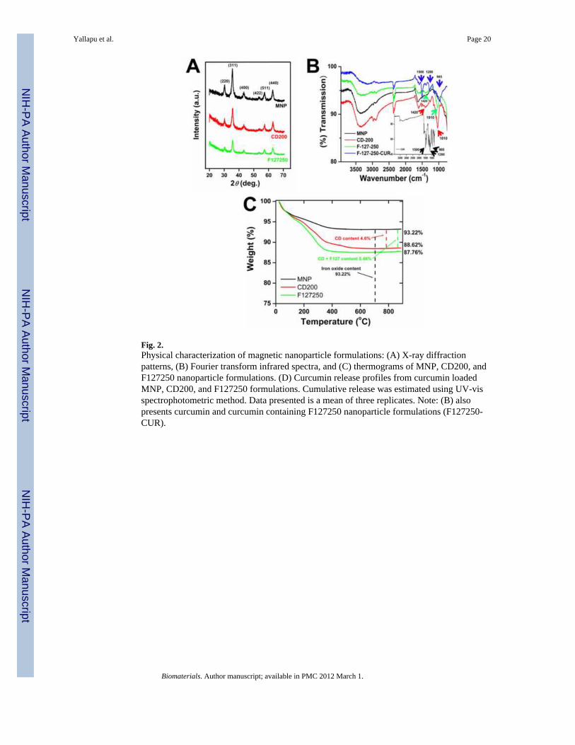
Fig. 2.Physical characterization of magnetic nanoparticle formulations: (A) X-ray diffractionpatterns, (B) Fourier transform infrared spectra, and (C) thermograms of MNP, CD200, andF127250 nanoparticle formulations. (D) Curcumin release profiles from curcumin loadedMNP, CD200, and F127250 formulations. Cumulative release was estimated using UV-visspectrophotometric method. Data presented is a mean of three replicates. Note: (B) alsopresents curcumin and curcumin containing F127250 nanoparticle formulations (F127250-CUR).
Yallapu et al. Page 20
Biomaterials. Author manuscript; available in PMC 2012 March 1.
NIH
-PA Author Manuscript
NIH
-PA Author Manuscript
NIH
-PA Author Manuscript
Fig. 3.(A) Hysteresis loops of MNP, CD200 and F127250 nanoparticle formulations at roomtemperature. (B) Time course of the raised temperature of MNP, CD200 and F127250nanoparticle formulations under an alternating magnetic field operating at 300 kHz. (C)Temperature of various concentrations of F127250 nanoparticles in solution and agarosegels after altering magnetic field applied for 15 min.
Yallapu et al. Page 21
Biomaterials. Author manuscript; available in PMC 2012 March 1.
NIH
-PA Author Manuscript
NIH
-PA Author Manuscript
NIH
-PA Author Manuscript
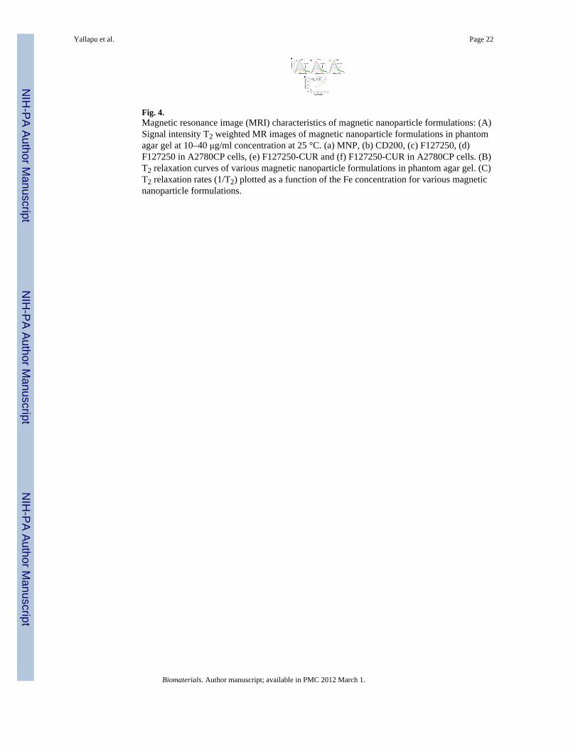
Fig. 4.Magnetic resonance image (MRI) characteristics of magnetic nanoparticle formulations: (A)Signal intensity T2 weighted MR images of magnetic nanoparticle formulations in phantomagar gel at 10–40 μg/ml concentration at 25 °C. (a) MNP, (b) CD200, (c) F127250, (d)F127250 in A2780CP cells, (e) F127250-CUR and (f) F127250-CUR in A2780CP cells. (B)T2 relaxation curves of various magnetic nanoparticle formulations in phantom agar gel. (C)T2 relaxation rates (1/T2) plotted as a function of the Fe concentration for various magneticnanoparticle formulations.
Yallapu et al. Page 22
Biomaterials. Author manuscript; available in PMC 2012 March 1.
NIH
-PA Author Manuscript
NIH
-PA Author Manuscript
NIH
-PA Author Manuscript
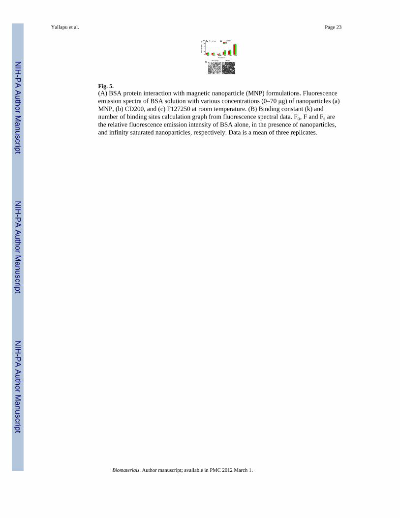
Fig. 5.(A) BSA protein interaction with magnetic nanoparticle (MNP) formulations. Fluorescenceemission spectra of BSA solution with various concentrations (0–70 μg) of nanoparticles (a)MNP, (b) CD200, and (c) F127250 at room temperature. (B) Binding constant (k) andnumber of binding sites calculation graph from fluorescence spectral data. Fo, F and Fs arethe relative fluorescence emission intensity of BSA alone, in the presence of nanoparticles,and infinity saturated nanoparticles, respectively. Data is a mean of three replicates.
Yallapu et al. Page 23
Biomaterials. Author manuscript; available in PMC 2012 March 1.
NIH
-PA Author Manuscript
NIH
-PA Author Manuscript
NIH
-PA Author Manuscript

Fig. 6.Cellular uptake of magnetic nanoparticle formulations in cancer cells. (A) Side scatteredmeasurements of nanoparticles uptake by cancer cells using FACS. (B) The quantitativeinternalization of magnetic nanoparticle formulations in A2780CP (cisplatin resistantovarian cancer cells), MDA-MB-231 (metastatic breast cancer cells) and MCF-7 (non-metastatic breast cancer cells) are based on the side scattered fluorescence height values.Data represents mean of 3 repeats for each treatment. (C) Transmission electronmicrographs of F127250 nanoparticle uptake in (a) A2780CP, (b) MDA-MB-231 and (c)MCF-7 cancer cells. Arrow points indicate F127250 nanoparticles internalization with adistinct contrast.
Yallapu et al. Page 24
Biomaterials. Author manuscript; available in PMC 2012 March 1.
NIH
-PA Author Manuscript
NIH
-PA Author Manuscript
NIH
-PA Author Manuscript
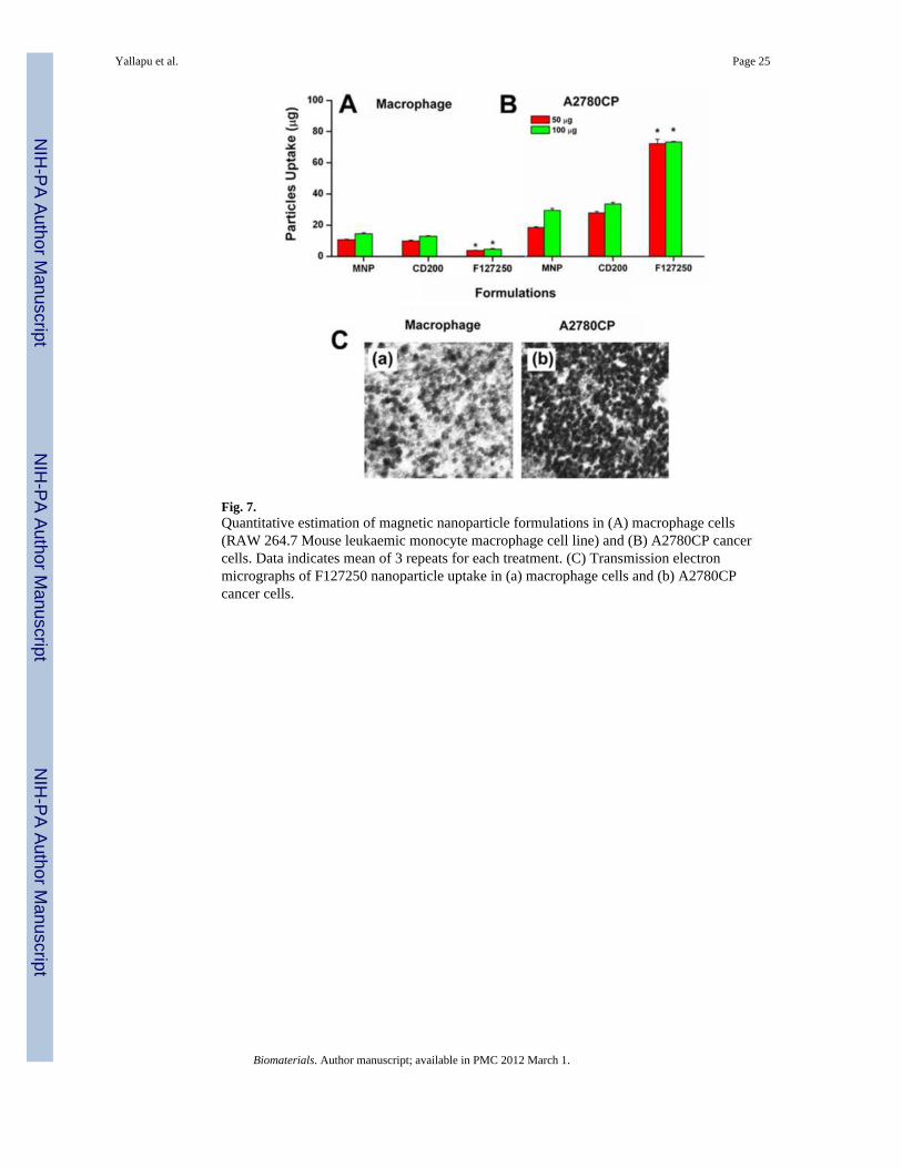
Fig. 7.Quantitative estimation of magnetic nanoparticle formulations in (A) macrophage cells(RAW 264.7 Mouse leukaemic monocyte macrophage cell line) and (B) A2780CP cancercells. Data indicates mean of 3 repeats for each treatment. (C) Transmission electronmicrographs of F127250 nanoparticle uptake in (a) macrophage cells and (b) A2780CPcancer cells.
Yallapu et al. Page 25
Biomaterials. Author manuscript; available in PMC 2012 March 1.
NIH
-PA Author Manuscript
NIH
-PA Author Manuscript
NIH
-PA Author Manuscript
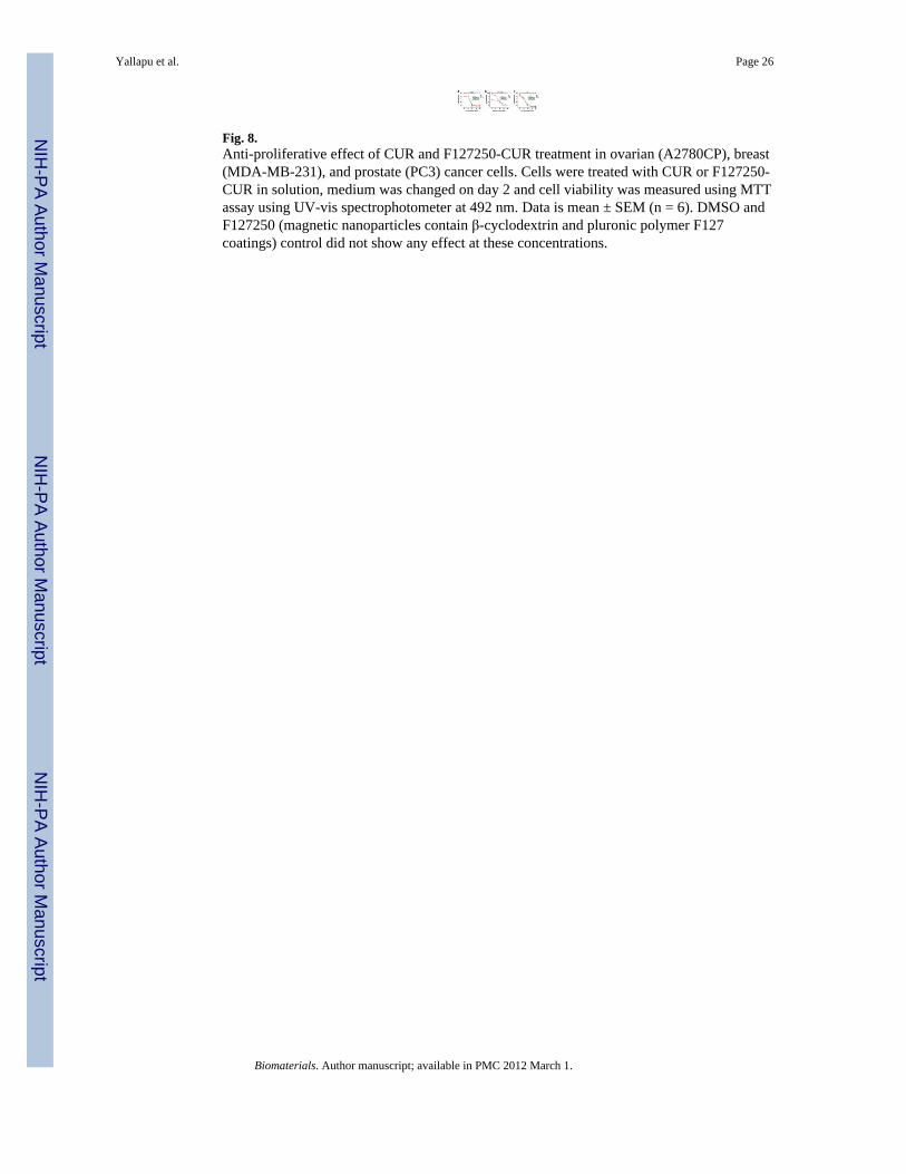
Fig. 8.Anti-proliferative effect of CUR and F127250-CUR treatment in ovarian (A2780CP), breast(MDA-MB-231), and prostate (PC3) cancer cells. Cells were treated with CUR or F127250-CUR in solution, medium was changed on day 2 and cell viability was measured using MTTassay using UV-vis spectrophotometer at 492 nm. Data is mean ± SEM (n = 6). DMSO andF127250 (magnetic nanoparticles contain β-cyclodextrin and pluronic polymer F127coatings) control did not show any effect at these concentrations.
Yallapu et al. Page 26
Biomaterials. Author manuscript; available in PMC 2012 March 1.
NIH
-PA Author Manuscript
NIH
-PA Author Manuscript
NIH
-PA Author Manuscript
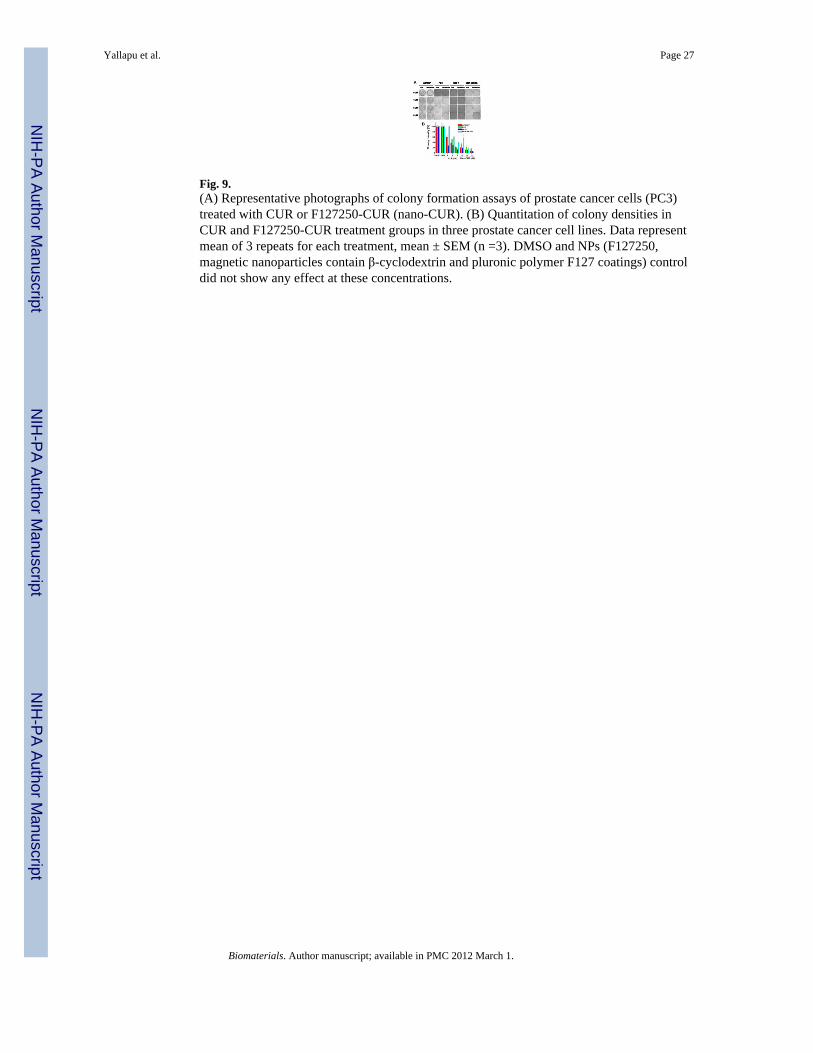
Fig. 9.(A) Representative photographs of colony formation assays of prostate cancer cells (PC3)treated with CUR or F127250-CUR (nano-CUR). (B) Quantitation of colony densities inCUR and F127250-CUR treatment groups in three prostate cancer cell lines. Data representmean of 3 repeats for each treatment, mean ± SEM (n =3). DMSO and NPs (F127250,magnetic nanoparticles contain β-cyclodextrin and pluronic polymer F127 coatings) controldid not show any effect at these concentrations.
Yallapu et al. Page 27
Biomaterials. Author manuscript; available in PMC 2012 March 1.
NIH
-PA Author Manuscript
NIH
-PA Author Manuscript
NIH
-PA Author Manuscript
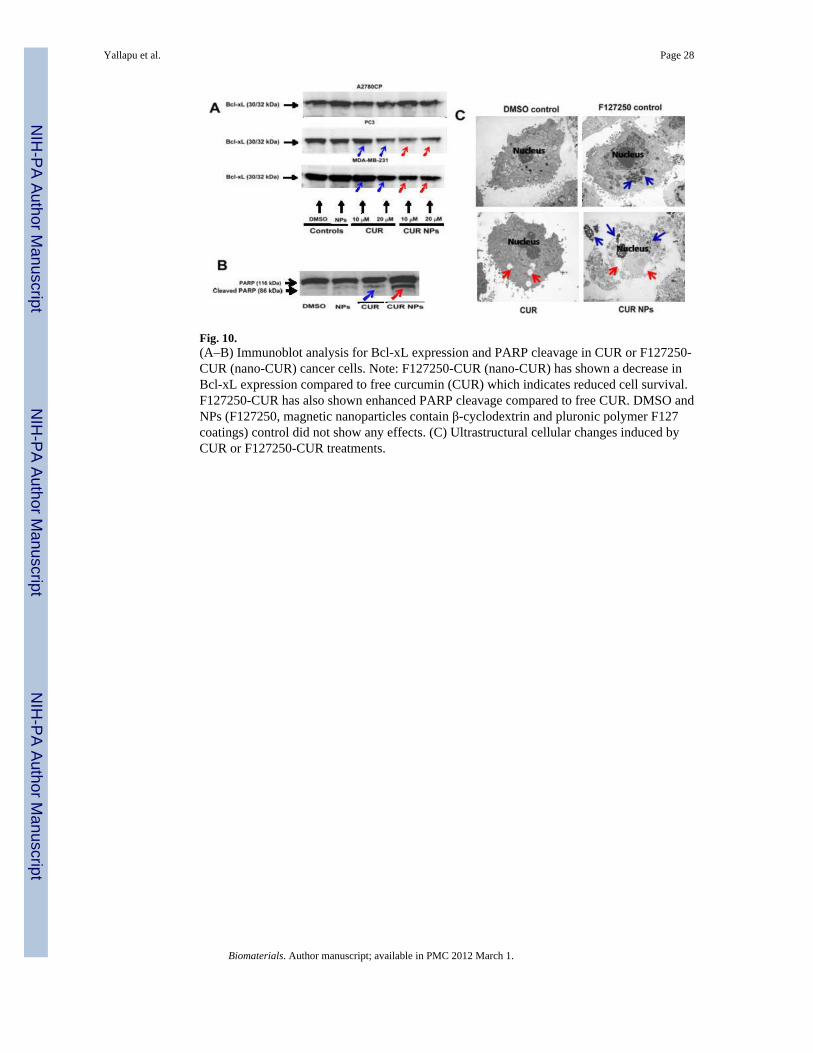
Fig. 10.(A–B) Immunoblot analysis for Bcl-xL expression and PARP cleavage in CUR or F127250-CUR (nano-CUR) cancer cells. Note: F127250-CUR (nano-CUR) has shown a decrease inBcl-xL expression compared to free curcumin (CUR) which indicates reduced cell survival.F127250-CUR has also shown enhanced PARP cleavage compared to free CUR. DMSO andNPs (F127250, magnetic nanoparticles contain β-cyclodextrin and pluronic polymer F127coatings) control did not show any effects. (C) Ultrastructural cellular changes induced byCUR or F127250-CUR treatments.
Yallapu et al. Page 28
Biomaterials. Author manuscript; available in PMC 2012 March 1.
NIH
-PA Author Manuscript
NIH
-PA Author Manuscript
NIH
-PA Author Manuscript
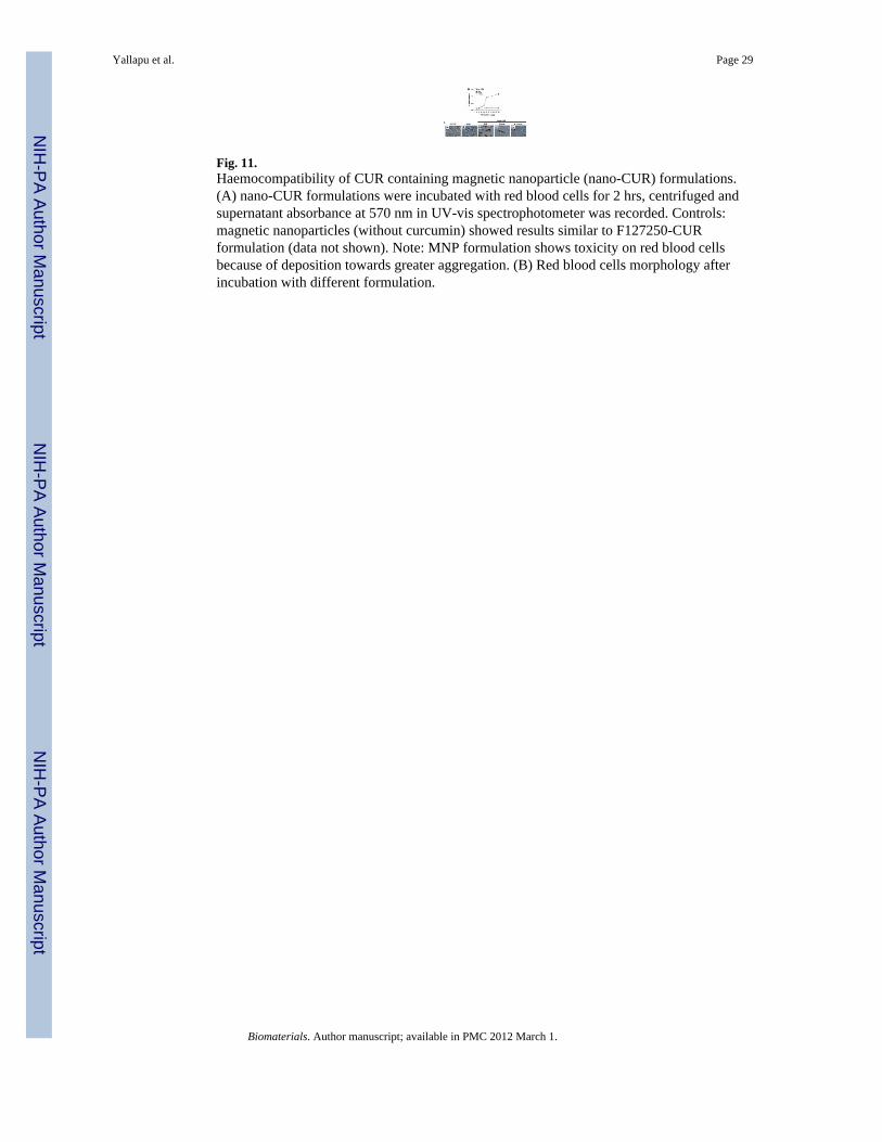
Fig. 11.Haemocompatibility of CUR containing magnetic nanoparticle (nano-CUR) formulations.(A) nano-CUR formulations were incubated with red blood cells for 2 hrs, centrifuged andsupernatant absorbance at 570 nm in UV-vis spectrophotometer was recorded. Controls:magnetic nanoparticles (without curcumin) showed results similar to F127250-CURformulation (data not shown). Note: MNP formulation shows toxicity on red blood cellsbecause of deposition towards greater aggregation. (B) Red blood cells morphology afterincubation with different formulation.
Yallapu et al. Page 29
Biomaterials. Author manuscript; available in PMC 2012 March 1.
NIH
-PA Author Manuscript
NIH
-PA Author Manuscript
NIH
-PA Author Manuscript
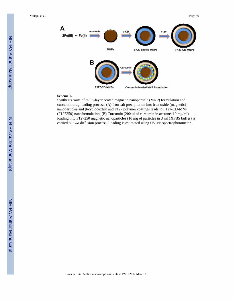
Scheme 1.Synthesis route of multi-layer coated magnetic nanoparticle (MNP) formulation andcurcumin drug loading process. (A) Iron salt precipitation into iron oxide (magnetic)nanoparticles and β-cyclodextrin and F127 polymer coatings leads to F127-CD-MNP(F127250) nanoformulation. (B) Curcumin (200 μl of curcumin in acetone, 10 mg/ml)loading into F127250 magnetic nanoparticles (10 mg of particles in 3 ml 1XPBS buffer) iscarried out via diffusion process. Loading is estimated using UV-vis spectrophotometer.
Yallapu et al. Page 30
Biomaterials. Author manuscript; available in PMC 2012 March 1.
NIH
-PA Author Manuscript
NIH
-PA Author Manuscript
NIH
-PA Author Manuscript
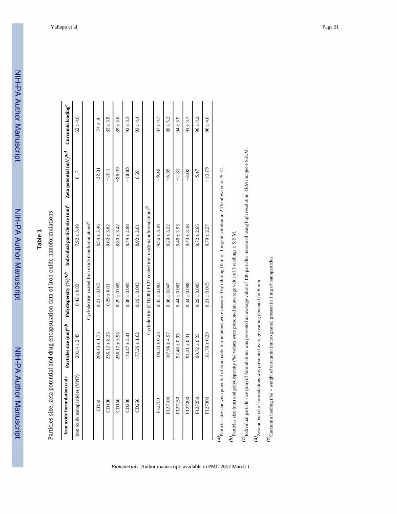
NIH
-PA Author Manuscript
NIH
-PA Author Manuscript
NIH
-PA Author Manuscript
Yallapu et al. Page 31
Tabl
e 1
Parti
cles
size
, zet
a po
tent
ial a
nd d
rug
enca
psul
atio
n da
ta o
f iro
n ox
ide
nano
form
ulat
ions
Iron
oxi
de fo
rmul
atio
n co
dePa
rtic
les s
ize
(nm
)a,b
Poly
disp
ersi
ty (%
)a,b
Indi
vidu
al p
artic
le si
ze (n
m)c
Zet
a po
tent
ial (
mV
)a,d
Cur
cum
in lo
adin
ge
Iron
oxi
de n
anop
artic
les (
MN
P)20
1.4
± 2.
450.
42 ±
0.0
27.
92 ±
3.4
96.
1762
± 4
.6
Cyc
lode
xtrin
coa
ted
Iron
oxi
de n
anof
orm
ulat
ions
a
CD
5020
8.43
± 1
.75
0.21
± 0
.015
8.54
± 2
.46
−32.31
74 ±
.9
CD
100
236.
12 ±
8.2
50.
29 ±
0.0
38.
62 ±
3.6
2−20.1
82 ±
3.8
CD
150
256.
17 ±
3.9
50.
29 ±
0.0
058.
80 ±
3.4
2−26.09
89 ±
3.6
CD
200
174.
47 ±
2.4
10.
38 ±
0.0
058.
79 ±
2.9
8−18.85
92 ±
3.3
CD
250
177.
26 ±
1.6
20.
19 ±
0.0
038.
92 ±
2.6
10.
5993
± 4
.4
Cyc
lode
xtrin
(CD
200)
-F12
7 co
ated
iron
oxi
de n
anof
orm
ulat
ions
b
F127
5010
8.33
± 0
.23
0.35
± 0
.003
9.56
± 3
.18
−9.42
87 ±
4.7
F127
100
107.
96 ±
4.9
70.
36 ±
0.0
479.
29 ±
3.2
2−8.55
89 ±
5.2
F127
150
92.4
6 ±
0.93
0.44
± 0
.002
9.46
± 2
.93
−2.31
94 ±
3.9
F127
200
91.2
1 ±
0.31
0.34
± 0
.008
9.73
± 3
.16
−8.02
93 ±
3.7
F127
250
90.7
2 ±
0.23
0.29
± 0
.005
9.72
± 2
.65
−5.47
96 ±
4.2
F127
300
181.
76 ±
0.2
30.
21 ±
0.0
159.
79 ±
2.2
7−10.79
96 ±
4.6
(a) Pa
rticl
es si
ze a
nd z
eta
pote
ntia
l of i
ron
oxid
e fo
rmul
atio
ns w
ere
mea
sure
d by
dilu
ting
10 μ
l of 1
mg/
ml s
olut
ion
in 2
.75
ml w
ater
at 2
5 °C
.
(b) Pa
rticl
es si
ze (n
m) a
nd p
olyd
ispe
rsity
(%) v
alue
s wer
e pr
esen
ted
an a
vera
ge v
alue
of 3
read
ings
± S
.E.M
.
(c) In
divi
dual
par
ticle
size
(nm
) of f
orm
ulat
ions
was
pre
sent
ed a
n av
erag
e va
lue
of 1
00 p
artic
les m
easu
red
usin
g hi
gh re
solu
tion
TEM
imag
es ±
S.E
.M.
(d) Ze
ta p
oten
tial o
f for
mul
atio
ns w
as p
rese
nted
ave
rage
read
ing
obta
ined
for 6
min
.
(e) C
urcu
min
load
ing
(%) =
wei
ght o
f cur
cum
in (m
icro
gra
ms)
pre
sent
in 1
mg
of n
anop
artic
les.
Biomaterials. Author manuscript; available in PMC 2012 March 1.