lxr and ppar activators stimulate cholesterol sulfotransferase type 2 isoform 1b in human...
TRANSCRIPT

Copyright © 2005 by the American Society for Biochemistry and Molecular Biology, Inc.
This article is available online at http://www.jlr.org
Journal of Lipid Research
Volume 46, 2005
2657
LXR and PPAR activators stimulate cholesterolsulfotransferase type 2 isoform 1b inhuman keratinocytes
Yan J. Jiang,* Peggy Kim,* Peter M. Elias,
†,§
and Kenneth R. Feingold
1,
*
,†,
**
Departments of Medicine* and Dermatology,
†
University of California San Francisco, San Francisco, CA 94121; and Dermatology Service
§
and Metabolism Section,** Veterans Affairs Medical Center, San Francisco, CA 94121
Abstract Liver X receptors (LXRs) and peroxisome prolifer-ator-activated receptors (PPARs) are potent regulators of ke-ratinocyte proliferation, differentiation, and epidermal per-meability barrier homeostasis. Cholesterol sulfotransferasetype 2B isoform 1b (SULT2B1b) is a key enzyme in the syn-thesis of cholesterol sulfate (CS), a critical regulator of kerati-nocyte differentiation and desquamation, as well as a media-tor of barrier homeostasis. In this study, we assessed theeffect of activators of LXR, PPAR
�
, PPAR
�
/
�
, and PPAR
�
on SULT2B1b gene expression and enzyme activity in cul-tured human keratinocytes (CHKs). Our results demonstratethat PPAR and LXR activators increase SULT2B1b mRNA lev-els, with the most dramatic effect (a 26-fold increase) inducedby the PPAR
�
activator ciglitazone. Ciglitazone upregulatesSULT2B1b mRNA in a dose- and time-dependent manner.Moreover, the stimulation of SULT2B1b gene expression byLXR and PPAR activators occurs in both undifferentiated anddifferentiated CHKs. The upregulation of SULT2B1b mRNAby ciglitazone appears to occur at a transcriptional level, be-cause the degradation of SULT2B1b is not accelerated by ci-glitazone. In addition, cycloheximide almost completely blocksthe ciglitazone-induced increase in SULT2B1b mRNA, suggest-ing that the transcription of SULTB1b mRNA is dependent onnew protein synthesis. Finally, LXR and PPAR activators alsoincreased the activity of cholesterol sulfotransferase.Thus, LXR and PPAR activators regulate the expression ofSULT2B1b, the key enzyme in the synthesis of CS, which is apotent regulator of epidermal differentiation and corneocytedesquamation.
—Jiang, Y. J., P. Kim, P. M. Elias, and K. R. Fein-gold.
LXR and PPAR activators stimulate cholesterol sul-fotransferase type 2 isoform 1b in human keratinocytes.
J.Lipid Res.
2005.
46:
2657–2666.
Supplementary key words
cholesterol sulfate
•
liver X receptor
•
per-oxisome proliferator-activated receptor
�
/
�
/
�
Cholesterol sulfate (CS) plays a number of key roles inregulating epidermal function. It is widely recognized as a
critical regulator of several keratinocyte corneocyte func-tions, most notably corneocyte desquamation (1). It alsostimulates keratinocyte differentiation via a number ofpotential pathways, including the activation of protein ki-nase C (2), indirectly by binding sphingosine (3), anddirectly by affecting gene transcription (4). CS also re-portedly modulates antimicrobial defense (5) and regu-lates keratinocyte cholesterol synthesis (6). Moreover, CSis catabolized to cholesterol and sulfate in the stratum cor-neum (SC), a process catalyzed by the enzyme steroid sul-fatase (6), and cholesterol is crucial for the formation ofthe cutaneous permeability barrier (7, 8). Thus, identifi-cation of factors that regulate epidermal CS synthesiscould be of potential importance for several keratinocytefunctions.
CS is synthesized in the basal and spinous layers of theepidermis by the cytosolic enzyme cholesterol sulfotrans-ferase (CSTase), resulting in peak levels of CS in the gran-ular layer of the epidermis (1, 9, 10). CSTase belongs to asuperfamily of cytosolic sulfotransferases (SULTs) that cat-alyze the sulfoconjugation of hormones and neurotrans-mitters as well as certain drugs and xenobiotics (11, 12).Five families of SULTs have been identified, with SULT2primarily engaged in the sulfoconjugation of neutral ste-roids and sterols (13). Cholesterol sulfotransferase type 2Bisoform 1b (SULT2B1b) selectively functions as a CSTase(14), and recent studies have shown that this isozyme cata-lyzes the synthesis of CS in epidermis/keratinocytes (12).SULT2B1b mRNA and protein levels increase during Ca
2
�
-induced terminal differentiation in parallel with an increasein CSTase activity (15). Despite the great importance of CS
Abbreviations: CHK, cultured human keratinocyte; CS, cholesterolsulfate; CSTase, cholesterol sulfotransferase; LXR, liver X receptor; 25-OH, 25-hydroxycholesterol; PPAR, peroxisome proliferator-activatedreceptor; 22R, 22(
R
)-hydroxycholesterol; RAR, retinoic acid receptor;RXR, retinoid X receptor; SC, stratum corneum; SULT2B1b, choles-terol sulfotransferase type 2B isoform 1b.
1
To whom correspondence should be addressed.e-mail: [email protected]
Manuscript received 9 June 2005 and in revised form 26 August 2005.
Published, JLR Papers in Press, September 8, 2005.DOI 10.1194/jlr.M500235-JLR200
by guest, on February 23, 2016
ww
w.jlr.org
Dow
nloaded from

2658 Journal of Lipid Research
Volume 46, 2005
synthesis for the epidermis, there is a paucity of informa-tion on the factors that regulate SULT2B1b expression inkeratinocytes. We have previously shown in rat fetal skinexplants that CSTase activity increases in fetal epidermislate in fetal development, in parallel with the formation ofa differentiated epidermis (16). Interestingly, CS levels in-crease dramatically when rabbit tracheal epithelial cellsundergo squamous differentiation, owing to a 20- to 30-fold increase in CSTase activity in the absence of retinoicacid, and CS levels decline by
�
25-fold in the presence ofexogenous retinoic acid (17). Similar phenomena occurin cultured human keratinocytes (CHKs) treated withretinoic acid (18, 19). Retinoic acid is a natural ligand ofthe type 2 nuclear hormone receptor retinoic acid recep-tor (RAR), and activation of RAR affects many importantcellular processes, such as proliferation and differentia-tion (20).
Recent studies have shown that the nuclear hormonereceptors peroxisome proliferator-activated receptor (PPAR)
�
,
�
/
�
, and
�
and liver X receptor (LXR)
�
and
�
are ex-pressed in the epidermis/keratinocytes (21–27). These re-ceptors heterodimerize with retinoid X receptor (RXR),after which they can activate the expression of manygenes, including those involved in lipid metabolism (20,28, 29). PPARs are activated by fatty acids, fatty acidmetabolites, or various drugs (PPAR
�
-fibrates or WY14643,PPAR
�
/
�
-GW501516, PPAR
�
-ciglitazone or troglitazone),whereas LXRs are activated by certain oxysterols [22(
R
)-hydroxysterol, 24,25-hydroxysterol] (30–32). Activation ofPPARs and LXRs stimulates keratinocyte differentiation,accelerates the development of the epidermis in fetal rats,and improves epidermal permeability barrier homeostasisin normal adult mice (21, 26, 27, 33, 34). In rat fetal skinexplants, we have shown that treatment with both PPAR
�
and LXR activators increases epidermal steroid sulfataseactivity (21). Therefore, we assessed whether PPARs andLXR activators regulate CSTase expression and activity innormal human keratinocytes.
MATERIALS AND METHODS
Materials
The LXR activators 22(
R
)-hydroxycholesterol (22R) and 25-hydroxycholesterol (25-OH) and the PPAR
�
activators clofibricacid and WY14643 were purchased from Sigma Chemical Co.(St. Louis, MO). Actinomycin D and cycloheximide were alsopurchased from Sigma. The PPAR
�
activator ciglitazone was pur-chased from Cayman Chemical Co. (Ann Arbor, MI); troglita-zone was obtained from Biomol Research Laboratories, Inc. (Ply-mouth Meeting, PA). GI262570 was kindly provided by Dr. TimWillson (GlaxoSmithKline, Pittsburgh, PA). The PPAR
�
activatorGW501516 was purchased from Calbiochem, Inc. (San Diego,CA). Molecular grade chemicals such as TRI reagent were ob-tained from either Sigma or Fisher Scientific (Fairlawn, NJ).[
�
-
32
P]dCTP (3,000 Ci/mmol) was purchased from New EnglandNuclear (Boston, MA). [
32
S]phosphoadenosine phosphosulfatewas purchased from Perkin-Elmer (Boston, MA). The MultiprimeLabeling System was purchased from Amersham (Amersham, UK).Mini-spin columns (G-50) were purchased from Worthington (Free-
hold, NJ). Oligo(dT)-cellulose, type 77F, was purchased fromPharmacia (Uppsala, Sweden). Nytran Plus membranes werepurchased from Schleicher and Schuell (Keene, NH). Spin-Xcentrifuge filters were purchased from Corning Costar (Cam-bridge, MA).
Keratinocyte culture
Human foreskin keratinocytes, second passage, were seededand maintained in 0.07 mM Ca
2
�
serum-free keratinocyte growthmedium (Clonetics, San Diego, CA). Once the cells attached, theculture medium was changed to either 0.03 mM (low) or 1.2 mM(high) Ca
2
�
. In the presence of 1.2 mM Ca
2
�
, keratinocytes wereinduced to differentiate. In a typical experiment, cells were har-vested at 80–100% confluence. Cells were treated with each activa-tor at their optimized concentration at preconfluence (60–70%)in either low (0.03 mM) or high (1.2 mM) calcium conditions.Alternatively, cells were coincubated with individual activatorand 10
�
g/ml cycloheximide, a classical protein synthesis inhibi-
Fig. 1. Basal levels of retinoid X receptor � (RXR�), liver X re-ceptor (LXR), and peroxisome proliferator-activated receptor(PPAR) mRNAs in undifferentiated versus differentiated culturedhuman keratinocytes (CHKs). Cells at second passage were platedin normal medium containing 0.07 mM Ca2� overnight and thenswitched to either low-Ca2� (0.03 mM) or high-Ca2� (1.2 mM) me-dium for 24 h. Total RNA was isolated and subjected to Northernblot analyses. RXR�, LXR�, LXR�, PPAR�, and PPAR� mRNAswere examined by Northern blot as described in Materials andMethods. PPAR� and internal control cyclophilin mRNAs were ex-amined by real-time PCR. Equal loading, the integrity of RNA, andthe densitometry value were verified and normalized by ethidiumbromide staining of 28S rRNA. The mRNA level of PPAR� was nor-malized by cyclophilin. Data are expressed as percentages of vehiclecontrol and presented as means � SEM (n 3) from at least twoindependent experiments. A representative blot is shown (A) and aplot is shown for PPAR� (B).
by guest, on February 23, 2016
ww
w.jlr.org
Dow
nloaded from

Jiang et al.
LXR and PPAR activators stimulate cholesterol sulfotransferase 2659
tor, or they were coincubated with 22R and ciglitazone. Controlkeratinocytes were treated with vehicle (0.05% ethanol).
Total RNA isolation, cDNA probes, and Northern blotting
Total RNA was isolated using TRI reagent according to themanufacturer’s protocol, and Northern blot analysis was per-formed to measure SULT2B1b mRNA. Briefly, Northern blotswere prepared, and equal loading, to test the integrity of RNA,was verified by ethidium bromide staining of 28S rRNA beforetransfer to Nytran membranes. Blots were then hybridized with
32
P-labeled probes of SULT2B1b overnight at 65
C and washed.Alternatively, poly(A
�
) RNA was isolated using oligo(dT) cellu-lose, and Northern blot analysis was performed to measuremRNA levels of RXR
�
, LXR
�
, LXR
�
, PPAR
�
, and PPAR
�
. Hu-man Sult2B1b probe was prepared using PCR, and the PCRprimer pairs used were: SULT2B1b, upper (5
�
-TCT CGG AAATCA GCC AGA AGTT-3
�
) and lower (5
�
-ATA GTG GAT GCTCCT CGA CGT-3
�
). Human RXR
�
cDNA was a gift from Dr.D. D. Bikle (University of California San Francisco). LXR
�
andLXR
�
were kindly provided by Dr. D. J. Mangelsdorf (University
of Texas Southwestern Medical Center, Dallas, TX). PPAR
�
andPPAR
�
cDNAs were gifts from Dr. A. Bass (University of Califor-nia San Francisco). Subsequently, the blots were exposed toX-ray films for various durations to ensure that measurementswere performed on the linear portion of the curve, and the tar-get bands were quantitated by densitometry (Bio-Rad Laborato-ries, Hercules, CA). Some blots were probed with cyclophilin asan internal control signal. The densitometry quantitation of thetarget gene was adjusted for either cyclophilin or 28S level andexpressed as a percentage of control, with the control as 100%.
Quantitative real-time PCR
Total RNA was extracted from keratinocytes using TRIZOL re-agent (Sigma). First-strand cDNA for PCR was synthesized usingan Advantage RT-for-PCR kit according to the manufacturer’sprotocol. Briefly, cDNA was synthesized from 1
�
g of total RNAusing Moloney murine leukemia virus reverse transcriptase withrandom hexamer primer at 42
C for 60 min. Relative mRNA lev-els of the target gene (PPAR
�
) and an invariant transcript, cyclo-philin, were determined using the Mx3000P™ Real-Time PCR
Fig. 2. LXR and PPAR activators increase cholesterol sulfotransferase type 2B isoform 1b (SULT2B1b) mRNA levels in undifferentiatedkeratinocytes. Cells were incubated in low-Ca2� (0.03 mM) medium in the presence or absence of each LXR/PPAR activator alone for24 h: 10 �M 25-hydroxycholesterol (25OH) (A); 400 �M clofibrate (Clo) (B); 8 �M GW501516 (GW) (C); or 10 �M troglitazone (Tro), 5 �MGI262570 (GI), or 5 �M ciglitazone (Cig) (D). Total RNA was isolated and subjected to Northern blot analyses. SULT2B1b mRNA was ex-amined as described in Materials and Methods. Equal loading, the integrity of RNA, and the densitometry value were verified and normal-ized by ethidium bromide staining of 28S rRNA. Data are expressed as percentages of vehicle control and presented as means � SEM (n 3) from at least two independent experiments. A representative blot is shown above each plot for the corresponding activator. * P � 0.05,** P � 0.01.
by guest, on February 23, 2016
ww
w.jlr.org
Dow
nloaded from
2660 Journal of Lipid Research
Volume 46, 2005
System (110 V) with a Notebook Computer (Stratagene Corp.,La Jolla, CA). This system uses SYBR Green chemistry for highlyaccurate quantification of mRNA levels. Individual PCRs werecarried out in a mixture of 20
�
l containing 20 ng of cDNA, 450nM forward or reverse primer, and 10
�
l of 2
SYBR GreenQ-PCR Master Mix (Applied Biosystems, Foster City, CA). The se-quences for human PAAR
�
primers were 5
�
-ATA TCT CCC TTTTTG TGG CTG CTA-3
�
(sense) and 5
�
-TCC GAC TCC GTC TTCTTG ATG A-3
�
(antisense). The reaction was performed at 50
Cfor 2 min, 95
C for 10 min, and then 40 cycles of amplificationwith melting at 95
C for 30 s, annealing at 60
C for 30 s, and ex-tension at 72
C for 45 s on Mx3000P 96-well plates (Stratagene).PCR was performed in triplicate and replicated in three inde-pendent experiments. Gel electrophoresis and melting curveanalyses were performed to confirm correct PCR product sizesand the absence of nonspecific bands. The expression level ofPPAR
�
was normalized against cyclophilin using the comparativeC
T
method according to the manufacturer’s protocols.
mRNA stability
Cells were seeded at 60% confluence in low-calcium mediumovernight and then incubated with the medium containing 5
�
Mciglitazone or vehicle alone (ethanol) for 24 h. Cells were thentreated with 2
�
g/ml actinomycin D for various periods of time(0, 2, 4, 6, 8, and 10 h). Cells were harvested at each time pointand subjected to Northern blot analysis, and the SULT2B1b mRNAlevels were determined. The interpolation, calculation, and graph-ing of mRNA values were performed using SigmaPlot 8.0 soft-ware (Systat Software, Inc., Point Richmond, CA).
Enzyme source preparation
After treatment, cells were washed twice with ice-cold PBS andharvested. Cell pellets were subsequently homogenized in abuffer consisting of 10 mM Tris(hydoxymethyl)aminomethane,pH 7.5, 0.15 M sucrose, and 2 mM ethylenediaminetetraaceticacid. Crude homogenates were first centrifuged at 10,000
g for10 min, followed by 100,000 g for 60 min, both at 4C. TheCSTase activity was measured in the cytosolic fraction (superna-tant after 100,000 g spin). Protein concentration was assayed bythe method of Bradford (Bio-Rad Laboratories).
CSTase activity assayCSTase activity was measured as described previously (16, 35).
Briefly, cholesterol (100 �g in benzene) was evaporated to dry-ness and suspended by sonication in 0.05 ml of homogenizationbuffer. [32S]phosphoadenosine phosphosulfate (50,000 cpm/tubein 0.025 ml) was then added to an equal volume of the enzymepreparation (50–100 �g). Reactions were carried out at 37C for1 h and were terminated by the addition of 2.0 ml of chloroform-methanol (2:1, v/v) and 1.0 ml of chloroform-methanol-water-KCl (15 ml:240 ml:235 ml:35.5 g), forming the lower and upperphases, respectively, to separate the substrate from sulfated prod-uct. The upper aqueous phase was saved in a separate tube, andthe lower phase was extracted once more with 1.0 ml of upperphase. The upper phase was discarded, and an aliquot of thelower organic phase containing the sulfated product was thendried and counted by scintillation spectrophotometry.
StatisticsAll data are expressed as means � SEM. Comparison between
two groups was undertaken using two-tailed and unpaired t-tests.In the multigroup experiments, data were compared by one-wayANOVA and Tukey’s test. Differences in values were consideredsignificant at P � 0.05.
RESULTS
The steady-state expression levels of LXRs and PPARs in undifferentiated versus differentiated CHKs
To compare the basal levels of LXR and PPAR expres-sion in undifferentiated versus differentiated CHKs, we mea-sured the mRNA levels of RXR�, LXR�, LXR�, PPAR�, andPPAR� by Northern blot (Fig. 1A) and PPAR� by real-timePCR analysis (Fig. 1B). As reported by others, RXR� andPPAR� mRNA levels increase with keratinocyte differenti-ation (23, 36, 37). In addition, both LXR� and LXR�mRNAs are expressed in CHKs (34), and in this study weshow that their levels increase with calcium-induced kera-tinocyte differentiation (Fig. 1A). In contrast, PPAR� andPPAR� mRNA levels do not significantly increase withshort-term (24 h) differentiation (Fig. 1A, B). These re-sults demonstrate that these receptors are expressed inCHKs and in some instances are regulated by keratinocytedifferentiation.
LXR and PPAR activators increase SULT2B1b mRNA levels in undifferentiated keratinocytes
We next measured the changes in mRNA levels ofSULT2B1b by Northern blot analysis in the second pas-sage of undifferentiated CHK treated with LXR and PPARactivators. Before these experiments, a set of preliminarystudies was conducted to optimize the dose for each acti-vator. As depicted in Fig. 2, activators of LXR, PPAR�,PPAR�/�, and PPAR� all upregulate SULT2B1b mRNAsignificantly. For LXR activators, we used two natural ligands,22R and 25-OH. 25-OH (10 �M) increased SULT2B1b mRNA
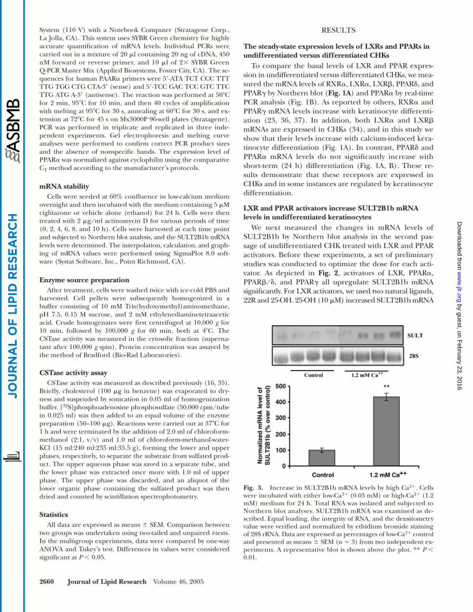
Fig. 3. Increase in SULT2B1b mRNA levels by high Ca2�. Cellswere incubated with either low-Ca2� (0.03 mM) or high-Ca2� (1.2mM) medium for 24 h. Total RNA was isolated and subjected toNorthern blot analyses. SULT2B1b mRNA was examined as de-scribed. Equal loading, the integrity of RNA, and the densitometryvalue were verified and normalized by ethidium bromide stainingof 28S rRNA. Data are expressed as percentages of low-Ca2� controland presented as means � SEM (n 3) from two independent ex-periments. A representative blot is shown above the plot. ** P �0.01.
by guest, on February 23, 2016
ww
w.jlr.org
Dow
nloaded from
Jiang et al. LXR and PPAR activators stimulate cholesterol sulfotransferase 2661
levels by 2.9-fold (Fig. 2A), and 22R increased them by 7.1-fold (data not shown). Similarly, an activator of PPAR�,clofibric acid, also modestly increased SULT2B1b mRNAlevels (39.9%) at 400 �M (Fig. 2B), but the change in-duced by a lower concentration (200 �M) did not archivestatistical significance (data not shown). In contrast, a 9.8-fold increase in SULT2B1b mRNA occurred with thePPAR� synthetic activator GW501516 (Fig. 2C). Finally,whereas all three PPAR� activators stimulated SULT2B1bexpression (Fig. 2D), their potencies differed: ciglitazoneincreased expression by 25.1-fold, troglitazone increasedit by 2.4-fold, and GI262570 increased it by 6.8-fold (Fig.2D). Thus, LXR and PPAR activators increase SULT2B1bexpression in undifferentiated CHKs.
LXR and PPAR activators increase SULT2B1b expression in differentiated keratinocytes
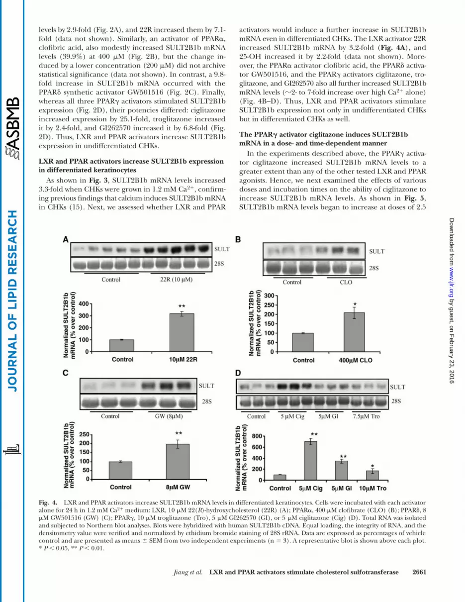
As shown in Fig. 3, SULT2B1b mRNA levels increased3.3-fold when CHKs were grown in 1.2 mM Ca2�, confirm-ing previous findings that calcium induces SULT2B1b mRNAin CHKs (15). Next, we assessed whether LXR and PPAR
activators would induce a further increase in SULT2B1bmRNA even in differentiated CHKs. The LXR activator 22Rincreased SULT2B1b mRNA by 3.2-fold (Fig. 4A), and25-OH increased it by 2.2-fold (data not shown). More-over, the PPAR� activator clofibric acid, the PPAR� activa-tor GW501516, and the PPAR� activators ciglitazone, tro-glitazone, and GI262570 also all further increased SULT2B1bmRNA levels (�2- to 7-fold increase over high Ca2� alone)(Fig. 4B–D). Thus, LXR and PPAR activators stimulateSULT2B1b expression not only in undifferentiated CHKsbut in differentiated CHKs as well.
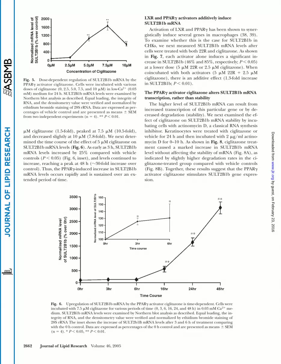
The PPAR� activator ciglitazone induces SULT2B1b mRNA in a dose- and time-dependent manner
In the experiments described above, the PPAR� activa-tor ciglitazone increased SULT2B1b mRNA levels to agreater extent than any of the other tested LXR and PPARagonists. Hence, we next examined the effects of variousdoses and incubation times on the ability of ciglitazone toincrease SULT2B1b mRNA levels. As shown in Fig. 5,SULT2B1b mRNA levels began to increase at doses of 2.5
Fig. 4. LXR and PPAR activators increase SULT2B1b mRNA levels in differentiated keratinocytes. Cells were incubated with each activatoralone for 24 h in 1.2 mM Ca2� medium: LXR, 10 �M 22(R)-hydroxycholesterol (22R) (A); PPAR�, 400 �M clofibrate (CLO) (B); PPAR�, 8�M GW501516 (GW) (C); PPAR�, 10 �M troglitazone (Tro), 5 �M GI262570 (GI), or 5 �M ciglitazone (Cig) (D). Total RNA was isolatedand subjected to Northern blot analyses. Blots were hybridized with human SULT2B1b cDNA. Equal loading, the integrity of RNA, and thedensitometry value were verified and normalized by ethidium bromide staining of 28S rRNA. Data are expressed as percentages of vehiclecontrol and are presented as means � SEM from two independent experiments (n 3). A representative blot is shown above each plot.* P � 0.05, ** P � 0.01.
by guest, on February 23, 2016
ww
w.jlr.org
Dow
nloaded from
2662 Journal of Lipid Research Volume 46, 2005
�M ciglitazone (1.5-fold), peaked at 7.5 �M (10.5-fold),and decreased slightly at 10 �M (7.8-fold). We next deter-mined the time course of the effect of 5 �M ciglitazone onSULT2B1b mRNA levels (Fig. 6). As early as 3 h, SULT2B1bmRNA levels increased by 25% compared with vehiclecontrols (P � 0.05) (Fig. 6, inset), and levels continued toincrease, reaching a peak at 48 h (�30-fold increase overcontrol). Thus, the PPAR�-induced increase in SULT2B1bmRNA levels occurs rapidly and is sustained over an ex-tended period of time.
LXR and PPAR� activators additively induceSULT2B1b mRNA
Activation of LXR and PPAR� has been shown to syner-gistically induce several genes in macrophages (38, 39).To examine whether this is the case for SULT2B1b inCHKs, we next measured SULT2B1b mRNA levels aftercells were treated with both 22R and ciglitazone. As shownin Fig. 7, each activator alone induces a significant in-crease in SULT2B1b (46% and 85%, respectively; P � 0.05)at a lower dose (5 �M 22R or 2.5 �M ciglitazone). Whencoincubated with both activators (5 �M 22R � 2.5 �Mciglitazone), there is an additive effect (1.3-fold increasein SULT2B1b; P � 0.01).
The PPAR� activator ciglitazone alters SULT2B1b mRNA transcription, rather than stability
The higher level of SULT2B1b mRNA can result fromincreased transcription of this particular gene or by de-creased degradation (stability). We next examined the ef-fect of ciglitazone on SULT2B1b mRNA stability by incu-bating cells with actinomycin D, a classical RNA synthesisinhibitor. Keratinocytes were treated with ciglitazone orvehicle for 24 h and then incubated with 2 �g/ml actino-mycin D for 0–10 h. As shown in Fig. 8, ciglitazone treat-ment caused a marked increase in SULT2B1b mRNAlevel without affecting the stability of mRNA (Fig. 8A), asindicated by slightly higher degradation rates in the ci-glitazone-treated group compared with vehicle controls(Fig. 8B). Together, these results suggest that the PPAR�activator ciglitazone stimulates SULT2B1b gene expres-sion.
Fig. 5. Dose-dependent regulation of SULT2B1b mRNA by thePPAR� activator ciglitazone. Cells were incubated with variousdoses of ciglitazone (0, 2.5, 5.0, 7.5, and 10 �M) in low-Ca2� (0.03mM) medium for 24 h. SULT2B1b mRNA levels were examined byNorthern blot analysis as described. Equal loading, the integrity ofRNA, and the densitometry value were verified and normalized byethidium bromide staining of 28S rRNA. Data are expressed as per-centages of vehicle control and are presented as means � SEMfrom two independent experiments (n 4). ** P � 0.01.
Fig. 6. Upregulation of SULT2B1b mRNA by the PPAR� activator ciglitazone is time-dependent. Cells wereincubated with 7.5 �M ciglitazone for various periods of time (0, 3, 6, 16, 24, and 48 h) in 0.03 mM Ca2� me-dium. SULT2B1b mRNA levels were examined by Northern blot analysis as described. Equal loading, the in-tegrity of RNA, and the densitometry value were verified and normalized by ethidium bromide staining of28S rRNA The inset shows the increase of SULT2b1B mRNA levels after 3 and 6 h of treatment comparingwith the 0 h control. Data are expressed as percentages of the 0 h control and are presented as means � SEM(n 4). * P � 0.05, ** P � 0.01.
by guest, on February 23, 2016
ww
w.jlr.org
Dow
nloaded from

Jiang et al. LXR and PPAR activators stimulate cholesterol sulfotransferase 2663
Ciglitazone-induced increase in SULT2B1b mRNA is dependent on protein synthesis
To examine whether the ciglitazone-induced increasein SULT2B1b mRNA is dependent on protein synthesis,we next assessed the effect of cycloheximide. Whereas 5 �Mciglitazone alone induced a robust increase in SULT2B1bmRNA, coincubation of cells with 10 �g/ml cycloheximidefor 16 h largely abrogated the expected increase in SULT2B1bmRNA (Fig. 9). These results indicate that the ciglitazone-induced increase in SULT2B1b mRNA is dependent onnew protein synthesis. In contrast, cyclophilin (a house-keeping protein) mRNA expression was unaffected by ei-ther ciglitazone or cycloheximide (Fig. 9).
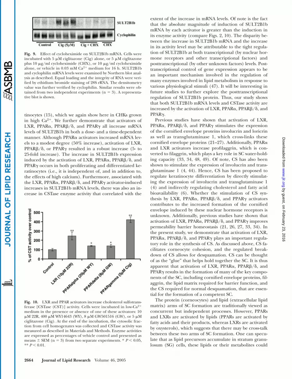
LXR and PPAR activators increase CSTase activityWe next examined the effect of PPAR and LXR activa-
tors on CSTase activity in undifferentiated CHKs. As shownin Fig. 10, activators of LXR, PPAR�, PPAR�/�, and PPAR�all significantly increased CSTase activity. In accordancewith the changes in SULT2B1b mRNA levels, those acti-vators that increase mRNA levels maximally likewise in-crease enzyme activity to the greatest extent. Thus, the in-crease in SULT2B1b mRNA levels, induced by PPAR andLXR activators, correlated with an increase in CSTase en-zyme activity.
DISCUSSION
The SC is composed of corneocytes (bricks) embeddedin an extracellular lipid-rich matrix (mortar). CS is an im-portant lipid in the extracellular lipid matrix, where it
plays a major role in regulating corneocyte desquamation(40). In patients with X-linked ichthyosis, mutations in thesteroid sulfatase gene, the enzyme responsible for the ca-tabolism of CS in the SC, lead to an increase in CS levels(�10-fold increase) in the SC, in parallel with a reductionin rates of corneocyte desquamation (1, 41–43). The ap-parent mechanism that accounts for the CS-induced inhi-bition of corneocyte desquamation is likely an inhibitionof serine protease activity in the SC (44, 45), a generalcharacteristic of CS. In addition to its key role in regulat-ing corneocyte desquamation, CS also has been shown tostimulate keratinocyte differentiation (4), to play a role inantimicrobial defense (46), and to regulate cholesterolsynthesis in keratinocytes (6, 46). Finally, although CSdegradation to cholesterol contributes to the extracellularpool of cholesterol, this source of sterol is not required forthe maintenance of permeability homeostasis (7).
CS is synthesized in the nucleated epidermis by CSTase.Of the three enzymes with CSTase activity, SULT2B1b isthe only isozyme detected in either CHKs or epidermis(12). Because of the multiple roles of CS in epidermalphysiology, the factors that regulate SULT2B1b could beof considerable importance for epidermal homeostasis,but these factors have not yet been completely described.Induction of keratinocyte differentiation by calcium in-creases SULT2B1b expression and CSTase activity in kera-
Fig. 7. LXR and PPAR� activators additively induce SULT2B1bmRNA. Cells were coincubated with or without 22R and ciglitazone(Cig) (5 �M 22R � 2.5 �M ciglitazone) in 0.03 mM Ca2� mediumfor 24 h. SULT2B1b mRNA levels were examined by Northern blotanalysis as described. Equal loading, the integrity of RNA, and thedensitometry value were verified and normalized by ethidium bro-mide staining of 28S rRNA. Data are expressed as percentages ofcontrol and are presented as means � SEM (n 3). * P � 0.05,** P � 0.01.
Fig. 8. Effect of actinomycin D on SULT2B1b mRNA stability.Cells were incubated with 5 �M ciglitazone or vehicle for 24 h andsubsequently challenged with 2 �g/ml actinomycin D for the indi-cated periods of time (0, 2, 4, 6, 8, and 10 h). SULT2B1b mRNA lev-els were examined by Northern blot analysis as described. Equalloading, the integrity of RNA, and the densitometry value were ver-ified and normalized by ethidium bromide staining of 28S rRNA.Similar results were obtained from two independent experiments(n 2). A representative blot is shown above the leaner regressionplot. by guest, on F
ebruary 23, 2016w
ww
.jlr.orgD
ownloaded from
2664 Journal of Lipid Research Volume 46, 2005
tinocytes (15), which we again show here in CHKs grownin high Ca2�. We further demonstrate that activators ofLXR, PPAR�, PPAR�/�, and PPAR� all increase mRNAlevels of SULT2B1b in both a dose- and a time-dependentmanner. Although PPAR� activators increased mRNA lev-els to a modest degree (50% increase), activation of LXR,PPAR�/�, or PPAR� resulted in a robust increase (5- to25-fold increase). The increase in SULT2B1b expressioninduced by the activation of LXR, PPAR�, PPAR�/�, andPPAR� occurs in both proliferating and differentiated ke-ratinocytes (i.e., it is independent of, and in addition to,the effects of high calcium). Furthermore, associated withthe LXR, PPAR�, PPAR�/�, and PPAR� activator-inducedincreases in SULT2B1b mRNA levels, there was also an in-crease in CSTase enzyme activity that correlated with the
extent of the increase in mRNA levels. Of note is the factthat the absolute magnitude of induction of SULT2B1bmRNA by each activator is greater than the induction inits enzyme activity (compare Figs. 2, 10). The disparity be-tween the increase in SULT2B1b mRNA and the increasein its activity level may be attributable to the tight regula-tion of SULT2B1b at both transcriptional (by nuclear hor-mone receptors and other transcriptional factors) andposttranscriptional (by other unknown factors) levels. Post-transcriptional control of gene expression appears to bean important mechanism involved in the regulation ofmany enzymes involved in lipid metabolism in response tovarious physiological stimuli (47). It will be interesting infuture studies to further explore the posttranscriptionalregulation of SULT2B1b protein. Thus, our study showsthat both SULT2B1b mRNA levels and CSTase activity areincreased by the activation of LXR, PPAR�, PPAR�/�, andPPAR�.
Previous studies have shown that activation of LXR,PPAR�, PPAR�/�, and PPAR� stimulates the expressionof the cornified envelope proteins involucrin and loricrinas well as transglutaminase 1, which cross-links thesecornified envelope proteins (21–27). Additionally, PPAR�and LXR activators increase profilaggrin, which is con-verted to filaggrin, which plays a key role in SC water-hold-ing capacity (33, 34, 48, 49). Of note, CS has also beenshown to stimulate the expression of involucrin and trans-glutaminase 1 (4, 44). Hence, CS has been proposed toregulate keratinocyte differentiation by directly stimulat-ing the expression of involucrin and transglutaminase 1(4) and indirectly regulating cholesterol and fatty acidbioavailability (6). Whether the stimulation of CS syn-thesis by LXR, PPAR�, PPAR�/�, and PPAR� activatorscontributes to the increased formation of the cornifiedenvelope induced by these nuclear hormone receptors isunknown. Additionally, previous studies have shown thatactivation of LXR, PPAR�, PPAR�/�, and PPAR� improvespermeability barrier homeostasis (21, 26, 27, 33, 34). Inthe present study, we demonstrate that activation of LXR,PPAR�, PPAR�/�, and PPAR� plays an important regula-tory role in the synthesis of CS. As discussed above, CS fa-cilitates corneocyte cohesion, and the regulated break-down of CS allows for desquamation. CS can be thoughtof as the “glue” that helps hold together the SC. It is thusapparent that activation of LXR, PPAR�, PPAR�/�, andPPAR� results in the formation of many of the key compo-nents of the SC, including cornified envelope proteins, fil-aggrin, the lipid matrix required for barrier function, andthe CS required for normal desquamation, that are essen-tial for the formation of a competent SC.
The protein (corneocytes) and lipid (extracellular lipidmatrix) arms of SC formation are traditionally viewed asconcurrent but independent processes. However, PPARsand LXRs are activated by lipids (PPARs are activated byfatty acids and their products, whereas LXRs are activatedby oxysterols), which suggests that there may be cross-talkbetween these two arms of SC formation. One can specu-late that as lipid precursors accumulate in stratum granu-losum (SG) cells, these lipids or their metabolites could
Fig. 9. Effect of cycloheximide on SULT2B1b mRNA. Cells wereincubated with 5 �M ciglitazone (Cig) alone, or 5 �M ciglitazoneplus 10 �g/ml cycloheximide (CHX), or 10 �g/ml cycloheximidealone, or vehicle in 0.03 mM Ca2� medium for 16 h. SULT2B1band cyclophilin mRNA levels were examined by Northern blot anal-ysis as described. Equal loading and the integrity of RNA were veri-fied by ethidium bromide staining of 28S rRNA. The densitometryvalue was further verified by cyclophilin. Similar results were ob-tained from two independent experiments (n 3). A representa-tive blot is shown.
Fig. 10. LXR and PPAR activators increase cholesterol sulfotrans-ferase [CSTase (CST)] activity. Cells were incubated in low-Ca2�
medium in the presence or absence of one of these activators: 10�M 22R, 400 �M WY14643 (WY), 8 �M GW501516 (GW), or 5 �Mciglitazone (Cig). At the end of the incubation, the cytosolic frac-tion from cell homogenates was collected and CSTase activity wasmeasured as described in Materials and Methods. Enzyme activitiesare expressed as percentages of vehicle control and presented asmeans � SEM (n 3) from two separate experiments. * P � 0.05,** P � 0.01.
by guest, on February 23, 2016
ww
w.jlr.org
Dow
nloaded from

Jiang et al. LXR and PPAR activators stimulate cholesterol sulfotransferase 2665
serve as endogenous activators of these liposensor recep-tors, which, in turn, could stimulate the formation of thevarious components required for SC formation, includ-ing, as shown here, CS.
The mechanism by which activation of LXR, PPAR�,PPAR�/�, and PPAR� increases SULT2B1b expression isunknown. Our study demonstrates that the upregulationof SULT2B1b mRNA by PPAR� activator occurs at thelevel of transcription, because ciglitazone treatment failsto stabilize SULT2B1b mRNA (Fig. 8). Our data also showthat the ciglitazone-induced SULT2B1b mRNA increasedepends on protein synthesis, because coincubation withcycloheximide, a classical protein synthesis inhibitor, re-sults in a complete blockage of the increase in SULT2B1bexpression in these cells (Fig. 9). This finding suggeststhat, rather than directly activating a response element inthe promoter region of SULT2B1b, ciglitazone activationof PPAR� instead may stimulate the production of othertranscription factors, which then activate response ele-ments in the promoter of SULT2B1b. Alternatively, thisresult could indicate that 1) a labile cofactor is required,or 2) an autoinduction of PPAR/LXR is required for theeffects to occur. In searching the promoter sequence ofSULT2B1b (GenBank accession number NM_94315) us-ing the Transcription Element Searching System, we didnot find either a LXR or a PPAR response element at the5� proximal promoter region (�1 to �1,951 bp), furthersupporting our hypothesis that upregulation of SULT2B1bmRNA by ciglitazone and other activators occurs indi-rectly. In previous studies, we have shown that LXR andPPAR� activators increase the expression of involucrinand that this increase is mediated at an AP-1 site in the dis-tal portion of the involucrin promoter (�2,117 to �2,111)(4). Subsequent studies have shown that LXR activatorsincrease the levels of AP-1 factors, particularly Fra-1, butalso Jun D and c-FOS, which leads to increased binding ofAP-1 factors to AP-1 response elements, thereby increas-ing the expression of AP-1-regulated genes (50). Whethersimilar indirect effects of the activation of LXR, PPAR�,PPAR�/�, and PPAR� on AP-1 or other transcription fac-tors account for the increase in SULT2B1b expression re-mains to be determined.
Previous studies have demonstrated that retinoids in-hibit CS production as well as the expected increase inCSTase activity that accompanies keratinocyte differentia-tion (18, 19). This is not surprising because it is well rec-ognized that activation of RAR inhibits keratinocyte dif-ferentiation (51). Depending upon the specific retinoidused, one can activate RAR and/or RXR (many retinoidsactivate both receptors). In many respects, activation ofRAR results in effects that are opposite the effects inducedby activation of PPARs or LXR (RAR activation inhibitsdifferentiation and adversely affects permeability barrierfunction, whereas PPAR and LXR activation stimulates dif-ferentiation and improves permeability barrier homeosta-sis). However, retinoids that predominantly activate RXRalso decrease CSTase activity (51). It is well known thatRXR ligands activate PPAR/RXR and LXR/RXR het-erodimers, so one might expect that RXR ligands would
increase rather than decrease CSTase activity. There are anumber of potential explanations for this paradox. First,RXR ligands activate RXR/RXR homodimers, which candirectly regulate gene expression. Second, RXR formsheterodimers with a large number of nuclear hormone re-ceptors, and it is possible that under certain circumstancesRXR ligands could activate these heterodimers, therebyinhibiting CSTase activity (e.g., activation of RAR/RXRheterodimers). Lastly, retinoids could affect CSTase activ-ity independently of their effects on nuclear hormone re-ceptors. At present, the precise molecular mechanisms bywhich retinoids inhibit and activators of PPARs and LXRincrease CSTase activity are unknown.
In conclusion, this study demonstrates that activation ofLXR, PPAR�, PPAR�/�, and PPAR� stimulates the expres-sion of SULT2B1b and increases CSTase activity in kerati-nocytes. The resulting increase in CS would provide a keyfactor required for the formation of a normal SC.
This study was supported by National Institutes of HealthGrants AR-050629 and AR-39448 and by the Research Serviceof the Department of Veterans Affairs at San Francisco. The au-thors thank Dr. Biao Lu for valuable discussion and Ms. SallyPennypacker for expert assistance with cell culture.
REFERENCES
1. Epstein, E. H., M. L. Williams, and P. M. Elias. 1984. The epider-mal cholesterol sulfate cycle. J. Am. Acad. Dermatol. 10: 866–868.
2. Denning, M. F., M. G. Kazanietz, P. M. Blumberg, and S. H. Yuspa.1995. Cholesterol sulfate activates multiple protein kinase C isoen-zymes and induces granular cell differentiation in cultured mu-rine keratinocytes. Cell Growth Differ. 6: 1619–1626.
3. Downing, D. T., R. W. Dose, and W. Abraham. 1993. Interactionbetween sphingosine and cholesteryl sulfate in epidermal lipids. J.Lipid Res. 34: 563–569.
4. Hanley, K., L. Wood, D. C. Ng, S. S. He, P. Lau, A. Moser, P. M.Elias, D. D. Bikle, M. L. Williams, and K. R. Feingold. 2001. Choles-terol sulfate stimulates involucrin transcription in keratinocytes byincreasing Fra-1, Fra-2, and Jun D. J. Lipid Res. 42: 390–398.
5. Payne, C. D., T. L. Ray, and D. T. Downing. 1996. Cholesterol sul-fate protects Candida albicans from inhibition by sphingosine invitro. J. Invest. Dermatol. 106: 549–552.
6. Williams, M. L., S. L. Rutherford, and K. R. Feingold. 1987. Effectsof cholesterol sulfate on lipid metabolism in cultured human kera-tinocytes and fibroblasts. J. Lipid Res. 28: 955–967.
7. Zettersten, E. M., R. Ghadially, K. R. Feingold, D. Crumrine, andP. M. Elias. 1997. Optimal ratios of topical stratum corneum lipidsimprove barrier recovery in chronologically aged skin. J. Am. Acad.Dermatol. 37: 403–408.
8. Feingold, K. R., M. Q. Man, G. K. Menon, S. S. Cho, B. E. Brown,and P. M. Elias. 1990. Cholesterol synthesis is required for cutane-ous barrier function in mice. J. Clin. Invest. 86: 1738–1745.
9. Lampe, M. A., M. L. Williams, and P. M. Elias. 1983. Human epi-dermal lipids: characterization and modulations during differenti-ation. J. Lipid Res. 24: 131–140.
10. Elias, P. M., M. L. Williams, M. E. Maloney, J. A. Bonifas, B. E.Brown, S. Grayson, and E. H. Epstein, Jr. 1984. Stratum corneumlipids in disorders of cornification. Steroid sulfatase and choles-terol sulfate in normal desquamation and the pathogenesis of re-cessive X-linked ichthyosis. J. Clin. Invest. 74: 1414–1421.
11. Strott, C. A. 2002. Sulfonation and molecular action. Endocr. Rev.23: 703–732.
12. Strott, C. A., and Y. Higashi. 2003. Cholesterol sulfate in humanphysiology: what’s it all about? J. Lipid Res. 44: 1268–1278.
13. Nagata, K., and Y. Yamazoe. 2000. Pharmacogenetics of sulfotrans-ferase. Annu. Rev. Pharmacol. Toxicol. 40: 159–176.
by guest, on February 23, 2016
ww
w.jlr.org
Dow
nloaded from

2666 Journal of Lipid Research Volume 46, 2005
14. Fuda, H., Y. C. Lee, C. Shimizu, N. B. Javitt, and C. A. Strott. 2002.Mutational analysis of human hydroxysteroid sulfotransferaseSULT2B1 isoforms reveals that exon 1B of the SULT2B1 gene pro-duces cholesterol sulfotransferase, whereas exon 1A yields preg-nenolone sulfotransferase. J. Biol. Chem. 277: 36161–36166.
15. Higashi, Y., H. Fuda, H. Yanai, Y. Lee, T. Fukushige, T. Kanzaki,and C. A. Strott. 2004. Expression of cholesterol sulfotransferase(SULT2B1b) in human skin and primary cultures of human epi-dermal keratinocytes. J. Invest. Dermatol. 122: 1207–1213.
16. Hanley, K., Y. Jiang, C. Katagiri, K. R. Feingold, and M. L. Williams.1997. Epidermal steroid sulfatase and cholesterol sulfotransferaseare regulated during late gestation in the fetal rat. J. Invest. Derma-tol. 108: 871–875.
17. Rearick, J. I., P. W. Albro, and A. M. Jetten. 1987. Increase in cho-lesterol sulfotransferase activity during in vitro squamous differen-tiation of rabbit tracheal epithelial cells and its inhibition by reti-noic acid. J. Biol. Chem. 262: 13069–13074.
18. Jetten, A. M., M. A. George, G. R. Pettit, C. L. Herald, and J. I.Rearick. 1989. Action of phorbol esters, bryostatins, and retinoicacid on cholesterol sulfate synthesis: relation to the multistep pro-cess of differentiation in human epidermal keratinocytes. J. Invest.Dermatol. 93: 108–115.
19. Jetten, A. M., M. A. George, C. Nervi, L. R. Boone, and J. I. Rearick.1989. Increased cholesterol sulfate and cholesterol sulfotransferaseactivity in relation to the multi-step process of differentiation in hu-man epidermal keratinocytes. J. Invest. Dermatol. 92: 203–209.
20. Mangelsdorf, D. J., and R. M. Evans. 1995. The RXR heterodimersand orphan receptors. Cell. 83: 841–850.
21. Hanley, K., L. G. Komuves, N. M. Bass, S. S. He, Y. Jiang, D. Crum-rine, R. Appel, M. Friedman, J. Bettencourt, K. Min, et al. 1999.Fetal epidermal differentiation and barrier development in vivo isaccelerated by nuclear hormone receptor activators. J. Invest. Der-matol. 113: 788–795.
22. Komuves, L. G., K. Hanley, Y. Jiang, C. Katagiri, P. M. Elias, M. L.Williams, and K. R. Feingold. 1999. Induction of selected lipidmetabolic enzymes and differentiation-linked structural proteinsby air exposure in fetal rat skin explants. J. Invest. Dermatol. 112:303–309.
23. Westergaard, M., J. Henningsen, M. L. Svendsen, C. Johansen, U. B.Jensen, H. D. Schroder, I. Kratchmarova, R. K. Berge, L. Iversen,L. Bolund, et al. 2001. Modulation of keratinocyte gene expres-sion and differentiation by PPAR-selective ligands and tetradec-ylthioacetic acid. J. Invest. Dermatol. 116: 702–712.
24. Schmuth, M., C. M. Haqq, W. J. Cairns, J. C. Holder, S. Dorsam,S. Chang, P. Lau, A. J. Fowler, G. Chuang, A. H. Moser, et al.2004. Peroxisome proliferator-activated receptor (PPAR)-beta/deltastimulates differentiation and lipid accumulation in keratinocytes.J. Invest. Dermatol. 122: 971–983.
25. Mao-Qiang, M., A. J. Fowler, M. Schmuth, P. Lau, S. Chang, B. E.Brown, A. H. Moser, L. Michalik, B. Desvergne, W. Wahli, et al.2004. Peroxisome-proliferator-activated receptor (PPAR)-gammaactivation stimulates keratinocyte differentiation. J. Invest. Derma-tol. 123: 305–312.
26. Hanley, K., Y. Jiang, S. S. He, M. Friedman, P. M. Elias, D. D. Bikle,M. L. Williams, and K. R. Feingold. 1998. Keratinocyte differentia-tion is stimulated by activators of the nuclear hormone receptorPPARalpha. J. Invest. Dermatol. 110: 368–375.
27. Hanley, K., D. C. Ng, S. S. He, P. Lau, K. Min, P. M. Elias, D. D.Bikle, D. J. Mangelsdorf, M. L. Williams, and K. R. Feingold. 2000.Oxysterols induce differentiation in human keratinocytes and in-crease Ap-1-dependent involucrin transcription. J. Invest. Dermatol.114: 545–553.
28. Schoonjans, K., B. Staels, and J. Auwerx. 1996. Role of the peroxi-some proliferator-activated receptor (PPAR) in mediating the ef-fects of fibrates and fatty acids on gene expression. J. Lipid Res. 37:907–925.
29. Mangelsdorf, D. J., C. Thummel, M. Beato, P. Herrlich, G. Schutz,K. Umesono, B. Blumberg, P. Kastner, M. Mark, P. Chambon, et al.1995. The nuclear receptor superfamily: the second decade. Cell.83: 835–839.
30. Forman, B. M., J. Chen, and R. M. Evans. 1997. Hypolipidemicdrugs, polyunsaturated fatty acids, and eicosanoids are ligands forperoxisome proliferator-activated receptors alpha and delta. Proc.Natl. Acad. Sci. USA. 94: 4312–4317.
31. Janowski, B. A., P. J. Willy, T. R. Devi, J. R. Falck, and D. J. Mangels-
dorf. 1996. An oxysterol signalling pathway mediated by the nu-clear receptor LXR alpha. Nature. 383: 728–731.
32. Willy, P. J., K. Umesono, E. S. Ong, R. M. Evans, R. A. Heyman, andD. J. Mangelsdorf. 1995. LXR, a nuclear receptor that defines adistinct retinoid response pathway. Genes Dev. 9: 1033–1045.
33. Komuves, L. G., K. Hanley, M. Q. Man, P. M. Elias, M. L. Williams,and K. R. Feingold. 2000. Keratinocyte differentiation in hyper-proliferative epidermis: topical application of PPARalpha activa-tors restores tissue homeostasis. J. Invest. Dermatol. 115: 361–367.
34. Komuves, L. G., M. Schmuth, A. J. Fowler, P. M. Elias, K. Hanley,M. Q. Man, A. H. Moser, J. M. Lobaccaro, M. L. Williams, D. J.Mangelsdorf, et al. 2002. Oxysterol stimulation of epidermal dif-ferentiation is mediated by liver X receptor-beta in murine epider-mis. J. Invest. Dermatol. 118: 25–34.
35. Epstein, E. H., Jr., J. M. Bonifas, T. C. Barber, and M. Haynes. 1984.Cholesterol sulfotransferase of newborn mouse epidermis. J. In-vest. Dermatol. 83: 332–335.
36. Rivier, M., I. Safonova, P. Lebrun, C. E. Griffiths, G. Ailhaud, andS. Michel. 1998. Differential expression of peroxisome prolifera-tor-activated receptor subtypes during the differentiation of hu-man keratinocytes. J. Invest. Dermatol. 111: 1116–1121.
37. Segaert, S., M. Garmyn, H. Degreef, and R. Bouillon. 2000. Sup-pression of vitamin D receptor and induction of retinoid X recep-tor alpha expression during squamous differentiation of culturedkeratinocytes. J. Invest. Dermatol. 114: 494–501.
38. Szanto, A., S. Benko, I. Szatmari, B. L. Balint, I. Furtos, R. Ruhl, S.Molnar, L. Csiba, R. Garuti, S. Calandra, et al. 2004. Transcrip-tional regulation of human CYP27 integrates retinoid, peroxisomeproliferator-activated receptor, and liver X receptor signaling inmacrophages. Mol. Cell. Biol. 24: 8154–8166.
39. Li, A. C., and C. K. Glass. 2004. PPAR- and LXR-dependent path-ways controlling lipid metabolism and the development of athero-sclerosis. J. Lipid Res. 45: 2161–2173.
40. Long, S. A., P. W. Wertz, J. S. Strauss, and D. T. Downing. 1985. Hu-man stratum corneum polar lipids and desquamation. Arch. Derma-tol. Res. 277: 284–287.
41. Marinkovic-Ilsen, A., J. G. Koppe, A. C. Jobsis, and W. P. de Groot.1978. Enzymatic basis of typical X-linked ichthyosis. Lancet. 2:1097.
42. Shapiro, L. J., R. Weiss, M. M. Buxman, J. Vidgoff, R. L. Dimond,J. A. Roller, and R. S. Wells. 1978. Enzymatic basis of typical X-linkedicthyosis. Lancet. 2: 756–757.
43. Webster, D., J. T. France, L. J. Shapiro, and R. Weiss. 1978.X-linked ichthyosis due to steroid-sulphatase deficiency. Lancet. 1:70–72.
44. Kawabe, S., T. Ikuta, M. Ohba, K. Chida, E. Ueda, K. Yamanishi,and T. Kuroki. 1998. Cholesterol sulfate activates transcription oftransglutaminase 1 gene in normal human keratinocytes. J. Invest.Dermatol. 111: 1098–1102.
45. Ohba, M., K. Ishino, M. Kashiwagi, S. Kawabe, K. Chida, N. H. Huh,and T. Kuroki. 1998. Induction of differentiation in normal humankeratinocytes by adenovirus-mediated introduction of the eta anddelta isoforms of protein kinase C. Mol. Cell. Biol. 18: 5199–5207.
46. Bibel, D. J., R. Aly, and H. R. Shinefield. 1992. Antimicrobial activ-ity of sphingosines. J. Invest. Dermatol. 98: 269–273.
47. Yu, Y. H., Y. Zhang, P. Oelkers, S. L. Sturley, D. J. Rader, and H. N.Ginsberg. 2002. Posttranscriptional control of the expression andfunction of diacylglycerol acyltransferase-1 in mouse adipocytes. J.Biol. Chem. 277: 50876–50884.
48. Komuves, L. G., K. Hanley, A. M. Lefebvre, M. Q. Man, D. C. Ng,D. D. Bikle, M. L. Williams, P. M. Elias, J. Auwerx, and K. R. Fein-gold. 2000. Stimulation of PPARalpha promotes epidermal kerati-nocyte differentiation in vivo. J. Invest. Dermatol. 115: 353–360.
49. Hanley, K., L. G. Komuves, D. C. Ng, K. Schoonjans, S. S. He, P.Lau, D. D. Bikle, M. L. Williams, P. M. Elias, J. Auwerx, et al. 2000.Farnesol stimulates differentiation in epidermal keratinocytes viaPPARalpha. J. Biol. Chem. 275: 11484–11491.
50. Schmuth, M., P. M. Elias, K. Hanley, P. Lau, A. Moser, T. M. Will-son, D. D. Bikle, and K. R. Feingold. 2004. The effect of LXR acti-vators on AP-1 proteins in keratinocytes. J. Invest. Dermatol. 123:41–48.
51. Rearick, J. I., and A. M. Jetten. 1986. Accumulation of cholesterol3-sulfate during in vitro squamous differentiation of rabbit tra-cheal epithelial cells and its regulation by retinoids. J. Biol. Chem.261: 13898–13904.
by guest, on February 23, 2016
ww
w.jlr.org
Dow
nloaded from