long-range regulatory interactions at the 4q25 atrial fibrillation risk locus involve pitx2c and...
TRANSCRIPT

Aguirre et al. BMC Biology (2015) 13:26 DOI 10.1186/s12915-015-0138-0
RESEARCH ARTICLE Open Access
Long-range regulatory interactions at the 4q25atrial fibrillation risk locus involve PITX2c andENPEPLuis A Aguirre1, M Eva Alonso1, Claudio Badía-Careaga1, Isabel Rollán1, Cristina Arias1, Ana Fernández-Miñán2,Elena López-Jiménez1, Amelia Aránega3, José Luis Gómez-Skarmeta2, Diego Franco3 and Miguel Manzanares1*
Abstract
Background: Recent genome-wide association studies have uncovered genomic loci that underlie an increased riskfor atrial fibrillation, the major cardiac arrhythmia in humans. The most significant locus is located in a gene desertat 4q25, approximately 170 kilobases upstream of PITX2, which codes for a transcription factor involved in embryonicleft-right asymmetry and cardiac development. However, how this genomic region functionally and structurally relatesto PITX2 and atrial fibrillation is unknown.
Results: To characterise its function, we tested genomic fragments from 4q25 for transcriptional activity in a mouseatrial cardiomyocyte cell line and in transgenic mouse embryos, identifying a non-tissue-specific potentiator regulatoryelement. Chromosome conformation capture revealed that this region physically interacts with the promoter ofthe cardiac specific isoform of Pitx2. Surprisingly, this regulatory region also interacts with the promoter of the nextneighbouring gene, Enpep, which we show to be expressed in regions of the developing mouse heart essential forcardiac electrical activity.
Conclusions: Our data suggest that de-regulation of both PITX2 and ENPEP could contribute to an increased riskof atrial fibrillation in carriers of disease-associated variants, and show the challenges that we face in the functionalanalysis of genome-wide disease associations.
Keywords: Atrial fibrillation, Chromosome conformation, ENPEP, PITX2, Regulatory element
BackgroundGenome-wide association studies (GWAS) have expo-nentially increased our knowledge of the genetic com-ponent of human disorders, revealing unsuspected locithat harbour variants linked to an increased risk ofdisease [1]. However, the majority of GWAS signals fallin non-coding regions of the genome, which has madetheir functional analysis particularly challenging [2,3].Even the identification of the genes targeted by disease-associated variants is not straightforward, as mereproximity can result in incorrect identification of theculprit gene [4].
* Correspondence: [email protected] Nacional de Investigaciones Cardiovasculares (CNIC), MelchorFernández Almagro 3, 28029 Madrid, SpainFull list of author information is available at the end of the article
© 2015 Aguirre et al.; licensee BioMed CentralCommons Attribution License (http://creativecreproduction in any medium, provided the orDedication waiver (http://creativecommons.orunless otherwise stated.
Atrial fibrillation (AF) is the most common cardiacarrhythmia in humans [5] although its pathophysiologicbasis is still not clearly understood, presenting a challengefor cardiovascular research and therapy. AF is defined as asupraventricular tachyarrhythmia characterised by un-coordinated atrial activation, and is frequently observedas a consequence of various systemic and cardiac disor-ders (syndromic AF) [6]. However, in 10% to 20% of casesAF is not associated with other cardiovascular disease, andthus is dubbed ‘idiopathic’ or ‘lone’ AF that mostly occursin patients under 60. The strong association of AF onsetwith risk factors, such as age, sex, ethnicity, hypertensionand other heart diseases [7], originally suggested it beinga non-genetic disorder [8]. Nevertheless, in the last twodecades several epidemiological studies pointed to a sig-nificant incidence of genetic factors [9]. Furthermore, rare
. This is an Open Access article distributed under the terms of the Creativeommons.org/licenses/by/4.0), which permits unrestricted use, distribution, andiginal work is properly credited. The Creative Commons Public Domaing/publicdomain/zero/1.0/) applies to the data made available in this article,

Aguirre et al. BMC Biology (2015) 13:26 Page 2 of 13
mutations in a dozen genes, mostly encoding ion channelsubunits [8], are associated with AF as part of widercardiac electrical syndromes.At least ten loci have been linked to AF by GWAS in
large cohorts of non-related patients of distinct ethnicbackgrounds [10-12]. The most highly AF associatedvariants identified in all studies are located on chromo-some 4q25 [11], 170 kilobases (kb) distal to PITX2 andwithin a 1.5 megabases (Mb) intergenic gene desert.PITX2 encodes an evolutionarily conserved homeodo-main transcription factor that is involved in the estab-lishment of left-right asymmetry and cardiovasculardevelopment in the vertebrate embryo. In mice andhumans the PITX2 gene generates several isoforms.PITX2a and PITX2b are alternative splicing variantsproduced from a common promoter, whereas PITX2c isthe product of an alternative promoter and is the mainisoform expressed in the heart [13].Mouse Pitx2c is first expressed in the left lateral plate
mesoderm of the early embryo, as part of the networkregulating the establishment of left-right asymmetryduring development [14], and is then expressed in theleft side of the heart at early stages [15]. At later stages,expression follows a dynamic pattern, being present inthe left atrium, the myocardium sleeves of the pulmon-ary veins, the atrio-ventricular cushions or the base ofthe ventricles [16,17]. Germline homozygous deletion ofPitx2 results in embryonic lethality and numerouscardiac malformations, such as right atrial isomerismand outflow tract defects, varying from double outletright ventricle or transposition of the great arteries topersistent truncus arteriosus [18]. More recent researchshowed that Pitx2c is expressed in adult mice and humanhearts, predominantly in the left atrium (levels in the rightatrium and the ventricles are 100-fold lower), and that itslevels of expression decrease in atria of AF patients[13,19]. Furthermore, Pitx2c heterozygous or atrial specificdeletion of Pitx2c display molecular and physiological hall-marks of human AF [13,19,20], which also is observedwhen Pitx2 is deleted in adult mice [21]. Altogether, thesedata support the hypothesis that PITX2 could play a causalrole in the pathogenesis of AF and that its function couldbe altered by genomic elements located in the vicinity ofthe single nucleotide polymorphisms (SNP) in 4q25 thatcorrelate with of AF.In this study, we explored whether the 4q25 region
spanning the AF-associated variants identified by GWASharbours putative regulatory elements that could beacting on neighbouring genes. By tissue culture, in vivotransgenics and analysis of chromatin structure, we havefound that this region contains potentiator cis-regulatoryelements that interact with the promoters of Pitx2c and,unexpectedly, Enpep, the next gene located downstreamof Pitx2. Given the expression of Enpep in the sinoatrial
node (SAN) and the co-expression of Pitx2 and Enpepin pro-arrhythmogenic regions of the embryonic heart,such as the sleeves of the pulmonary veins, our datasuggest that de-regulation of these genes could underlieincreased risk of AF.
ResultsGenomic analysis of the 4q25 AF-associated locusTo identify putative cis-regulatory elements located in the4q25 region we analysed the evolutionary conservation[22] and distribution of histone modifications associatedwith active elements [23] (H3K4me1) in an 85 kb windowcontaining the main SNPs that have been associated withan increased risk of AF by GWAS (Figure 1). This windowis centred on the lead rs2200733 SNP [11] that has repeat-edly been identified as the most significant variant asso-ciated with AF [10-12,24-26], and spans a region thatincludes other distal SNPs (rs2634073 and rs17570669)that lie in the proximity of sequences highly conservedbetween human and mouse (Figure 1A). The region in-cluding the majority of AF-associated SNPs in 4q25 isconfined to a linkage disequilibrium (LD) block [11],separated from that containing the coding exons of thegene and from adjacent LD blocks in the 1.5 Mb genedesert located centromeric to PITX2 (Figure 1A). It isalso noteworthy that the selected SNPs and the PITX2gene are contained together in a single topologically as-sociated domain (TAD; Additional file 1A), as definedby Hi-C in three different human cell lines [27].We selected seven genomic fragments (AF1-7) for
further analysis (Figure 1B). Fragments AF1 and AF2 liein close proximity to rs2634073 and show high evolu-tionary conservation. These fragments are included in aregion (hs930) tested as part of a large scale screen fortissue specific human enhancers by transgenesis in themouse embryo [28] and drive reporter expression in thenervous system and limbs but not in the heart. Thisregion was also tested in transgenic zebrafish, drivingexpression in similar patterns but again not in the devel-oping heart [29]. AF3 to AF5 are a set of overlappingfragments that include the lead rs2200733 variant andother highly associated SNPs, in a region with high con-servation among placental mammals and H3K4me1marks of active regulatory elements. Finally, AF6 andAF7 map to a region conserved in vertebrates includingrs17570669, which has been associated with AF but isindependent of rs2200733 [12].
The 4q25 AF-associated locus contains active regulatoryelementsWe tested the regulatory activity of these fragments, cor-responding to the none-risk haplotype at rs2200733 froma commercial source of human DNA, by linking themto a human minimal beta-globin promoter [30] and the
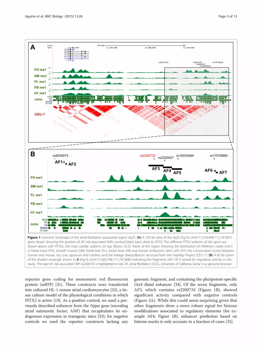
Figure 1 Genomic landscape of the atrial-fibrillation associated region 4q25. (A) A 230 kb view of the 4q25 (hg19; chr4:111,516,448-111,747,857)gene desert, showing the position of AF risk-associated SNPs (vertical black bars) distal to PITX2. The different PITX2 isoforms of the gene areshown above with PITX2c, the main cardiac isoform, on top. Below, UCSC tracks of the region showing the distribution of H3K4me1 marks (me1)in foetal heart (FH), smooth muscle (SM), foetal liver (FL), foetal brain (FB) and human embryonic stem cells (H1); the conservation (cons) betweenhuman and mouse, rat, cow, opossum and chicken; and the linkage disequilibrium structure from the HapMap Project (CEU r2). (B) A 85 kb zoomof the shaded rectangle shown in A (hg19; chr4:111,662,786-111,747,668) indicating the fragments (AF1-AF7) tested for regulatory activity in thisstudy. The lead AF risk associated SNP rs2200733 is highlighted in red. AF, atrial fibrillation; UCSC, University of California Santa Cruz genome browser.
Aguirre et al. BMC Biology (2015) 13:26 Page 3 of 13
reporter gene coding for monomeric red fluorescentprotein (mRFP) [31]. These constructs were transfectedinto cultured HL-1 mouse atrial cardiomyocytes [32], a tis-sue culture model of the physiological conditions in whichPITX2 is active [19]. As a positive control, we used a pre-viously described enhancer from the Nppa gene (encodingatrial natriuretic factor; ANF) that recapitulates its en-dogenous expression in transgenic mice [33]; for negativecontrols we used the reporter constructs lacking any
genomic fragment, and containing the pluripotent-specificOct4 distal enhancer [34]. Of the seven fragments, onlyAF3, which contains rs2200733 (Figure 1B), showedsignificant activity compared with negative controls(Figure 2A). While this could seem surprising given thatother fragments show a more robust signal for histonemodifications associated to regulatory elements (for ex-ample AF4, Figure 1B), enhancer prediction based onhistone marks is only accurate in a fraction of cases [35].
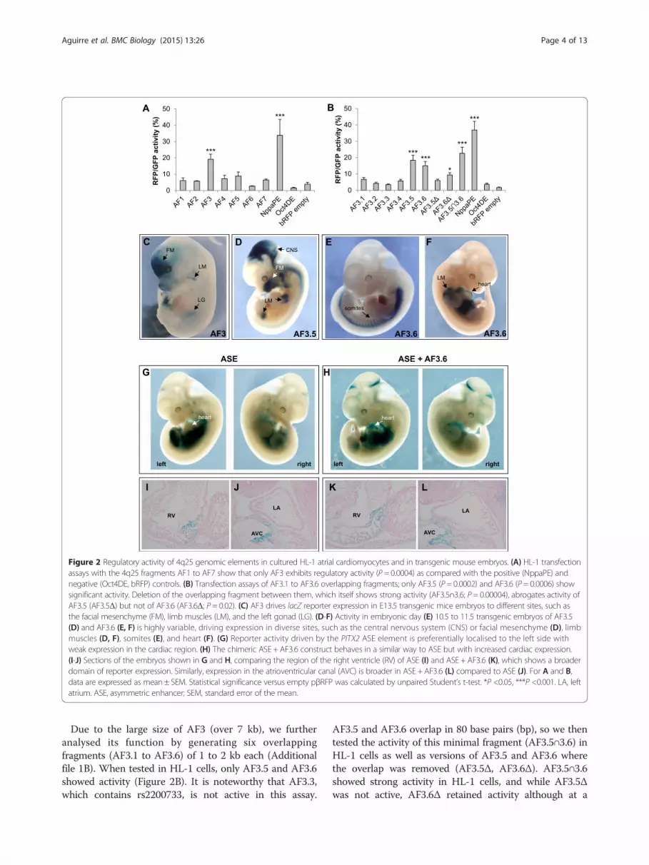
Figure 2 Regulatory activity of 4q25 genomic elements in cultured HL-1 atrial cardiomyocytes and in transgenic mouse embryos. (A) HL-1 transfectionassays with the 4q25 fragments AF1 to AF7 show that only AF3 exhibits regulatory activity (P = 0.0004) as compared with the positive (NppaPE) andnegative (Oct4DE, bRFP) controls. (B) Transfection assays of AF3.1 to AF3.6 overlapping fragments; only AF3.5 (P = 0.0002) and AF3.6 (P = 0.0006) showsignificant activity. Deletion of the overlapping fragment between them, which itself shows strong activity (AF3.5∩3.6; P = 0.00004), abrogates activity ofAF3.5 (AF3.5Δ) but not of AF3.6 (AF3.6Δ; P = 0.02). (C) AF3 drives lacZ reporter expression in E13.5 transgenic mice embryos to different sites, such asthe facial mesenchyme (FM), limb muscles (LM), and the left gonad (LG). (D-F) Activity in embryonic day (E) 10.5 to 11.5 transgenic embryos of AF3.5(D) and AF3.6 (E, F) is highly variable, driving expression in diverse sites, such as the central nervous system (CNS) or facial mesenchyme (D), limbmuscles (D, F), somites (E), and heart (F). (G) Reporter activity driven by the PITX2 ASE element is preferentially localised to the left side withweak expression in the cardiac region. (H) The chimeric ASE + AF3.6 construct behaves in a similar way to ASE but with increased cardiac expression.(I-J) Sections of the embryos shown in G and H, comparing the region of the right ventricle (RV) of ASE (I) and ASE + AF3.6 (K), which shows a broaderdomain of reporter expression. Similarly, expression in the atrioventricular canal (AVC) is broader in ASE + AF3.6 (L) compared to ASE (J). For A and B,data are expressed as mean ± SEM. Statistical significance versus empty pβRFP was calculated by unpaired Student’s t-test. *P <0.05, ***P <0.001. LA, leftatrium. ASE, asymmetric enhancer; SEM, standard error of the mean.
Aguirre et al. BMC Biology (2015) 13:26 Page 4 of 13
Due to the large size of AF3 (over 7 kb), we furtheranalysed its function by generating six overlappingfragments (AF3.1 to AF3.6) of 1 to 2 kb each (Additionalfile 1B). When tested in HL-1 cells, only AF3.5 and AF3.6showed activity (Figure 2B). It is noteworthy that AF3.3,which contains rs2200733, is not active in this assay.
AF3.5 and AF3.6 overlap in 80 base pairs (bp), so we thentested the activity of this minimal fragment (AF3.5∩3.6) inHL-1 cells as well as versions of AF3.5 and AF3.6 wherethe overlap was removed (AF3.5Δ, AF3.6Δ). AF3.5∩3.6showed strong activity in HL-1 cells, and while AF3.5Δwas not active, AF3.6Δ retained activity although at a

Aguirre et al. BMC Biology (2015) 13:26 Page 5 of 13
reduced level (Figure 2B). It is interesting to note thatthis 80 bp minimal fragment is highly conserved be-tween human and mouse, and that its sequence corre-sponds to a short interspersed nuclear element of theMIR3 family. It has been shown that repeat sequences canact as enhancers in experimental assays [36], althoughtheir putative function in vivo is still under debate [37].Therefore, we can conclude that the regulatory activity ofAF3 in this assay is located in AF3.6, and that additionalactivity may be present in the overlapping fragment ofAF3.5 and AF3.6.Next, we assayed the activity of fragments AF3-5 and
AF7 in transgenic mouse embryos, using the lacZ geneas a reporter. Again, only AF3 showed enhancer activity(Figure 2C, Additional file 2), confirming the results ofthe tissue culture assays. AF3 drives reporter expressionin facial mesenchyme, limb muscles, and the left gonad,some of which are sites of expression of endogenousPitx2 [14,38]. Contrary to expectations, AF3 did notdrive expression in the developing heart. We reasonedthat regulatory elements from PITX2 underlying theassociation with AF might not be active during develop-ment, and instead drive cardiac-specific expression ofPITX2 in the adult. We therefore generated transgenicmice and examined reporter expression in the heart atpostnatal day 3. Again, we found no expression incardiac tissues (Additional file 2).When tested in mouse transgenic assays, both AF3.5
and AF3.6 showed activity (Figure 2D to F). Sites ofexpression include facial mesenchyme, limb muscles,somites, or pericardium, but we did not observe a repro-ducible pattern driven by these fragments. We can ruleout the possibility that this heterogeneity is due to non-specific reporter activation as a consequence of integra-tion site of the transgene, because of the very low per-centage of lacZ positive embryos (out of the totalnumber of transgenics as assessed by genotyping) ob-tained for genomic fragments tested showing no activity(0% to 5%) as compared to those that do (20% to 45%;Additional file 2).We also tested activity in transgenicembryos of the minimal AF3.5∩3.6 fragment, findingthat it was not active (1 weak lacZ+ embryo out of 17transgenics; Additional file 2).
The 4q25 regulatory elements show non-specificpotentiator activityThe above results suggest that these 4q25 elements,while they have regulatory potential, do not confer tissuespecificity. To test this hypothesis further, we transfectedfragments AF3, AF3.5 and AF3.6 into two cell types unre-lated to the cardiac lineage: the mouse teratocarcinoma-derived pluripotent cell line P19 and human embryonickidney (HEK) cells. We found that all three fragmentswere active in both cell types, closely matching the degree
of activation in HL-1 cells (Additional file 3). As expected,the Oct4-DE was active in P19 but not in HEK cells; incontrast, the Nppa enhancer was not active in P19 butshowed activity in HEK cells, as expected given theendogenous expression of NPPA in human kidney [39].Overall, our results suggest that the regulatory elementsdetected in 4q25 do not act as cell type-specificenhancers, but rather as accessory elements that canpotentiate the activity of tissue-specific enhancerslocated elsewhere in the locus.To further prove the putative potentiator activity of
4q25 elements, we assessed the effect of AF3.6 on theactivity of a previously identified intronic enhancer fromPitx2, which drives left-sided expression in the embryo[40]. This asymmetric enhancer (ASE) is evolutionarilyconserved in sequence and function, but it is noteworthythat the ASE from human PITX2 only drives weak ex-pression in the mice heart compared with its mousehomologue [41]. We generated a chimeric constructcontaining both human AF3.6 and ASE and comparedits activity to that of ASE alone in transgenic mouseembryos at 10.5 (Figure 2G-L). We first observed thatwhen using the chimeric ASE + AF3.6 construct, thevariability associated with AF3.6 alone is lost, and allembryos show the characteristic left-sided expressiondescribed for the ASE (Figure 2G, H). Importantly, wefound that there is no additive effect of both genomicfragments as that observed when placing together differ-ent enhancers in the same transgenic construct [42]. Infact, we observed that adding AF3.6 to the ASE appar-ently increased the levels of reporter expression in thecardiac region (Figure 2G, H), as seen in sections wheredomains of reporter activity in the right ventricle and inthe atrio-ventricular canal are expanded in ASE + AF3.6embryos compared to ASE (Figure 2I-L). When weexamined in detail reporter expression in the developingheart for all transgenic embryos, we found that AF3.6increases the number of embryos expressing lacZ in theleft atrium (two out of five for ASE, as compared toseven out of ten for ASE + AF3.6; Additional file 4). Theresults of these assays further suggest that 4q25elements have an accessory role in defining PITX2expression acting in conjunction with other regulatoryelements.
The three-dimensional architecture of the Pitx2 locusidentifies promoter-specific long range interactionsAlthough the above evidence shows that 4q25 includesregulatory elements, there is no direct evidence that thisgenomic region acts on PITX2 or that, if it does, it showsany specificity regarding the cardiac and non-cardiac iso-forms produced from two alternative promoters. To an-swer these questions, we analysed the three-dimensionalorganisation of the locus by chromosome conformation
Aguirre et al. BMC Biology (2015) 13:26 Page 6 of 13
capture (3C) [43,44], to address if these regions physicallycontact the promoters of PITX2. Because we aimed to per-form the assays in the physiological context of the heart,we selected the mouse genome region syntenic to human4q25 and analysed chromatin from the atria and ventricles
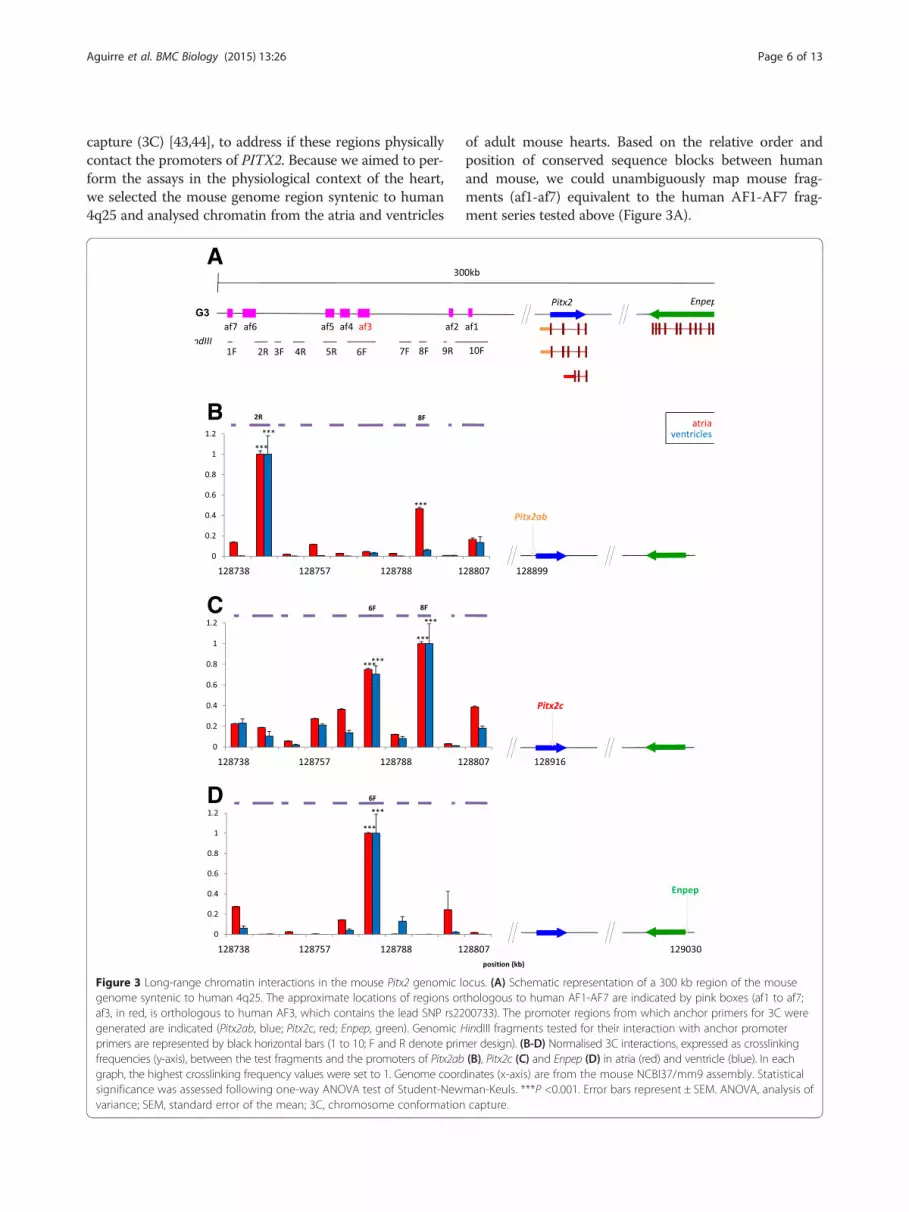
Figure 3 Long-range chromatin interactions in the mouse Pitx2 genomic lgenome syntenic to human 4q25. The approximate locations of regions oraf3, in red, is orthologous to human AF3, which contains the lead SNP rs22generated are indicated (Pitx2ab, blue; Pitx2c, red; Enpep, green). Genomic Hprimers are represented by black horizontal bars (1 to 10; F and R denote primfrequencies (y-axis), between the test fragments and the promoters of Pitx2abgraph, the highest crosslinking frequency values were set to 1. Genome coordsignificance was assessed following one-way ANOVA test of Student-Newvariance; SEM, standard error of the mean; 3C, chromosome conformation
of adult mouse hearts. Based on the relative order andposition of conserved sequence blocks between humanand mouse, we could unambiguously map mouse frag-ments (af1-af7) equivalent to the human AF1-AF7 frag-ment series tested above (Figure 3A).
ocus. (A) Schematic representation of a 300 kb region of the mousethologous to human AF1-AF7 are indicated by pink boxes (af1 to af7;00733). The promoter regions from which anchor primers for 3C wereindIII fragments tested for their interaction with anchor promoterer design). (B-D) Normalised 3C interactions, expressed as crosslinking(B), Pitx2c (C) and Enpep (D) in atria (red) and ventricle (blue). In eachinates (x-axis) are from the mouse NCBI37/mm9 assembly. Statisticalman-Keuls. ***P <0.001. Error bars represent ± SEM. ANOVA, analysis ofcapture.
Aguirre et al. BMC Biology (2015) 13:26 Page 7 of 13
We probed the interaction of HindIII restriction frag-ments containing the Pitx2a,b or Pitx2c promoter withten fragments spanning over 100 kb of the distal regionon mouse chromosome 3 syntenic to the AF-associated4q25 locus in humans (Figure 3A). Using atria and ven-tricles from adult mice we observed a clear pattern oflong range interactions, with regions interacting specific-ally with the Pitx2a,b promoter (fragment 2R; Figure 3B),the Pitx2c promoter (fragment 6F; Figure 3C), or with both(fragment 8F; Figure 3B, C). The latter result prompted usto ask if fragment 8F had regulatory activity in HL-1 cells,since it is not included in any of the previously testedfragments. This was not the case, suggesting that thisgenomic region has other architectural roles in config-uring the regulatory landscape of Pitx2. To furtherexamine the specificity of chromatin interaction be-tween the AF-associated region and Pitx2c, we checkedthe interaction of a fragment containing the promoterof Enpep, the next neighbouring gene distal to Pitx2 inboth mouse and humans (Figure 3A). To our surprise,we found a robust interaction between fragment 6F andEnpep (Figure 3D), suggesting that the 4q25 regulatorylandscape is partially shared between PITX2 and ENPEP.The specificity of the interactions of fragments 2R, 6F and8F with Pitx2ab, Pitx2c and Enpep was tested by using aseries of control primers located upstream and down-stream of the promoters, which showed no interactions(Additional file 5).We next asked if the interactions we observed showed
regional differences in atria, as AF constitutes a disorderof the left atrium and this is the region where PITX2 isprominently expressed [13]. While fragment 2R inter-acted specifically with the Pitx2ab promoter in both leftand right atrium, fragment 6F interacts only in leftatrium with Pitx2c and Enpep. On the other hand, frag-ment 8F shows interaction with Pitx2c in both atria butonly in the right atrium with Enpep (Figure 4A). Giventhe fact that the promoters of Pitx2c and Enpep sharemany of the interactions tested, we examined if theywere physically associated and if this was region-specific.We found a robust and specific promoter-promoterinteraction in both atria and in ventricles (Figure 4B),therefore independent of transcription and in line with re-cent observations on the role of pre-existing promoter-promoter interactions for structuring the genome [45].It should be noted that fragment 6F contains the
region conserved with human fragment AF3, thereby sug-gesting that the region with regulatory activity and thatcontains the lead SNP associated with AF (rs2200733) in-teracts in a specific manner with the promoters of thecardiac-specific isoform of Pitx2 and the neighbouringgene, Enpep. The 3C analysis of the mouse Pitx2/Enpeplocus thus revealed an unexpected complexity of specificand shared chromatin interactions between the regions
containing the potentiator elements and the different pro-moters studied that could be related to their function.
Enpep is expressed in arrhythmogenic sites in theembryonic heartENPEP encodes aminopeptidase A, which cleaves angio-tensin II to produce angiotensin III as part of the renin-angiotensin system [46]. Therefore ENPEP is involvedin the control of blood pressure, and accordingly it isexpressed in the renal system and endothelial cells, andknockout mice for Enpep develop hypertension [47].However, at present there is no report for expression ora role of Enpep in the heart. In light of our results, weexamined the expression of Enpep in the E14.5 mouseembryos by in situ hybridization on tissue sections(Figure 5). Enpep is strongly expressed in the endo-thelial lining of the lungs, but also in a specific and re-stricted pattern in the developing heart (Figure 5A, D).We compared Enpep expression with that of Pitx2(Figure 5B, E) and Hcn4 (Figure 5C, F), which encodes avoltage-gated ion channel and at this stage is a markerof most of the cardiac conduction system [48,49]. Thisanalysis showed that Enpep is co-expressed with Pitx2in the pulmonary veins but not in the myocardium ofthe left atrium (Figure 5A, B, D, E), and is co-expressedwith Hcn4 in the left and right superior venae cavae andin the SAN (Figure 5A, C, D, F). Enpep is thus expressedin the embryonic mouse heart in key components of thecardiac conduction system such as the SAN. Moreover,Enpep is also expressed at the base of the pulmonaryveins and the junction of the caval veins, regions proneto initiate ectopic electrical beats, which lead in manycases to the onset of AF [7].
DiscussionThe advent of GWAS has radically changed our pers-pective on the genetic analysis of common diseases inhumans. On the one hand, a plethora of novel locilinked to increased disease risk have been uncovered,which await further analysis before possible translationto the clinic [2]. On the other hand, the vast majority ofrisk variants are located in non-coding genomic se-quences, pointing to a fundamental role for variation incis-regulatory elements as the basis of common diseases[50,51]. Understanding the role and function of thesegenomic elements will be fundamental to making themost of the discoveries of GWAS.The genomic analysis of AF is a prime example in this
regard. All GWAS carried out to date have shown thatthe major loci for AF lie in an intergenic gene desert in4q25, located distal to the developmental regulatorPITX2 [10-12]. Despite its early role in establishing theleft-right patterning of the heart and its prominent ex-pression in the left atrium, no evidence suggested a role

Figure 4 Differential chromatin interactions of Pitx2 and Enpep in left and right atrium. (A) Normalised 3C interactions, expressed as crosslinkingfrequencies (y-axis), between fragments 2R, 6F and 8F, and the promoters of Pitx2ab, Pitx2c and Enpep, in left (red) and right (yellow) atrium. (B)3C interaction between the Pitx2c and Enpep promoters, including control regions upstream (c4) and downstream (c5) of Enpep (see Additionalfile 5), in left (red) and right (yellow) atrium, as well as in ventricles (blue). In each graph, the highest crosslinking frequency values were set to 1.Statistical significance was assessed following one-way ANOVA test of Student-Newman-Keuls. *P <0.05, **P <0.01, ***P <0.001. Error bars represent ±SEM. ANOVA, analysis of variance; SEM, standard error of the mean; 3C, chromosome conformation capture.
Aguirre et al. BMC Biology (2015) 13:26 Page 8 of 13
for PITX2 in the pathophysiology of AF [9]. Subsequentanalysis of loss-of-function mouse models of Pitx2 haveconfirmed that it plays a pivotal role in regulating differ-ent atrial phenotypes by distinguishing electrical fromworking myocardium in the right and left atria respect-ively [13,19,20]. However, no reports to date have providedevidence as to how distal variants in 4q25 act on PITX2.Even more surprisingly, a recent report showed that 4q25variants do not correlate with PITX2 expression in atrialtissue from human patients [52]. This evidences that vari-ants identified by GWAS may have extremely subtle ef-fects, which fall below the threshold of detection ofcurrent analytical tools and approaches.Our analysis of the regulatory structure of the 4q25
locus shows that genomic sequences in close proximity toAF-linked variants can act as transcriptional regulatory
elements both in tissue culture and in mouse embryos.Rather unexpectedly, and contrasting with other cases ofGWAS-related enhancers in the cardiovascular system[53,54], these elements are not specific to cardiac celltypes, either in culture or in vivo. The 4q25 elements showequal activity when transfected into cell types of differentorigin. Furthermore, in transgenic mouse embryos theseelements drive highly variable patterns of reporter expres-sion. These results suggest that the cis-regulatory elementsin 4q25 do not act as classical tissue-specific enhancers,but as potentiator elements that would act in cooperationwith elements located elsewhere in the locus that dictatetissue restricted expression. In the case of mouse Pitx2, anintronic enhancer (ASE) has been described that drivesleft-side specific expression in the early embryo and laterin the heart, liver and other organs [41]. It is conceivable

Figure 5 Enpep is expressed in the embryonic mouse heart. (A to F) Expression of Enpep (A, D), Pitx2 (B, E) and Hcn4 (C, F) in E14.5 mouseembryos shown by in situ hybridization on two sets of consecutive sections (A to C and D to F). In addition to strong expression in the endotheliallining of the lungs, Enpep is expressed in a restricted pattern in the heart (A, D), where it is co-expressed with Pitx2 in the pulmonary veins (PV; zoomin A and B) and with Hcn4 in the leaflet of the venous valve (VV) and left superior vena cava (LSVC; zoom in A and C) as well as in the right superiorvena cava (RSVC) and sinoatrial node (SAN; zoom in D and F). It is noteworthy that Enpep is not expressed in the myocardium of the left atria (LA), as isPitx2 (zoom in B and E). AVN-His, atrial ventricular node-bundle of His; RA, right atria. Scale bars, 1 mm; close-ups, 200 μm. E, embryonic day.
Aguirre et al. BMC Biology (2015) 13:26 Page 9 of 13
that precise control of spatial and quantitative expressionof PITX2 requires interplay of the ASE and the 4q25potentiator. In fact, when placed together and tested bytransgenics, the variability of 4q25 elements is lost and itcan modulate the activity of ASE. It is possible that thispotentiator could also modulate the activity of other yet tobe identified regulatory elements from the locus.By analysing the physical interaction between promoters
and intergenic sequences of the mouse region syntenic to4q25, we have found a further degree of complexity in thechromatin structure of the region. First, there is a clearspecificity in the interaction of distal elements with thealternative promoters of the different Pitx2 isoforms,despite their being separated by less than 10 kb. In thisregard, it is noteworthy that the region containing the po-tentiator activity we have described interacts specificallywith the promoter of the cardiac-specific Pitx2c isoform.Furthermore, this interaction occurs specifically in the leftatrium. These results provide additional support to thespecific role of the region identified by GWAS in regulat-ing PITX2 in the heart.
More surprising was the fact that this same region phys-ically interacts with the promoter of Enpep, the neighbour-ing gene located distal to Pitx2. This opens the possibilitythat Enpep could also be a transcriptional target of theidentified cis-regulatory elements. ENPEP, as part of therenin-angiotensin system, has been shown to control bloodpressure, and hypertension is a known risk factor for AF[55]. However, 4q25 variants are associated with lone AF,with no co-occurrence of hypertension [25], and independ-ent variants located in the proximity of ENPEP but not inthe 4q25 AF loci are associated with changes in blood pres-sure [56]. Furthermore, there is no reported correlationbetween expression of ENPEP and 4q25 variants in theblood or adipose tissue [11]. We can therefore concludethat the possible regulation of ENPEP by the 4q25 potenti-ator elements would be unrelated to its known role in thecontrol of blood pressure. Our re-evaluation of Enpep ex-pression in the developing mouse heart by in situhybridization reveals co-expression with Pitx2 in the pul-monary veins, a region with pro-arrhythmogenic potential[57], and in the SAN of the right atria, a key component of

Aguirre et al. BMC Biology (2015) 13:26 Page 10 of 13
the cardiac conduction system where the electrical impulseis generated. Our preliminary observations suggest that in-correct regulation of ENPEP in these locations could belinked to AF. The precise role of ENPEP in the heart re-mains to be identified and could offer novel insight intothe pathogenesis of AF.
ConclusionsWe have shown that novel cis-regulatory elements arelocated in the region of 4q25 associated with an in-creased risk for AF. These elements establish complexlong-distance interactions with the promoters of bothPitx2c and Enpep, and therefore could regulate the tran-scription of these genes. A potential limitation of ourstudy is the fact that while we have used human gen-omic DNA for regulatory assays, the chromatin struc-ture of the Pitx2/Enpep locus and the expression ofEnpep in the heart was carried out in mouse. However,the sequence conservation in the regions studied, as wellas conserved synteny of the locus and of gene functionsstrongly suggests that regulatory mechanisms will alsobe conserved between human and mouse. Overall, ourresults suggest that de-regulation of either one or bothPITX2 and ENPEP might have a causal role in the devel-opment of AF. Future work will be needed to identify thecausal variants and the upstream regulatory factors thatact through the potentiator elements described here.Our study also highlights the challenges we face in the
functional analysis of genetic variation identified byGWAS. Our understanding of the nature and functionof non-coding genomic elements is still incomplete, des-pite the wealth of genome-wide data available throughENCODE and similar projects [58,59]. We are greatlylimited by the breadth and specificity of available assaysto interrogate the function of a DNA fragment. We canhypothesise that only a fraction of GWAS hits will rep-resent classical tissue-specific enhancers, whose charac-terisation is feasible with current tools. Many cases willaffect other regulatory elements with not such a clear-cutand easily identifiable role in gene transcription, such aspotentiators or modulators (as we have identified here),but also silencers, insulators or stabilisers. Novel tools andassays will need to be devised to fully understand the regu-latory variation underlying common human disease.
MethodsCloningCommercial Clontech (Mountain View, California, USA)human DNA was used for PCR amplification of all thetested genome fragments from chromosome 4q25(for primers used see Additional file 6). We used thepGem-T Easy Promega (Madison, Wisconsin, USA) vec-tor for initial cloning of the PCR products, followed bydigestion with NotI New England BioLabs (Ipswich,
Massachusetts, USA) and subsequent cloning inenhancer-detection vectors containing the human min-imal beta-globin promoter and either monomeric redfluorescence protein (pβRFP) or lacZ (p1230) reportergenes.
Cell culture and transfectionsMouse HL-1 atrial cardiomyocytes were cultured inClaycomb medium Sigma (St. Louis, Missouri, USA)supplemented with 10% (v/v) inactive (56°C, 30 mi-nutes) fetal bovine serum (FBS) (Sigma), 4 mmol/L L-glutamine (Sigma), 100 μmol/L norepinephrine (Sigma)and 100 U/mL penicillin-streptomycin (Sigma). Allseeding supports were previously coated for 24 hourswith a solution of gelatin (0.02% w/v, Sigma) and fibro-nectin (25 μg/mL, Sigma). Mouse P19 embryonic terato-carcinoma cells (a kind gift from Christine Mummery,Leiden University Medical Center, The Netherlands)were cultivated in α-minimal essential medium (α-MEM, Gibco (Grand Island, New York, USA)) contain-ing 10% FBS, 100 U/mL penicillin-streptomycin and 4mmol/L L-glutamine. HEK293T human embryonic kid-ney cells were cultured in Dulbecco’s modified Eagle’smedium (DMEM, Sigma) supplemented with 10% FBS,4 mmol/L L-glutamine and 100 U/mL penicillin-streptomycin.One day before transfections, cells were counted and
plated at a density of 5 × 10⁵ cells per p12 well (HL-1cells) or p6 (P19 and HEK293T) with complete growthmedium and no antibiotics. Cells were co-transfectedwith 2 μg of pβRFP vector containing the appropriate4q25 fragment and 1 μg of pCAGGS-GFP (a kind giftfrom Joaquín Rodríguez-León, University of Extremadura,Badajoz, Spain) as an internal transfection efficiencycontrol; co-transfections were performed with 6 μL ofLipofectamine 2000 Invitrogen (Waltham, Massachusetts,USA). Cells were transferred to complete medium withantibiotics after five hours. The empty vector pβRFP wasused as a negative control.Forty-eight hours after transfection, cultures were
photographed (Zeiss) and fluorescent cells automaticallycounted (ImageJ) in twelve independent random fieldsper well (for the transfections of AF1 to AF7 in HL-1cells; Figure 2A), or were measured by fluorescence acti-vated cell sorting (FACS) (LSRFortessa (BD Biosciences;Franklin Lakes, New Jersey, USA) Flow Cytometer) in allother transfections. Three independent experiments withthree technical replicates each were quantified in allcases. Relative regulatory enhancer activity was then cal-culated as the ratio of red cells (RFP+) to total green(GFP+) control cells, expressed as mean ± standard errorof the mean (SEM) and statistically analysed by unpairedStudent’s t-test (Prism5), with the significance thresholdset at P <0.05.

Aguirre et al. BMC Biology (2015) 13:26 Page 11 of 13
Transient transgenic micep1230-derived constructs were digested with SacII andSalI (New England BioLabs) to remove the plasmidbackbone, and the fragment was purified using theQiagen gel extraction kit. DNA fragments were dilutedin microinjection buffer (10 mmol/L Tris–HCl, pH7.4,0.1 mmol/L ethylenediaminetetraacetic acid (EDTA))at 5 to 7 ng/μL and injected into zygote pronuclei ob-tained from crosses of (C57BL/6xCBA/J)F1 mice. Injectedzygotes were transferred to CD1 foster mothers, followingstandard procedures [60]. At the desired stage, mice wereeuthanised and embryos dissected and stained for β-galactosidase activity [60]. All embryos were genotyped forlacZ by PCR, using primers for Myogenin (Additionalfile 6) as an internal control for calculating transgenicefficiency and the percentage of embryos expressinglacZ (Additional file 2).Animal studies were approved by the local ethics com-
mittee. All animal procedures conformed to EU Directive2010/63EU and Recommendation 2007/526/EC regardingthe protection of animals used for experimental and otherscientific purposes, enforced in Spanish law under RealDecreto 1201/2005.
Chromosome conformation capture (3C) assaysThe 3C protocol was performed essentially as described[61]. Hearts from adult (C57BL/6xCBA/J)F1 female micewere dissected into atria and ventricles. After mincingwith a scalpel, tissue was mechanically disrupted in 10volumes of cold PBS, centrifuged at 3,000 g, and thecell supernatants cross-linked with 2% formaldehyde foreight minutes at room temperature. Nuclei were ex-tracted with nuclear extraction buffer and the chromatinwas digested with HindIII on a shaking platform at 37°Covernight. The cross-linked and digested chromatinproducts were ligated with T4 ligase (100 Weiss units) at15°C for 12 hours in 7 mL 1 × ligation buffer. Samplequality was measured by semi-qPCR of the XPB/Eccr3locus, as a control of non-tissue-specific chromatin con-formation (see Additional file 6 for primer sequences).Only samples with more than 70% amplification effi-ciency were used as experimental templates. BAC clones(20 μg) containing XPB/Eccr3 (MRC Geneservice, clone344-C18), Pitx2 (CHORI, clone RP24-215O15), Enpep(CHORI, clone RP24-172B1) and 3:G3 tested region(CHORI, clone RP23-356C23) were treated in parallel,to generate the control templates.All primers used (Additional file 6) were designed in
an approximately 300 kb region of mouse 3:G3 chromo-some spanning the syntenic human 4q25 locus in whichthe GWAS-identified AF-related variants, PITX2 andENPEP genes are located. Anchor primers were designedwithin the Pitx2ab, Pitx2c and Enpep promoter se-quences (Figure 3). Three technical replicates of three
independent experiments were performed for all sets oftest-anchor primers for each tissue. Physical interactionsamong anchor and test primers, in the experimental andcontrol templates, were measured by qPCR (SYBR® Green)and resulting frequencies were calculated and normalisedusing the XPB/Eccr3 locus as control [61,62]. Statisticalanalysis, assuming a normal distribution of data, was per-formed by one-way analysis of variance (ANOVA) test ofStudent-Newman-Keuls of the significance of differencesamong biological samples; the significance threshold wasset at P <0.05. Error bars represent the SEM for the threebiological replicates.
In situ hybridizationIn situ hybridization was performed on sections of E14.5embryos essentially as previously described [63]. A pan-Pitx2 probe was kindly provided by José Luis de la Pompa(CNIC, Madrid, Spain). While this probe recognises allPitx2 isoforms, only Pitx2c is expressed in the heart [64].Hcn4 and Enpep dsDNA were amplified by PCR fromC57Bl/6 J DNA with primers containing T7 or SP6 RNApolymerase initiation sites (Additional file 6). Sense andanti-sense RNA probes were prepared by PCR usingdigoxigenin-labelled dsDNA as template Roche (Basel,Switzerland); sense probes were used as negative controls.Embryos used for different probes were processed in par-allel in all assays.
Additional files
Additional file 1: Genomic analysis of 4q25. (A) TAD structure of the4q25 genomic region (hg19; chr4:110,940,551-113,100,551). Hi-C analysisin three different human cell lines (IMR90, lung fibroblasts; hES, embryonicstem cells; GM12878, lymphoblastoid cells) identifies stable TADs in the genedesert surrounding PITX2, one of which includes both the gene promoterand the AF-associated SNPs (highlighted in green). Hi-C data were obtainedfrom http://yuelab.org/hi-c/ [27] (B) Overlapping fragments from AF3.Detailed view of the genomic landscape and evolutionary conservationof the region surrounding fragment AF3 and of the sub-fragments(AF3.1 to AF3.6) used in this study (hg19; chr4:111,706,648-111,715,384).Legend as in Figure 1.
Additional file 2: Results of transgenic experiments.
Additional file 3: 4q25 regulatory elements do not show cell-typespecificity. Activity of 4q25 regulatory elements (AF3, AF3.5 and AF3.6)in mouse HL-1 cardiomyocytes (dark grey), compared to mouse P19teratocarcinoma (light grey) and human HEK293T embryonic kidney(black) cells. Nppa proximal (NppaPE) and Oct4 distal (Oct4DE) enhancerswere used as controls of cell type specificity and the empty pβRFP as acontrol of basal activity. Data are expressed as mean ± SEM. Statisticalsignificance versus empty pβRFP was calculated with the unpaired Student’st-test. *P <0.05, **P <0.01 and ***P <0.001.
Additional file 4: Assessment of reporter activity in ASE andASE+AF3.6 transgenic embryos.
Additional file 5: Specificity of 3C interactions with Pitx2 and Enpeppromoters. (A) Schematic representation of the interacting regions(2R, 6F and 8F; Figure 3) and of the Pitx2 and Enpep genes showing thelocation of promoter specific anchor primers (Pitx2ab, Pitx2c, Enpep) andcontrol anchor primers located upstream of Pitx2ab (c1), in betweenPitx2ab and Pitx2c (c2), downstream of Pitx2c (c3), and upstream (c4) or

Aguirre et al. BMC Biology (2015) 13:26 Page 12 of 13
downstream (c5) of Enpep. (B-D) Normalised 3C interactions, expressed asrelative crosslinking frequencies (y-axis), between 2R (B), 6F (C) and 8F (D)fragments and controls (c1-c5) and promoters (Pitx2ab, Pitx2c andEnpep), in atria (red) and ventricles (blue). In each graph, the highestcrosslinking frequency values were set to 1. Statistical significance wasassessed following one-way ANOVA test of Student-Newman-Keuls.**P <0.01, ***P <0.001. Error bars represent ± SEM.
Additional file 6: Primers used in this study.
Abbreviations3C: chromosome conformation capture; AF: atrial fibrillation; ASE: asymmetricenhancer; bp: base pair; E: embryonic day; GFP: green fluorescent protein;GWAS: genome-wide association studies; HEK: human embryonic kidney;kb: kilobase; LD: linkage disequilibrium; Mb: megabase; mRFP: monomericred fluorescent protein; PBS: phosphate-buffered saline; SAN: sinoatrial node;SEM: standard error of the mean; SNP: single nucleotide polymorphism;TAD: topologically associated domain.
Competing interestsThe authors declare that they have no competing interests.
Authors’ contributionsLAA, JLGS, DF and MM designed the study. LAA performed the majority ofexperiments with contributions from MEA and IR (transgenics); CBC (histologyand in situs); CA, ELJ and AA (cell transfections); and AFM and JLGS (3C). LAAand MM wrote the manuscript with input from AA, JLGS and DF. All authorsread and approved the final manuscript.
AcknowledgmentsWe thank Miguel Torres and members of the Manzanares lab for supportand comments; Christine Mummery, José Luis de la Pompa and JoaquínRodríguez-León for reagents; the CNIC Transgenic Unit for generation ofembryos; Stuart Pocock for statistical advice; and Simon Bartlett for Englishediting. This study was funded by the CNIC Translational Grant Programme(CNIC-08-2009 to MM and DF), the Spanish Ministerio de Economia yCompetitividad (grants BFU2011-23083 to MM, BFU2013-41322-P to JLGS,BFU2012-38111 to AA, and CSD2007-00008 to JLGS and MM), the ComunidadAutónoma de Madrid (grant CELLDD-CM to MM), and the AndalusianGovernment (grant BIO-396 to JLGS). The CNIC is supported by the SpanishMinisterio de Economia y Competitividad and the Pro-CNIC Foundation.
Author details1Centro Nacional de Investigaciones Cardiovasculares (CNIC), MelchorFernández Almagro 3, 28029 Madrid, Spain. 2Centro Andaluz de Biología delDesarrollo (CABD), CSIC-Universidad Pablo de Olavide-Junta de Andalucía,ctra. de Utrera km1, 41013 Seville, Spain. 3Department of ExperimentalBiology, Faculty of Experimental Sciences, University of Jaen, Paraje de lasLagunillas s/n, 23071 Jaén, Spain.
Received: 23 January 2015 Accepted: 10 April 2015
References1. Manolio TA. Genomewide association studies and assessment of the risk of
disease. N Engl J Med. 2010;363:166–76.2. Manolio TA. Bringing genome-wide association findings into clinical use.
Nat Rev Genet. 2013;14:549–58.3. Paul DS, Soranzo N, Beck S. Functional interpretation of non-coding sequence
variation: concepts and challenges. Bioessays. 2014;36:191–9.4. Smemo S, Tena JJ, Kim KH, Gamazon ER, Sakabe NJ, Gomez-Marin C, et al.
Obesity-associated variants within FTO form long-range functional connectionswith IRX3. Nature. 2014;507:371–5.
5. Kirchhof P, Curtis AB, Skanes AC, Gillis AM, Samuel Wann L, John CA. Atrialfibrillation guidelines across the Atlantic: a comparison of the currentrecommendations of the European Society of Cardiology/European HeartRhythm Association/European Association of Cardiothoracic Surgeons, theAmerican College of Cardiology Foundation/American Heart Association/Heart Rhythm Society, and the Canadian Cardiovascular Society. Eur Heart J.2013;34:1471–4.
6. Fuster V, Ryden LE, Cannom DS, Crijns HJ, Curtis AB, Ellenbogen KA, et al.ACCF/AHA/HRS focused updates incorporated into the ACC/AHA/ESC 2006guidelines for the management of patients with atrial fibrillation: a report ofthe American College of Cardiology Foundation/American Heart Associationtask force on practice guidelines developed in partnership with theEuropean Society of Cardiology and in collaboration with the EuropeanHeart Rhythm Association and the Heart Rhythm Society. J Am Coll Cardiol.2011;2011:e101–98.
7. Allessie MA, Boyden PA, Camm AJ, Kleber AG, Lab MJ, Legato MJ, et al.Pathophysiology and prevention of atrial fibrillation. Circulation.2001;103:769–77.
8. Andalib A, Brugada R, Nattel S. Atrial fibrillation: evidence for geneticallydetermined disease. Curr Opin Cardiol. 2008;23:176–83.
9. Clauss S, Kaab S. Is Pitx2 growing up? Circ Cardiovasc Genet. 2011;4:105–7.10. Ellinor PT, Lunetta KL, Albert CM, Glazer NL, Ritchie MD, Smith AV, et al.
Meta-analysis identifies six new susceptibility loci for atrial fibrillation. NatGenet. 2012;44:670–5.
11. Gudbjartsson DF, Arnar DO, Helgadottir A, Gretarsdottir S, Holm H,Sigurdsson A, et al. Variants conferring risk of atrial fibrillation onchromosome 4q25. Nature. 2007;448:353–7.
12. Lubitz SA, Sinner MF, Lunetta KL, Makino S, Pfeufer A, Rahman R, et al.Independent susceptibility markers for atrial fibrillation on chromosome4q25. Circulation. 2010;122:976–84.
13. Kirchhof P, Kahr PC, Kaese S, Piccini I, Vokshi I, Scheld HH, et al. PITX2c isexpressed in the adult left atrium, and reducing Pitx2c expression promotesatrial fibrillation inducibility and complex changes in gene expression. CircCardiovasc Genet. 2011;4:123–33.
14. Campione M, Steinbeisser H, Schweickert A, Deissler K, van Bebber F, Lowe LA,et al. The homeobox gene Pitx2: mediator of asymmetric left-right signaling invertebrate heart and gut looping. Development. 1999;126:1225–34.
15. Campione M, Ros MA, Icardo JM, Piedra E, Christoffels VM, Schweickert A,et al. Pitx2 expression defines a left cardiac lineage of cells: evidence foratrial and ventricular molecular isomerism in the iv/iv mice. Dev Biol.2001;231:252–64.
16. Christoffels VM, Smits GJ, Kispert A, Moorman AF. Development of thepacemaker tissues of the heart. Circ Res. 2010;106:240–54.
17. Furtado MB, Biben C, Shiratori H, Hamada H, Harvey RP. Characterization ofPitx2c expression in the mouse heart using a reporter transgene. Dev Dyn.2011;240:195–203.
18. Franco D, Christoffels VM, Campione M. Homeobox transcription factorPitx2: The rise of an asymmetry gene in cardiogenesis andarrhythmogenesis. Trends Cardiovasc Med. 2014;24:23–31.
19. Chinchilla A, Daimi H, Lozano-Velasco E, Dominguez JN, Caballero R, DelponE, et al. PITX2 insufficiency leads to atrial electrical and structural remodelinglinked to arrhythmogenesis. Circ Cardiovasc Genet. 2011;4:269–79.
20. Wang J, Klysik E, Sood S, Johnson RL, Wehrens XH, Martin JF. Pitx2 preventssusceptibility to atrial arrhythmias by inhibiting left-sided pacemakerspecification. Proc Natl Acad Sci U S A. 2010;107:9753–8.
21. Tao Y, Zhang M, Li L, Bai Y, Zhou Y, Moon AM, et al. Pitx2, an atrialfibrillation predisposition gene, directly regulates ion transport andintercalated disc genes. Circ Cardiovasc Genet. 2014;7:23–32.
22. Wasserman WW, Sandelin A. Applied bioinformatics for the identification ofregulatory elements. Nat Rev Genet. 2004;5:276–87.
23. Heintzman ND, Hon GC, Hawkins RD, Kheradpour P, Stark A, Harp LF, et al.Histone modifications at human enhancers reflect global cell-type-specificgene expression. Nature. 2009;459:108–12.
24. Benjamin EJ, Rice KM, Arking DE, Pfeufer A, van Noord C, Smith AV, et al.Variants in ZFHX3 are associated with atrial fibrillation in individuals ofEuropean ancestry. Nat Genet. 2009;41:879–81.
25. Ellinor PT, Lunetta KL, Glazer NL, Pfeufer A, Alonso A, Chung MK, et al.Common variants in KCNN3 are associated with lone atrial fibrillation. NatGenet. 2010;42:240–4.
26. Gudbjartsson DF, Holm H, Gretarsdottir S, Thorleifsson G, Walters GB,Thorgeirsson G, et al. A sequence variant in ZFHX3 on 16q22 associateswith atrial fibrillation and ischemic stroke. Nat Genet. 2009;41:876–8.
27. Dixon JR, Selvaraj S, Yue F, Kim A, Li Y, Shen Y, et al. Topological domains inmammalian genomes identified by analysis of chromatin interactions.Nature. 2012;485:376–80.
28. Visel A, Minovitsky S, Dubchak I, Pennacchio LA. VISTA Enhancer Browser–adatabase of tissue-specific human enhancers. Nucleic Acids Res.2007;35:D88–92.

Aguirre et al. BMC Biology (2015) 13:26 Page 13 of 13
29. Volkmann BA, Zinkevich NS, Mustonen A, Schilter KF, Bosenko DV, Reis LM,et al. Potential novel mechanism for Axenfeld-Rieger syndrome: deletion ofa distant region containing regulatory elements of PITX2. Invest OphthalmolVis Sci. 2011;52:1450–9.
30. Yee SP, Rigby PW. The regulation of myogenin gene expression during theembryonic development of the mouse. Genes Dev. 1993;7:1277–89.
31. Campbell RE, Tour O, Palmer AE, Steinbach PA, Baird GS, Zacharias DA, et al.A monomeric red fluorescent protein. Proc Natl Acad Sci U S A.2002;99:7877–82.
32. Claycomb WC, Lanson Jr NA, Stallworth BS, Egeland DB, Delcarpio JB,Bahinski A, et al. HL-1 cells: a cardiac muscle cell line that contracts andretains phenotypic characteristics of the adult cardiomyocyte. Proc NatlAcad Sci U S A. 1998;95:2979–84.
33. Horsthuis T, Houweling AC, Habets PE, de Lange FJ, el Azzouzi H, Clout DE,et al. Distinct regulation of developmental and heart disease-induced atrialnatriuretic factor expression by two separate distal sequences. Circ Res.2008;102:849–59.
34. Yeom YI, Fuhrmann G, Ovitt CE, Brehm A, Ohbo K, Gross M, et al. Germlineregulatory element of Oct-4 specific for the totipotent cycle of embryonalcells. Development. 1996;122:881–94.
35. Kwasnieski JC, Fiore C, Chaudhari HG, Cohen BA. High-throughputfunctional testing of ENCODE segmentation predictions. Genome Res.2014;24:1595–602.
36. Rebollo R, Romanish MT, Mager DL. Transposable elements: an abundantand natural source of regulatory sequences for host genes. Annu RevGenet. 2012;46:21–42.
37. de Souza FS, Franchini LF, Rubinstein M. Exaptation of transposableelements into novel cis-regulatory elements: is the evidence always strong?Mol Biol Evol. 2013;30:1239–51.
38. Piedra ME, Icardo JM, Albajar M, Rodriguez-Rey JC, Ros MA. Pitx2 participatesin the late phase of the pathway controlling left-right asymmetry. Cell.1998;94:319–24.
39. Annilo T, Kepp K, Laan M. Natural antisense transcript of natriuretic peptideprecursor A (NPPA): structural organization and modulation of NPPAexpression. BMC Mol Biol. 2009;10:81.
40. Shiratori H, Sakuma R, Watanabe M, Hashiguchi H, Mochida K, Sakai Y, et al.Two-step regulation of left-right asymmetric expression of Pitx2: initiationby nodal signaling and maintenance by Nkx2. Mol Cell. 2001;7:137–49.
41. Shiratori H, Yashiro K, Shen MM, Hamada H. Conserved regulation and roleof Pitx2 in situs-specific morphogenesis of visceral organs. Development.2006;133:3015–25.
42. Visel A, Akiyama JA, Shoukry M, Afzal V, Rubin EM, Pennacchio LA.Functional autonomy of distant-acting human enhancers. Genomics.2009;93:509–13.
43. de Wit E, de Laat W. A decade of 3C technologies: insights into nuclearorganization. Genes Dev. 2012;26:11–24.
44. Simonis M, Kooren J, de Laat W. An evaluation of 3C-based methods tocapture DNA interactions. Nat Methods. 2007;4:895–901.
45. Jin F, Li Y, Dixon JR, Selvaraj S, Ye Z, Lee AY, et al. A high-resolution map ofthe three-dimensional chromatin interactome in human cells. Nature.2013;503:290–4.
46. Mizutani S, Ishii M, Hattori A, Nomura S, Numaguchi Y, Tsujimoto M, et al.New insights into the importance of aminopeptidase A in hypertension.Heart Fail Rev. 2008;13:273–84.
47. Mitsui T, Nomura S, Okada M, Ohno Y, Kobayashi H, Nakashima Y, et al.Hypertension and angiotensin II hypersensitivity in aminopeptidaseA-deficient mice. Mol Med. 2003;9:57–62.
48. Herrmann S, Layh B, Ludwig A. Novel insights into the distribution ofcardiac HCN channels: an expression study in the mouse heart. J Mol CellCardiol. 2011;51:997–1006.
49. Mommersteeg MT, Hoogaars WM, Prall OW, de Gier-de VC, Wiese C, CloutDE, et al. Molecular pathway for the localized formation of the sinoatrialnode. Circ Res. 2007;100:354–62.
50. Sakabe NJ, Savic D, Nobrega MA. Transcriptional enhancers in developmentand disease. Genome Biol. 2012;13:238.
51. Visel A, Rubin EM, Pennacchio LA. Genomic views of distant-actingenhancers. Nature. 2009;461:199–205.
52. Gore-Panter SR, Hsu J, Hanna P, Gillinov AM, Pettersson G, Newton DW,et al. Atrial Fibrillation associated chromosome 4q25 variants are notassociated with PITX2c expression in human adult left atrial appendages.PLoS One. 2014;9:e86245.
53. Arnolds DE, Liu F, Fahrenbach JP, Kim GH, Schillinger KJ, Smemo S, et al.TBX5 drives Scn5a expression to regulate cardiac conduction systemfunction. J Clin Invest. 2012;122:2509–18.
54. van den Boogaard M, Wong LY, Tessadori F, Bakker ML, Dreizehnter LK,Wakker V, et al. Genetic variation in T-box binding element functionallyaffects SCN5A/SCN10A enhancer. J Clin Invest. 2012;122:2519–30.
55. Healey JS, Connolly SJ. Atrial fibrillation: hypertension as a causative agent,risk factor for complications, and potential therapeutic target. Am J Cardiol.2003;91:9G–14G.
56. Kato N, Takeuchi F, Tabara Y, Kelly TN, Go MJ, Sim X, et al. Meta-analysis ofgenome-wide association studies identifies common variants associatedwith blood pressure variation in east Asians. Nat Genet. 2011;43:531–8.
57. Mommersteeg MT, Brown NA, Prall OW, de Gier-de VC, Harvey RP, MoormanAF, et al. Pitx2c and Nkx2-5 are required for the formation and identity ofthe pulmonary myocardium. Circ Res. 2007;101:902–9.
58. ENCODE Project Consortium. An integrated encyclopedia of DNA elementsin the human genome. Nature. 2012;489:57–74.
59. Yue F, Cheng Y, Breschi A, Vierstra J, Wu W, Ryba T, et al. A comparativeencyclopedia of DNA elements in the mouse genome. Nature.2014;515:355–64.
60. Nagy A, Gertsensten M, Vintersten K, Behringer R. Manipulating the mouseembryo: a laboratory manual. 3rd ed. Cold Spring Harbor, NY: Cold SpringHarbor Laboratory Press; 2003.
61. Hagege H, Klous P, Braem C, Splinter E, Dekker J, Cathala G, et al. Quantitativeanalysis of chromosome conformation capture assays (3C-qPCR). Nat Protoc.2007;2:1722–33.
62. Palstra RJ, Tolhuis B, Splinter E, Nijmeijer R, Grosveld F, de Laat W.The beta-globin nuclear compartment in development and erythroiddifferentiation. Nat Genet. 2003;35:190–4.
63. Acloque H, Wilkinson DG, Nieto MA. In situ hybridization analysis of chickembryos in whole-mount and tissue sections. Methods Cell Biol.2008;87:169–85.
64. Schweickert A, Campione M, Steinbeisser H, Blum M. Pitx2 isoforms:involvement of Pitx2c but not Pitx2a or Pitx2b in vertebrate left-rightasymmetry. Mech Dev. 2000;90:41–51.
Submit your next manuscript to BioMed Centraland take full advantage of:
• Convenient online submission
• Thorough peer review
• No space constraints or color figure charges
• Immediate publication on acceptance
• Inclusion in PubMed, CAS, Scopus and Google Scholar
• Research which is freely available for redistribution
Submit your manuscript at www.biomedcentral.com/submit