long noncoding rna fam201a involves in radioresistance of
TRANSCRIPT
5802
Abstract. – OBJECTIVE: The aberrant expres-sion of long noncoding RNAs (lncRNAs) is in-volved in the molecular regulation of non-small cell lung cancer (NSCLC). This study aims to in-vestigate the biological interaction of lnc-FA-M201A and its downstream factors and their im-pacts on the radiotherapy response of NSCLC.
PATIENTS AND METHODS: Quantitative Poly-merase Chain Reaction (qPCR) was used to de-termine the expression of FAM201A in NSCLC tissues. The Chi-square tests explored the as-sociation between FAM201A level and the poor clinicopathological characteristics (including radioresistance) of NSCLC. Univariate and mul-tivariate Cox proportional hazards regression analyses were used to evaluate various prog-nostic factors for overall survival (OS). The ef-fect of FAM201A on OS was tested by the log-rank test. A549/SK-MES-1 cell lines transfect-ed with short hairpin RNA (shRNA) were used to verify the promoting effects of FAM201A on ra-diotherapy resistance in vitro and in vivo. Cell apoptosis (analyzed by flow cytometry), cell proliferation (determined by Cell Counting Kit-8), and mice xenograft models were performed to confirm the results. The downstream targets of FAM201A were predicted by bioinformatics tools. Additionally, the Dual-luciferase report-er assay, qPCR, and Western blotting were per-formed to confirm their interaction.
RESULTS: FAM201A was significantly upreg-ulated in tissues obtained from NSCLC patients resistant to radiotherapy. Increased FAM201A expression was strongly associated with radio-resistance and inferior survival in NSCLC, as demonstrated by clinical data. The silence of FAM201A could inhibit cell proliferation and fur-ther cell apoptosis of NSCLC cells under X-ray irradiation both in vitro and in vivo. Moreover, by competitively targeting miR‐370, FAM201A elevated the epidermal growth factor receptor (EGFR) and the hypoxia-inducible factor 1alpha
(HIF-1α) levels. After FAM201A knockdown, EG-FR and HIF-1α were repressed with enhanced ra-diosensitivity.
CONCLUSIONS: The interference of FAM201A impairs its suppression of miR‐370, resulting in the upregulation of EGFR and HIF-1α and enhancement of radiosensitivity in NSCLC pa-tients. Collectively, our results indicated that this regulatory axis might serve as a potential therapeutic target to increase the sensitivity of radiotherapy in NSCLC patients.
Key Words:Non-small-cell lung cancer, LncRNA family with
sequence similarity 201-member A, MiR-370, Epider-mal growth factor receptor, Hypoxia-inducible fac-tor 1alpha.
Introduction
Lung cancer (LC) has the highest incidence among all cancers and is the leading cause of can-cer-related deaths globally1,2. A relatively small percentage of patients with LC are identified in the early stage of the disease and receive radical excision. The majority of them are often diag-nosed late as advanced metastatic non-small cell lung cancer (NSCLC). A multi-disciplinary ther-apy3, involving radiation concurrent with chemo-therapy, is considered a promising treatment for unresectable NSCLC4,5.
Extensive irradiation can cause inflammation and necrosis of the tissue cells adjacent to cancer, triggering various radiological complications, while a poor therapeutic effect can cause mod-erate apoptosis of the tumor cells6. Recent novel techniques, including intensity-modulated radia-
European Review for Medical and Pharmacological Sciences 2019; 23: 5802-5814
A.-M. LIU1,2, Y. ZHU1,2, Z.-W. HUANG1,2, L. LEI1,2, S.-Z. FU3, Y. CHEN1,2
1Department of Nuclear Medicine, Affiliated Hospital of Southwest Medical University, Luzhou, China2Nuclear Medicine and Molecular Imaging Key Laboratory of Sichuan Province, Luzhou, China3Department of Oncology, Affiliated Hospital of Southwest Medical University, Luzhou, China
Corresponding Author: Yue Chen, MD; e-mail: [email protected]
Long noncoding RNA FAM201A involves inradioresistance of non-small-cell lung cancer by enhancing EGFR expression via miR-370
LncRNA FAM201A involves in NSCLC radioresistance by enhancing EGFR expression
5803
tion therapy and three-dimensional conformal ra-diation therapy, can provide structure-conformed dose to stationary target regions, thereby boost-ing treatment accuracy7. However, the efficacy of radiotherapy is altered by cell heterogeneity, resistance empowering mutations, and negative immune responses in the microenvironment8,9. Clinically, even though the side effects of these therapies lower the quality of life, acceptable doses might also be associated with local-re-gional failure. Since radiotherapy tolerance in NSCLC patients continues to hamper clinical applications10,11, a biology-based optimization in therapeutic strategy is urgently required.
In the present work, we found a significant differ-ence in the expression levels of lncRNA family with sequence similarity 201-member A (FAM201A), and between the radiotherapy-sensitive and radio-therapy-resistant NSCLC patients. Moreover, the multivariate analysis revealed that FAM201A was an independent predictor for radioresistance in NS-CLC. Our results suggest a plausible role for FA-M201A in the prognosis of NSCLC. Moreover, the understanding of the downstream epigenetic regu-lation of FAM201A may assist in finding a novel therapeutic target for treating NSCLC.
Patients and Methods
Patients, Radiotherapy, Therapy Responses and Tissue Specimens
Totally, 69 NSCLC patients were enrolled be-tween June 2016 and September 2018. All pa-tients received transbronchial lung biopsy fol-lowed by radiotherapy. Tissue specimens were firstly used for histopathological diagnosis, then frozen in -80°C.
The inclusion criteria were as follows: lung squamous cell carcinoma confirmed by histology (biopsy), Karnofsky Performance Status scores above 70, completive radiotherapy treatment ful-filled. The exclusion criteria for this study were as follows: clinical stage of M1 (the 7th AJCC TNM staging system) proved by bone scan or magnetic resonance imaging, incomplete follow-up data, other conditions that required medical treatment.
A total dose of 60-70 Gy radiotherapy was implemented to all these patients with 6-7 weeks treatment duration (2 Gy per fraction per day, 5 days per week). An enhanced computed tomogra-phy was done before the first-time radiotherapy, while another one was done 3 weeks after the last-time radiotherapy to assess the radiation re-
sponse. Basing on the previous description about tumor response12, the radiosensitive group (n=37, including complete response, partial response) and the radioresistant group (n=32, including stable disease, progressive disease) were divided by RECIST1.1. A follow-up was administered to evaluate the overall survival of patients. This investigation was approved by the Affiliated Hos-pital of Southwest Medical University Ethics Committee. We obtained the consent from each patient before their treatment.
RNA Extraction and Quantitative Polymerase Chain Reaction (qPCR)
According to the manufacturer’s protocol, ei-ther lung cancer tissues or cell lines were homog-enized for total RNAs extraction using TRIzol reagent (Thermo Fisher Scientific, Waltham, MA, USA). All extracted RNAs were qualified (A260: A280 ratio ≥ 2.0) and quantificationally measured by NanoDrop 2000 spectrophotometer (Thermo Fisher Scientific, Waltham, MA, USA). cDNA synthesis was performed by PrimeScriptTM RT kit (TaKaRa Biotechnology, Dalian, China), which is complemented with gDNA Eraser for puta-tive target RNAs following the manufacturer’s instruction. The quantitative test of target RNA in tissues and cells was done using TB Green™ Premix Ex Taq™ (Tli RNaseH Plus; TaKaRa Biotechnology, Dalian, China) in accordance with the protocol on the Roche LightCyber 480 System (Roche Molecular Systems, Mannheim, Germany). The target RNA expression was cal-culated utilizing the 2-ΔΔCt method in a relative way (GAPDH as the endogenous control). All primer sequences and oligonucleotides used for transfection (Invitrogen, Waltham, MA, USA) were presented in the Supplemental Table I.
Cell Lines and CultureThe NSCLC cell line A549 and SK-MES-1
were purchased from the Chinese Academy of Sciences (Shanghai Institute of Cell Biology, Shanghai, China). The cell lines were cultured in DMEM medium (Sigma-Aldrich, St. Louis, MO, USA) supplemented with 10% (v/v) fetal bovine serum (Gibco, Waltham, MA, USA) in a circum-stance of 5 % CO2 at 37°C.
Cell Transfection The short hairpin RNAs (shRNAs) for FA-
M201A knockdown were constructed (Invitro-gen, Carlsbad, CA, USA) and expressed using pGFP271-puro-RNAi expression vector (Ad-
A.-M. Liu, Y. Zhu, Z.-W. Huang, L. Lei, S.-Z. Fu, Y. Chen
5804
dgene, Watertown, MA, USA). The lung cancer cells were seeded in six-well plates with 1 × 10⁶ per well, and were transfected with RNAi expres-sion vector overnight, using Lipofectamine 2000 (Thermo Fisher Scientific, Waltham, MA, USA) in accordance with the protocol. The efficacy of the transfection must be verified by PCR.
Cells Irradiation The cells with a concentration of 5×10³ /well
were cultured for 48 h. Then, an exposure to ra-diation with a gradient dose (0, 2, 4, 6, and 8 Gy) was made using a 6-MV X-ray linear accelerator (ELEKTA, Beijing, China). The cells were placed in the incubator, and the samples were collected at the indicated time points (0, 1, 12, and 24 h).
Cell Proliferation AssayThe viability of the irradiated cells was assessed
by Cell Counting Kit-8 (CCK-8; MedChem, Lex-ington, MA, USA) following the manufacturer’s instructions. The absorbance representing each viability was measured by a spectrophotometer at 450 nm.
Apoptosis AssayThe apoptosis analysis of the transfected GC
cells (after 48 h culture) was performed Utilizing Annexin V Apoptosis Detection Kit (eBioscienc-es, Waltham, MA, USA), the apoptosis rate of the irradiated cells was tested after every dose. The stained cells were analyzed using BD FACS Ari-aII Flow Cytometry (BD Biosciences, Franklin, NJ, USA).
Mice Model Experiments5-week-old nude mice, purchased from the
Laboratory Animal Center of Southwest Medical University, were injected subcutaneously with
FAM201A shRNA and the control vector-trans-fected A549/SK-MES-1 cells, respectively at the concentration of 5×10⁵ /ml (100 ul). The mice (n=5 per group) were kept under specific pathogen-free conditions with an atmosphere of 12 h light/dark cycle. After the injection, a rest of two weeks al-lowed the growth for tumor nudes. A treatment of X-ray at 10 Gy was performed to each mouse. The tumor sizes were recorded every week. Six weeks after inoculation, the tumor nodes were resected for weight assessments following the sacrifice of mice. All these animal experiments were ap-proved by the Animal Care and Use Committee of the Affiliated Hospital of Southwest Medical University, following the Institutional Guide for the Care and Use of Laboratory Animals.
Dual-Luciferase Reporter AnalysisThe wild-type target lncRNA or the one con-
taining a mutant miRNA-binding area were con-structed (Invitrogen, Carlsbad, CA, USA). Both of these lncRNAs were cloned with a Luciferase gene in the pGL3 vector (Promega, Madison, WI, USA). The synthetic vectors, the Renilla luciferase reporter vector, and miRNA mimic were co-transfected into the cells using the Li-pofectamine 2000 kit (Thermo Fisher Scientific, Waltham, MA, USA) following the protocol pro-vided by the manufacturer, 48 h later cells were seeded into 96-well plates. The Luciferase activ-ity of Renilla plasmid (as the endogenic control, Promega, Madison, WI, USA) and target gene was assessed via Dual-Luciferase Reporter Assay Kit (Promega, Madison, WI, USA).
Western Blot AssayA RIPA buffer containing protease inhibi-
tor (Beyotime, Shanghai, China) was used for proteins exaction from cells and tissues. The
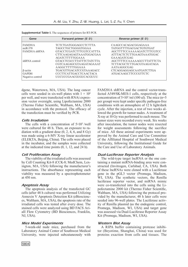
Supplemental Table I. The sequences of primers for RT-PCR.
Gene Forward primer (5’-3’) Reverse primer (5’-3’)
FAM201A TCTCTGATGGGAGCCTCTTTA CAAGCCACAGACGGAGAAAmiR-370 TAGCCTGCTGGGGTGGAA TATGGTTTTGACGACTGTGTGATFAM201A shRNA GTACCTCGATCTTTCGTCCATTTA AGCTTTTCCAAAAAGATCTTTCGTCC CTTCAAGAGAGTAAATGGACGAA ATTTACTCTCTTGAAGTAAATGGAC AGATCTTTTTGGAAA GAAAGATCGAGshRNA control GTACCTCGCCTTATTTCTATCTTA AGCTTTTCCAAAAAGCCTTATTTCTA CGTCAAGAGCGTAAGATAGAAAT TCTTACGCTCTTGACGTAAGATAGA AAGGCTTTTTGGAAA AATAAGGCGAGβ-actin TGACGTTGACATCCGTAAAGACC CTCAGGAGGAGCAATGATCTTGAGAPDH CCCTTCATTGACCTCAACTACA ATGACAAGCTTCCCGTTCTCNegative control UUCUCCGAACGUGUCACGUUU
LncRNA FAM201A involves in NSCLC radioresistance by enhancing EGFR expression
5805
protein concentrations were measured using the Pierce BCA Protein Assay Kit (Thermo Fisher Scientific, Waltham, MA, USA). The electrophoresis on SDS-PAGE gel (Thermo Fisher Scientific, Waltham, MA, USA) sep-arated the kinds of proteins in each sample, followed by a transfer to polyvinylidene difluo-ride membranes (PVDF) membranes (Thermo Fisher Scientific, Waltham, MA, USA). The membranes were incubated with the primary antibodies (Anti-EGFR, 1:2500; anti-HIF-1α, 1:2000; β-actin, 1:5000; Abcam, San Francisco, CA, USA) at 4°C overnight after incubation with PBS (5% dry milk) at room temperature for 1 h. Then, the membrane with blotting was incubated with a secondary antibody conju-gated with HRP (1:5000 dilution, Santa Cruz Biotechnology, Santa Cruz, CA, USA). An ECLTM chemiluminescence detection system (Pierce, Waltham, MA, USA) was used to com-pare the protein levels reflected by the blotting.
Statistical Analysis All experiments were conducted independent-
ly in triplicate. The data are presented as mean ± standard deviation (SD). The comparison within the groups was performed using the indepen-dent Student’s t-test. Univariate and multivariate analyses of the prognostic factors for overall survival using Cox proportional hazards regres-sion model. GraphPad Prism software (San Di-ego, CA, USA) was used for statistical analysis. The binding site prediction between the target miRNA and FAM201A were performed by Star-Base 2.0 (http://starbase.sysu.edu.cn/starbase2/
index.php). p-value < 0.05 was considered sta-tistically significant.
Results
FAM201A Elevation is Associated to Resistance to Radiotherapy and Poor Prognosis in NSCLC Patients
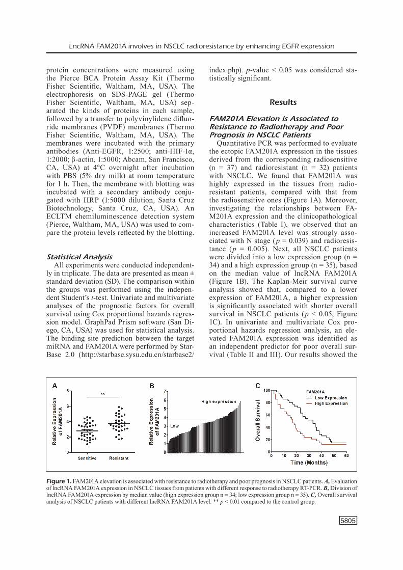
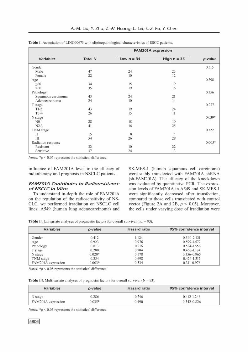
Quantitative PCR was performed to evaluate the ectopic FAM201A expression in the tissues derived from the corresponding radiosensitive (n = 37) and radioresistant (n = 32) patients with NSCLC. We found that FAM201A was highly expressed in the tissues from radio-resistant patients, compared with that from the radiosensitive ones (Figure 1A). Moreover, investigating the relationships between FA-M201A expression and the clinicopathological characteristics (Table I), we observed that an increased FAM201A level was strongly asso-ciated with N stage (p = 0.039) and radioresis-tance (p = 0.005). Next, all NSCLC patients were divided into a low expression group (n = 34) and a high expression group (n = 35), based on the median value of lncRNA FAM201A (Figure 1B). The Kaplan-Meir survival curve analysis showed that, compared to a lower expression of FAM201A, a higher expression is significantly associated with shorter overall survival in NSCLC patients (p < 0.05, Figure 1C). In univariate and multivariate Cox pro-portional hazards regression analysis, an ele-vated FAM201A expression was identified as an independent predictor for poor overall sur-vival (Table II and III). Our results showed the
Figure 1. FAM201A elevation is associated with resistance to radiotherapy and poor prognosis in NSCLC patients. A, Evaluation of lncRNA FAM201A expression in NSCLC tissues from patients with different response to radiotherapy RT-PCR. B, Division of lncRNA FAM201A expression by median value (high expression group n = 34; low expression group n = 35). C, Overall survival analysis of NSCLC patients with different lncRNA FAM201A level. ** p < 0.01 compared to the control group.
A.-M. Liu, Y. Zhu, Z.-W. Huang, L. Lei, S.-Z. Fu, Y. Chen
5806
influence of FAM201A level in the efficacy of radiotherapy and prognosis in NSCLC patients.
FAM201A Contributes to Radioresistance of NSCLC In Vitro
To understand in-depth the role of FAM201A on the regulation of the radiosensitivity of NS-CLC, we performed irradiation on NSCLC cell lines; A549 (human lung adenocarcinoma) and
SK-MES-1 (human squamous cell carcinoma) were stably transfected with FAM201A shRNA (sh-FAM201A). The efficacy of the knockdown was evaluated by quantitative PCR. The expres-sion levels of FAM201A in A549 and SK-MES-1 were significantly decreased after transfection, compared to those cells transfected with control vector (Figure 2A and 2B, p < 0.05). Moreover, the cells under varying dose of irradiation were
Notes: *p < 0.05 represents the statistical difference.
Table I. Association of LINC00675 with clinicopathological characteristics of ESCC patients.
FAM201A expression
Variables Total N Low n = 34 High n = 35 p-value
Gender 0.315 Male 47 24 23 Female 22 10 12 Age 0.398 ≤60 34 15 19 >60 35 19 16 Pathology 0.356 Squamous carcinoma 45 24 21 Adenocarcinoma 24 10 14 T stage 0.277 T1-2 43 19 24 T3-4 26 15 11 N stage 0.039* N0-1 28 18 10 N2-3 41 16 25 TNM stage 0.722 II 15 8 7 III 54 26 28 Radiation response 0.005* Resistant 32 10 22 Sensitive 37 24 13
Table II. Univariate analyses of prognostic factors for overall survival (no. = 93).
Variables p-value Hazard ratio 95% confidence interval
Gender 0.412 1.124 0.540-2.131Age 0.923 0.976 0.599-1.577Pathology 0.813 0.916 0.524-1.556T stage 0.280 0.704 0.456-1.184N stage 0.020* 0.578 0.356-0.965TNM stage 0.354 0.698 0.424-1.317FAM201A expression 0.003* 0.534 0.311-0.976
Notes: *p < 0.05 represents the statistical difference.
Table III. Multivariate analyses of prognostic factors for overall survival (N = 93).
Variables p-value Hazard ratio 95% confidence interval
N stage 0.286 0.746 0.412-1.246FAM201A expression 0.035* 0.490 0.342-0.826
Notes: *p < 0.05 represents the statistical difference.

LncRNA FAM201A involves in NSCLC radioresistance by enhancing EGFR expression
5807
Figure 2. FAM201A contributes to radioresistance of NSCLC in vitro. (A) SK-MES-1 and (B) A549 were transfected with a synthesized shRNA targeting FAM201A to decrease expression. (C) Proliferation of SK-MES-1 and (D) A549 under various dose of irradiation measured by CCK-8 assay. (E) Apoptotic rate of SK-MES-1 and (F) A549 transfected with shRNA or controls tested by flow cytometry. * p < 0.05, ** p < 0.01 compared to the control group.
A.-M. Liu, Y. Zhu, Z.-W. Huang, L. Lei, S.-Z. Fu, Y. Chen
5808
analyzed by methylthiazol tetrazolium assay (MTT), and the results showed that a decrease in FAM201A expression, both in A549 and SK-MES-1 cell, was associated with a lower cell viability rate, compared to normal FAM201A expression (Figures 2C and 2D, p < 0.05). Ra-diation-induced apoptosis was considered as an indicator for radiosensitivity. Our flow cytometry results showed that the radiation-induced apop-tosis was higher in FAM201A knockdown cells (both in A549 and SK-MES-1), compared with the control vector-transfected cells (Figures 2E and 2F, p < 0.05). Collectively, our data indicated that FAM201A reduction ameliorates radioresis-tance of NSCLC in vitro.
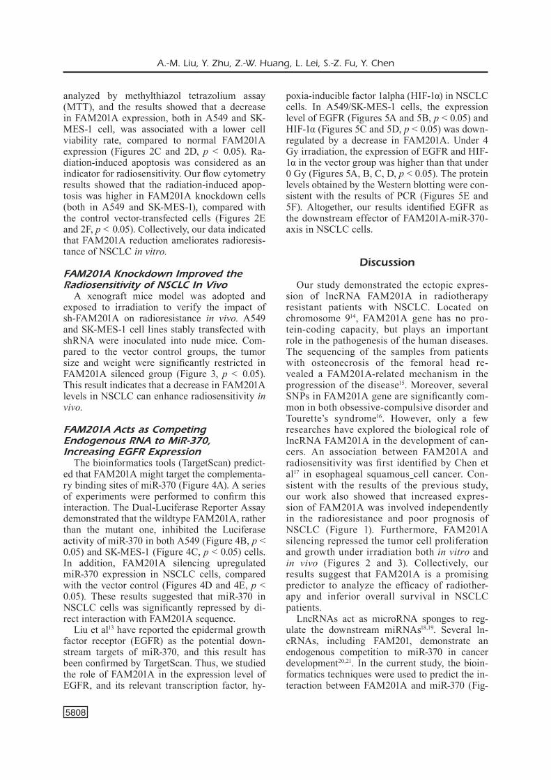
FAM201A Knockdown Improved the Radiosensitivity of NSCLC In Vivo
A xenograft mice model was adopted and exposed to irradiation to verify the impact of sh-FAM201A on radioresistance in vivo. A549 and SK-MES-1 cell lines stably transfected with shRNA were inoculated into nude mice. Com-pared to the vector control groups, the tumor size and weight were significantly restricted in FAM201A silenced group (Figure 3, p < 0.05). This result indicates that a decrease in FAM201A levels in NSCLC can enhance radiosensitivity in vivo.
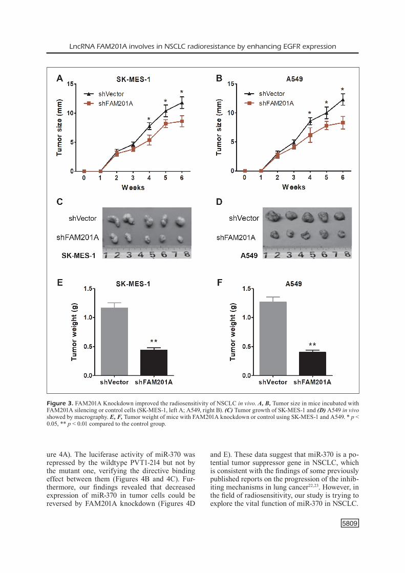
FAM201A Acts as Competing Endogenous RNA to MiR-370, Increasing EGFR Expression
The bioinformatics tools (TargetScan) predict-ed that FAM201A might target the complementa-ry binding sites of miR-370 (Figure 4A). A series of experiments were performed to confirm this interaction. The Dual-Luciferase Reporter Assay demonstrated that the wildtype FAM201A, rather than the mutant one, inhibited the Luciferase activity of miR-370 in both A549 (Figure 4B, p < 0.05) and SK-MES-1 (Figure 4C, p < 0.05) cells. In addition, FAM201A silencing upregulated miR-370 expression in NSCLC cells, compared with the vector control (Figures 4D and 4E, p < 0.05). These results suggested that miR-370 in NSCLC cells was significantly repressed by di-rect interaction with FAM201A sequence.
Liu et al13 have reported the epidermal growth factor receptor (EGFR) as the potential down-stream targets of miR-370, and this result has been confirmed by TargetScan. Thus, we studied the role of FAM201A in the expression level of EGFR, and its relevant transcription factor, hy-
poxia-inducible factor 1alpha (HIF-1α) in NSCLC cells. In A549/SK-MES-1 cells, the expression level of EGFR (Figures 5A and 5B, p < 0.05) and HIF-1α (Figures 5C and 5D, p < 0.05) was down-regulated by a decrease in FAM201A. Under 4 Gy irradiation, the expression of EGFR and HIF-1α in the vector group was higher than that under 0 Gy (Figures 5A, B, C, D, p < 0.05). The protein levels obtained by the Western blotting were con-sistent with the results of PCR (Figures 5E and 5F). Altogether, our results identified EGFR as the downstream effector of FAM201A-miR-370-axis in NSCLC cells.
Discussion
Our study demonstrated the ectopic expres-sion of lncRNA FAM201A in radiotherapy resistant patients with NSCLC. Located on chromosome 914, FAM201A gene has no pro-tein-coding capacity, but plays an important role in the pathogenesis of the human diseases. The sequencing of the samples from patients with osteonecrosis of the femoral head re-vealed a FAM201A-related mechanism in the progression of the disease15. Moreover, several SNPs in FAM201A gene are significantly com-mon in both obsessive-compulsive disorder and Tourette’s syndrome16. However, only a few researches have explored the biological role of lncRNA FAM201A in the development of can-cers. An association between FAM201A and radiosensitivity was first identified by Chen et al17 in esophageal squamous cell cancer. Con-sistent with the results of the previous study, our work also showed that increased expres-sion of FAM201A was involved independently in the radioresistance and poor prognosis of NSCLC (Figure 1). Furthermore, FAM201A silencing repressed the tumor cell proliferation and growth under irradiation both in vitro and in vivo (Figures 2 and 3). Collectively, our results suggest that FAM201A is a promising predictor to analyze the efficacy of radiother-apy and inferior overall survival in NSCLC patients.
LncRNAs act as microRNA sponges to reg-ulate the downstream miRNAs18,19. Several ln-cRNAs, including FAM201, demonstrate an endogenous competition to miR-370 in cancer development20,21. In the current study, the bioin-formatics techniques were used to predict the in-teraction between FAM201A and miR-370 (Fig-
LncRNA FAM201A involves in NSCLC radioresistance by enhancing EGFR expression
5809
ure 4A). The luciferase activity of miR-370 was repressed by the wildtype PVT1-214 but not by the mutant one, verifying the directive binding effect between them (Figures 4B and 4C). Fur-thermore, our findings revealed that decreased expression of miR-370 in tumor cells could be reversed by FAM201A knockdown (Figures 4D
and E). These data suggest that miR-370 is a po-tential tumor suppressor gene in NSCLC, which is consistent with the findings of some previously published reports on the progression of the inhib-iting mechanisms in lung cancer22,23. However, in the field of radiosensitivity, our study is trying to explore the vital function of miR-370 in NSCLC.
Figure 3. FAM201A Knockdown improved the radiosensitivity of NSCLC in vivo. A, B, Tumor size in mice incubated with FAM201A silencing or control cells (SK-MES-1, left A; A549, right B). (C) Tumor growth of SK-MES-1 and (D) A549 in vivo showed by macrography. E, F, Tumor weight of mice with FAM201A knockdown or control using SK-MES-1 and A549. * p < 0.05, ** p < 0.01 compared to the control group.
A.-M. Liu, Y. Zhu, Z.-W. Huang, L. Lei, S.-Z. Fu, Y. Chen
5810
A significant proportion of advanced NSCLC patients (40-50%) in the East Asian population possess oncogenic mutations in EGFR gene24. Exon 19 deletion and exon 21-point mutation, the most common alterations in EGFR, are widely
used as a therapeutic target of EGFR tyrosine kinase inhibitor (EGFR TKI)25. However, the role of EGFR in the radiosensitivity of cancer and its definitive molecular mechanisms have not yet been elucidated. Investigators26,27 origi-
Figure 4. FAM201A acts as competing endogenous RNA to miR-370. A, The bioinformatics tool reveals the complementary binding sites within FAM201A and miR-370. B, C, Luciferase reporter assay confirmed the molecular binding between FAM201A and miR-370 in SK-MES-1 and A549. D, E, qPCR showed the miR-370 expression in SK-MES-1 and A549 transfected with shFAM201A or controls under 0 or 4 Gy irradiation. ** p < 0.01 compared to the control group.

LncRNA FAM201A involves in NSCLC radioresistance by enhancing EGFR expression
5811
nally found the EGFR as a therapeutic switch to radiation sensitivity in cervical cancer through different inducing molecules. Furthermore, both PERK-eIF2α-GRP94 and IRE1α-XBP1-GRP78 are potentially involved in EGFR-induced radio-resistance in oropharyngeal carcinoma28. EGFR
conspires with ZEB1 to enhance the radioresis-tance which can be reversed by miR-875 in pros-tate cancer29. Recently, the connection between microRNA and EGFR in cancer radiosensitivity is attracting increased attention. In our study, ln-cRNA FAM201A-miR370 upregulated the EGFR
Figure 5. FAM201A inhibits miR-370 resulting in EGFR up-regulation. A, B, qPCR showed the EGFR level in SK-MES-1 and A549 transfected with shFAM201A or controls under 0 or 4 Gy irradiation. C, D, qPCR showed the HIF-1α level in SK-MES-1 and A549 transfected with shFAM201A or controls under 0 or 4 Gy irradiation. E, F, Western blot assay showed the EGFR and HIF-1α protein expression in SK-MES-1 and A549 transfected with shFAM201A or controls under 0 or 4 Gy irradiation. *p < 0.05, ** p < 0.01 compared to the control group.
A.-M. Liu, Y. Zhu, Z.-W. Huang, L. Lei, S.-Z. Fu, Y. Chen
5812
and HIF-1α expression in radio-resistant NSCLC cell lines (Figure 5C). Our results indicate that hypoxia-related EGFR functions as an effector in radiation modulation. Consistently, Liu et al30 found that EGFR loss resulted from hypoxia-in-duced autophagy which can cause cell death and enhance radiosensitivity in NSCLC. Lee et al31 showed that hypoxia/reoxygenation-induced the activation of Nrf2 and EGFR which can further evoke radioresistance in A549 cell.
Conclusion
We demonstrated that lncRNA FAM201A in-duces cell proliferation and tumor growth un-der radiation treatment in vitro and in vivo. In addition, the interference of FAM201A impairs its suppression of miR-370, resulting in EGFR and HIF-1α upregulation and radiosensitivity en-hancement. Our results indicate that this regu-latory axis may be a potential therapeutic target to enhance the sensitivity response of NSCLC to radiotherapy.
Conflict of InterestThe Authors declare that they have no conflict of interests.
Disclosure of Financial ArrangementsThe research and manuscript preparation are funded by Yue Chen.
Authors’ ContributionAnmin Liu made a contribution to molecular biological as-says, data analysis, and manuscript preparation. Yan Zhu assisted in performing the literature research and clinical enrollment. Lei Lei helped Anmin Liu to perform the an-imal experiment. Shaozhi Fu analyzed the survival data from NSCLC patients. Zhanwen Huang reviewed the pa-per and gave advice. Yue Chen directed all these research-es and reviewed this manuscript. All authors read and ap-proved the final manuscript.
References
1) Chen WQ, Zheng RS, Baade Pd, Zhang SW, Zeng hM, BRay F, JeMal a, yu XQ, he J. Cancer statistics in Chi-na, 2015. CA Cancer J Clin 2016; 66: 115-132.
2) BRay F, FeRlay J, SoeRJoMataRaM I, SIegel Rl, toRRe la, JeMal a. Global cancer statistics 2018: GLOBO-CAN estimates of incidence and mortality world-wide for 36 cancers in 185 countries. CA Cancer J Clin 2018; 68: 394-424.
3) SIegel Rl, MIlleR Kd, JeMal a. Cancer Statistics, 2018. CA Cancer J Clin 2018; 68: 7-30.
4) BRyan dS, donIngton JS. The role of surgery in management of locally advanced non-small cell lung cancer. Curr Treat Options Oncol 2019; 20: 27.
5) Zhao y, Wang W, lIang h, yang CJ, d’aMICo t, ng CSh, lIu CC, PeteRSen Rh, RoCCo g, BRunellI a, lIu J, he J, huang W, lIang W, he J, aMe thoRaCIC SuR-geRy CollaBoRatIve gRouP. The optimal treatment for stage IIIA-N2 non-small cell lung cancer: a network meta-analysis. Ann Thorac Surg 2019; 107: 1866-1875.
6) Moon Sh, Cho Kh, lee Cg, KeuM KC, KIM yS, Wu hg, KIM Jh, ahn yC, oh d, lee Jh. IMRT vs. 2D-radiotherapy or 3D-conformal radiotherapy of nasopharyngeal carcinoma survival outcome in a Korean multi-institutional retrospective study (KROG 11-06). Strahlenther Onkol 2016; 192: 377-385.
7) Ren XC, lIu ye, lI J, lIn Q. Progress in image-guid-ed radiotherapy for the treatment of non-small cell lung cancer. World J Radiol 2019; 11: 46-54.
8) ohRI n. Radiotherapy dosing for locally ad-vanced non-small cell lung carcinoma: “MTD” or “ALARA”? Front Oncol 2017; 7: 205.
9) nIyaZI M, MaIhoeFeR C, KRauSe M, Rodel C, BudaCh W, BelKa C. Radiotherapy and “new” drugs-new side effects? Radiat Oncol 2011; 6: 177.
10) BRadley Jd, PauluS R, KoMaKI R, MaSteRS g, BluMen-SCheIn g, SChIld S, BogaRt J, hu C, FoRSteR K, Ma-glIoCCo a, KavadI v, gaRCeS yI, naRayan S, IyengaR P, RoBInSon C, Wynn RB, KoPRoWSKI C, Meng J, BeItleR J, gauR R, CuRRan W, Choy h. Standard-dose ver-sus high-dose conformal radiotherapy with con-current and consolidation carboplatin plus pa-clitaxel with or without cetuximab for patients with stage IIIA or IIIB non-small-cell lung cancer (RTOG 0617): a randomised, two-by-two factori-al phase 3 study. Lancet Oncology 2015; 16: 187-199.
11) FaRR KP, KhalIl aa, KnaP MM, MølleR dS, gRau C. Development of radiation pneumopathy and gen-eralised radiological changes after radiotherapy are independent negative prognostic factors for survival in non-small cell lung cancer patients. Radiother Oncol 2013; 107: 382-388.
12) eISenhaueR ea, theRaSSe P, BogaeRtS J, SChWaRtZ lh, SaRgent d, FoRd R, danCey J, aRBuCK S, gWytheR S, Mooney M, RuBInSteIn l, ShanKaR l, dodd l, KaPlan R, laCoMBe d, veRWeIJ J. New response evaluation criteria in solid tumours: Revised RECIST guide-line (version 1.1). Eur J Cancer 2009; 45: 228-247.
13) lIu X, huang yg, JIn Cg, Zhou yC, Chen XQ, lI J, Chen y, lI M, yao Q, lI K, lan M, ye Jg, Wang XC. MicroRNA-370 inhibits the growth and metasta-sis of lung cancer by down-regulating epidermal growth factor receptor expression. Oncotarget 2017; 8: 88139-88151.
14) huMPhRay SJ, olIveR K, hunt aR, PluMB RW, love-land Je, hoWe Kl, andReWS td, SeaRle S, hunt Se,
LncRNA FAM201A involves in NSCLC radioresistance by enhancing EGFR expression
5813
SCott Ce, JoneS MC, aInSCough R, alMeIda JP, aM-BRoSe Kd, aShWell RIS, BaBBage aK, BaBBage S, Bag-guley Cl, BaIley J, BaneRJee R, BaRKeR dJ, BaRloW KF, BateS K, BeaSley h, BeaSley o, BIRd CP, BRay-allen S, BRoWn aJ, BRoWn Jy, BuRFoRd d, BuRRIll W, BuRton J, CaRdeR C, CaRteR nP, ChaPMan JC, Chen y, ClaRKe g, ClaRK Sy, Clee CM, Clegg S, CollIeR Re, CoRBy n, CRoSIeR M, CuMMIngS at, davIeS J, dhaMI P, dunn M, dutta I, dyeR lW, eaRthRoWl Me, FaulKneR l, FleM-Ing CJ, FRanKISh a, FRanKland Ja, FRenCh l, FRICK-eR dg, gaRneR P, gaRnett J, ghoRI J, gIlBeRt JgR, glISon C, gRaFhaM dv, gRIBBle S, gRIFFIthS C, JoneS Sg, gRoCoCK R, guy J, hall Re, haMMond S, haR-ley Jl, haRRISon eSI, haRt ea, heath Pd, hendeRSon Cd, hoPKInS Bl, hoWaRd PJ, hoWden PJ, huCKle e, JohnSon C, JohnSon d, Joy aa, Kay M, Keenan S, KeRShaW JK, KIMBeRley aM, KIng a, KnIghtS a, laIRd gK, langFoRd C, laWloR S, leongaMoRnleRt da, le-veRSha M, lloyd C, lloyd dM, lovell J, MaRtIn S, MaShReghI-MohaMMadI M, MattheWS l, MClaRen S, MClay Ke, MCMuRRay a, MIlne S, nICKeRSon t, nIS-Bett J, noRdSIeK g, PeaRCe av, PeCK aI, PoRteR KM, PandIan R, Pelan S, PhIllIMoRe B, Povey S, RaMSey y, Rand v, SChaRFe M, SehRa hK, ShoWnKeen R, SIMS SK, SKuCe Cd, SMIth M, SteWaRd Ca, SWaRBReCK d, SyC-aMoRe n, teSteR J, thoRPe a, tRaCey a, tRoManS a, thoMaS dW, Wall M, WallIS JM, WeSt aP, WhItehead Sl, WIlley dl, WIllIaMS Sa, WIlMIng l, WRay PW, young l, aShuRSt Jl, CoulSon a, BloCKeR h, duRBIn R, SulSton Je, huBBaRd t, JaCKSon MJ, Bentley dR, BeCK S, RogeRS J, dunhaM I. DNA sequence and analysis of human chromosome 9. Nature 2004; 429: 369-374.
15) yu dM, MatheWS Ca, SChaRF JM, neale BM, davIS lK, gaMaZon eR, deRKS eM, evanS P, edlund CK, CRane J, oSIeCKI l, gallagheR P, geRBeR g, haddad S, IllMann C, MCgRath lM, MayeRFeld C, aRePallI S, BaRlaSSIna C, BaRR Cl, BellodI l, BenaRRoCh F, BeR-RIo gB, BIenvenu oJ, BlaCK dW, BloCh Mh, BRen-tanI h, BRuun Rd, BudMan Cl, CaMaRena B, CaMP-Bell dd, CaPPI C, SIlgado JCC, CavallInI MC, Cha-vIRa da, ChouInaRd S, CooK eh, CooKSon MR, Co-RIC v, Cullen B, CuSI d, deloRMe R, denyS d, dIon y, eaPen v, egBeRtS K, FalKaI P, FeRnandeZ t, FouRnIeR e, gaRRIdo h, gelleR d, gIlBeRt dl, gIRaRd Sl, gRaBe hJ, gRadoS Ma, gReenBeRg Bd, gRoSS-tSuR v, gRun-Blatt e, haRdy J, heIMan ga, heMMIngS SMJ, heRRe-Ra ld, heZel dM, hoeKStRa PJ, JanKovIC J, Kennedy Jl, KIng Ra, KonKaShBaev aI, KReMeyeR B, KuRlan R, lanZagoRta n, leBoyeR M, leCKMan JF, lenneRtZ l, lIu Cy, loChneR C, loWe tl, luPolI S, MaCCIaRdI F, MaIeR W, Manunta P, MaRConI M, MCCRaCKen Jt, ReStRePo SCM, MoeSSneR R, MooRJanI P, MoRgan J, MulleR h, MuRPhy dl, naaRden al, nuRMI e, oChoa WC, oPhoFF Ra, PaKStIS aJ, Pato Mt, Pato Cn, PIaCentI-nI J, PIttengeR C, PollaK y, RauCh Sl, RenneR t, Re-uS vI, RIChteR Ma, RIddle Ma, RoBeRtSon MM, RoMe-Ro R, RoSaRIo MC, RoSenBeRg d, RuhRMann S, SaBat-tI C, SalvI e, SaMPaIo aS, SaMuelS J, SandoR P, SeRvICe SK, ShePPaRd B, SIngeR hS, SMIt Jh, SteIn dJ, StReng-Man e, tISChFIeld Ja, tuRIel M, duaRte avv, valla-da h, veenStRa-vandeRWeele J, WalItZa S, Wang y, Weale M, WeISS R, Wendland JR, WeStenBeRg hgM, ShugaRt yy, hounIe ag, MIguel eC, nIColInI h, Wag-
neR M, RuIZ-lInaReS a, Cath dC, MCMahon W, PoSt-huMa d, ooStRa Ba, neStadt g, Routeau ga, PuR-Cell S, JenIKe Ma, heutInK P, hanna gl, ContI dv, aRnold Pd, FReIMeR nB, SteWaRt e, KnoWleS Ja, CoX nJ, PaulS dl. Cross-disorder Ggnome-wide anal-yses suggest a complex genetic relationship be-tween Tourette’s syndrome and OCD. Am J Psy-chiat 2015; 172: 82-93.
16) huang gy, Zhao gl, XIa J, WeI yB, Chen Fy, Chen J, ShI JS. FGF2 and FAM201A affect the develop-ment of osteonecrosis of the femoral head after femoral neck fracture. Gene 2018; 652: 39-47.
17) Chen M, lIu P, Chen y, Chen Z, Shen M, lIu X, lI X, lI a, lIn y, yang R, nI W, Zhou X, Zhang l, tIan y, lI J, Chen J. Long noncoding RNA FAM201A medi-ates the radiosensitivity of esophageal squamous cell cancer by regulating ATM and mTOR expres-sion via miR-101. Front Genet 2018; 9: 611.
18) Chen hh, Zong J, Wang SJ. LncRNA GAPLINC promotes the growth and metastasis of glioblas-toma by sponging miR-331-3p. Eur Rev Med Pharmacol Sci 2019; 23: 262-270.
19) WeI n, WeI h, Zhang h. Long non-coding RNA ZEB1-AS1 promotes glioma cell proliferation, mi-gration and invasion through regulating miR-577. Eur Rev Med Pharmacol Sci 2018; 22: 3085-3093.
20) JIa B, Wang ZP, Sun X, Chen J, Zhao JJ, QIu Xl. Long noncoding RNA LINC00707 sponges miR-370-3p to promote osteogenesis of human bone marrow-derived mesenchymal stem cells through upregulating WNT2B. Stem Cell Res Ther 2019; 10: 67.
21) lI J, huang yy, deng XJ, luo Ml, Wang XF, hu hy, lIu Cd, Zhong M. Long noncoding RNA H19 pro-motes transforming growth factor-beta-induced epithelial-mesenchymal transition by acting as a competing endogenous RNA of miR-370-3p in ovarian cancer cells. Oncotargets Ther 2018; 11: 427-440.
22) lI CC, ge QQ, lIu JX, Zhang QS, Wang Ch, CuI K, Chen Z. Effects of miR-1236-3p and miR-370-5p on activation of p21 in various tumors and its in-hibition on the growth of lung cancer cells. Tumor Biol 2017; 39: 1010428317710824.
23) Chen t, gao F, Feng S, yang t, Chen M. MicroR-NA-370 inhibits the progression of non-small cell lung cancer by downregulating oncogene TRAF4. Oncol Rep 2015; 34: 461-468.
24) huang SF, lIu hP, lI lh, Ku yC, Fu yn, tSaI hy, Chen yt, lIn yF, Chang WC, Kuo hP, Wu yC, Chen yR, tSaI SF. High frequency of epidermal growth fac-tor receptor mutations with complex patterns in non-small cell lung cancers related to gefitinib re-sponsiveness in Taiwan. Clin Cancer Res 2004; 10: 8195-8203.
25) MaeMondo M, Inoue a, KoBayaShI K, SugaWaRa S, oI-ZuMI S, ISoBe h, geMMa a, haRada M, yoShIZaWa h, KInoShIta I, FuJIta y, oKInaga S, hIRano h, yoShIMoRI K, haRada t, oguRa t, ando M, MIyaZaWa h, tanaKa t, SaIJo y, hagIWaRa K, MoRIta S, nuKIWa t; noRth-eaSt JaPan Study gRouP. Gefitinib or chemotherapy
A.-M. Liu, Y. Zhu, Z.-W. Huang, L. Lei, S.-Z. Fu, Y. Chen
5814
for non-small-cell lung cancer with mutated EG-FR. N Engl J Med 2010; 362: 2380-2388.
26) XIong h, nIe X, Zou y, gong C, lI y, Wu h, QIu h, yang l, Zhuang l, Zhang P, Zhang J, Wang y, XIong h. Twist1 enhances hypoxia induced radioresis-tance in cervical cancer cells by promoting nucle-ar EGFR localization. J Cancer 2017; 8: 345-353.
27) Wang X, gu y, lIu h, ShI l, Sun X. Icotinib hydro-chloride enhances chemo- and radiosensitivity by inhibiting EGFR signaling and attenuating RAD51 expression and function in Hela S3 cells. Onco Targets Ther 2018; 11: 1245-1258.
28) Zhang M, han n, JIang yJ, Wang J, lI gy, lv Xt, lI g, QIao Q. EGFR confers radioresistance in hu-man oropharyngeal carcinoma by activating en-doplasmic reticulum stress signaling PERK-eIF2 α-GRP94 and IRE1 α-XBP1-GRP78. Cancer Med 2018; 7: 6234-6246.
29) el BeZaWy R, CoMInettI d, FendeRICo n, ZuCo v, Be-Retta gl, dugo M, aRRIghettI n, StuCChI C, RanCatI t, valdagnI R, ZaFFaRonI n, gandellInI P. MiR-875-5p counteracts epithelial-to-mesenchymal transi-tion and enhances radiation response in prostate cancer through repression of the EGFR-ZEB1 ax-is. Cancer Lett 2017; 395: 53-62.
30) lIu B, han d, Zhang t, Cheng g, lu y, Wang J, Zhao h, Zhao Z. Hypoxia-induced autophagy promotes EGFR loss in specific cell contexts, which leads to cell death and enhanced radiosensitivity. Int J Biochem Cell Biol 2019; 111: 12-18.
31) lee Sl, Ryu h, Son aR, Seo B, KIM J, Jung Sy, Song Jy, hWang Sg, ahn J. TGF-β and hypoxia/re-oxygenation promote radioresistance of A549 lung cancer cells through activation of Nrf2 and EGFR. Oxid Med Cell Longev 2016; 2016: 6823471.