copper homeostasis in aspergillus nidulans involves ... - addi
TRANSCRIPT
BIOKIMIKA II
Applied Chemistry
Faculty of Chemistry
Donostia
Copper Homeostasis in the Filamentous Ascomycete
Aspergillus nidulans.
Applied Chemistry and Polymeric Materials PhD program
Martzel Antsotegi Uskola
2021
Supervisor:
Unai Ona Ugalde Martinez
Senior Lecturer
Faculty of Chemistry
EHU/UPV
(cc)2021 MARTZEL ANTSOTEGI USKOLA (cc by-nc-sa 4.0)

This PhD project has been carried out in the Faculty of
Chemistry of the University of the Basque Country (EHU/UPV), under
the supervision of the senior lecturer Dr. Unai Ona Ugalde Martinez.
This work has been co-supported by grant IT599-13 and S-
PC13UN041 from the Basque Government, funds from the University
of the Basque Country, both to the Dr. Ugalde; and the PhD
scholarship from the Basque Government to Martzel Antsotegi
Uskola, the doctorate candidate.

Hauts hartatikan ustekabean
Hor agertzen da… Txirritaa
Ur pistol batez...
Fuuurra fuurraaa...
Soineko txuriz jantzita!!
Momentu zailenetan ere barre bat
atera didazuen guztiontzat...
Zuen alde

Acknowledgment
Acknowledgment
Bueno, ba iritsi da hainbestetan amestutako momentua, iritsi da abentura honen
bukaera. Bost urte pasatxoren ostean, iritsi da tesia entregatu eta bizitzako etapa
honi bukaera emateko unea. Prozesu guzti honen parte izan zareten guztioi edo
gehienoi behintzat eskerrak emateko tartea hartu nahiko nuke hurrengo lerro
hauetan.
Lehenik eta behin, prozesu honetan ezinbestekoak izan diren bi pertsonetaz
oroitzea dagokit, Unai eta Ane. Unai, bide guzti honetan zure gidaritza, aholkuak
eta bideak zabaltzeko ahalegina asko estimatzen dut. Tesi gidari bezala nire lan
erritmoak errespetatu dituzu beti, edozein arazoren aurrean zure bulegoko ateak
ireki dizkidazu eta bulegotik ateratzerakoan beti lasaiago eta gauzak argiago
nituela atera izan naiz. Benetan, mila esker. Ane, “my mentor”, zuri zer esango
dizut ba. Urteak dira laborategia utzi zenuenetik, baina denok oroitzen gara
zutaz. Zure alaitasuna, lanerako grina, beti laguntzeko prest bueltan ezer espero
gabe… askotan pentsatu izan dut, “nondik ateratzen ote du emakume honek
hainbesteko energia?”. Nire partetik esan behar dut nik tesi hau aurrera atera
izanaren merituaren zati haundi bat zurea dela. Zurekin ikasi nuen zer zen
laborategiko lana, lanarekiko konpromezua eta lan ona egitearen garrantzia.
Askok pentsa lezakeena baino eragin handiagoa izan duzu nire bizitzan. Zerua
irabazia duzu, Ane.
Bueno, eta laborategiko beste jendilajeaz ere ezin ahaztu. Nahiz eta proiektu
berean aritu ez, zuek ere guzti honen parte zarete. DJ Oilier, zuri ere gauza asko
eskertu behar dizkizut, aholku eske joan naizenean beti izan duzu erantzun zintzo
bat eta animo hitzak egoera edozein delarik ere. Summer lab day eta
soziedadeko bazkari guztien saltsa. Hi haiz hi tipoa! Eli O eta Eli P, zuek ere
aspalditxo utzi zenuten laborategia eta badakit nire laborategiko hasiera hartan
txotxolo samarra nintzela, baina zuengandik ere asko ikasi nuen. Azkenik, nola
ez, “mis compañeros de armas”, Mikel eta Ainara, a ze bi. Zuekin dena izan da
Acknowledgment
eramangarriagoa, momentu on eta ez hain onetan ere beti elkarri adarra jotzeko
prest, laneko tentsioak baretzeko barre terapia lagunak. Bakoitzak orain bere
bidea hartuko dugu, baina beti izango dugu lotura estu bat. Biologiako
laborategiko jendeaz ere ezingo naiz ahaztu. Maite, Goretti eta Ana plazer bat
izan da zuek ezagutzea.
I would also like to remember everyone that has been part of my journeys
abroad. Roberta, Emiliano, Mattia, Mickey, Anton and many more… It’s been a
pleasure and an honor to meet you, work with you and learn from and with you.
Lantokitik kanpoko jendeaz oroitu nahiko nuke. Gurasoek ezin aipatu gabe utzi.
Beraien esku dagoen guztia eta gehiago egingo luketela behin baino gehiagotan
erakutsi didate. Milioi bat aldiz eskerrak emanagatik ere labur geldituko
nintzateke. Koadrilakoei ere eskerrak, dena eramangarriagoa egiten duzue,
zuekin egote hutsak barrua pozten dit eta arazoak ahaztarazi. And last but not
least, Ainho. Eskerrik asko zaren bezalakoa izateagatik, ura eta olioa nahastu
daitezkeela erakusteagatik eta nire aurpegi zurbilean argitasuna sortzeko duzun
gaitasunagatik.
Bizitako guztiagatik, bihotz-bihotzez, eskerrak denoi!!!
Abbreviations
Abbreviations
ACM Aspergillus complete medium
A Alanine
AMBER Assisted Model Building with Energy Refinement
AMM Aspergillus minimal medium
AspGD Aspergillus Genome Database
ATP Adenosine Triphosphate
BCS Bathocuproinedisulfonic acid
BLASTp Basic Local Alignment Search Tool for proteins
bp Base pair oC Celsius degrees
C-term Carboxy-terminal
Cat Catalase
CDF Cation Diffusion Facilitator
CHARMM Chemistry at Harvard Macromolecular Mechanics
cm Centimeter
Co Cobalt
Crp Copper response P-type ATPase
Ctr Copper Transporter
Cu Copper
C/Cys Cysteine
DIG Digoxigenin
DNA Deoxyribonucleic Acid
EDTA Ethylenediaminetetraacetic acid
ER Endoplasmic Reticulum
Fe Iron
g gram
g g-force
G1 Growth
G2 Growth & Mitosis preparation
gDNA genomic DNA
GFP Green Fluorescent protein
GO Gene Onthology
GROMACS GROningen MAchine for Chemical Simulations
Gsp Gene specific primer
h hours
HA Residues 98-106 of the human flu virus hemagglutinin
HBB 5aS,10aS)-1,3,8,10a-tetrahydroxy-2-[hydroxy(methyl)BLAHyl]-5a-(3-methylbut-2-enyl)benzofuro[3,2-b]c
Hg Mercury
H/His Histidine
HS Hot Start
Abbreviations
K Potassium
kDa kiloDalton
l liter
M Molar
M Mitosis
MD Molecular Dynamics
min minutes
μg microgram
μl microliter
m micrometer
mg milligram
mJ milliJoule
ml milliliter
mm millimeter
Mn manganese
MOPS 3-(N-Morpholino)-propanesulfonic acid
MRE Metal Response/Regulatory Element
mRFP monomeric Red Fluorescent Protein
mRNA messenger RNA
MT metallothionein
MVB Multivesicular Body
NE Nuclear Envelope
Ni nickel
N-terminal Amino-terminal
PCR Polymerase Chain Reaction
PEG Polyethylene glycol
PM Plasma Membrane
PREG pregn-5-ene-3-ol derivative
OPM Orientations of Proteins in Membranes
qPCR quantitative Polymerase Chain Reaction
RT-PCR Real-Time PCR
RMM Regeneration Minimal Medium
RMM-TOP Regeneration Minimal Medium-TOP
RMSD Root Mean Square Deviation
RNA: Ribonucleic acid
RNase Ribonuclease
RNA-seq RNA-sequencing
ROS Reactive Oxygen Species
rpm Revolutions per minute
rRNA ribosomal RNA
S DNA synthesis
s seconds
SDS Sodium dodecyl sulfate
Sod Superoxide Dismutase
Abbreviations
TCA Trichloroacetic acid
TGN Trans-Golgi Network
TM Transmembrane
TMD Transmembrane domain
TF Transcription factor
to Time zero
Tris Tris(hydroxymethyl)aminomethane
UTR Untranslated Region
UV Ultraviolet
WMM Watch Minimal Medium
WT wild-type
Zn Zinc
ZINC Zinc Is Not Commercial


Index

Index
Introduction _____________________________________ 1
1- Aspergillus nidulans ___________________________________ 3
2- Copper an Essential Element for Life ______________________ 9
3- Copper Import Across Membranes ______________________ 10
4- Copper Detoxification _________________________________ 12
5- Genetic Regulation of Copper Homeostasis ________________ 13
6- Copper in Fungal Biology and Justification for its Study ______ 16
Objectives ______________________________________ 21
Materials & Methods _____________________________ 25
1- Strains, Primers and Plasmids ___________________________ 27
1.1- Aspergillus nidulans Strains _______________________________________ 27
1.2- Plasmids ______________________________________________________ 29
1.3- Primers _______________________________________________________ 30
2- Media and growth conditions___________________________ 34
3- Construction of Replacement Cassettes for Null and Tagged
Protein Strain Generation ________________________________ 37
3.1- Construction of Replacement Cassettes for Null Mutant Strain Generation _ 37
3.2- Construction of Replacement Cassettes for GFP/HA Tagged Protein Expressing
Strain Generation __________________________________________________ 38
3.3- Construction of Replacement Cassettes to Generate Specific Cysteine
Mutations and C-terminal Truncations of GFP Expressing Strains _____________ 38
4- Specific Protocols Applied in Aspergillus nidulans ___________ 39
4.1- A. nidulans Transformation _______________________________________ 39
4.2- Conidia Count __________________________________________________ 41
5- Molecular Biology, Biochemistry and Proteomic Techniques
Applied in Aspergillus nidulans ____________________________ 41
5.1- Genomic DNA Extraction _________________________________________ 41
5.2- DNA Analysis by Southern-Blot ____________________________________ 43
5.3- Membrane Protein Extraction _____________________________________ 44
5.4- Western-Blot __________________________________________________ 45
Index
5.5- Fluorescence Microscopy ________________________________________ 46
5.6- RNA Extraction _________________________________________________ 46
5.7- Northern-Blot __________________________________________________ 47
5.8- cDNA ________________________________________________________ 48
5.9- Real-Time PCR _________________________________________________ 48
6- Bioinformatics _______________________________________ 49
The High-Affinity Copper Uptake System in A. nidulans _ 51
1- Introduction ________________________________________ 53
2- Results _____________________________________________ 55
2.1- Gene Expression Analysis Under Copper Toxicity Conditions _____________ 55
2.2- Screening for Potential Copper Transport Proteins _____________________ 61
2.3- Functional Characterization of AnCtr Mutants ________________________ 64
2.4- AnCtr Protein Expression Dynamics _________________________________ 69
2.5- AnCtr Localization ______________________________________________ 72
2.6- C-Terminal Mutations of AnCtrA and AnCtrC _________________________ 75
2.7- Screening for Cupric Metalloreductases _____________________________ 78
3- Discussion __________________________________________ 80
The Copper Detoxification System in A. nidulans _______ 87
1- Introduction ________________________________________ 89
2- Results _____________________________________________ 90
2.1- Identification of Copper Resistance Determinants in A. nidulans. _________ 90
2.2- Functional Characterization _______________________________________ 93
2.3- Expression Dynamics ____________________________________________ 96
2.4- AnCrpA Dynamic Localization ____________________________________ 101
2.5- Expression Regulation of AnCrpA and AnCrdA by the Transcription Factor
AnAceA _________________________________________________________ 103
3- Discussion _________________________________________ 106
Molecular Modelling Approach to Screen for AnCrpA
Inhibitors ______________________________________ 111
1- Introduction _______________________________________ 113
Index
2- Materials & Methods ________________________________ 115
2.1- Protein Preparation ____________________________________________ 115
2.2- Grid Generation _______________________________________________ 115
2.3- Virtual Screening and Focused Docking _____________________________ 116
2.4- Molecular Dynamics____________________________________________ 117
3- Results & Discussion _________________________________ 118
3.1- The template Model LpCopA _____________________________________ 118
3.2- Model Validation ______________________________________________ 123
3.3- Virtual Screening ______________________________________________ 124
3.4- Focused Docking and Molecular Dynamics __________________________ 125
3.5- Grid 1: ZINC95099399 (HBB) _____________________________________ 127
3.6-Grid 2: ZINC35877687 (PREG) _____________________________________ 131
Final Discussion _________________________________ 135
Conclusions ____________________________________ 151
References _____________________________________ 155
Appendices ____________________________________ 173


1
Introduction

2
Introduction
3
1- Aspergillus nidulans
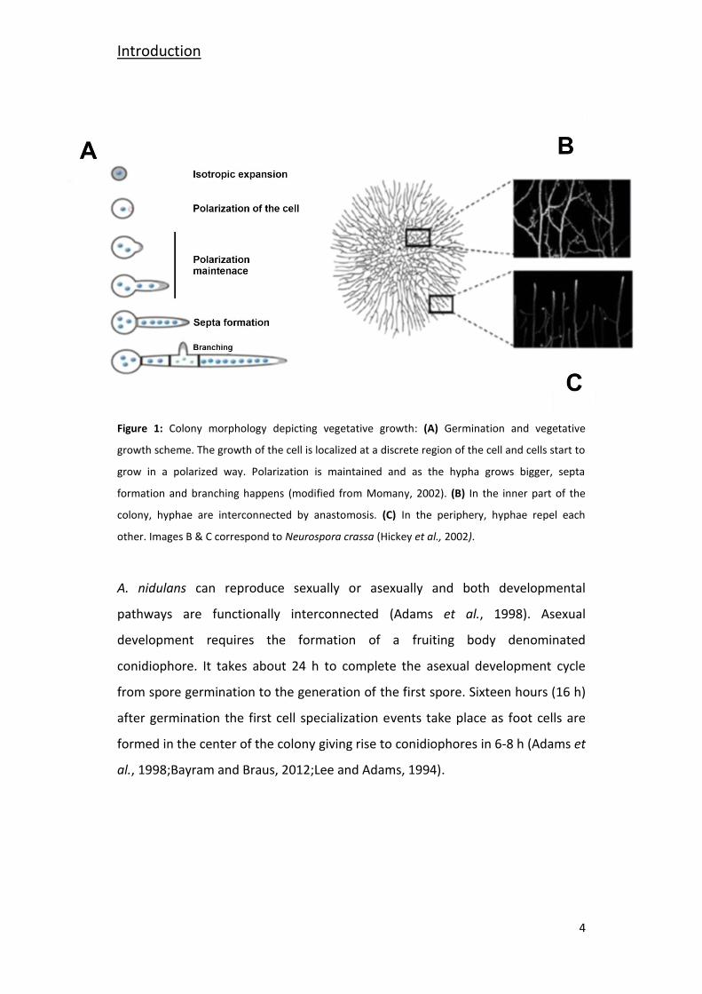
Aspergillus nidulans (Emericella nidulans) is a filamentous fungus that belongs to
the Ascomycota division. Vegetative cells, denominated hyphae, have a tubular
shape and grow in a hyperpolarized manner by apical extension and lateral
branching, forming an interconnected cellular network denominated mycelium
(Harris et al., 2005;Lee and Adams, 1994). The extension of hyphae results in
radial expansion of the fungal colony (Harris and Momany, 2004) (Figure 1B & C).
The clearest images of a fungal mycelium have been obtained in Neurospora
crassa, as shown in Figure 1B & C. The fact that A. nidulans mycelium is very
compact makes it difficult to obtain clear images; however the mycelium
organization follows a comparable pattern in both organisms. The Aspergillus
mycelium is coenocytic, meaning that its hyphae are multinucleated syncytia,
generally haploid, and divided by perforated septa that permit the selective
exchange of nutrients and effectors between cellular compartments (Glass et al.,
2004;Hickey et al., 2002;Momany, 2002;Riquelme et al., 2003).
The A. nidulans life cycle is constituted by four different stages: vegetative
growth, asexual development, sexual development and the parasexual cycle
(Todd et al., 2007). The cycle is initiated by uninucleate haploid asexual spore or
sexual ascospore germination. The spores are metabolically dormant cells
retained in phase G1 (Growth) (Bergen and Morris, 1983). A. nidulans has a
relatively short cell-cycle and the timing of the distinct phases of the cell cycle
follow as stated: G1 (Growth), 15 minutes; S (Synthesis), 25 minutes; G2 (Growth
and Mitosis preparation), 30 minutes; and M (Mitosis), around 5 minutes at 37
ºC (Bergen & Morris, 1983). Depending on environmental conditions the
duration of the G1 and G2 phases is variable (Doonan, 1992). After spore
germination, with an isotropic expansion and establishment of the polarity of the
cell, the addition of new cellular material to the polarized tip of the germling
leads to the mature vegetative cell, denominated hypha (Harris and Momany,
2004) (Figure 1A).
Introduction
4
Figure 1: Colony morphology depicting vegetative growth: (A) Germination and vegetative
growth scheme. The growth of the cell is localized at a discrete region of the cell and cells start to
grow in a polarized way. Polarization is maintained and as the hypha grows bigger, septa
formation and branching happens (modified from Momany, 2002). (B) In the inner part of the
colony, hyphae are interconnected by anastomosis. (C) In the periphery, hyphae repel each
other. Images B & C correspond to Neurospora crassa (Hickey et al., 2002).
A. nidulans can reproduce sexually or asexually and both developmental
pathways are functionally interconnected (Adams et al., 1998). Asexual
development requires the formation of a fruiting body denominated
conidiophore. It takes about 24 h to complete the asexual development cycle
from spore germination to the generation of the first spore. Sixteen hours (16 h)
after germination the first cell specialization events take place as foot cells are
formed in the center of the colony giving rise to conidiophores in 6-8 h (Adams et
al., 1998;Bayram and Braus, 2012;Lee and Adams, 1994).
Introduction
5
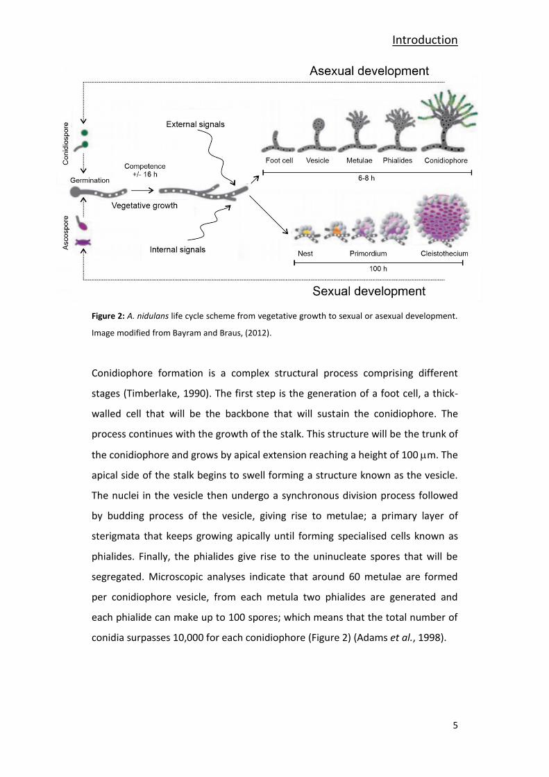
Figure 2: A. nidulans life cycle scheme from vegetative growth to sexual or asexual development.
Image modified from Bayram and Braus, (2012).
Conidiophore formation is a complex structural process comprising different
stages (Timberlake, 1990). The first step is the generation of a foot cell, a thick-
walled cell that will be the backbone that will sustain the conidiophore. The
process continues with the growth of the stalk. This structure will be the trunk of
the conidiophore and grows by apical extension reaching a height of 100 m. The
apical side of the stalk begins to swell forming a structure known as the vesicle.
The nuclei in the vesicle then undergo a synchronous division process followed
by budding process of the vesicle, giving rise to metulae; a primary layer of
sterigmata that keeps growing apically until forming specialised cells known as
phialides. Finally, the phialides give rise to the uninucleate spores that will be
segregated. Microscopic analyses indicate that around 60 metulae are formed
per conidiophore vesicle, from each metula two phialides are generated and
each phialide can make up to 100 spores; which means that the total number of
conidia surpasses 10,000 for each conidiophore (Figure 2) (Adams et al., 1998).
Introduction
6
A. nidulans spores possess a characteristic dark green pigment. Spore
pigmentation is a multistep process ending in a yellow pigment precursor turning
into dark green pigment. This step is catalyzed by laccase AnYA, which, as other
members of the laccase family, requires copper ions as a cofactor (Aramayo and
Timberlake, 1990;Clutterbuck, 1972;Scherer and Fischer, 2001).
Conidiophore production is a very demanding process, both materially and
energetically speaking. The fungus goes from vegetative hyphae of 2-3 m in
diameter to a complex structure that is larger than 100 m tall and this process
takes place repeatedly and simultaneously at distal locations of the colony in
synchrony with vegetative growth at the colony margin (Adams et al., 1998). The
colony largely provides for the requirements of conidiogenesis by recycling
autophagy (Kikuma et al., 2007;Richie and Askew, 2008). Autophagy is a self-
degradation process that proves to be very important to obtain energy sources in
response to nutrient stress and it is a widespread response to nutrient limitation
in eukaryotic organisms (Cebollero and Reggiori, 2009). The organelles and
cytoplasm are taken up into vacuoles to obtain reusable nutrients from them
(Pollack et al., 2009;Talbot and Kershaw, 2009). The hydrolyzed materials of the
cells are transferred to adjacent cells serving as a nutrient source. Then, these
nutrients can be transported wherever necessary, for example to conidiophore
production points (Darrah et al., 2006). One of the main objectives of autophagy
is to provide nutrients for sporulation (Kikuma et al., 2007;Richie and Askew,
2008). When speaking about nutrients, we don’t mean carbon or nitrogen
exclusively; it is documented that autophagy is also connected to metal ion
homeostasis (Richie and Askew, 2008), which means that also pigmentation
could be related to autophagy. Thus, the lack of a specific nutrient in a secondary
level might not only compromise sporulation, it might also compromise other
processes like pigmentation. In conditions of lack of first level supply, colony
development itself will be severely limited.
Introduction
7
Sexual development is an alternative path to asexual development but may be
brought about at the end of the asexual development (Adams et al., 1998). A.
nidulans is a homotalic organism that possesses the two mating genes (HMG &
α). This makes possible the sexual crossing between two different strains, but
also between two different cells within a colony (homothallism) in A. nidulans.
Sexual reproduction begins when two homokaryotic hyphae fuse by anastomosis
constituting a heterokaryon cell. The different nuclei are fused and a transitory
diploid nucleus is formed. This nucleus is then divided by meiosis followed by
mitosis resulting in 8 ascospores. During the ascospore maturation process,
another nuclear division happens giving birth to binucleate haploid ascospores
(Bokor et al., 2019;Han, 2009;Yager, 1992). Sexual development occurs within
specific fruiting bodies termed cleistothecia (which means, a closed container)
and these structures are surrounded by specialized protective cells named Hülle
cells (Han, 2009). A vegetative hypha will emerge from each germinated
ascospore (Adams et al., 1998).
Finally, the parasexual cycle comprises the fusion of two vegetative hyphae
resulting in an exchange of genetic material by anastomosis (Glass et al., 2004).
As a result, a heterokaryon with haploid genomic dotation is generated. A fusion
between two haploid nuclei can also happen giving birth to diploid nuclei. The
chromosomic dotation is restored by a haploidization (PONTECORVO et al.,
1953).
A. nidulans has been subjected to numerous live microscopy studies which have
led to the detailed assessment of its cellular structures. A. nidulans is a
multinucleated organism and these nuclei are spaced through the whole length
of hyphae. The polar growth of hyphae conditions the distribution of structures
and organelles within the cell. As the colony is developing, the organism needs a
good communication system to keep the whole colony growing in an organized
manner as the distances between the different growing tips increase (Markina-
Inarrairaegui et al., 2013;Pantazopoulou and Penalva, 2009). For that purpose, A.
Introduction
8
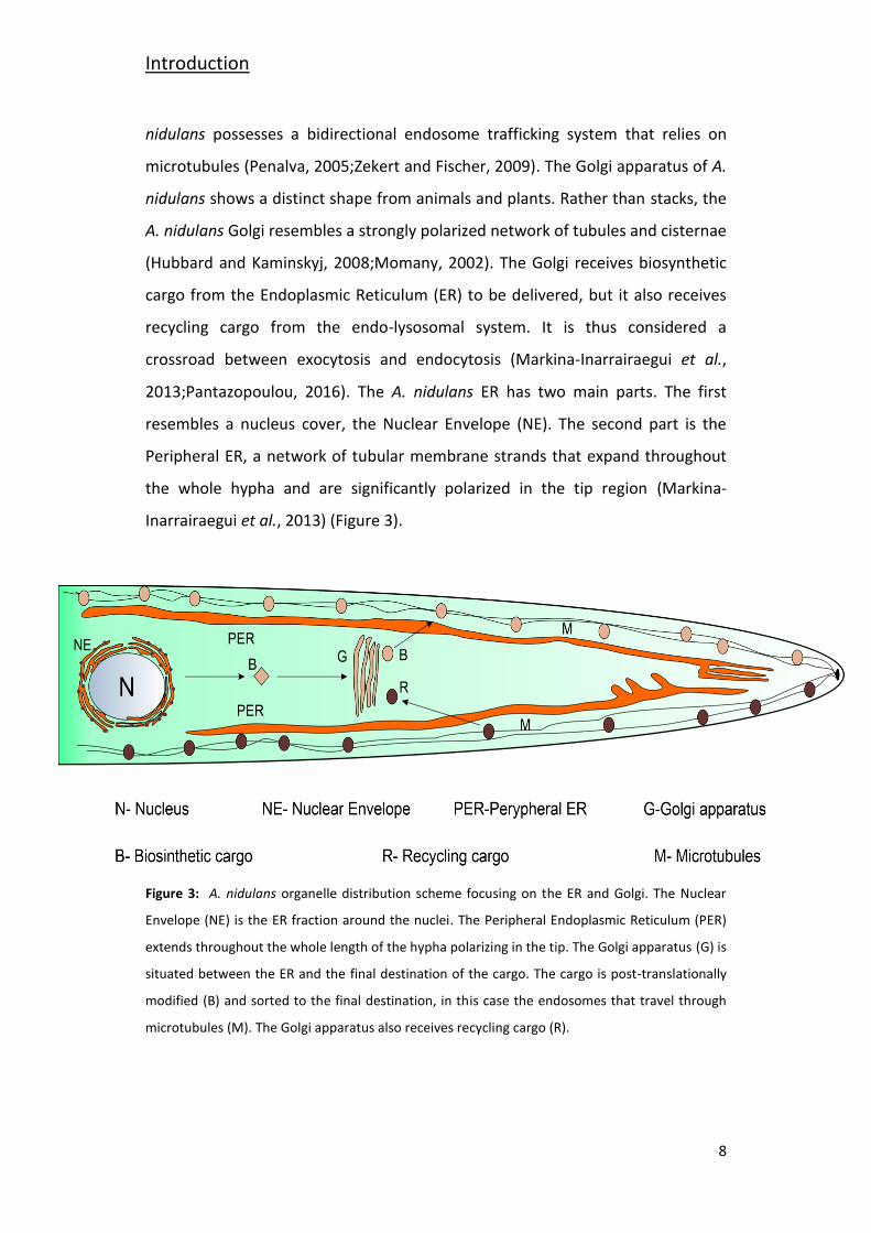
nidulans possesses a bidirectional endosome trafficking system that relies on
microtubules (Penalva, 2005;Zekert and Fischer, 2009). The Golgi apparatus of A.
nidulans shows a distinct shape from animals and plants. Rather than stacks, the
A. nidulans Golgi resembles a strongly polarized network of tubules and cisternae
(Hubbard and Kaminskyj, 2008;Momany, 2002). The Golgi receives biosynthetic
cargo from the Endoplasmic Reticulum (ER) to be delivered, but it also receives
recycling cargo from the endo-lysosomal system. It is thus considered a
crossroad between exocytosis and endocytosis (Markina-Inarrairaegui et al.,
2013;Pantazopoulou, 2016). The A. nidulans ER has two main parts. The first
resembles a nucleus cover, the Nuclear Envelope (NE). The second part is the
Peripheral ER, a network of tubular membrane strands that expand throughout
the whole hypha and are significantly polarized in the tip region (Markina-
Inarrairaegui et al., 2013) (Figure 3).
Figure 3: A. nidulans organelle distribution scheme focusing on the ER and Golgi. The Nuclear
Envelope (NE) is the ER fraction around the nuclei. The Peripheral Endoplasmic Reticulum (PER)
extends throughout the whole length of the hypha polarizing in the tip. The Golgi apparatus (G) is
situated between the ER and the final destination of the cargo. The cargo is post-translationally
modified (B) and sorted to the final destination, in this case the endosomes that travel through
microtubules (M). The Golgi apparatus also receives recycling cargo (R).
Introduction
9
The A. nidulans genome contains 30 million base pairs, approximately 10,982
genes distributed in eight chromosomes (Galagan et al., 2005). The multiple
molecular techniques applied in this fungus give the possibility to study biological
questions with many different experimental approaches like gene replacement
(Nayak et al., 2006), protein tagging, subcellular localization analysis of proteins
(Yang et al., 2004) or proteomic analysis. This is one of the reasons why it has
been used as a model organism to study stress-related cellular responses in
eukaryotes for more than 30 years.
2- Copper an Essential Element for Life
All organisms depend on metal ions as catalysts, structural elements in proteins,
in electron transfer reactions, or as messengers. The first-row transition metals
cobalt (Co), copper (Cu), iron (Fe), manganese (Mn), and nickel (Ni), possess the
appropriate Redox potential characteristics for key biological processes (Gerwien
et al., 2018;Nevitt et al., 2012). The intracellular concentration of these metals is
tightly controlled, as an excess of unbound metal would result in uncontrolled
and deleterious side-reactions (Blatzer and Latge, 2017). Thus, tightly regulated
mechanisms are in place to control to intracellular metal fluctuations of each
metal ion species. This is achieved through accurate sensing of intracellular and
extracellular metal levels coupled with the activation of various mechanisms that
maintain free intracellular metal levels within a safety range (Nevitt et al., 2012).
Copper (Cu) is an indispensable trace element for living organisms. Its capacity to
adopt an oxidized (Cu2+) and a reduced (Cu+) state is exploited by many enzymes
to act as Redox cofactors in enzyme-catalyzed processes (Nevitt et al., 2012);
cytochrome c, a key component of mitochondrial cellular respiration process;
superoxide dismutase, for ROS neutralization; and laccases, an abovementioned
protein family involved in fungal pigment synthesis (Ding et al., 2014;Lutsenko,
2010;Scherer and Fischer, 2001;Smith et al., 2017). However, free intracellular
copper can interfere with Redox processes generating Reactive Oxygen Species
Introduction
10
(ROS) or cause metalloprotein dysfunction by displacement of other bound metal
ions (Besold et al., 2016;Fridovich, 1983;Macomber and Imlay, 2009;Smith et al.,
2017). Hence, all organisms have elaborate mechanisms that secure copper
bioavailability, yet maintain free copper levels below the toxicity threshold. They
include copper uptake, intracellular traffic, storage, and detoxification processes
(Balamurugan and Schaffner, 2006;Nevitt et al., 2012).
Most studies on copper homeostasis have been carried out in mammalian cells,
bacteria, and yeast model systems. The model yeast Saccharomyces cerevisiae
has served as an important reference for other lower eukaryotes (Balamurugan
and Schaffner, 2006), including pathogens like Cryptococcus neoformans and
Candida albicans (Ballou and Wilson, 2016). In these models, copper is a
recognized virulence factor (Zhang et al., 2016). A number of recent publications
have described copper import and export mechanisms in Aspergillus fumigatus, a
common airborne fungal pathogen (Blatzer and Latge, 2017), responsible for
severe invasive aspergillosis in immunocompromised patients (Cai et al., 2017).
The following sections present an overview of recently published findings on the
different aspects of copper homeostasis in filamentous fungi.
3- Copper Import Across Membranes
The lipid bilayer of the plasma membrane is impenetrable to charged ions,
including copper and this generates the necessity for copper import machinery
through the plasma membrane. A functional copper transporter is required to be
highly specific, in order not to transport other heavy metal cations. On the other
hand, it needs display high-affinity binding in order to scavenge the cation
selectively under commonly low availability conditions (Smith et al., 2017).
Introduction
11
High-affinity transporters are denominated Copper Transporter (Ctr) proteins.
They are omnipresent in eukaryotes and display high specificity for copper
(Petris, 2004;Puig et al., 2002).
Saccharomyces cerevisiae has long been the subject of many studies on copper
metabolism. The high-affinity copper uptake system consists of two plasma
membrane metalloreductases responsible for converting Cu2+ to Cu+, ScFre1 and
ScFre2, and two plasma membrane copper transporters, ScCtr1 and ScCtr3, that
internalize the reduced extracellular copper (Balamurugan and Schaffner,
2006;Nevitt et al., 2012;Smith et al., 2017). The regulation of the high-affinity
copper system is copper-dependent and relies on the TF ScMac1 (Pena et al.,
1999). After import, the reduced copper is delivered to specific target proteins by
specific copper chaperones. ScAtx1 is a chaperone that delivers copper to the P-
type ATPase ScCcc2, a protein responsible for intracellular copper delivery;
ScCcs1 delivers copper to the Cu/Zn Superoxide dismutase (Sod); and finally,
ScCox17 delivers copper to the mitochondria (Balamurugan and Schaffner,
2006;Nevitt et al., 2012;Smith et al., 2017).
Complementation studies in yeast for the Scctr1 and Scctr3 double mutant led
the way to identify Ctr homologs in different species. In Candida albicans copper
import is mediated by the high-affinity copper transporter CaCtr1 and the copper
reductases CaFre7 and CaFre10. The expression of the protein is induced by the
transcription factor CaMac1 under low copper availability conditions (Marvin et
al., 2004).
The high-affinity copper uptake system in filamentous fungi contains all the
elements mentioned above. In A. fumigatus, four Ctr proteins have been
identified: AfCtrA1, AfCtrA2, AfCtrB and AfCtrC (Park et al., 2014). According to
the authors, phylogenetic analysis showed that these four proteins are closely
related to S. cerevisiae Ctr1. AfCtrB showed higher homology to ScCtr2, a copper
transporter that pumps stored copper out of the vacuole in conditions of copper
Introduction
12
scarcity (Rees et al., 2004). AfCtrA2 and AfCtrC are able to complement the
disruption of ctr1 in S. cerevisiae (Park et al., 2014) and their expression is under
the control of the TF AfMac1 (Cai et al., 2017;Kusuya et al., 2017;Park et al.,
2017).
4- Copper Detoxification
Even if copper import is a strictly controlled process, there are circumstances in
which intracellular copper levels may reach toxic levels. In these circumstances,
detoxification/sequestration mechanisms are activated to restore cellular copper
balance. In contrast to the similarity in proteins and mechanisms described for
copper uptake in yeast and filamentous fungi, copper detoxification is conducted
by two different mechanisms. The details of this division are as presented below.
The first method relies on copper sequestration by metallothioneins (MTs) and
has been exhaustively described in S. cerevisiae. It involves two copper-specific
MTs, ScCup1 and ScCrs5, and these two proteins are responsible for copper
tolerance (Culotta et al., 1994;Jensen et al., 1996). Metallothioneins, cysteine
(C)-rich low molecular weight polypeptides characterized by high-affinity for
diverse metals (Cu, Zn, Cd, Hg, etc.), are found in all eukaryotes and some
prokaryotes (Balamurugan and Schaffner, 2006;Sutherland and Stillman, 2011).
The synthesis of these proteins is induced in response to high levels of exposure
to metals. They bind metals with high-affinity, thereby buffering them and
lowering their intracellular concentrations.
The second mechanism, termed detoxification, which is absent in S. cerevisiae,
has been described in filamentous fungi. It relies on PIB-type ATPases: ATP-
dependent heavy metal translocators that are deeply conserved from archaea to
mammals (Palmgren and Nissen, 2011). These copper extrusion pumps represent
the main detoxification mechanism in bacteria (Ladomersky and Petris, 2015),
and in eukaryotes, they are also involved in copper compartmentalization into
Introduction
13
the secretory network where copper is incorporated into different Cu-dependent
proteins as a cofactor. In humans, two PIB-ATPases hATP7A (Menkes disease
protein) and hATP7B (Wilson disease protein) are responsible for the delivery of
copper to the trans-Golgi compartment (TGN) (Lutsenko et al., 2007). In
response to Cu toxicity, both transporters change their location from the TGN to
the cell membrane to act as detoxification (export) pumps conferring copper
resistance to the cell (Petris et al., 1996;Suzuki and Gitlin, 1999). In the dimorphic
fungus C. albicans, each task is fulfilled by a different PIB-ATPase; CaCcc2 is
involved in copper compartmentalization into the TGN and CaCrp1p is
responsible for copper detoxification at the plasma membrane (Riggle and
Kumamoto, 2000;Weissman et al., 2000). Despite the predominance of the
detoxification mechanism in C. albicans, it has been reported to possess
metallothioneins which also contribute to copper homeostasis (Weissman et al.,
2000).
C. albicans has become a benchmark for recent studies in filamentous fungi.
Aspergilli rely on copper extrusion pumps as the main heavy metal detoxification
mechanism (Antsotegi-Uskola et al., 2017;Wiemann et al., 2017) and it has been
recently described that some species of the Aspergillus possess two CaCrp1p
homologs (Yang et al., 2018). The presence of a metallothionein has been
described in A. nidulans (Antsotegi-Uskola et al., 2017;Cai et al., 2018).
5- Genetic Regulation of Copper Homeostasis
Copper homeostasis is principally regulated at the transcriptional level. Metal
responsive transcription factors are able to sense the copper concentration
within the cell and orchestrate a response by activating the copper import or
copper detoxification, as required. These two regulatory pathways are under the
control of two separate transcription factors (Keller et al., 2005) which were first
discovered in S. cerevisiae as Mac1 and Ace1 (Balamurugan and Schaffner, 2006).
Introduction
14
The high-affinity copper uptake system is controlled by the transcription factor
Mac1. Most ascomycetes possess the characteristic functional domains of this
copper sensing TF. The N-terminal region contains an RGHR and GRP amino acid
motifs and the “Cu fist” domain, all together, they are responsible for the Cu-
dependent DNA binding of the TF (Cai et al., 2017). A C-terminal copper-binding
domain with two cysteine-rich motifs is responsible for copper sensing (Kusuya
et al., 2017). Site-directed mutagenesis studies in A. fumigatus and A. nidulans
have demonstrated that Cys residues of the N-terminal “Cu fist” domain are
essential for Mac1 DNA binding (Cai et al., 2019). Under copper limitation,
AfMac1 binds copper response elements 5′-TGTGCTCA-3′ in the gene promoter
regions and enables the transcription of the Ctr proteins for copper uptake. On
the other hand, copper accumulation leads to AfMac1 inactivation. The C-
terminal copper-binding domain acts as an auto-inhibitory domain (Park et al.,
2017).
The copper detoxification process is orchestrated by the transcription factor
Ace1. In A. fumigatus, AfAce1 is also involved in zinc detoxification (Cai et al.,
2018). The characteristic domains that identify this TF are the “Cu fist” DNA
binding domain, and the numerous cysteine residues arranged in CxC-CxxC
segments through the protein sequence, identified as necessary for function in S.
cerevisiae (Hu et al., 1990). Within the DNA binding domain, two different motifs
are found, a 3 x Cys-His zinc finger and the KGRP amino acid motif for DNA
binding stabilization. A single cysteine-rich domain is located downstream of the
DNA binding domain. Under excess copper conditions, four Cu+ atoms bind the
Cys-rich domain to form a tetra-copper-thiolate cluster (Dameron et al., 1991).
Cluster formation leads to a conformational change that enables Ace1-DNA
binding.
Introduction
15
Introduction
16
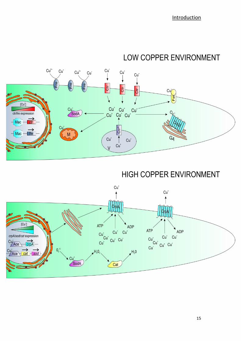
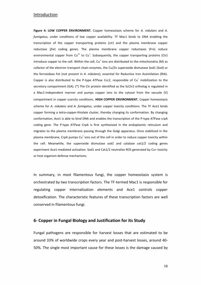
Figure 4: LOW COPPER ENVIRONMENT. Copper homeostasis scheme for A. nidulans and A.
fumigatus, under conditions of low copper availability. TF Mac1 binds to DNA enabling the
transcription of the copper transporting proteins (ctr) and the plasma membrane copper
reductase (fre) coding genes. The plasma membrane copper reductases (Fre) reduce
environmental copper from Cu2+
to Cu+. Subsequently, the copper transporting proteins (Ctr)
introduce copper to the cell. Within the cell, Cu+ ions are distributed to the mitochondria (M) as
cofactor of the electron transport chain enzymes, the Cu/Zn superoxide dismutase Sod1 (Sod) or
the ferroxidase Fet (not present in A. nidulans), essential for Reductive Iron Assimilation (RIA).
Copper is also distributed to the P-type ATPase Ccc2, responsible of Cu+ mobilization to the
secretory compartment (GA). (*) The Ctr protein identified as the ScCtr2 ortholog is regulated in
a Mac1-independent manner and pumps copper ions to the cytosol from the vacuole (V)
compartment in copper scarcity conditions. HIGH COPPER ENVIRONMENT. Copper homeostasis
scheme for A. nidulans and A. fumigatus, under copper toxicity conditions. The TF Ace1 binds
copper forming a tetra-copper-thiolate cluster, thereby changing its conformation. By changing
conformation, Ace1 is able to bind DNA and enables the transcription of the P-type ATPase crpA
coding gene. The P-type ATPase CrpA is first synthesized in the endoplasmic reticulum and
migrates to the plasma membrane passing through the Golgi apparatus. Once stabilized in the
plasma membrane, CrpA pumps Cu+ ions out of the cell in order to reduce copper toxicity within
the cell. Meanwhile, the superoxide dismutase sod1 and catalase cat1/2 coding genes
experiment Ace1-mediated activation. Sod1 and Cat1/2 neutralize ROS generated by Cu+ toxicity
or host organism defense mechanisms.
In summary, in most filamentous fungi, the copper homeostasis system is
orchestrated by two transcription factors. The TF-termed Mac1 is responsible for
regulating copper internalization elements and Ace1 controls copper
detoxification. The characteristic features of these transcription factors are well
conserved in filamentous fungi.
6- Copper in Fungal Biology and Justification for its Study
Fungal pathogens are responsible for harvest losses that are estimated to be
around 33% of worldwide crops every year and post-harvest losses, around 40-
50%. The single most important cause for these losses is the damage caused by
Introduction
17
fungal pathogens (Alkan and Fortes, 2015). On the other hand, fungal infections
affecting humans by organisms like Candida, Aspergillus, Cryptococcus or
Histoplasma cause about half a million deaths each year (Brown et al., 2012). In
the bulk of the cases pathogenic fungi are filamentous or make use of the
transient filamentous form to invade host cells. This is the case in human
(Aspergillus fumigatus, Hystoplasma capsulatum, Candida albicans...) and plant
pathogens (Botrytis cinerea, Fusarium spp. or Alternaria spp., to name a few;
Bastmeyer et al., 2002;Doehlemann et al., 2017).
Host organisms have developed defense strategies against fungal pathogens that
target copper availability. By scavenging Cu+ in the infection area, copper
deprivation can be induced in the pathogen. On the contrary, host innate
immune cells, such as macrophages, are able to mobilize copper to invade fungal
tissue as a defense mechanism. The generation of Reactive Oxygen Species (ROS)
is another defense mechanism employed by the innate immune system (Garcia-
Santamarina and Thiele, 2015).
A. fumigatus is one of the notorious airborne fungal pathogens responsible for
severe invasive aspergillosis (IA), especially in immunocompromised individuals
(Cai et al., 2017). Copper is a recognized virulence factor, as it is the cofactor of
many enzymes that contribute to virulence, such as laccases or superoxide
dismutase (Sod). Enzymes of the Sod family are copper-dependent enzymes
responsible for neutralizing ROS. The Cu/Zn Sod enzyme is considered as an
important virulence factor in pathogenic in organisms, like C. albicans and C.
neoformans (Frohner et al., 2009;Narasipura et al., 2005). Laccases are involved
in melanin biosynthesis and they are copper-dependent enzymes (Upadhyay et
al., 2013). Melanin confers a non-immunogenic status to the fungus and a
protective layer to the action of host-derived ROS (Jahn et al., 2000;Pihet et al.,
2009). When copper uptake is impaired, laccase and Sod activity are substantially
reduced in A. fumigatus (Park et al., 2014).
Introduction
18
The A. fumigatus Ace1 homolog deletion represses the expression of catalases
such as Afcat1 and Afcat2 involved in hydrogen peroxide neutralization and the
TF AfatfA which is known to be involved in spore maturation and ROS-response
(Hagiwara et al., 2014;Wiemann et al., 2017). Moreover, it also inhibits the
expression of the copper extrusion pump AfcrpA (Wiemann et al., 2017), the
main copper detoxification system in A. fumigatus, leaving the fungus exposed to
copper mobilization to fungal tissue by host innate immune system cells. Thus,
the copper detoxification machinery is a key factor in pathogen viability during
infection.
Botrytis cinerea is a necrotrophic fungal plant pathogen of worldwide
distribution, capable of infecting a wide range of hosts. Copper-dependent
proteins play a central role in many aspects of the B. cinerea, including
pathogenesis. The P-type ATPase BcCcc2, an ortholog of the S. cerevisiae Ccc2
copper transporting P-type ATPase that delivers Cu to the secretory
compartment for subsequent protein modification (Smith et al., 2017), is crucial
for virulence in B. cinerea (Saitoh et al., 2010). The absence of BcCcc2 results in a
defective BcSod1 function (Lopez-Cruz et al., 2017), as well as other proteins,
proving the importance of the copper homeostasis system for B. cinerea
virulence.
In summary, copper homeostasis plays an essential role in pathogenic fungi
virulence development. The high-affinity copper uptake system enables the
maturation of many copper-dependent enzymes for virulence. On the other
hand, the detoxification system confers the organism with the necessary
resistance for survival. In the case of phytopathogens, the intracellular copper
delivery system is crucial for successful virulence development.
Copper has long been used as an antimicrobial agent in many different
agricultural practices (Judet-Correia et al., 2011) and the strategy based on the
intensive use of fungicides based on copper is maintained. However, copper
Introduction
19
accumulation causes harmful effects on soil and fresh water ecosystems, as well
as drinking water quality. Moreover, this excessive use has also brought about
increasing levels of copper resistance in microbial pathogens (Lamichane et al.,
2018;Santo et al., 2010).
The mechanisms of copper resistance in fungi need a thorough assessment in
order to find new and effective methods to complement or replace copper in the
control of fungal disease. New details on the mechanism of copper toxicity on
fungal cells may shed light on the potential use of synergists affecting copper
homeostasis that could help lower the currently required copper dose. The
acquisition of copper by fungi in the soil, and its exchange between
microorganisms and plants in a symbiotic relationship is another aspect which
should be considered, to ensure sustainable agricultural and environmental
conservation programs.
A. nidulans is a model filamentous ascomycete that remains as one of the most
studied lower eukaryotes. Gathering knowledge about the cellular response to
copper concentrations in A. nidulans could be a significant step towards
understanding the cellular response in related pathogenic fungi (Alternaria,
Botrytis, Magnaporthe, etc.) and thus achieve the abovementioned objective. In
this study, some key elements of copper homeostasis will be explained in A.
nidulans. In the first chapter, the transcriptomic behavior of A. nidulans in copper
toxicity conditions is discussed, along with a deep characterization of the high-
affinity copper uptake proteins AnCtrA and AnCtrC. In the second chapter, the A.
nidulans copper detoxification machinery will be exposed, focusing primarily on
the PIB-type ATPase AnCrpA. Finally, in the third chapter, a model of the CrpA
protein generated by homology modelling will be presented. With this protein
model virtual screening tests, followed by molecular docking and molecular
dynamic tests were made to test possible inhibitors of the protein.

20

21
Objectives

22
Objectives
23
Objectives
The objectives pursued by this thesis are the following ones:
Characterization of the high-affinity copper transporters AnCtrA and
AnCtrC in A. nidulans. Determine their function, hierarchy, expression
dynamics, subcellular localization and possible regulation mechanisms.
Characterization of the copper detoxification system in Aspergillus
nidulans. Identify the main agents of the system, to study their impact on
copper resistance and learn about their expression patterns, subcellular
localization and the transcription factor regulating the system.
To test molecular modelling techniques as new tools to search for
inhibitory compounds of different targets of the copper homeostasis
system of A. nidulans.

24

25
Materials & Methods

26
Materials & Methods
27
1- Strains, Primers and Plasmids
1.1- Aspergillus nidulans Strains
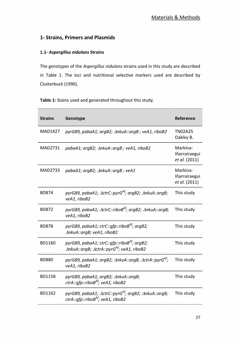
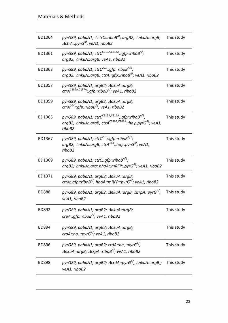
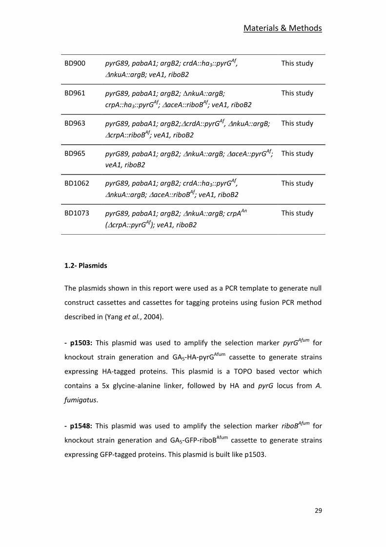
The genotypes of the Aspergillus nidulans strains used in this study are described
in Table 1. The loci and nutritional selective markers used are described by
Clutterbuck (1990).
Table 1: Stains used and generated throughout this study.
Strains Genotype Reference
MAD1427 pyrG89, pabaA1; argB2; nkuA::argB ; veA1, riboB2 TN02A25 Oakley B.
MAD2731 pabaA1; argB2;nkuA::argB ; veA1, riboB2 Markina-Iñarrairaegui et al. (2011)
MAD2733 pabaA1; argB2;nkuA::argB ; veA1 Markina-Iñarrairaegui et al. (2011)
BD874 pyrG89, pabaA1; ctrC::pyrGAf; argB2; nkuA::argB; veA1, riboB2
This study
BD872 pyrG89, pabaA1; ctrC::riboBAf; argB2;nkuA::argB; veA1, riboB2
This study
BD878 pyrG89, pabaA1; ctrC::gfp::riboBAf; argB2;
nkuA::argB; veA1, riboB2
This study
BD1160 pyrG89, pabaA1; ctrC::gfp::riboBAf; argB2;
nkuA::argB; ctrA::pyrGAf; veA1, riboB2
This study
BD880 pyrG89, pabaA1; argB2; nkuA::argB; ctrA::pyrGAf; veA1, riboB2
This study
BD1158 pyrG89, pabaA1; argB2; nkuA::argB; ctrA::gfp::riboBAf; veA1, riboB2
This study
BD1162 pyrG89, pabaA1; ctrC::pyrGAf; argB2;nkuA::argB; ctrA::gfp::riboBAf; veA1, riboB2
This study
Materials & Methods
28
BD1064 pyrG89, pabaA1; ctrC::riboBAf; argB2;nkuA::argB;
ctrA::pyrGAf; veA1, riboB2
This study
BD1361 pyrG89, pabaA1; ctrCC213A,C214A::gfp::riboBAf;
argB2;nkuA::argB; veA1, riboB2
This study
BD1363 pyrG89, pabaA1; ctrC201::gfp::riboBAf1;
argB2;nkuA::argB; ctrA::gfp::riboBAf; veA1, riboB2
This study
BD1357 pyrG89, pabaA1; argB2;nkuA::argB; ctrAC186A,C187A::gfp::riboBAf; veA1, riboB2
This study
BD1359 pyrG89, pabaA1; argB2;nkuA::argB; ctrA164::gfp::riboBAf; veA1, riboB2
This study
BD1365 pyrG89, pabaA1; ctrCC213A,C214A::gfp::riboBAf1;
argB2;nkuA::argB; ctrAC186A,C187A::ha3::pyrGAf; veA1, riboB2
This study
BD1367 pyrG89, pabaA1; ctrC201::gfp::riboBAf1;
argB2;nkuA::argB; ctrA164::ha3::pyrGAf; veA1, riboB2
This study
BD1369 pyrG89, pabaA1; ctrC::gfp::riboBAf1;
argB2;nkuA::arg; hhoA::mRFP::pyrGAf; veA1, riboB2
This study
BD1371 pyrG89, pabaA1; argB2;nkuA::argB; ctrA::gfp::riboBAf, hhoA::mRFP::pyrGAf; veA1, riboB2
This study
BD888 pyrG89, pabaA1; argB2; nkuA::argB; crpA::pyrGAf;
veA1, riboB2
This study
BD892 pyrG89, pabaA1; argB2;nkuA::argB;
crpA::gfp::riboBAf; veA1, riboB2
This study
BD894 pyrG89, pabaA1; argB2;nkuA::argB;
crpA::ha3::pyrGAf; veA1, riboB2
This study
BD896 pyrG89, pabaA1; argB2; crdA::ha3::pyrGAf,
nkuA::argB;crpA::riboBAf; veA1, riboB2
This study
BD898 pyrG89, pabaA1; argB2;crdA::pyrGAf, nkuA::argB;;
veA1, riboB2
This study
Materials & Methods
29
BD900 pyrG89, pabaA1; argB2; crdA::ha3::pyrGAf,
nkuA::argB; veA1, riboB2
This study
BD961 pyrG89, pabaA1; argB2;nkuA::argB;
crpA::ha3::pyrGAf; aceA::riboBAf; veA1, riboB2
This study
BD963 pyrG89, pabaA1; argB2;crdA::pyrGAf,nkuA::argB;
crpA::riboBAf; veA1, riboB2
This study
BD965 pyrG89, pabaA1; argB2;nkuA::argB; aceA::pyrGAf;
veA1, riboB2
This study
BD1062 pyrG89, pabaA1; argB2; crdA::ha3::pyrGAf,
nkuA::argB; aceA::riboBAf; veA1, riboB2
This study
BD1073 pyrG89, pabaA1; argB2;nkuA::argB; crpAAn
(crpA::pyrGAf); veA1, riboB2
This study
1.2- Plasmids
The plasmids shown in this report were used as a PCR template to generate null
construct cassettes and cassettes for tagging proteins using fusion PCR method
described in (Yang et al., 2004).
- p1503: This plasmid was used to amplify the selection marker pyrGAfum for
knockout strain generation and GA5-HA-pyrGAfum cassette to generate strains
expressing HA-tagged proteins. This plasmid is a TOPO based vector which
contains a 5x glycine-alanine linker, followed by HA and pyrG locus from A.
fumigatus.
- p1548: This plasmid was used to amplify the selection marker riboBAfum for
knockout strain generation and GA5-GFP-riboBAfum cassette to generate strains
expressing GFP-tagged proteins. This plasmid is built like p1503.
Materials & Methods
30
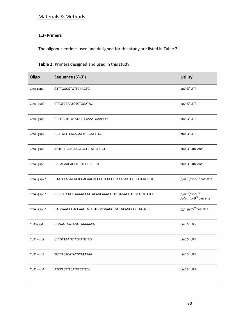
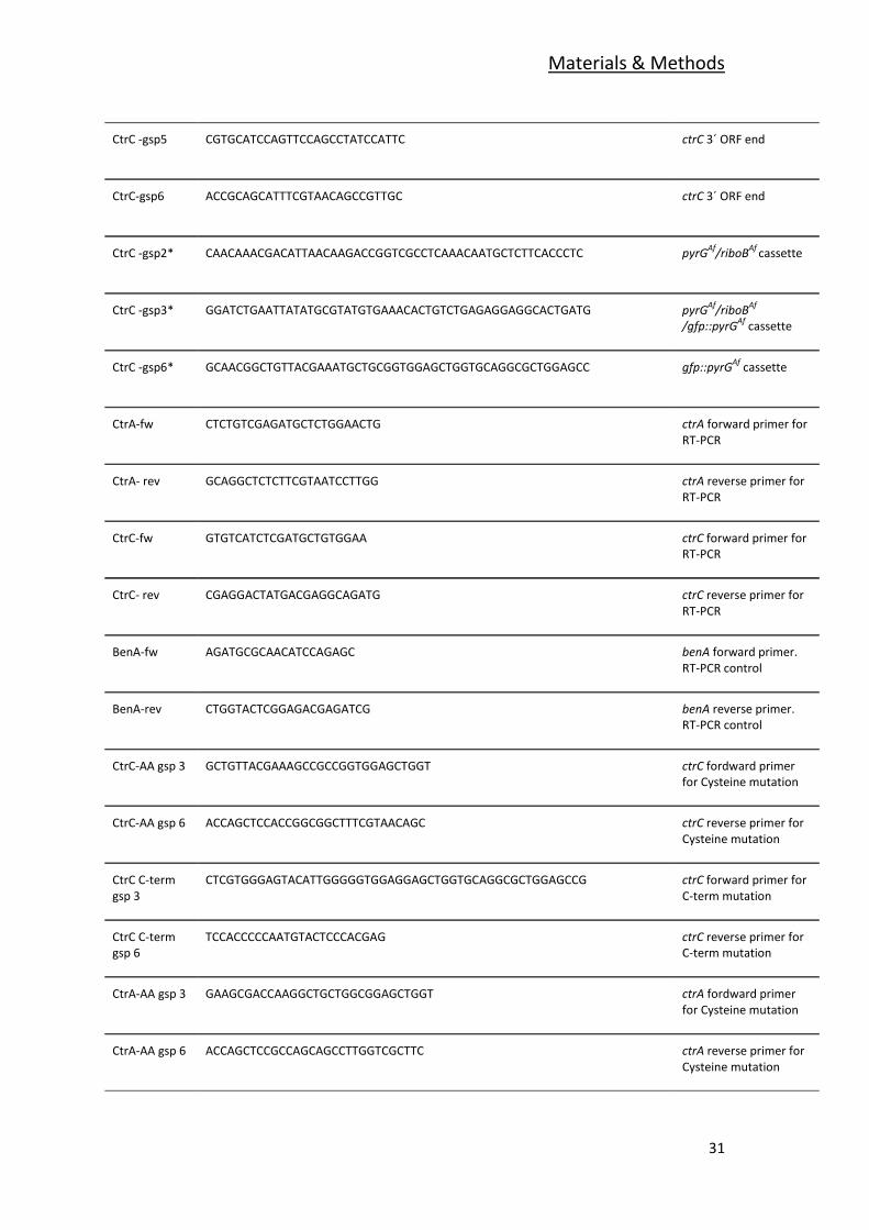
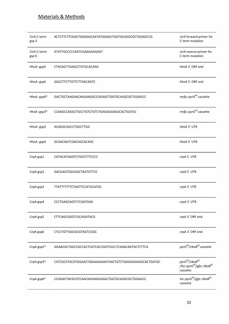
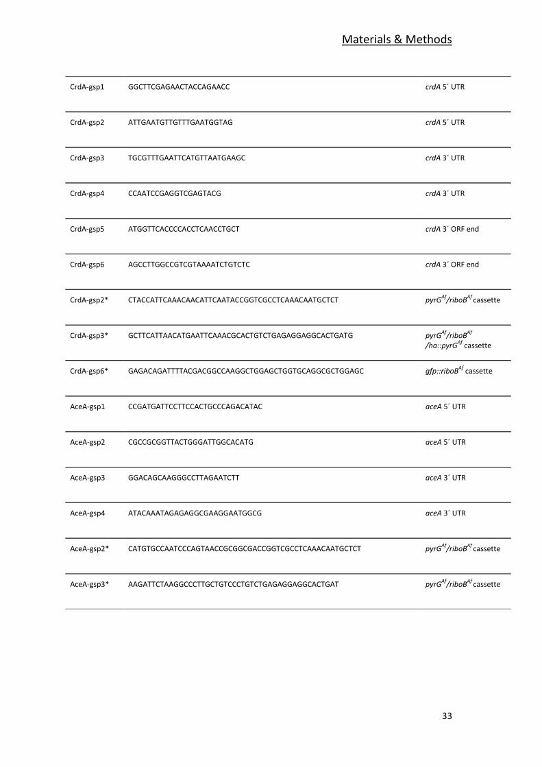
1.3- Primers
The oligonucleotides used and designed for this study are listed in Table 2.
Table 2: Primers designed and used in this study
Oligo Sequence (5´-3´) Utility
CtrA-gsp1 GTTTGGCGTGTTGAAGTG ctrA 5´ UTR
CtrA -gsp2 CTTGTCGAATGTCTGGATAC ctrA 5´ UTR
CtrA -gsp3 CTTTGCTGTATATATTTTAAATGAAGCGC ctrA 3´ UTR
CtrA -gsp4 GCTTATTTCACAGATTGGGATTTCC ctrA 3´ UTR
CtrA -gsp5 AGTCTTCAAGAAGCGCTTTATCATTCT ctrA 3´ ORF end
CtrA -gsp6 GCCACAACACTTGGTCGCTTCCTC ctrA 3´ ORF end
CtrA -gsp2* GTATCCAGACATTCGACAAGACCGGTCGCCTCAAACAATGCTCTTCACCCTC pyrGAf
/riboBAf
cassette
CtrA -gsp3* GCGCTTCATTTAAAATATATACAGCAAAGGTCTGAGAGGAGGCACTGATGC pyrGAf
/riboBAf
/gfp::riboBAf
cassette
CtrA -gsp6* GAGGAAGCGACCAAGTGTTGTGGCGGAGCTGGTGCAGGCGCTGGAGCC gfp::pyrGAf
cassette
CtrC-gsp1 GGGAGTGATAGGTAAAAACA ctrC 5´ UTR
CtrC -gsp2 CTTGTTAATGTCGTTTGTTG ctrC 5´ UTR
CtrC -gsp3 TGTTTCACATACGCATATAA ctrC 3´ UTR
CtrC -gsp4 ATCCTCTTTCATCTCTTTCC ctrC 3´ UTR
Materials & Methods
31
CtrC -gsp5 CGTGCATCCAGTTCCAGCCTATCCATTC ctrC 3´ ORF end
CtrC-gsp6 ACCGCAGCATTTCGTAACAGCCGTTGC ctrC 3´ ORF end
CtrC -gsp2* CAACAAACGACATTAACAAGACCGGTCGCCTCAAACAATGCTCTTCACCCTC pyrGAf
/riboBAf
cassette
CtrC -gsp3* GGATCTGAATTATATGCGTATGTGAAACACTGTCTGAGAGGAGGCACTGATG pyrGAf
/riboBAf
/gfp::pyrGAf
cassette
CtrC -gsp6* GCAACGGCTGTTACGAAATGCTGCGGTGGAGCTGGTGCAGGCGCTGGAGCC gfp::pyrGAf
cassette
CtrA-fw CTCTGTCGAGATGCTCTGGAACTG ctrA forward primer for RT-PCR
CtrA- rev GCAGGCTCTCTTCGTAATCCTTGG ctrA reverse primer for RT-PCR
CtrC-fw GTGTCATCTCGATGCTGTGGAA ctrC forward primer for RT-PCR
CtrC- rev CGAGGACTATGACGAGGCAGATG ctrC reverse primer for RT-PCR
BenA-fw AGATGCGCAACATCCAGAGC benA forward primer. RT-PCR control
BenA-rev CTGGTACTCGGAGACGAGATCG benA reverse primer. RT-PCR control
CtrC-AA gsp 3 GCTGTTACGAAAGCCGCCGGTGGAGCTGGT ctrC fordward primer for Cysteine mutation
CtrC-AA gsp 6 ACCAGCTCCACCGGCGGCTTTCGTAACAGC ctrC reverse primer for Cysteine mutation
CtrC C-term gsp 3
CTCGTGGGAGTACATTGGGGGTGGAGGAGCTGGTGCAGGCGCTGGAGCCG ctrC forward primer for C-term mutation
CtrC C-term gsp 6
TCCACCCCCAATGTACTCCCACGAG ctrC reverse primer for C-term mutation
CtrA-AA gsp 3 GAAGCGACCAAGGCTGCTGGCGGAGCTGGT ctrA fordward primer for Cysteine mutation
CtrA-AA gsp 6 ACCAGCTCCGCCAGCAGCCTTGGTCGCTTC ctrA reverse primer for Cysteine mutation
Materials & Methods
32
CtrA C-term gsp 3
ACTCTTCTTCGACTGGGGGCAATATGGAGCTGGTGCAGGCGCTGGAGCCG ctrA forward primer for C-term mutation
CtrA C-term-gsp 6
ATATTGCCCCCAGTCGAAGAAGAGT ctrA reverse primer for C-term mutation
HhoA -gsp5 CTACAGTTGAGCCTATGCACAAG hhoA 3´ ORF end
HhoA -gsp6 GGCCTTCTTGTTCTTAGCAGTC hhoA 3´ ORF end
HhoA -gsp6* GACTGCTAAGAACAAGAAGGCCGGAGCTGGTGCAGGCGCTGGAGCC mrfp::pyrGAf
cassette
HhoA -gsp3* CCAAGCCAGGCTGCCTGTCTGTCTGAGAGGAGGCACTGATGC mrfp::pyrGAf
cassette
HhoA -gsp3 ACAGGCAGCCTGGCTTGG hhoA 3´ UTR
HhoA -gsp4 GCAACAGTCGACAGCACAGC hhoA 3´ UTR
CrpA-gsp1 CGTACATGGGTCTGGTCTTCCCC crpA 5´ UTR
CrpA-gsp2 GACGAGTGGCGGCTAGTGTTCC crpA 5´ UTR
CrpA-gsp3 TTATTTTTTTCTAGTTCCATGCATGC crpA 3´ UTR
CrpA-gsp4 CCCTGAGCAGTCTCGATGAG crpA 3´ UTR
CrpA-gsp5 CTTCAGCGGGTCGCAGATACG crpA 3´ ORF end
CrpA-gsp6 CTCCTGTTGACGCGTAGTCCGG crpA 3´ ORF end
CrpA-gsp2* GGAACACTAGCCGCCACTCGTCACCGGTCGCCTCAAACAATGCTCTTCA pyrGAf
/riboBAf
cassette
CrpA-gsp3* CATCGCATGCATGGAACTAGAAAAAAATAACTGTCTGAGAGGAGGCACTGATGC pyrGAf
/riboBAf
/ha::pyrGAf
/gfp::riboBAf
cassette
CrpA-gsp6* CCGGACTACGCGTCAACAGGAGGGAGCTGGTGCAGGCGCTGGAGCC ha::pyrGAf
/gfp::riboBAf
cassette
Materials & Methods
33
CrdA-gsp1 GGCTTCGAGAACTACCAGAACC crdA 5´ UTR
CrdA-gsp2 ATTGAATGTTGTTTGAATGGTAG crdA 5´ UTR
CrdA-gsp3 TGCGTTTGAATTCATGTTAATGAAGC crdA 3´ UTR
CrdA-gsp4 CCAATCCGAGGTCGAGTACG crdA 3´ UTR
CrdA-gsp5 ATGGTTCACCCCACCTCAACCTGCT crdA 3´ ORF end
CrdA-gsp6 AGCCTTGGCCGTCGTAAAATCTGTCTC crdA 3´ ORF end
CrdA-gsp2* CTACCATTCAAACAACATTCAATACCGGTCGCCTCAAACAATGCTCT pyrGAf
/riboBAf
cassette
CrdA-gsp3* GCTTCATTAACATGAATTCAAACGCACTGTCTGAGAGGAGGCACTGATG pyrGAf
/riboBAf
/ha::pyrGAf
cassette
CrdA-gsp6* GAGACAGATTTTACGACGGCCAAGGCTGGAGCTGGTGCAGGCGCTGGAGC gfp::riboBAf
cassette
AceA-gsp1 CCGATGATTCCTTCCACTGCCCAGACATAC aceA 5´ UTR
AceA-gsp2 CGCCGCGGTTACTGGGATTGGCACATG aceA 5´ UTR
AceA-gsp3 GGACAGCAAGGGCCTTAGAATCTT aceA 3´ UTR
AceA-gsp4 ATACAAATAGAGAGGCGAAGGAATGGCG aceA 3´ UTR
AceA-gsp2* CATGTGCCAATCCCAGTAACCGCGGCGACCGGTCGCCTCAAACAATGCTCT pyrGAf
/riboBAf
cassette
AceA-gsp3* AAGATTCTAAGGCCCTTGCTGTCCCTGTCTGAGAGGAGGCACTGAT pyrGAf
/riboBAf
cassette
Materials & Methods
34
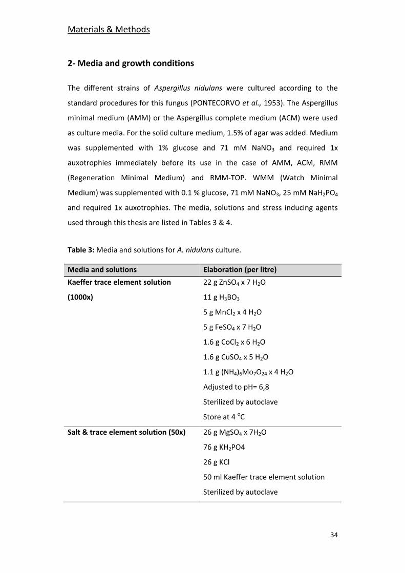
2- Media and growth conditions
The different strains of Aspergillus nidulans were cultured according to the
standard procedures for this fungus (PONTECORVO et al., 1953). The Aspergillus
minimal medium (AMM) or the Aspergillus complete medium (ACM) were used
as culture media. For the solid culture medium, 1.5% of agar was added. Medium
was supplemented with 1% glucose and 71 mM NaNO3 and required 1x
auxotrophies immediately before its use in the case of AMM, ACM, RMM
(Regeneration Minimal Medium) and RMM-TOP. WMM (Watch Minimal
Medium) was supplemented with 0.1 % glucose, 71 mM NaNO3, 25 mM NaH2PO4
and required 1x auxotrophies. The media, solutions and stress inducing agents
used through this thesis are listed in Tables 3 & 4.
Table 3: Media and solutions for A. nidulans culture.
Media and solutions Elaboration (per litre)
Kaeffer trace element solution 22 g ZnSO4 x 7 H2O
(1000x) 11 g H3BO3
5 g MnCl2 x 4 H2O
5 g FeSO4 x 7 H2O
1.6 g CoCl2 x 6 H2O
1.6 g CuSO4 x 5 H2O
1.1 g (NH4)6Mo7O24 x 4 H2O
Adjusted to pH= 6,8
Sterilized by autoclave
Store at 4 oC
Salt & trace element solution (50x) 26 g MgSO4 x 7H2O
76 g KH2PO4
26 g KCl
50 ml Kaeffer trace element solution
Sterilized by autoclave
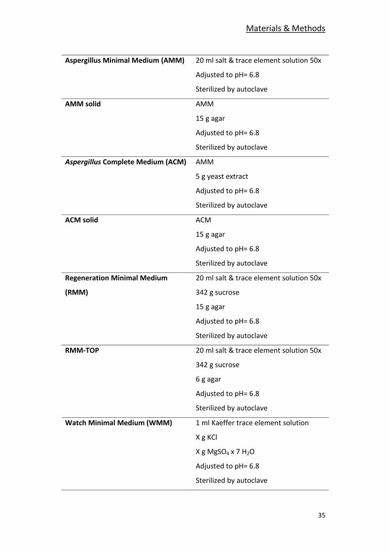
Materials & Methods
35
Aspergillus Minimal Medium (AMM) 20 ml salt & trace element solution 50x
Adjusted to pH= 6.8
Sterilized by autoclave
AMM solid AMM
15 g agar
Adjusted to pH= 6.8
Sterilized by autoclave
Aspergillus Complete Medium (ACM) AMM
5 g yeast extract
Adjusted to pH= 6.8
Sterilized by autoclave
ACM solid ACM
15 g agar
Adjusted to pH= 6.8
Sterilized by autoclave
Regeneration Minimal Medium 20 ml salt & trace element solution 50x
(RMM) 342 g sucrose
15 g agar
Adjusted to pH= 6.8
Sterilized by autoclave
RMM-TOP 20 ml salt & trace element solution 50x
342 g sucrose
6 g agar
Adjusted to pH= 6.8
Sterilized by autoclave
Watch Minimal Medium (WMM) 1 ml Kaeffer trace element solution
X g KCl
X g MgSO4 x 7 H2O
Adjusted to pH= 6.8
Sterilized by autoclave
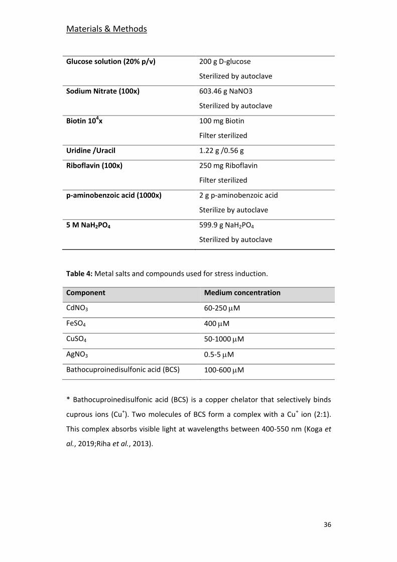
Materials & Methods
36
Glucose solution (20% p/v) 200 g D-glucose
Sterilized by autoclave
Sodium Nitrate (100x) 603.46 g NaNO3
Sterilized by autoclave
Biotin 104x 100 mg Biotin
Filter sterilized
Uridine /Uracil 1.22 g /0.56 g
Riboflavin (100x) 250 mg Riboflavin
Filter sterilized
p-aminobenzoic acid (1000x) 2 g p-aminobenzoic acid
Sterilize by autoclave
5 M NaH2PO4 599.9 g NaH2PO4
Sterilized by autoclave
Table 4: Metal salts and compounds used for stress induction.
Component Medium concentration
CdNO3 60-250 M
FeSO4 400 M
CuSO4 50-1000 M
AgNO3 0.5-5 M
Bathocuproinedisulfonic acid (BCS) 100-600 M
* Bathocuproinedisulfonic acid (BCS) is a copper chelator that selectively binds
cuprous ions (Cu+). Two molecules of BCS form a complex with a Cu+ ion (2:1).
This complex absorbs visible light at wavelengths between 400-550 nm (Koga et
al., 2019;Riha et al., 2013).
Materials & Methods
37
3- Construction of Replacement Cassettes for Null and Tagged
Protein Strain Generation
Different strains were generated by homologous recombination using linear
constructions of DNA obtained by PCR amplification (Yang et al., 2004). The
enzyme used for this purpose was the Prime Star(R) HS DNA polymerase
(Takara), with 3’-5’/5’-3’ proofreading activity. The PCR conditions were set
following Takara´s protocol, with some variations (time and annealing
temperature) depending on the used primers and the size of the fragment
amplified.
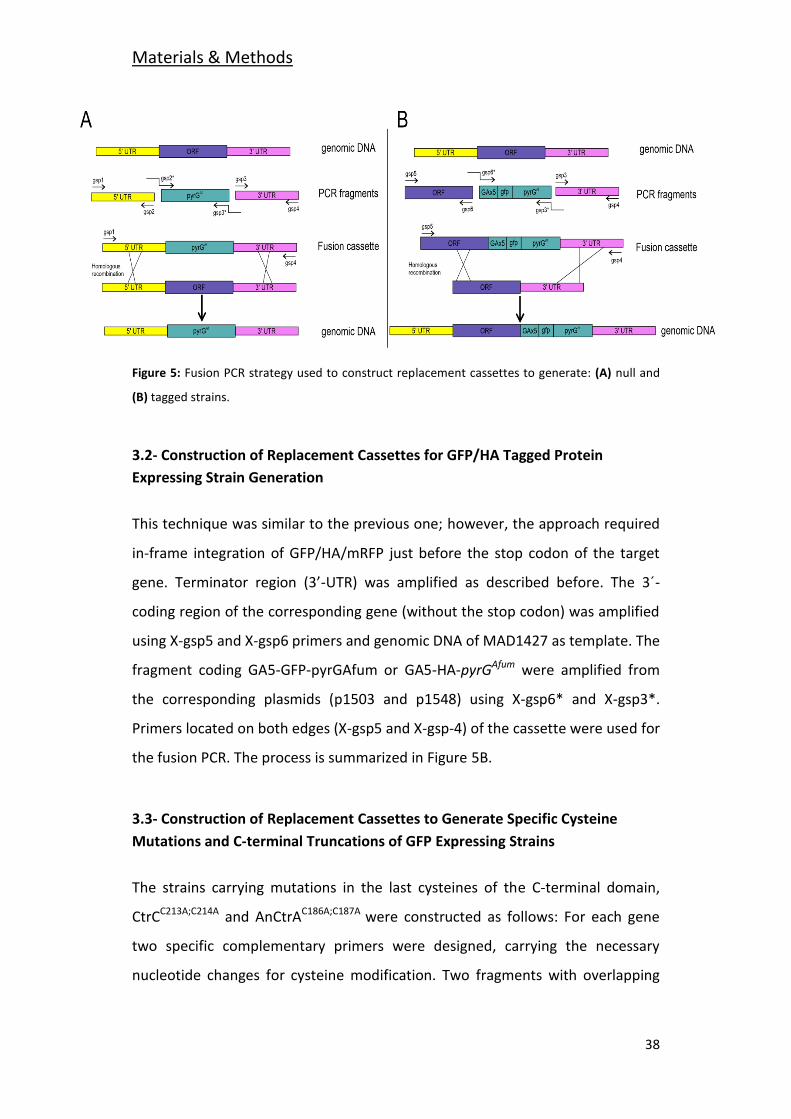
3.1- Construction of Replacement Cassettes for Null Mutant Strain Generation
The different steps of the cassette construction process are shown in Figure 5A.
In the first step, by standard PCR reaction the promoter (5’-UTR) and the
terminator (3’-UTR) regions flanking the target loci were amplified, using
genomic DNA of the wild type strain (MAD1427) as template. The fragment
corresponding to the promoter, obtained with the X-gsp1 and X-gsp2 primers, is
approximately 1,500 bp long upstream of the gen. The terminator, around 1,500
bp long, corresponds to the downstream region starting immediately after the
stop codon and was obtained with primers X-gsp3 and X-gsp4. For the
construction of each knockout cassette the consequent nutritional selection
marker, pyrGAfum or riboBAfum, was amplified with primers X-gsp2* and X-gsp3*
from the corresponding plasmid (p1503 and p1548).
The amplicons have overlapping regions that permit their fusion, using primers
X-gsp1 and X-gsp4, in a new PCR reaction. The resulting product is the
replacement cassette used in the generation of the null strains.
Materials & Methods
38
Figure 5: Fusion PCR strategy used to construct replacement cassettes to generate: (A) null and
(B) tagged strains.
3.2- Construction of Replacement Cassettes for GFP/HA Tagged Protein
Expressing Strain Generation
This technique was similar to the previous one; however, the approach required
in-frame integration of GFP/HA/mRFP just before the stop codon of the target
gene. Terminator region (3’-UTR) was amplified as described before. The 3´-
coding region of the corresponding gene (without the stop codon) was amplified
using X-gsp5 and X-gsp6 primers and genomic DNA of MAD1427 as template. The
fragment coding GA5-GFP-pyrGAfum or GA5-HA-pyrGAfum were amplified from
the corresponding plasmids (p1503 and p1548) using X-gsp6* and X-gsp3*.
Primers located on both edges (X-gsp5 and X-gsp-4) of the cassette were used for
the fusion PCR. The process is summarized in Figure 5B.
3.3- Construction of Replacement Cassettes to Generate Specific Cysteine
Mutations and C-terminal Truncations of GFP Expressing Strains
The strains carrying mutations in the last cysteines of the C-terminal domain,
CtrCC213A;C214A and AnCtrAC186A;C187A were constructed as follows: For each gene
two specific complementary primers were designed, carrying the necessary
nucleotide changes for cysteine modification. Two fragments with overlapping
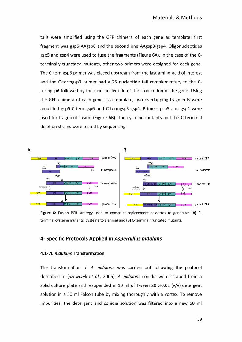
Materials & Methods
39
tails were amplified using the GFP chimera of each gene as template; first
fragment was gsp5-AAgsp6 and the second one AAgsp3-gsp4. Oligonucleotides
gsp5 and gsp4 were used to fuse the fragments (Figure 6A). In the case of the C-
terminally truncated mutants, other two primers were designed for each gene.
The C-termgsp6 primer was placed upstream from the last amino-acid of interest
and the C-termgsp3 primer had a 25 nucleotide tail complementary to the C-
termgsp6 followed by the next nucleotide of the stop codon of the gene. Using
the GFP chimera of each gene as a template, two overlapping fragments were
amplified gsp5-C-termgsp6 and C-termgsp3-gsp4. Primers gsp5 and gsp4 were
used for fragment fusion (Figure 6B). The cysteine mutants and the C-terminal
deletion strains were tested by sequencing.
Figure 6: Fusion PCR strategy used to construct replacement cassettes to generate: (A) C-
terminal cysteine mutants (cysteine to alanine) and (B) C-terminal truncated mutants.
4- Specific Protocols Applied in Aspergillus nidulans
4.1- A. nidulans Transformation
The transformation of A. nidulans was carried out following the protocol
described in (Szewczyk et al., 2006). A. nidulans conidia were scraped from a
solid culture plate and resupended in 10 ml of Tween 20 %0.02 (v/v) detergent
solution in a 50 ml Falcon tube by mixing thoroughly with a vortex. To remove
impurities, the detergent and conidia solution was filtered into a new 50 ml
Materials & Methods
40
Falcon tube using sterile Miracloth (Calbiochem, 475855) and centrifuged at
4,000 rpm for 10 min. After centrifugation the supernatant was discarded and
the conidia concentrated in the pellet was resuspended in fresh Tween 20 %0.02
(v/v) detergent solution. Conidia concentration was measured by counting with a
hemocytometer (Thoma) under the microscope (Optihot, Nikon) using a 40x
objective. 2.5 x 10⁶ conidiospores/ml were inoculated in 150 ml of liquid ACM
containing the carbon and nitrogen sources together with the required
supplements. After 14 h of growth at 30 oC the mycelium was recollected by
filtration. One gram (1g) of mycelium was resuspended in 16 ml of ACM
containing the carbon and nitrogen sources together with the required
supplements and 16 ml of Protoplasting solution (1.1 M KCl, 0.1 M citric acid, pH
5.5, 2.048 g of VinoTaste® (Novozymes)), an enzyme for digestion of the cell wall,
was added. The mixture was incubated at 30 oC from 1 h to 2 h with gentle
agitation. Protoplast formation was checked by observation of samples under an
optical microscope.
During the protoplast formation process, two 50 ml falcon tubes with 16 ml of
1.2 M sucrose were prepared. The protoplast suspension was divided into equal
volumes and gently added onto the 16 ml of 1.2 M sucrose solution to create
two different phases. After centrifuging the biphasic solution at 4 oC for 15 min
at 1,800 g, the protoplast fraction concentrated between the two phases was
collected with a sterile Pasteur pipette. The collected protoplast fraction was
diluted by adding two volumes of 0.6 M KCl. The suspension was again
centrifuged at 4 oC for 10 min at 1,800 g. The pellet containing the protoplasts
was washed three times with 0.6 M KCl and finally concentrated in 1 ml of 0.6 M
KCl, 50 mM CaCl2.
The transformation process starts when 100 l protoplasts were mixed with DNA
(transformation cassette, 1-3 μg of DNA) and 50 μl of Solution 8 (PEG 6,000 60%
(p/v), 10 mM Tris-HCl pH 7.5, 10 mM CaCl2). After incubating the mixture in ice
for 20 min, 1 ml of Solution 8 was added and incubated at room temperature for
Materials & Methods
41
another 5 min. Finally, 5 ml of Solution 7 (1 M Sorbitol, 10 mM Tris-HCl pH 7.5,
10 mM CaCl2) and 15 ml of warm RMM-TOP regeneration medium were added.
The mixture was plated over the Petri plates containing selective regeneration
minimal medium. The colonies grown from the transformed protoplasts were
observed after 3-4 days of incubation at 37 oC. After conidia purification in
selective medium, homokaryotic transformants were maintained. The presence
of the recombinant nuclei was confirmed by PCR and Southern-blot technique as
described in (Sambrook et al., 1989).
4.2- Conidia Count
For conidia count experiments, the different strains were inoculated by point-
inoculation in 55 mm Petri dishes in the selected conditions. Then they were
incubated for 72 h at 37 oC. After the incubation time the area of the colonies
was calculated using the Digimizer Image Analysis Software. Then the colony was
collected into 50 ml falcon tubes in 40 ml Tween 20 0.02 % (v/v). The Falcon tube
was agitated thoroughly using a vortex for approximately 2 min. The
conidiospores were then counted using a hemocytometer (Thoma) under the
microscope (Optihot, Nikon) using a 40x objective. Using the area measurements
and calculated area of each strain, the conidia density per area was calculated
(conidia/cm2). Three replicates of each strain were made and the results were
statistically treated. The mean values with their standard deviation values are
shown in the graphs.
5- Molecular Biology, Biochemistry and Proteomic Techniques
Applied in Aspergillus nidulans
5.1- Genomic DNA Extraction
DNA extraction was performed following the method described by Herrero-
García (2013). In 100 ml Erlenmeyer flasks 30 ml of supplemented AMM were
Materials & Methods
42
prepared and inoculated with a 106 conidia/ml concentration (following the
procedure explained in section 4.1). The inoculated media was then incubated at
37 oC at 250 rpm for 16 h. Approximately 300 mg of mycelium was collected by
filtration using Miracloth and lyophilized for 10 h in a lyophilizer. The lyophilized
mycelium was homogenyzed in a mini bead beater and resuspended in 1 ml of
lysis buffer (25 mM Tris-HCl pH 8.0, 0.25 M sucrose and 20 mM EDTA). After the
addition of 100 μl of 10% SDS the suspension was incubated at 65 oC for 15 min.
One milliliter (1 ml) of phenol/chloroform/isoamylalcohol (50:48:2) mix was
added to the fungal extract. The suspension was shaken vigorously for 10 min
and immediately after centrifuged for 5 min at 14,000 rpm. The aqueous phase
was placed in a new Eppendorf. This procedure was repeated twice.
The genomic DNA of the aqueous phase was precipitated by adding 1/10 of the
volume of Sodium Acetate (NaAc 3 M pH 6) and 0,6 volume of Isopropanol to the
supernatant. The mixture was incubated for 15 min at room temperature. The
mixture was centrifuged for 5 min at 14,000 rpm and the pellet was washed with
1 ml of 80% ethanol. After a second centrifugation, the supernatant was
eliminated and the dried pellet was resuspended in 300-500 μl of milli-Q water.
To prevent any RNA contamination every sample was treated with RNase-A
(Roche) incubating the reaction at 37 oC for 1 h (mixing every 10-15 min). The
genomic DNA was precipitated using NaAc and isopropanol and sedimented as
mentioned above. The genomic DNA was washed again with 0.5 ml of ethanol
80% (v/v), as indicated before. Once the pellet was dry, the DNA was
resuspended in 100-200 μl of milli-Q water and stored at -20 oC. The quality and
concentration of the extracted DNA was tested by electrophoresis in a 0.8 %
(w/v) agarose gel.
Materials & Methods
43
5.2- DNA Analysis by Southern-Blot
After the extracted gDNAs were digested with the appropriate restriction
enzyme, the digestion products were run in a 0.8 % (w/v) agarose gel for 2 hours
at 90 Volts.
After 10 min exposure to UV light (320nm, Vilber Lourmat), the gel was
incubated for 45 min in denaturing solution (1.5 M NaCl, 0.5 M NaOH) and twice
(2 x 30 min) in neutralizing solution (0.5 M Tris-HCl pH 7.5, 3 M NaCl). The gel
was homogenized with 20x SSC solution (3 M NaCl, 300 mM Sodium Citrate, pH
7) it was transferred to a nylon membrane (Zeta-Probe® blotting, Bio-Rad) by
following the standard protocol of capillarity transference (Sambrook et al.,
1989). The transferred DNA was fixed to the nylon membrane by exposing the
membrane to two 120 mJ UV light pulses (Vilber Lourmat BLX-E254, 254 nm).
The membrane was pre hybridized by incubating for 2 h at 42 oC in Church buffer
(Church and Gilbert, 1984), before hybridizing with a specific DNA probe marked
with Digoxigenin-dUTP prepared the day earlier (DIG High Prime DNA LABELLING
AND DETECTION STARTER KIT II (Roche)).
For DNA probe generation, 1 μg were diluted in 16 μl. The DNA was first
denatured by incubating for 10 min at 95 °C. Then it was cooled in ice for 5 min
and finally, 4 l of DIG High Prime were added and the probe was incubated at
37 oC overnight. The reaction was stopped by adding 2 μl of EDTA (0.2 M pH 8.0)
and heating the sample for 10 min at 65 °C. For DNA hybridization, 5 l of the
probe were taken and denatured by incubating 5 min at 95 °C and then cooled in
ice for 1 min before adding to the hybridization tube. The hybridization process
was carried out at 42°C overnight.
The membrane was incubated with the marked probe for 16 h at 42 °C. To
eliminate probe excess two washing steps (30 ml of 2x SSC, 0.1 % SDS) of 5 min
and two more of 15 min at 65 oC (40 ml of 0.5x SSC, 0.1 % SDS) were carried out.
Materials & Methods
44
For the correct blocking the membrane was incubated for 30 min with 40 ml of
blocking solution (stock 10x from the kit diluted in a maleic acid solution: 0.1 M
Maleic acid, 0.15 M NaCl, pH 7.5). Two microliters (2 l) of Anti-Digoxigenin-AP
1:10,000 were added to 20 ml of blocking solution and incubated for 30 min. The
membrane was washed (two 20 min washing steps in 0.1 M Maleic acid, 0.15 M
NaCl, 0.3% tween 20, pH 7.5 solution) and balanced with detection buffer (5 min
balancing step in 0.1 M NaCl, 0.1 M Tris-HCl pH 9.5). The membrane was placed
in a plastic surface. The chromogenic agent CSPD ready-to-use (Roche) was
added on the DNA containing face of the membrane, expanded uniformly and
incubated for 5 min. After the incubation the membrane was dried and
incubated at 37 °C for 10 min to improve the luminescent reaction. The images
of the detection were taken with a XR GelDoc chemiluminescence Analyzer and
were processed with the ImageLab program.
5.3- Membrane Protein Extraction
Procedure described in Hervás-Aguilar and Peñalva (2010). Samples were
incubated as described in section 5.1. The samples were collected by filtering
with Miracloth, collected into an Eppendorf tube and immediately snap frozen by
submerging in liquid nitrogen. Then, the frozen mycelium was lyophilized for 10
h. The lyophilized mycelium was crushed with a metal bead in a mini bead beater
placed inside a fridge (4 oC). Approximately 6-7 mg of mycelium was
resuspended in 1 ml of pre-cooled lysis buffer (0.2 M NaOH, 0.2% β-
mercaptoethanol) by vigorous agitation for 1.5 min. To precipitate the proteins,
7.5% (w/v) TCA was added. The suspension was mixed gently and incubated for
10 min in ice. After the mixture was centrifuged at 4 oC for 5 min at 14,000 rpm
the supernatant was taken away. A second centrifugation was performed in
order to make sure that all TCA was removed. The pellet was partially
resuspended by adding 100 l of 1 M TrisBase and 200 l of Laemmli buffer.
Samples were stored at -20 oC.
Materials & Methods
45
5.4- Western-Blot
The precipitated protein samples were denatured at 95 oC and run in 10% (w/v)
SDS-polyacrylamide gels (MiniProtean 3 system, Biorad) (Laemmli, 1970). The
proteins of the gel were transferred to a nitrocellulose membrane. Prior to
blocking the membrane a Ponceau staining was conducted as a loading control.
The membrane was washed three times for 5 min with distilled water in
agitation; then, the Ponceau stain (0.5% (w/v) Ponceau S dissolved in 1% (v/v)
acetic acid) was added and incubated for 1 min. Multiple washes with water
were carried out until the protein bands were visible. Then, a picture of the
membrane was taken. To remove the Ponceau staining, the membrane was
incubated in a 200 μM NaOH, 20% (v/v) acetonitrile solution for 1 min. After
some additional washes with distilled water the membrane was ready to block.
After blocking, tagged proteins were detected depending on the antigen or
epitope, with their respective primary antibody (Table 5). In all the cases the
secondary antibody was conjugated to horseradish peroxidase. The peroxidase
activity was detected with Clarity TM Western chemiluminescence system
(Biorad) in a XR GelDoc chemiluminescence Analyzer. ImageLab software was
used to process and analyze the images taken.
Table 5: Antibodies used in the study
ANTIBODY DILUTION SOURCE REFERENCE
1º α-GFP 1:5000 mouse ROCHE
α-HA 1:10000 mouse Santa Cruz
α-Hexokinase 1:80000 rabbit Cemicon Intemat Inc
2º α-mouse IgG 1:4000 goat Jackson InmunoResearch
α-rabbit IgG 1:10000 ass Sigma
Materials & Methods
46
5.5- Fluorescence Microscopy
Aspergillus nidulans strains expressing CprA::GFP, CtrC::GFP-Hhoa::mRFP and
CtrA::GFP-HhoA::mRFP in vegetative state were studied. A phosphate-
supplemented of low phosphate minimal medium (“WATCH” minimal medium,
WMM) version was used for culture. The basal medium containing 17 mM KCl, 5
mM MgSO4 and 1/400 of Kaeffer’s trace element solution was supplemented
with 0.1% glucose (w/v), 71 mM NaNO3 and 25 mM NaH2PO4, which resulted in a
pH of 5.5. Conidiospores were cultured on Ibidi-plates containing 3 ml of
medium. Plates were incubated for 22 h at 25 °C before visualization under the
microscope.
Fluorescence images were obtained from these in vivo cultures in an Axio
Observer Z1 inverted microscope, equipped with a 63× Plan-Apochromat 1.4 oil
immersion Lens, and fitted with filters number 38 (green fluorescence: excitation
470 nm; emission 525 nm. Red fluorescence: excitation 545/25; emission
605/70). The images were processed with ImageJ software.
5.6- RNA Extraction
Samples were incubated in the same conditions as described in section 5.1 with
certain differences. The sample was incubated in a 2 L (liter) fermenter with an
aeration rate of 1 volume per unit or medium volume per minute and agitation.
The samples were collected by filtering with Miracloth, collected into cryovials
and immediately submerged into liquid nitrogen to avoid heating of the sample.
Then, the samples were crushed in a mortar with liquid nitrogen to avoid heating
of the sample. About 150 mg of each sample were separated into an Eppendorf
tube for total RNA extraction. For total RNA extraction, the Macherey-Nagel™
NucleoSpin™ RNA Plant Kit (Ref: 740949.50) was used following the
manufacturer’s instructions.
Materials & Methods
47
The quality and concentration of RNA was tested using two methods. First, the
sample were run in a 1.2% (w/v) agarose gel and stained in ethidium bromide to
check the integrity of the rRNA. Second, the purity and concentration of the
samples was checked using the NanoDrop 2000c Spectrophotometer (Thermo
Fisher Scientific) with 260 nm wavelength.
5.7- Northern-Blot
The RNA samples were run in a 1.2 % (w/v) agarose and 6.8% (v/v) formaldehyde
gel. For a 200 ml gel, 2.4 g agarose were dissolved in 144 ml of RNase free water
by boiling. Then 20 ml of MOPS 10x (0.4 M MOPS, Sigma-Aldrich; 0.1 M Sodium
Acetate; 0.01 M EDTA) were added and left to cool down. When the mix was
cooled (around 45 oC), 36 ml of formaldehyde 36.5-38 % (v/v; Sigma-Aldrich)
were added. Finally, the whole mix was poured into the gel cuvette.
The samples to be loaded were prepared in the following steps: 7 μg of RNA
were taken from each sample and diluted to 10 μl with RNase free water. Then
to each sample 12.5 μl of N,N-dimethylformamide, 4 μl de formaldehyde (36,5-
38% v/v), and 2.5 μl de MOPS 10x were added. The samples were incubated at
60 oC for 10 min. Then 3 μl of Gel Loading Buffer were added to each sample and
samples were loaded. The gel was run at 10 Volts for 15-16 h. After the
electrophoresis, the gel was stained in ethidium bromide to check the integrity
and the concentration of the RNA samples. The gel was unstained by doing
several washes in RNase free water and transferred to a nylon membrane
following the protocol described in section 5.2. The transferred RNA was fixed to
the nylon membrane (Zeta-Probe® blotting, Bio-Rad) by exposing the membrane
to three 120 mJ UV light pulses (Vilber Lourmat BLX-E254, 254 nm).
After Cross-linking the RNA to the membrane the methylene blue staining was
used as a loading control. The membrane was incubated in 5% (v/v) acetic acid
for 15 minutes, then rinsed with RNase free water and incubated in a 0.04%
Materials & Methods
48
methylene blue in 0.5 M sodium acetate (pH 5.2) solution for 5 minutes; finally,
the membrane was destained by washing the membrane with RNase free water
until the rRNA bands were clearly visible. A picture of the rRNA bands was taken
as a loading control of the membrane. After this we proceeded to the
hybridization step.
For these experiments, DNA probes were generated as specified in section 5.2.
The hybridization process was also identical except that it was carried out at 65
oC. The washing steps of the hybridized nylon membrane were the same as in
section 5.2 except for two things, the 0.5x SSC, 0.1 % SDS buffer was changed for
a 0.1x SSC, 0.1 % SDS and the last incubation at 37 oC was skipped due to
background problems. The images of the detection were taken with a XR GelDoc
chemiluminescence Analyzer and were processed with the ImageLab program.
5.8- cDNA
First strand cDNA of each sample was synthesized using the reverse transcriptase
PrimeScriptTM RT reagent Kit (Takara) following manufacturer’s instructions.
5.9- Real-Time PCR
qPCR assays were performed in an ABI 7500 system according to the
manufacturer’s instructions (Applied Biosystems, Foster, CA) using 5x PyroTaq
EvaGreen qPCR Mix Plus (CMB, Cultek Molecular Bioline), an analog of SYBR
Green. Three biological replicates of each condition and two technical replicates
of each biological replicate were tested. Relative expression to the to sample was
studied in each case and AnbenA was used as internal reference to normalize
gene expression levels. The 2- CT method was used to determine the relative
expression level ratio. Oligonucleotides used in this assay are listed in Table 2.
Materials & Methods
49
6- Bioinformatics
Alignments were performed using the predicted protein sequences released in
the National Centre for Biotechnology Information (NCBI) database. Multiple
sequence alignments were performed and analyzed using Clustal Omega
application in EBI (http://www.ebi.ac.uk/Tools/msa/clustalomega).
Transmembrane domains were predicted using Hidden Markov Models (HMM)
in the Institute Pasteur Mobyle server (http://mobyle.pasteur.fr). Phylogenetic
analyses were carried out using the Molecular Evolutionary Genetics Analysis
version 7 (MEGA 7) software (https://www.megasoftware.net/home). The used
statistical method was neighbor-joining, with 500 bootstrap phylogeny test and
p-distance as substitution model. For gene construct designing the VectorNTI
11.5 software was used.
To check Gene Onthology classification and possible gene function of our
organism the Aspergillus Genome database (http://aspgd.org) and FungiDB
(https://fungidb.org/fungidb/app) databases were mostly used. For gene
homology relationship searches the Ensembl fungi
(https://fungi.ensembl.org/index.html) database was used.
Other databases were also used assiduously, for example: Uniprot
(https://www.uniprot.org/), GenBank (https://www.ncbi.nlm.nih.gov/genbank/)
and RCSB Protein Data Bank (https://www.rcsb.org/).

50
51
I. Chapter-
The High-Affinity Copper Uptake
System in A. nidulans

52
I.The High-Affinity Copper Uptake System in A. nidulans
53
1- Introduction
Previous studies have reported the high-affinity copper uptake system in
eukaryotes to be composed of effectors denominated Copper Transporter (Ctr)
proteins. They are relatively small proteins that contain up to three
transmembrane domains (TMD) and show high copper specificity for reduced
copper (Cu+) (Petris, 2004;Puig et al., 2002). ScCtr1 has multiple Cu-binding
methionines arranged in MxM or MxxM motifs (Mets motifs) in the extracellular
N-terminal for facilitated copper import (Dancis et al., 1994). ScCtr3 on the other
hand, has 11 cysteine residues, another Cu-binding ligand, throughout the
sequence (Pena et al., 2000). The conserved M-xxx-M motif in the
transmembrane domain is essential for copper ion translocation (Puig et al.,
2002). In order to be internalized by Ctr proteins, environmental copper is
reduced from Cu2+ to Cu+ by cell surface metalloreductases ScFre1 and ScFre2
prior to uptake (Rees and Thiele, 2004;Rutherford and Bird, 2004). Cu+ transport
across membranes requires Ctr proteins to assemble as trimers and generate a
pore in the plasma membrane (De Feo et al., 2009). Cu+ transport by Ctr proteins
does not require ATP hydrolysis (Lee et al., 2002), as Cu+ enters the cell by a
passive transport mechanism. Extracellular K+ and the extremely low intracellular
copper concentration facilitate copper import (Balamurugan and Schaffner,
2006).
The Ctr proteins responsible for high-affinity copper uptake were described in A.
fumigatus by Park et al., (2014). Studies also demonstrated that copper
acquisition through the high-affinity copper uptake machinery was critical for
growth and conidiation in low Cu environments (Kusuya et al., 2017). As in many
other pathogenic organisms, Cu uptake and virulence are also closely related in
A. fumigatus (Cai et al., 2017). Expression of Ctr proteins is up-regulated when A.
fumigatus conidia are challenged by human neutrophils (Sugui et al., 2008).
Moreover, it has been reported that, in the absence of the Ctr proteins A.
I. The High-Affinity Copper Uptake System in A. nidulans
54
fumigatus fails to grow in infected tissues (Cai et al., 2017), corroborating the
importance of Ctr protein expression for virulence.
However, no high-affinity copper transporters had been reported in A. nidulans.
With the RNA-seq data and previous data on copper homeostasis, two putative
high-affinity copper transporters were identified, AN3209 and AN3813. While
the characterization process of these putative copper transporters was ongoing,
Cai et al., (2019) reported AN3209 and AN3813 as Ctr proteins. Thus, the
investigation was taken a step further in the characterization of the Ctr proteins.
The aim of this study was to gain insight on the effect of copper toxicity on gene
expression, as well as to complement and expand the first characterization of the
copper transport proteins by elucidating their role in general copper homeostasis
in Aspergillus nidulans. For the first purpose, an RNA-seq experiment was
performed which informed on gene expression changes in the most altered
biological processes, structural components and molecular functions. For the
second purpose, exhaustive functional analysis experiments permitted us to
discern the individual roles of each Ctr protein in the copper uptake process.
Apparently, AnCtrC plays a major role in high-affinity copper uptake in mild
copper deficiency conditions. Its deletion causes secondary level copper
limitation effects like spore and pigmentation deficiencies, most likely due to Cu
deficiency of the conidial laccase AnYA, which can be ascribed to limiting copper
stores in the cells due to impaired copper uptake (Scherer and Fischer, 2001). On
the other hand, AnCtrA prevails as the most effective copper uptake protein in
conditions of more extreme copper deficiency. Its deletion causes primary
copper limitation effects like growth limitation and lack of pigmentation.
Nevertheless, individual deletion of either Ctr protein reveals that both function
in a complementary manner. To further complement the characterization of the
high-affinity copper uptake system, a putative plasma membrane copper
reductase is presented, AnFreC.
I.The High-Affinity Copper Uptake System in A. nidulans
55
2- Results
2.1- Gene Expression Analysis Under Copper Toxicity Conditions
An RNA-seq analysis was performed to compare the transcriptome of vegetative
hyphae grown for 16 h in liquid medium (nutritional condition; acronym NC) with
that of a hyphae shifted to medium with 100 M CuSO4, a high enough
concentration to activate the copper toxicity response (copper toxicity; acronym
CT), 1 h before harvest. The reads of mRNAs expressed under NC and CT
conditions mapped 87.4% and 89.7% of a total of 10,943 genes predicted by the
AspGD, respectively. Based on the gene expression analysis criteria applied in
this study (2-fold change, p-value < 0.05) the expression of 13.7% (1,494) of
genes was modified between the two conditions: 7.3% (796) of the genes were
up-regulated (higher transcript levels in CT than in NT), while 6.4% (698) were
down-regulated. The copper toxicity response therefore exhibited a balanced
gene down-regulation and up-regulation rate (Figure 7A).
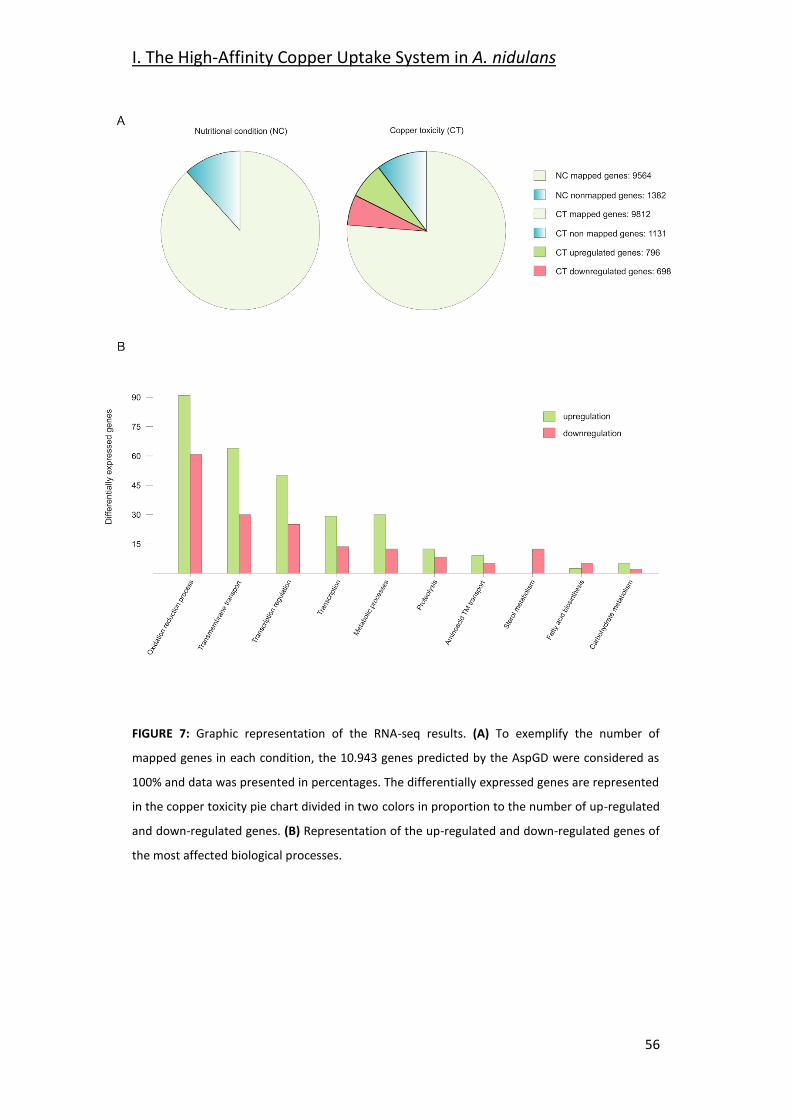
I. The High-Affinity Copper Uptake System in A. nidulans
56
FIGURE 7: Graphic representation of the RNA-seq results. (A) To exemplify the number of
mapped genes in each condition, the 10.943 genes predicted by the AspGD were considered as
100% and data was presented in percentages. The differentially expressed genes are represented
in the copper toxicity pie chart divided in two colors in proportion to the number of up-regulated
and down-regulated genes. (B) Representation of the up-regulated and down-regulated genes of
the most affected biological processes.
I.The High-Affinity Copper Uptake System in A. nidulans
57
Table 6 shows the expression of most of the genes related to copper
homeostasis, either characterized or predicted, and also some copper containing
proteins. The TFs that orchestrate the copper homeostasis process, AnaceA and
Anmac1, showed no significant variations in response to copper; however, the
genes that code for copper mobilizing proteins, either uptake or detoxification,
showed significant changes in their expression. The copper detoxification gene
AncrpA was up-regulated in response to an external copper load. On the other
hand, the already described Ctr protein coding genes AnctrA and AnctrC
(elaborated on below) were significantly down-regulated under copper-toxicity
condition; however, the putative Ctr protein coding gene, AnctrB (elaborated on
below), was unaltered. Homology searches revealed the putative homologs of
the S. cerevisiae Atx1, Cox17 and Ccs1 genes. A very important element for
proper copper uptake is the plasma membrane oxidoreductase that reduces
copper prior to internalization. In Table 6 a characterized metalloreductase,
AnfreA is shown, along with two other predicted plasma membrane
oxidoreductases, AN0773 and AnfreC (elaborated on below), which are co-
regulated with the Ctr proteins and thus, could be possible metalloreductase
candidates. Finally, the expression of the intracellular copper trafficking P-type
ATPase AnygA shows significant up-regulation under excess copper conditions.
Besides copper homeostasis proteins, many other copper related proteins
showed expression changes, as copper is a required cofactor of many enzymes
(Table 6). The Cu/Zn superoxide dismutase AnsodA showed no difference in
expression in response to the external copper load. On the other hand, laccases,
a copper-containing enzyme-family required for melanin synthesis, displayed a
divergent response: AnyA and AnlccD were down-regulated, while AntilA was up-
regulated.
I. The High-Affinity Copper Uptake System in A. nidulans
58
Table 6: RNA-seq data of the copper homeostasis genes and the copper related protein coding
genes.
To interpret the biological significance of the registered changes in gene
expression, we conducted a GO (Gene Onthology) classification analysis of the
results. The identified changes in gene expression occurring in response to toxic
copper levels were indicative of modifications in various processes (Figure 7B).
Redox processes were the most affected. Copper has a relatively high reduction
potential (Eo0 = -0.34V), thus, it may interfere with ongoing oxidation-reduction
processes, leading to changes in the expression levels of the corresponding
enzymes. For example, seven oxidoreductases with predicted roles in sterol
metabolism were significantly down-regulated (AN8907, AN4094, AN6506,
AN6973, AN3638, AN10648, and AN3817). Catalases AncatB, AncatC and
AncpeA/AncatD were slightly up-regulated. On the other hand, the expression of
superoxide dismutases and the oxidative stress transcription factors AnnapA and
Gene log2 fold change
Value_1 Value_2 q value Significant Description
AN1924 -0,554556 250,162 170,327 0,269576 no AnaceA. Copper detoxification TF
AN0658 0,417535 20,518 27,4048 0,503204 no Anmac1. Copper uptake TF
AN3117 6,4078 11,9725 1016,54 0 yes AncrpA. Copper-exporting P-type ATPase.
AN2934 -0,284851 71,0897 58,3523 0,686464 no AnctrB. Copper ion transmembrane transporter activity.
AN3209 -4,18902 24,4975 1,34308 2,75E-08 yes AnctrA. High-affinity copper transporter
AN3813 -2,97333 134,667 17,1475 3,34E-12 yes AnctrC. High-affinity copper transporter.
AN3624 1,01501 20,1871 40,7964 0,015651 yes AnygA. ScCcc2 homologue. Predicted copper transporter.
AN6045 1,95069 76,4666 295,588 2,09E-07 yes Ccs1 Sod chaperone ortholog,
AN1390 0,137135 110,504 121,523 0,853005 no Atx1 chaperone homologue.
AN4863 0,397399 78,3646 103,216 0,541532 no Cox17 Cytochrome C oxidase chaperone homologue.
AN7662 -1,11534 35,8567 16,5508 0,018818 yes AnfreA. Predicted iron metaloreductase.
AN0773 -2,14409 114,6 25,9269 4,15E-08 yes Predicted metalloreductase.
AN3208 -4,54749 14,5531 0,622335 2,56E-14 yes AnfreC. Predicted metalloreductase.
AN0241 -0,342918 1361,11 1073,16 0,57373 no AnsodA. Cu/Zn-superoxide dismutase.
AN6635 -1,48008 4,56506 1,63643 0,034958 yes AnyA. Conidial laccase involved in green pigment production.
AN6830 1,79e+308 0 0,145851 1 no AnlccA. Putative laccase.
AN9170 0 0 0 1 no AnlccB. Extracellular laccase.
AN5379 -0,611597 0,055515 0,0363333 1 no AnlccC. Extracellular laccase.
AN0878 -1,92869 1,5301 0,401909 0,0384564 yes AnlccD. Extracellular laccase.
AN0901 1,89527 6,9476 25,8445 6,10E-05 yes AntilA. Hyphal tip laccase.
I.The High-Affinity Copper Uptake System in A. nidulans
59
AnatfA were not altered. Transmembrane (TM) transport of various substrates,
as sugars, oligopeptides and metals was modified: the xylose transporters AnxtrA
and AnxtrB, the putative high-affinity nickel uptake protein AN6115, the putative
Cation Diffusion Facilitator (CDF) transporter. Finally, transcriptional regulation
was also notably altered, based on the changes in the expression of transcription
factors. Most of them were unidentified.
Analyzing the repercussion of copper-toxicity on gene expression, certain gene
clusters were affected by copper addition. For example, the siderophore
biosynthesis gene cluster (AnsidA, AnsidC, AnsidD, AnsidF, AnsidG, AnsidH, and
AnsidI) was notably down-regulated, together with the iron homeostasis TF,
AnhapX. Besides the siderophore biosynthesis cluster, most members of the
Emericellamide antibiotic biosynthesis gene cluster were down-regulated
(AneasB, AneasC, and AneasD). A set of genes (AN2558, AN2559, AN2560,
AN2561, and AN2562) with no confirmed function was drastically up-regulated.
However, by sequence analysis tools, we found that AN2562 was the homolog of
the N-acetylglucosamine transporter Ngt1 from C. albicans; AN2560 was the
homolog of the high-affinity methionine and cysteine transporter Mup1 from S.
cerevisiae and C. albicans; AN2558 was the homolog of the flavin-containing
monooxygenases CaIfk1 and ScFmo1; and AN2559 and AN2561 were a predicted
oxidoreductase and a predicted methyltransferase, respectively (Table 7).
In summary, copper toxicity altered the expression of 13.7% of the genes with a
balanced ratio between up-regulated and down-regulated genes (Figure 7A).
Changes in copper concentration derived in a strong modification of the copper
homeostasis system, the copper dependent enzymes and multiple biological
processes, especially oxidation reduction processes and predictably, in cellular
components such as biological membrane composition. As a remarkable feature,
in most of these processes, more genes were up-regulated than down-regulated
(Figure 7B).
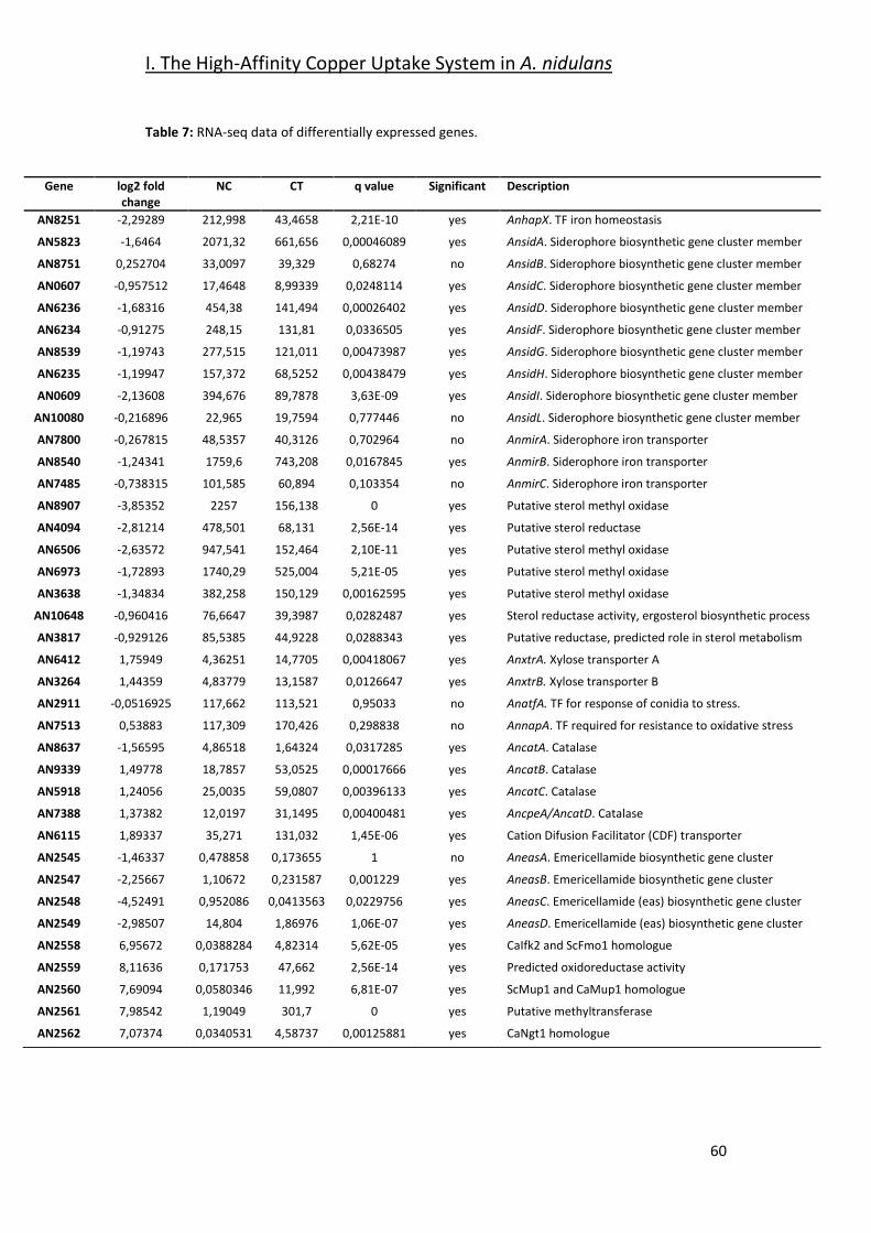
I. The High-Affinity Copper Uptake System in A. nidulans
60
Table 7: RNA-seq data of differentially expressed genes.
Gene log2 fold change
NC CT q value Significant Description
AN8251 -2,29289 212,998 43,4658 2,21E-10 yes AnhapX. TF iron homeostasis
AN5823 -1,6464 2071,32 661,656 0,00046089 yes AnsidA. Siderophore biosynthetic gene cluster member
AN8751 0,252704 33,0097 39,329 0,68274 no AnsidB. Siderophore biosynthetic gene cluster member
AN0607 -0,957512 17,4648 8,99339 0,0248114 yes AnsidC. Siderophore biosynthetic gene cluster member
AN6236 -1,68316 454,38 141,494 0,00026402 yes AnsidD. Siderophore biosynthetic gene cluster member
AN6234 -0,91275 248,15 131,81 0,0336505 yes AnsidF. Siderophore biosynthetic gene cluster member
AN8539 -1,19743 277,515 121,011 0,00473987 yes AnsidG. Siderophore biosynthetic gene cluster member
AN6235 -1,19947 157,372 68,5252 0,00438479 yes AnsidH. Siderophore biosynthetic gene cluster member
AN0609 -2,13608 394,676 89,7878 3,63E-09 yes AnsidI. Siderophore biosynthetic gene cluster member
AN10080 -0,216896 22,965 19,7594 0,777446 no AnsidL. Siderophore biosynthetic gene cluster member
AN7800 -0,267815 48,5357 40,3126 0,702964 no AnmirA. Siderophore iron transporter
AN8540 -1,24341 1759,6 743,208 0,0167845 yes AnmirB. Siderophore iron transporter
AN7485 -0,738315 101,585 60,894 0,103354 no AnmirC. Siderophore iron transporter
AN8907 -3,85352 2257 156,138 0 yes Putative sterol methyl oxidase
AN4094 -2,81214 478,501 68,131 2,56E-14 yes Putative sterol reductase
AN6506 -2,63572 947,541 152,464 2,10E-11 yes Putative sterol methyl oxidase
AN6973 -1,72893 1740,29 525,004 5,21E-05 yes Putative sterol methyl oxidase
AN3638 -1,34834 382,258 150,129 0,00162595 yes Putative sterol methyl oxidase
AN10648 -0,960416 76,6647 39,3987 0,0282487 yes Sterol reductase activity, ergosterol biosynthetic process
AN3817 -0,929126 85,5385 44,9228 0,0288343 yes Putative reductase, predicted role in sterol metabolism
AN6412 1,75949 4,36251 14,7705 0,00418067 yes AnxtrA. Xylose transporter A
AN3264 1,44359 4,83779 13,1587 0,0126647 yes AnxtrB. Xylose transporter B
AN2911 -0,0516925 117,662 113,521 0,95033 no AnatfA. TF for response of conidia to stress.
AN7513 0,53883 117,309 170,426 0,298838 no AnnapA. TF required for resistance to oxidative stress
AN8637 -1,56595 4,86518 1,64324 0,0317285 yes AncatA. Catalase
AN9339 1,49778 18,7857 53,0525 0,00017666 yes AncatB. Catalase
AN5918 1,24056 25,0035 59,0807 0,00396133 yes AncatC. Catalase
AN7388 1,37382 12,0197 31,1495 0,00400481 yes AncpeA/AncatD. Catalase
AN6115 1,89337 35,271 131,032 1,45E-06 yes Cation Difusion Facilitator (CDF) transporter
AN2545 -1,46337 0,478858 0,173655 1 no AneasA. Emericellamide biosynthetic gene cluster
AN2547 -2,25667 1,10672 0,231587 0,001229 yes AneasB. Emericellamide biosynthetic gene cluster
AN2548 -4,52491 0,952086 0,0413563 0,0229756 yes AneasC. Emericellamide (eas) biosynthetic gene cluster
AN2549 -2,98507 14,804 1,86976 1,06E-07 yes AneasD. Emericellamide (eas) biosynthetic gene cluster
AN2558 6,95672 0,0388284 4,82314 5,62E-05 yes CaIfk2 and ScFmo1 homologue
AN2559 8,11636 0,171753 47,662 2,56E-14 yes Predicted oxidoreductase activity
AN2560 7,69094 0,0580346 11,992 6,81E-07 yes ScMup1 and CaMup1 homologue
AN2561 7,98542 1,19049 301,7 0 yes Putative methyltransferase
AN2562 7,07374 0,0340531 4,58737 0,00125881 yes CaNgt1 homologue
I.The High-Affinity Copper Uptake System in A. nidulans
61
2.2- Screening for Potential Copper Transport Proteins
In order to select putative copper transporters we searched for putative
orthologs of Saccharomyces cerevisiae Ctr transporters among down-regulated
genes under copper toxicity. Blastp searches using the full-length of the ScCtr
protein sequences yielded three results, the already identified AN3209 and
AN3813 and another putative copper transporter, AN2934, termed AnctrB.
AnctrB is the top hit of the copper transporter ScCtr2, which encodes a vacuolar
Cu transporter (Liu et al., 2012;Qi et al., 2012). Cai et al., (2019) recently
identified AN3209 and AN3813 as copper uptake proteins in A. nidulans, and
AnMac1 as the TF controlling their expression. In this work, AN3209 and AN3813
were named AnCtrA2 and AnCtrC, respectively, claiming they were AfCtrA2 and
AfCtrC homologs, but the protein sequence analysis results support a different
interpretation. BLASTp analyses revealed that AnCtrA and AnCtrC are significant
hits of AfCtrC (42% and 60% protein identity, respectively); however, there is no
significant hit of AfCtrA2 (24% and 22% protein identity, respectively) in the A.
nidulans proteome. Thus, instead of naming AN3209 as AnCtrA2, we considered
the name AnCtrA.
Sequence data revealed that AnctrA and AnctrC share great similarity.
Furthermore, out of the 127 species that possess an AnctrA ortholog, 94 also
possess an AnctrC ortholog (Figure 14).
According to the phylogeny analyses shown in Figure 8, three major clusters can
be differentiated. Most Ctr proteins of filamentous fungi are related to ScCtr3
(Red); A. fumigatus, A. nidulans, and A. niger possess Ctr proteins closely related
to the vacuole Cu transporter ScCtr2 (Blue); Out of the 27 proteins of the
phylogeny test, only four proteins are related to ScCtr1 (Green). The results
reflect that ScCtr3 orthologs comprise the biggest group of proteins and in this
group we can find most Ctr proteins of filamentous fungi.
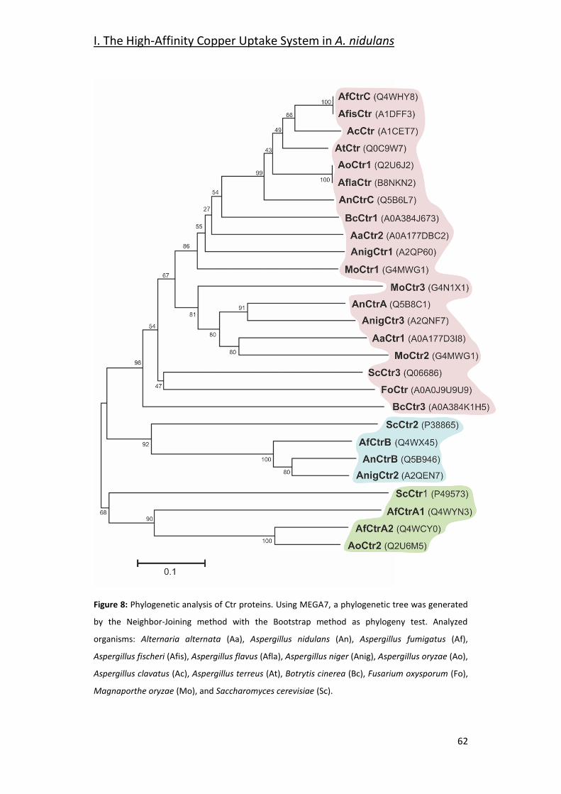
I. The High-Affinity Copper Uptake System in A. nidulans
62
Figure 8: Phylogenetic analysis of Ctr proteins. Using MEGA7, a phylogenetic tree was generated
by the Neighbor-Joining method with the Bootstrap method as phylogeny test. Analyzed
organisms: Alternaria alternata (Aa), Aspergillus nidulans (An), Aspergillus fumigatus (Af),
Aspergillus fischeri (Afis), Aspergillus flavus (Afla), Aspergillus niger (Anig), Aspergillus oryzae (Ao),
Aspergillus clavatus (Ac), Aspergillus terreus (At), Botrytis cinerea (Bc), Fusarium oxysporum (Fo),
Magnaporthe oryzae (Mo), and Saccharomyces cerevisiae (Sc).
I.The High-Affinity Copper Uptake System in A. nidulans
63
A multiple sequence alignment of A. nidulans Ctr proteins is shown in Figure 9.
Strong similarities in amino acid sequences could be observed in certain regions
of the N- and C-terminal, but not in the loop between transmembrane domains
(TMD) 1 and 2, which showed higher variability. Detailed sequence analysis
showed the presence of common conserved motifs predicted to code for copper
binding. AnCtrA, AnCtrC, AfCtrC and CnCtr4 contain a Met motif arranged as M-
xx-M-x-M in the amino-terminal region (Nt) (Met motif shown in blue box). Three
additional methionine residues are conserved among the Ctr protein sequences
aligned: a methionine in the amino-terminal, located 22 amino acids upstream
from the TMD1 (M30 in AnCtrA, M23 in AnCtrC, M20 in ScCtr3, M58 in AfCtrC
and M41 CnCtr4) and an M-xxx-M motif in the predicted TMD2 (M134-M138 in
AnCtrA, M168-M172 in AnCtrC, M185-M189 in ScCtr3, M197-M201 in AfCtrC and
M156-M160 CnCtr4). The methionine residue located prior to TMD1 is
equidistant in A. nidulans and other fungi, as noted by Puig et al. (2002). As
shown in Figure 9A, there are two conserved cysteine residues flanking this
functional residue (Cys residues are highlighted in red for their copper handling
capacity). Additional methionine and cysteine residues are widespread within
the sequence. Finally, in all the proteins analyzed close to the terminus of the
carboxy-terminal region, a putative copper handling motif that could be involved
in copper coordination and transport was identified; a di-cystine motif in the
case of ScCtr3, CnCtr4, AnCtrA and AnCtrC, and a M-x-C motif in the case of
AfCtrC. Based on computer algorithms (TMHMM and HMMTOP predictions), as
shown in Figure 9B, AnCtrA was predicted to contain two transmembrane
regions while AnCtrC contained three. According to these predictions, the amino
tail would be oriented to the outside of the cell and the carboxyl tail into the
cytosol, in the case of AnCtrC. In the case of AnCtrA, however, both protein ends
would be oriented to the outside.
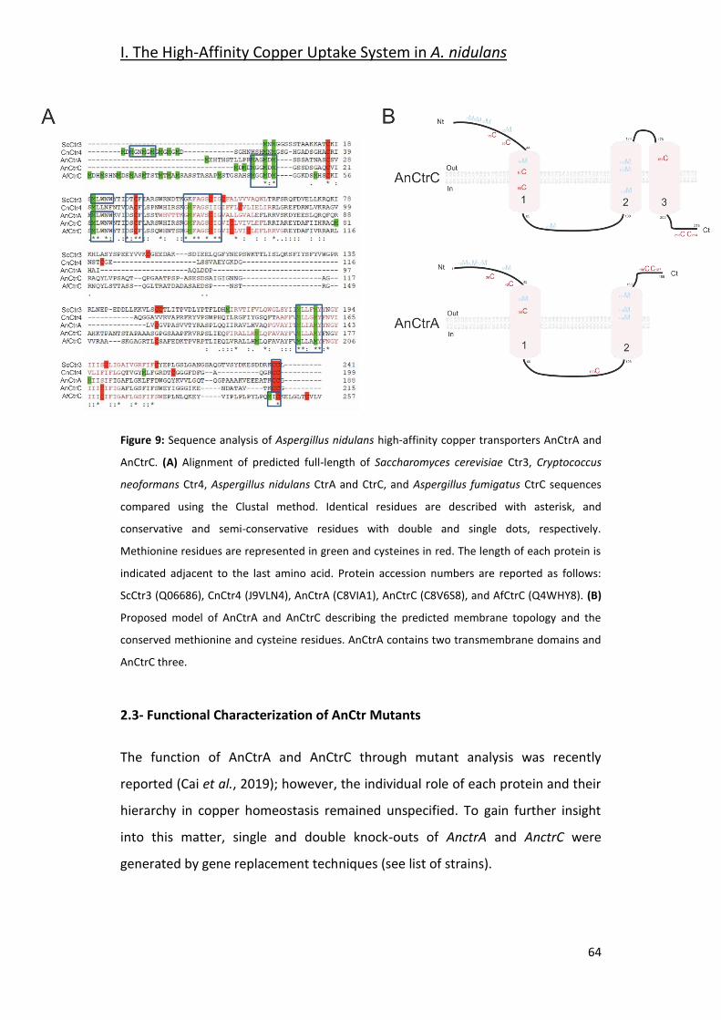
I. The High-Affinity Copper Uptake System in A. nidulans
64
Figure 9: Sequence analysis of Aspergillus nidulans high-affinity copper transporters AnCtrA and
AnCtrC. (A) Alignment of predicted full-length of Saccharomyces cerevisiae Ctr3, Cryptococcus
neoformans Ctr4, Aspergillus nidulans CtrA and CtrC, and Aspergillus fumigatus CtrC sequences
compared using the Clustal method. Identical residues are described with asterisk, and
conservative and semi-conservative residues with double and single dots, respectively.
Methionine residues are represented in green and cysteines in red. The length of each protein is
indicated adjacent to the last amino acid. Protein accession numbers are reported as follows:
ScCtr3 (Q06686), CnCtr4 (J9VLN4), AnCtrA (C8VIA1), AnCtrC (C8V6S8), and AfCtrC (Q4WHY8). (B)
Proposed model of AnCtrA and AnCtrC describing the predicted membrane topology and the
conserved methionine and cysteine residues. AnCtrA contains two transmembrane domains and
AnCtrC three.
2.3- Functional Characterization of AnCtr Mutants
The function of AnCtrA and AnCtrC through mutant analysis was recently
reported (Cai et al., 2019); however, the individual role of each protein and their
hierarchy in copper homeostasis remained unspecified. To gain further insight
into this matter, single and double knock-outs of AnctrA and AnctrC were
generated by gene replacement techniques (see list of strains).
I.The High-Affinity Copper Uptake System in A. nidulans
65
The capacity of these strains to grow under different copper availability
conditions was tested. We first reproduced the experiment conducted by Cai et
al., (2019), by testing all generated mutant strains in 0 M Cu media, with
identical results: the single deletion mutants showed a WT-like phenotype and
the only strain showing growth disruption was the double null mutant AnctrA-
AnctrC (Figure 10A). These results provided evidence that, under copper
deficiency conditions, AnCtrA and AnCtrC complemented each other. Addition of
copper to the medium rescued the double deletion mutant defective phenotype.
Similar results were obtained in liquid media as only the double null mutant
showed a significantly lower growth rate than the WT strain (Figure 10B).
The above results showed that removing Cu from the trace element solution did
not help discern phenotypic differences between single null mutants. Thus, we
used the copper chelator Bathocuproinedisulfonic acid (BCS) in our experiments.
As shown in Figure 10C, null mutant colonies did not exhibit appreciable
alterations in radial growth or conidial pigmentation on regular solid minimal
medium. Under copper starvation conditions (AMM supplemented with BCS),
AnctrC exhibited secondary level copper limitation effects at 80 M
concentration, displaying yellowish conidia which could be a result of the
inactivity of the copper-dependent conidial laccase AnYA due to Cu deficiency
(Scherer and Fischer, 2001). In contrast, the AnctrA mutant presented no
appreciable copper limitation effects. When both mutations were combined,
primary level copper limitation effects were visible. Radial growth of the double
knock-out strain was reduced to 40% and no pigmentation was appreciated.
Upon Cu supplementation (100 M BCS + 100 M CuSO4), normal growth and
morphology were restored. It has been described that copper and iron
metabolism are related in fungi, and iron can recover mac1 colony morphology
-very similar to the double knock-out strain shown in Figure 10C- to some extent
in A. fumigatus (Cai et al., 2017). Therefore, we tested whether addition of FeSO4
could rescue the colony defects observed in Anctr mutants grown under copper
I. The High-Affinity Copper Uptake System in A. nidulans
66
starvation conditions. Fe supplementation (100 M BCS + 400 M FeSO4) did not
rescue the defects in null mutants (Figure 10C). Since Sods are also copper
dependent enzymes, we hypothesized that a menadione (a widely used oxidative
stress inducing agent) experiment could probably help clarify the nature of the
differences observed so far between the two transporter null mutants. However,
with 60 M menadione, the phenotype was similar; the AnctrC strain was
visibly more affected by the oxidative damage generated by menadione,
suggesting that in the condition tested Sod copper supply depended more on
AnCtrC.
In addition to the above colony growth inhibition and pigmentation defects,
hyphal growth in liquid medium and conidia production were also studied. To
investigate the effect of the deletion of the Anctr coding genes on conidial
production, we harvested and compared the number of conidiospores produced
by each strain, under basal and low copper availability conditions (Figure 10D).
Under basal conditions, single deletion of Anctr genes had no apparent effect on
conidia production. Under copper deficiency conditions AnctrA deletion had a
slight effect, but AnctrC deletion had a remarkable impact; conidia numbers
were halved by comparison to the wild-type strain. Numbers of conidia are
connected with the amount of biomass and the AnctrC colony grown in solid
AMM medium with 100 M BCS was not smaller than the colony grown in AMM.
Thus, copper scarcity had no effect on colony expansion, but on colony density,
an effect that has also been described under other stresses conditions such as
carbon or nitrogen starvation (Richie et al., 2007). Deletion of both AnCtr
proteins had a slight effect at control conditions. Under copper starvation
conditions, conidia quantity was, 37-fold lower than the wildtype strain (Figure
10D).
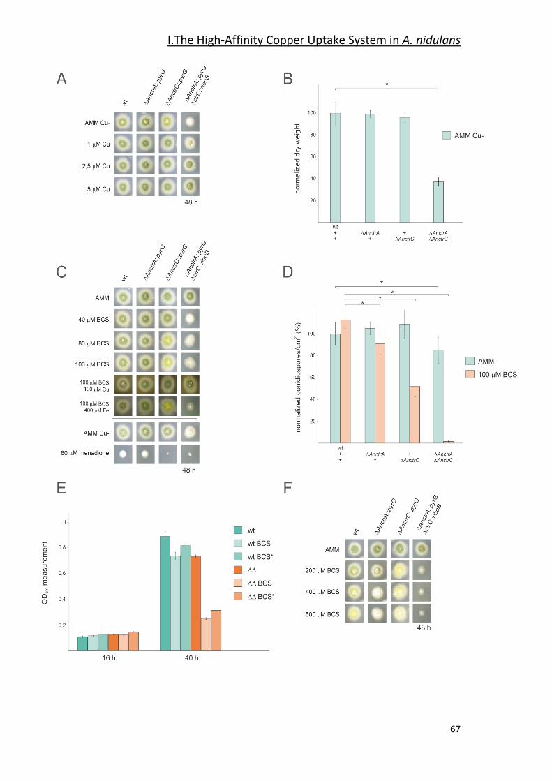
I.The High-Affinity Copper Uptake System in A. nidulans
67
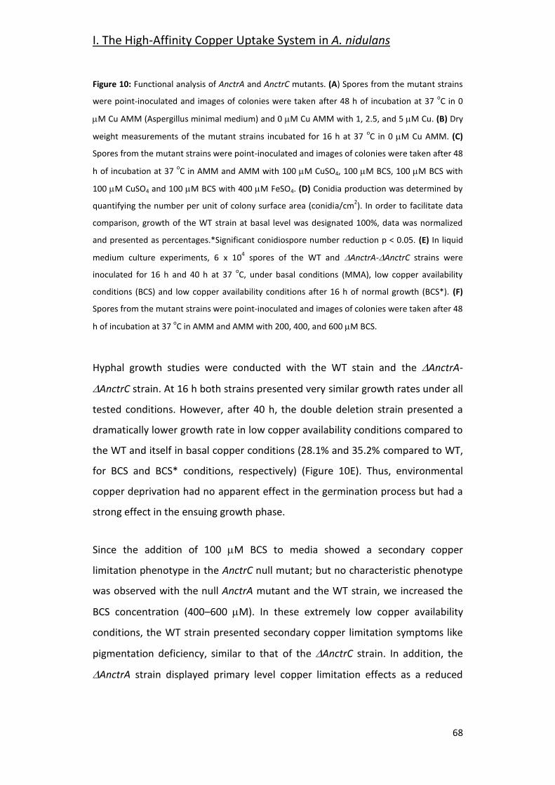
I. The High-Affinity Copper Uptake System in A. nidulans
68
Figure 10: Functional analysis of AnctrA and AnctrC mutants. (A) Spores from the mutant strains
were point-inoculated and images of colonies were taken after 48 h of incubation at 37 oC in 0
M Cu AMM (Aspergillus minimal medium) and 0 M Cu AMM with 1, 2.5, and 5 M Cu. (B) Dry
weight measurements of the mutant strains incubated for 16 h at 37 oC in 0 M Cu AMM. (C)
Spores from the mutant strains were point-inoculated and images of colonies were taken after 48
h of incubation at 37 oC in AMM and AMM with 100 M CuSO4, 100 M BCS, 100 M BCS with
100 M CuSO4 and 100 M BCS with 400 M FeSO4. (D) Conidia production was determined by
quantifying the number per unit of colony surface area (conidia/cm2). In order to facilitate data
comparison, growth of the WT strain at basal level was designated 100%, data was normalized
and presented as percentages.*Significant conidiospore number reduction p < 0.05. (E) In liquid
medium culture experiments, 6 x 10⁴ spores of the WT and AnctrA-AnctrC strains were
inoculated for 16 h and 40 h at 37 oC, under basal conditions (MMA), low copper availability
conditions (BCS) and low copper availability conditions after 16 h of normal growth (BCS*). (F)
Spores from the mutant strains were point-inoculated and images of colonies were taken after 48
h of incubation at 37 oC in AMM and AMM with 200, 400, and 600 M BCS.
Hyphal growth studies were conducted with the WT stain and the AnctrA-
AnctrC strain. At 16 h both strains presented very similar growth rates under all
tested conditions. However, after 40 h, the double deletion strain presented a
dramatically lower growth rate in low copper availability conditions compared to
the WT and itself in basal copper conditions (28.1% and 35.2% compared to WT,
for BCS and BCS* conditions, respectively) (Figure 10E). Thus, environmental
copper deprivation had no apparent effect in the germination process but had a
strong effect in the ensuing growth phase.
Since the addition of 100 M BCS to media showed a secondary copper
limitation phenotype in the AnctrC null mutant; but no characteristic phenotype
was observed with the null AnctrA mutant and the WT strain, we increased the
BCS concentration (400–600 M). In these extremely low copper availability
conditions, the WT strain presented secondary copper limitation symptoms like
pigmentation deficiency, similar to that of the AnctrC strain. In addition, the
AnctrA strain displayed primary level copper limitation effects as a reduced
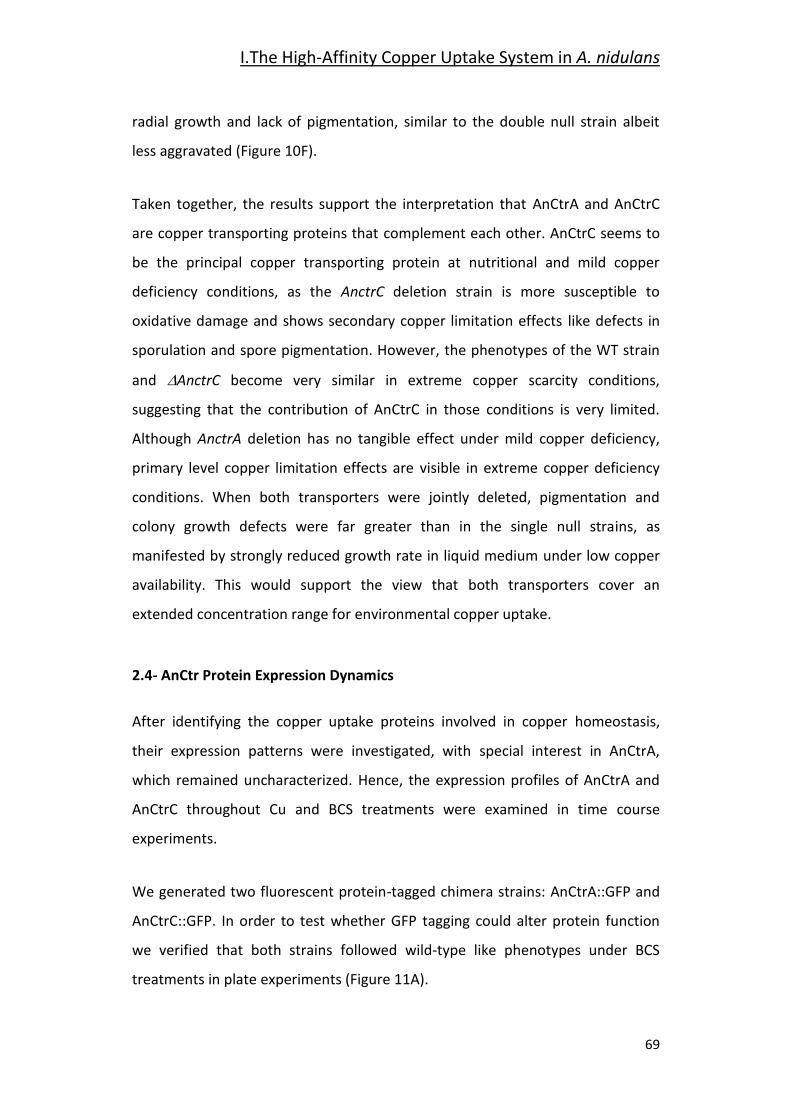
I.The High-Affinity Copper Uptake System in A. nidulans
69
radial growth and lack of pigmentation, similar to the double null strain albeit
less aggravated (Figure 10F).
Taken together, the results support the interpretation that AnCtrA and AnCtrC
are copper transporting proteins that complement each other. AnCtrC seems to
be the principal copper transporting protein at nutritional and mild copper
deficiency conditions, as the AnctrC deletion strain is more susceptible to
oxidative damage and shows secondary copper limitation effects like defects in
sporulation and spore pigmentation. However, the phenotypes of the WT strain
and AnctrC become very similar in extreme copper scarcity conditions,
suggesting that the contribution of AnCtrC in those conditions is very limited.
Although AnctrA deletion has no tangible effect under mild copper deficiency,
primary level copper limitation effects are visible in extreme copper deficiency
conditions. When both transporters were jointly deleted, pigmentation and
colony growth defects were far greater than in the single null strains, as
manifested by strongly reduced growth rate in liquid medium under low copper
availability. This would support the view that both transporters cover an
extended concentration range for environmental copper uptake.
2.4- AnCtr Protein Expression Dynamics
After identifying the copper uptake proteins involved in copper homeostasis,
their expression patterns were investigated, with special interest in AnCtrA,
which remained uncharacterized. Hence, the expression profiles of AnCtrA and
AnCtrC throughout Cu and BCS treatments were examined in time course
experiments.
We generated two fluorescent protein-tagged chimera strains: AnCtrA::GFP and
AnCtrC::GFP. In order to test whether GFP tagging could alter protein function
we verified that both strains followed wild-type like phenotypes under BCS
treatments in plate experiments (Figure 11A).
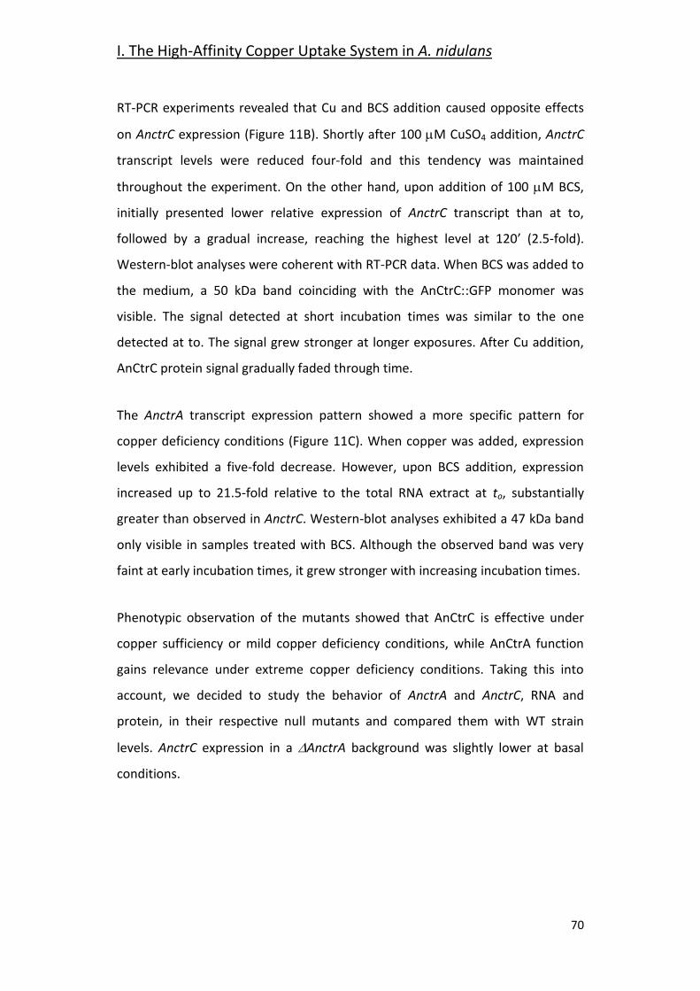
I. The High-Affinity Copper Uptake System in A. nidulans
70
RT-PCR experiments revealed that Cu and BCS addition caused opposite effects
on AnctrC expression (Figure 11B). Shortly after 100 M CuSO4 addition, AnctrC
transcript levels were reduced four-fold and this tendency was maintained
throughout the experiment. On the other hand, upon addition of 100 M BCS,
initially presented lower relative expression of AnctrC transcript than at to,
followed by a gradual increase, reaching the highest level at 120’ (2.5-fold).
Western-blot analyses were coherent with RT-PCR data. When BCS was added to
the medium, a 50 kDa band coinciding with the AnCtrC::GFP monomer was
visible. The signal detected at short incubation times was similar to the one
detected at to. The signal grew stronger at longer exposures. After Cu addition,
AnCtrC protein signal gradually faded through time.
The AnctrA transcript expression pattern showed a more specific pattern for
copper deficiency conditions (Figure 11C). When copper was added, expression
levels exhibited a five-fold decrease. However, upon BCS addition, expression
increased up to 21.5-fold relative to the total RNA extract at to, substantially
greater than observed in AnctrC. Western-blot analyses exhibited a 47 kDa band
only visible in samples treated with BCS. Although the observed band was very
faint at early incubation times, it grew stronger with increasing incubation times.
Phenotypic observation of the mutants showed that AnCtrC is effective under
copper sufficiency or mild copper deficiency conditions, while AnCtrA function
gains relevance under extreme copper deficiency conditions. Taking this into
account, we decided to study the behavior of AnctrA and AnctrC, RNA and
protein, in their respective null mutants and compared them with WT strain
levels. AnctrC expression in a AnctrA background was slightly lower at basal
conditions.
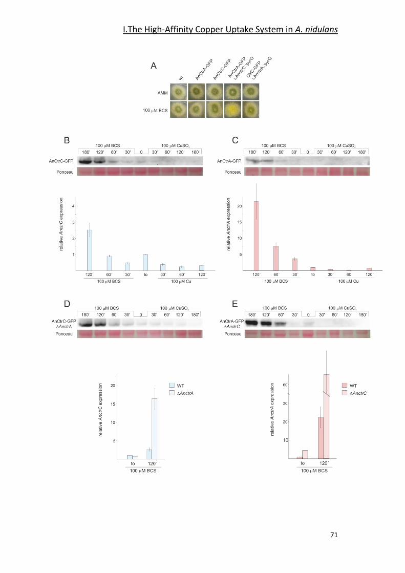
I.The High-Affinity Copper Uptake System in A. nidulans
71
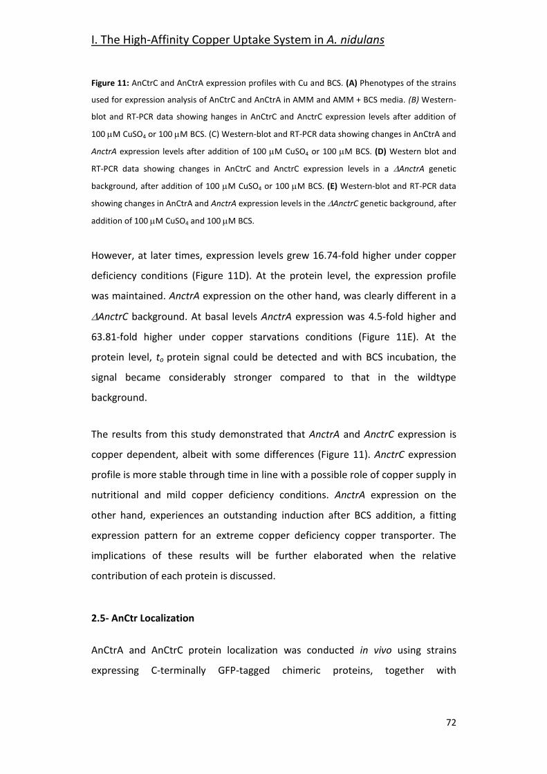
I. The High-Affinity Copper Uptake System in A. nidulans
72
Figure 11: AnCtrC and AnCtrA expression profiles with Cu and BCS. (A) Phenotypes of the strains
used for expression analysis of AnCtrC and AnCtrA in AMM and AMM + BCS media. (B) Western-
blot and RT-PCR data showing hanges in AnCtrC and AnctrC expression levels after addition of
100 M CuSO4 or 100 M BCS. (C) Western-blot and RT-PCR data showing changes in AnCtrA and
AnctrA expression levels after addition of 100 M CuSO4 or 100 M BCS. (D) Western blot and
RT-PCR data showing changes in AnCtrC and AnctrC expression levels in a AnctrA genetic
background, after addition of 100 M CuSO4 or 100 M BCS. (E) Western-blot and RT-PCR data
showing changes in AnCtrA and AnctrA expression levels in the AnctrC genetic background, after
addition of 100 M CuSO4 and 100 M BCS.
However, at later times, expression levels grew 16.74-fold higher under copper
deficiency conditions (Figure 11D). At the protein level, the expression profile
was maintained. AnctrA expression on the other hand, was clearly different in a
AnctrC background. At basal levels AnctrA expression was 4.5-fold higher and
63.81-fold higher under copper starvations conditions (Figure 11E). At the
protein level, to protein signal could be detected and with BCS incubation, the
signal became considerably stronger compared to that in the wildtype
background.
The results from this study demonstrated that AnctrA and AnctrC expression is
copper dependent, albeit with some differences (Figure 11). AnctrC expression
profile is more stable through time in line with a possible role of copper supply in
nutritional and mild copper deficiency conditions. AnctrA expression on the
other hand, experiences an outstanding induction after BCS addition, a fitting
expression pattern for an extreme copper deficiency copper transporter. The
implications of these results will be further elaborated when the relative
contribution of each protein is discussed.
2.5- AnCtr Localization
AnCtrA and AnCtrC protein localization was conducted in vivo using strains
expressing C-terminally GFP-tagged chimeric proteins, together with
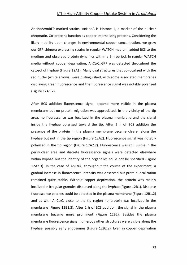
I.The High-Affinity Copper Uptake System in A. nidulans
73
AnHhoA::mRFP marked strains. AnHhoA is Histone 1, a marker of the nuclear
chromatin. Ctr proteins function as copper internalizing proteins. Considering the
likely mobility upon changes in environmental copper concentration, we grew
our GFP chimera expressing strains in regular WATCH medium, added BCS to the
medium and observed protein dynamics within a 2 h period. In regular WATCH
media without copper deprivation, AnCtrC::GFP was detected throughout the
cytosol of hyphae (Figure 12A1). Many oval structures that co-localized with the
red nuclei (white arrows) were distinguished, with some associated membranes
displaying green fluorescence and the fluorescence signal was notably polarized
(Figure 12A1.2).
After BCS addition fluorescence signal became more visible in the plasma
membrane but no protein migration was appreciated. In the vicinity of the tip
area, no fluorescence was localized in the plasma membrane and the signal
inside the hyphae polarized toward the tip. After 2 h of BCS addition the
presence of the protein in the plasma membrane became clearer along the
hyphae but not in the tip region (Figure 12A2). Fluorescence signal was notably
polarized in the tip region (Figure 12A2.2). Fluorescence was still visible in the
perinuclear area and discrete fluorescence signals were detected elsewhere
within hyphae but the identity of the organelles could not be specified (Figure
12A2.3). In the case of AnCtrA, throughout the course of the experiment, a
gradual increase in fluorescence intensity was observed but protein localization
remained quite stable. Without copper deprivation, the protein was mainly
localized in irregular granules dispersed along the hyphae (Figure 12B1). Disperse
fluorescence patches could be detected in the plasma membrane (Figure 12B1.2)
and as with AnCtrC, close to the tip region no protein was localized in the
membrane (Figure 12B1.3). After 2 h of BCS addition, the signal in the plasma
membrane became more prominent (Figure 12B2). Besides the plasma
membrane fluorescence signal numerous other structures were visible along the
hyphae, possibly early endosomes (Figure 12B2.2). Even in copper deprivation
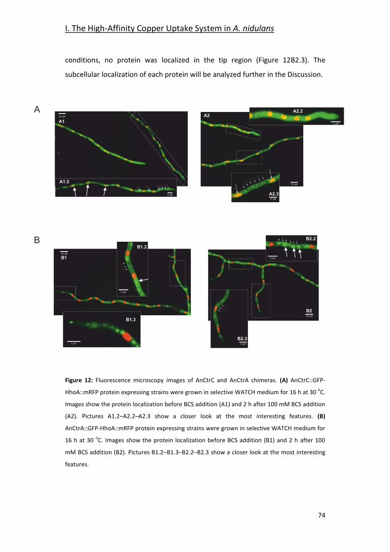
I. The High-Affinity Copper Uptake System in A. nidulans
74
conditions, no protein was localized in the tip region (Figure 12B2.3). The
subcellular localization of each protein will be analyzed further in the Discussion.
Figure 12: Fluorescence microscopy images of AnCtrC and AnCtrA chimeras. (A) AnCtrC::GFP-
HhoA::mRFP protein expressing strains were grown in selective WATCH medium for 16 h at 30 oC.
Images show the protein localization before BCS addition (A1) and 2 h after 100 mM BCS addition
(A2). Pictures A1.2–A2.2–A2.3 show a closer look at the most interesting features. (B)
AnCtrA::GFP-HhoA::mRFP protein expressing strains were grown in selective WATCH medium for
16 h at 30 oC. Images show the protein localization before BCS addition (B1) and 2 h after 100
mM BCS addition (B2). Pictures B1.2–B1.3–B2.2–B2.3 show a closer look at the most interesting
features.
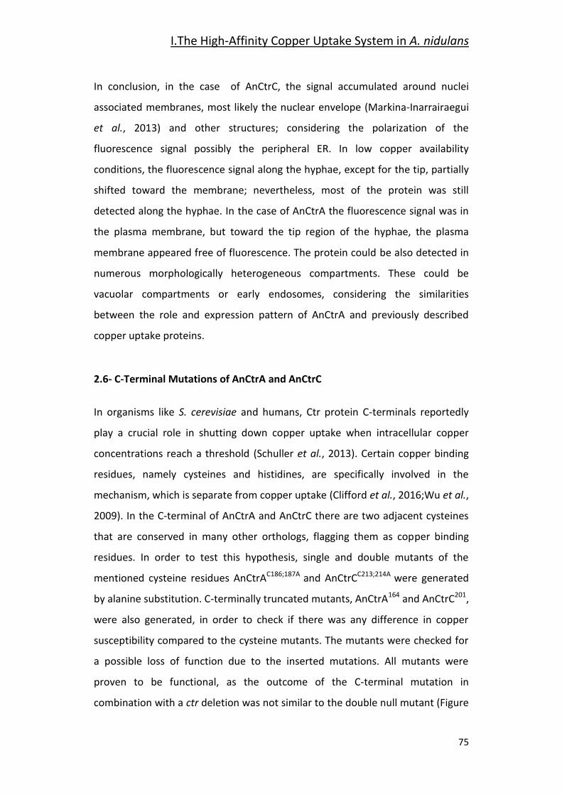
I.The High-Affinity Copper Uptake System in A. nidulans
75
In conclusion, in the case of AnCtrC, the signal accumulated around nuclei
associated membranes, most likely the nuclear envelope (Markina-Inarrairaegui
et al., 2013) and other structures; considering the polarization of the
fluorescence signal possibly the peripheral ER. In low copper availability
conditions, the fluorescence signal along the hyphae, except for the tip, partially
shifted toward the membrane; nevertheless, most of the protein was still
detected along the hyphae. In the case of AnCtrA the fluorescence signal was in
the plasma membrane, but toward the tip region of the hyphae, the plasma
membrane appeared free of fluorescence. The protein could be also detected in
numerous morphologically heterogeneous compartments. These could be
vacuolar compartments or early endosomes, considering the similarities
between the role and expression pattern of AnCtrA and previously described
copper uptake proteins.
2.6- C-Terminal Mutations of AnCtrA and AnCtrC
In organisms like S. cerevisiae and humans, Ctr protein C-terminals reportedly
play a crucial role in shutting down copper uptake when intracellular copper
concentrations reach a threshold (Schuller et al., 2013). Certain copper binding
residues, namely cysteines and histidines, are specifically involved in the
mechanism, which is separate from copper uptake (Clifford et al., 2016;Wu et al.,
2009). In the C-terminal of AnCtrA and AnCtrC there are two adjacent cysteines
that are conserved in many other orthologs, flagging them as copper binding
residues. In order to test this hypothesis, single and double mutants of the
mentioned cysteine residues AnCtrAC186;187A and AnCtrCC213;214A were generated
by alanine substitution. C-terminally truncated mutants, AnCtrA164 and AnCtrC201,
were also generated, in order to check if there was any difference in copper
susceptibility compared to the cysteine mutants. The mutants were checked for
a possible loss of function due to the inserted mutations. All mutants were
proven to be functional, as the outcome of the C-terminal mutation in
combination with a ctr deletion was not similar to the double null mutant (Figure
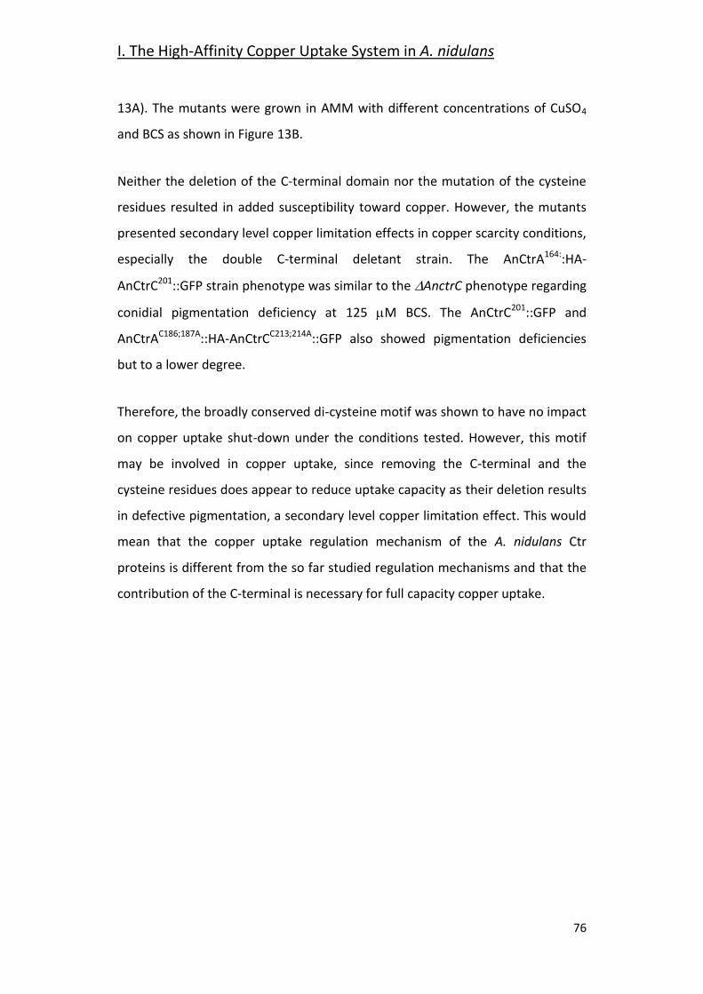
I. The High-Affinity Copper Uptake System in A. nidulans
76
13A). The mutants were grown in AMM with different concentrations of CuSO4
and BCS as shown in Figure 13B.
Neither the deletion of the C-terminal domain nor the mutation of the cysteine
residues resulted in added susceptibility toward copper. However, the mutants
presented secondary level copper limitation effects in copper scarcity conditions,
especially the double C-terminal deletant strain. The AnCtrA164::HA-
AnCtrC201::GFP strain phenotype was similar to the AnctrC phenotype regarding
conidial pigmentation deficiency at 125 M BCS. The AnCtrC201::GFP and
AnCtrAC186;187A::HA-AnCtrCC213;214A::GFP also showed pigmentation deficiencies
but to a lower degree.
Therefore, the broadly conserved di-cysteine motif was shown to have no impact
on copper uptake shut-down under the conditions tested. However, this motif
may be involved in copper uptake, since removing the C-terminal and the
cysteine residues does appear to reduce uptake capacity as their deletion results
in defective pigmentation, a secondary level copper limitation effect. This would
mean that the copper uptake regulation mechanism of the A. nidulans Ctr
proteins is different from the so far studied regulation mechanisms and that the
contribution of the C-terminal is necessary for full capacity copper uptake.
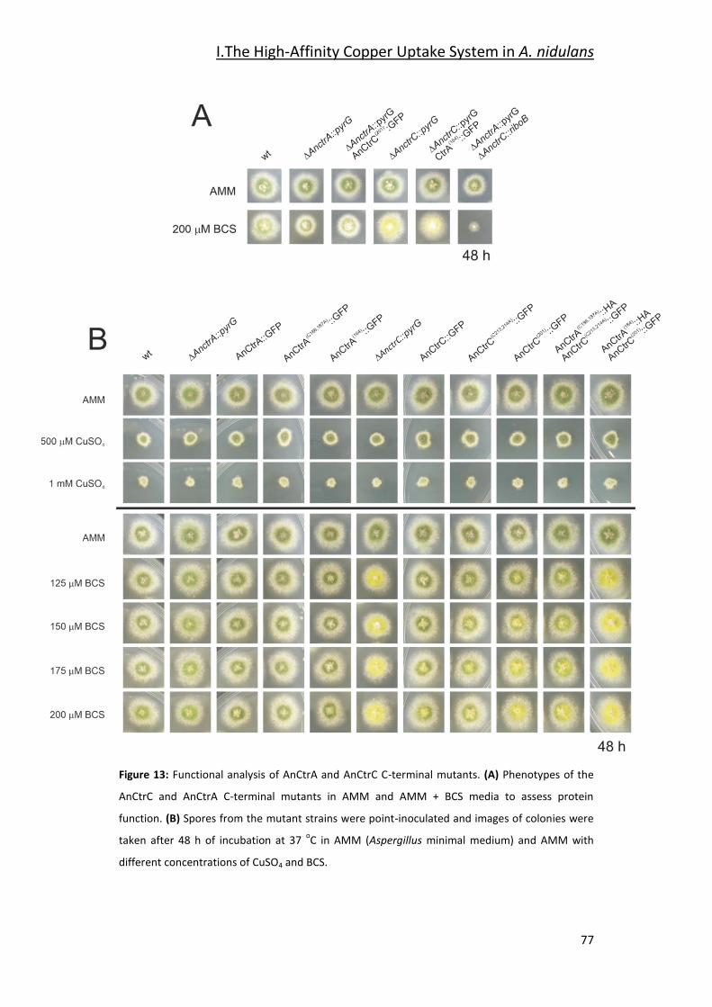
I.The High-Affinity Copper Uptake System in A. nidulans
77
Figure 13: Functional analysis of AnCtrA and AnCtrC C-terminal mutants. (A) Phenotypes of the
AnCtrC and AnCtrA C-terminal mutants in AMM and AMM + BCS media to assess protein
function. (B) Spores from the mutant strains were point-inoculated and images of colonies were
taken after 48 h of incubation at 37 oC in AMM (Aspergillus minimal medium) and AMM with
different concentrations of CuSO4 and BCS.
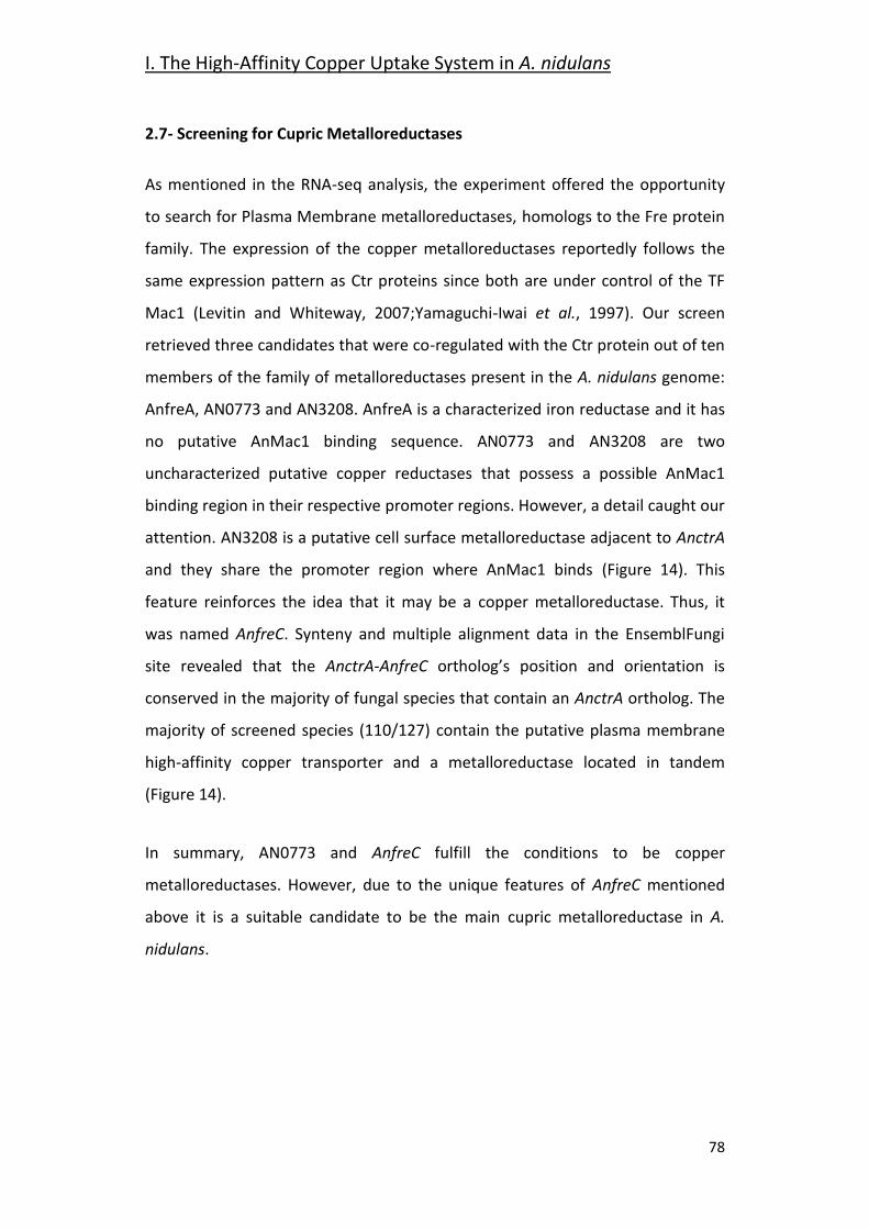
I. The High-Affinity Copper Uptake System in A. nidulans
78
2.7- Screening for Cupric Metalloreductases
As mentioned in the RNA-seq analysis, the experiment offered the opportunity
to search for Plasma Membrane metalloreductases, homologs to the Fre protein
family. The expression of the copper metalloreductases reportedly follows the
same expression pattern as Ctr proteins since both are under control of the TF
Mac1 (Levitin and Whiteway, 2007;Yamaguchi-Iwai et al., 1997). Our screen
retrieved three candidates that were co-regulated with the Ctr protein out of ten
members of the family of metalloreductases present in the A. nidulans genome:
AnfreA, AN0773 and AN3208. AnfreA is a characterized iron reductase and it has
no putative AnMac1 binding sequence. AN0773 and AN3208 are two
uncharacterized putative copper reductases that possess a possible AnMac1
binding region in their respective promoter regions. However, a detail caught our
attention. AN3208 is a putative cell surface metalloreductase adjacent to AnctrA
and they share the promoter region where AnMac1 binds (Figure 14). This
feature reinforces the idea that it may be a copper metalloreductase. Thus, it
was named AnfreC. Synteny and multiple alignment data in the EnsemblFungi
site revealed that the AnctrA-AnfreC ortholog’s position and orientation is
conserved in the majority of fungal species that contain an AnctrA ortholog. The
majority of screened species (110/127) contain the putative plasma membrane
high-affinity copper transporter and a metalloreductase located in tandem
(Figure 14).
In summary, AN0773 and AnfreC fulfill the conditions to be copper
metalloreductases. However, due to the unique features of AnfreC mentioned
above it is a suitable candidate to be the main cupric metalloreductase in A.
nidulans.
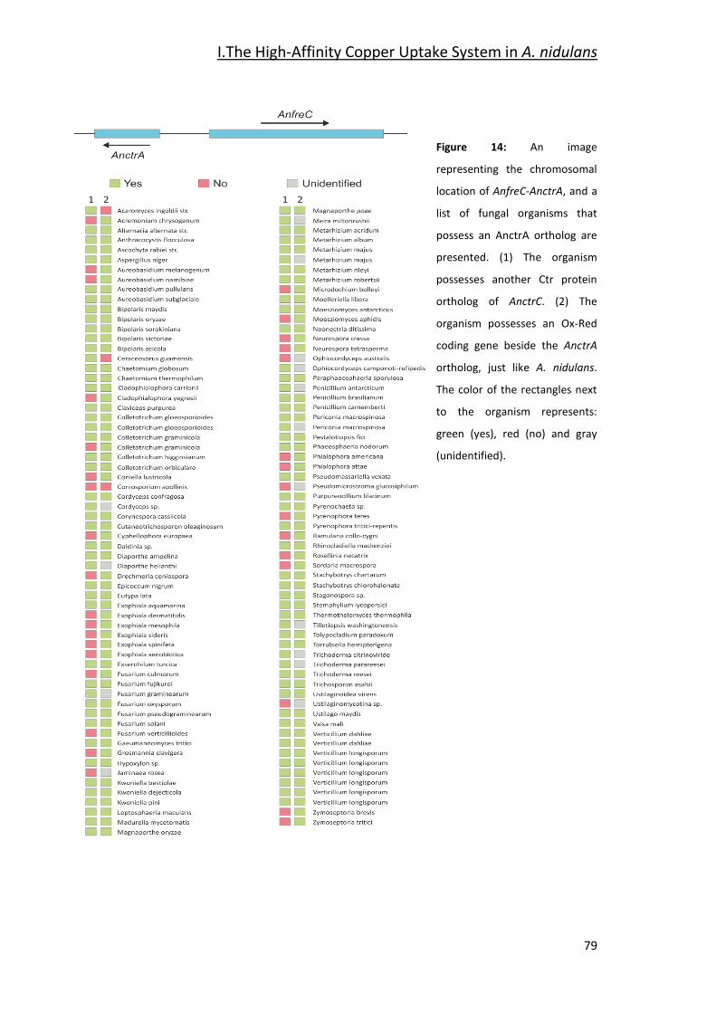
I.The High-Affinity Copper Uptake System in A. nidulans
79
Figure 14: An image
representing the chromosomal
location of AnfreC-AnctrA, and a
list of fungal organisms that
possess an AnctrA ortholog are
presented. (1) The organism
possesses another Ctr protein
ortholog of AnctrC. (2) The
organism possesses an Ox-Red
coding gene beside the AnctrA
ortholog, just like A. nidulans.
The color of the rectangles next
to the organism represents:
green (yes), red (no) and gray
(unidentified).
I. The High-Affinity Copper Uptake System in A. nidulans
80
3- Discussion
This study provides additional information on the expression and function of the
high-affinity copper uptake system reported in yeast and filamentous fungi of
clinical interest. The study of gene expression profiles under basal copper
availability conditions, copper deprivation and copper toxicity has shed new light
on the overall adaptation of A. nidulans to copper fluctuations, as well as
targeting the dynamics of copper transporters. The subsequent analysis through
targeted mutagenesis of the principal copper transporters has enabled an
interpretation of their individual contributions in the copper uptake process,
their expression patterns and their subcellular localization dynamics. In addition,
new inquiries into the role of transporter C-terminal domains together with the
identification of putative membrane copper reductases have been initiated.
The response to copper toxicity in A. nidulans involves the differential expression
of 13.7% of its genes. Significant changes were recorded on recognized copper
homeostasis genes. Even though the expression of the TFs regulating the process
was not significantly altered, that of proteins responsible of copper uptake and
detoxification was very notable. The gene coding for the copper exporting P-type
ATPase AnCrpA (Antsotegi-Uskola et al., 2017;Levitin and Whiteway,
2007;Yamaguchi-Iwai et al., 1997), was up-regulated. On the other hand, the two
characterized copper transporter genes AnctrA and AnctrC (Cai et al., 2019) were
down-regulated in line with the objective of reducing the copper uptake to the
minimum. Finally, the significant up-regulation of the intracellular copper
trafficking P-type ATPase AnygA (Clutterbuck, 1990) could be a response to the
presence of high amounts of copper ions within the cell that have to be
distributed or compartmentalized. Out of the identified three putative copper
chaperones only the putative Cu/Zn Sod chaperone AN6045 was significantly up-
regulated. The S. cerevisiae functional counterpart, chaperone Atx1, responsible
for copper distribution within the cell, binds excess cytosolic copper (Lin and
Culotta, 1995) and could act as a first level scavenger at low levels of toxicity.
I.The High-Affinity Copper Uptake System in A. nidulans
81
This effect may have taken place under our experimental conditions, as
discussed below.
Free cytoplasmic copper is known to produce ROS through Fenton-type reactions
(Sutton and Winterbourn, 1989;Timoshnikov et al., 2019). Superoxide
dismutases, accept electrons from reduced metals or superoxides, yielding
hydrogen peroxide, which, along with small organic peroxides, is the substrate of
catalases (2 H2O2 2 H2O + O2) (Zamocky et al., 2008). Hence, the reported
copper toxicity response involves the activation of both groups of enzymes
(Wiemann et al., 2017). In our experiments, however, we observed that catalases
AncatB (2.82-fold), AncatC (2.36-fold) and AncpeA/AncatD (2.59-fold) were up-
regulated, but the expression of the Cu/Zn superoxide dismutase AnsodA
(Holdom et al., 1996) (reported in Table 6), an enzyme activated in response to
copper and zinc-derived ROS toxicity, remained unchanged, along with that of
the ROS responsive transcription factors AnatfA and AnnapA (Table 7). These
variations may stem from the differences in copper concentrations and the
relative sensitivities of the strains used by (Wiemann et al., 2017) and this study.
In our experiments, 100 M Cu concentrations were used, a dose that exerted
no visible effect in A. nidulans colony development (Antsotegi-Uskola et al.,
2017). On the other hand, Wiemann et al., (2017) used a 200 M copper
concentration, in an organism at which 50 M already compromised colony
development. It is therefore likely that, under conditions which elicit no
phenotypic symptoms of copper toxicity, the AnCrpA detoxification system could
have removed the free cytosolic copper, rendering ROS neutralization
unnecessary. On the other hand, Wiemann et al., (2017) combined higher copper
concentrations and greater susceptibility may have overwhelmed the capacity of
AfCrpA, thus triggering the up-regulation catalases to quench ROS and H2O2.
Both results may depict the response to two different levels of copper toxicity. It
may be hypothesized that catalases are heme-containing enzymes (Scherer et al.,
2002) and it is well known that intracellular free copper ions can displace
I. The High-Affinity Copper Uptake System in A. nidulans
82
protein-bound iron. The selective up-regulation of the catalases in this case may
be a response to this phenomenon, leading to the greater demand for functional
catalases. More evidence will be required to confirm this interpretation.
Aside from catalases, the copper toxicity response also modified the expression
of many other oxidoreductases. Laccases are blue-copper enzymes involved in
lignin degradation or pigment biosynthesis (Scherer and Fischer, 2001). However,
laccases don’t show a specific expression pattern in response to copper toxicity
(Table 6). Other oxidoreductases corresponded to membrane function-related
metabolic processes such as sterol metabolism. Sterols, especially ergosterol, are
the most abundant lipids in fungal cell membranes (Rodrigues, 2018), with roles
in integral protein stabilization, membrane fluidity and permeability (Abe and
Hiraki, 2009;Alvarez et al., 2007). This could be a result of copper accumulation
in the Plasma membrane (Cervantes and Gutierrez-Corona, 1994). Under copper
toxicity, the expression of proteins involved in transmembrane traffic
augmented; for example, many putative sugar transporters were up-regulated.
This could be ascribed to a heightened requirement for energy (ATP) and
reducing power (NADH), both principal products of aerobic sugar catabolism. For
example, the ATP-dependent copper detoxification protein AnCrpA was highly
up-regulated in this study, in line with earlier observations (Antsotegi-Uskola et
al., 2017). Changes in differential expression of various heavy metal transporters
may indicate that a change of copper concentration could cause an imbalance in
heavy metal homeostasis, as described in A. fumigatus (Cai et al., 2017). Finally,
a stress mechanism with such a wide ranging impact on cellular metabolism
should also alter the regulation of a core regulatory process as transcription. A TF
that experienced notable lower expression under copper toxicity conditions was
AnhapX and this, in turn, is consistent with the down-regulation of the
siderophore biosynthesis cluster that it has been reported to regulate (Schrettl et
al., 2004). The same result was obtained with the antimicrobial compound
emericellamide biosynthesis cluster (Table 7). Finally, analyzing the data of the
RNA-seq we found out a highly up-regulated set of genes. The up-regulation of
I.The High-Affinity Copper Uptake System in A. nidulans
83
an N-acetylglucosamide transporter, AN2562, could be an adaptive response to
the high copper environment as this compound proves to be important for
survival in the phagosome (Vesely et al., 2017) where copper toxicity is used to
kill by the innate immune system (Li et al., 2019). Secondly, it is very convenient
that a cysteine and methionine transporter, AN2560, would be up-regulated in
such conditions as they are two amino-acids that participate in copper binding
motifs (Pena et al., 2000). The flavin-containing monooxygenase might play an
important role in creating an optimal Redox environment threatened by the
presence of copper in the cytosol (Suh et al., 2000). These three genes together
with the predicted oxidoreductase AN2559 and the putative methyltransferase
AN2561 might comprise a defensive gene cluster against copper toxicity.
The phylogenetic characteristics of two A. nidulans Ctr proteins, AnCtrA and
AnCtrC, have been examined in this study. Although the reference Ctr sequence
used to screen for orthologs in fungi had been ScCtr1, none were found in the A.
nidulans genome with that criterion. According to the Ensembl fungi ortholog
predictions, homologs of ScCtr1 are only found in the Saccharomycetales order.
Moreover, most Ctr proteins of filamentous fungi shared higher similarity with
ScCtr3 (Figure 8). Unfortunately, Scctr3 is poorly documented as it is disrupted by
a Ty2 transposon in many laboratory strains (Knight et al., 1996). AnCtrC showed
phylogenetic relationship with numerous Ctr orthologs in A. fumigatus, S.
cerevisiae and various other fungi. On the other hand, AnCtrA showed similarity
with other putative Ctr proteins that have not been characterized.
Both proteins possess the characteristic features of Ctr proteins (Petris,
2004;Rees and Thiele, 2004;Zhou et al., 2003). Ctr family members generally
harbor three putative TMDs (Puig et al., 2002), but according to topology
software predictions, AnCtrA has only two TMDs. This has also been reported in
A. fumigatus and C. neoformans (Park et al., 2014;Waterman et al., 2012).
Cysteine residues are present throughout the sequence and cysteines are known
for their affinity to copper (Pena et al., 2000). Both proteins contain two cysteine
I. The High-Affinity Copper Uptake System in A. nidulans
84
residues flanking the methionine residue adjacent to TMD1. This soft Lewis base
may be important to coordinate copper since it has been described that metal
binding residues located close to the plasma membrane seem to play a more
important role in copper transport (Puig et al., 2002).
Functional characterization analyses in this study showed that the two Ctr
proteins worked independently and the range of copper concentrations they
covered were partially complementary, as derived from the obtained
phenotypes. The phenotypes observed were broadly interpreted in terms of
acute or partial deficiencies as follows. Normal growth rates and WT-like green
conidial pigmentation were interpreted as an indication of adequate copper
supply for basic growth and developmental functions. Defects in spore
pigmentation and sporulation that did not affect growth rate were interpreted as
the result of a partial limitation in copper. Conidiogenesis is strongly reliant on
autophagy-derived resources (Kikuma et al., 2007;Richie and Askew, 2008). The
yellowish spore pigmentation could be explained by the inactivity of the copper-
dependent conidial laccase AnYA; the phenotype corresponds with the yA2
phenotype (Hermann et al., 1983;Scherer and Fischer, 2001). On the other hand,
acute copper deficiency was interpreted to affect extracellular copper transport
with direct consequences on vegetative growth. Following these criteria, we
surmised that AnCtrC functions as the principal copper uptake protein under
copper sufficiency and mild copper deficiency conditions. AnCtrA may
complement the lack of AnCtrC at moderate copper deficiencies but is
specifically required for extreme copper deficiency scenarios.
As shown in this study, conditions of low copper availability induce a gradual up-
regulation in Ctr expression, while high copper levels induce the opposite effect.
The rapid shutdown of the copper uptake system under high extracellular copper
concentrations is of great importance to avoid copper toxicity (Bertinato and
L'Abbe, 2004;Dong et al., 2013). The expression patterns of AnCtrA and AnCtrC
under similar testing conditions share certain similarities with S. cerevisiae Ctr
I.The High-Affinity Copper Uptake System in A. nidulans
85
proteins; a Ctr protein with a more stable expression pattern (ScCtr3) and
another one that is uniquely expressed under low copper availability conditions
(ScCtr1) (Pena et al., 2000). AnCtrC is the only high-affinity copper transporter
expressed at basal copper levels, which means it is likely to cover for copper
intake under ordinary copper availability (Figure 11B). On the other hand, AnCtrA
expression pattern seems to be designed for extreme copper scarcity conditions,
when AnCtrC transport cannot meet the cell’s copper requirements. These
results are in line with the conclusions from phenotypic analyses of selective null
mutants discussed above.
In terms of protein localization dynamics, Ctr proteins are known to follow a very
dynamic trafficking pathway. Pena et al., (2000) described that ScCtr3 traveled to
the plasma membrane through the Trans-Golgi network and the fluorescence
signal of tagged proteins accumulated in the ER after inhibiting protein traffic
from ER to Golgi. However, when copper concentration rose, Ctr proteins like
ScCtr1, moved from the plasma membrane to the vacuole (Liu et al., 2007), or
early endosomes, as in the case of hCtr1 (Clifford et al., 2016;Eisses and Kaplan,
2002). The predominant ER localization of AnCtrC (Markina-Inarrairaegui et al.,
2013) and its partial migration to the plasma membrane in copper deficiency
conditions support its role as a copper uptake protein covering transport under
copper sufficiency or mildly limiting conditions. The predominant plasma
membrane and vesicular localization of AnCtrA is in line with reported data of
other proteins that respond to extreme copper deficiency scenarios such as
described for ScCtr1 or hCtr1 (Clifford et al., 2016;Eisses and Kaplan, 2002;Liu et
al., 2007).
The shutdown of copper transport as a measure to avoid toxicity has been
documented many instances. Previous reports on this toxicity-preventing
mechanism refer to certain residues, mostly cysteines, in the C-terminus of Ctr
proteins as key elements in uptake modulation (Liu et al., 2007;Schuller et al.,
2013;Wu et al., 2009). Copper has been postulated to bind C-terminal cysteines,
I. The High-Affinity Copper Uptake System in A. nidulans
86
purportedly triggering the mechanism. The possible role of the Ctr proteins’ C-
terminal in activity modulation has not been studied in filamentous fungi.
However, the C-terminal di-cysteine motif, a common heavy metal binding
sequence (Lekeux et al., 2018) can be found in Ctr proteins of Alternaria
alternata, Aspergillus fumigatus, Botrytis cinerea, Fusarium graminearum,
Histoplasma capsulatum and Magnaporthe oryzae. We first hypothesized that
this di-cysteine motif could act as copper uptake modulator, as the ScCtr1 C-
terminal cysteine residues or the hCtr1 HCH amino acid motif. However, our
results do not indicate any copper dependent uptake modulation or
internalization role. Moreover, unlike the C-terminus of ScCtr1, that is
dispensable for full copper uptake capacity (Wu et al., 2009), deletion of AnCtrC
C-terminus results in secondary copper deficiency effects manifested as a
pigmentation deficiency (Figure 13B). Thus, the C-terminus of the Ctr proteins
plays some role in the copper uptake process. Further studies will be needed to
clarify this question.
Plasma membrane metalloreductases are crucial components of the copper
uptake process as they provide Ctr transporting proteins with the substrate in a
transport-compatible reduced state (Labbe et al., 1997;Petris, 2004). There are
documented reductases that reduce iron and also copper (Georgatsou et al.,
1997); however, AnfreA was discarded for lacking a putative Mac1 binding
sequence. AN0773 and AnfreC both shared copper reductase features, but the
phylogenetically preserved genomic location of AnfreC in tandem with AnctrA
tipped the scales in its favor. The genes coding for the reductive iron assimilation
complex composed by the oxidoreductase FetC and the iron transporter FtrA
(Schrettl et al., 2004) are located in tandem, exactly like AnfreC-AnctrA, in
Fusarium species (Park et al., 2007;Wiemann et al., 2012) and other Aspergillus
species like A. fumigatus, A. clavatus and A. fischeri for example. This fact points
out to AnfreC as the main copper oxidoreductase.
87
II. Chapter-
The Copper Detoxification System
in A. nidulans

88
II. The Copper Detoxification System in A. nidulans
89
1- Introduction
Having covered the transport of copper into fungal cells in the previous chapter,
we now turn our attention to the element of copper homeostasis dealing with
the internalized fraction of the cation that may be exerting toxicity. This may be
implemented through several mechanisms.
The RNA-seq data presented in the previous chapter offered the opportunity to
identify putative actors of the copper detoxification process: metallothioneins,
acting as copper quenching molecules and P-type ATPases that could pump the
cation into specific compartments or out of the cell. Thus, we screened for genes
that coded for metallothioneins and for P-type ATPases that could be up-
regulated under copper toxicity conditions. Two P-type ATPase coding genes
were identified in the screen.
P-type ATPases constitute a superfamily of membrane cargo transporters that
transport different kinds of cargo, from cations to lipids. Phylogenetically, they
are classified into 5 main subfamilies, from PI to P5, and in each family there are
also subgroups divided. The heavy metal transporting ATPases belong to the PIB
subgroup. PIB-type ATPases have peculiar terminal extensions (N- or C-terminal)
which contain metal (copper) binding domains (MBDs), rich in Cys and
sometimes His. PIB-ATPases possess 6 to 8 transmembrane domains (TMDs),
metal-binding sequences in their TMDs, regulatory MBDs, a structure involved in
enzyme phosphorylation (P-domain), a nucleotide-binding domain (N-domain),
and an energy transduction domain (A-domain) (Palmgren and Nissen, 2011).
One of the identified genes was an already annotated gene, AnyA, with
intracellular copper delivery function (Clutterbuck, 1990). The other one,
AN3117, had not been characterized yet. From previous work in C. albicans and
humans, P-type ATPases are known to either change location to perform
different functions within the copper homeostasis system, copper delivery and

II. The Copper Detoxification System in A. nidulans
90
detoxification (Lutsenko et al., 2007;Petris et al., 1996;Suzuki and Gitlin, 1999);
or the two processes can be achieved by two different P-type ATPases (Riggle
and Kumamoto, 2000;Weissman et al., 2000). A. nidulans follows the second
model and AN3117 was named AnCrpA, a putative detoxification protein.
Even though no metallothioneins were found among the up-regulated genes in
the RNA-seq, a single putative metallothionein coding gene was identified by
Blastp sequence analysis.
This chapter describes the identification and characterization of the major
copper detoxification determinant in A. nidulans, the copper transporting PI-type
ATPase, AnCrpA. AnCrpA expression is highly inducible and dynamic in response
to prolonged copper exposure and localizes close to the cellular surface as a
result of an organized trafficking process. In addition, metal-dependent induction
of AnCrpA is under the control of AnAceA, a transcription factor activator of
genes involved in copper detoxification. A third gene encoding a
metallothionein-like protein has been identified, AnCrdA, with as yet
unidentified function. Our results indicate that copper detoxification in A.
nidulans, as in the dimorphic fungus C. albicans, relies mainly in copper
excretion.
2- Results
2.1- Identification of Copper Resistance Determinants in A. nidulans.
The RNA-seq experiment described in chapter one, provided a spectrum of up-
regulated genes under copper toxicity. Detailed examination for candidates
responsible for copper detoxification resulted in the identification of AN3117, a
gene that encodes a 1211 amino acid (3636 bp) protein with all the signature
motifs of heavy metal transporting P-type ATPases. The protein is encoded by a
single exon containing gene located at chromosome VI. Blastp searches were
II. The Copper Detoxification System in A. nidulans
91
carried out in order to establish homology relations. AN3117 was classified as a
homologue of the main copper detoxification determinant in Candida albicans,
the P-type ATPase CaCrp1 (35% identity, 55% similarity and 95% query cover)
(Riggle and Kumamoto, 2000;Weissman et al., 2000). Thus, AN3117 was named
AnCrpA (copper resistance P-type ATPase). An already annotated gene termed
AnygA coding for a P-type ATPase was also detected within the up-regulated
genes. The protein coded by this gene shared great homology with the ScCcc2 P-
type ATPases from C. albicans and S. cerevisiae (Fu et al., 1995;Weissman et al.,
2000). This P-type ATPase has been attributed the role of copper transport to the
secretory pathway. The absence of this protein reportedly causes a pigmentation
defect resulting in yellow conidia (Clutterbuck, 1990).
From literature references and bioinformatic tools, the signature domains
identified in other copper transporting ATPases from human, bacteria, yeast and
archaea (Barry et al., 2010;Mandal et al., 2002;Migocka, 2015;Rosenzweig and
Argüello, 2012;Smith et al., 2016;Solioz and Odermatt, 1995) were recognized in
AnCrpA (Figure 15A). This kind of ATPases have 8 transmembrane helixes (TM),
TM 1-5 belong to the denominated Transport domain, TMDs 6-8 belong to the
Support domain and there is an invariant CPC copper translocation motif located
within the 6th TMD. Five adjacent cysteine rich metal binding motifs (MBD) are
present in the cytoplasmic N-terminal, two CxxC motifs and three GMxCxxC
motifs. As shown in Figure 15A, the configuration of the N-terminal MBDs is very
variable; however, AnCrpA and CaCrp1 share the same configuration. Finally, the
three signature motifs present in all P-type ATPses, the aspartyl kinase domain
(DKTG) containing the transiently phosphorylated aspartate residue during the
catalytic cycle, the ATP binding sequence (GDGVND) and the phosphatase
domain (TGES; Inesi et al., 2014;Palmgren and Nissen, 2011).
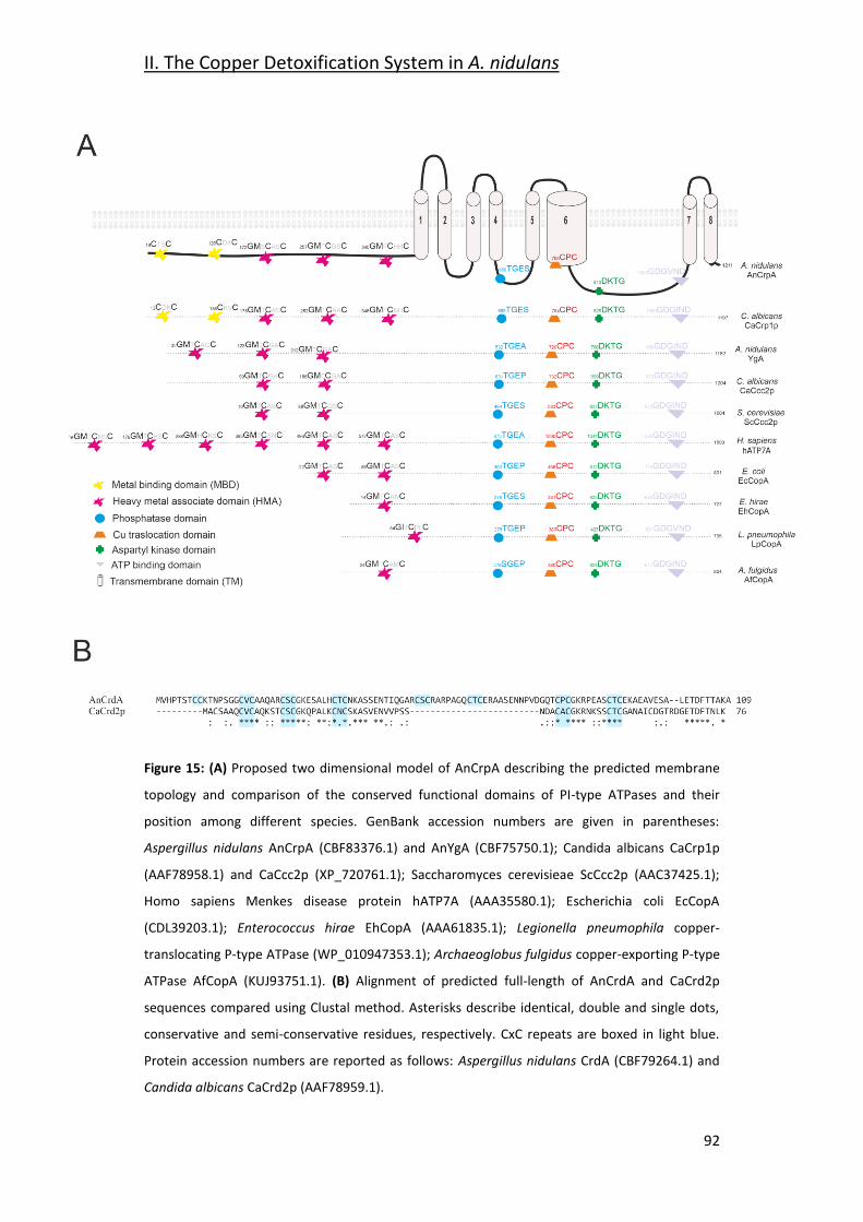
II. The Copper Detoxification System in A. nidulans
92
Figure 15: (A) Proposed two dimensional model of AnCrpA describing the predicted membrane
topology and comparison of the conserved functional domains of PI-type ATPases and their
position among different species. GenBank accession numbers are given in parentheses:
Aspergillus nidulans AnCrpA (CBF83376.1) and AnYgA (CBF75750.1); Candida albicans CaCrp1p
(AAF78958.1) and CaCcc2p (XP_720761.1); Saccharomyces cerevisieae ScCcc2p (AAC37425.1);
Homo sapiens Menkes disease protein hATP7A (AAA35580.1); Escherichia coli EcCopA
(CDL39203.1); Enterococcus hirae EhCopA (AAA61835.1); Legionella pneumophila copper-
translocating P-type ATPase (WP_010947353.1); Archaeoglobus fulgidus copper-exporting P-type
ATPase AfCopA (KUJ93751.1). (B) Alignment of predicted full-length of AnCrdA and CaCrd2p
sequences compared using Clustal method. Asterisks describe identical, double and single dots,
conservative and semi-conservative residues, respectively. CxC repeats are boxed in light blue.
Protein accession numbers are reported as follows: Aspergillus nidulans CrdA (CBF79264.1) and
Candida albicans CaCrd2p (AAF78959.1).
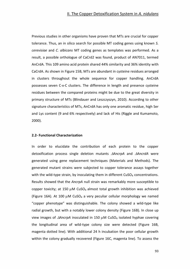
II. The Copper Detoxification System in A. nidulans
93
Previous studies in other organisms have proven that MTs are crucial for copper
tolerance. Thus, an in silico search for possible MT coding genes using known S.
cerevisiae and C. albicans MT coding genes as templates was performed. As a
result, a possible orthologue of CaCrd2 was found, product of AN7011, termed
AnCrdA. This 109 amino acid protein shared 44% similarity and 36% identity with
CaCrdA. As shown in Figure 15B, MTs are abundant in cysteine residues arranged
in clusters throughout the whole sequence for copper handling. AnCrdA
possesses seven C-x-C clusters. The difference in length and presence cysteine
residues between the compared proteins might be due to the great diversity in
primary structure of MTs (Blindauer and Leszczyszyn, 2010). According to other
signature characteristics of MTs, AnCrdA has only one aromatic residue, high Ser
and Lys content (9 and 6% respectively) and lack of His (Riggle and Kumamoto,
2000).
2.2- Functional Characterization
In order to elucidate the contribution of each protein to the copper
detoxification process single deletion mutants AncrpA and AncrdA were
generated using gene replacement techniques (Materials and Methods). The
generated mutant strains were subjected to copper tolerance assays together
with the wild-type strain, by inoculating them in different CuSO4 concentrations.
Results showed that the AncrpA null strain was remarkably more susceptible to
copper toxicity; at 150 M CuSO4 almost total growth inhibition was achieved
(Figure 16A). At 100 M CuSO4 a very peculiar cellular morphology we named
“copper phenotype” was distinguishable. The colony showed a wild-type like
radial growth, but with a notably lower colony density (Figure 16B). In close up
view images of AncrpA inoculated in 150 M CuSO4 isolated hyphae covering
the longitudinal area of wild-type colony size were detected (Figure 16B,
magenta dotted line). With additional 24 h incubation the poor cellular growth
within the colony gradually recovered (Figure 16C, magenta line). To assess the
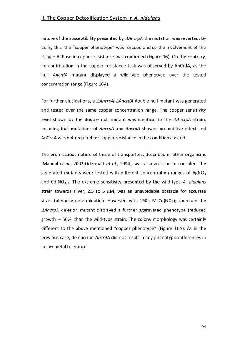
II. The Copper Detoxification System in A. nidulans
94
nature of the susceptibility presented by AncrpA the mutation was reverted. By
doing this, the “copper phenotype” was rescued and so the involvement of the
PI-type ATPase in copper resistance was confirmed (Figure 16). On the contrary,
no contribution in the copper resistance task was observed by AnCrdA, as the
null AncrdA mutant displayed a wild-type phenotype over the tested
concentration range (Figure 16A).
For further elucidations, a AncrpA-AncrdA double null mutant was generated
and tested over the same copper concentration range. The copper sensitivity
level shown by the double null mutant was identical to the AncrpA strain,
meaning that mutations of AncrpA and AncrdA showed no additive effect and
AnCrdA was not required for copper resistance in the conditions tested.
The promiscuous nature of these of transporters, described in other organisms
(Mandal et al., 2002;Odermatt et al., 1994), was also an issue to consider. The
generated mutants were tested with different concentration ranges of AgNO3
and Cd(NO3)2. The extreme sensitivity presented by the wild-type A. nidulans
strain towards silver, 2.5 to 5 M, was an unavoidable obstacle for accurate
silver tolerance determination. However, with 150 M Cd(NO3)2 cadmium the
AncrpA deletion mutant displayed a further aggravated phenotype (reduced
growth ∼ 50%) than the wild-type strain. The colony morphology was certainly
different to the above mentioned “copper phenotype” (Figure 16A). As in the
previous case, deletion of AncrdA did not result in any phenotypic differences in
heavy metal tolerance.
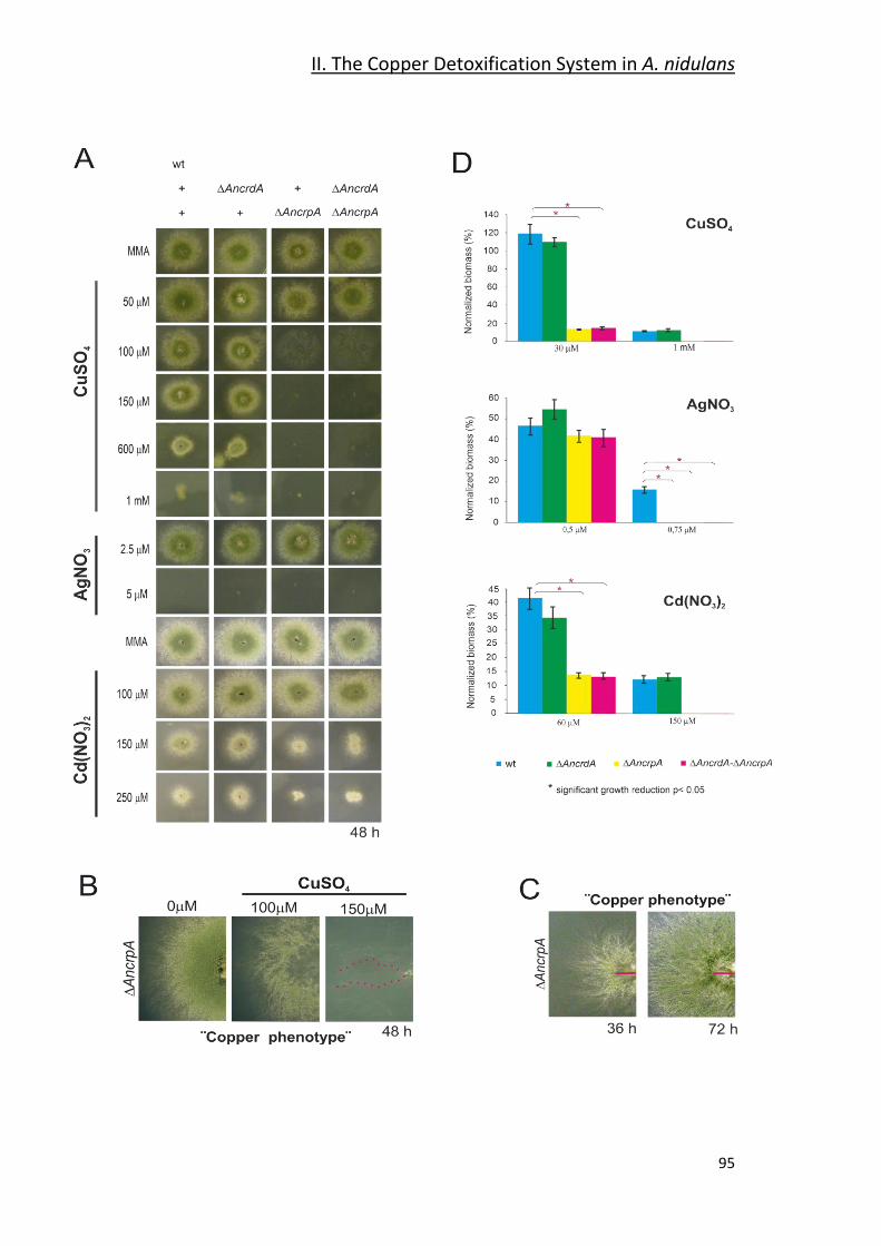
II. The Copper Detoxification System in A. nidulans
95
II. The Copper Detoxification System in A. nidulans
96
Figure 16: Phenotypic analysis of AncrpA and AncrdA mutant strains. Spores of strains having
the indicated relevant genotypes were point-inoculated on standard MMA. Images of colonies
were taken after 2 days of incubation at 37 oC. (A) Mutant characterization in solid medium
supplemented with indicated concentrations of metal salts. (B) Close-up views of the
morphological colony alterations in the central region of the colony caused by the deletion of
AncrpA when exposed to 100 μM CuSO4, denominated “Copper phenotype.” At 150 μM CuSO4
isolated hyphae were observed (magenta dotted line). (C) Recovery of cellular growth in the
central region of the colony over the time (magenta lines). Images of colonies were taken after
36 and 72 h of incubation. (D) Cellular growth in liquid MMA was monitored by determining the
dry weight (biomass) of cells grown 24 h at 37 oC with the indicated concentrations of metal salts.
In order to facilitate data comparison growth of each strain at basal level (no stress) was
designated 100%, data was normalized and presented as percentages. Graphs show the means ±
standard deviation (SD) of triplicate experiments (n = 3). * Significant growth reduction p < 0.05.
Liquid mycelial biomass experiments further corroborated the results obtained in
solid medium. Cellular growth in the presence of cadmium and copper was
strongly conditioned by AncrpA deletion, whereas in the case of AncrdA deletion,
no significant difference was observed. Biomass production of AncrpA and
AncrpA-AncrdA decreased up to 15% and 14%, respectively, compared to the
wild-type with 30 M CuSO4 and 60 M Cd(NO3)2. In the case of silver, no
differences were observed as all strains were unable to grow at 0.75 M AgNO3.
In conclusion, the obtained results highlight the importance of AnCrpA for
copper resistance in A. nidulans and possibly other metals. On the other hand, in
the conditions tested, the contribution of AnCrdA in metal ion toxicity resistance
is negligible. The possible role of this protein remains to be elucidated.
2.3- Expression Dynamics
Earlier studies on these proteins reported that MT expression is boosted by a
heavy metal load (Ehrensberger and Bird, 2011;García et al., 2002;Liu and Thiele,
1997;Peña et al., 1998) and copper detoxification by P-type ATPases is activated
by either inducing expression (Riggle and Kumamoto, 2000;Weissman et al.,
II. The Copper Detoxification System in A. nidulans
97
2000) or changing their subcellular localization (Cobbold et al., 2002;Suzuki and
Gitlin, 1999).
To confirm whether AncrpA followed the premise, an AnCrpA::HA3 chimera
expressing strain was generated. In order to rule out any possible alterations in
protein function due to HA3 tagging, the copper and cadmium tolerance levels of
the tagged strains were tested. The results showed that AnCrpA::HA3 was
functional (Fig 17). For the experiment, 16 h cultures were harvested according
to the established sampling times. The t0 sample was taken after 16 h of
incubation, then 100 M of CuSO4 was added to the medium and samples were
taken in the indicated times (15-360 min) (Fig 17B). Gene and protein expression
results are presented in a graphic normalized with respect rRNA and hexokinase
respectively (Fig 17C).
Northern-blot results show that AncrpA trasncripts were not detectable in
resting cells. Gene expression was first detected, 15 min after Cu-addition
reaching the peak of expression at 60 min. Then, the expression decreased
gradually throughout the course of the experiment reaching similar expression
level to that observed at 15 min (Fig 17B). These results reinforce the idea that
AncrpA expression follows the abovementioned pattern: after copper addition,
expression is transiently induced and in long copper exposures, low transcript
levels are maintained. The gene expression pattern matched that of protein
expression, except for the fact that the band corresponding to the chimera was
first detected at 30 min after copper exposure (Figure 17B). Something worth
mentioning is the presence of dispersed high molecular weight AnCrpA::HA3
species and a barely visible smaller band in the 60 min sample, which could be
indicative of post-translational modifications.
In order to assess the previously reported metal promiscuity of AnCrpA, the
protein expression experiment was repeated with silver and cadmium stress. In
the presence of 2.5 M AgNO3, the magnitude of the response was considerably
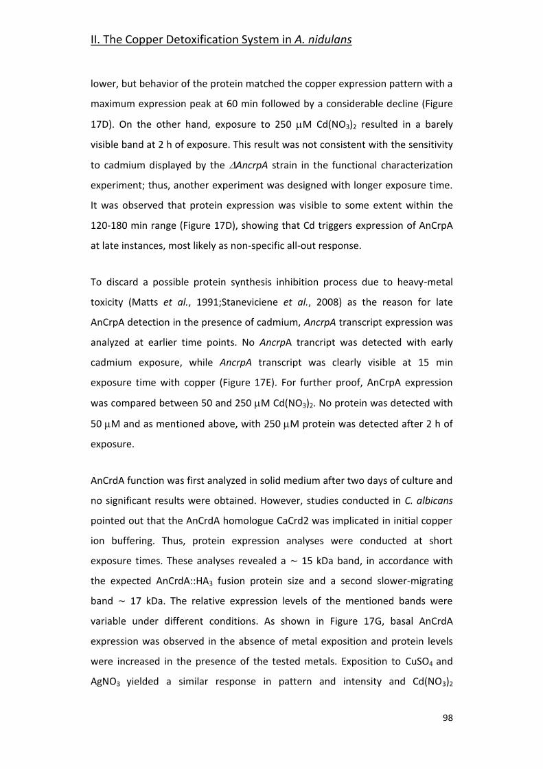
II. The Copper Detoxification System in A. nidulans
98
lower, but behavior of the protein matched the copper expression pattern with a
maximum expression peak at 60 min followed by a considerable decline (Figure
17D). On the other hand, exposure to 250 M Cd(NO3)2 resulted in a barely
visible band at 2 h of exposure. This result was not consistent with the sensitivity
to cadmium displayed by the AncrpA strain in the functional characterization
experiment; thus, another experiment was designed with longer exposure time.
It was observed that protein expression was visible to some extent within the
120-180 min range (Figure 17D), showing that Cd triggers expression of AnCrpA
at late instances, most likely as non-specific all-out response.
To discard a possible protein synthesis inhibition process due to heavy-metal
toxicity (Matts et al., 1991;Staneviciene et al., 2008) as the reason for late
AnCrpA detection in the presence of cadmium, AncrpA transcript expression was
analyzed at earlier time points. No AncrpA trancript was detected with early
cadmium exposure, while AncrpA transcript was clearly visible at 15 min
exposure time with copper (Figure 17E). For further proof, AnCrpA expression
was compared between 50 and 250 M Cd(NO3)2. No protein was detected with
50 M and as mentioned above, with 250 M protein was detected after 2 h of
exposure.
AnCrdA function was first analyzed in solid medium after two days of culture and
no significant results were obtained. However, studies conducted in C. albicans
pointed out that the AnCrdA homologue CaCrd2 was implicated in initial copper
ion buffering. Thus, protein expression analyses were conducted at short
exposure times. These analyses revealed a ∼ 15 kDa band, in accordance with
the expected AnCrdA::HA3 fusion protein size and a second slower-migrating
band ∼ 17 kDa. The relative expression levels of the mentioned bands were
variable under different conditions. As shown in Figure 17G, basal AnCrdA
expression was observed in the absence of metal exposition and protein levels
were increased in the presence of the tested metals. Exposition to CuSO4 and
AgNO3 yielded a similar response in pattern and intensity and Cd(NO3)2
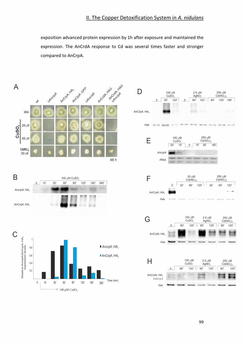
II. The Copper Detoxification System in A. nidulans
99
exposition advanced protein expression by 1h after exposure and maintained the
expression. The AnCrdA response to Cd was several times faster and stronger
compared to AnCrpA.
II. The Copper Detoxification System in A. nidulans
100
FIGURE 17: AnCrpA-HA3 and AnCrdA-HA3 induction by heavy metal ions. (A) Copper sensibility
assay of tagged AnCrpA and AnCrdA mutant strains. (B) Northern-blot and Western-blot showing
changes in AncrpA::HA3 transcript and AnCrpA::HA3 expression levels after addition of 100 μM of
CuSO4 and monitored at the indicated times. Images correspond to the area of interest. (C) The
graph shows AncrpA::HA3 transcript and AnCrpA::HA3 protein expressions relative to the
corresponding loading controls, rRNA and hexokinase respectively. Average pixel intensity for
each band was calculated with Image J (version 4.0; Fujifilm, Valhalla, NY). (D) Comparison of
AnCrpA::HA3 induction pattern by different metal salts [100 μM CuSO4, 2.5 μM AgNO3, and 250
μM Cd(NO3)2]. Images correspond to the area of interest. (E) Northern-blot showing changes in
AncrpA::HA3 expression after addition of 100 μM of CuSO4 but not during 1 h treatment with 250
μM of Cd(NO3)2. (F) Western-blot comparing Cd-induced AnCrpA::HA3 expression after 50 μM
and 250 μM of Cd(NO3)2 addition. Western-blot showing changes in HA3 tagged version of
AnCrdA in a wild-type (G) and null AncrpA (H) background induced by different metal ions.
Expression of AnCrdA was then studied in a AncrpA genetic background,
hypothesizing that the deletion of the major copper resistance determinant in
the organism may result in heightened AnCrdA expression in order to increase
the copper buffering capacity. For this purpose, a AncrpA strain expressing the
AnCrdA::HA3 chimera was generated (Figure 17A). Comparing protein expression
between the AncrpA and the wild-type background, the signal detected in
copper and silver exposure samples was significantly lower and, in the case of
cadmium, it was similar. Taken together, the results indicate that AnCrdA
expression is negatively affected by the absence of AncrpA.
Overall, the obtained results show that AncrpA has a dynamic expression pattern
during long copper treatments, a strong early response followed by a notable
decay reaching low expression levels that are maintained through time. The
experiments performed with different metal salts suggest that this ATPase
functions principally as a copper transporter, but it may also have a role in silver
transport. On the other hand, AnCrdA responds to metal toxicity, but its role in
heavy metal tolerance remains uncertain.
II. The Copper Detoxification System in A. nidulans
101
2.4- AnCrpA Dynamic Localization
The results obtained so far are consistent with at a copper extrusion role of the
P-type ATPase AnCrpA; this implied plasma membrane (PM) localization, not
excluding other compartments. To test this hypothesis, an AnCrpA::GFP fusion
protein expressing strain was generated. The strain displayed a wild-type like
phenotype in the tested conditions (Figure 18A) and the protein expression
analyses revealed similar protein dynamics to those previously seen AnCrpA::HA3
strain (Figure 18A).
For life in vivo microscopy experiments, cells were grown overnight without
copper stress and then shifted to new media containing 100 M CuSO4. The
protein was first visible at 30 min upon the medium shift. It was localized
throughout the hyphae in structures reminiscent of the ER (Figure 18B.A1,A2;
arrowed), surrounding oval structures that resembled the nucleus and strands
associated to the PM (Markina-Inarrairaegui et al., 2013). Taking a closer view to
the tip region, a finger-like protrusion towards the tip could be distinguished
(Figures 18B.A2,A4). After 1h of copper exposition, AnCrpA localization changed.
A homogeneous fluorescence signal in the cytosol was detected, but the most
notable feature was the strong fluorescence signal throughout the cellular
periphery, suggesting a predominant PM localization of AnCrpA (Figures
18B.B1,B2). In addition, it was found that the fluorescence signal was polarized
at the tip region in a large number of cells (Figures 18B.B3, B4). Linescan
measures were made in order to quantify signal accumulation throughout the
drawn lines and prove that the signal distribution is stronger in the PM and the
tip region.
After two hours of copper exposition, AnCrpA distribution was again modified. A
large part of the fluorescence remained at the PM. However, different
aggregates of variable morphologies were visible: punctate and ring-shaped
structures reminiscent of the Golgi body were dispersed in the cytosol
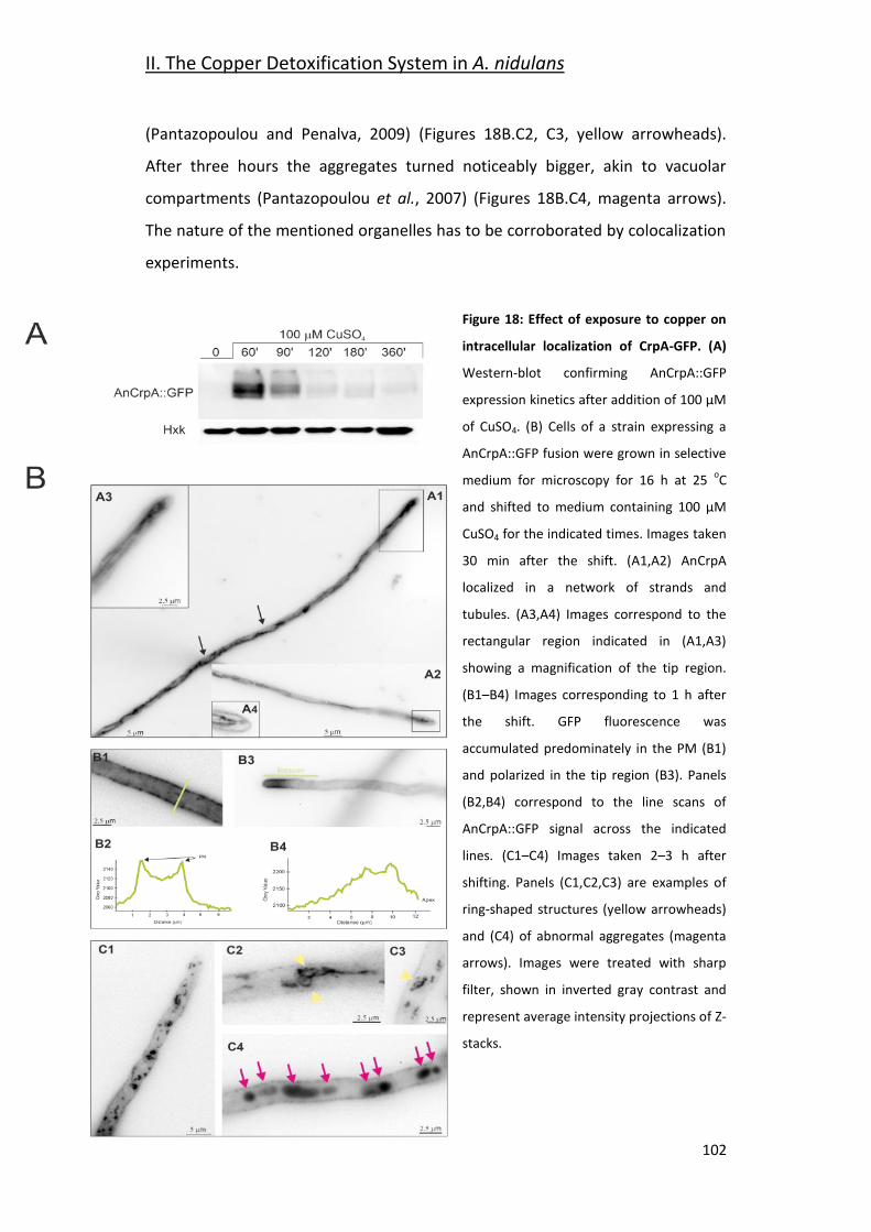
II. The Copper Detoxification System in A. nidulans
102
(Pantazopoulou and Penalva, 2009) (Figures 18B.C2, C3, yellow arrowheads).
After three hours the aggregates turned noticeably bigger, akin to vacuolar
compartments (Pantazopoulou et al., 2007) (Figures 18B.C4, magenta arrows).
The nature of the mentioned organelles has to be corroborated by colocalization
experiments.
Figure 18: Effect of exposure to copper on
intracellular localization of CrpA-GFP. (A)
Western-blot confirming AnCrpA::GFP
expression kinetics after addition of 100 μM
of CuSO4. (B) Cells of a strain expressing a
AnCrpA::GFP fusion were grown in selective
medium for microscopy for 16 h at 25 oC
and shifted to medium containing 100 μM
CuSO4 for the indicated times. Images taken
30 min after the shift. (A1,A2) AnCrpA
localized in a network of strands and
tubules. (A3,A4) Images correspond to the
rectangular region indicated in (A1,A3)
showing a magnification of the tip region.
(B1–B4) Images corresponding to 1 h after
the shift. GFP fluorescence was
accumulated predominately in the PM (B1)
and polarized in the tip region (B3). Panels
(B2,B4) correspond to the line scans of
AnCrpA::GFP signal across the indicated
lines. (C1–C4) Images taken 2–3 h after
shifting. Panels (C1,C2,C3) are examples of
ring-shaped structures (yellow arrowheads)
and (C4) of abnormal aggregates (magenta
arrows). Images were treated with sharp
filter, shown in inverted gray contrast and
represent average intensity projections of Z-
stacks.
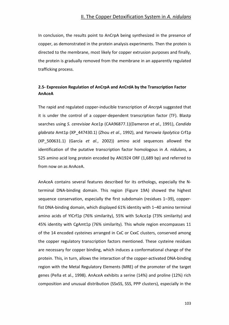
II. The Copper Detoxification System in A. nidulans
103
In conclusion, the results point to AnCrpA being synthesized in the presence of
copper, as demonstrated in the protein analysis experiments. Then the protein is
directed to the membrane, most likely for copper extrusion purposes and finally,
the protein is gradually removed from the membrane in an apparently regulated
trafficking process.
2.5- Expression Regulation of AnCrpA and AnCrdA by the Transcription Factor
AnAceA
The rapid and regulated copper-inducible transcription of AncrpA suggested that
it is under the control of a copper-dependent transcription factor (TF). Blastp
searches using S. cerevisiae Ace1p (CAA96877.1)(Dameron et al., 1991), Candida
glabrata Amt1p (XP_447430.1) (Zhou et al., 1992), and Yarrowia lipolytica Crf1p
(XP_500631.1) (García et al., 2002)) amino acid sequences allowed the
identification of the putative transcription factor homologous in A. nidulans, a
525 amino acid long protein encoded by AN1924 ORF (1,689 bp) and referred to
from now on as AnAceA.
AnAceA contains several features described for its orthologs, especially the N-
terminal DNA-binding domain. This region (Figure 19A) showed the highest
sequence conservation, especially the first subdomain (residues 1–39), copper-
fist DNA-binding domain, which displayed 61% identity with 1–40 amino terminal
amino acids of YlCrf1p (76% similarity), 55% with ScAce1p (73% similarity) and
45% identity with CgAmt1p (76% similarity). This whole region encompasses 11
of the 14 encoded cysteines arranged in CxC or CxxC clusters, conserved among
the copper regulatory transcription factors mentioned. These cysteine residues
are necessary for copper binding, which induces a conformational change of the
protein. This, in turn, allows the interaction of the copper-activated DNA-binding
region with the Metal Regulatory Elements (MRE) of the promoter of the target
genes (Peña et al., 1998). AnAceA exhibits a serine (14%) and proline (12%) rich
composition and unusual distribution (SSxSS, SSS, PPP clusters), especially in the
II. The Copper Detoxification System in A. nidulans
104
C-terminal half of the protein, similar to YlCrf1p of Y. lipolytica (García et al.,
2002).
The conservation of motifs is consistent with the view that AnAceA may be the
transcriptional factor involved in copper tolerance in A. nidulans. To investigate
its role, a single-knockout mutant of AnaceA locus was generated (BD965) and
tested in the same range of metal concentrations used before. Growth analyses
demonstrated that not only did the AnaceA strain display an identical “copper
phenotype” to that observed in AncrpA, but it also exhibited a more
pronounced sensitivity response to CuSO4, since colony defects were observed at
lower concentration (Figure 19B). Elevated cadmium content provoked a
reduction of colony growth, nevertheless in a similar degree to the previously
observed in the AncrpA strain. Biomass measurements supported these results
(Figure 19C). The AnaceA mutant strain was significantly more sensitive to
CuSO4 than AncrpA strain (p < 0.05). In contrast, the tolerance to Cd+2 and Ag+
was almost identical in both strains. These results confirm the implication of
AnAceA in metal tolerance, playing a key role in copper resistance.
We hypothesized that AnAceA was the copper dependent transcriptional
activator of genes involved in copper detoxification, thus the strain bearing a
mutation in the AnaceA gene should be sensitive to copper primarily due to a
defect in copper-inducible transcription. In order to prove this hypothesis, we
analyzed by Western-blot extracts of strain BD961, which expressed the
AnCrpA::HA3 fusion protein in an AnaceA-deletant background (Figure 19B).
Using anti-HA3 antibody, we did not detect the 134 kDa band observed
previously, as in extracts from the strain expressing AnCrpA::HA3 and loaded as
control (Figure 19D). The lack of AnCrpA::HA3 detection demonstrated that
AnAceA is necessary for metal induction of the PIB-type pump transcription.
Subsequently, the HA3 tagged metallothionein like protein in response to metal
salts in the absence of AnaceA allele (BD1062) was analyzed. AnCrdA::HA3 was
detected in the AnaceA -deleted strain (Figure 19E). Moreover, results indicated
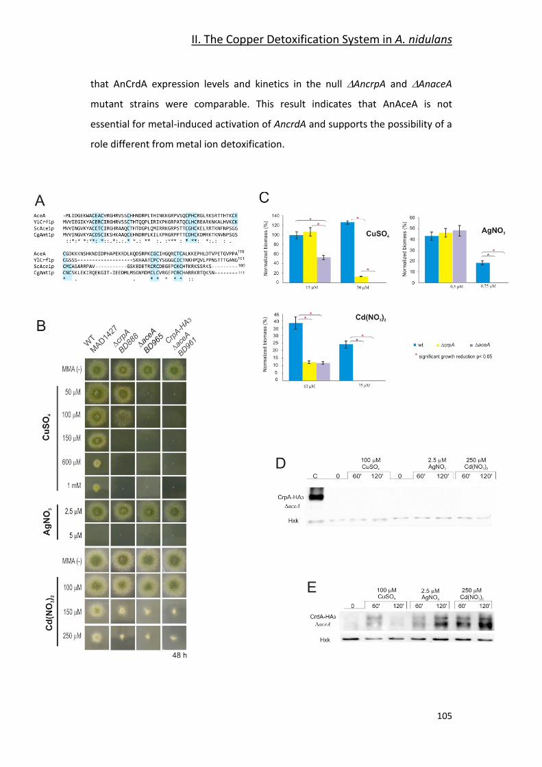
II. The Copper Detoxification System in A. nidulans
105
that AnCrdA expression levels and kinetics in the null AncrpA and AnaceA
mutant strains were comparable. This result indicates that AnAceA is not
essential for metal-induced activation of AncrdA and supports the possibility of a
role different from metal ion detoxification.
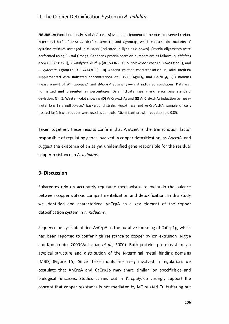
II. The Copper Detoxification System in A. nidulans
106
FIGURE 19: Functional analysis of AnAceA. (A) Multiple alignment of the most conserved region,
N-terminal half, of AnAceA, YlCrf1p, ScAce1p, and CgAmt1p, which contains the majority of
cysteine residues arranged in clusters (indicated in light blue boxes). Protein alignments were
performed using Clustal Omega. Genebank protein accesion numbers are as follows: A. nidulans
AceA (CBF85835.1), Y. lipolytica YlCrf1p (XP_500631.1), S. cerevisiae ScAce1p (CAA96877.1), and
C. glabrata CgAmt1p (XP_447430.1). (B) AnaceA mutant characterization in solid medium
supplemented with indicated concentrations of CuSO4, AgNO3, and Cd(NO3)2. (C) Biomass
measurement of WT, AnaceA and AncrpA strains grown at indicated conditions. Data was
normalized and presented as percentages. Bars indicate means and error bars standard
deviation. N = 3. Western-blot showing (D) AnCrpA::HA3 and (E) AnCrdA::HA3 induction by heavy
metal ions in a null AnaceA background strain. Hexokinase and AnCrpA::HA3 sample of cells
treated for 1 h with copper were used as controls. *Significant growth reduction p < 0.05.
Taken together, these results confirm that AnAceA is the transcription factor
responsible of regulating genes involved in copper detoxification, as AncrpA, and
suggest the existence of an as yet unidentified gene responsible for the residual
copper resistance in A. nidulans.
3- Discussion
Eukaryotes rely on accurately regulated mechanisms to maintain the balance
between copper uptake, compartmentalization and detoxification. In this study
we identified and characterized AnCrpA as a key element of the copper
detoxification system in A. nidulans.
Sequence analysis identified AnCrpA as the putative homolog of CaCrp1p, which
had been reported to confer high resistance to copper by ion extrusion (Riggle
and Kumamoto, 2000;Weissman et al., 2000). Both proteins proteins share an
atypical structure and distribution of the N-terminal metal binding domains
(MBD) (Figure 15). Since these motifs are likely involved in regulation, we
postulate that AnCrpA and CaCrp1p may share similar ion specificities and
biological functions. Studies carried out in Y. lipolytica strongly support the
concept that copper resistance is not mediated by MT related Cu buffering but
II. The Copper Detoxification System in A. nidulans
107
by efflux in this species (García et al., 2002;Ito et al., 2007). Further blast
searches in this dimorphic fungus revealed a putative homolog YALI0B02684p
(GeneBank accession number XP_500433.1), which displayed a similar N-
terminal extension. These results indicate that ATPase-based copper extrusion
may be found in a wider range of fungi than those reported so far. It is also
important to underscore the crucial role of copper resistance for virulence of
clinically important fungal pathogens as C. albicans (Mackie et al., 2016),
Cryptococcus neoformans (Ding et al., 2013a;Samanovic et al., 2012) and A.
fumigatus (Dietl et al., 2016;Wiemann et al., 2017).
Evidence of the direct participation of AnCrpA in copper resistance was obtained
by the extreme sensitivity exhibited by AnCrpA null mutants. An atypical aspect
of the phenotype of the null AncrpA mutation was the poor growth in the central
region of the colony, which overcame toxicity over time. Similar adaptive growth
patterns have been described previosuly (Valix et al., 2001). As the colony
matures, copper may bind to new cellular material locally, resulting in reduction
of free copper concentration, and increased growth.
Besides copper, AncrpA also exhibited sensitivity to cadmium; however, the
divergent phenotype reflected either affinity of metals for different targets
and/or distinct detoxification paths (Mendoza-Cózatl et al., 2010). Neither
AncrpA Cd-induction nor Cd-protein expression under low Cd concentration was
observed. In addition, AnCrpA was detected only in the presence of high
cadmium loads. Together, these results support an indirect PI-type ATPase
activation taking place, possibly due to the saturation of the main cadmium
detoxification system, a Cd2+ pump (Shiraishi et al., 2000;Thorsen et al., 2009) or
glutathione-derived peptides denominated phytochelatins (Mendoza-Cózatl et
al., 2005;Mendoza-Cózatl et al., 2010).
Regarding silver toxicity, our data showed that A. nidulans is sensitive to this
metal at a very low (2.5–5 μM) concentration range. Biomass measurements
II. The Copper Detoxification System in A. nidulans
108
indicate that AnCrpA may participate in silver resistance. On the other hand, it
was observed that silver induced expression of AnCrpA which matched copper
kinetics, although to a lesser extent. Based on these results, we propose that
copper is the principal substrate of the A. nidulans ATPase, however silver may
also be transported. Indeed, this is consistent with the fact that the orthologs
described above are also reported Cu+/Ag+ transporters. Although we did not
ascertain it in this study, AnCrpA likely exports Cu+ and not Cu2+ since; (a) in the
reduced cytosolic environment Cu+2 is found in Cu+ form and (b) substitution of
Cu+ by Ag+ is possible in proteins with CxxC containing MBDs, but not Cu2+ (Petris
et al., 1996).
Long term copper exposure induces an early, strong, and transient expression
followed by a basal maintenance expression level of AnCrpA. Regarding the
effect of copper exposure on AncrpA expression over time, it has been
demonstrated that AncrpA induction is rapid (within 15 min), transiently strong
(57-fold increase in 1 h) and sustained throughout the period of exposure to
copper, since mRNA levels were detectable along the whole experiment.
Hypothesizing that AncrpA mRNA levels may reflect intracellular ion
concentrations, it is plausible that, under long term high copper exposure, cells
would firstly synthesize an elevated amount of Cu-exporter in order to rapidly
reduce the initial burst of copper incorporated via high-affinity transporters.
Subsequently, pump levels would be down-regulated and then maintained at a
certain level during the period in which copper continues to enter chronically to
the cell via low-affinity transporters, similar to S. cerevisiae (Yu et al., 1996).
Regulation studies are ongoing at present in our laboratory to verify whether
AnCrpA post-translational modifications (PTM) are involved in AnCrpA activity
control besides the transcriptional regulation described in this work.
AnCrpA localization to the cell membrane in response to copper addition
sustains the expected copper extrusion role of this ATPase. Detailed evaluation
of the subcellular distribution indicated that AnCrpA is under the control of a
II. The Copper Detoxification System in A. nidulans
109
highly orchestrated trafficking process that resembles membrane transporter
regulation (Lauwers et al., 2010;Pantazopoulou and Diallinas, 2007). It may be
envisaged that, in response to increased cytosolic Cu+, de novo synthesized
AnCrpA may traffic from the ER to the Golgi compartment and then to the
plasma membrane (PM). Upon a purported decrease in cytosolic copper
concentration, a fraction of PM AnCrpA would be internalized by endocytosis
and sorted to the multivesicular body (MVB). AnCrpA could then be recycled
back to the PM via the TGN as reported in human cells (Pase et al., 2004) where
the PM AnCrpA pool could be the result of two different protein delivery
pathways; a newly synthesized and a recycled AnCrpA pool. Post-translational
modification and organelle trafficking mutant studies would be required to
establish a model of AnCrpA sorting and regulation. Solving the molecular
mechanism of the PI-type ATPase trafficking in A. nidulans could provide novel
insights into how cells with a polarized architecture regulate copper ATPase
activity.
The active role of AnAceA in copper resistance gene regulation was
demonstrated by the extreme copper-sensitive phenotype of the knock-out
mutant and the absence of expression of AnCrpA in the AnaceA strain. In fact,
the increased sensitivity of AnaceA with respect to AncrpA mutants
highlighted two major points: firstly, the participation of an additional element in
the copper detoxification path and secondly, the predominant role of AnCrpA
over the minor participation of the other player in the system. This second
participant could be either a copper-metallothionein (MT) that has not been
identified or a copper scavenging phytochelatin, like in Schizosaccahoromyces
pombe (Clemens et al., 1999). Identification of new metallothioneins, since the
primary structure is highly diverse, could be a laborious task. It may be
worthwhile to attempt a different approach: the characterization of the AnAceA
binding metal-response element (MRE) on the AnCrpA promoter might propose
a consensus MRE sequence for A. nidulans copper detoxification genes that
could facilitate new searches.
II. The Copper Detoxification System in A. nidulans
110
AnCrdA expression was induced by copper, silver and cadmium within 1 h,
indicating a role in metal homeostasis. In summary, we found that: (a) a AncrdA
strain did not exhibit a metal sensitivity phenotype, (b) a non-additive effect of
AncrpA and AncrdA deletion was observed, (c) the deletion of AncrpA did not
increase AncrdA expression, and (d) AnAceA was not required to activate AnCrdA
expression. Taken together, these results do not support the participation of
AnCrdA in AnCrpA-mediated metal detoxification. A likely role for this putative
metallothionein may be as an antioxidant (Blindauer and Leszczyszyn,
2010;Palmiter, 1998). Further studies will confirm whether the principal AnAceA-
regulated PI-type ATPase (AnCrpA) system is connected with AnCrdA mechanism
in response to metal stress.
111
III. Chapter-
Molecular Modelling Approach to
Screen for AnCrpA Inhibitors

112
III. Molecular Modelling Approach to Screen for AnCrpA Inhibitors
113
1- Introduction
The two previous chapters of this thesis cover the high-affinity copper uptake
system and the copper detoxification system of A. nidulans. The original aim of
this project was to gain insight on copper homeostasis in order to find possible
ways to complement or replace copper as an agricultural fungicide.
There are some different approaches to improve the efficiency of antifungal
preparations and one of them is by synergistic interaction, the use of an
ingredient that complements or amplifies the antifungal activity of the active
ingredient, the synergist. If the synergist can exert an impact on the functionality
of the copper homeostasis system, this could lead to higher susceptibility to
copper. Four processes where synergists could exert the desired effect on copper
homeostasis are: uptake, delivery, buffering and detoxification. This chapter
presents an attempt of a molecular modelling approach to find active
compounds that interfere with copper detoxification as proof of concept.
Current knowledge on copper homeostasis is limited to the uptake and the
detoxification systems. The Ctr proteins AnCtrA and AnCtrC are two possible
targets. Their purpose is to bind Cu+ with high-affinity and transport it inside the
cell when the cell senses that copper reservoir is low. However, their expression
profile is specific for low copper environments and the literature has provided
evidence that these proteins shut down when copper concentrations rise in the
environment. If more details of this mechanism were available, specific means to
interfere with the mechanism could be attempted. This is not currently feasible.
The best understood target currently is AnCrpA. As shown in chapter 2, it is the
main detoxification agent in A. nidulans and inhibiting its activity, either totally or
partially could make a significant difference to potentiate the effect of copper-
based fungicides. In addition, phylogenetic analyses have shown that a wide
range of phytopathogenic fungi possess a homolog protein of AnCrpA.
III. Molecular Modelling Approach to Screen for AnCrpA Inhibitors
114
In this chapter the procedure followed to find inhibitory compounds for AnCrpA
using computational techniques will be described. First, a structural model of
AnCrpA was generated based on the crystal structure of the Legionella
pheumophila copper extruding P-type ATPase LpCopA. This is called homology
modelling (Waterhouse et al., 2018). From the literature we determined the
most interesting areas for AnCrpA inhibition and different compound libraries
were screened by docking in the selected areas of the AnCrpA model. This
process is called virtual screening, a high-throughput approach to find the best
candidates according to their binding-energy values. From the virtual screening
experiments a set of candidates were selected depending on the binding-energy
(Jaghoori et al., 2016). However, the virtual screening gives no information about
the orientation of the molecule (binding pose) in the grid (delimited binding
area) and being a process that manipulates a lot of information in short time is
also prone to error. Thus, individual docking experiments were made to check
the results of virtual screening and to obtain the orientation of the molecule that
corresponds with the best binding energy possible (Rizvi et al., 2013). Finally,
molecular dynamics experiments were carried out with each molecule to check
protein-ligand complex stability (Mangiaterra et al., 2017).
The whole process was carried out under the supervision of Dr. Roberta Galeazzi
at the Department of Live and Environmental Sciences (DiSVA) in “Università
Politecnica delle Marche” for their experience in similar procedures (Mangiaterra
et al., 2017).
III. Molecular Modelling Approach to Screen for AnCrpA Inhibitors
115
2- Materials & Methods
2.1- Protein Preparation
The protein structure of AnCrpA was obtained by homology modelling. The PDB
file of the template was downloaded from the Protein Data Bank (PDB). The
Legionella pneumophila CopA protein structure “4bbj” obtained by protein
crystallization was used as a template (35.8% identity). This structure has a 2.75
Å resolution and is delineated in the E2P state (Andersson et al., 2014). The
homology modelling was carried out in the fully automated protein structure
homology-modelling server SWISS-MODEL using the “User template” option.
Once the structure of the protein was obtained hydrogens were added to the
new model and the template and the Root Mean Square Deviation (RMSD)
between the template and our structure was calculated using Chimera software
(Pettersen et al., 2004;Waterhouse et al., 2018).
Protein salinity was settled in pH 6.5, which corresponds to the internal pH of the
organism (Caddick et al., 1986). Then the macromolecule was minimized using a
standardized protocol (Galeazzi et al., 2014;Scire et al., 2013) (Mohamadi et al.,
1990;Suite., 2011), using AMBER force field (Duan et al., 2003). By doing this the
software makes the net inter-atomic force closest to zero as possible.
2.2- Grid Generation
For the virtual screening experiments the area in which we want to dock the
compound library has to be delimited. Each delimited area is called a receptor
grid. To generate the receptor grids to test, “Receptor Grid Generation” tool
from Autodock Vina software was used (Morris et al., 2009;Sanner, 1999). For
grid selection, data available on Legionella pneumophila CopA was used. Two sites were specifically selected: The ATP binding site and the putative chaperone
binding region.
III. Molecular Modelling Approach to Screen for AnCrpA Inhibitors
116
Grid 1 (The ATP binding site): 70 x 70 x 76 Å.
Grid 2 (Putative chaperone binding site): 62 x 100 x 86 Å.
2.3- Virtual Screening and Focused Docking
Molecular docking experiments were carried out in the AutoDock Vina version
1.1.2 software (Trott and Olson, 2010). The in-house compound libraries were
screened using the VINA software against the two selected receptor grids of the
AnCrpA model (most compounds are from the database ZINC12). The PDB file of
our model was converted to PDBQT and all compounds were docked in the
selected grids using standard parameters. The procedure was repeated three
times, each one in a different computer. Only the compounds that appear in the
top 10 binding energy score on the three replicates of each library were used for
further procedures (Jaghoori et al., 2016).
After choosing the compounds with best binding energy score from the Virtual
Screening, Autodock 4.2.1 (AD4) was used to check the virtual screening results
and in case they were good, obtain the best binding pose (orientation of the
molecule in the binding grid) and relative energy (Chang et al., 2010;Morris et al.,
1998). The virtual screening process binds the compounds 10 times in the
delimited grid and shows the best binding-energy of each compound, but it gives
no information about how the molecule is oriented within the grid (binding pose)
and the residues that it interacts with. With Autodock 4.2.1 the molecules are
docked individually and the number of binding times was increased from 10 to
1000 within each grid. As a result of this process different binding poses of each
molecule that would differ in binding-energy and frequency would be obtained.
The binding pose with best energy and frequency ratio was chosen in each case
(Rizvi et al., 2013).
III. Molecular Modelling Approach to Screen for AnCrpA Inhibitors
117
2.4- Molecular Dynamics
Molecular dynamics simulations are a tool to predict the behavior of the protein
and the ligand when they are docked. This approach shows if they are likely to
form a stable complex or not. As AnCrpA is localized in the plasma membrane,
for the molecular dynamics simulation an AnCrpA-Plasma Membrane complex
was designed. For such purpose, AnCrpA orientation in the membrane was
obtained by the OPM server (https://opm.phar.umich.edu/ppm_server) and the
Protein-Plasma membrane (PM) complex was built using CHARMM GUI
(http://www.charmm-gui.org/). The membrane was composed of 25%
ergosterol, 42% palmitoyl oleyl phosphaditylcholine and 33% N-palmitoyl
sphingomyelin. The result was a Protein-PM complex with a 91 x 91 x 160 Å size,
appropriately solvated with water and ionized (0.15 M NaCl). AMBER99SB-ILDN
force field parameters were used for the proteins and lipids (Guy et al., 2012)
and the TIP3P model (Jorgensen, 1998) model for solvent as implemented in
GROMACS 5 (Hess et al., 2008). First the Protein-PM model was minimized,
subjected to six equilibration steps and then the molecular dynamic simulation
of 10 ns was carried out on its own. The same procedure was repeated with the
Protein-PM complex with each compound docked in the grid with the best
binding orientation obtained with Autodock 4.2.1. By doing this the stability of
the ligand-receptor complex is studied (Mangiaterra et al., 2017).
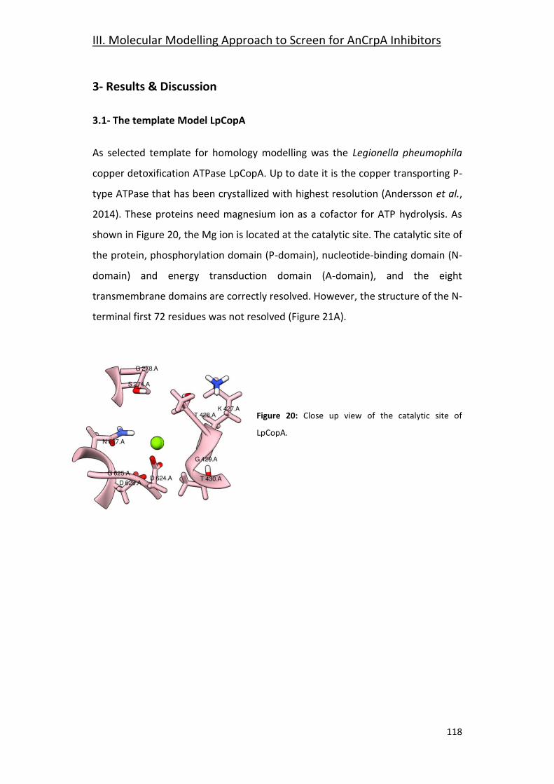
III. Molecular Modelling Approach to Screen for AnCrpA Inhibitors
118
3- Results & Discussion
3.1- The template Model LpCopA
As selected template for homology modelling was the Legionella pheumophila
copper detoxification ATPase LpCopA. Up to date it is the copper transporting P-
type ATPase that has been crystallized with highest resolution (Andersson et al.,
2014). These proteins need magnesium ion as a cofactor for ATP hydrolysis. As
shown in Figure 20, the Mg ion is located at the catalytic site. The catalytic site of
the protein, phosphorylation domain (P-domain), nucleotide-binding domain (N-
domain) and energy transduction domain (A-domain), and the eight
transmembrane domains are correctly resolved. However, the structure of the N-
terminal first 72 residues was not resolved (Figure 21A).
Figure 20: Close up view of the catalytic site of
LpCopA.
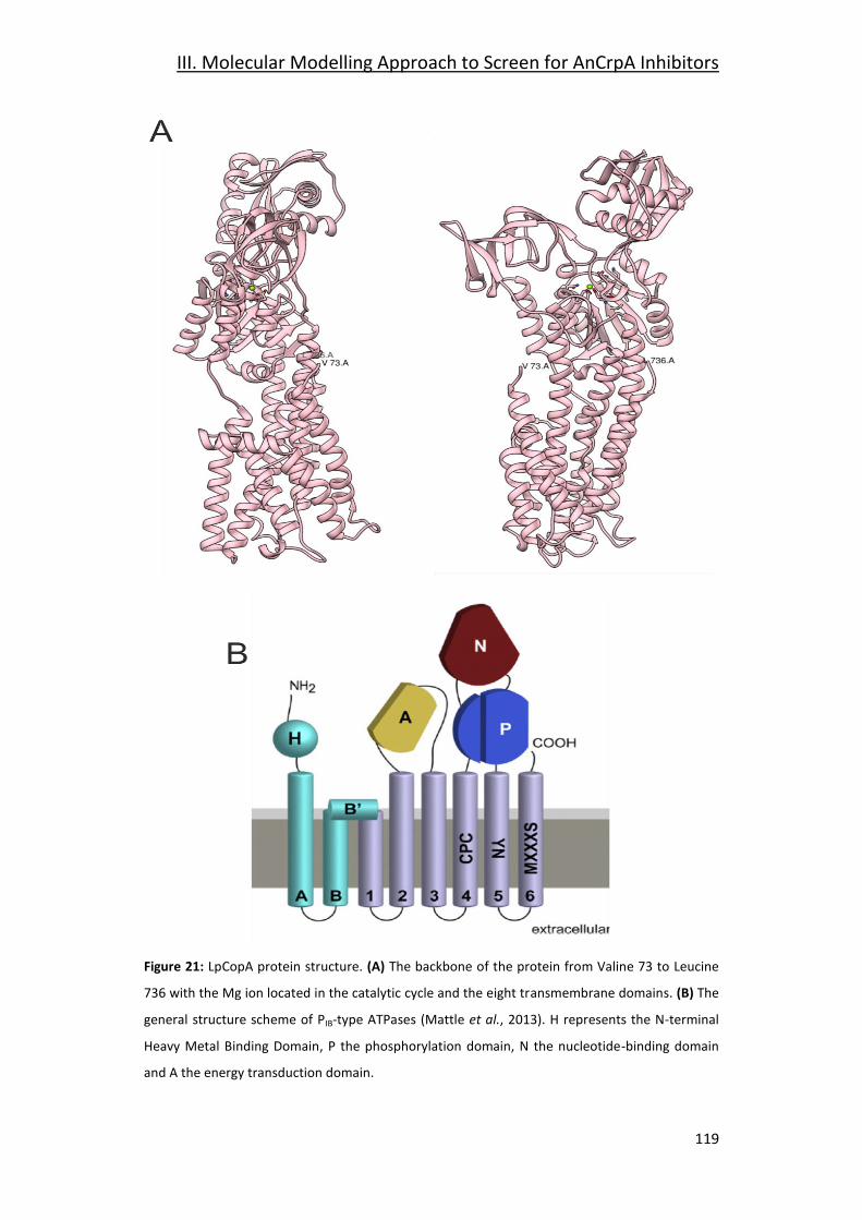
III. Molecular Modelling Approach to Screen for AnCrpA Inhibitors
119
Figure 21: LpCopA protein structure. (A) The backbone of the protein from Valine 73 to Leucine
736 with the Mg ion located in the catalytic cycle and the eight transmembrane domains. (B) The
general structure scheme of PIB-type ATPases (Mattle et al., 2013). H represents the N-terminal
Heavy Metal Binding Domain, P the phosphorylation domain, N the nucleotide-binding domain
and A the energy transduction domain.
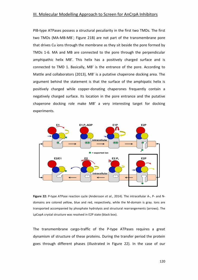
III. Molecular Modelling Approach to Screen for AnCrpA Inhibitors
120
PIB-type ATPases possess a structural peculiarity in the first two TMDs. The first
two TMDs (MA-MB-MB´; Figure 21B) are not part of the transmembrane pore
that drives Cu ions through the membrane as they sit beside the pore formed by
TMDs 1-6. MA and MB are connected to the pore through the perpendicular
amphipathic helix MB’. This helix has a positively charged surface and is
connected to TMD 1. Basically, MB’ is the entrance of the pore. According to
Mattle and collaborators (2013), MB’ is a putative chaperone docking area. The
argument behind the statement is that the surface of the amphipatic helix is
positively charged while copper-donating chaperones frequently contain a
negatively charged surface. Its location in the pore entrance and the putative
chaperone docking role make MB’ a very interesting target for docking
experiments.
Figure 22: P-type ATPase reaction cycle (Andersson et al., 2014). The intracellular A-, P- and N-
domains are colored yellow, blue and red, respectively, while the M-domain is gray. Ions are
transported accompanied by phosphate hydrolysis and structural rearrangements (arrows). The
LpCopA crystal structure was resolved in E2P state (black box).
The transmembrane cargo-traffic of the P-type ATPases requires a great
dynamism of structure of these proteins. During the transfer period the protein
goes through different phases (illustrated in Figure 22). In the case of our
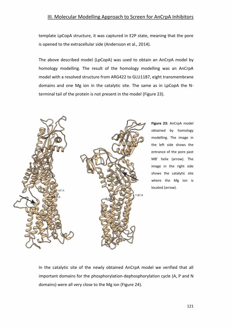
III. Molecular Modelling Approach to Screen for AnCrpA Inhibitors
121
template LpCopA structure, it was captured in E2P state, meaning that the pore
is opened to the extracellular side (Andersson et al., 2014).
The above described model (LpCopA) was used to obtain an AnCrpA model by
homology modelling. The result of the homology modelling was an AnCrpA
model with a resolved structure from ARG422 to GLU1187, eight transmembrane
domains and one Mg ion in the catalytic site. The same as in LpCopA the N-
terminal tail of the protein is not present in the model (Figure 23).
Figure 23: AnCrpA model
obtained by homology
modelling. The image in
the left side shows the
entrance of the pore past
MB’ helix (arrow). The
image in the right side
shows the catalytic site
where the Mg ion is
located (arrow).
In the catalytic site of the newly obtained AnCrpA model we verified that all
important domains for the phosphorylation-dephosphorylation cycle (A, P and N
domains) were all very close to the Mg ion (Figure 24).
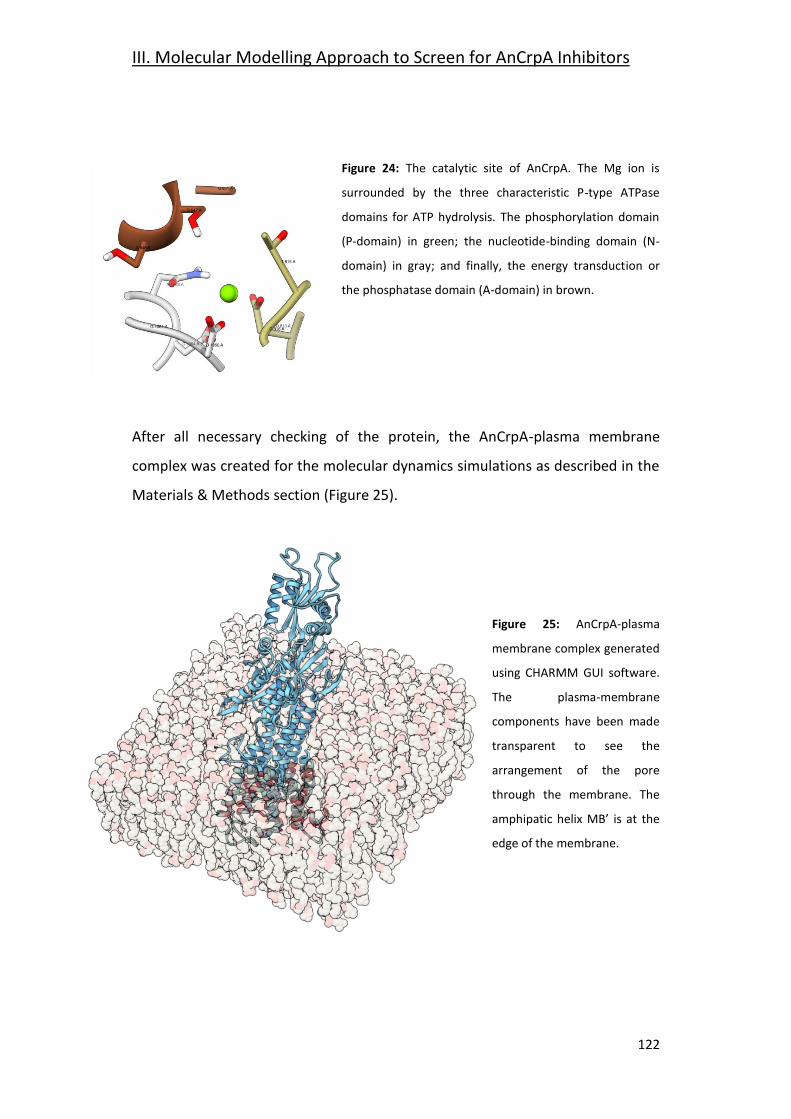
III. Molecular Modelling Approach to Screen for AnCrpA Inhibitors
122
Figure 24: The catalytic site of AnCrpA. The Mg ion is
surrounded by the three characteristic P-type ATPase
domains for ATP hydrolysis. The phosphorylation domain
(P-domain) in green; the nucleotide-binding domain (N-
domain) in gray; and finally, the energy transduction or
the phosphatase domain (A-domain) in brown.
After all necessary checking of the protein, the AnCrpA-plasma membrane
complex was created for the molecular dynamics simulations as described in the
Materials & Methods section (Figure 25).
Figure 25: AnCrpA-plasma
membrane complex generated
using CHARMM GUI software.
The plasma-membrane
components have been made
transparent to see the
arrangement of the pore
through the membrane. The
amphipatic helix MB’ is at the
edge of the membrane.
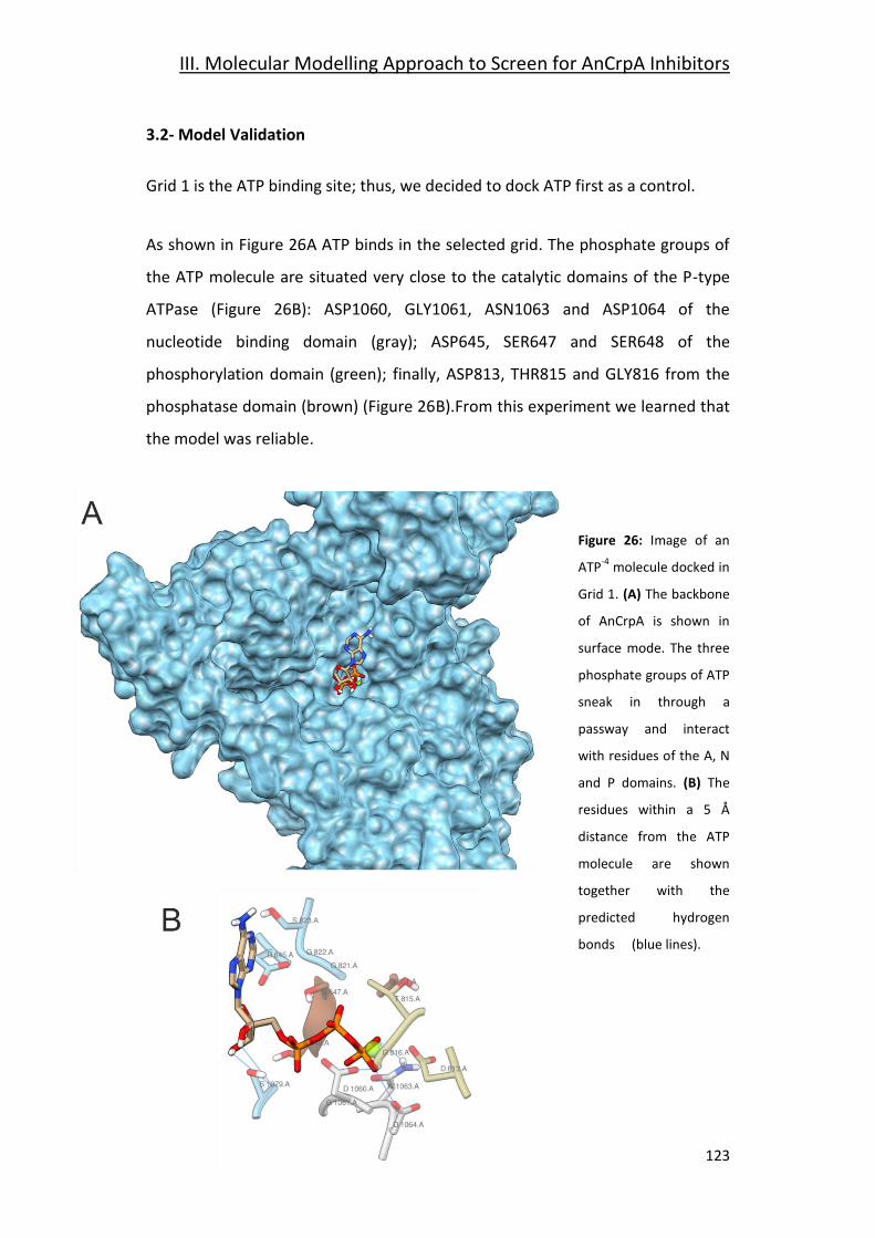
III. Molecular Modelling Approach to Screen for AnCrpA Inhibitors
123
3.2- Model Validation
Grid 1 is the ATP binding site; thus, we decided to dock ATP first as a control.
As shown in Figure 26A ATP binds in the selected grid. The phosphate groups of
the ATP molecule are situated very close to the catalytic domains of the P-type
ATPase (Figure 26B): ASP1060, GLY1061, ASN1063 and ASP1064 of the
nucleotide binding domain (gray); ASP645, SER647 and SER648 of the
phosphorylation domain (green); finally, ASP813, THR815 and GLY816 from the
phosphatase domain (brown) (Figure 26B).From this experiment we learned that
the model was reliable.
Figure 26: Image of an
ATP-4
molecule docked in
Grid 1. (A) The backbone
of AnCrpA is shown in
surface mode. The three
phosphate groups of ATP
sneak in through a
passway and interact
with residues of the A, N
and P domains. (B) The
residues within a 5 Å
distance from the ATP
molecule are shown
together with the
predicted hydrogen
bonds (blue lines).
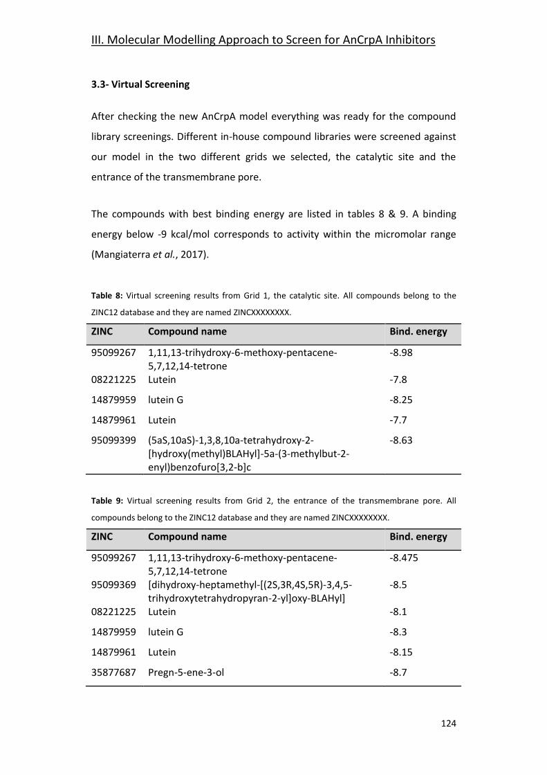
III. Molecular Modelling Approach to Screen for AnCrpA Inhibitors
124
3.3- Virtual Screening
After checking the new AnCrpA model everything was ready for the compound
library screenings. Different in-house compound libraries were screened against
our model in the two different grids we selected, the catalytic site and the
entrance of the transmembrane pore.
The compounds with best binding energy are listed in tables 8 & 9. A binding
energy below -9 kcal/mol corresponds to activity within the micromolar range
(Mangiaterra et al., 2017).
Table 8: Virtual screening results from Grid 1, the catalytic site. All compounds belong to the
ZINC12 database and they are named ZINCXXXXXXXX.
ZINC Compound name Bind. energy
95099267 1,11,13-trihydroxy-6-methoxy-pentacene-5,7,12,14-tetrone
-8.98
08221225 Lutein -7.8
14879959 lutein G -8.25
14879961 Lutein -7.7
95099399 (5aS,10aS)-1,3,8,10a-tetrahydroxy-2-[hydroxy(methyl)BLAHyl]-5a-(3-methylbut-2-enyl)benzofuro[3,2-b]c
-8.63
Table 9: Virtual screening results from Grid 2, the entrance of the transmembrane pore. All
compounds belong to the ZINC12 database and they are named ZINCXXXXXXXX.
ZINC Compound name Bind. energy
95099267 1,11,13-trihydroxy-6-methoxy-pentacene-5,7,12,14-tetrone
-8.475
95099369 [dihydroxy-heptamethyl-[(2S,3R,4S,5R)-3,4,5-trihydroxytetrahydropyran-2-yl]oxy-BLAHyl]
-8.5
08221225 Lutein -8.1
14879959 lutein G -8.3
14879961 Lutein -8.15
35877687 Pregn-5-ene-3-ol -8.7
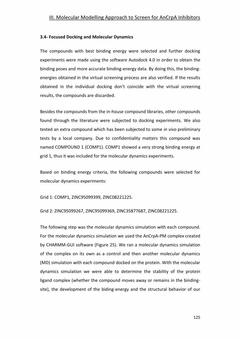
III. Molecular Modelling Approach to Screen for AnCrpA Inhibitors
125
3.4- Focused Docking and Molecular Dynamics
The compounds with best binding energy were selected and further docking
experiments were made using the software Autodock 4.0 in order to obtain the
binding poses and more accurate binding-energy data. By doing this, the binding-
energies obtained in the virtual screening process are also verified. If the results
obtained in the individual docking don’t coincide with the virtual screening
results, the compounds are discarded.
Besides the compounds from the in-house compound libraries, other compounds
found through the literature were subjected to docking experiments. We also
tested an extra compound which has been subjected to some in vivo preliminary
tests by a local company. Due to confidentiality matters this compound was
named COMPOUND 1 (COMP1). COMP1 showed a very strong binding energy at
grid 1, thus it was included for the molecular dynamics experiments.
Based on binding energy criteria, the following compounds were selected for
molecular dynamics experiments:
Grid 1: COMP1, ZINC95099399, ZINC08221225.
Grid 2: ZINC95099267, ZINC95099369, ZINC35877687, ZINC08221225.
The following step was the molecular dynamics simulation with each compound.
For the molecular dynamics simulation we used the AnCrpA-PM complex created
by CHARMM-GUI software (Figure 25). We ran a molecular dynamics simulation
of the complex on its own as a control and then another molecular dynamics
(MD) simulation with each compound docked on the protein. With the molecular
dynamics simulation we were able to determine the stability of the protein
ligand complex (whether the compound moves away or remains in the binding-
site), the development of the biding-energy and the structural behavior of our
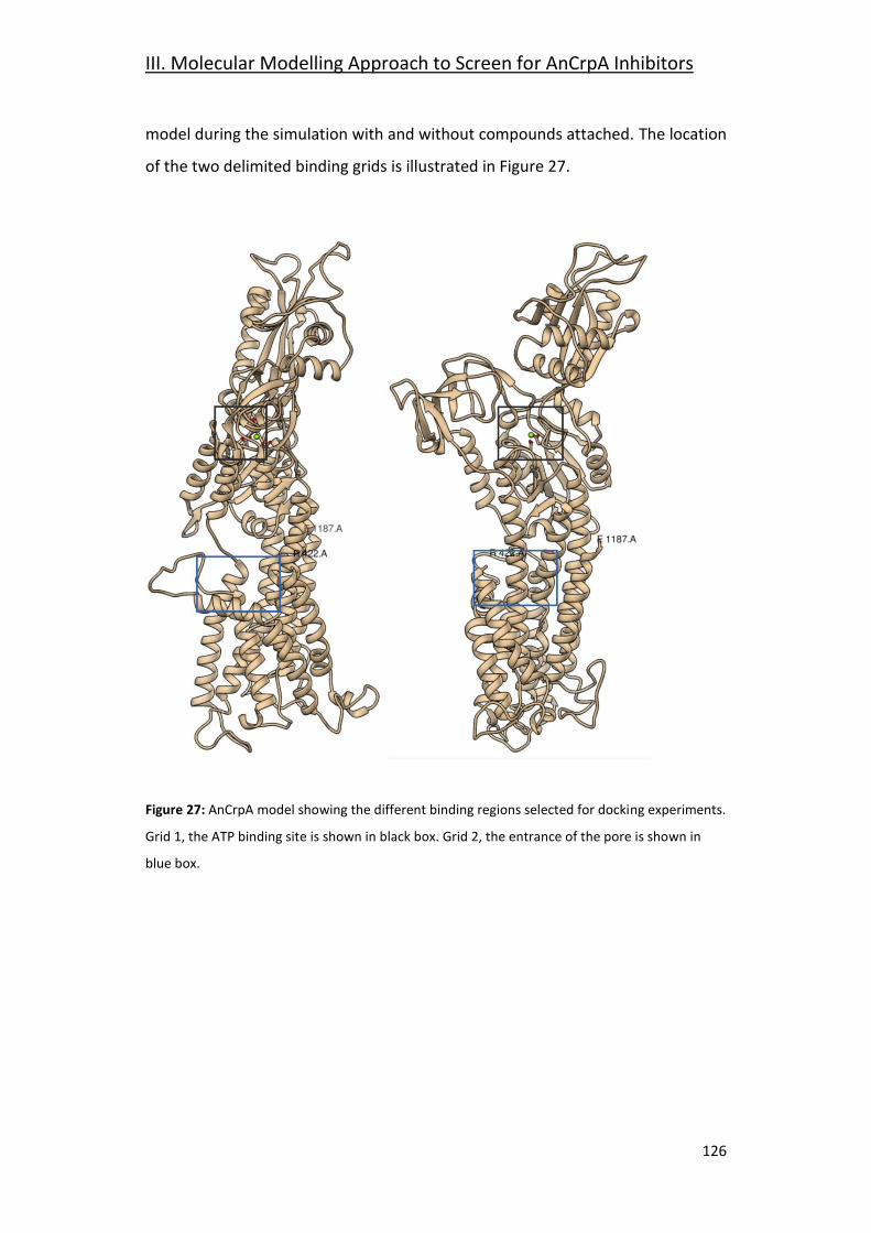
III. Molecular Modelling Approach to Screen for AnCrpA Inhibitors
126
model during the simulation with and without compounds attached. The location
of the two delimited binding grids is illustrated in Figure 27.
Figure 27: AnCrpA model showing the different binding regions selected for docking experiments.
Grid 1, the ATP binding site is shown in black box. Grid 2, the entrance of the pore is shown in
blue box.
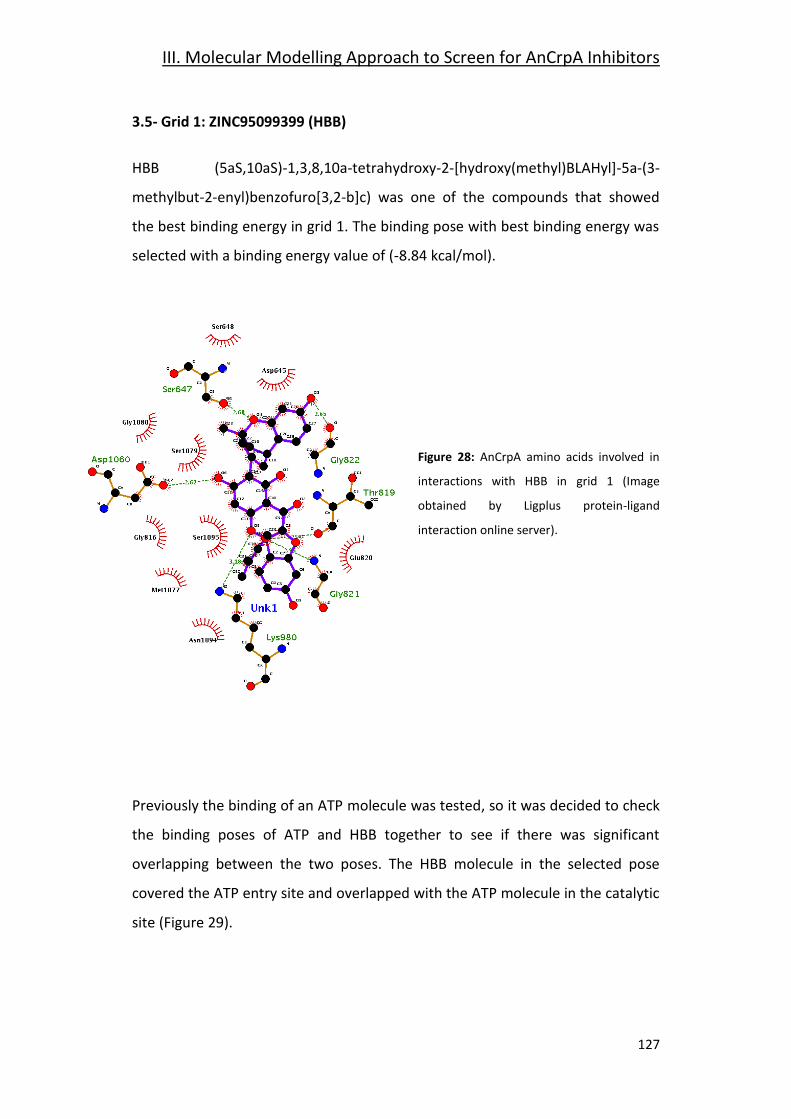
III. Molecular Modelling Approach to Screen for AnCrpA Inhibitors
127
3.5- Grid 1: ZINC95099399 (HBB)
HBB (5aS,10aS)-1,3,8,10a-tetrahydroxy-2-[hydroxy(methyl)BLAHyl]-5a-(3-
methylbut-2-enyl)benzofuro[3,2-b]c) was one of the compounds that showed
the best binding energy in grid 1. The binding pose with best binding energy was
selected with a binding energy value of (-8.84 kcal/mol).
Figure 28: AnCrpA amino acids involved in
interactions with HBB in grid 1 (Image
obtained by Ligplus protein-ligand
interaction online server).
Previously the binding of an ATP molecule was tested, so it was decided to check
the binding poses of ATP and HBB together to see if there was significant
overlapping between the two poses. The HBB molecule in the selected pose
covered the ATP entry site and overlapped with the ATP molecule in the catalytic
site (Figure 29).
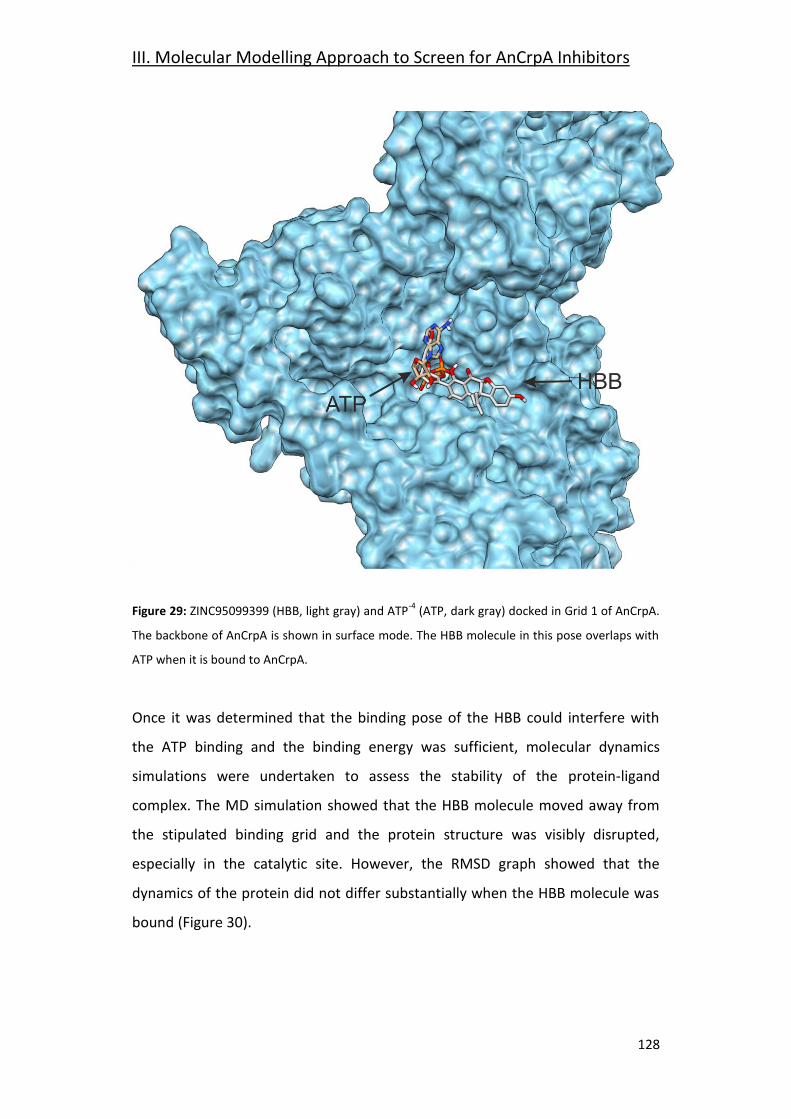
III. Molecular Modelling Approach to Screen for AnCrpA Inhibitors
128
Figure 29: ZINC95099399 (HBB, light gray) and ATP-4
(ATP, dark gray) docked in Grid 1 of AnCrpA.
The backbone of AnCrpA is shown in surface mode. The HBB molecule in this pose overlaps with
ATP when it is bound to AnCrpA.
Once it was determined that the binding pose of the HBB could interfere with
the ATP binding and the binding energy was sufficient, molecular dynamics
simulations were undertaken to assess the stability of the protein-ligand
complex. The MD simulation showed that the HBB molecule moved away from
the stipulated binding grid and the protein structure was visibly disrupted,
especially in the catalytic site. However, the RMSD graph showed that the
dynamics of the protein did not differ substantially when the HBB molecule was
bound (Figure 30).
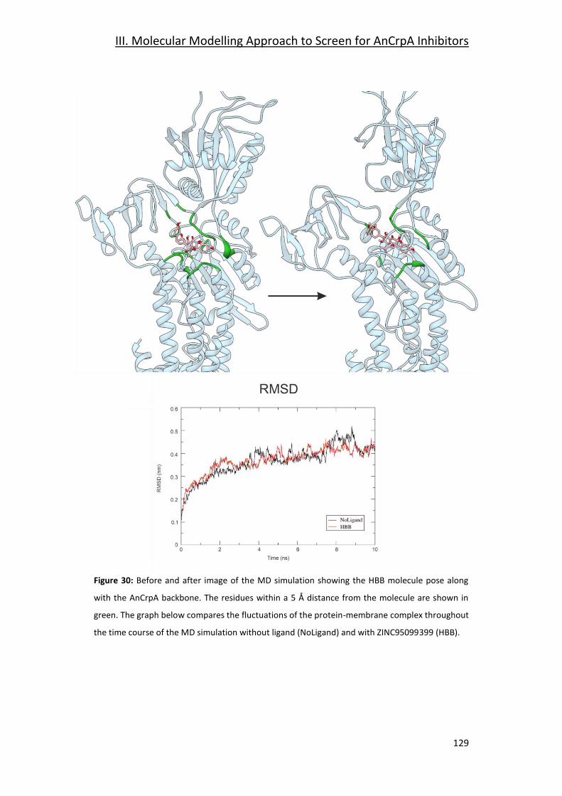
III. Molecular Modelling Approach to Screen for AnCrpA Inhibitors
129
Figure 30: Before and after image of the MD simulation showing the HBB molecule pose along
with the AnCrpA backbone. The residues within a 5 Å distance from the molecule are shown in
green. The graph below compares the fluctuations of the protein-membrane complex throughout
the time course of the MD simulation without ligand (NoLigand) and with ZINC95099399 (HBB).
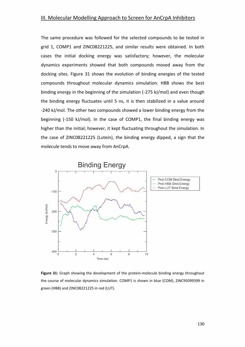
III. Molecular Modelling Approach to Screen for AnCrpA Inhibitors
130
The same procedure was followed for the selected compounds to be tested in
grid 1, COMP1 and ZINC08221225, and similar results were obtained. In both
cases the initial docking energy was satisfactory; however, the molecular
dynamics experiments showed that both compounds moved away from the
docking sites. Figure 31 shows the evolution of binding energies of the tested
compounds throughout molecular dynamics simulation. HBB shows the best
binding energy in the beginning of the simulation (-275 kJ/mol) and even though
the binding energy fluctuates until 5 ns, it is then stabilized in a value around
-240 kJ/mol. The other two compounds showed a lower binding energy from the
beginning (-150 kJ/mol). In the case of COMP1, the final binding energy was
higher than the initial; however, it kept fluctuating throughout the simulation. In
the case of ZINC08221225 (Lutein), the binding energy dipped, a sign that the
molecule tends to move away from AnCrpA.
Figure 31: Graph showing the development of the protein-molecule binding energy throughout
the course of molecular dynamics simulation. COMP1 is shown in blue (COM), ZINC95099399 in
green (HBB) and ZINC08221225 in red (LUT).
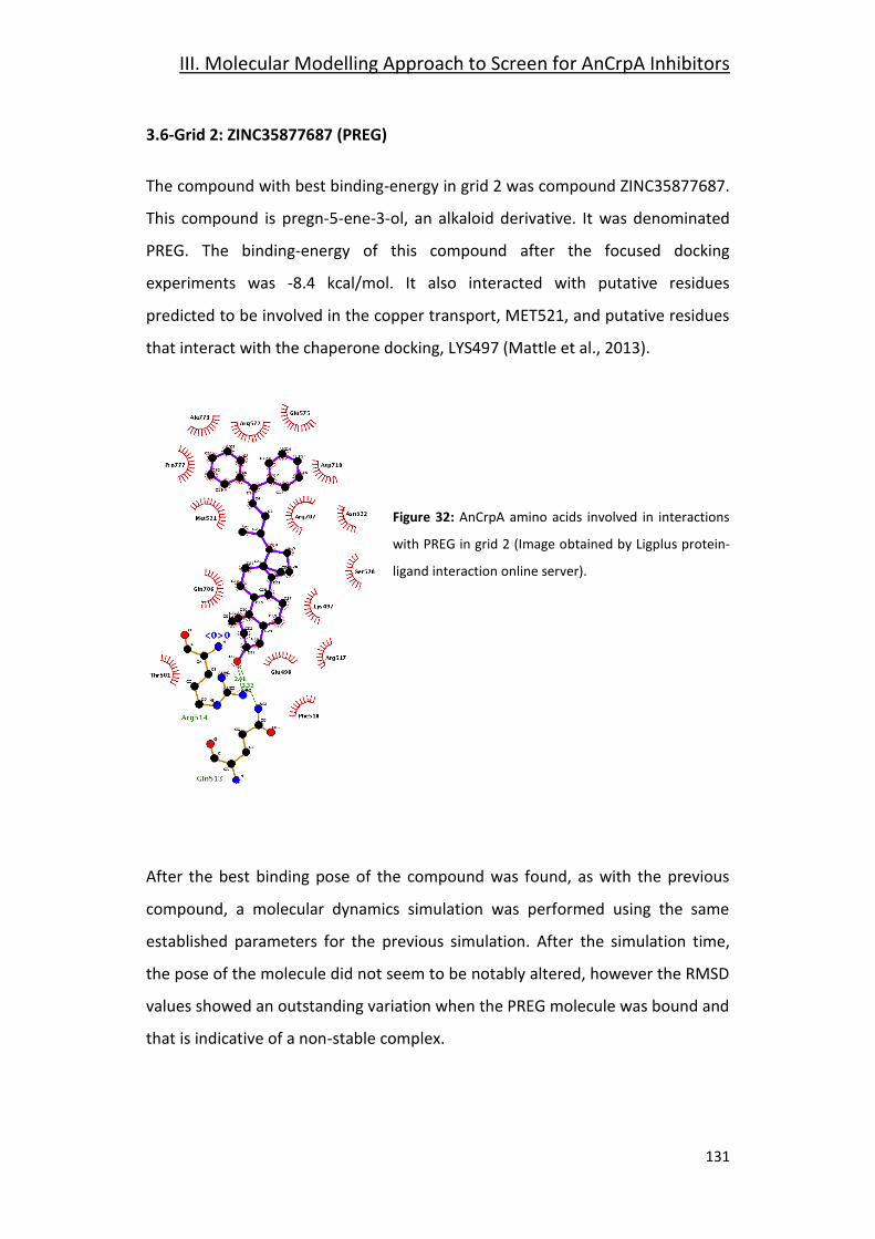
III. Molecular Modelling Approach to Screen for AnCrpA Inhibitors
131
3.6-Grid 2: ZINC35877687 (PREG)
The compound with best binding-energy in grid 2 was compound ZINC35877687.
This compound is pregn-5-ene-3-ol, an alkaloid derivative. It was denominated
PREG. The binding-energy of this compound after the focused docking
experiments was -8.4 kcal/mol. It also interacted with putative residues
predicted to be involved in the copper transport, MET521, and putative residues
that interact with the chaperone docking, LYS497 (Mattle et al., 2013).
Figure 32: AnCrpA amino acids involved in interactions
with PREG in grid 2 (Image obtained by Ligplus protein-
ligand interaction online server).
After the best binding pose of the compound was found, as with the previous
compound, a molecular dynamics simulation was performed using the same
established parameters for the previous simulation. After the simulation time,
the pose of the molecule did not seem to be notably altered, however the RMSD
values showed an outstanding variation when the PREG molecule was bound and
that is indicative of a non-stable complex.
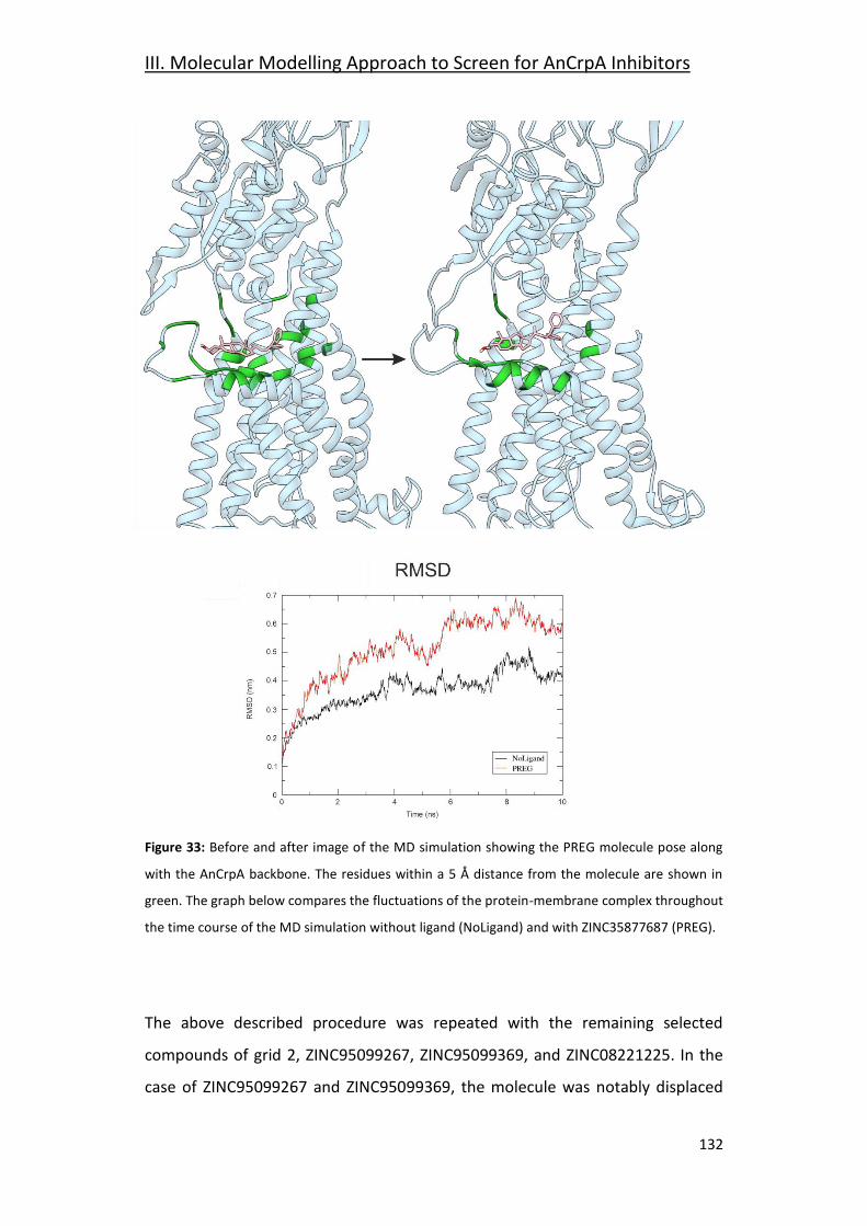
III. Molecular Modelling Approach to Screen for AnCrpA Inhibitors
132
Figure 33: Before and after image of the MD simulation showing the PREG molecule pose along
with the AnCrpA backbone. The residues within a 5 Å distance from the molecule are shown in
green. The graph below compares the fluctuations of the protein-membrane complex throughout
the time course of the MD simulation without ligand (NoLigand) and with ZINC35877687 (PREG).
The above described procedure was repeated with the remaining selected
compounds of grid 2, ZINC95099267, ZINC95099369, and ZINC08221225. In the
case of ZINC95099267 and ZINC95099369, the molecule was notably displaced
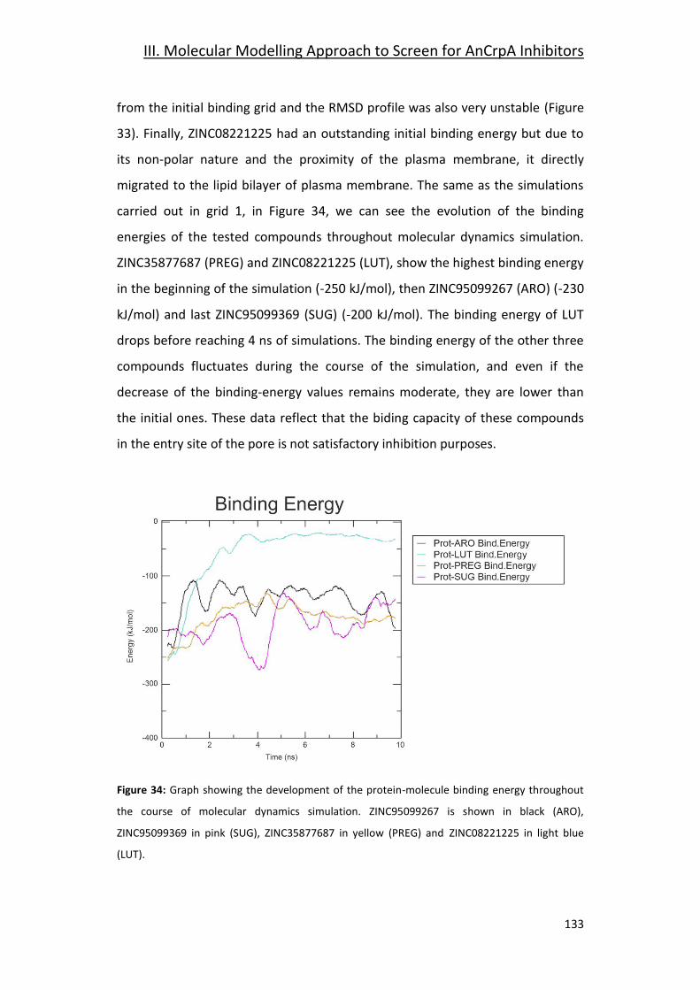
III. Molecular Modelling Approach to Screen for AnCrpA Inhibitors
133
from the initial binding grid and the RMSD profile was also very unstable (Figure
33). Finally, ZINC08221225 had an outstanding initial binding energy but due to
its non-polar nature and the proximity of the plasma membrane, it directly
migrated to the lipid bilayer of plasma membrane. The same as the simulations
carried out in grid 1, in Figure 34, we can see the evolution of the binding
energies of the tested compounds throughout molecular dynamics simulation.
ZINC35877687 (PREG) and ZINC08221225 (LUT), show the highest binding energy
in the beginning of the simulation (-250 kJ/mol), then ZINC95099267 (ARO) (-230
kJ/mol) and last ZINC95099369 (SUG) (-200 kJ/mol). The binding energy of LUT
drops before reaching 4 ns of simulations. The binding energy of the other three
compounds fluctuates during the course of the simulation, and even if the
decrease of the binding-energy values remains moderate, they are lower than
the initial ones. These data reflect that the biding capacity of these compounds
in the entry site of the pore is not satisfactory inhibition purposes.
Figure 34: Graph showing the development of the protein-molecule binding energy throughout
the course of molecular dynamics simulation. ZINC95099267 is shown in black (ARO),
ZINC95099369 in pink (SUG), ZINC35877687 in yellow (PREG) and ZINC08221225 in light blue
(LUT).
III. Molecular Modelling Approach to Screen for AnCrpA Inhibitors
134
The molecular dynamics simulations results in grid 2, the entrance of the pore,
are not sufficient to justify continued efforts in their study. All tested compounds
were displaced from the initial binding site and the binding poses are notably
changed after the simulation time, the RMSD graphs show a substantial
difference when the compound is bound to the AnCrpA-membrane complex, the
binding energy values are low in comparison to required standards and they are
not stable during molecular dynamics simulation. All this data together supports
the interpretation that these compounds do not form a stable complex with the
protein and thus they are not good candidates to achieve AnCrpA inhibition. The
main reason could be that the template model was crystallized in the E2P
conformation. This means the transmembrane pore is closed in the intracellular
side, the docking site, and opened in the extracellular area, making it more
difficult to access the residues of the pore from the intracellular side.
Out of these experiments the best results were obtained in grid 1, the catalytic
site. HBB showed the most satisfactory results in terms of binding energy, RMSD
profile and stability in the binding grid. However, is not a commercially available
compound and it would have to be synthesized. COMP1 was the second best
match. Even though the overall results of the compound were not as adequate
as HBB, the fact that it is a commercially available compound, makes it a
candidate worth further testing.
In conclusion, with the work conducted under the supervision of Dr. Galeazzi we
accomplished our objectives: first, we can state that a molecular modelling
approach is valid to find inhibitory compounds for our copper detoxification P-
type ATPase AnCrpA and second, the doctorate student has been introduced to
Molecular Modelling.

135
Final Discussion

136
Final Discussion
137
Final Discussion
Copper homeostasis in A. nidulans has been the overall subject of this study,
with special focus on uptake and detoxification of the cation. An RNA-seq
experiment comparing the transcriptome under copper toxicity to standard
laboratory nutritional conditions provided the starting point of the investigation.
The obtained results allowed for the identification of genes and biological
processes involved in the response to copper toxicity. Moreover, reference to
previous literature and sequence analysis tools helped identify the genes
involved in the copper homeostasis at different stages of the toxicity-response
process, especially focusing on high-affinity uptake and detoxification.
The A. nidulans high-affinity copper uptake system shares similarities with
counterparts in Cryptococcus, Saccharomyces and A. fumigatus. The uptake
system is regulated by the transcription factor AnMac1 that exclusively controls
the expression of copper uptake proteins. Plasma-membrane metalloreductases
reduce environmental copper from Cu2+ to Cu+ (Cai et al., 2019). Even though
these elements have not been characterized, A. nidulans possesses nine putative
plasma membrane reductases related to the S. cerevisiae metalloreductase
family ScFre. Finally, two high-affinity copper transporters, one of which covers
for the acquisition of copper under most environmental conditions (ScCtr3,
CnCtr1 and AnCtrC) and the other specifically covers for in high copper-
deficiency scenarios (ScCtr1, CnCtr4 and AnCtrA) (Antsotegi-Uskola et al., 2020).
Thus, our current understanding is that the high-affinity copper uptake system in
fungi is conserved in the species so far examined.
Despite this apparent uniformity, there are specific aspects that need to be
covered, such as details on the role of AnCtrB. According to the RNA-seq data
obtained in this study, the expression of this gene is not significantly changed
under copper toxicity conditions. In addition, there are some discrepancies on
Final Discussion
138
the regulation of different homologs of this protein. The first articles published in
the S. cerevisiae homolog Ctr2 state that it is solely regulated by iron availability
(Rees and Thiele, 2007); on the other hand, more recent articles report that the
ScMac1 is involved in its regulation (Liu et al., 2012;Qi et al., 2012). Reports
describing the A. fumigatus homolog AfCtrB claim that neither the
environmental copper level nor the deletion of AfMac1 have any role in its
regulation (Cai et al., 2017;Kusuya et al., 2017). Despite regulation controversies,
the function of this protein is well specified in yeast as a copper transporter that
mobilizes vacuolar copper to cytosolic copper chaperones (Balamurugan and
Schaffner, 2006;Nevitt et al., 2012;Smith et al., 2017) and its single deletion
makes no difference on colony development in high and low copper availability
environments (Kampfenkel et al., 1995). However, in filamentous fungi its
function remains poorly understood and Cai and collaborators (2017) reported
that deletion of AfCtrB renders the colony unable to develop, in high, regular and
low copper availability conditions. Further work is necessary to accurately define
AnCtrB function.
Some discrepancies on functional hierarchy of the Ctr proteins in the high-affinity
copper uptake system have emerged in two different studies. Park and
collaborators (2014) presented the first characterization of Ctr protein in A.
fumigatus proposing that AfctrA2 and AfctrC are functionally equivalent since
individual deletion mutants exhibited a wild type–like phenotype with 100 μM
BCS. On the other hand, Cai et al., (2017) reported that the AfctrC deletion
mutant showed defective colony morphology with 100 μM BCS, suggesting that
this protein plays a dominant role in copper homeostasis. This thesis concludes
that the lack of copper deficiency phenotype (in this case a secondary level)
under mild copper deficiency conditions in a single deletion strain does not
necessarily prove that any one of the Ctr proteins has a dominant role over the
other(Page 67-68, Figure 10). The possibility still remains that one of the Ctr
proteins may be more active under the tested conditions or that the other Ctr
protein responds in more specific scenarios.
Final Discussion
139
Copper addition reverted the defective phenotypes of the Anctr deletant strains,
corroborating that the cause of the phenotypes was indeed copper deficiency. It
is well known that copper and iron metabolism are closely related and a recent
study in A. fumigatus has revealed that the TF Mac1, responsible for the
regulation of the high-affinity copper uptake system, also plays a role in
regulating iron homeostasis (Park et al., 2018). Apparently, Afmac1 deletion
down-regulates the expression of siderophores and reductive iron transporters.
Along with this, it has been described that iron can partially rescue the copper
starvation phenotype generated by deletion of the TF Afmac1 in A. fumigatus
(Cai et al., 2017). Usually, when the deletion of a transcription factor
compromises the high-affinity uptake of a heavy metal, addition of this metal to
the medium reverts or at least partially recovers the depletion phenotype via
low-affinity transporters or other mechanisms. This is the case with the A.
nidulans copper transporters and the Anmac1 deletion phenotypes when copper
is added (Cai et al., 2019). However, in contrast to the A. fumigatus mac1
phenotype, the A. nidulans mac1 copper depletion phenotype (very similar to
the double knock-out strain shown in Figure 10C, page 67-68) shows no recovery
when iron is supplemented to the medium (not shown). The high-affinity iron
uptake systems present substantial differences (Blatzer et al., 2011;Eisendle et
al., 2003;Schrettl et al., 2004) between these two organisms. Whether the
regulation of iron uptake differs between A. fumigatus and A. nidulans remains
to be investigated.
A common feature of AnCtrA that raised attention was the number of TM
domains predicted by the TMHMM topology prediction software (Page 64,
Figure 9B). These predictions need to be corroborated, but similar models have
been documented for A. fumigatus and C. neoformans Ctr proteins (Park et al.,
2014;Waterman et al., 2012). If these predictions were confirmed, it would imply
that the oligomerization mechanism may differ from the one described by De
Feo and collaborators (2012). Instead of a trimer with nine combined trans-
membrane domains, the oligomer of Ctr monomers with an even number of
Final Discussion
140
trans-membrane domains would always possess an even number of trans-
membrane domains probably altering the oligomer formation.
Finally, our attempt to characterize the putative copper uptake shutdown
mechanism of the AnCtr proteins had an unexpected outcome. Our C-terminal
mutations did not have any effect on copper susceptibility; the only effect we
were able to record was reduced copper uptake capacity as secondary level
copper limitation effects were observed in mutant strains (Page 77, Figure 13B).
Based on the observed results, we postulate that, in the conditions tested, the
cysteines object of study and the C-terminal do not act as copper uptake
modulators but they have a role in copper transport. The secondary level copper
limitation effect, the yellow pigmentation, is most likely due to an impaired AnYA
laccase function that correlates with an insufficient copper supply, as laccases
are copper dependent enzymes (Hermann et al., 1983;Scherer and Fischer,
2001). Considering the distal localization of this motif it is rather difficult for it to
participate in transmembrane copper mobilization. The key for the copper
deficiency phenotype might be that, mutating the di-cysteine motif or deleting
the C-terminal might be taking away the linker for interaction between the Ctr
protein and the chaperone as it has been described in human and yeast (Kahra et
al., 2016;Xiao and Wedd, 2002). Thus, the distribution effected by the chaperone
route might be blocked or substantially altered, with the corresponding
phenotypic manifestation of copper deficiency.
Following the Ctr protein regulation mechanism in A. nidulans, it is clear that the
C-terminal deletion does not exert an impact on copper susceptibility, so it
would be reasonable to consider whether there is any regulation mechanism at
all that blocks or controls transmembrane copper traffic in the event of a copper
fluctuation in the environment, since the only known mechanism relies solely on
the C-terminal (Clifford et al., 2016;Schuller et al., 2013;Wu et al., 2009).
Final Discussion
141
In contrast with the conservation of the copper uptake system in fungi,
resistance to copper appears to be achieved by two alternative mechanisms. The
first mechanism is through metallothioneins. These cysteine-rich small proteins
chelate free copper ions of the cytosol in organisms like S. cerevisiae and C.
neoformans (Balamurugan and Schaffner, 2006;Ding et al., 2013b;Nevitt et al.,
2012;Smith et al., 2017). The second mechanism relies on heavy metal pumping
PIB-type ATPases. These heavy metal pumps extrude copper ions from the cytosol
to the extracellular space in organisms like, A. nidulans, A. fumigatus and C.
albicans (Antsotegi-Uskola et al., 2017;Riggle and Kumamoto, 2000;Weissman et
al., 2000;Wiemann et al., 2017). This divergence in metal detoxification
mechanism by different fungal species may be the result of an adaptation to
their respective ecological niches. S. cerevisiae has been isolated from a range of
environments (Goddard and Greig, 2015), many of which are specific, such as
fruits and flowers. In contrast, C. albicans, like most filamentous fungi, is an all-
rounder found in environments ranging from the digestive tracts of animals to
soils, where competition and antagonism are common. In these environments,
PIB-ATPase (CaCrp1p) mediated copper export confers superior tolerance in
comparison to MTs, despite the greater cost in energy required to produce it and
operate it. MTs, on the other hand, may allow for the immobilization of greater
amounts of copper, as a resource, in media which may be especially poor in
copper, such as flower nectar. In the same medium and growing conditions, S.
cerevisiae is able to grow in up to 2 mM of CuSO4, whereas C. albicans tolerates
20 mM of CuSO4 (Weissman et al., 2000).
The synthesis time of metallothioneins versus the P-type ATPases favours the
metallothioneins. Actually the ScCup1 metallothionein synthesis time is almost
half of AnCrpA. ScCup1 synthesis is clearly detectable 10 min after 100 M CuSO4
addition and by 25 min, the expression level is outstanding (Pena et al., 1998).
Thus, the reason behind P-type ATPases providing a superior tolerance is not an
earlier reaction time, which leaves two different possibilities: firstly, that the
amount of synthesized P-type ATPases outscores the metallothioneins and thus
Final Discussion
142
they confer a higher resistance; or secondly, that the strategy of pumping copper
out is simply more effective against copper toxicity than the buffering strategy.
In this study, a single metallothionein coding gene was identified in A. nidulans,
but its contribution to heavy metal toxicity resistance was not demonstrated. In
the conditions tested, (Cu, Cd and Ag) the presence of the metallothionein did
not confer resistance to heavy metal toxicity. In an article published by Cai and
collaborators (2018), the authors found that the of AnCrdA homolog in A.
fumigatus contributed minimally to Cu tolerance when AfAce1 and AfCrpA were
both mutated and AfCrdA was overexpressed. Thus, CrdA contribution in
laboratory conditions, where the organism is well accommodated, appears
rather low or negligible. However, it might make a difference in the natural
environment, where every resistance mechanism counts, as a second mechanism
over the pump, for example.
Throughout this investigation, the obtained results were compared to data
obtained by Wiemann and collaborators (2017) in A. fumigatus. This led to the
realization that the level copper toxicity induced determined the characteristics
of the response. This opened the possibility to envisage the existence of a
multilevel resistance mechanism in Aspergilli. The first level of copper toxicity
response would involve copper detoxification through PIB-type ATPase CrpA. As
described in this study, the fungus activates a rapid and intense expression of
AnCrpA and the metallothionein AnCrdA at 100 M CuSO4. However, the
expression is transient and the RNA-seq data indicate that other defense
mechanisms like ROS neutralization are not activated, purportedly signaling that
no uncontrolled ROS are intracellularly detected. The second level copper
toxicity response would be the one described in A. fumigatus by Wienmann and
collaborators (2017) at 200 M CuSO4. This would involve not only the activation
of CrpA and CrdA but also the up-regulation of all the ROS neutralization
machinery, catalase and superoxide dismutase. Having such and multilevel
copper toxicity response would be an efficient system capable of deploying the
Final Discussion
143
required cellular mechanisms to the level of toxicity encountered by the
organism. This proposal would require experimental validation through
experimentation at different copper toxicity levels, to verify the activation of the
different response levels.
Finally, AnCrpA is clearly the principal copper detoxification agent in A. nidulans;
however, AnCrpA is not exclusively activated by copper. Other studies have
corroborated that the P-type ATPase CrpA and the TF AceA, also contribute to
other heavy metal resistance, such as zinc in A. fumigatus. Thus, the P-type
ATPase CrpA has versatile detoxification role as described in other organisms
(Mandal et al., 2002;Odermatt et al., 1994).
This study started at the time when only a single publication was available about
the copper homeostasis system in filamentous fungi. At the time of its final
presentation, several contributions provide evidence to propose a preliminary
model to describe the state of the cell in regular environmental conditions.
The average copper concentration in soil according to a study conducted in
United States (Agency for Toxic Substances & Disease Registry, 2004) is about 50
parts per million (ppm) parts soil. According to the Geochemical Mapping of
Agricultural and Grazing Land Soil in Europe (GEMAS) project that analyzed
thousands of soil samples across Europe, the mean copper value was 20 ppm
parts soil (Ballabio et al., 2018). Analyzing these figures it seems that copper is
generally available in sufficient concentration for a proper maintenance of life;
however, the soil is the living niche for thousands or organisms that co-exist in
the environment and have to compete for the available resources. Thus, high-
affinity uptake mechanisms would be a consequence of such competition.
In this condition, the TF regulating AnctrA and AnctrC expression, AnMac1 would
actively bind to their promoters, enabling transcription, as well as that of the
plasma-membrane metalloreductase AnfreC (at least AnfreC and most likely
Final Discussion
144
other Fre protein coding genes). In this “regular” environmental situation one
would expect a mild copper deficiency situation. Thus, AnctrC transcription
would likely be very active and AnctrA transcription would be barely active. The
plasma-membrane metalloreductases and the copper transporters would be
located in the plasma-membrane to make sure that copper is internalized to the
cell. Once the copper is internalized to the cell the copper chaperones AN6045,
AN1390 and AN4863 would bind the internalized copper. These chaperones
perform a double function: First, they bind the incoming ion preventing possible
deleterious effects due to uncontrolled copper accumulation in the cytosol.
Second, they are responsible for copper delivery within the cell. By homology
based predictions we infer that: AN6045, homolog of the ScCcs1, would be
responsible for copper delivery to the Cu/Zn Superoxide Dismutase AnSodA;
AN1390, homolog of ScAtx1, would deliver copper to the P-type ATPase AnYgA
responsible for delivery into the trans-Golgi network; AN4863, homolog of the
ScCox17, would deliver copper to the cytochrome oxidase complex at the inner
membrane of mitochondria (Balamurugan and Schaffner, 2006;Nevitt et al.,
2012;Smith et al., 2017). Finally, the copper reservoir in the vacuoles also plays a
role in a copper store management under scarcity conditions. Although the role
of vacuolar AnCtrB is not yet fully understood, based on the literature it may be
inferred that it may be actively pumping copper from within the vacuolar copper
reservoir to supplement external copper import deficit (Figure 35).
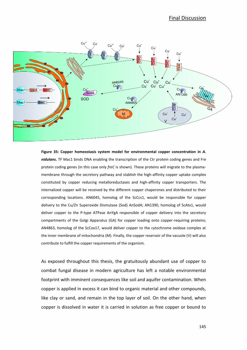
Final Discussion
145
Figure 35: Copper homeostasis system model for environmental copper concentration in A.
nidulans. TF Mac1 binds DNA enabling the transcription of the Ctr protein coding genes and Fre
protein coding genes (in this case only freC is shown). These proteins will migrate to the plasma-
membrane through the secretory pathway and stablish the high-affinity copper uptake complex
constituted by copper reducing metalloreductases and high-affinity copper transporters. The
internalized copper will be received by the different copper chaperones and distributed to their
corresponding locations. AN6045, homolog of the ScCcs1, would be responsible for copper
delivery to the Cu/Zn Superoxide Dismutase (Sod) AnSodA; AN1390, homolog of ScAtx1, would
deliver copper to the P-type ATPase AnYgA responsible of copper delivery into the secretory
compartments of the Golgi Apparatus (GA) for copper loading onto copper-requiring proteins;
AN4863, homolog of the ScCox17, would deliver copper to the cytochrome oxidase complex at
the inner membrane of mitochondria (M). Finally, the copper reservoir of the vacuole (V) will also
contribute to fulfill the copper requirements of the organism.
As exposed throughout this thesis, the gratuitously abundant use of copper to
combat fungal disease in modern agriculture has left a notable environmental
footprint with imminent consequences like soil and aquifer contamination. When
copper is applied in excess it can bind to organic material and other compounds,
like clay or sand, and remain in the top layer of soil. On the other hand, when
copper is dissolved in water it is carried in solution as free copper or bound to
Final Discussion
146
suspended particles and it can reach ground waters, riverbeds, lakes and
estuaries (Agency for Toxic Substances & Disease Registry, 2004).
Moreover, copper-tolerances from the disease-causing strains have also been
documented to steadily increase, making it clear that copper based antimicrobial
preparations need a thorough reassessment.
Thus, the formulation of new antifungal preparations with lower copper doses
has become an issue of growing importance to regulatory authorities. Different
approaches to tackle the challenge of copper dose reduction have emerged in
the recent years and one of the most notable ones is the encapsulation in
nanoparticles. Metallic nanomaterials (silver, gold, copper and titanium) exhibit
specific characteristics that result in enhanced antimicrobial activity (Vimbela et
al., 2017). Micron-size particles allow for the control of parameters such as
density, malleability, stability and reactivity which make a significant difference
to antimicrobial activity. Field studies in different environments with copper
nanoparticles have been reported satisfactory results against bacteria and fungi
(Carvalho et al., 2019;Hermida-Montero et al., 2019;Ingle and Rai, 2017;Munoz-
Escobar and Reyes-Lopez, 2020). Thus, the application of copper in nanoscale
format is a promising strategy and it could become a viable solution against
multidrug resistant bacteria and fungi for surface treatment or medical purposes.
However, the use of copper nanoparticles in agriculture may entail prohibitive
manufacturing costs in comparison with traditional dissolved copper salt
applications.
A different method to enhance copper toxicity could be the use of co-formulants
that exert a synergistic interaction with copper. This strategy has been
successfully used to fight antibiotic-resistant bacteria in the clinical field (Gomara
and Ramon-Garcia, 2019;Rozenbaum et al., 2019;Wittekind and Schuch,
2016;Zerweck et al., 2017). We believe that finding a synergistic counterpart for
Final Discussion
147
copper is plausible now that we have more knowledge about the copper
homeostasis system in filamentous fungi.
We ourselves favored this second option and this was the reason for the visit to
Dr. Galeazzi’s laboratory was first proposed. It was proposed as to proof the
concept that molecular modelling principles and techniques could be applied to
find inhibitory compounds for certain elements of the copper homeostasis
system. This would make the search of a synergistic counterpart for copper much
easier because it can be done by a computational approach and then the
candidates can be verified experimentally.
With AnCrpA we proved that this approach is valid by testing two compounds,
ZINC95099399 and COMP1, as potential AnCrpA inhibitors. The objective of
inhibiting AnCrpA is to deactivate the A. nidulans main copper detoxification
mechanism that is also present in many phytopathogens (Antsotegi-Uskola et al.,
2017). We hypothesize that by doing this the necessary amount to kill the
organism would be notably lower.
Apart from AnCrpA, there are some other possible targets that by inhibiting or
modulating their activity could potentiate the toxic effect of copper. For
example, copper chaperones can be an interesting target. According to some
investigations in ScCtr1 and hCtr1 proteins (Kahra et al., 2016;Xiao and Wedd,
2002), the copper chaperones receive copper ions directly from the Ctr proteins
C-terminal avoiding the threat of free copper ions in the cytosol. By inhibiting or
blocking the copper delivering chaperones we can create an opening to
maximize the impact of free copper ions entered to the cell.
Another strategy to maximize copper damage would be to target the TFs
controlling the copper homeostasis system. Out of the two TF the most
interesting would be AnAce1, the one controlling the copper detoxification
system (Antsotegi-Uskola et al., 2017). ScAceA1 studies show that this TF has a
Final Discussion
148
cysteine-rich copper binding domain denominated the “DNA Binding Domain”
that binds four copper ions forming a tetra-copper-thiolate cluster (Cai et al.,
2018;Smith et al., 2017). The formation of this cluster triggers a conformational
change that enables DNA binding and thus, the transcription of the whole copper
detoxification machinery. This domain is largely conserved in Aspergilli spp. and
other genera like Botrytis, Magnaporthe and Fusarium. A compound that would
form a stable complex with AnAce1 would be likely to prevent DNA binding of
the TF and thus the copper detoxification machinery would not be activated,
maximizing the damage of internalized copper ions.
Finally, another hypothetical option would be related to the as yet
undemonstrated Ctr copper uptake protein shutdown mechanism. In case there
is such mechanism it could be a good possibility to exploit this avenue. By
delaying the copper uptake shutdown the toxic effect of the copper present in
the antifungal formulations would be increased. This approach would first
require a full characterization of the Ctr protein regulation mechanism.
The development of resistance to copper by fungal strains has been long
documented (Cervantes & Gutierrez-Corona, 1994; Ashida, 1965). Copper
resistance in fungi is achieved by metal trapping by cell-wall components, altered
copper uptake, extracellular chelation or precipitation by secreted metabolites,
intracellular copper chelation by metallothioneins or phytochelatins; and finally
intracellular copper extrusion by P-type ATPases (Antsotegi-Uskola et al.,
2017;Cervantes and Gutierrez-Corona, 1994;Kovacec et al., 2017;Wiemann et al.,
2017). It has been reported that copper-resistant S. cerevisiae strains possess
multiple ScCup1 metallothionein coding gene copies while regular copper-
sensitive strains bear a single copy (Cervantes and Gutierrez-Corona, 1994).
However, taking it to agriculture, where copper-resistant phytopathogens are a
real problem, the cause or causes of these resistances remain unclear. Judet-
Correia and collaborators (2011) documented the MIC (Minimal Inhibitory
Concentration) values of two Botrytis cinerea strains demonstrating that some
Final Discussion
149
isolates of these molds can develop resistance. Following the S. cerevisiae
example it might be that these copper-resistant phytopathogens possess more
than one copy of the AnCrpA homolog as documented in Aspergillus species
(Yang et al., 2018). Otherwise it could be an overall improvement of all above
commented defense mechanisms that confers these strains an unusual capacity
to resist copper. Whatever the case is, it would be a key point to know the
source of these resistances in order to find the right target.
While this thesis was ongoing, apart from our contributions in the model
organism A. nidulans, there have been some contributions in the pathogenic
fungus A. fumigatus, but many details remain to be uncovered. Considering the
repercussion of copper homeostasis on fungal biology, it is a topic worth
studying in more depth and a wider range of species. The copper uptake and
detoxification mechanisms and their respective transcription factors have been
the focus of recent publications (Antsotegi-Uskola et al., 2017;Antsotegi-Uskola
et al., 2020;Cai et al., 2019;Cai et al., 2017;Kusuya et al., 2017;Park et al.,
2017;Park et al., 2014;Wiemann et al., 2017), identifying the principal
participants of each process, but the copper buffering and intracellular
distribution mechanisms remain practically unknown. Phylogenetic analyses
have revealed the presence of proteins that act as copper scavenging and
trafficking vehicles in other organisms (Ding et al., 2014), but so far, those
proteins have not been investigated. This evidence calls for further investigation
covering intracellular copper trafficking and storage in oncoming years.

150

151
Conclusions

152
Conclusions
153
Conclusions
The main conclusions extracted from this work are the following:
Three proteins involved in copper homeostasis have been identified in
Aspergillus nidulans: a PIB-type ATPase, AnCrpA, and two copper
transporter (Ctr) proteins, AnCtrA and AnCtrC.
AnCtrA and AnCtrC are copper transporting proteins taking part in the
high-affinity copper uptake system. AnCtrC covers copper uptake for
nutritional requirements and AnCtrA is more important in high copper
deficiency scenarios; however, the two proteins work in a
complementary manner.
AnCrpA is the major copper resistance determinant in A. nidulans and is
expressed tightly and transiently in the presence of high copper levels.
AnAceA is a TF which regulates copper tolerance key elements. Besides
activating AncrpA expression, regulates an additional element which has
a minor role in copper detoxification process that has not been yet
identified.
The molecular modelling approach is suitable to find AnCrpA inhibitors.
ZINC95099399 and COMP1 showed the most satisfactory results. These
compounds have to be tested experimentally.

154

155
References

156
References
157
References
Abe,F. and Hiraki,T. (2009). Mechanistic role of ergosterol in membrane rigidity and cycloheximide resistance in Saccharomyces cerevisiae. Biochim. Biophys. Acta 1788, 743-752.
Adams,T.H., Wieser,J.K., and Yu,J.H. (1998). Asexual sporulation in Aspergillus nidulans. Microbiol. Mol. Biol. Rev. 62, 35-54.
Agency for Toxic Substances & Disease Registry (2004). Public Health Statement. In: Toxicological Profile for Copper.
Alkan,N. and Fortes,A.M. (2015). Insights into molecular and metabolic events associated with fruit response to post-harvest fungal pathogens. Front Plant Sci. 6, 889.
Alvarez,F.J., Douglas,L.M., and Konopka,J.B. (2007). Sterol-rich plasma membrane domains in fungi. Eukaryot. Cell 6, 755-763.
Andersson,M., Mattle,D., Sitsel,O., Klymchuk,T., Nielsen,A.M., Moller,L.B., White,S.H., Nissen,P., and Gourdon,P. (2014). Copper-transporting P-type ATPases use a unique ion-release pathway. Nat. Struct. Mol. Biol. 21, 43-48.
Antsotegi-Uskola,M., Markina-Inarrairaegui,A., and Ugalde,U. (2017). Copper Resistance in Aspergillus nidulans Relies on the PI-Type ATPase CrpA, Regulated by the Transcription Factor AceA. Front Microbiol. 8, 912.
Antsotegi-Uskola,M., Markina-Inarrairaegui,A., and Ugalde,U. (2020). Copper Homeostasis in Aspergillus nidulans Involves Coordinated Transporter Function, Expression and Cellular Dynamics. Front Microbiol. 11, 555306.
Aramayo,R. and Timberlake,W.E. (1990). Sequence and molecular structure of the Aspergillus nidulans yA (laccase I) gene. Nucleic Acids Res. 18, 3415.
Balamurugan,K. and Schaffner,W. (2006). Copper homeostasis in eukaryotes: teetering on a tightrope. Biochim. Biophys. Acta 1763, 737-746.
Ballabio,C., Panagos,P., Lugato,E., Huang,J.H., Orgiazzi,A., Jones,A., Fernandez-Ugalde,O., Borrelli,P., and Montanarella,L. (2018). Copper distribution in European topsoils: An assessment based on LUCAS soil survey. Sci. Total Environ. 636, 282-298.
Ballou,E.R. and Wilson,D. (2016). The roles of zinc and copper sensing in fungal pathogenesis. Curr. Opin. Microbiol. 32, 128-134.
Barry,A.N., Shinde,U., and Lutsenko,S. (2010). Structural organization of human Cu-transporting ATPases: learning from building blocks. J. Biol. Inorg. Chem. 15, 47-59.
Bastmeyer,M., Deising,H.B., and Bechinger,C. (2002). Force exertion in fungal infection. Annu. Rev. Biophys. Biomol. Struct. 31, 321-341.
References
158
Bayram,O. and Braus,G.H. (2012). Coordination of secondary metabolism and development in fungi: the velvet family of regulatory proteins. FEMS Microbiol. Rev. 36, 1-24.
Bergen,L.G. and Morris,N.R. (1983). Kinetics of the nuclear division cycle of Aspergillus nidulans. J. Bacteriol. 156, 155-160.
Bertinato,J. and L'Abbe,M.R. (2004). Maintaining copper homeostasis: regulation of copper-trafficking proteins in response to copper deficiency or overload. J. Nutr. Biochem. 15, 316-322.
Besold,A.N., Culbertson,E.M., and Culotta,V.C. (2016). The Yin and Yang of copper during infection. J. Biol. Inorg. Chem. 21, 137-144.
Blatzer,M., Binder,U., and Haas,H. (2011). The metalloreductase FreB is involved in adaptation of Aspergillus fumigatus to iron starvation. Fungal. Genet. Biol. 48, 1027-1033.
Blatzer,M. and Latge,J.P. (2017). Metal-homeostasis in the pathobiology of the opportunistic human fungal pathogen Aspergillus fumigatus. Curr. Opin. Microbiol. 40, 152-159.
Blindauer,C.A. and Leszczyszyn,O.I. (2010). Metallothioneins: unparalleled diversity in structures and functions for metal ion homeostasis and more. Nat. Prod. Rep. 27, 720-741.
Bokor,E., Amon,J., Keisham,K., Karacsony,Z., Vagvolgyi,C., and Hamari,Z. (2019). HMGB proteins are required for sexual development in Aspergillus nidulans. PLoS. One. 14, e0216094.
Brown,G.D., Denning,D.W., Gow,N.A., Levitz,S.M., Netea,M.G., and White,T.C. (2012). Hidden killers: human fungal infections. Sci. Transl. Med. 4, 165rv13.
Caddick,M.X., Brownlee,A.G., and Arst,H.N., Jr. (1986). Regulation of gene expression by pH of the growth medium in Aspergillus nidulans. Mol. Gen. Genet. 203, 346-353.
Cai,Z., Du,W., Liu,L., Pan,D., and Lu,L. (2019). Molecular Characteristics of the Conserved Aspergillus nidulans Transcription Factor Mac1 and Its Functions in Response to Copper Starvation. mSphere. 4.
Cai,Z., Du,W., Zeng,Q., Long,N., Dai,C., and Lu,L. (2017). Cu-sensing transcription factor Mac1 coordinates with the Ctr transporter family to regulate Cu acquisition and virulence in Aspergillus fumigatus. Fungal. Genet. Biol. 107, 31-43.
Cai,Z., Du,W., Zhang,Z., Guan,L., Zeng,Q., Chai,Y., Dai,C., and Lu,L. (2018). The Aspergillus fumigatus transcription factor AceA is involved not only in Cu but also in Zn detoxification through regulating transporters CrpA and ZrcA. Cell Microbiol. e12864.
Carvalho,R., Duman,K., Jones,J.B., and Paret,M.L. (2019). Bactericidal Activity of Copper-Zinc Hybrid Nanoparticles on Copper-Tolerant Xanthomonas perforans. Sci. Rep. 9, 20124.
References
159
Cebollero,E. and Reggiori,F. (2009). Regulation of autophagy in yeast Saccharomyces cerevisiae. Biochim. Biophys. Acta 1793, 1413-1421.
Cervantes,C. and Gutierrez-Corona,F. (1994). Copper resistance mechanisms in bacteria and fungi. FEMS Microbiol. Rev. 14, 121-137.
Chang,M.W., Ayeni,C., Breuer,S., and Torbett,B.E. (2010). Virtual screening for HIV protease inhibitors: a comparison of AutoDock 4 and Vina. PLoS. One. 5, e11955.
Church,G.M. and Gilbert,W. (1984). Genomic sequencing. Proc. Natl. Acad. Sci. U. S. A 81, 1991-1995.
Clemens,S., Kim,E.J., Neumann,D., and Schroeder,J.I. (1999). Tolerance to toxic metals by a gene family of phytochelatin synthases from plants and yeast. EMBO J. 18, 3325-3333.
Clifford,R.J., Maryon,E.B., and Kaplan,J.H. (2016). Dynamic internalization and recycling of a metal ion transporter: Cu homeostasis and CTR1, the human Cu(+) uptake system. J. Cell Sci. 129, 1711-1721.
Clutterbuck,A.J. (1972). Absence of laccase from yellow-spored mutants of Aspergillus nidulans. J. Gen. Microbiol. 70, 423-435.
Clutterbuck,A.J. (1990). The genetics of conidiophore pigmentation in Aspergillus nidulans. J. Gen. Microbiol. 136, 1731-1738.
Cobbold,C., Ponnambalam,S., Francis,M.J., and Monaco,A.P. (2002). Novel membrane traffic steps regulate the exocytosis of the Menkes disease ATPase. Hum. Mol. Genet. 11, 2855-2866.
Culotta,V.C., Howard,W.R., and Liu,X.F. (1994). CRS5 encodes a metallothionein-like protein in Saccharomyces cerevisiae. J. Biol. Chem. 269, 25295-25302.
Dameron,C.T., Winge,D.R., George,G.N., Sansone,M., Hu,S., and Hamer,D. (1991). A copper-thiolate polynuclear cluster in the ACE1 transcription factor. Proc. Natl. Acad. Sci. U. S. A 88, 6127-6131.
Dancis,A., Haile,D., Yuan,D.S., and Klausner,R.D. (1994). The Saccharomyces cerevisiae copper transport protein (Ctr1p). Biochemical characterization, regulation by copper, and physiologic role in copper uptake. J. Biol. Chem. 269, 25660-25667.
Darrah,P.R., Tlalka,M., Ashford,A., Watkinson,S.C., and Fricker,M.D. (2006). The vacuole system is a significant intracellular pathway for longitudinal solute transport in basidiomycete fungi. Eukaryot. Cell 5, 1111-1125.
De Feo,C.J., Aller,S.G., Siluvai,G.S., Blackburn,N.J., and Unger,V.M. (2009). Three-dimensional structure of the human copper transporter hCTR1. Proc. Natl. Acad. Sci. U. S. A 106, 4237-4242.
References
160
Dietl,A.M., Amich,J., Leal,S., Beckmann,N., Binder,U., Beilhack,A., Pearlman,E., and Haas,H. (2016). Histidine biosynthesis plays a crucial role in metal homeostasis and virulence of Aspergillus fumigatus. Virulence. 7, 465-476.
Ding,C., Festa,R.A., Chen,Y.L., Espart,A., Palacios,Ò., Espin,J., Capdevila,M., Atrian,S., Heitman,J., and Thiele,D.J. (2013a). Cryptococcus neoformans copper detoxification machinery is critical for fungal virulence. Cell Host. Microbe 13, 265-276.
Ding,C., Festa,R.A., Chen,Y.L., Espart,A., Palacios,O., Espin,J., Capdevila,M., Atrian,S., Heitman,J., and Thiele,D.J. (2013b). Cryptococcus neoformans copper detoxification machinery is critical for fungal virulence. Cell Host. Microbe 13, 265-276.
Ding,C., Festa,R.A., Sun,T.S., and Wang,Z.Y. (2014). Iron and copper as virulence modulators in human fungal pathogens. Mol. Microbiol. 93, 10-23.
Doehlemann,G., Okmen,B., Zhu,W., and Sharon,A. (2017). Plant Pathogenic Fungi. Microbiol. Spectr. 5.
Dong,K., Addinall,S.G., Lydall,D., and Rutherford,J.C. (2013). The yeast copper response is regulated by DNA damage. Mol. Cell Biol. 33, 4041-4050.
Doonan,J.H. (1992). Cell division in Aspergillus. J. Cell Sci. 103 ( Pt 3), 599-611.
Duan,Y. et al. (2003). A point-charge force field for molecular mechanics simulations of proteins based on condensed-phase quantum mechanical calculations. J. Comput. Chem. 24, 1999-2012.
Ehrensberger,K.M. and Bird,A.J. (2011). Hammering out details: regulating metal levels in eukaryotes. Trends Biochem. Sci. 36, 524-531.
Eisendle,M., Oberegger,H., Zadra,I., and Haas,H. (2003). The siderophore system is essential for viability of Aspergillus nidulans: functional analysis of two genes encoding l-ornithine N 5-monooxygenase (sidA) and a non-ribosomal peptide synthetase (sidC). Mol. Microbiol. 49, 359-375.
Eisses,J.F. and Kaplan,J.H. (2002). Molecular characterization of hCTR1, the human copper uptake protein. J. Biol. Chem. 277, 29162-29171.
Fridovich,I. (1983). Superoxide radical: an endogenous toxicant. Annu. Rev. Pharmacol. Toxicol. 23, 239-257.
Frohner,I.E., Bourgeois,C., Yatsyk,K., Majer,O., and Kuchler,K. (2009). Candida albicans cell surface superoxide dismutases degrade host-derived reactive oxygen species to escape innate immune surveillance. Mol. Microbiol. 71, 240-252.
Fu,D., Beeler,T.J., and Dunn,T.M. (1995). Sequence, mapping and disruption of CCC2, a gene that cross-complements the Ca(2+)-sensitive phenotype of csg1 mutants and encodes a P-type ATPase belonging to the Cu(2+)-ATPase subfamily. Yeast 11, 283-292.
Galagan,J.E. et al. (2005). Sequencing of Aspergillus nidulans and comparative analysis with A. fumigatus and A. oryzae. Nature 438, 1105-1115.
References
161
Galeazzi,R., Massaccesi,L., Piva,F., Principato,G., and Laudadio,E. (2014). Insights into the influence of 5-HT2c aminoacidic variants with the inhibitory action of serotonin inverse agonists and antagonists. J. Mol. Model. 20, 2120.
García,S., Prado,M., Dégano,R., and Domínguez,A. (2002). A copper-responsive transcription factor, CRF1, mediates copper and cadmium resistance in Yarrowia lipolytica. J. Biol. Chem. 277, 37359-37368.
Garcia-Santamarina,S. and Thiele,D.J. (2015). Copper at the Fungal Pathogen-Host Axis. J. Biol. Chem. 290, 18945-18953.
Georgatsou,E., Mavrogiannis,L.A., Fragiadakis,G.S., and Alexandraki,D. (1997). The yeast Fre1p/Fre2p cupric reductases facilitate copper uptake and are regulated by the copper-modulated Mac1p activator. J. Biol. Chem. 272, 13786-13792.
Gerwien,F., Skrahina,V., Kasper,L., Hube,B., and Brunke,S. (2018). Metals in fungal virulence. FEMS Microbiol. Rev. 42.
Glass,N.L., Rasmussen,C., Roca,M.G., and Read,N.D. (2004). Hyphal homing, fusion and mycelial interconnectedness. Trends Microbiol. 12, 135-141.
Goddard,M.R. and Greig,D. (2015). Saccharomyces cerevisiae: a nomadic yeast with no niche? FEMS Yeast Res. 15.
Gomara,M. and Ramon-Garcia,S. (2019). The FICI paradigm: Correcting flaws in antimicrobial in vitro synergy screens at their inception. Biochem. Pharmacol. 163, 299-307.
Guy,A.T., Piggot,T.J., and Khalid,S. (2012). Single-stranded DNA within nanopores: conformational dynamics and implications for sequencing; a molecular dynamics simulation study. Biophys. J. 103, 1028-1036.
Hagiwara,D., Suzuki,S., Kamei,K., Gonoi,T., and Kawamoto,S. (2014). The role of AtfA and HOG MAPK pathway in stress tolerance in conidia of Aspergillus fumigatus. Fungal. Genet. Biol. 73, 138-149.
Han,K.H. (2009). Molecular Genetics of Emericella nidulans Sexual Development. Mycobiology. 37, 171-182.
Harris,S.D. and Momany,M. (2004). Polarity in filamentous fungi: moving beyond the yeast paradigm. Fungal. Genet. Biol. 41, 391-400.
Harris,S.D., Read,N.D., Roberson,R.W., Shaw,B., Seiler,S., Plamann,M., and Momany,M. (2005). Polarisome meets spitzenkorper: microscopy, genetics, and genomics converge. Eukaryot. Cell 4, 225-229.
Hermann,T.E., Kurtz,M.B., and Champe,S.P. (1983). Laccase localized in hulle cells and cleistothecial primordia of Aspergillus nidulans. J. Bacteriol. 154, 955-964.
Hermida-Montero,L.A., Pariona,N., Mtz-Enriquez,A.I., Carrion,G., Paraguay-Delgado,F., and Rosas-Saito,G. (2019). Aqueous-phase synthesis of nanoparticles of copper/copper
References
162
oxides and their antifungal effect against Fusarium oxysporum. J. Hazard. Mater. 380, 120850.
Herrero-García,E. Desarrollo asexual en Aspergillus nidulans: regulación molecular y celular de la actividad del bZIP FlbB. 2013. Ref Type: Thesis/Dissertation
Hervás-Aguilar, A. and Peñalva, M.A. (2010). Endocytic machinery protein SlaB is dispensable for polarity establishment but necessary for polarity maintenance in hyphal tip cells of Aspergillus nidulans. Eukaryot Cell. 9, 1504–1518.
Hess,B., Kutzner,C., van der Spoel,D., and Lindahl,E. (2008). GROMACS 4: Algorithms for Highly Efficient, Load-Balanced, and Scalable Molecular Simulation. J. Chem. Theory. Comput. 4, 435-447.
Hickey,P.C., Jacobson,D., Read,N.D., and Glass,N.L. (2002). Live-cell imaging of vegetative hyphal fusion in Neurospora crassa. Fungal. Genet. Biol. 37, 109-119.
Holdom,M.D., Hay,R.J., and Hamilton,A.J. (1996). The Cu,Zn superoxide dismutases of Aspergillus flavus, Aspergillus niger, Aspergillus nidulans, and Aspergillus terreus: purification and biochemical comparison with the Aspergillus fumigatus Cu,Zn superoxide dismutase. Infect. Immun. 64, 3326-3332.
Hu,S., Furst,P., and Hamer,D. (1990). The DNA and Cu binding functions of ACE1 are interdigitated within a single domain. New Biol. 2, 544-555.
Hubbard,M.A. and Kaminskyj,S.G.W. (2008). Rapid tip-directed movement of Golgi equivalents in growing Aspergillus nidulans hyphae suggests a mechanism for delivery of growth-related materials. Microbiology (Reading. ) 154, 1544-1553.
Inesi,G., Pilankatta,R., and Tadini-Buoninsegni,F. (2014). Biochemical characterization of P-type copper ATPases. Biochem. J. 463, 167-176.
Ingle,A.P. and Rai,M. (2017). Copper nanoflowers as effective antifungal agents for plant pathogenic fungi. IET. Nanobiotechnol. 11, 546-551.
Ito,H., Inouhe,M., Tohoyama,H., and Joho,M. (2007). Characteristics of copper tolerance in Yarrowia lipolytica. Biometals 20, 773-780.
Jaghoori,M.M., Bleijlevens,B., and Olabarriaga,S.D. (2016). 1001 Ways to run AutoDock Vina for virtual screening. J. Comput. Aided Mol. Des 30, 237-249.
Jahn,B., Boukhallouk,F., Lotz,J., Langfelder,K., Wanner,G., and Brakhage,A.A. (2000). Interaction of human phagocytes with pigmentless Aspergillus conidia. Infect. Immun. 68, 3736-3739.
Jensen,L.T., Howard,W.R., Strain,J.J., Winge,D.R., and Culotta,V.C. (1996). Enhanced effectiveness of copper ion buffering by CUP1 metallothionein compared with CRS5 metallothionein in Saccharomyces cerevisiae. J. Biol. Chem. 271, 18514-18519.
References
163
Jorgensen,W.L. (1998). Temperature dependence of TIP3P, SPC, and TIP4P water from NPT Monte Carlo simulations: seeking temperatures of maximum density. J. Comput. Chem. 19, 1179-1186.
Judet-Correia,D., Charpentier,C., Bensoussan,M., and Dantigny,P. (2011). Modelling the inhibitory effect of copper sulfate on the growth of Penicillium expansum and Botrytis cinerea. Lett. Appl. Microbiol. 53, 558-564.
Kahra,D., Kovermann,M., and Wittung-Stafshede,P. (2016). The C-Terminus of Human Copper Importer Ctr1 Acts as a Binding Site and Transfers Copper to Atox1. Biophys. J. 110, 95-102.
Kampfenkel,K., Kushnir,S., Babiychuk,E., Inze,D., and Van,M.M. (1995). Molecular characterization of a putative Arabidopsis thaliana copper transporter and its yeast homologue. J. Biol. Chem. 270, 28479-28486.
Keller,G., Bird,A., and Winge,D.R. (2005). Independent metalloregulation of Ace1 and Mac1 in Saccharomyces cerevisiae. Eukaryot. Cell 4, 1863-1871.
Kikuma,T., Arioka,M., and Kitamoto,K. (2007). Autophagy during conidiation and conidial germination in filamentous fungi. Autophagy. 3, 128-129.
Knight,S.A., Labbe,S., Kwon,L.F., Kosman,D.J., and Thiele,D.J. (1996). A widespread transposable element masks expression of a yeast copper transport gene. Genes Dev. 10, 1917-1929.
Koga,T., Sakata,Y., and Terasaki,N. (2019). Accumulation and Analysis of Cuprous Ions in a Copper Sulfate Plating Solution. J. Vis. Exp.
Kovacec,E., Regvar,M., van Elteren,J.T., Arcon,I., Papp,T., Makovec,D., and Vogel-Mikus,K. (2017). Biotransformation of copper oxide nanoparticles by the pathogenic fungus Botrytis cinerea. Chemosphere 180, 178-185.
Kusuya,Y., Hagiwara,D., Sakai,K., Yaguchi,T., Gonoi,T., and Takahashi,H. (2017). Transcription factor Afmac1 controls copper import machinery in Aspergillus fumigatus. Curr. Genet. 63, 777-789.
Labbe,S., Zhu,Z., and Thiele,D.J. (1997). Copper-specific transcriptional repression of yeast genes encoding critical components in the copper transport pathway. J. Biol. Chem. 272, 15951-15958.
Ladomersky,E. and Petris,M.J. (2015). Copper tolerance and virulence in bacteria. Metallomics. 7, 957-964.
Lamichane,J.R., Osdaghi,E., Behlau,F., et al. (2018). Thirteen decades of antimicrobial
copper compounds applied in agriculture. Agron. Sustain. Dev. 38.
Lauwers,E., Erpapazoglou,Z., Haguenauer-Tsapis,R., and André,B. (2010). The ubiquitin code of yeast permease trafficking. Trends Cell Biol. 20, 196-204.
References
164
Lee,B.N. and Adams,T.H. (1994). Overexpression of flbA, an early regulator of Aspergillus asexual sporulation, leads to activation of brlA and premature initiation of development. Mol. Microbiol. 14, 323-334.
Lee,J., Pena,M.M., Nose,Y., and Thiele,D.J. (2002). Biochemical characterization of the human copper transporter Ctr1. J. Biol. Chem. 277, 4380-4387.
Lekeux,G. et al. (2018). di-Cysteine motifs in the C-terminus of plant HMA4 proteins confer nanomolar affinity for zinc and are essential for HMA4 function in vivo. J. Exp. Bot. 69, 5547-5560.
Levitin,A. and Whiteway,M. (2007). The effect of prostaglandin E2 on transcriptional responses of Candida albicans. Microbiol. Res. 162, 201-210.
Li,C., Li,Y., and Ding,C. (2019). The Role of Copper Homeostasis at the Host-Pathogen Axis: From Bacteria to Fungi. Int. J. Mol. Sci. 20.
Lin,S.J. and Culotta,V.C. (1995). The ATX1 gene of Saccharomyces cerevisiae encodes a small metal homeostasis factor that protects cells against reactive oxygen toxicity. Proc. Natl. Acad. Sci. U. S. A 92, 3784-3788.
Liu,J., Sitaram,A., and Burd,C.G. (2007). Regulation of copper-dependent endocytosis and vacuolar degradation of the yeast copper transporter, Ctr1p, by the Rsp5 ubiquitin ligase. Traffic. 8, 1375-1384.
Liu,L., Qi,J., Yang,Z., Peng,L., and Li,C. (2012). Low-affinity copper transporter CTR2 is regulated by copper-sensing transcription factor Mac1p in Saccharomyces cerevisiae. Biochem. Biophys. Res. Commun. 420, 600-604.
Liu,X.D. and Thiele,D.J. (1997). Yeast metallothionein gene expression in response to metals and oxidative stress. Methods 11, 289-299.
Lopez-Cruz,J., Oscar,C.S., Emma,F.C., Pilar,G.A., and Carmen,G.B. (2017). Absence of Cu-Zn superoxide dismutase BCSOD1 reduces Botrytis cinerea virulence in Arabidopsis and tomato plants, revealing interplay among reactive oxygen species, callose and signalling pathways. Mol. Plant Pathol. 18, 16-31.
Lutsenko,S. (2010). Human copper homeostasis: a network of interconnected pathways. Curr. Opin. Chem. Biol. 14, 211-217.
Lutsenko,S., Barnes,N.L., Bartee,M.Y., and Dmitriev,O.Y. (2007). Function and regulation of human copper-transporting ATPases. Physiol Rev. 87, 1011-1046.
Mackie,J., Szabo,E.K., Urgast,D.S., Ballou,E.R., Childers,D.S., MacCallum,D.M., Feldmann,J., and Brown,A.J. (2016). Host-Imposed Copper Poisoning Impacts Fungal Micronutrient Acquisition during Systemic Candida albicans Infections. PLoS. One. 11, e0158683.
Macomber,L. and Imlay,J.A. (2009). The iron-sulfur clusters of dehydratases are primary intracellular targets of copper toxicity. Proc. Natl. Acad. Sci. U. S. A 106, 8344-8349.
References
165
Mandal,A.K., Cheung,W.D., and Argüello,J.M. (2002). Characterization of a thermophilic P-type Ag+/Cu+-ATPase from the extremophile Archaeoglobus fulgidus. J. Biol. Chem. 277, 7201-7208.
Mangiaterra,G., Laudadio,E., Cometti,M., et al. (2017). Inhibitors of multidrug efflux pumps of Pseudomonas aeruginosa from natural sources: An in silico high-throughput virtual screening and in vitro validation. Med. Chem. Res. 26, 414-430.
Markina-Inarrairaegui,A., Pantazopoulou,A., Espeso,E.A., and Penalva,M.A. (2013). The Aspergillus nidulans peripheral ER: disorganization by ER stress and persistence during mitosis. PLoS. One. 8, e67154.
Marvin,M.E., Mason,R.P., and Cashmore,A.M. (2004). The CaCTR1 gene is required for high-affinity iron uptake and is transcriptionally controlled by a copper-sensing transactivator encoded by CaMAC1. Microbiology 150, 2197-2208.
Mattle,D., Sitsel,O., Autzen,H.E., Meloni,G., Gourdon,P., and Nissen,P. (2013). On allosteric modulation of P-type Cu(+)-ATPases. J. Mol. Biol. 425, 2299-2308.
Matts,R.L., Schatz,J.R., Hurst,R., and Kagen,R. (1991). Toxic heavy metal ions activate the heme-regulated eukaryotic initiation factor-2 alpha kinase by inhibiting the capacity of hemin-supplemented reticulocyte lysates to reduce disulfide bonds. J. Biol. Chem. 266, 12695-12702.
Mendoza-Cózatl,D., Loza-Tavera,H., Hernández-Navarro,A., and Moreno-Sanchéz,R. (2005). Sulfur assimilation and glutathione metabolism under cadmium stress in yeast, protists and plants. FEMS Microbiol. Rev. 29, 653-671.
Mendoza-Cózatl,D.G. et al. (2010). Tonoplast-localized Abc2 transporter mediates phytochelatin accumulation in vacuoles and confers cadmium tolerance. J. Biol. Chem. 285, 40416-40426.
Migocka,M. (2015). Copper-transporting ATPases: The evolutionarily conserved machineries for balancing copper in living systems. IUBMB. Life 67, 737-745.
Mohamadi,G., Richard,N.G., Guida,W.C., Liskamp,R., Lipton,M., Caufield,C., Chang,G., Hendrickson,T., and Still,W.C. (1990). MacroModel - an integrated software system for modeling organic and bioorganic molecules using molecular mechanics. J. Comput. Chem. 11, 440-467.
Momany,M. (2002). Polarity in filamentous fungi: establishment, maintenance and new axes. Curr. Opin. Microbiol. 5, 580-585.
Morris,G.M., Goodsell,D.S., Halliday,R.S., Huey,R., Hart,W.E., Belew,R.K., and Olson,A.J. (1998). Automated docking using a Lamarckian genetic algorithm and an empirical binding free energy function. J. Comput. Chem. 19, 1639-1662.
Morris,G.M., Huey,R., Lindstrom,W., Sanner,M.F., Belew,R.K., Goodsell,D.S., and Olson,A.J. (2009). AutoDock4 and AutoDockTools4: Automated docking with selective receptor flexibility. J. Comput. Chem. 30, 2785-2791.
References
166
Munoz-Escobar,A. and Reyes-Lopez,S.Y. (2020). Antifungal susceptibility of Candida species to copper oxide nanoparticles on polycaprolactone fibers (PCL-CuONPs). PLoS. One. 15, e0228864.
Narasipura,S.D., Chaturvedi,V., and Chaturvedi,S. (2005). Characterization of Cryptococcus neoformans variety gattii SOD2 reveals distinct roles of the two superoxide dismutases in fungal biology and virulence. Mol. Microbiol. 55, 1782-1800.
Nayak,T., Szewczyk,E., Oakley,C.E., Osmani,A., Ukil,L., Murray,S.L., Hynes,M.J., Osmani,S.A., and Oakley,B.R. (2006). A versatile and efficient gene-targeting system for Aspergillus nidulans. Genetics 172, 1557-1566.
Nevitt,T., Ohrvik,H., and Thiele,D.J. (2012). Charting the travels of copper in eukaryotes from yeast to mammals. Biochim. Biophys. Acta 1823, 1580-1593.
Odermatt,A., Krapf,R., and Solioz,M. (1994). Induction of the putative copper ATPases, CopA and CopB, of Enterococcus hirae by Ag+ and Cu2+, and Ag+ extrusion by CopB. Biochem. Biophys. Res. Commun. 202, 44-48.
Palmgren,M.G. and Nissen,P. (2011). P-type ATPases. Annu. Rev. Biophys. 40, 243-266.
Palmiter,R.D. (1998). The elusive function of metallothioneins. Proc. Natl. Acad. Sci. U. S. A 95, 8428-8430.
Pantazopoulou,A. (2016). The Golgi apparatus: insights from filamentous fungi. Mycologia. 108, 603-622.
Pantazopoulou,A. and Diallinas,G. (2007). Fungal nucleobase transporters. FEMS Microbiol. Rev. 31, 657-675.
Pantazopoulou,A., Lemuh,N.D., Hatzinikolaou,D.G., Drevet,C., Cecchetto,G., Scazzocchio,C., and Diallinas,G. (2007). Differential physiological and developmental expression of the UapA and AzgA purine transporters in Aspergillus nidulans. Fungal. Genet. Biol. 44, 627-640.
Pantazopoulou,A. and Penalva,M.A. (2009). Organization and dynamics of the Aspergillus nidulans Golgi during apical extension and mitosis. Mol. Biol. Cell 20, 4335-4347.
Park,Y.S., Kang,S., Seo,H., and Yun,C.W. (2018). A copper transcription factor, AfMac1, regulates both iron and copper homeostasis in the opportunistic fungal pathogen Aspergillus fumigatus. Biochem. J. 475, 2831-2845.
Park,Y.S., Kim,J.H., Cho,J.H., Chang,H.I., Kim,S.W., Paik,H.D., Kang,C.W., Kim,T.H., Sung,H.C., and Yun,C.W. (2007). Physical and functional interaction of FgFtr1-FgFet1 and FgFtr2-FgFet2 is required for iron uptake in Fusarium graminearum. Biochem. J. 408, 97-104.
Park,Y.S., Kim,T.H., and Yun,C.W. (2017). Functional characterization of the copper transcription factor AfMac1 from Aspergillus fumigatus. Biochem. J. 474, 2365-2378.
References
167
Park,Y.S., Lian,H., Chang,M., Kang,C.M., and Yun,C.W. (2014). Identification of high-affinity copper transporters in Aspergillus fumigatus. Fungal. Genet. Biol. 73, 29-38.
Pase,L., Voskoboinik,I., Greenough,M., and Camakaris,J. (2004). Copper stimulates trafficking of a distinct pool of the Menkes copper ATPase (ATP7A) to the plasma membrane and diverts it into a rapid recycling pool. Biochem. J. 378, 1031-1037.
Peña,M.M., Koch,K.A., and Thiele,D.J. (1998). Dynamic regulation of copper uptake and detoxification genes in Saccharomyces cerevisiae. Mol. Cell Biol. 18, 2514-2523.
Pena,M.M., Koch,K.A., and Thiele,D.J. (1998). Dynamic regulation of copper uptake and detoxification genes in Saccharomyces cerevisiae. Mol. Cell Biol. 18, 2514-2523.
Pena,M.M., Lee,J., and Thiele,D.J. (1999). A delicate balance: homeostatic control of copper uptake and distribution. J. Nutr. 129, 1251-1260.
Pena,M.M., Puig,S., and Thiele,D.J. (2000). Characterization of the Saccharomyces cerevisiae high affinity copper transporter Ctr3. J. Biol. Chem. 275, 33244-33251.
Penalva,M.A. (2005). Tracing the endocytic pathway of Aspergillus nidulans with FM4-64. Fungal. Genet. Biol. 42, 963-975.
Petris,M.J. (2004). The SLC31 (Ctr) copper transporter family. Pflugers Arch. 447, 752-755.
Petris,M.J., Mercer,J.F., Culvenor,J.G., Lockhart,P., Gleeson,P.A., and Camakaris,J. (1996). Ligand-regulated transport of the Menkes copper P-type ATPase efflux pump from the Golgi apparatus to the plasma membrane: a novel mechanism of regulated trafficking. EMBO J. 15, 6084-6095.
Pettersen,E.F., Goddard,T.D., Huang,C.C., Couch,G.S., Greenblatt,D.M., Meng,E.C., and Ferrin,T.E. (2004). UCSF Chimera--a visualization system for exploratory research and analysis. J. Comput. Chem. 25, 1605-1612.
Pihet,M., Vandeputte,P., Tronchin,G., Renier,G., Saulnier,P., Georgeault,S., Mallet,R., Chabasse,D., Symoens,F., and Bouchara,J.P. (2009). Melanin is an essential component for the integrity of the cell wall of Aspergillus fumigatus conidia. BMC. Microbiol. 9, 177.
Pollack,J.K., Harris,S.D., and Marten,M.R. (2009). Autophagy in filamentous fungi. Fungal. Genet. Biol. 46, 1-8.
PONTECORVO,G., ROPER,J.A., HEMMONS,L.M., MACDONALD,K.D., and BUFTON,A.W. (1953). The genetics of Aspergillus nidulans. Adv. Genet. 5, 141-238.
Puig,S., Lee,J., Lau,M., and Thiele,D.J. (2002). Biochemical and genetic analyses of yeast and human high affinity copper transporters suggest a conserved mechanism for copper uptake. J. Biol. Chem. 277, 26021-26030.
Qi,J., Han,A., Yang,Z., and Li,C. (2012). Metal-sensing transcription factors Mac1p and Aft1p coordinately regulate vacuolar copper transporter CTR2 in Saccharomyces cerevisiae. Biochem. Biophys. Res. Commun. 423, 424-428.
References
168
Rees,E.M., Lee,J., and Thiele,D.J. (2004). Mobilization of intracellular copper stores by the ctr2 vacuolar copper transporter. J. Biol. Chem. 279, 54221-54229.
Rees,E.M. and Thiele,D.J. (2004). From aging to virulence: forging connections through the study of copper homeostasis in eukaryotic microorganisms. Curr. Opin. Microbiol. 7, 175-184.
Rees,E.M. and Thiele,D.J. (2007). Identification of a vacuole-associated metalloreductase and its role in Ctr2-mediated intracellular copper mobilization. J. Biol. Chem. 282, 21629-21638.
Richie,D.L. and Askew,D.S. (2008). Autophagy: a role in metal ion homeostasis? Autophagy. 4, 115-117.
Richie,D.L., Fuller,K.K., Fortwendel,J., Miley,M.D., McCarthy,J.W., Feldmesser,M., Rhodes,J.C., and Askew,D.S. (2007). Unexpected link between metal ion deficiency and autophagy in Aspergillus fumigatus. Eukaryot. Cell 6, 2437-2447.
Riggle,P.J. and Kumamoto,C.A. (2000). Role of a Candida albicans P1-type ATPase in resistance to copper and silver ion toxicity. J. Bacteriol. 182, 4899-4905.
Riha,M., Karlickova,J., Filipsky,T., Macakova,K., Hrdina,R., and Mladenka,P. (2013). Novel method for rapid copper chelation assessment confirmed low affinity of D-penicillamine for copper in comparison with trientine and 8-hydroxyquinolines. J. Inorg. Biochem. 123, 80-87.
Riquelme,M., Fischer,R., and Bartnicki-Garcia,S. (2003). Apical growth and mitosis are independent processes in Aspergillus nidulans. Protoplasma 222, 211-215.
Rizvi,S.M., Shakil,S., and Haneef,M. (2013). A simple click by click protocol to perform docking: AutoDock 4.2 made easy for non-bioinformaticians. EXCLI. J. 12, 831-857.
Rodrigues,M.L. (2018). The Multifunctional Fungal Ergosterol. MBio. 9.
Rosenzweig,A.C. and Argüello,J.M. (2012). Toward a molecular understanding of metal transport by P(1B)-type ATPases. Curr. Top. Membr. 69, 113-136.
Rozenbaum,R.T. et al. (2019). Antimicrobial synergy of monolaurin lipid nanocapsules with adsorbed antimicrobial peptides against Staphylococcus aureus biofilms in vitro is absent in vivo. J. Control Release 293, 73-83.
Rutherford,J.C. and Bird,A.J. (2004). Metal-responsive transcription factors that regulate iron, zinc, and copper homeostasis in eukaryotic cells. Eukaryot. Cell 3, 1-13.
Saitoh,Y., Izumitsu,K., Morita,A., and Tanaka,C. (2010). A copper-transporting ATPase BcCCC2 is necessary for pathogenicity of Botrytis cinerea. Mol. Genet. Genomics 284, 33-43.
Samanovic,M.I., Ding,C., Thiele,D.J., and Darwin,K.H. (2012). Copper in microbial pathogenesis: meddling with the metal. Cell Host. Microbe 11, 106-115.
References
169
Sambrook,J., Fritsch,E.F., and Maniatis,T. (1989). Molecular Cloning: A Laboratory Manual, Cold Spring Harbor Laboratory Press.
Sanner,M.F. (1999). Python: a programming language for software integration and development. J. Mol. Graph. Model. 17, 57-61.
Santo,C.E., Morais,P.V., and Grass,G. (2010). Isolation and characterization of bacteria resistant to metallic copper surfaces. Appl. Environ. Microbiol. 76, 1341-1348.
Scherer,M. and Fischer,R. (2001). Molecular characterization of a blue-copper laccase, TILA, of Aspergillus nidulans. FEMS Microbiol. Lett. 199, 207-213.
Scherer,M., Wei,H., Liese,R., and Fischer,R. (2002). Aspergillus nidulans catalase-peroxidase gene (cpeA) is transcriptionally induced during sexual development through the transcription factor StuA. Eukaryot. Cell 1, 725-735.
Schrettl,M., Bignell,E., Kragl,C., Joechl,C., Rogers,T., Arst,H.N., Jr., Haynes,K., and Haas,H. (2004). Siderophore biosynthesis but not reductive iron assimilation is essential for Aspergillus fumigatus virulence. J. Exp. Med. 200, 1213-1219.
Schuller,A., Auffermann,G., Zoschke,K., Schmidt,U., Ostermann,K., and Rodel,G. (2013). Overexpression of ctr1Delta300, a high-affinity copper transporter with deletion of the cytosolic C-terminus in Saccharomyces cerevisiae under excess copper, leads to disruption of transition metal homeostasis and transcriptional remodelling of cellular processes. Yeast 30, 201-218.
Scire,A., Baldassarre,M., Galeazzi,R., and Tanfani,F. (2013). Fibrillation properties of human alpha(1)-acid glycoprotein. Biochimie 95, 158-166.
Shiraishi,E., Inouhe,M., Joho,M., and Tohoyama,H. (2000). The cadmium-resistant gene, CAD2, which is a mutated putative copper-transporter gene (PCA1), controls the intracellular cadmium-level in the yeast S. cerevisiae. Curr. Genet. 37, 79-86.
Smith,A.D., Logeman,B.L., and Thiele,D.J. (2017). Copper Acquisition and Utilization in Fungi. Annu. Rev. Microbiol. 71, 597-623.
Smith,N., Wei,W., Zhao,M., Qin,X., Seravalli,J., Kim,H., and Lee,J. (2016). Cadmium and Secondary Structure-dependent Function of a Degron in the Pca1p Cadmium Exporter. J. Biol. Chem. 291, 12420-12431.
Solioz,M. and Odermatt,A. (1995). Copper and silver transport by CopB-ATPase in membrane vesicles of Enterococcus hirae. J. Biol. Chem. 270, 9217-9221.
Staneviciene,I., Sadauskiene,I., Lesauskaite,V., Ivanoviene,L., Kasauskas,A., and Ivanov,L. (2008). Subacute effects of cadmium and zinc ions on protein synthesis and cell death in mouse liver. Medicina (Kaunas. ) 44, 131-138.
Sugui,J.A., Kim,H.S., Zarember,K.A., Chang,Y.C., Gallin,J.I., Nierman,W.C., and Kwon-Chung,K.J. (2008). Genes differentially expressed in conidia and hyphae of Aspergillus fumigatus upon exposure to human neutrophils. PLoS. One. 3, e2655.
References
170
Suh,J.K., Poulsen,L.L., Ziegler,D.M., and Robertus,J.D. (2000). Redox regulation of yeast flavin-containing monooxygenase. Arch. Biochem. Biophys. 381, 317-322.
Suite. (2011). MacroModel, Version 9.9. Schrödinger, LLC, New York, NY.
Sutherland,D.E. and Stillman,M.J. (2011). The "magic numbers" of metallothionein. Metallomics. 3, 444-463.
Sutton,H.C. and Winterbourn,C.C. (1989). On the participation of higher oxidation states of iron and copper in Fenton reactions. Free Radic. Biol. Med. 6, 53-60.
Suzuki,M. and Gitlin,J.D. (1999). Intracellular localization of the Menkes and Wilson's disease proteins and their role in intracellular copper transport. Pediatr. Int. 41, 436-442.
Szewczyk,E., Nayak,T., Oakley,C.E., Edgerton,H., Xiong,Y., Taheri-Talesh,N., Osmani,S.A., and Oakley,B.R. (2006). Fusion PCR and gene targeting in Aspergillus nidulans. Nat. Protoc. 1, 3111-3120.
Talbot,N.J. and Kershaw,M.J. (2009). The emerging role of autophagy in plant pathogen attack and host defence. Curr. Opin. Plant Biol. 12, 444-450.
Thorsen,M., Perrone,G.G., Kristiansson,E., Traini,M., Ye,T., Dawes,I.W., Nerman,O., and Tamás,M.J. (2009). Genetic basis of arsenite and cadmium tolerance in Saccharomyces cerevisiae. BMC. Genomics 10, 105.
Timberlake,W.E. (1990). Molecular genetics of Aspergillus development. Annu. Rev. Genet. 24, 5-36.
Timoshnikov,V.A., Kobzeva,T., Selyutina,O.Y., Polyakov,N.E., and Kontoghiorghes,G.J. (2019). Effective inhibition of copper-catalyzed production of hydroxyl radicals by deferiprone. J. Biol. Inorg. Chem. 24, 331-341.
Todd,R.B., Davis,M.A., and Hynes,M.J. (2007). Genetic manipulation of Aspergillus nidulans: heterokaryons and diploids for dominance, complementation and haploidization analyses. Nat. Protoc. 2, 822-830.
Trott,O. and Olson,A.J. (2010). AutoDock Vina: improving the speed and accuracy of docking with a new scoring function, efficient optimization, and multithreading. J. Comput. Chem. 31, 455-461.
Upadhyay,S., Torres,G., and Lin,X. (2013). Laccases involved in 1,8-dihydroxynaphthalene melanin biosynthesis in Aspergillus fumigatus are regulated by developmental factors and copper homeostasis. Eukaryot. Cell 12, 1641-1652.
Valix,M., Tang,J.Y., and Malik,R. (2001). Heavy metal tolerance of fungi . Minerals engineering 14, 499-505.
Vesely,E.M., Williams,R.B., Konopka,J.B., and Lorenz,M.C. (2017). N-Acetylglucosamine Metabolism Promotes Survival of Candida albicans in the Phagosome. mSphere. 2.
References
171
Vimbela,G.V., Ngo,S.M., Fraze,C., Yang,L., and Stout,D.A. (2017). Antibacterial properties and toxicity from metallic nanomaterials. Int. J. Nanomedicine. 12, 3941-3965.
Waterhouse,A. et al. (2018). SWISS-MODEL: homology modelling of protein structures and complexes. Nucleic Acids Res. 46, W296-W303.
Waterman,S.R., Park,Y.D., Raja,M., Qiu,J., Hammoud,D.A., O'Halloran,T.V., and Williamson,P.R. (2012). Role of CTR4 in the Virulence of Cryptococcus neoformans. MBio. 3.
Weissman,Z., Berdicevsky,I., Cavari,B.Z., and Kornitzer,D. (2000). The high copper tolerance of Candida albicans is mediated by a P-type ATPase. Proc. Natl. Acad. Sci. U. S. A 97, 3520-3525.
Wiemann,P., Albermann,S., Niehaus,E.M., Studt,L., von Bargen,K.W., Brock,N.L., Humpf,H.U., Dickschat,J.S., and Tudzynski,B. (2012). The Sfp-type 4'-phosphopantetheinyl transferase Ppt1 of Fusarium fujikuroi controls development, secondary metabolism and pathogenicity. PLoS. One. 7, e37519.
Wiemann,P. et al. (2017). Aspergillus fumigatus Copper Export Machinery and Reactive Oxygen Intermediate Defense Counter Host Copper-Mediated Oxidative Antimicrobial Offense. Cell Rep. 19, 2174-2176.
Wittekind,M. and Schuch,R. (2016). Cell wall hydrolases and antibiotics: exploiting synergy to create efficacious new antimicrobial treatments. Curr. Opin. Microbiol. 33, 18-24.
Wu,X., Sinani,D., Kim,H., and Lee,J. (2009). Copper transport activity of yeast Ctr1 is down-regulated via its C terminus in response to excess copper. J. Biol. Chem. 284, 4112-4122.
Xiao,Z. and Wedd,A.G. (2002). A C-terminal domain of the membrane copper pump Ctr1 exchanges copper(I) with the copper chaperone Atx1. Chem. Commun. (Camb. ) 588-589.
Yager,L.N. (1992). Early developmental events during asexual and sexual sporulation in Aspergillus nidulans. Biotechnology 23, 19-41.
Yamaguchi-Iwai,Y., Serpe,M., Haile,D., Yang,W., Kosman,D.J., Klausner,R.D., and Dancis,A. (1997). Homeostatic regulation of copper uptake in yeast via direct binding of MAC1 protein to upstream regulatory sequences of FRE1 and CTR1. J. Biol. Chem. 272, 17711-17718.
Yang,K., Shadkchan,Y., Tannous,J., Landero Figueroa,J.A., Wiemann,P., Osherov,N., Wang,S., and Keller,N.P. (2018). Contribution of ATPase copper transporters in animal but not plant virulence of the crossover pathogen Aspergillus flavus. Virulence. 9, 1273-1286.
Yang,L., Ukil,L., Osmani,A., Nahm,F., Davies,J., De Souza,C.P., Dou,X., Perez-Balaguer,A., and Osmani,S.A. (2004). Rapid production of gene replacement constructs and
References
172
generation of a green fluorescent protein-tagged centromeric marker in Aspergillus nidulans. Eukaryot. Cell 3, 1359-1362.
Yu,W., Farrell,R.A., Stillman,D.J., and Winge,D.R. (1996). Identification of SLF1 as a new copper homeostasis gene involved in copper sulfide mineralization in Saccharomyces cerevisiae. Mol. Cell Biol. 16, 2464-2472.
Zamocky,M., Furtmuller,P.G., and Obinger,C. (2008). Evolution of catalases from bacteria to humans. Antioxid. Redox. Signal. 10, 1527-1548.
Zekert,N. and Fischer,R. (2009). The Aspergillus nidulans kinesin-3 UncA motor moves vesicles along a subpopulation of microtubules. Mol. Biol. Cell 20, 673-684.
Zerweck,J., Strandberg,E., Kukharenko,O., Reichert,J., Burck,J., Wadhwani,P., and Ulrich,A.S. (2017). Molecular mechanism of synergy between the antimicrobial peptides PGLa and magainin 2. Sci. Rep. 7, 13153.
Zhang,P., Zhang,D., Zhao,X., Wei,D., Wang,Y., and Zhu,X. (2016). Effects of CTR4 deletion on virulence and stress response in Cryptococcus neoformans. Antonie Van Leeuwenhoek 109, 1081-1090.
Zhou,H., Cadigan,K.M., and Thiele,D.J. (2003). A copper-regulated transporter required for copper acquisition, pigmentation, and specific stages of development in Drosophila melanogaster. J. Biol. Chem. 278, 48210-48218.
Zhou,P., Szczypka,M.S., Sosinowski,T., and Thiele,D.J. (1992). Expression of a yeast metallothionein gene family is activated by a single metalloregulatory transcription factor. Mol. Cell Biol. 12, 3766-3775.

173
Appendices

174
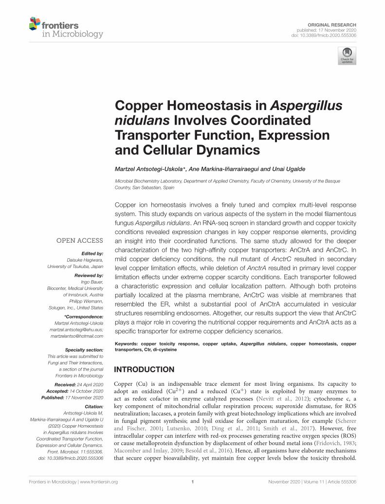
fmicb-11-555306 November 12, 2020 Time: 16:47 # 1
ORIGINAL RESEARCHpublished: 17 November 2020
doi: 10.3389/fmicb.2020.555306
Edited by:Daisuke Hagiwara,
University of Tsukuba, Japan
Reviewed by:Ingo Bauer,
Biocenter, Medical Universityof Innsbruck, Austria
Philipp Wiemann,Solugen, Inc., United States
*Correspondence:Martzel Antsotegi-Uskola
[email protected];[email protected]
Specialty section:This article was submitted toFungi and Their Interactions,
a section of the journalFrontiers in Microbiology
Received: 24 April 2020Accepted: 14 October 2020
Published: 17 November 2020
Citation:Antsotegi-Uskola M,
Markina-Iñarrairaegui A and Ugalde U(2020) Copper Homeostasis
in Aspergillus nidulans InvolvesCoordinated Transporter Function,Expression and Cellular Dynamics.
Front. Microbiol. 11:555306.doi: 10.3389/fmicb.2020.555306
Copper Homeostasis in Aspergillusnidulans Involves CoordinatedTransporter Function, Expressionand Cellular DynamicsMartzel Antsotegi-Uskola* , Ane Markina-Iñarrairaegui and Unai Ugalde
Microbial Biochemistry Laboratory, Department of Applied Chemistry, Faculty of Chemistry, University of the BasqueCountry, San Sebastian, Spain
Copper ion homeostasis involves a finely tuned and complex multi-level responsesystem. This study expands on various aspects of the system in the model filamentousfungus Aspergillus nidulans. An RNA-seq screen in standard growth and copper toxicityconditions revealed expression changes in key copper response elements, providingan insight into their coordinated functions. The same study allowed for the deepercharacterization of the two high-affinity copper transporters: AnCtrA and AnCtrC. Inmild copper deficiency conditions, the null mutant of AnctrC resulted in secondarylevel copper limitation effects, while deletion of AnctrA resulted in primary level copperlimitation effects under extreme copper scarcity conditions. Each transporter followeda characteristic expression and cellular localization pattern. Although both proteinspartially localized at the plasma membrane, AnCtrC was visible at membranes thatresembled the ER, whilst a substantial pool of AnCtrA accumulated in vesicularstructures resembling endosomes. Altogether, our results support the view that AnCtrCplays a major role in covering the nutritional copper requirements and AnCtrA acts as aspecific transporter for extreme copper deficiency scenarios.
Keywords: copper toxicity response, copper uptake, Aspergillus nidulans, copper homeostasis, coppertransporters, Ctr, di-cysteine
INTRODUCTION
Copper (Cu) is an indispensable trace element for most living organisms. Its capacity toadopt an oxidized (Cu2+) and a reduced (Cu+) state is exploited by many enzymes toact as redox cofactor in enzyme catalyzed processes (Nevitt et al., 2012); cytochrome c, akey component of mitochondrial cellular respiration process; superoxide dismutase, for ROSneutralization; laccases, a protein family with great biotechnology implications which are involvedin fungal pigment synthesis; and lysil oxidase for collagen maturation, for example (Schererand Fischer, 2001; Lutsenko, 2010; Ding et al., 2011; Smith et al., 2017). However, freeintracellular copper can interfere with red-ox processes generating reactive oxygen species (ROS)or cause metalloprotein dysfunction by displacement of other bound metal ions (Fridovich, 1983;Macomber and Imlay, 2009; Besold et al., 2016). Hence, all organisms have elaborate mechanismsthat secure copper bioavailability, yet maintain free copper levels below the toxicity threshold.
Frontiers in Microbiology | www.frontiersin.org 1 November 2020 | Volume 11 | Article 555306
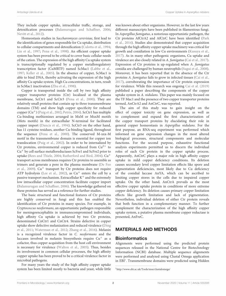
fmicb-11-555306 November 12, 2020 Time: 16:47 # 2
Antsotegi-Uskola et al. Copper Uptake in Aspergillus nidulans
They include copper uptake, intracellular traffic, storage, anddetoxification processes (Balamurugan and Schaffner, 2006;Nevitt et al., 2012).
Homeostasis studies in Saccharomyces cerevisiae, first lead tothe identification of genes responsible for Cu uptake, distributionto cellular compartments and detoxification (Culotta et al., 1994;Lin et al., 1997; Pena et al., 1998). An efficient copper uptakesystem has been proved to be critical to cover basic cellular needsof the cation. The expression of the high affinity Cu uptake systemis transcriptionally regulated by a copper metalloregulatorytranscription factor (CuMRTF) named ScMac1 (Labbe et al.,1997; Keller et al., 2005). In the absence of copper, ScMac1 isable to bind DNA, thereby activating the expression of the highaffinity Cu uptake system. High Cu concentrations, in turn, resultin ScMac1 inactivation (Zhu et al., 1998).
Copper is transported inside the cell by two high affinitycopper transporter proteins (Ctr) localized at the plasmamembrane, ScCtr1 and ScCtr3 (Pena et al., 2000). They arerelatively small proteins that contain up to three transmembranedomains (TM) and show high copper specificity for reducedcopper (Cu+) (Puig et al., 2002; Petris, 2004). ScCtr1 has multipleCu-binding methionines arranged in MxM or MxxM motifs(Mets motifs) in the extracellular N-terminal for facilitatedcopper import (Dancis et al., 1994). ScCtr3 on the other hand,has 11 cysteine residues, another Cu-binding ligand, throughoutthe sequence (Pena et al., 2000). The conserved M-xxx-Mmotif in the transmembrane domain is essential for copper iontranslocation (Puig et al., 2002). In order to be internalized byCtr proteins, environmental copper is reduced from Cu2+ toCu+ by cell surface metalloreductases ScFre1 and ScFre2 prior touptake (Rees and Thiele, 2004; Rutherford and Bird, 2004). Cu+transport across membranes requires Ctr proteins to assemble astrimers and generate a pore in the plasma membrane (De Feoet al., 2009). Cu+ transport by Ctr proteins does not requireATP hydrolysis (Lee et al., 2002), as Cu+ enters the cell by apassive transport mechanism. Extracellular K+ and the extremelylow intracellular copper concentration facilitate copper import(Balamurugan and Schaffner, 2006). The knowledge gathered onthese proteins has served as a reference for further studies.
The basic structural and functional features of Ctr proteinsare highly conserved in fungi and this has enabled theidentification of Ctr proteins in many species. For example, inCryptococcus neoformans, an opportunistic pathogen responsiblefor meningoencephalitis in immunocompromised individuals,high affinity Cu uptake is achieved by two Ctr proteins,denominated CnCtr1 and CnCtr4. Strains defective in copperuptake show defective melanization and reduced virulence (Dinget al., 2011; Waterman et al., 2012; Zhang et al., 2016). Melaninis a recognized virulence factor in C. neoformans and thelaccases involved in melanin biosynthesis require Cu+ as acofactor, thus copper acquisition from the host cell environmentis necessary for virulence (Walton et al., 2005). Thus, besidesits involvement in common cellular processes, the high affinitycopper uptake has been proved to be a critical virulence factor inmicrobial pathogens.
For many years the study of the high affinity copper uptakesystem has been limited mostly to bacteria and yeast, while little
was known about other organisms. However, in the last few yearsdifferent manuscripts have been published in filamentous fungi.In Aspergillus fumigatus, a notorious opportunistic pathogen, theCtr proteins AfCtrA2 and AfCtrC have been identified (Parket al., 2014). Studies also demonstrated that copper acquisitionthrough the high affinity copper uptake machinery was critical forgrowth and conidiation in low Cu environments (Kusuya et al.,2017). As in many other pathogenic organisms, Cu uptake andvirulence are also closely related in A. fumigatus (Cai et al., 2017).Expression of Ctr proteins is up-regulated when A. fumigatusconidia are challenged by human neutrophils (Sugui et al., 2008).Moreover, it has been reported that in the absence of the Ctrproteins A. fumigatus fails to grow in infected tissues (Cai et al.,2017), corroborating the importance of Ctr protein expressionfor virulence. While this research was ongoing Cai et al. (2019)published a paper describing the components of the copperuptake system in A. nidulans. This paper was mainly focused onthe TF Mac1 and the presence of two copper transporter proteinstermed, AnCtrA2 and AnCtrC, was reported.
The aim of this study was to gain insight on theeffect of copper toxicity on gene expression, as well asto complement and expand the first characterization ofthe copper transport proteins by elucidating their role ingeneral copper homeostasis in Aspergillus nidulans. For thefirst purpose, an RNA-seq experiment was performed whichinformed on gene expression changes in the most alteredbiological processes, structural components and molecularfunctions. For the second purpose, exhaustive functionalanalysis experiments permitted us to discern the individualroles of each Ctr protein in the copper uptake process.Apparently, AnCtrC plays a major role in high affinity copperuptake in mild copper deficiency conditions. Its deletioncauses secondary level copper limitation effects like spore andpigmentation deficiencies, most likely due to Cu deficiencyof the conidial laccase AnYA, which can be ascribed tolimiting copper stores in the cells due to impaired copperuptake. On the other hand, AnCtrA prevails as the mosteffective copper uptake protein in conditions of more extremecopper deficiency. Its deletion causes primary copper limitationeffects like growth limitation and lack of pigmentation.Nevertheless, individual deletion of either Ctr protein revealsthat both function in a complementary manner. To furthercomplement the characterization of the high affinity copperuptake system, a putative plasma membrane copper reductase ispresented, AnFreC.
MATERIALS AND METHODS
BioinformaticsAlignments were performed using the predicted proteinsequences released in the National Centre for BiotechnologyInformation (NCBI) database. Multiple sequence alignmentswere performed and analyzed using Clustal Omega applicationin EBI1. Transmembrane domains were predicted using Hidden
1http://www.ebi.ac.uk/Tools/msa/clustalomega/
Frontiers in Microbiology | www.frontiersin.org 2 November 2020 | Volume 11 | Article 555306
fmicb-11-555306 November 12, 2020 Time: 16:47 # 3
Antsotegi-Uskola et al. Copper Uptake in Aspergillus nidulans
Markov Models (HMM) in the Institute Pasteur Mobyle server2.Phylogenetic analyses were carried out using the MolecularEvolutionary Genetics Analysis version 7 (MEGA 7) software3.
Strains, Media and Growth ConditionsA list of all Aspergillus nidulans strains used in this study ispresented in Supplementary Table S1. MAD1427 (wild-type,WT) strain was used to generate single and double knock-outand GFP-tagged strains. All colonies were grown in pH 6.8buffered solid Aspergillus minimal medium (AMM) containingKäfer’s trace elements (Kafer, 1965) 1% (w/v) D-glucose, 71 µMsodium nitrate and appropriately supplemented according to theprocedure described in Pontecorvo et al. (1953). No agar wasadded to the medium for liquid AMM preparation. For lowand high copper availability conditions, 100 µM bathocuproinedisulfonic acid (BCS) Cu chelator and 100 µM CuSO4 wereadded to AMM. For the oxidative stress condition, 2-methyl-1,4-naphthoquinone (menadione) was used. AMM media preparedwith Käfer’s trace elements contains 1.6 µM copper (copper-nutritional condition). For the 0 µM Cu AMM, copper wasremoved from the trace elements. Colony growth tests werecarried out by inoculating conidiospores on solid AMM andincubating for 48 h at 37◦C. Dry weight experiments wereperformed by inoculating conidiospores on liquid 0 µM CuAMM, incubating for 16 h at 37◦C.
Fungal growth tests were carried out in a 96-weel plate byinoculating 60 µl of a 1.106 spore/ml Tween 20 0.1% suspensionon 160 µl liquid AMM or AMM + 100 µM BCS and incubatingfor 40 h at 37◦C. For BCS addition after 16 h of incubation,the required volume was added from a 12.5 µM BCS solution.Measurements are averages of three biological replicates andthree technical replicates of each condition. The error barsrepresent the standard deviation. OD600 was measured with aniMarkTM microplate absorbance reader (Bio-Rad).
To study the effect of gene deletion on conidia production,conidiospores of mutant strains were inoculated by pointinoculation in solid AMM with and without 100 µM BCS. Afterincubation at 37◦C for 3 days, colony diameter was measuredusing the Digimizer image analysis software. Thereafter, inorder to collect the conidia, the agar including mycelia andproduced spores was vortexed in 40 µL 0.2% Tween 20 solutionfor 3 min. Conidia were quantified using a hemocytometer.Four biological replicates and two technical replicates of eachcondition/strain were performed. Two-tailed Student’s t-test forunpaired samples was used for the statistical analysis to comparequantitative counts of conidia (conidia/cm2) between the WTand mutant strains.
Generation of Null and Tagged StrainsDeletion and C-terminally tagging cassettes were constructedfollowing fusion-PCR technique described in Yang et al. (2004)and Markina-Inarrairaegui et al. (2011). Deletion cassettes wereused for null mutant generation and contain the Aspergillusfumigatus pyrG or riboB selection marker which was amplified
2http://mobyle.pasteur.fr/3https://www.megasoftware.net/home
using the plasmid p1439 (pyrGAf ) and p1548 (riboBAf ) as atemplate with oligonucleotides gsp2∗ and gsp3∗. 1500 bp ofthe flanking 5′UTR and 3′UTR regions of the target gene wereamplified from A. nidulans genomic DNA using specific primerpairs; gsp1-gsp2 (5′ UTR) and gsp3-gsp4 (3′ UTR). Hindsight,the fragments were fused using gsp1-gsp4 primers. The cassettefor the generation of strains carrying C-terminally tagged fusionproteins were constructed as follow. The gfp:riboBAf fragmentfrom plasmid p1548 and mrfp:pyrGAf fragment was amplifiedfrom plasmid p184 using oligonucleotides gsp6∗-gsp3∗. The 3′end (∼1500 bp) of the gene and the 3′UTR regions were amplifiedfrom gDNA using oligonucleotides gsp5-gsp6 and gsp3-gsp4,respectively. Finally, oligonucleotides gsp5 and gsp4 were usedto fuse the fragments. Oligonucleotides used in this study aresummarized in Supplementary Table S2.
The purified fused products were then used to transformAspergillus nidulans MAD1427 recipient strain following theprocedure detailed in Antsotegi-Uskola et al. (2017). pyrG+and riboB+ transformants were isolated and single integrationof construct was confirmed by Southern blot technique(Supplementary Figure S1). To generate the double knock-out, strains combining a deleted allele and C-terminally taggedexpressing protein strain or a double tagged strain step-by-steptransformation procedure was used with individual cassettes.
The strains carrying mutations in the last cysteines of theC-terminal domain, CtrCC213A,C214A and AnCtrAC186A,C187A
were constructed as follows: For each gene two specificcomplementary primers were designed, carrying the necessarynucleotide modifications for cysteine modification. Twofragments were amplified using the GFP chimera of each geneas template; first fragment was gsp5-AAreverse and the secondone AAforward-gsp4. Oligonucleotides gsp5 and gsp4 were usedto fuse the fragments. In the case of the C-terminally truncatedmutants, other two primers were designed for each gene. TheC-reverse primer was placed upstream from the last amino-acidof interest and the ∗C-forward primer was placed in the nextnucleotide of the stop codon of the gene (5′ tail complementaryto the C-reverse primer). Using the GFP chimera of each geneas a template, two fragments were amplified gsp5-C-reverse and∗C-forward-gsp4. Primers gsp5 and gsp4 were used for fragmentfusion. The cysteine mutants and the C-terminal deletion strainswere tested by sequencing.
RNA Isolation and RT-PCR Analysis1.106 conidia/ml were inoculated into a two liter flask withair steam containing liquid AMM and incubated at 37◦C for16 h with magnetic stirring. RNA was extracted from cellsharvested prior (0 min) and after transfer to fresh liquid AMMcontaining 100 µM BCS or 100 µM CuSO4 for the indicatedperiod. Mycelia was collected by filtration and ground in liquidnitrogen. Total RNA was isolated from one hundred milligramsof mycelia of three independent biological replicates using a RNAIsolation Kit, Nucleospin R© RNA plant, following instructionsfrom the manufacturer (Macherey-Nagel GmbH & Co. KG). Theintegrity of the RNA was examined by gel electrophoresis andconcentration was calculated using a Nanodrop 2000c system
Frontiers in Microbiology | www.frontiersin.org 3 November 2020 | Volume 11 | Article 555306

fmicb-11-555306 November 12, 2020 Time: 16:47 # 4
Antsotegi-Uskola et al. Copper Uptake in Aspergillus nidulans
(Thermo Fisher Scientific, Waltham, MA, United States). First-strand cDNA of each sample was synthesized using the reversetranscriptase PrimeScriptTM RT reagent Kit (Takara) and thencDNA was used for evaluating gene expression levels. Each cDNAsample was tested in duplicate. qPCR assays were performed inan ABI 7500 system according to the manufacturer’s instructions(Applied Biosystems, Foster, CA) using 5x PyroTaq EvaGreenqPCR Mix Plus (CMB, Cultek Molecular Bioline). benA was usedas internal reference to normalize gene expression levels. The2−1 CT method was used to determine the relative expressionlevel ratio. Oligonucleotides used in this assay are listed inSupplementary Table S2.
RNA-SeqFor the RNA-seq experiment 1:10 diluted supplemented AMMwas used to grow MAD1427 strain. The transcriptome ofvegetative hyphae grown for 16 h in liquid medium wascompared with those cultured under identical conditions butshifted to 100µM CuSO4 medium (IC50 in 1:10 dilutedmedia) 1 h before cell harvest. Three biological replicates wereprocessed for each culture condition. RNA isolation, mRNAlibrary construction, Illumina sequencing and data analysiswas performed as described in Garzia et al. (2013). Briefly,total RNA was extracted based on TRIzol reagent (Invitrogen,Carlsbad, CA, United States), samples were purified using theRNeasy Mini Kit (QIAGEN, Valencia, CA, United States) andlibraries were prepared following Illumina standard protocols(Illumina, San Diego, CA, United States). Sequencing wasperformed in a pair-end-read and 50-base mode on an IlluminaHiSeq Sequencer running nine samples per lane (multiplexing).Sequences were demultiplexed and quality of reads was analyzedwith FastQC v0.10.1. Reads with quality values higher thanQ30 were introduced for mapping using CUTADAPT v1.2and Aspergillus nidulans genome version s07-m02-r07 as thetemplate4. TopHat version 2.0.6 was used for mapping readsand Cuffdiff version 2.0.2 to detect genes differentially expressedbetween different samples. For the determination of differentiallyexpressed genes a q-value below 0.05 was used as a FDRthreshold. RNA-seq results were visualized with CummeRbundversion 1.99.2 software. Gene ontology (GO) terms for eachA. nidulans gene were obtained from the Aspergillus genomedatabase5 and were related with terms downloaded fromOBO6. The GO project provided a standardized set of termsdescribing the molecular functions of genes. We used the topGOsoftware package from the Bioconductor project7 to identifyoverrepresented GO terms from a set of differentially expressedgenes. The Python programming language8 was used to preparethe data, utilizing rpy29 to call R for the statistical analysis.The raw Illumina sequencing data were submitted to the NCBI
4http://www.aspergillusgenome.org/download/sequence/A_nidulans_FGSC_A4/archive/5http://www.aspgd.org/download/go/gene_association.aspgd.gz6http://www.geneontology.org/ontology/obo_format_1_2/gene_ontology_ext.obo7http://www.bioconductor.org/packages/release/bioc/html/topGO.html8http://www.python.org/9http://rpy.sourceforge.net/rpy2.html
Sequence Read Archive (SRA) and deposited under AccessionNumber PRJNA623550.
Protein Isolation and Western Blot1.106 conidia were inoculated in liquid AMM and incubatedon a rotary shaker at 220 rpm and 37◦C for 16 h. Cells wereharvested (0 min), transferred to fresh liquid AMM followed byinduction with 100 µM BCS and 100 µM CuSO4 for the periodindicated in the figures. Mycelia was collected, lyophilized for16 h and ground in a minibeater. Protein samples were extractedby alkaline-lysis buffer (0.2 M NaOH, 0.2% β-mercaptoethanol),as described in Hervas-Aguilar and Penalva (2010).
Five micro liters of each protein sample were loaded into eachwell of 10% SDS-polyacrylamide gels and electrotransfered toImmun-Blot R© PVDF membranes by TransBlot Turbo TransferSystem (Bio-Rad). Ponceau S staining was used as a loadingcontrol (0.1% Ponceau S, 5% acetic acid). Ponceau staining wasremoved using a 20% acetonitrile, 200 µM NaOH solution.For Western blotting the following primary antibodies wereused: monoclonal mouse anti-GFP antibody (1/5000; Roche).As secondary antibodies peroxidase-conjugated goat anti-mouseIgG immunoglobulin (1/4000 dilution; Jackson ImmunoresearchLab.) is used. Proteins were detected using ClarityTM WesternECL Substrate (Bio-Rad) in a Chemidoc + XRS system (Bio-Rad). Signal intensity was measured with Image Lab 3.0software (Bio-Rad).
Fluorescence MicroscopyFor localization of AnCtrA and AnCtrC, conidiosporesof GFP-tagged strains were cultured in Ibidi µ-dishes,35 µm high (Ibidi GmbH, Germany; 2 µl of mediumper well) for 16 h at 30◦C in liquid AspergillusMinimal WATCH medium (Penalva, 2005) adequatelysupplemented and containing 0.1% D-glucose, 71 µMsodium nitrate, 25 µM sodium phosphate monobasic. Forlow copper availability conditions, 100 µM bathocuproinedisulfonic acid (BCS) Cu chelator were added to theWATCH media.
Fluorescence microscopy was performed using a Zeiss AxioObserver Z1 inverted microscope (63 Plan Apochromat 1.4oil immersion Lens) and Axiocam MRm Rev.3 camera. Forobservation of GFP, a filter set 38 HE (Ex BP 470/40; FT 495;Em BP 525/50) was used. The images shown are representativeof at least five experiments repeats. Levels of fluorescence wereanalyzed using open source ImageJ software10 (U. S. NationalInstitutes of Health, Bethesda, MD, United States).
RESULTS
Gene Expression Analysis Under CopperToxicity ConditionsAn RNA-seq analysis was performed to compare thetranscriptome of vegetative hyphae grown for 16 h in liquidmedium (nutritional condition; acronym NC) with that of
10http://imagej.nih.gov/ij
Frontiers in Microbiology | www.frontiersin.org 4 November 2020 | Volume 11 | Article 555306
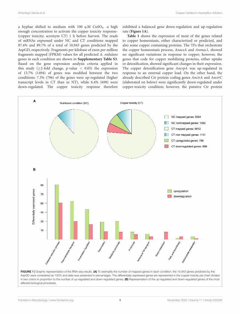
fmicb-11-555306 November 12, 2020 Time: 16:47 # 5
Antsotegi-Uskola et al. Copper Uptake in Aspergillus nidulans
a hyphae shifted to medium with 100 µM CuSO4 -a highenough concentration to activate the copper toxicity response-(copper toxicity; acronym CT) 1 h before harvest. The readsof mRNAs expressed under NC and CT conditions mapped87.4% and 89.7% of a total of 10,943 genes predicted by theAspGD, respectively. Fragments per kilobase of exon per millionfragments mapped (FPKM) values for all predicted A. nidulansgenes in each condition are shown in Supplementary Table S3.Based on the gene expression analysis criteria applied inthis study (≥2-fold change, p-value < 0.05) the expressionof 13.7% (1494) of genes was modified between the twoconditions: 7.3% (796) of the genes were up-regulated (highertranscript levels in CT than in NT), while 6.4% (698) weredown-regulated. The copper toxicity response therefore
exhibited a balanced gene down-regulation and up-regulationrate (Figure 1A).
Table 1 shows the expression of most of the genes relatedto copper homeostasis, either characterized or predicted, andalso some copper containing proteins. The TFs that orchestratethe copper homeostasis process, AnaceA and Anmac1, showedno significant variations in response to copper; however, thegenes that code for copper mobilizing proteins, either uptakeor detoxification, showed significant changes in their expression.The copper detoxification gene AncrpA was up-regulated inresponse to an external copper load. On the other hand, thealready described Ctr protein coding genes AnctrA and AnctrC(elaborated on below) were significantly down-regulated undercopper-toxicity condition; however, the putative Ctr protein
FIGURE 1 | Graphic representation of the RNA-seq results. (A) To exemplify the number of mapped genes in each condition, the 10.943 genes predicted by theAspGD were considered as 100% and data was presented in percentages. The differentially expressed genes are represented in the copper toxicity pie chart dividedin two colors in proportion to the number of up-regulated and down-regulated genes. (B) Representation of the up-regulated and down-regulated genes of the mostaffected biological processes.
Frontiers in Microbiology | www.frontiersin.org 5 November 2020 | Volume 11 | Article 555306
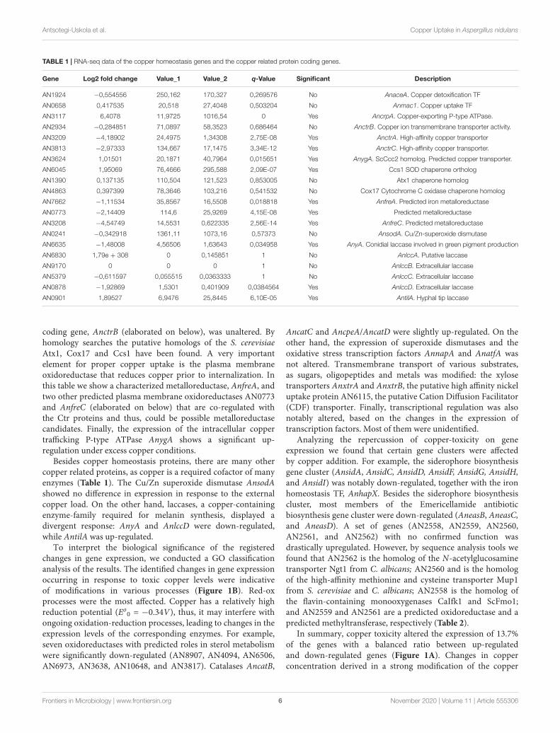
fmicb-11-555306 November 12, 2020 Time: 16:47 # 6
Antsotegi-Uskola et al. Copper Uptake in Aspergillus nidulans
TABLE 1 | RNA-seq data of the copper homeostasis genes and the copper related protein coding genes.
Gene Log2 fold change Value_1 Value_2 q-Value Significant Description
AN1924 −0,554556 250,162 170,327 0,269576 No AnaceA. Copper detoxification TF
AN0658 0,417535 20,518 27,4048 0,503204 No Anmac1. Copper uptake TF
AN3117 6,4078 11,9725 1016,54 0 Yes AncrpA. Copper-exporting P-type ATPase.
AN2934 −0,284851 71,0897 58,3523 0,686464 No AnctrB. Copper ion transmembrane transporter activity.
AN3209 −4,18902 24,4975 1,34308 2,75E-08 Yes AnctrA. High-affinity copper transporter
AN3813 −2,97333 134,667 17,1475 3,34E-12 Yes AnctrC. High-affinity copper transporter.
AN3624 1,01501 20,1871 40,7964 0,015651 Yes AnygA. ScCcc2 homolog. Predicted copper transporter.
AN6045 1,95069 76,4666 295,588 2,09E-07 Yes Ccs1 SOD chaperone ortholog
AN1390 0,137135 110,504 121,523 0,853005 No Atx1 chaperone homolog
AN4863 0,397399 78,3646 103,216 0,541532 No Cox17 Cytochrome C oxidase chaperone homolog
AN7662 −1,11534 35,8567 16,5508 0,018818 Yes AnfreA. Predicted iron metalloreductase
AN0773 −2,14409 114,6 25,9269 4,15E-08 Yes Predicted metalloreductase
AN3208 −4,54749 14,5531 0,622335 2,56E-14 Yes AnfreC. Predicted metalloreductase
AN0241 −0,342918 1361,11 1073,16 0,57373 No AnsodA. Cu/Zn-superoxide dismutase
AN6635 −1,48008 4,56506 1,63643 0,034958 Yes AnyA. Conidial laccase involved in green pigment production
AN6830 1,79e + 308 0 0,145851 1 No AnlccA. Putative laccase
AN9170 0 0 0 1 No AnlccB. Extracellular laccase
AN5379 −0,611597 0,055515 0,0363333 1 No AnlccC. Extracellular laccase
AN0878 −1,92869 1,5301 0,401909 0,0384564 Yes AnlccD. Extracellular laccase
AN0901 1,89527 6,9476 25,8445 6,10E-05 Yes AntilA. Hyphal tip laccase
coding gene, AnctrB (elaborated on below), was unaltered. Byhomology searches the putative homologs of the S. cerevisiaeAtx1, Cox17 and Ccs1 have been found. A very importantelement for proper copper uptake is the plasma membraneoxidoreductase that reduces copper prior to internalization. Inthis table we show a characterized metalloreductase, AnfreA, andtwo other predicted plasma membrane oxidoreductases AN0773and AnfreC (elaborated on below) that are co-regulated withthe Ctr proteins and thus, could be possible metalloreductasecandidates. Finally, the expression of the intracellular coppertrafficking P-type ATPase AnygA shows a significant up-regulation under excess copper conditions.
Besides copper homeostasis proteins, there are many othercopper related proteins, as copper is a required cofactor of manyenzymes (Table 1). The Cu/Zn superoxide dismutase AnsodAshowed no difference in expression in response to the externalcopper load. On the other hand, laccases, a copper-containingenzyme-family required for melanin synthesis, displayed adivergent response: AnyA and AnlccD were down-regulated,while AntilA was up-regulated.
To interpret the biological significance of the registeredchanges in gene expression, we conducted a GO classificationanalysis of the results. The identified changes in gene expressionoccurring in response to toxic copper levels were indicativeof modifications in various processes (Figure 1B). Red-oxprocesses were the most affected. Copper has a relatively highreduction potential (Eo
0 = −0.34V), thus, it may interfere withongoing oxidation-reduction processes, leading to changes in theexpression levels of the corresponding enzymes. For example,seven oxidoreductases with predicted roles in sterol metabolismwere significantly down-regulated (AN8907, AN4094, AN6506,AN6973, AN3638, AN10648, and AN3817). Catalases AncatB,
AncatC and AncpeA/AncatD were slightly up-regulated. On theother hand, the expression of superoxide dismutases and theoxidative stress transcription factors AnnapA and AnatfA wasnot altered. Transmembrane transport of various substrates,as sugars, oligopeptides and metals was modified: the xylosetransporters AnxtrA and AnxtrB, the putative high affinity nickeluptake protein AN6115, the putative Cation Diffusion Facilitator(CDF) transporter. Finally, transcriptional regulation was alsonotably altered, based on the changes in the expression oftranscription factors. Most of them were unidentified.
Analyzing the repercussion of copper-toxicity on geneexpression we found that certain gene clusters were affectedby copper addition. For example, the siderophore biosynthesisgene cluster (AnsidA, AnsidC, AnsidD, AnsidF, AnsidG, AnsidH,and AnsidI) was notably down-regulated, together with the ironhomeostasis TF, AnhapX. Besides the siderophore biosynthesiscluster, most members of the Emericellamide antibioticbiosynthesis gene cluster were down-regulated (AneasB, AneasC,and AneasD). A set of genes (AN2558, AN2559, AN2560,AN2561, and AN2562) with no confirmed function wasdrastically upregulated. However, by sequence analysis tools wefound that AN2562 is the homolog of the N-acetylglucosaminetransporter Ngt1 from C. albicans; AN2560 and is the homologof the high-affinity methionine and cysteine transporter Mup1from S. cerevisiae and C. albicans; AN2558 is the homolog ofthe flavin-containing monooxygenases CaIfk1 and ScFmo1;and AN2559 and AN2561 are a predicted oxidoreductase and apredicted methyltransferase, respectively (Table 2).
In summary, copper toxicity altered the expression of 13.7%of the genes with a balanced ratio between up-regulatedand down-regulated genes (Figure 1A). Changes in copperconcentration derived in a strong modification of the copper
Frontiers in Microbiology | www.frontiersin.org 6 November 2020 | Volume 11 | Article 555306
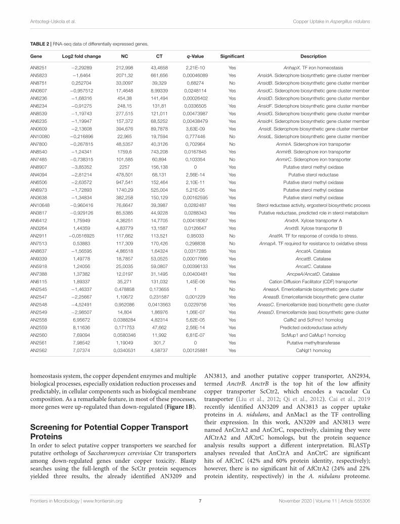
fmicb-11-555306 November 12, 2020 Time: 16:47 # 7
Antsotegi-Uskola et al. Copper Uptake in Aspergillus nidulans
TABLE 2 | RNA-seq data of differentially expressed genes.
Gene Log2 fold change NC CT q-Value Significant Description
AN8251 −2,29289 212,998 43,4658 2,21E-10 Yes AnhapX. TF iron homeostasis
AN5823 −1,6464 2071,32 661,656 0,00046089 Yes AnsidA. Siderophore biosynthetic gene cluster member
AN8751 0,252704 33,0097 39,329 0,68274 No AnsidB. Siderophore biosynthetic gene cluster member
AN0607 −0,957512 17,4648 8,99339 0,0248114 Yes AnsidC. Siderophore biosynthetic gene cluster member
AN6236 −1,68316 454,38 141,494 0,00026402 Yes AnsidD. Siderophore biosynthetic gene cluster member
AN6234 −0,91275 248,15 131,81 0,0336505 Yes AnsidF. Siderophore biosynthetic gene cluster member
AN8539 −1,19743 277,515 121,011 0,00473987 Yes AnsidG. Siderophore biosynthetic gene cluster member
AN6235 −1,19947 157,372 68,5252 0,00438479 Yes AnsidH. Siderophore biosynthetic gene cluster member
AN0609 −2,13608 394,676 89,7878 3,63E-09 Yes AnsidI. Siderophore biosynthetic gene cluster member
AN10080 −0,216896 22,965 19,7594 0,777446 No AnsidL. Siderophore biosynthetic gene cluster member
AN7800 −0,267815 48,5357 40,3126 0,702964 No AnmirA. Siderophore iron transporter
AN8540 −1,24341 1759,6 743,208 0,0167845 Yes AnmirB. Siderophore iron transporter
AN7485 −0,738315 101,585 60,894 0,103354 No AnmirC. Siderophore iron transporter
AN8907 −3,85352 2257 156,138 0 Yes Putative sterol methyl oxidase
AN4094 −2,81214 478,501 68,131 2,56E-14 Yes Putative sterol reductase
AN6506 −2,63572 947,541 152,464 2,10E-11 Yes Putative sterol methyl oxidase
AN6973 −1,72893 1740,29 525,004 5,21E-05 Yes Putative sterol methyl oxidase
AN3638 −1,34834 382,258 150,129 0,00162595 Yes Putative sterol methyl oxidase
AN10648 −0,960416 76,6647 39,3987 0,0282487 Yes Sterol reductase activity, ergosterol biosynthetic process
AN3817 −0,929126 85,5385 44,9228 0,0288343 Yes Putative reductase, predicted role in sterol metabolism
AN6412 1,75949 4,36251 14,7705 0,00418067 Yes AnxtrA. Xylose transporter A
AN3264 1,44359 4,83779 13,1587 0,0126647 Yes AnxtrB. Xylose transporter B
AN2911 −0,0516925 117,662 113,521 0,95033 No AnatfA. TF for response of conidia to stress.
AN7513 0,53883 117,309 170,426 0,298838 No AnnapA. TF required for resistance to oxidative stress
AN8637 −1,56595 4,86518 1,64324 0,0317285 Yes AncatA. Catalase
AN9339 1,49778 18,7857 53,0525 0,00017666 Yes AncatB. Catalase
AN5918 1,24056 25,0035 59,0807 0,00396133 Yes AncatC. Catalase
AN7388 1,37382 12,0197 31,1495 0,00400481 Yes AncpeA/AncatD. Catalase
AN6115 1,89337 35,271 131,032 1,45E-06 Yes Cation Diffusion Facilitator (CDF) transporter
AN2545 −1,46337 0,478858 0,173655 1 No AneasA. Emericellamide biosynthetic gene cluster
AN2547 −2,25667 1,10672 0,231587 0,001229 Yes AneasB. Emericellamide biosynthetic gene cluster
AN2548 −4,52491 0,952086 0,0413563 0,0229756 Yes AneasC. Emericellamide (eas) biosynthetic gene cluster
AN2549 −2,98507 14,804 1,86976 1,06E-07 Yes AneasD. Emericellamide (eas) biosynthetic gene cluster
AN2558 6,95672 0,0388284 4,82314 5,62E-05 Yes CaIfk2 and ScFmo1 homolog
AN2559 8,11636 0,171753 47,662 2,56E-14 Yes Predicted oxidoreductase activity
AN2560 7,69094 0,0580346 11,992 6,81E-07 Yes ScMup1 and CaMup1 homolog
AN2561 7,98542 1,19049 301,7 0 Yes Putative methyltransferase
AN2562 7,07374 0,0340531 4,58737 0,00125881 Yes CaNgt1 homolog
homeostasis system, the copper dependent enzymes and multiplebiological processes, especially oxidation reduction processes andpredictably, in cellular components such as biological membranecomposition. As a remarkable feature, in most of these processes,more genes were up-regulated than down-regulated (Figure 1B).
Screening for Potential Copper TransportProteinsIn order to select putative copper transporters we searched forputative orthologs of Saccharomyces cerevisiae Ctr transportersamong down-regulated genes under copper toxicity. Blastpsearches using the full-length of the ScCtr protein sequencesyielded three results, the already identified AN3209 and
AN3813, and another putative copper transporter, AN2934,termed AnctrB. AnctrB is the top hit of the low affinitycopper transporter ScCtr2, which encodes a vacuolar Cutransporter (Liu et al., 2012; Qi et al., 2012). Cai et al., 2019recently identified AN3209 and AN3813 as copper uptakeproteins in A. nidulans, and AnMac1 as the TF controllingtheir expression. In this work, AN3209 and AN3813 werenamed AnCtrA2 and AnCtrC, respectively, claiming they wereAfCtrA2 and AfCtrC homologs, but the protein sequenceanalysis results support a different interpretation. BLASTpanalyses revealed that AnCtrA and AnCtrC are significanthits of AfCtrC (42% and 60% protein identity, respectively);however, there is no significant hit of AfCtrA2 (24% and 22%protein identity, respectively) in the A. nidulans proteome.
Frontiers in Microbiology | www.frontiersin.org 7 November 2020 | Volume 11 | Article 555306
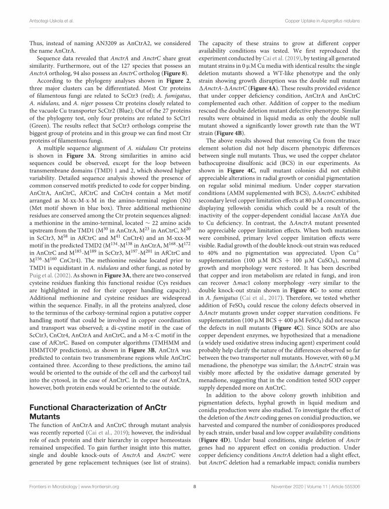
fmicb-11-555306 November 12, 2020 Time: 16:47 # 8
Antsotegi-Uskola et al. Copper Uptake in Aspergillus nidulans
Thus, instead of naming AN3209 as AnCtrA2, we consideredthe name AnCtrA.
Sequence data revealed that AnctrA and AnctrC share greatsimilarity. Furthermore, out of the 127 species that possess anAnctrA ortholog, 94 also possess an AnctrC ortholog (Figure 8).
According to the phylogeny analyses shown in Figure 2,three major clusters can be differentiated. Most Ctr proteinsof filamentous fungi are related to ScCtr3 (red); A. fumigatus,A. nidulans, and A. niger possess Ctr proteins closely related tothe vacuole Cu transporter ScCtr2 (Blue); Out of the 27 proteinsof the phylogeny test, only four proteins are related to ScCtr1(Green). The results reflect that ScCtr3 orthologs comprise thebiggest group of proteins and in this group we can find most Ctrproteins of filamentous fungi.
A multiple sequence alignment of A. nidulans Ctr proteinsis shown in Figure 3A. Strong similarities in amino acidsequences could be observed, except for the loop betweentransmembrane domains (TMD) 1 and 2, which showed highervariability. Detailed sequence analysis showed the presence ofcommon conserved motifs predicted to code for copper binding.AnCtrA, AnCtrC, AfCtrC and CnCtr4 contain a Met motifarranged as M-xx-M-x-M in the amino-terminal region (Nt)(Met motif shown in blue box). Three additional methionineresidues are conserved among the Ctr protein sequences aligned:a methionine in the amino-terminal, located ∼ 22 amino acidsupstream from the TMD1 (M30 in AnCtrA, M23 in AnCtrC, M20
in ScCtr3, M58 in AfCtrC and M41 CnCtr4) and an M-xxx-Mmotif in the predicted TMD2 (M134-M138 in AnCtrA, M168-M172
in AnCtrC and M185-M189 in ScCtr3, M197-M201 in AfCtrC andM156-M160 CnCtr4). The methionine residue located prior toTMD1 is equidistant in A. nidulans and other fungi, as noted byPuig et al. (2002). As shown in Figure 3A, there are two conservedcysteine residues flanking this functional residue (Cys residuesare highlighted in red for their copper handling capacity).Additional methionine and cysteine residues are widespreadwithin the sequence. Finally, in all the proteins analyzed, closeto the terminus of the carboxy-terminal region a putative copperhandling motif that could be involved in copper coordinationand transport was observed; a di-cystine motif in the case ofScCtr3, CnCtr4, AnCtrA and AnCtrC, and a M-x-C motif in thecase of AfCtrC. Based on computer algorithms (TMHMM andHMMTOP predictions), as shown in Figure 3B, AnCtrA waspredicted to contain two transmembrane regions while AnCtrCcontained three. According to these predictions, the amino tailwould be oriented to the outside of the cell and the carboxyl tailinto the cytosol, in the case of AnCtrC. In the case of AnCtrA,however, both protein ends would be oriented to the outside.
Functional Characterization of AnCtrMutantsThe function of AnCtrA and AnCtrC through mutant analysiswas recently reported (Cai et al., 2019); however, the individualrole of each protein and their hierarchy in copper homeostasisremained unspecified. To gain further insight into this matter,single and double knock-outs of AnctrA and AnctrC weregenerated by gene replacement techniques (see list of strains).
The capacity of these strains to grow at different copperavailability conditions was tested. We first reproduced theexperiment conducted by Cai et al. (2019), by testing all generatedmutant strains in 0 µM Cu media with identical results: the singledeletion mutants showed a WT-like phenotype and the onlystrain showing growth disruption was the double null mutant1AnctrA-1AnctrC (Figure 4A). These results provided evidencethat under copper deficiency condition, AnCtrA and AnCtrCcomplemented each other. Addition of copper to the mediumrescued the double deletion mutant defective phenotype. Similarresults were obtained in liquid media as only the double nullmutant showed a significantly lower growth rate than the WTstrain (Figure 4B).
The above results showed that removing Cu from the traceelement solution did not help discern phenotypic differencesbetween single null mutants. Thus, we used the copper chelatorbathocuproine disulfonic acid (BCS) in our experiments. Asshown in Figure 4C, null mutant colonies did not exhibitappreciable alterations in radial growth or conidial pigmentationon regular solid minimal medium. Under copper starvationconditions (AMM supplemented with BCS), 1AnctrC exhibitedsecondary level copper limitation effects at 80 µM concentration,displaying yellowish conidia which could be a result of theinactivity of the copper-dependent conidial laccase AnYA dueto Cu deficiency. In contrast, the 1AnctrA mutant presentedno appreciable copper limitation effects. When both mutationswere combined, primary level copper limitation effects werevisible. Radial growth of the double knock-out strain was reducedto 40% and no pigmentation was appreciated. Upon Cu+supplementation (100 µM BCS + 100 µM CuSO4), normalgrowth and morphology were restored. It has been describedthat copper and iron metabolism are related in fungi, and ironcan recover 1mac1 colony morphology -very similar to thedouble knock-out strain shown in Figure 4C- to some extentin A. fumigatus (Cai et al., 2017). Therefore, we tested whetheraddition of FeSO4 could rescue the colony defects observed in1Anctr mutants grown under copper starvation conditions. Fesupplementation (100 µM BCS+ 400 µM FeSO4) did not rescuethe defects in null mutants (Figure 4C). Since SODs are alsocopper dependent enzymes, we hypothesized that a menadione(a widely used oxidative stress inducing agent) experiment couldprobably help clarify the nature of the differences observed so farbetween the two transporter null mutants. However, with 60 µMmenadione, the phenotype was similar; the 1AnctrC strain wasvisibly more affected by the oxidative damage generated bymenadione, suggesting that in the condition tested SOD coppersupply depended more on AnCtrC.
In addition to the above colony growth inhibition andpigmentation defects, hyphal growth in liquid medium andconidia production were also studied. To investigate the effect ofthe deletion of the Anctr coding genes on conidial production, weharvested and compared the number of conidiospores producedby each strain, under basal and low copper availability conditions(Figure 4D). Under basal conditions, single deletion of Anctrgenes had no apparent effect on conidia production. Undercopper deficiency conditions AnctrA deletion had a slight effect,but AnctrC deletion had a remarkable impact; conidia numbers
Frontiers in Microbiology | www.frontiersin.org 8 November 2020 | Volume 11 | Article 555306
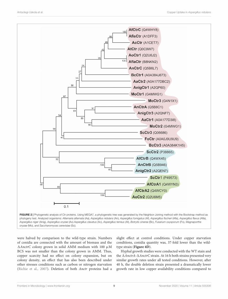
fmicb-11-555306 November 12, 2020 Time: 16:47 # 9
Antsotegi-Uskola et al. Copper Uptake in Aspergillus nidulans
FIGURE 2 | Phylogenetic analysis of Ctr proteins. Using MEGA7, a phylogenetic tree was generated by the Neighbor-Joining method with the Bootstrap method asphylogeny test. Analyzed organisms: Alternaria alternata (Aa), Aspergillus nidulans (An), Aspergillus fumigatus (Af), Aspergillus fischeri (Afis), Aspergillus flavus (Afla),Aspergillus niger (Anig), Aspergillus oryzae (Ao) Aspergillus clavatus (Ac), Aspergillus terreus (At), Botrytis cinerea (Bc), Fusarium oxysporum (Fo), Magnaportheoryzae (Mo), and Saccharomyces cerevisiae (Sc).
were halved by comparison to the wild-type strain. Numbersof conidia are connected with the amount of biomass and the1AnctrC colony grown in solid AMM medium with 100 µMBCS was not smaller than the colony grown in AMM. Thus,copper scarcity had no effect on colony expansion, but oncolony density, an effect that has also been described underother stresses conditions such as carbon or nitrogen starvation(Richie et al., 2007). Deletion of both Anctr proteins had a
slight effect at control conditions. Under copper starvationconditions, conidia quantity was, 37-fold lower than the wild-type strain (Figure 4D).
Hyphal growth studies were conducted with the WT stain andthe 1AnctrA-1AnctrC strain. At 16 h both strains presented verysimilar growth rates under all tested conditions. However, after40 h, the double deletion strain presented a dramatically lowergrowth rate in low copper availability conditions compared to
Frontiers in Microbiology | www.frontiersin.org 9 November 2020 | Volume 11 | Article 555306
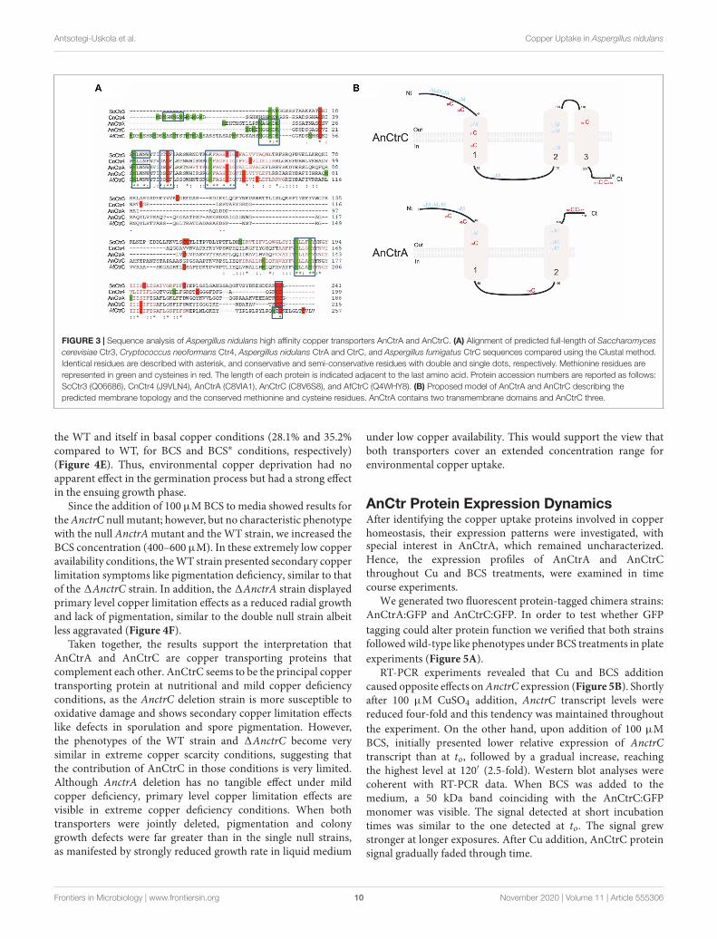
fmicb-11-555306 November 12, 2020 Time: 16:47 # 10
Antsotegi-Uskola et al. Copper Uptake in Aspergillus nidulans
FIGURE 3 | Sequence analysis of Aspergillus nidulans high affinity copper transporters AnCtrA and AnCtrC. (A) Alignment of predicted full-length of Saccharomycescerevisiae Ctr3, Cryptococcus neoformans Ctr4, Aspergillus nidulans CtrA and CtrC, and Aspergillus fumigatus CtrC sequences compared using the Clustal method.Identical residues are described with asterisk, and conservative and semi-conservative residues with double and single dots, respectively. Methionine residues arerepresented in green and cysteines in red. The length of each protein is indicated adjacent to the last amino acid. Protein accession numbers are reported as follows:ScCtr3 (Q06686), CnCtr4 (J9VLN4), AnCtrA (C8VIA1), AnCtrC (C8V6S8), and AfCtrC (Q4WHY8). (B) Proposed model of AnCtrA and AnCtrC describing thepredicted membrane topology and the conserved methionine and cysteine residues. AnCtrA contains two transmembrane domains and AnCtrC three.
the WT and itself in basal copper conditions (28.1% and 35.2%compared to WT, for BCS and BCS∗ conditions, respectively)(Figure 4E). Thus, environmental copper deprivation had noapparent effect in the germination process but had a strong effectin the ensuing growth phase.
Since the addition of 100 µM BCS to media showed results forthe AnctrC null mutant; however, but no characteristic phenotypewith the null AnctrA mutant and the WT strain, we increased theBCS concentration (400–600 µM). In these extremely low copperavailability conditions, the WT strain presented secondary copperlimitation symptoms like pigmentation deficiency, similar to thatof the 1AnctrC strain. In addition, the 1AnctrA strain displayedprimary level copper limitation effects as a reduced radial growthand lack of pigmentation, similar to the double null strain albeitless aggravated (Figure 4F).
Taken together, the results support the interpretation thatAnCtrA and AnCtrC are copper transporting proteins thatcomplement each other. AnCtrC seems to be the principal coppertransporting protein at nutritional and mild copper deficiencyconditions, as the AnctrC deletion strain is more susceptible tooxidative damage and shows secondary copper limitation effectslike defects in sporulation and spore pigmentation. However,the phenotypes of the WT strain and 1AnctrC become verysimilar in extreme copper scarcity conditions, suggesting thatthe contribution of AnCtrC in those conditions is very limited.Although AnctrA deletion has no tangible effect under mildcopper deficiency, primary level copper limitation effects arevisible in extreme copper deficiency conditions. When bothtransporters were jointly deleted, pigmentation and colonygrowth defects were far greater than in the single null strains,as manifested by strongly reduced growth rate in liquid medium
under low copper availability. This would support the view thatboth transporters cover an extended concentration range forenvironmental copper uptake.
AnCtr Protein Expression DynamicsAfter identifying the copper uptake proteins involved in copperhomeostasis, their expression patterns were investigated, withspecial interest in AnCtrA, which remained uncharacterized.Hence, the expression profiles of AnCtrA and AnCtrCthroughout Cu and BCS treatments, were examined in timecourse experiments.
We generated two fluorescent protein-tagged chimera strains:AnCtrA:GFP and AnCtrC:GFP. In order to test whether GFPtagging could alter protein function we verified that both strainsfollowed wild-type like phenotypes under BCS treatments in plateexperiments (Figure 5A).
RT-PCR experiments revealed that Cu and BCS additioncaused opposite effects on AnctrC expression (Figure 5B). Shortlyafter 100 µM CuSO4 addition, AnctrC transcript levels werereduced four-fold and this tendency was maintained throughoutthe experiment. On the other hand, upon addition of 100 µMBCS, initially presented lower relative expression of AnctrCtranscript than at to, followed by a gradual increase, reachingthe highest level at 120′ (2.5-fold). Western blot analyses werecoherent with RT-PCR data. When BCS was added to themedium, a 50 kDa band coinciding with the AnCtrC:GFPmonomer was visible. The signal detected at short incubationtimes was similar to the one detected at to. The signal grewstronger at longer exposures. After Cu addition, AnCtrC proteinsignal gradually faded through time.
Frontiers in Microbiology | www.frontiersin.org 10 November 2020 | Volume 11 | Article 555306
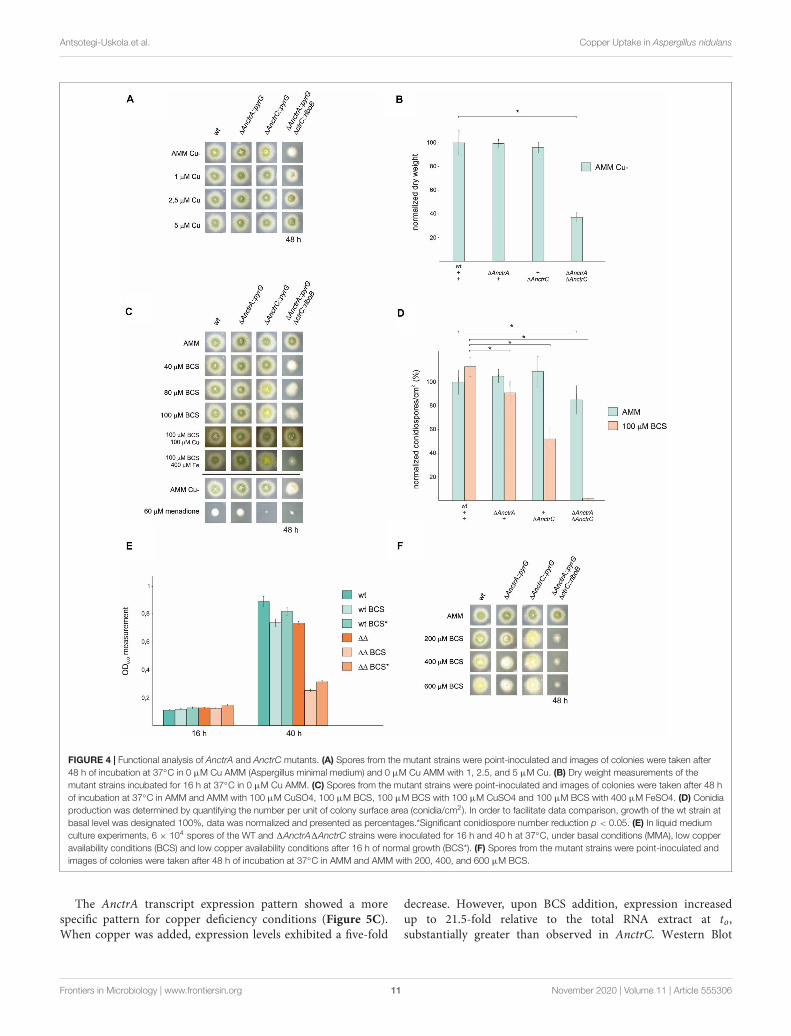
fmicb-11-555306 November 12, 2020 Time: 16:47 # 11
Antsotegi-Uskola et al. Copper Uptake in Aspergillus nidulans
FIGURE 4 | Functional analysis of AnctrA and AnctrC mutants. (A) Spores from the mutant strains were point-inoculated and images of colonies were taken after48 h of incubation at 37◦C in 0 µM Cu AMM (Aspergillus minimal medium) and 0 µM Cu AMM with 1, 2.5, and 5 µM Cu. (B) Dry weight measurements of themutant strains incubated for 16 h at 37◦C in 0 µM Cu AMM. (C) Spores from the mutant strains were point-inoculated and images of colonies were taken after 48 hof incubation at 37◦C in AMM and AMM with 100 µM CuSO4, 100 µM BCS, 100 µM BCS with 100 µM CuSO4 and 100 µM BCS with 400 µM FeSO4. (D) Conidiaproduction was determined by quantifying the number per unit of colony surface area (conidia/cm2). In order to facilitate data comparison, growth of the wt strain atbasal level was designated 100%, data was normalized and presented as percentages.*Significant conidiospore number reduction p < 0.05. (E) In liquid mediumculture experiments, 6 × 10⁴ spores of the WT and 1AnctrA1AnctrC strains were inoculated for 16 h and 40 h at 37◦C, under basal conditions (MMA), low copperavailability conditions (BCS) and low copper availability conditions after 16 h of normal growth (BCS*). (F) Spores from the mutant strains were point-inoculated andimages of colonies were taken after 48 h of incubation at 37◦C in AMM and AMM with 200, 400, and 600 µM BCS.
The AnctrA transcript expression pattern showed a morespecific pattern for copper deficiency conditions (Figure 5C).When copper was added, expression levels exhibited a five-fold
decrease. However, upon BCS addition, expression increasedup to 21.5-fold relative to the total RNA extract at to,substantially greater than observed in AnctrC. Western Blot
Frontiers in Microbiology | www.frontiersin.org 11 November 2020 | Volume 11 | Article 555306
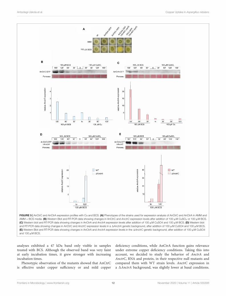
fmicb-11-555306 November 12, 2020 Time: 16:47 # 12
Antsotegi-Uskola et al. Copper Uptake in Aspergillus nidulans
FIGURE 5 | AnCtrC and AnCtrA expression profiles with Cu and BCS. (A) Phenotypes of the strains used for expression analysis of AnCtrC and AnCtrA in AMM andAMM + BCS media. (B) Western Blot and RT-PCR data showing changes in AnCtrC and AnctrC expression levels after addition of 100 µM CuSO4 or 100 µM BCS.(C) Western blot and RT-PCR data showing changes in AnCtrA and AnctrA expression levels after addition of 100 µM CuSO4 and 100 µM BCS. (D) Western blotand RT-PCR data showing changes in AnCtrC and AnctrC expression levels in a 1AnctrA genetic background, after addition of 100 µM CuSO4 and 100 µM BCS.(E) Western Blot and RT-PCR data showing changes in AnCtrA and AnctrA expression levels in the 1AnctrC genetic background, after addition of 100 µM CuSO4and 100 µM BCS.
analyses exhibited a 47 kDa band only visible in samplestreated with BCS. Although the observed band was very faintat early incubation times, it grew stronger with increasingincubation times.
Phenotypic observation of the mutants showed that AnCtrCis effective under copper sufficiency or and mild copper
deficiency conditions, while AnCtrA function gains relevanceunder extreme copper deficiency conditions. Taking this intoaccount, we decided to study the behavior of AnctrA andAnctrC, RNA and protein, in their respective null mutants andcompared them with WT strain levels. AnctrC expression ina 1AnctrA background, was slightly lower at basal conditions.
Frontiers in Microbiology | www.frontiersin.org 12 November 2020 | Volume 11 | Article 555306
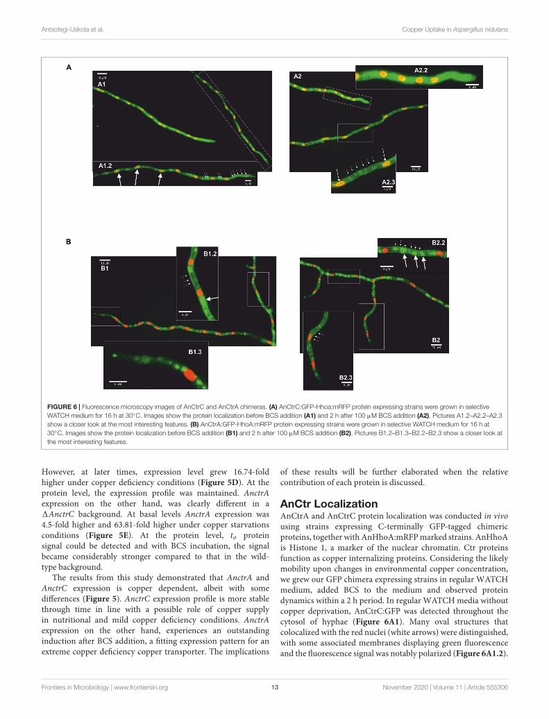
fmicb-11-555306 November 12, 2020 Time: 16:47 # 13
Antsotegi-Uskola et al. Copper Uptake in Aspergillus nidulans
FIGURE 6 | Fluorescence microscopy images of AnCtrC and AnCtrA chimeras. (A) AnCtrC:GFP-Hhoa:mRFP protein expressing strains were grown in selectiveWATCH medium for 16 h at 30◦C. Images show the protein localization before BCS addition (A1) and 2 h after 100 µM BCS addition (A2). Pictures A1.2–A2.2–A2.3show a closer look at the most interesting features. (B) AnCtrA:GFP-HhoA:mRFP protein expressing strains were grown in selective WATCH medium for 16 h at30◦C. Images show the protein localization before BCS addition (B1) and 2 h after 100 µM BCS addition (B2). Pictures B1.2–B1.3–B2.2–B2.3 show a closer look atthe most interesting features.
However, at later times, expression level grew 16.74-foldhigher under copper deficiency conditions (Figure 5D). At theprotein level, the expression profile was maintained. AnctrAexpression on the other hand, was clearly different in a1AnctrC background. At basal levels AnctrA expression was4.5-fold higher and 63.81-fold higher under copper starvationsconditions (Figure 5E). At the protein level, to proteinsignal could be detected and with BCS incubation, the signalbecame considerably stronger compared to that in the wild-type background.
The results from this study demonstrated that AnctrA andAnctrC expression is copper dependent, albeit with somedifferences (Figure 5). AnctrC expression profile is more stablethrough time in line with a possible role of copper supplyin nutritional and mild copper deficiency conditions. AnctrAexpression on the other hand, experiences an outstandinginduction after BCS addition, a fitting expression pattern for anextreme copper deficiency copper transporter. The implications
of these results will be further elaborated when the relativecontribution of each protein is discussed.
AnCtr LocalizationAnCtrA and AnCtrC protein localization was conducted in vivousing strains expressing C-terminally GFP-tagged chimericproteins, together with AnHhoA:mRFP marked strains. AnHhoAis Histone 1, a marker of the nuclear chromatin. Ctr proteinsfunction as copper internalizing proteins. Considering the likelymobility upon changes in environmental copper concentration,we grew our GFP chimera expressing strains in regular WATCHmedium, added BCS to the medium and observed proteindynamics within a 2 h period. In regular WATCH media withoutcopper deprivation, AnCtrC:GFP was detected throughout thecytosol of hyphae (Figure 6A1). Many oval structures thatcolocalized with the red nuclei (white arrows) were distinguished,with some associated membranes displaying green fluorescenceand the fluorescence signal was notably polarized (Figure 6A1.2).
Frontiers in Microbiology | www.frontiersin.org 13 November 2020 | Volume 11 | Article 555306
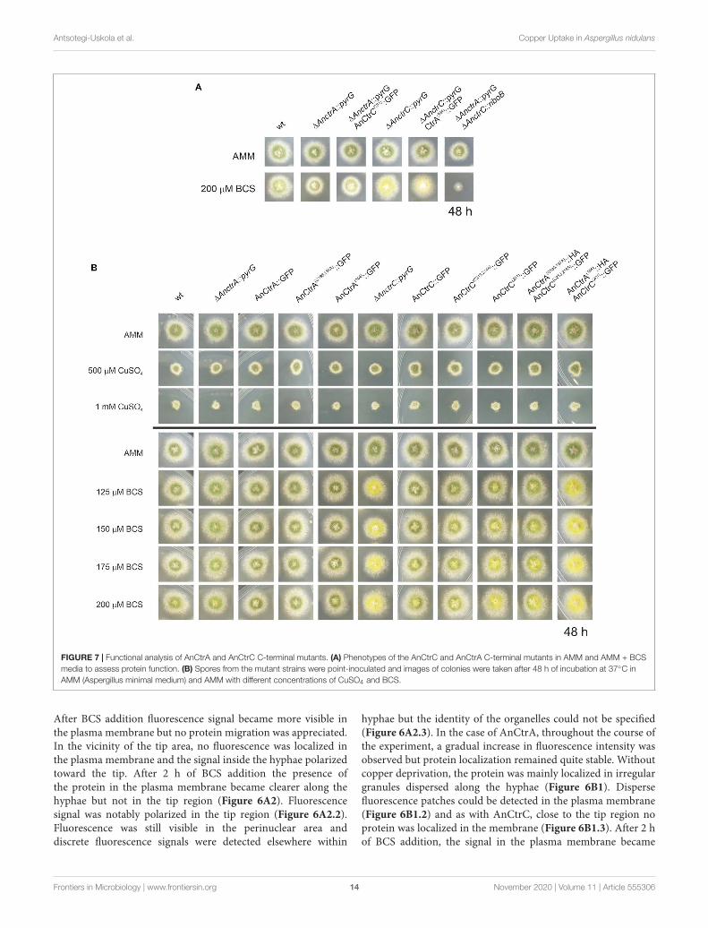
fmicb-11-555306 November 12, 2020 Time: 16:47 # 14
Antsotegi-Uskola et al. Copper Uptake in Aspergillus nidulans
FIGURE 7 | Functional analysis of AnCtrA and AnCtrC C-terminal mutants. (A) Phenotypes of the AnCtrC and AnCtrA C-terminal mutants in AMM and AMM + BCSmedia to assess protein function. (B) Spores from the mutant strains were point-inoculated and images of colonies were taken after 48 h of incubation at 37◦C inAMM (Aspergillus minimal medium) and AMM with different concentrations of CuSO4 and BCS.
After BCS addition fluorescence signal became more visible inthe plasma membrane but no protein migration was appreciated.In the vicinity of the tip area, no fluorescence was localized inthe plasma membrane and the signal inside the hyphae polarizedtoward the tip. After 2 h of BCS addition the presence ofthe protein in the plasma membrane became clearer along thehyphae but not in the tip region (Figure 6A2). Fluorescencesignal was notably polarized in the tip region (Figure 6A2.2).Fluorescence was still visible in the perinuclear area anddiscrete fluorescence signals were detected elsewhere within
hyphae but the identity of the organelles could not be specified(Figure 6A2.3). In the case of AnCtrA, throughout the course ofthe experiment, a gradual increase in fluorescence intensity wasobserved but protein localization remained quite stable. Withoutcopper deprivation, the protein was mainly localized in irregulargranules dispersed along the hyphae (Figure 6B1). Dispersefluorescence patches could be detected in the plasma membrane(Figure 6B1.2) and as with AnCtrC, close to the tip region noprotein was localized in the membrane (Figure 6B1.3). After 2 hof BCS addition, the signal in the plasma membrane became
Frontiers in Microbiology | www.frontiersin.org 14 November 2020 | Volume 11 | Article 555306
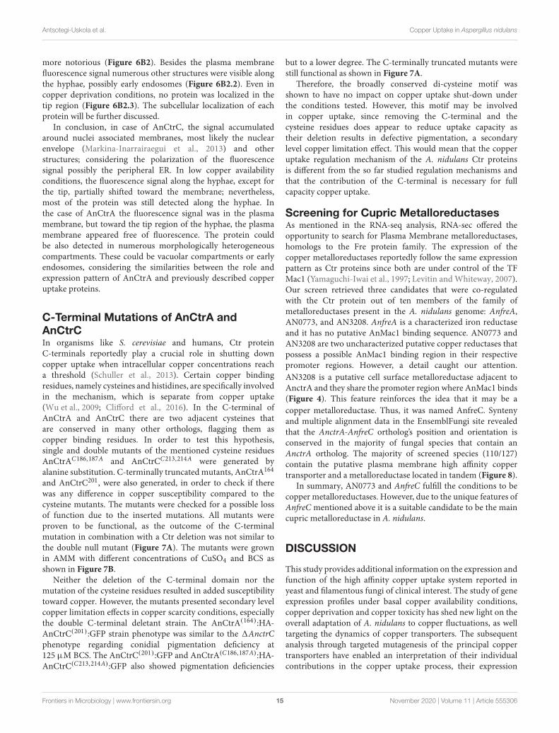
fmicb-11-555306 November 12, 2020 Time: 16:47 # 15
Antsotegi-Uskola et al. Copper Uptake in Aspergillus nidulans
more notorious (Figure 6B2). Besides the plasma membranefluorescence signal numerous other structures were visible alongthe hyphae, possibly early endosomes (Figure 6B2.2). Even incopper deprivation conditions, no protein was localized in thetip region (Figure 6B2.3). The subcellular localization of eachprotein will be further discussed.
In conclusion, in case of AnCtrC, the signal accumulatedaround nuclei associated membranes, most likely the nuclearenvelope (Markina-Inarrairaegui et al., 2013) and otherstructures; considering the polarization of the fluorescencesignal possibly the peripheral ER. In low copper availabilityconditions, the fluorescence signal along the hyphae, except forthe tip, partially shifted toward the membrane; nevertheless,most of the protein was still detected along the hyphae. Inthe case of AnCtrA the fluorescence signal was in the plasmamembrane, but toward the tip region of the hyphae, the plasmamembrane appeared free of fluorescence. The protein couldbe also detected in numerous morphologically heterogeneouscompartments. These could be vacuolar compartments or earlyendosomes, considering the similarities between the role andexpression pattern of AnCtrA and previously described copperuptake proteins.
C-Terminal Mutations of AnCtrA andAnCtrCIn organisms like S. cerevisiae and humans, Ctr proteinC-terminals reportedly play a crucial role in shutting downcopper uptake when intracellular copper concentrations reacha threshold (Schuller et al., 2013). Certain copper bindingresidues, namely cysteines and histidines, are specifically involvedin the mechanism, which is separate from copper uptake(Wu et al., 2009; Clifford et al., 2016). In the C-terminal ofAnCtrA and AnCtrC there are two adjacent cysteines thatare conserved in many other orthologs, flagging them ascopper binding residues. In order to test this hypothesis,single and double mutants of the mentioned cysteine residuesAnCtrAC186,187A and AnCtrCC213,214A were generated byalanine substitution. C-terminally truncated mutants, AnCtrA164
and AnCtrC201, were also generated, in order to check if therewas any difference in copper susceptibility compared to thecysteine mutants. The mutants were checked for a possible lossof function due to the inserted mutations. All mutants wereproven to be functional, as the outcome of the C-terminalmutation in combination with a Ctr deletion was not similar tothe double null mutant (Figure 7A). The mutants were grownin AMM with different concentrations of CuSO4 and BCS asshown in Figure 7B.
Neither the deletion of the C-terminal domain nor themutation of the cysteine residues resulted in added susceptibilitytoward copper. However, the mutants presented secondary levelcopper limitation effects in copper scarcity conditions, especiallythe double C-terminal deletant strain. The AnCtrA(164):HA-AnCtrC(201):GFP strain phenotype was similar to the 1AnctrCphenotype regarding conidial pigmentation deficiency at125 µM BCS. The AnCtrC(201):GFP and AnCtrA(C186,187A):HA-AnCtrC(C213,214A):GFP also showed pigmentation deficiencies
but to a lower degree. The C-terminally truncated mutants werestill functional as shown in Figure 7A.
Therefore, the broadly conserved di-cysteine motif wasshown to have no impact on copper uptake shut-down underthe conditions tested. However, this motif may be involvedin copper uptake, since removing the C-terminal and thecysteine residues does appear to reduce uptake capacity astheir deletion results in defective pigmentation, a secondarylevel copper limitation effect. This would mean that the copperuptake regulation mechanism of the A. nidulans Ctr proteinsis different from the so far studied regulation mechanisms andthat the contribution of the C-terminal is necessary for fullcapacity copper uptake.
Screening for Cupric MetalloreductasesAs mentioned in the RNA-seq analysis, RNA-sec offered theopportunity to search for Plasma Membrane metalloreductases,homologs to the Fre protein family. The expression of thecopper metalloreductases reportedly follow the same expressionpattern as Ctr proteins since both are under control of the TFMac1 (Yamaguchi-Iwai et al., 1997; Levitin and Whiteway, 2007).Our screen retrieved three candidates that were co-regulatedwith the Ctr protein out of ten members of the family ofmetalloreductases present in the A. nidulans genome: AnfreA,AN0773, and AN3208. AnfreA is a characterized iron reductaseand it has no putative AnMac1 binding sequence. AN0773 andAN3208 are two uncharacterized putative copper reductases thatpossess a possible AnMac1 binding region in their respectivepromoter regions. However, a detail caught our attention.AN3208 is a putative cell surface metalloreductase adjacent toAnctrA and they share the promoter region where AnMac1 binds(Figure 4). This feature reinforces the idea that it may be acopper metalloreductase. Thus, it was named AnfreC. Syntenyand multiple alignment data in the EnsemblFungi site revealedthat the AnctrA-AnfreC ortholog’s position and orientation isconserved in the majority of fungal species that contain anAnctrA ortholog. The majority of screened species (110/127)contain the putative plasma membrane high affinity coppertransporter and a metalloreductase located in tandem (Figure 8).
In summary, AN0773 and AnfreC fulfill the conditions to becopper metalloreductases. However, due to the unique features ofAnfreC mentioned above it is a suitable candidate to be the maincupric metalloreductase in A. nidulans.
DISCUSSION
This study provides additional information on the expression andfunction of the high affinity copper uptake system reported inyeast and filamentous fungi of clinical interest. The study of geneexpression profiles under basal copper availability conditions,copper deprivation and copper toxicity has shed new light on theoverall adaptation of A. nidulans to copper fluctuations, as welltargeting the dynamics of copper transporters. The subsequentanalysis through targeted mutagenesis of the principal coppertransporters have enabled an interpretation of their individualcontributions in the copper uptake process, their expression
Frontiers in Microbiology | www.frontiersin.org 15 November 2020 | Volume 11 | Article 555306
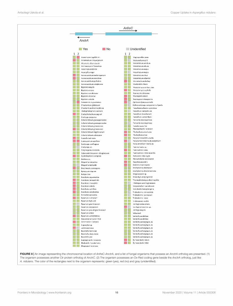
fmicb-11-555306 November 12, 2020 Time: 16:47 # 16
Antsotegi-Uskola et al. Copper Uptake in Aspergillus nidulans
FIGURE 8 | An image representing the chromosomal location of AnfreC-AnctrA, and a list of fungal organisms that possess an AnctrA ortholog are presented. (1)The organism possesses another Ctr protein ortholog of AnctrC. (2) The organism possesses an Ox-Red coding gene beside the AnctrA ortholog, just likeA. nidulans. The color of the rectangles next to the organism represents: green (yes), red (no) and gray (unidentified).
Frontiers in Microbiology | www.frontiersin.org 16 November 2020 | Volume 11 | Article 555306
fmicb-11-555306 November 12, 2020 Time: 16:47 # 17
Antsotegi-Uskola et al. Copper Uptake in Aspergillus nidulans
patterns and their subcellular localization dynamics. In addition,new inquiries into the role of transporter C-terminal domainstogether with the identification of putative membrane copperreductases have been initiated.
The response to copper toxicity in A. nidulans involves thedifferential expression of 13.7% of its genes. Significant changeswere recorded on recognized copper homeostasis genes. Eventhough the expression of the TFs regulating the process wasnot significantly altered, that of proteins responsible of copperuptake and detoxification was very notable. The gene codingfor the copper exporting P-type ATPase AnCrpA (Antsotegi-Uskola et al., 2017), was up-regulated. On the other hand, thetwo characterized copper transporter genes AnctrA and AnctrC(Cai et al., 2019) were down-regulated, in line with the objectiveof reducing the copper uptake to the minimum. Finally, thenotable upregulation of the intracellular copper trafficking P-typeATPase AnygA (Clutterbuck, 1990) might be a response tothe presence of high amounts of copper ions within the cellthat have to be distributed or compartmentalized. Out of theidentified three putative copper chaperones only the putativeCu/Zn SOD chaperone AN6045 was significantly up-regulated.The functional counterpart in S. cerevisiae, chaperone Atx1,responsible for copper distribution within the cell, binds excesscytosolic copper (Lin and Culotta, 1995) and could act as a firstlevel scavenger at low levels of toxicity. This effect may have beenrelevant under our experimental conditions, as discussed below.
Free cytoplasmic copper is known to produce ROS throughFenton-type reactions (Sutton and Winterbourn, 1989;Timoshnikov et al., 2019). Superoxide dismutases, acceptelectrons from reduced metals or superoxides, yielding hydrogenperoxide, which, along with small organic peroxides, is thesubstrate of catalases (2 H2O2→ 2 H2O + O2) (Zamocky et al.,2008). Hence, the reported copper toxicity response involvesthe activation of both groups of enzymes (Wiemann et al.,2017). In our experiments, however, we observed that catalasesAncatB (2.82-fold), AncatC (2.36-fold) and AncpeA/AncatD(2.59-fold) were up-regulated, but the expression of the Cu/Znsuperoxide dismutase AnsodA (Holdom et al., 1996) (reportedin Table 1), an enzyme activated in response to copper andzinc-derived ROS toxicity, remained unchanged, along with thatof the ROS responsive transcription factors AnatfA and AnnapA(Table 2). These variations may stem from the differencesin copper concentrations and the relative sensitivities of thestrains used by Wiemann et al. (2017) and this study. In ourexperiments, 100 µM Cu concentrations were used, a dose thatshowed no visible effect in A. nidulans colony development(Antsotegi-Uskola et al., 2017). On the other hand, Wiemannet al. (2017) used a 200 µM copper concentration, in an organismat which 50 µM already compromised colony development. It istherefore likely that, under conditions which elicit no phenotypicsymptoms of copper toxicity, the AnCrpA detoxification systemcould have removed the free cytosolic copper, rendering ROSneutralization unnecessary. On the other hand, Wiemann et al.(2017) combined higher copper concentrations and greatersusceptibility may have overwhelmed the capacity of AfCrpA,thus triggering the upregulation catalases to quench ROS andH2O2. Both results may thus depict the response to two different
levels of copper toxicity. We hypothesize that catalases are heme-containing enzymes (Scherer et al., 2002) and it is well knownthat intracellular free copper ions can displace protein-boundiron. The selective up-regulation of the catalases in this casemay be a response to this phenomenon, leading to the greaterdemand for functional catalases. More evidence will be requiredto confirm this interpretation.
Aside from catalases, the copper toxicity response alsomodified the expression of many other oxidoreductases. Laccasesare blue–copper enzymes involved in lignin degradation orpigment biosynthesis (Scherer and Fischer, 2001). However,laccases don’t show a specific expression pattern in response tocopper toxicity (Table 1). Other oxidoreductases correspondedto membrane function-related metabolic processes such assterol metabolism. Sterols, especially ergosterol, are the mostabundant lipids in fungal cell membranes (Rodrigues, 2018),with roles in integral protein stabilization, membrane fluidityand permeability (Alvarez et al., 2007; Abe and Hiraki, 2009).Under copper toxicity, the expression of proteins involved intransmembrane traffic augmented; for example, many putativesugar transporters were up-regulated. This could be ascribed toa heightened requirement for energy (ATP) and reducing power(NADH), both principal products of aerobic sugar catabolism.For example, the ATP-dependent copper detoxification proteinAnCrpA was highly up-regulated in this study, in line withearlier observations (Antsotegi-Uskola et al., 2017). Changesin differential expression of various heavy metal transportersmay indicate that a change of copper concentration couldcause an imbalance in heavy metal homeostasis, as described inA. fumigatus (Cai et al., 2017). Finally, a stress mechanism withsuch a wide ranging impact on cellular metabolism should alsoalter the regulation of a core regulatory process as transcription.A TF that experienced notable lower expression under coppertoxicity conditions was AnhapX and this, in turn, is consistentwith the down-regulation of the siderophore biosynthesis clusterthat it has been reported to regulate (Schrettl et al., 2010).The same result was obtained with the antimicrobial compoundemericellamide biosynthesis cluster (Table 2). Finally, analyzingthe data of the RNA-seq we found out a highly up-regulated set ofgenes. The upregulation of an N-acetylglucosamide transporter,AN2562, could be an adaptive response to the high copperenvironment as this compound proves to be important forsurvival in the phagosome (Vesely et al., 2017) where coppertoxicity is used to kill by the innate immune system (Liet al., 2019). Secondly, it is very convenient that a cysteineand methionine transporter, AN2560, would be upregulated insuch conditions as they are two amino-acids that participate incopper binding motifs (Pena et al., 2000). The Flavin-containingmonooxygenase might play an important role in creating anoptimal redox environment threatened by the presence of copperin the cytosol (Suh et al., 2000). These three genes togetherwith the predicted oxidoreductase AN2559 and the putativemethyltransferase AN2561 might comprise a defensive genecluster against copper toxicity.
The phylogenetic characteristics of two A. nidulans Ctrproteins, AnCtrA and AnCtrC, have been examined in thisstudy. Although the reference Ctr sequence used to screen
Frontiers in Microbiology | www.frontiersin.org 17 November 2020 | Volume 11 | Article 555306
fmicb-11-555306 November 12, 2020 Time: 16:47 # 18
Antsotegi-Uskola et al. Copper Uptake in Aspergillus nidulans
for orthologs in fungi had been ScCtr1, none were found inthe A. nidulans genome with that criterion. According to theEnsembl fungi ortholog predictions, homologs of ScCtr1 areonly found in the Saccharomycetales order. Moreover, most Ctrproteins of filamentous fungi shared higher similarity with ScCtr3(Figure 2). Unfortunately, Scctr3 is poorly documented as it isdisrupted by a Ty2 transposon in many laboratory strains (Knightet al., 1996). AnCtrC showed phylogenetic relationship withnumerous Ctr orthologs in A. fumigatus, S. cerevisiae and variousother fungi. On the other hand, AnCtrA showed similarity withother putative Ctr proteins that have not been characterized.
Both proteins possess the characteristic features of Ctrproteins (Zhou et al., 2003; Petris, 2004; Rees and Thiele, 2004).Ctr family members generally harbor three putative TMDs (Puiget al., 2002), but according to topology software predictions,AnCtrA has only two TMDs. This has also been reportedin A. fumigatus and C. neoformans (Waterman et al., 2012;Park et al., 2014). Cysteine residues are present throughout thesequence and cysteines are known for their affinity to copper(Pena et al., 2000). Both proteins contain two cysteine residuesflanking the methionine residue adjacent to TMD1. This softLewis base may be important to coordinate copper since it hasbeen described that metal binding residues located close to theplasma membrane seem to play a more important role in coppertransport (Puig et al., 2002).
Functional characterization analyses in this study showedthat the two Ctr proteins worked independently and therange of copper concentrations they covered were partiallycomplementary, as derived from the obtained phenotypes. Thephenotypes observed were broadly interpreted in terms of acuteor partial deficiencies as follows. Normal growth rates andWT-like green conidial pigmentation were interpreted as anindication of adequate copper supply for basic growth anddevelopmental functions. Defects in spore pigmentation andsporulation that did not affect growth rate were interpreted as theresult of a partial limitation in copper. Conidiogenesis is stronglyreliant on autophagy-based resources (Kikuma et al., 2007; Richieet al., 2007). The yellowish spore pigmentation could be explainedby the inactivity of the copper-dependent conidial laccase AnYA;the phenotype corresponds with the yA2 phenotype (Hermannet al., 1983; Scherer and Fischer, 2001). On the other hand,acute copper deficiency was interpreted to affect extracellularcopper transport with direct consequences on vegetative growth.Following these criteria, we surmised that AnCtrC functions asthe principal copper uptake protein under copper sufficiency andmild copper deficiency conditions. AnCtrA may complement thelack of AnCtrC at moderate copper deficiencies but is specificallyrequired for extreme copper deficiency scenarios.
As shown in this study, conditions of low copper availabilityinduce a gradual up-regulation in Ctr expression, while highcopper levels induce the opposite effect. The rapid shutdownof the copper uptake system under high extracellular copperconcentrations is of great importance to avoid copper toxicity(Bertinato and L’Abbe, 2004; Dong et al., 2013). The expressionpatterns of AnCtrA and AnCtrC under similar testing conditionsshare certain similarities with S. cerevisiae Ctr proteins; aCtr protein with a more stable expression pattern (ScCtr3)
and another one that is uniquely expressed under low copperavailability conditions (ScCtr1) (Pena et al., 2000). AnCtrC is theonly high-affinity copper transporter expressed at basal copperlevels, which means it is likely to cover for copper intake underordinary copper availability (Figure 5B). On the other hand,AnCtrA expression pattern seems to be designed for extremecopper scarcity conditions, when AnCtrC transport cannot meetthe cell’s copper requirements. These results are in line with theconclusions from phenotypic analyses of selective null mutantsdiscussed above.
In terms of protein localization dynamics, Ctr proteins areknown to follow a very dynamic trafficking pathway. Pena et al.(2000) described that ScCtr3 traveled to the plasma membranethrough the Trans-Golgi network and the fluorescence signal oftagged proteins accumulated in the ER after inhibiting proteintraffic from ER to Golgi. However, when copper concentrationrose, Ctr proteins like ScCtr1, moved from the plasma membraneto the vacuole (Liu et al., 2007), or early endosomes, as in thecase of hCtr1 (Eisses and Kaplan, 2002; Clifford et al., 2016). Thepredominant ER localization of AnCtrC (Markina-Inarrairaeguiet al., 2013) and its partial migration to the plasma membranein copper deficiency conditions support its role as a copperuptake protein covering transport under copper sufficiency ormildly limiting conditions. The predominant plasma membraneand vesicular localization of AnCtrA is in line with reporteddata of other proteins that respond to extreme copper deficiencyscenarios such as described for ScCtr1 or hCtr1 (Eisses andKaplan, 2002; Liu et al., 2007; Clifford et al., 2016).
The shutdown of copper transport as a measure to avoidtoxicity has been documented many instances. Previous reportson this toxicity-preventing mechanism refer to certain residues,mostly cysteines, in the C-terminus of Ctr proteins as keyelements in uptake modulation (Liu et al., 2007; Wu et al.,2009; Schuller et al., 2013). Copper has been postulated to bindC-terminal cysteines, purportedly triggering the mechanism.The possible role of the Ctr proteins’ C-terminal in activitymodulation has not been studied in filamentous fungi. However,the C-terminal di-cysteine motif, a common heavy metalbinding sequence (Lekeux et al., 2018) can be found in Ctrproteins of Alternaria alternata, Aspergillus fumigatus, Botrytiscinerea, Fusarium graminearum, Histoplasma capsulatum andMagnaporthe oryzae. We first hypothesized that this di-cysteine motif could act as copper uptake modulator, asthe ScCtr1 C-terminal cysteine residues or the hCtr1 HCHmotif. However, our results do not indicate any copperdependent uptake modulation or internalization role. Moreover,unlike the C-terminus of ScCtr1, that is dispensable forfull copper uptake capacity (Wu et al., 2009), deletion ofAnCtrC C-terminus results in secondary copper deficiencyeffects manifested as a pigmentation deficiency (Figure 2B).Thus, the C-terminus of the Ctr proteins plays some role inthe copper uptake process. Further studies will be needed toclarify this question.
Plasma membrane metalloreductases are crucial componentsof the copper uptake process as they provide Ctr transportingproteins with the substrate in a transport-compatible reducedstate (Labbe et al., 1997; Petris, 2004). There are documented
Frontiers in Microbiology | www.frontiersin.org 18 November 2020 | Volume 11 | Article 555306
fmicb-11-555306 November 12, 2020 Time: 16:47 # 19
Antsotegi-Uskola et al. Copper Uptake in Aspergillus nidulans
reductases that reduce iron and also copper (Georgatsou et al.,1997); however, AnfreA was discarded for lacking a putativeMac1 binding sequence. AN0773 and AnfreC both shared copperreductase features, but the phylogenetically preserved genomiclocation of AnfreC in tandem with AnctrA tipped the scales inits favor. The genes coding for the reductive iron assimilationcomplex composed by the oxidoreductase FetC and the irontransporter FtrA (Schrettl et al., 2004) are located in tandem,exactly like AnfreC-AnctrA, in Fusarium species (Park et al.,2007; Wiemann et al., 2012) and other Aspergillus species likeA. fumigatus, A. clavatus and A. fischeri for example. This factpoints out to AnfreC as the main copper oxidoreductase.
In conclusion, the RNA-seq experiments performed in thisstudy have clearly shown that copper concentration has a notableimpact on many cellular processes, especially the redox-state andtransmembrane transport. The absence of phenotypic impactof the treatments used on colony development suggests thatthe concentration used for the RNA-seq experiment is withinthe range of concentrations covered by the P-type ATPaseAnCrpA. The deep functional characterization of AnCtrA andAnCtrC has revealed that both Ctr proteins function in acomplementary manner but each protein has a role in copperuptake. AnCtrC works in copper uptake under copper sufficiencyconditions and AnCtrA is only activated under extreme copperdeficiency. Both proteins display a copper-dependent expressionbut with different patterns that reinforce the above-mentionedhypothesis. Interestingly, both proteins are upregulated when thecounterpart is missing but they are not able to fully complementeach other. Both proteins are partially located in the plasmamembrane; nevertheless, they are clearly dynamic proteins. TheC-terminus CC motif neither the C-terminus itself are part ofthe copper uptake modulation mechanism in copper toxicityconditions but they do have a role in copper uptake. Finally, theputative plasma membrane metalloreductase AnfreC possessesvery interesting features that make it a good candidate to be themain cupric metalloreductase.
DATA AVAILABILITY STATEMENT
The datasets presented in this study can be found in onlinerepositories. The names of the repository/repositories andaccession number(s) can be found below: https://www.ncbi.nlm.nih.gov/, PRJNA623550.
AUTHOR CONTRIBUTIONS
MA-U designed and conducted the experimental work, analyzedand interpreted the results, wrote the manuscript, and ensuredthe accuracy of the project as whole. AM-I made the conceptand designed the work, analyzed data and contributed to thewriting of the manuscript. UU con-conceived the work, ensuredthe scientific issue was appropriately investigated, ensured theintegrity of the work, revised and approved the final version tobe published. All authors contributed to the article and approvedthe submitted version.
FUNDING
This work was co-supported by grant IT599-13 from the BasqueGovernment and by funds from the University of the BasqueCountry, both to UU.
SUPPLEMENTARY MATERIAL
The Supplementary Material for this article can be foundonline at: https://www.frontiersin.org/articles/10.3389/fmicb.2020.555306/full#supplementary-material
Supplementary Figure 1 | Southern-blot validation of the mutant strainsgenerated in this study. AnHhoA:mRFP strains were validated by microscope.
REFERENCESAbe, F., and Hiraki, T. (2009). Mechanistic role of ergosterol in membrane rigidity
and cycloheximide resistance in Saccharomyces cerevisiae. Biochim. Biophys.Acta 1788, 743–752. doi: 10.1016/j.bbamem.2008.12.002
Alvarez, F. J., Douglas, L. M., and Konopka, J. B. (2007). Sterol-rich plasmamembrane domains in fungi. Eukaryot. Cell 6, 755–763. doi: 10.1128/ec.00008-07
Antsotegi-Uskola, M., Markina-Inarrairaegui, A., and Ugalde, U. (2017). Copperresistance in Aspergillus nidulans relies on the PI-Type ATPase CrpA, regulatedby the transcription factor AceA. Front. Microbiol. 8:912. doi: 10.3389/fmicb.2017.00912
Balamurugan, K., and Schaffner, W. (2006). Copper homeostasis in eukaryotes:teetering on a tightrope. Biochim. Biophys. Acta 1763, 737–746. doi: 10.1016/j.bbamcr.2006.05.001
Bertinato, J., and L’Abbe, M. R. (2004). Maintaining copper homeostasis:regulation of copper-trafficking proteins in response to copper deficiencyor overload. J. Nutr. Biochem. 15, 316–322. doi: 10.1016/j.jnutbio.2004.02.004
Besold, A. N., Culbertson, E. M., and Culotta, V. C. (2016). The Yin and Yang ofcopper during infection. J. Biol. Inorg. Chem. 21, 137–144. doi: 10.1007/s00775-016-1335-1
Cai, Z., Du, W., Liu, L., Pan, D., and Lu, L. (2019). Molecularcharacteristics of the conserved Aspergillus nidulans transcription factormac1 and its functions in response to copper starvation. mSphere4:e00670-18.
Cai, Z., Du, W., Zeng, Q., Long, N., Dai, C., and Lu, L. (2017). Cu-sensingtranscription factor Mac1 coordinates with the Ctr transporter family toregulate Cu acquisition and virulence in Aspergillus fumigatus. Fungal Genet.Biol. 107, 31–43. doi: 10.1016/j.fgb.2017.08.003
Clifford, R. J., Maryon, E. B., and Kaplan, J. H. (2016). Dynamic internalizationand recycling of a metal ion transporter: Cu homeostasis and CTR1, thehuman Cu(+) uptake system. J. Cell Sci. 129, 1711–1721. doi: 10.1242/jcs.173351
Clutterbuck, A. J. (1990). The genetics of conidiophore pigmentation in Aspergillusnidulans. J. Gen. Microbiol. 136, 1731–1738. doi: 10.1099/00221287-136-9-1731
Culotta, V. C., Howard, W. R., and Liu, X. F. (1994). CRS5 encodes ametallothionein-like protein in Saccharomyces cerevisiae. J. Biol. Chem. 269,25295–25302.
Dancis, A., Haile, D., Yuan, D. S., and Klausner, R. D. (1994). The Saccharomycescerevisiae copper transport protein (Ctr1p). Biochemical characterization,regulation by copper, and physiologic role in copper uptake. J. Biol. Chem. 269,25660–25667.
Frontiers in Microbiology | www.frontiersin.org 19 November 2020 | Volume 11 | Article 555306
fmicb-11-555306 November 12, 2020 Time: 16:47 # 20
Antsotegi-Uskola et al. Copper Uptake in Aspergillus nidulans
De Feo, C. J., Aller, S. G., Siluvai, G. S., Blackburn, N. J., and Unger, V. M. (2009).Three-dimensional structure of the human copper transporter hCTR1. Proc.Natl. Acad. Sci. U.S.A 106, 4237–4242. doi: 10.1073/pnas.0810286106
Ding, C., Yin, J., Tovar, E. M., Fitzpatrick, D. A., Higgins, D. G., and Thiele,D. J. (2011). The copper regulon of the human fungal pathogen Cryptococcusneoformans H99. Mol. Microbiol. 81, 1560–1576. doi: 10.1111/j.1365-2958.2011.07794.x
Dong, K., Addinall, S. G., Lydall, D., and Rutherford, J. C. (2013). The yeastcopper response is regulated by DNA damage. Mol. Cell Biol. 33, 4041–4050.doi: 10.1128/mcb.00116-13
Eisses, J. F., and Kaplan, J. H. (2002). Molecular characterization of hCTR1, thehuman copper uptake protein. J. Biol. Chem. 277, 29162–29171. doi: 10.1074/jbc.m203652200
Fridovich, I. (1983). Superoxide radical: an endogenous toxicant. Annu. Rev.Pharmacol. Toxicol. 23, 239–257. doi: 10.1146/annurev.pa.23.040183.001323
Garzia, A., Etxebeste, O., Rodriguez-Romero, J., Fischer, R., Espeso, E. A., andUgalde, U. (2013). Transcriptional changes in the transition from vegetativecells to asexual development in the model fungus Aspergillus nidulans.Eukaryot. Cell 12, 311–321. doi: 10.1128/ec.00274-12
Georgatsou, E., Mavrogiannis, L. A., Fragiadakis, G. S., and Alexandraki, D.(1997). The yeast Fre1p/Fre2p cupric reductases facilitate copper uptake andare regulated by the copper-modulated Mac1p activator. J. Biol. Chem. 272,13786–13792. doi: 10.1074/jbc.272.21.13786
Hermann, T. E., Kurtz, M. B., and Champe, S. P. (1983). Laccase localized inhulle cells and cleistothecial primordia of Aspergillus nidulans. J. Bacteriol. 154,955–964. doi: 10.1128/jb.154.2.955-964.1983
Hervas-Aguilar, A., and Penalva, M. A. (2010). Endocytic machinery protein SlaBis dispensable for polarity establishment but necessary for polarity maintenancein hyphal tip cells of Aspergillus nidulans. Eukaryot. Cell 9, 1504–1518. doi:10.1128/ec.00119-10
Holdom, M. D., Hay, R. J., and Hamilton, A. J. (1996). The Cu, Zn superoxidedismutases of Aspergillus flavus, Aspergillus niger, Aspergillus nidulans,and Aspergillus terreus: purification and biochemical comparison with theAspergillus fumigatus Cu, Zn superoxide dismutase. Infect. Immun. 64, 3326–3332. doi: 10.1128/iai.64.8.3326-3332.1996
Kafer, E. (1965). Origins of translocations in Aspergillus nidulans. Genetics 52,217–232.
Keller, G., Bird, A., and Winge, D. R. (2005). Independent metalloregulation ofAce1 and Mac1 in Saccharomyces cerevisiae. Eukaryot. Cell 4, 1863–1871. doi:10.1128/ec.4.11.1863-1871.2005
Kikuma, T., Arioka, M., and Kitamoto, K. (2007). Autophagy during conidiationand conidial germination in filamentous fungi. Autophagy 3, 128–129. doi:10.4161/auto.3560
Knight, S. A., Labbe, S., Kwon, L. F., Kosman, D. J., and Thiele, D. J. (1996). Awidespread transposable element masks expression of a yeast copper transportgene. Genes Dev. 10, 1917–1929. doi: 10.1101/gad.10.15.1917
Kusuya, Y., Hagiwara, D., Sakai, K., Yaguchi, T., Gonoi, T., and Takahashi, H.(2017). Transcription factor Afmac1 controls copper import machinery inAspergillus fumigatus. Curr. Genet. 63, 777–789. doi: 10.1007/s00294-017-0681-z
Labbe, S., Zhu, Z., and Thiele, D. J. (1997). Copper-specific transcriptionalrepression of yeast genes encoding critical components in the copper transportpathway. J. Biol. Chem. 272, 15951–15958. doi: 10.1074/jbc.272.25.15951
Lee, J., Pena, M. M., Nose, Y., and Thiele, D. J. (2002). Biochemical characterizationof the human copper transporter Ctr1. J. Biol. Chem. 277, 4380–4387. doi:10.1074/jbc.m104728200
Lekeux, G., Laurent, C., Joris, M., Jadou, A., Jiang, D., and Bosman, B. (2018). di-Cysteine motifs in the C-terminus of plant HMA4 proteins confer nanomolaraffinity for zinc and are essential for HMA4 function in vivo. J. Exp. Bot. 69,5547–5560.
Levitin, A., and Whiteway, M. (2007). The effect of prostaglandin E2 ontranscriptional responses of Candida albicans. Microbiol. Res. 162, 201–210.doi: 10.1016/j.micres.2007.02.001
Li, C., Li, Y., and Ding, C. (2019). The role of copper homeostasis at the host-pathogen axis: from bacteria to fungi. Int. J. Mol. Sci. 20:175. doi: 10.3390/ijms20010175
Lin, S. J., and Culotta, V. C. (1995). The ATX1 gene of Saccharomyces cerevisiaeencodes a small metal homeostasis factor that protects cells against reactive
oxygen toxicity. Proc. Natl. Acad. Sci. U.S.A 92, 3784–3788. doi: 10.1073/pnas.92.9.3784
Lin, S. J., Pufahl, R. A., Dancis, A., O’Halloran, T. V., and Culotta, V. C. (1997). Arole for the Saccharomyces cerevisiae ATX1 gene in copper trafficking and irontransport. J. Biol. Chem. 272, 9215–9220.
Liu, L., Qi, J., Yang, Z., Peng, L., and Li, C. (2012). Low-affinity coppertransporter CTR2 is regulated by copper-sensing transcription factor Mac1pin Saccharomyces cerevisiae. Biochem. Biophys. Res. Commun. 420, 600–604.doi: 10.1016/j.bbrc.2012.03.040
Liu, J., Sitaram, A., and Burd, C. G. (2007). Regulation of copper-dependentendocytosis and vacuolar degradation of the yeast copper transporter, Ctr1p,by the Rsp5 ubiquitin ligase. Traffic 8, 1375–1384. doi: 10.1111/j.1600-0854.2007.00616.x
Lutsenko, S. (2010). Human copper homeostasis: a network of interconnectedpathways. Curr. Opin. Chem. Biol. 14, 211–217. doi: 10.1016/j.cbpa.2010.01.003
Macomber, L., and Imlay, J. A. (2009). The iron-sulfur clusters of dehydratases areprimary intracellular targets of copper toxicity. Proc. Natl. Acad. Sci. U.S.A 106,8344–8349. doi: 10.1073/pnas.0812808106
Markina-Inarrairaegui, A., Etxebeste, O., Herrero-Garcia, E., Araujo-Bazan, L.,Fernandez-Martinez, J., Flores, J. A., et al. (2011). Nuclear transporters in amultinucleated organism: functional and localization analyses in Aspergillusnidulans. Mol. Biol. Cell 22, 3874–3886. doi: 10.1091/mbc.e11-03-0262
Markina-Inarrairaegui, A., Pantazopoulou, A., Espeso, E. A., and Penalva, M. A.(2013). The Aspergillus nidulans peripheral ER: disorganization by ER stressand persistence during mitosis. PLoS One 8:e67154. doi: 10.1371/journal.pone.0067154
Nevitt, T., Ohrvik, H., and Thiele, D. J. (2012). Charting the travels of copper ineukaryotes from yeast to mammals. Biochim. Biophys. Acta 1823, 1580–1593.doi: 10.1016/j.bbamcr.2012.02.011
Park, Y. S., Kim, J. H., Cho, J. H., Chang, H. I., Kim, S. W., Paik, H. D., et al.(2007). Physical and functional interaction of FgFtr1-FgFet1 and FgFtr2-FgFet2is required for iron uptake in Fusarium graminearum. Biochem. J. 408, 97–104.doi: 10.1042/bj20070450
Park, Y. S., Lian, H., Chang, M., Kang, C. M., and Yun, C. W. (2014). Identificationof high-affinity copper transporters in Aspergillus fumigatus. Fungal Genet. Biol.73, 29–38. doi: 10.1016/j.fgb.2014.09.008
Pena, M. M., Koch, K. A., and Thiele, D. J. (1998). Dynamic regulation of copperuptake and detoxification genes in Saccharomyces cerevisiae. Mol. Cell Biol. 18,2514–2523. doi: 10.1128/mcb.18.5.2514
Pena, M. M., Puig, S., and Thiele, D. J. (2000). Characterization of theSaccharomyces cerevisiae high affinity copper transporter Ctr3. J. Biol. Chem.275, 33244–33251. doi: 10.1074/jbc.m005392200
Penalva, M. A. (2005). Tracing the endocytic pathway of Aspergillus nidulans withFM4-64. Fungal Genet. Biol. 42, 963–975. doi: 10.1016/j.fgb.2005.09.004
Petris, M. J. (2004). The SLC31 (Ctr) copper transporter family.Pflugers. Arch. 447, 752–755. doi: 10.1007/s00424-003-1092-1
Pontecorvo, G., Roper, J. A., Hemmons, L. M., Macdonald, K. D., and Bufton, A. W.(1953). The genetics of Aspergillus nidulans. Adv. Genet. 5, 141–238.
Puig, S., Lee, J., Lau, M., and Thiele, D. J. (2002). Biochemical and genetic analysesof yeast and human high affinity copper transporters suggest a conservedmechanism for copper uptake. J. Biol. Chem. 277, 26021–26030. doi: 10.1074/jbc.m202547200
Qi, J., Han, A., Yang, Z., and Li, C. (2012). Metal-sensing transcription factorsMac1p and Aft1p coordinately regulate vacuolar copper transporter CTR2 inSaccharomyces cerevisiae. Biochem. Biophys. Res. Commun. 423, 424–428. doi:10.1016/j.bbrc.2012.05.150
Rees, E. M., and Thiele, D. J. (2004). From aging to virulence: forging connectionsthrough the study of copper homeostasis in eukaryotic microorganisms. Curr.Opin. Microbiol. 7, 175–184. doi: 10.1016/j.mib.2004.02.004
Richie, D. L., Fuller, K. K., Fortwendel, J., Miley, M. D., McCarthy, J. W.,Feldmesser, M., et al. (2007). Unexpected link between metal ion deficiencyand autophagy in Aspergillus fumigatus. Eukaryot. Cell 6, 2437–2447. doi:10.1128/ec.00224-07
Rodrigues, M. L. (2018). The multifunctional fungal ergosterol. mBio 9:e01755-18.Rutherford, J. C., and Bird, A. J. (2004). Metal-responsive transcription factors that
regulate iron, zinc, and copper homeostasis in eukaryotic cells. Eukaryot. Cell 3,1–13. doi: 10.1128/ec.3.1.1-13.2004
Frontiers in Microbiology | www.frontiersin.org 20 November 2020 | Volume 11 | Article 555306
fmicb-11-555306 November 12, 2020 Time: 16:47 # 21
Antsotegi-Uskola et al. Copper Uptake in Aspergillus nidulans
Scherer, M., and Fischer, R. (2001). Molecular characterization of a blue-copperlaccase, TILA, of Aspergillus nidulans. FEMS Microbiol. Lett. 199, 207–213.doi: 10.1111/j.1574-6968.2001.tb10676.x
Scherer, M., Wei, H., Liese, R., and Fischer, R. (2002). Aspergillus nidulans catalase-peroxidase gene (cpeA) is transcriptionally induced during sexual developmentthrough the transcription factor StuA. Eukaryot. Cell 1, 725–735. doi: 10.1128/ec.1.5.725-735.2002
Schrettl, M., Beckmann, N., Varga, J., Heinekamp, T., Jacobsen, I. D., Jöchl, C.,et al. (2010). HapX-mediated adaption to iron starvation is crucial for virulenceof Aspergillus fumigatus. PLoS Pathog. 6:e1001124. doi: 10.1371/journal.ppat.1001124
Schrettl, M., Bignell, E., Kragl, C., Joechl, C., Rogers, T., Arst, H. N., et al. (2004).Siderophore biosynthesis but not reductive iron assimilation is essential forAspergillus fumigatus virulence. J. Exp. Med. 200, 1213–1219. doi: 10.1084/jem.20041242
Schuller, A., Auffermann, G., Zoschke, K., Schmidt, U., Ostermann, K., and Rodel,G. (2013). Overexpression of ctr1Delta300, a high-affinity copper transporterwith deletion of the cytosolic C-terminus in Saccharomyces cerevisiae underexcess copper, leads to disruption of transition metal homeostasis andtranscriptional remodelling of cellular processes. Yeast 30, 201–218. doi: 10.1002/yea.2953
Smith, A. D., Logeman, B. L., and Thiele, D. J. (2017). Copper acquisition andutilization in fungi. Annu. Rev. Microbiol. 71, 597–623. doi: 10.1146/annurev-micro-030117-020444
Sugui, J. A., Kim, H. S., Zarember, K. A., Chang, Y. C., Gallin, J. I., Nierman,W. C., et al. (2008). Genes differentially expressed in conidia and hyphae ofAspergillus fumigatus upon exposure to human neutrophils. PLoS One 3:e2655.doi: 10.1371/journal.pone.0002655
Suh, J. K., Poulsen, L. L., Ziegler, D. M., and Robertus, J. D. (2000). Redoxregulation of yeast flavin-containing monooxygenase. Arch. Biochem. Biophys.381, 317–322. doi: 10.1006/abbi.2000.1965
Sutton, H. C., and Winterbourn, C. C. (1989). On the participation of higheroxidation states of iron and copper in Fenton reactions. Free Radic. Biol. Med.6, 53–60. doi: 10.1016/0891-5849(89)90160-3
Timoshnikov, V. A., Kobzeva, T., Selyutina, O. Y., Polyakov, N. E., andKontoghiorghes, G. J. (2019). Effective inhibition of copper-catalyzedproduction of hydroxyl radicals by deferiprone. J. Biol. Inorg. Chem. 24, 331–341. doi: 10.1007/s00775-019-01650-9
Vesely, E. M., Williams, R. B., Konopka, J. B., and Lorenz, M. C. (2017). N-acetylglucosamine metabolism promotes survival of Candida albicans in thephagosome. mSphere 2:e00357-17.
Walton, F. J., Idnurm, A., and Heitman, J. (2005). Novel gene functions required formelanization of the human pathogen Cryptococcus neoformans. Mol. Microbiol.57, 1381–1396. doi: 10.1111/j.1365-2958.2005.04779.x
Waterman, S. R., Park, Y. D., Raja, M., Qiu, J., Hammoud, D. A., O’Halloran, T. V.,et al. (2012). Role of CTR4 in the virulence of Cryptococcus neoformans. mBio3:e00285-12.
Wiemann, P., Albermann, S., Niehaus, E. M., Studt, L., von Bargen, K. W.,Brock, N. L., et al. (2012). The Sfp-type 4’-phosphopantetheinyl transferasePpt1 of Fusarium fujikuroi controls development, secondary metabolism andpathogenicity. PLoS One 7:e37519. doi: 10.1371/journal.pone.0037519
Wiemann, P., Perevitsky, A., Lim, F. Y., Shadkchan, Y., Knox, B. P., LanderoFigueora, J. A., et al. (2017). Aspergillus fumigatus copper export machinery andreactive oxygen intermediate defense counter host copper-mediated oxidativeantimicrobial offense. Cell. Rep. 19, 2174–2176. doi: 10.1016/j.celrep.2017.05.075
Wu, X., Sinani, D., Kim, H., and Lee, J. (2009). Copper transport activityof yeast Ctr1 is down-regulated via its C terminus in response toexcess copper. J. Biol. Chem. 284, 4112–4122. doi: 10.1074/jbc.m807909200
Yamaguchi-Iwai, Y., Serpe, M., Haile, D., Yang, W., Kosman, D. J., Klausner, R. D.,et al. (1997). Homeostatic regulation of copper uptake in yeast via direct bindingof MAC1 protein to upstream regulatory sequences of FRE1 and CTR1. J. Biol.Chem. 272, 17711–17718. doi: 10.1074/jbc.272.28.17711
Yang, L., Ukil, L., Osmani, A., Nahm, F., Davies, J., De Souza, C. P., et al.(2004). Rapid production of gene replacement constructs and generation of agreen fluorescent protein-tagged centromeric marker in Aspergillus nidulans.Eukaryot. Cell 3, 1359–1362. doi: 10.1128/ec.3.5.1359-1362.2004
Zamocky, M., Furtmuller, P. G., and Obinger, C. (2008). Evolution of catalases frombacteria to humans. Antioxid. Redox. Signal. 10, 1527–1548. doi: 10.1089/ars.2008.2046
Zhang, P., Zhang, D., Zhao, X., Wei, D., Wang, Y., and Zhu, X. (2016). Effects ofCTR4 deletion on virulence and stress response in Cryptococcus neoformans.Antonie Van Leeuwenhoek 109, 1081–1090. doi: 10.1007/s10482-016-0709-2
Zhou, H., Cadigan, K. M., and Thiele, D. J. (2003). A copper-regulatedtransporter required for copper acquisition, pigmentation, and specific stagesof development in Drosophila melanogaster. J. Biol. Chem. 278, 48210–48218.doi: 10.1074/jbc.m309820200
Zhu, Z., Labbe, S., Pena, M. M., and Thiele, D. J. (1998). Copperdifferentially regulates the activity and degradation of yeast Mac1transcription factor. J. Biol. Chem. 273, 1277–1280. doi: 10.1074/jbc.273.3.1277
Conflict of Interest: The authors declare that the research was conducted in theabsence of any commercial or financial relationships that could be construed as apotential conflict of interest.
Copyright © 2020 Antsotegi-Uskola, Markina-Iñarrairaegui and Ugalde. This is anopen-access article distributed under the terms of the Creative Commons AttributionLicense (CC BY). The use, distribution or reproduction in other forums is permitted,provided the original author(s) and the copyright owner(s) are credited and that theoriginal publication in this journal is cited, in accordance with accepted academicpractice. No use, distribution or reproduction is permitted which does not complywith these terms.
Frontiers in Microbiology | www.frontiersin.org 21 November 2020 | Volume 11 | Article 555306
ORIGINAL RESEARCHpublished: 30 May 2017
doi: 10.3389/fmicb.2017.00912
Frontiers in Microbiology | www.frontiersin.org 1 May 2017 | Volume 8 | Article 912
Edited by:
Alex Andrianopoulos,
University of Melbourne, Australia
Reviewed by:
Jesus Aguirre,
National Autonomous University of
Mexico, Mexico
Hubertus Haas,
Innsbruck Medical University, Austria
*Correspondence:
Ane Markina-Iñarrairaegui
Specialty section:
This article was submitted to
Fungi and Their Interactions,
a section of the journal
Frontiers in Microbiology
Received: 12 December 2016
Accepted: 04 May 2017
Published: 30 May 2017
Citation:
Antsotegi-Uskola M,
Markina-Iñarrairaegui A and Ugalde U
(2017) Copper Resistance in
Aspergillus nidulans Relies on the
PI-Type ATPase CrpA, Regulated by
the Transcription Factor AceA
Front. Microbiol. 8:912.
doi: 10.3389/fmicb.2017.00912
Copper Resistance in Aspergillusnidulans Relies on the PI-TypeATPase CrpA, Regulated by theTranscription Factor AceAMartzel Antsotegi-Uskola, Ane Markina-Iñarrairaegui * and Unai Ugalde
Microbial Biochemistry Laboratory, Department of Applied Chemistry, Faculty of Chemistry, University of the Basque Country,
San Sebastian, Spain
Copper homeostasis has been extensively studied in mammals, bacteria, and yeast, but
it has not been well-documented in filamentous fungi. In this report, we investigated
the basis of copper tolerance in the model fungus Aspergillus nidulans. Three genes
involved in copper homeostasis have been characterized. First, crpA the A. nidulans
ortholog of Candida albicans CaCRP1 gene encoding a PI-type ATPase was identified.
The phenotype of crpA deletion led to a severe sensitivity to Cu+2 toxicity and a
characteristic morphological growth defect in the presence of high copper concentration.
CrpA displayed some promiscuity regarding metal species response. The expression
pattern of crpA showed an initial strong elevation of mRNA and a low continuous gene
expression in response to long term toxic copper levels. Coinciding with maximum
protein expression level, CrpA was localized close to the cellular surface, however protein
distribution across diverse organelles suggests a complex regulated trafficking process.
Secondly, aceA gene, encoding a transcription factor was identified and deleted, resulting
in an even more extreme copper sensitivity than the ∆crpA mutant. Protein expression
assays corroborated that AceA was necessary for metal inducible expression of CrpA,
but not CrdA, a putative metallothionein the function of which has yet to be elucidated.
Keywords: copper resistance, Aspergillus nidulans, copper homeostasis, PI-type ATPase, transcription factor,
metallothionein
INTRODUCTION
Copper (Cu) is an essential trace element that functions as cofactor of enzymes involved in a widerange of biochemical processes including cellular respiration (cytochrome-c oxidase), free radicaldetoxification (superoxide dismutase), pigmentation (tyrosinase), and collagen maturation (lysyloxidase; Lutsenko, 2010; Nevitt et al., 2012). However, excess accumulation of copper promotes thegeneration of hydroxyl radicals that induce severe cellular damage (Halliwell and Gutteridge, 1990;Valko et al., 2005). Copper toxicity can also be triggered by displacement of other metals present inmolecules leading to protein dysfunction (Macomber and Imlay, 2009; Lemire et al., 2013).
Because environmental copper availability may fluctuate in ecological niches (soil, pollutants,etc.), all biological systems have developed homeostatic mechanisms that sense copper levelsand respond in order to maintain adequate cellular concentrations within the required thresholdlevels. To ensure this balance, cells tightly control copper uptake, cellular traffic, storage, anddetoxification (reviewed in Nevitt et al., 2012).
Antsotegi-Uskola et al. Copper Resistance in Aspergillus nidulans
In the model organism Saccharomyces cerevisiae, elevatedenvironmental copper concentrations (>1 µM) result in acopper-dependent inactivation of the transcription factor (TF)ScMac1p (Graden and Winge, 1997). In consequence, the genescoding proteins implicated in copper uptake, two high affinityCu+ transporters (ScCtr1p and ScCtr3p) and a surface Cu+2-metalloreductase (ScFre1p), are down-regulated (Graden andWinge, 1997; Labbé et al., 1997; Nevitt et al., 2012). Copperhas the opposite effect on the transcriptional factor ScAce1p(Keller et al., 2005; Ehrensberger and Bird, 2011). Formationof a tetra-copper cluster in the regulatory domain of the TFresults in a conformational change that leads to the activationof the inactive Apo-ScAce1p (Furst et al., 1988). As a result,ScAce1p is capable to bind the promoters and induce theexpression of detoxification genes, ScCUP1 and ScCRS5, whichencode two Cu-metallothioneins (MTs; Thiele, 1988; Culottaet al., 1994). Metallothioneins, cysteine (C)-rich low molecularweight polypeptides characterized by high-affinity for diversemetals (Cu, Zn, Cd, Hg, etc.), are found in all eukaryotes andsome prokaryotes (Balamurugan and Schaffner, 2006; Sutherlandand Stillman, 2011). These proteins are induced in responseto exposure to metals, buffering them and lowering theirintracellular concentrations.
Other known intracellular copper level regulators are the P-type ATPases, which are heavy-metal translocators conservedthrough all biological kingdoms. These copper extrusion pumpsare the major copper resistance determinant in bacteria(Odermatt et al., 1993; Solioz and Odermatt, 1995; Ladomerskyand Petris, 2015), while in eukaryotes, in general, they areinvolved in copper compartmentalization into the secretorynetwork for the subsequent metallation of newly synthesized Cu-dependent enzymes. In humans, hATP7A (MNK,Menkes diseaseprotein) and hATP7B (WND, Wilson disease protein) copperATPases carry out both, copper delivering and detoxificationfunctions. At basal Cu conditions hATP7A and hATP7B delivercopper to cuproenzymes in the trans-Golgi compartment (TGN;Lutsenko et al., 2007) but in response to Cu level increase, ATPasepumps traffic from the TGN to the cell surface in order to effluxcopper out of the cytoplasm, thus increasing resistance of cells tothis transition metal (Petris et al., 1996; Suzuki and Gitlin, 1999).In contrast, in Candida albicans, each function is achieved bydifferent PI-type ATPases; CaCcc2p is involved in cuproenzymebiosynthesis (Weissman et al., 2002) while CaCrp1p is committedto copper export (Riggle and Kumamoto, 2000; Weissman et al.,2000). Indeed, CaCrp1p is mainly responsible for the unusualhigh copper tolerance of this dimorphic yeast, in contrast to othereukaryotes were copper detoxification relies significantly onMTs.Recently, it has been shown in A. fumigatus that histidine is alsoinvolved in copper detoxification (Dietl et al., 2016).
The great majority of studies in copper homeostasis havefocused on mammalian, bacteria, and yeast cells, but not infilamentous fungi, a major group of microorganisms whichincludes species of agricultural, food, clinical and environmentalinterest. Aspergillus nidulans is one of the best characterizedfungal species and offers the opportunity to study in detailthe precise mechanisms involved in Cu balance. The presentreport describes the identification and characterization of the
major copper resistance determinant in A. nidulans, the coppertransporting PI-type ATPase, CrpA. We show that CrpAexpression is highly inducible and dynamic in response toprolongued copper exposure and localizes close to the cellularsurface as a result of an organized trafficking process. In addition,we demonstrate that metal-dependent induction of CrpA isunder the control of AceA, a transcription factor activator ofgenes involved in copper detoxification. A third gene encoding ametallothionein-like protein has been identified, CrdA, althoughfurther studies are needed to elucidate its function. Our resultsindicate that copper detoxification in A. nidulans, as in thedimorphic fungus C. albicans, relies mainly in copper excretion.
MATERIALS AND METHODS
BioinformaticAlignments were performed using the predicted proteinsequences released in the National Centre for BiotechnologyInformation (NCBI) database. Multiple sequence alignmentswere performed using BLAST and Clustal Omega application inEBI (http://www.ebi.ac.uk/Tools/msa/clustalomega/).Alignments visualization and domain analysis were performedwith Jalview program. Transmembrane domains were predictedusing Hidden Markov Models (HMM) in the Institute PasteurMobyle server (http://mobyle.pasteur.fr/).
Strains, Media, and Growth ConditionsA. nidulans strains used in this study are listed in Table 1.Strain MAD1427 was used as a recipient for transformationand generation of single/double-null and GFP/HA3-taggedstrains. Appropriately supplemented Käfer’s minimal (MMA)and complete (CMA) pH 6.8 buffered medium (Käfer, 1965)containing 1% (w/v) D-glucose and 71 mM sodium nitrateas main carbon and nitrogen source was used to cultivateA. nidulans. Liquid culture experiments were conducted withMMAwithout agar (liquidMMA). General molecular techniquesfollowed (Sambrook et al., 1989).
Colony growth tests were carried out by inoculatingconidiospores on solid MMA and incubating for 48 h at 37◦C.Phenotypes caused by the deletion of genes were studied underan array of metal stresses induced by addition of CuSO4 (50, 100,150, 200, 400, 600 and 1000 µM), AgNO3 (2.5 and 5 µM) andCd(NO3)2 (100, 150, and 250 µM). Radial extension and colonymorphology of mutant strains was always compared with that ofthe wild-typeMAD1427 strain. Close-up views of colonies grownfor 36, 48, and 72 h at 37◦C were observed and photographedwith a binocular microscope (Nikon SMZ800).
To quantify the effect of CuSO4, AgNO3, and Cd(NO3)2, onbiomass production liquid culture experiments were carried out.1.106 spores were inoculated in liquid MMA (30ml medium in100 ml flask) with or without metal agents and incubated for24 h at 37◦C in a rotary incubator at 200 rpm. The cultureswere collected, dried for 24 h at 100◦C, and their dry weightmeasured. Three technical replicates were performed. Two-tailedStudent’s t-test for unpaired samples was used for the statisticalanalysis to compare the cellular growth between two strains indifferent conditions.
Frontiers in Microbiology | www.frontiersin.org 2 May 2017 | Volume 8 | Article 912
Antsotegi-Uskola et al. Copper Resistance in Aspergillus nidulans
RNA and protein extracts were isolated from myceliumof strains cultivated in a fermenter for 16 h at 37◦C. Cellswere harvested before (0min) and after adding the metalagent to fresh MMA and incubating for the period indicatedin the figures. Mycelia was collected by filtration throughMiracloth (Calbiochem), squeezed to dry and frozen in liquidnitrogen. Samples for protein extraction were lyophilizedfor 16 h.
Generation of Null and Tagged StrainsGenomic cassettes for the generation of strains carrying nullalleles and GFP or HA3 C-terminally tagged fusion proteinswere constructed following fusion-PCR technique described inMarkina-Iñarrairaegui et al. (2011). Null mutants were generatedby using deletion cassettes containing the Aspergillus fumigatuspyrG or riboB as prototrophic selection marker flanked by 1,500bp of 5′ UTR and 3′ UTR regions of the target gene. Firstly,each fragment was amplified using specific oligonucleotide pairs;gsp1–gsp2 (5′ UTR), gsp3–gsp4 (3′ UTR), and gsp2∗–gsp3∗(selectable marker). The gsp2∗, gsp3∗, and gsp6∗ (see below)hold, in addition of the selectable marker sequence, a 24 bptail homologous to the gsp2, gsp3, and gsp6 gene specificprimers. The fragments containing the selectable marker wereamplified using a plasmid as a template. Afterwards, the three
TABLE 1 | Aspergillus nidulans strains used in this study.
Strains Genotype References
MAD1427 pyrG89, pabaA1; argB2; ∆nkuA::argB; veA1,
riboB2
TN02A25 Oakley B.
MAD2731 pabaA1; argB2; ∆nkuA::argB; veA1, riboB2 Markina-Iñarrairaegui
et al., 2011
MAD2733 pabaA1; argB2; ∆nkuA::argB; veA1 Markina-Iñarrairaegui
et al., 2011
BD888 pyrG89, pabaA1; argB2; ∆nkuA::argB;
∆crpA::pyrGAf ; veA1, riboB2
This study
BD892 pyrG89, pabaA1; argB2; ∆nkuA::argB;
crpA::gfp::riboBAf ; veA1, riboB2
This study
BD894 pyrG89, pabaA1; argB2; ∆nkuA::argB;
crpA::ha3::pyrGAf ; veA1, riboB2
This study
BD896 pyrG89, pabaA1; argB2; crdA::ha3::pyrGAf ,
∆nkuA::argB; ∆crpA::riboBAf ; veA1, riboB2
This study
BD898 pyrG89, pabaA1; argB2; ∆crdA::pyrGAf ,
∆nkuA::argB; veA1, riboB2
This study
BD900 pyrG89, pabaA1; argB2; crdA::ha3::pyrGAf ,
∆nkuA::argB; veA1, riboB2
This study
BD961 pyrG89, pabaA1; argB2; ∆nkuA::argB;
crpA::ha3::pyrGAf ; ∆aceA::riboBAf ; veA1,
riboB2
This study
BD963 pyrG89, pabaA1; argB2;∆crdA::pyrGAf ,
∆nkuA::argB; ∆crpA::riboBAf ; veA1, riboB2
This study
BD965 pyrG89, pabaA1; argB2; ∆nkuA::argB;
∆aceA::pyrGAf ; veA1, riboB2
This study
BD1062 pyrG89, pabaA1; argB2; crdA::ha3::pyrGAf ,
∆nkuA::argB; ∆aceA::riboBAf ; veA1, riboB2
This study
BD1073 pyrG89, pabaA1; argB2; ∆nkuA::argB;
crpAAn (∆crpA::pyrGAf ); veA1, riboB2
This study
fragments were fused using gsp1-gsp4 primers. For the C-terminal tagging ha3::pyrGAf and gfp::riboBAf fragments, the3′ end (∼1,500 bp) of the gene and the 3′ UTR regionswere amplified using oligonucleotides gsp6∗–gsp3∗, gsp5–gsp6,and gsp3–gsp4, respectively. Finally, the tagging cassettes wereconstructed using gsp5 and gsp4 oligonucleotides to fuse thefragments. Oligonucleotides used in this study are summarizedin Table 2.
The A. nidulans transformation procedure followed inthis study is an adaptation of the transformable protoplastsproduction protocol described in Szewczyk et al. (2006)and the protoplast transformation protocol described byTilburn et al. (1983). The purified genomic casettes obtainedabove were used to transform A. nidulans. Between 7 and15 transformants were obtained in each regeneration plate(4 plates/transformation). Among the pyrG+ and riboB+
transformants, Southern blot technique was used in order toidentify homokaryotic recombinant strains carrying a single-copy integration event. Of the initially screened 4 transformants,2 verified clones were characterized in this study. The double-null
TABLE 2 | Oligonucleotides used in this study.
Oligo Sequence (5′–3′)
CrpA-gsp1 CGTACATGGGTCTGGTCTTCCCC
CrpA-gsp2 GACGAGTGGCGGCTAGTGTTCC
CrpA-gsp3 TTATTTTTTTCTAGTTCCATGCATGC
CrpA-gsp4 CCCTGAGCAGTCTCGATGAG
CrpA-gsp5 CTTCAGCGGGTCGCAGATACG
CrpA-gsp6 CTCCTGTTGACGCGTAGTCCGG
CrpA-gsp2* GGAACACTAGCCGCCACTCGTCACCGGTCGCCTCAA
ACAATGCTCTTCA
CrpA-gsp3* CATCGCATGCATGGAACTAGAAAAAAATAACTGTCTG
AGAGGAGGCACTGATGC
CrpA-gsp6* CCGGACTACGCGTCAACAGGAGGGAGCTGGTGCA
GGCGCTGGAGCC
CrdA-gsp1 GGCTTCGAGAACTACCAGAACC
CrdA-gsp2 ATTGAATGTTGTTTGAATGGTAG
CrdA-gsp3 TGCGTTTGAATTCATGTTAATGAAGC
CrdA-gsp4 CCAATCCGAGGTCGAGTACG
CrdA-gsp5 ATGGTTCACCCCACCTCAACCTGCT
CrdA-gsp6 AGCCTTGGCCGTCGTAAAATCTGTCTC
CrdA-gsp2* CTACCATTCAAACAACATTCAATACCGGTCGCCTCA
AACAATGCTCT
CrdA-gsp3* GCTTCATTAACATGAATTCAAACGCACTGTCTGAGAGG
AGGCACTGATG
CrdA-gsp6* GAGACAGATTTTACGACGGCCAAGGCTGGAGCTGGTG
CAGGCGCTGGAGC
AceA-gsp1 CCGATGATTCCTTCCACTGCCCAGACATAC
AceA-gsp2 CGCCGCGGTTACTGGGATTGGCACATG
AceA-gsp3 GGACAGCAAGGGCCTTAGAATCTT
AceA-gsp4 ATACAAATAGAGAGGCGAAGGAATGGCG
AceA-gsp2* CATGTGCCAATCCCAGTAACCGCGGCGACCGGTCGCCT
CAAACAATGCTCT
AceA-gsp3* AAGATTCTAAGGCCCTTGCTGTCCCTGTCTGAGAG
GAGGCACTGAT
Frontiers in Microbiology | www.frontiersin.org 3 May 2017 | Volume 8 | Article 912
Antsotegi-Uskola et al. Copper Resistance in Aspergillus nidulans
mutant and strains combining a null allele and HA3-taggedprotein were generated by step-by-step transformation with eachfusion genomic cassette.
Revertant strain construction was achieved by transformingthe null crpA strain (BD888) with a DNA cassette containingthe crpA gene flanked by 1,500 bp up and downstream thecorresponding ORF. gsp1 and gsp4 oligonucleotides were usedto amplify the cassette using a WT strain genomic DNA asa template. Transformants containing the replacement of thenull locus were selected by the ability to resist the 2 mg/ml 5-fluoro-orotic acid (5-FOA; Apollo Scientific, Stockport, UnitedKingdom) that was added in the medium.
RNA Isolation and Northern BlotTotal RNA was isolated using TRIzol reagent following theInvitrogen protocol (Invitrogen, Carlsbad, CA) as described inGarzia et al. (2013) and RNA concentration was calculated usinga Nanodrop 2000 c system (Thermo Fisher Scientific, Waltham,MA). Ten micrograms of total RNA were loaded in 1.2% agarosegels, transferred to positively charged nylon filters, and analyzedby Northern blot which was carried out with a digoxigeninNorthern starter kit (Roche) essentially following manufacturer’sinstructions and using the hybridization solution (1% BSA, 1mMEDTA, 0.5MNaPO4, pH= 7.2, and 7% SDS; Church andGilbert,1984). Transcript of crpA was detected using a genomic probeamplified by PCR using oligonucleotides gsp5 and gsp6 listed inTable 2. Labeling of the probe was performed using DIG HighPrime DNA Labeling Starter Kit (Roche) and covered 43% of theORF. Equal loading of total RNA, which was used as internalcontrol for normalization, was evaluated by ethidium bromidestaining of rRNA. Digoxigenin bound probe was detected usingCSDP (Roche) in a Chemidoc+ XRS (Bio-Rad) system. Intensityof chemiluminescence signal of bands was measured with ImageLab 3.0 software (Bio-Rad).
Protein Isolation and Western BlotProtein extraction from lyophilized samples was performedby two different methods. The alkaline-lysis extraction (AL)procedure was used for CrpA extraction, using Lysis buffer (0.2M NaOH, 0.2% β-mercaptoethanol), as described in Hervás-Aguilar and Peñalva (2010). Protein concentration of extractwas estimated loading 5 µl of each sample on polyacrylamidegels. CrdA was extracted as described in Drubin et al. (1988)using Drubin buffer (5mM HEPES pH 7.5, 1 mM EDTA, 20mM KCl, 0.1% NP-40, 0.5 mM DTT) supplemented with EDTA-free protease-inhibitor cocktail tablets (1 tablet/50 ml of buffer;Roche). Protein quantification was carried out by Bradford assay(Bradford, 1976) using Bradford dye reagent (Alfa Aesar) andfollowing manufacturer’s instructions 12.5µg of total proteinsamples were loaded on gels.
Tagged protein expression was analyzed by Western blotting.Proteins were resolved in 8% (CrpA-HA3) or 12% (CrdA-HA3)SDS-polyacrylamide gels and electrotransfered to Immun-Blot R© PVDF membranes by TransBlot R© TurboTM TransferSystem (Bio-Rad). HA3-tagged proteins were detected usinganti-HA mouse antibody [1/10,000 (v/v) dilution for taggedCrpA and 1/1,000 for tagged CrdA detection; Santa Cruz
Biotechnology] and CrpA-GFP with mouse anti-GFP (1/5,000;Roche). Hexokinase, used as loading control, was detectedwith anti-hexokinase rabbit antibody (1/30,000; ChemiconIntemat Inc.). Peroxidase-conjugated goat anti-mouse IgGimmunoglobulin (1/4,000 for CrpA and 1/2,500 for CrdAdetection; Jackson Immunoresearch Lab) or donkey anti-rabbit(1/10,000; Sigma) cocktails were used as secondary antibodies.Peroxidase activity was detected using ClarityTM Western ECLSubstrate (Bio-Rad). Chemiluminescence was observed using aChemidoc + XRS system (Bio-Rad) and signal intensity wasmeasured with Image Lab 3.0 software (Bio-Rad).
Fluorescence MicroscopyA. nidulans conidiospores were cultured in uncoated glass-bottom dishes (Ibidi GmbH, Germany; 2.5 ml of mediumper well) for 16 h at 25◦C in adequately supplemented pH6.8 Käfer’s minimal medium containing 0.1% D-glucose, 71mM sodium nitrate and 25 mM sodium phosphate monobasic,similar to watch minimal medium (WMM; Peñalva, 2005).After this period, the medium was replaced with fresh mediumsupplemented with 100 µM CuSO4 to induce CrpA-GFPexpression.
Fluorescence images were acquired using a Zeiss AxioObserver Z1 inverted microscope equipped with a 63xPlan Apochromat 1.4 oil immersion Lens, Axiocam MRmRev.3 camera, an Zeiss HXP 120C external light source forepifluorescence excitation and fitted with filter set 38HE forgreen fluorescence (Ex BP 470/40; FT 495; Em BP 525/50) andfilter set 43HE for red fluorescence (Ex BP 545/25; FT 570; EmBP 605/70). Same exposure time and microscope settings wereapplied for all image acquirement. Numerous cells were observedfor each time before taking representative images. Fluorescencelevels were measured using ImageJ software (http://imagej.nih.gov/ij; U.S National Institutes of Health, Bethesda, Maryland,USA).
RESULTS
Identification of Aspergillus nidulansGenes Involved in Copper HomeostasisIn a RNA-seq experiment conducted in our lab (data notpublished) the expression of the AN3117 gene was significantlyincreased as a consequence of copper load. Blastp sequenceanalysis identified the 1,211 amino acid long protein encodedby AN3117 ORF (3,636 bp) as the putative ortholog of C.albicans copper-transporting PI-type ATPase, CaCrp1p (Riggleand Kumamoto, 2000; Weissman et al., 2000). Based on sequenceconservation (35% identity, 55% similarity, and 95% query cover)AN3117 was termed crpA (copper resistance P-type ATPase).The A. nidulans genome contains a second PI-type ATPasecoding gene, ygA, with high homology to CaCcc2p (37% identity;Weissman et al., 2002). YgA is predicted to transport Cu+
to the secretory pathway and its absence results in conidialpigmentation impairment (Clutterbuck, 1990).
As shown in Figure 1A, bioinformatic analyses predictedin CrpA the presence of the distinctive domains describedfor well-studied copper-transporting ATPases of yeast, human,
Frontiers in Microbiology | www.frontiersin.org 4 May 2017 | Volume 8 | Article 912
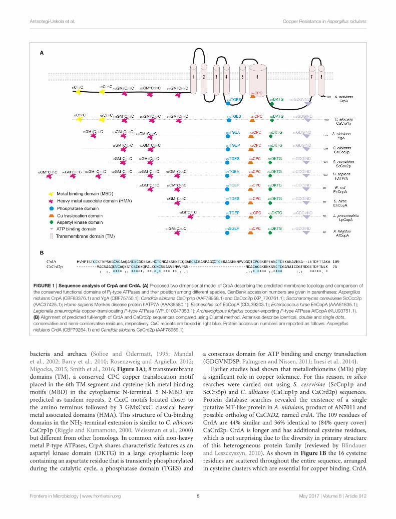
Antsotegi-Uskola et al. Copper Resistance in Aspergillus nidulans
FIGURE 1 | Sequence analysis of CrpA and CrdA. (A) Proposed two dimensional model of CrpA describing the predicted membrane topology and comparison of
the conserved functional domains of PI-type ATPases and their position among different species. GenBank accession numbers are given in parentheses: Aspergillus
nidulans CrpA (CBF83376.1) and YgA (CBF75750.1); Candida albicans CaCrp1p (AAF78958.1) and CaCcc2p (XP_720761.1); Saccharomyces cerevisieae ScCcc2p
(AAC37425.1); Homo sapiens Menkes disease protein hATP7A (AAA35580.1); Escherichia coli EcCopA (CDL39203.1); Enterococcus hirae EhCopA (AAA61835.1);
Legionella pneumophila copper-translocating P-type ATPase (WP_010947353.1); Archaeoglobus fulgidus copper-exporting P-type ATPase AfCopA (KUJ93751.1).
(B) Alignment of predicted full-length of CrdA and CaCrd2p sequences compared using Clustal method. Asterisks describe identical, double and single dots,
conservative and semi-conservative residues, respectively. CxC repeats are boxed in light blue. Protein accession numbers are reported as follows: Aspergillus
nidulans CrdA (CBF79264.1) and Candida albicans CaCrd2p (AAF78959.1).
bacteria and archaea (Solioz and Odermatt, 1995; Mandalet al., 2002; Barry et al., 2010; Rosenzweig and Argüello, 2012;Migocka, 2015; Smith et al., 2016; Figure 1A); 8 transmembranedomains (TM), a conserved CPC copper translocation motifplaced in the 6th TM segment and cysteine rich metal bindingmotifs (MBD) in the cytoplasmic N-terminal. 5 N-MBD arepredicted as tandem repeats, 2 CxxC motifs located closer tothe amino terminus followed by 3 GMxCxxC classical heavymetal associated domains (HMA). This structure of Cu-bindingdomains in the NH2-terminal extension is similar to C. albicansCaCrp1p (Riggle and Kumamoto, 2000; Weissman et al., 2000)but different from other homologs. In common with non-heavymetal P-type ATPases, CrpA shares characteristic features as anaspartyl kinase domain (DKTG) in a large cytoplasmic loopcontaining an aspartate residue that is transiently phosphorylatedduring the catalytic cycle, a phosphatase domain (TGES) and
a consensus domain for ATP binding and energy transduction(GDGVNDSP; Palmgren and Nissen, 2011; Inesi et al., 2014).
Earlier studies had shown that metallothioneins (MTs) playa significant role in copper tolerance. For this reason, in silicosearches were carried out using S. cerevisiae (ScCup1p andScCrs5p) and C. albicans (CaCup1p and CaCrd2p) sequences.Protein database searches revealed the existence of a singleputative MT-like protein in A. nidulans, product of AN7011 andpossible ortholog of CaCRD2, named crdA. The 109 residues ofCrdA are 44% similar and 36% identical to (84% query cover)CaCrd2p. CrdA is longer and has additional cysteine residues,which is not surprising due to the diversity in primary structureof this heterogeneous protein family (reviewed by Blindauerand Leszczyszyn, 2010). As shown in Figure 1B the 16 cysteineresidues are scattered throughout the entire sequence, arrangedin cysteine clusters which are essential for copper binding. CrdA
Frontiers in Microbiology | www.frontiersin.org 5 May 2017 | Volume 8 | Article 912
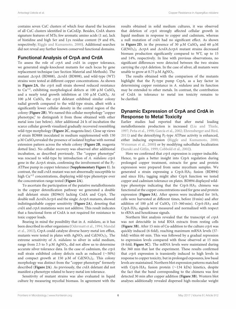
Antsotegi-Uskola et al. Copper Resistance in Aspergillus nidulans
contains seven CxC clusters of which four shared the locationof all CxC clusters identified in CaCrd2p. Besides, CrdA sharessignature features of MTs; few aromatic amino acids (1 aa), lackof histidine and high Ser and Lys residue content (9 and 6%,respectively; Riggle and Kumamoto, 2000). Additional searchesdid not reveal any further known conserved functional domains.
Functional Analysis of CrpA and CrdATo assess the role of crpA and crdA in copper tolerance,we generated single-knockout mutants for each locus by genereplacement technique (see Section Material and Methods). Themutant ∆crpA (BD888), ∆crdA (BD898), and wild-type (WT)strains were tested at different copper concentrations. As shownin Figure 2A, the crpA null strain showed reduced resistanceto Cu+2, exhibiting morphological defects at 100 µM CuSO4
and a nearly total growth inhibition at 150 µM CuSO4. At100 µM CuSO4 the crpA deletant exhibited similar colonyradial growth compared to the wild-type strain, albeit with asignificantly lower cellular density in the central region of thecolony (Figure 2B). We named this cellular morphology “copperphenotype,” to distinguish it from those obtained with othermetal ions (see below). After additional 24 h of incubation thescarce cellular growth visualized gradually recovered resemblingwild-type morphology (Figure 2C, magenta line). Close up viewsof strain BD888 inoculated in medium supplemented with 150µMCuSO4revealed the presence of isolated hyphae with normalextension pattern across the whole colony (Figure 2B, magentadotted line). No cellular recovery was observed after additionalincubation, as described previously. The “copper phenotype”was rescued to wild-type by introduction of A. nidulans crpAgene in the ∆crpA strain, confirming the involvement of the PI-ATPase pump in copper tolerance (Supplementary Figure 1). Incontrast, the null crdAmutant was not abnormally susceptible tohigh Cu+2 concentrations, displaying wild-type phenotype overthe concentration range tested (Figure 2A).
To ascertain the participation of the putative metallothioneinin the copper detoxification pathway we generated a doublenull deletant strain (BD963) lacking CrdA and CrpA. Thedouble null ∆crdA∆crpA and the single ∆crpAmutants, showedindistinguishable copper sensitivity (Figure 2A), denoting thatcrdA and crpAmutations were not additive. This result indicatesthat a functional form of CrdA is not required for resistance totoxic copper loads.
Bearing in mind the possibility that in A. nidulans, as it hasbeen described in other organisms (Odermatt et al., 1994;Mandalet al., 2002), CrpA could catalyze diverse heavy-metal ion efflux,mutants were tested in plates with AgNO3 and Cd(NO3)2. Theextreme sensitivity of A. nidulans to silver in solid medium,range from 2.5 to 5 µM AgNO3, did not allow us to determineaccurate silver tolerance data. In the case of cadmium, the crpAnull strain exhibited colony defects such as reduced (∼50%)and compact growth at 150 µM of Cd(NO3)2. This colonymorphology was distinct from the “copper phenotype” formerlydescribed (Figure 2A). As previously, the crdA deletant did notmanifest a phenotype related to heavy metal ion tolerance.
Sensitivity of mutant strains was also evaluated in liquidculture by measuring mycelial biomass. In agreement with the
results obtained in solid medium cultures, it was observedthat deletion of crpA strongly affected cellular growth inliquid medium in response to copper and cadmium, whereasa null mutation of crdA had no significant effect. As shownin Figure 2D, in the presence of 30 µM CuSO4 and 60 µMCd(NO3)2 ∆crpA and ∆crdA∆crpA mutant strains decreasedbiomass production significantly compared to WT, up to 15and 14%, respectively. In line with previous observations, nosignificant differences were detected between the two strainscarrying the crpA deletion. In the case of silver, all mutants wereunable to grow at 0.75 µMAgNO3.
The results obtained with the comparison of the mutantshighlight that the PI-type pump CrpA, as a key factor indetermining copper resistance in A. nidulans and its functionmay be extended to other metals. In contrast, the contributionof CrdA in tolerance to metal ion toxicity remains tobe clarified.
Dynamic Expression of CrpA and CrdA inResponse to Metal ToxicityEarlier studies had reported that after metal loadingmetallothionein production is increased (Liu and Thiele,1997; Peña et al., 1998; García et al., 2002; Ehrensberger and Bird,2011) and the detoxifying PI-type ATPase activity is enhanced,either inducing expression (Riggle and Kumamoto, 2000;Weissman et al., 2000) or by modifying subcellular localization(Suzuki and Gitlin, 1999; Cobbold et al., 2002).
Here we confirmed that crpA expression is copper-inducible.Hence, to gain a better insight into CrpA regulation duringprolonged copper treatment, extracts for gene and proteinexpression were prepared from same mycelium samples. Wegenerated a strain expressing a CrpA-HA3 fusion (BD894)and since HA3 tagging might alter CrpA function we testedcopper and cadmium tolerance on plates. BD894 displayed wild-type phenotype indicating that the CrpA-HA3 chimera wasfunctional at the copper concentrations used for gene and proteinexpression (Figure 3A). After spores were incubated for 16 h,cells were harvested at different times, before (0min) and afteraddition of 100 µM of CuSO4 (15–360min). CrpA-HA3 andCrpA-HA3 signals were measured and normalized with respectto rRNA and hexokinase signals.
Northern blot analysis revealed that the transcript of crpAwas not detectable in total RNA extracts from resting cells(Figure 3B). After 15 min of Cu-addition to the culture crpA wasquickly induced (6-fold), reaching maximum mRNA levels (57-fold) within 60 min. This was followed by a gradual repressionto expression levels compared with those observed at 15 min(8-fold; Figure 3C). The mRNA levels were maintained duringthe 360 min that last the experiment. These results confirmedthat crpA expression is transiently induced to high levels inresponse to copper toxicity, but in prolonged exposures, low basallevels aremaintained. Northern blot expression patternsmatchedwith CrpA-HA3 fusion protein (∼134 kDa) kinetics, despitethe fact that the band corresponding to the chimera was firstdetected 30 min after copper addition (Figure 3B). Western blotanalyses additionally revealed dispersed high-molecular weight
Frontiers in Microbiology | www.frontiersin.org 6 May 2017 | Volume 8 | Article 912
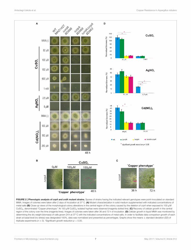
Antsotegi-Uskola et al. Copper Resistance in Aspergillus nidulans
FIGURE 2 | Phenotypic analysis of crpA and crdA mutant strains. Spores of strains having the indicated relevant genotypes were point-inoculated on standard
MMA. Images of colonies were taken after 2 days of incubation at 37◦C. (A) Mutant characterization in solid medium supplemented with indicated concentrations of
metal salts (B) Close-up views of the morphological colony alterations in the central region of the colony caused by the deletion of crpA when exposed to 100 µM
CuSO4, denominated “Copper phenotype.” At 150 µM CuSO4 isolated hyphae were observed (magenta dotted line). (C) Recovery of cellular growth in the central
region of the colony over the time (magenta lines). Images of colonies were taken after 36 and 72 h of incubation. (D) Cellular growth in liquid MMA was monitored by
determining the dry weight (biomass) of cells grown 24 h at 37◦C with the indicated concentrations of metal salts. In order to facilitate data comparison growth of each
strain at basal level (no stress) was designated 100%, data was normalized and presented as percentages. Graphs show the means ± standard deviation (SD) of
triplicate experiments (n = 3). *Significant growth reduction p < 0.05.
Frontiers in Microbiology | www.frontiersin.org 7 May 2017 | Volume 8 | Article 912
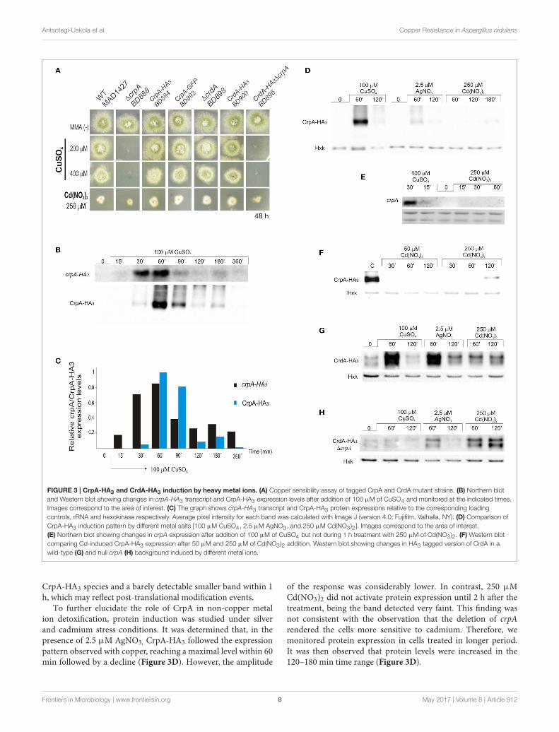
Antsotegi-Uskola et al. Copper Resistance in Aspergillus nidulans
FIGURE 3 | CrpA-HA3 and CrdA-HA3 induction by heavy metal ions. (A) Copper sensibility assay of tagged CrpA and CrdA mutant strains. (B) Northern blot
and Western blot showing changes in crpA-HA3 transcript and CrpA-HA3 expression levels after addition of 100 µM of CuSO4 and monitored at the indicated times.
Images correspond to the area of interest. (C) The graph shows crpA-HA3 transcript and CrpA-HA3 protein expressions relative to the corresponding loading
controls, rRNA and hexokinase respectively. Average pixel intensity for each band was calculated with Image J (version 4.0; Fujifilm, Valhalla, NY). (D) Comparison of
CrpA-HA3 induction pattern by different metal salts [100 µM CuSO4, 2.5 µM AgNO3, and 250 µM Cd(NO3)2]. Images correspond to the area of interest.
(E) Northern blot showing changes in crpA expression after addition of 100 µM of CuSO4 but not during 1 h treatment with 250 µM of Cd(NO3)2. (F) Western blot
comparing Cd-induced CrpA-HA3 expression after 50 µM and 250 µM of Cd(NO3)2 addition. Western blot showing changes in HA3 tagged version of CrdA in a
wild-type (G) and null crpA (H) background induced by different metal ions.
CrpA-HA3 species and a barely detectable smaller band within 1h, which may reflect post-translational modification events.
To further elucidate the role of CrpA in non-copper metalion detoxification, protein induction was studied under silverand cadmium stress conditions. It was determined that, in thepresence of 2.5 µM AgNO3, CrpA-HA3 followed the expressionpattern observed with copper, reaching a maximal level within 60min followed by a decline (Figure 3D). However, the amplitude
of the response was considerably lower. In contrast, 250 µMCd(NO3)2 did not activate protein expression until 2 h after thetreatment, being the band detected very faint. This finding wasnot consistent with the observation that the deletion of crpArendered the cells more sensitive to cadmium. Therefore, wemonitored protein expression in cells treated in longer period.It was then observed that protein levels were increased in the120–180 min time range (Figure 3D).
Frontiers in Microbiology | www.frontiersin.org 8 May 2017 | Volume 8 | Article 912
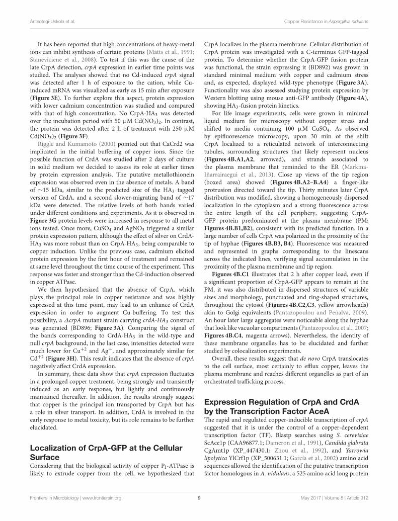
Antsotegi-Uskola et al. Copper Resistance in Aspergillus nidulans
It has been reported that high concentrations of heavy-metalions can inhibit synthesis of certain proteins (Matts et al., 1991;Staneviciene et al., 2008). To test if this was the cause of thelate CrpA detection, crpA expression in earlier time points wasstudied. The analyses showed that no Cd-induced crpA signalwas detected after 1 h of exposure to the cation, while Cu-induced mRNA was visualized as early as 15 min after exposure(Figure 3E). To further explore this aspect, protein expressionwith lower cadmium concentration was studied and comparedwith that of high concentration. No CrpA-HA3 was detectedover the incubation period with 50 µM Cd(NO3)2. In contrast,the protein was detected after 2 h of treatment with 250 µMCd(NO3)2 (Figure 3F).
Riggle and Kumamoto (2000) pointed out that CaCrd2 wasimplicated in the initial buffering of copper ions. Since thepossible function of CrdA was studied after 2 days of culturein solid medium we decided to assess its role at earlier timesby protein expression analysis. The putative metallothioneinexpression was observed even in the absence of metals. A bandof ∼15 kDa, similar to the predicted size of the HA3 taggedversion of CrdA, and a second slower-migrating band of ∼17kDa were detected. The relative levels of both bands variedunder different conditions and experiments. As it is observed inFigure 3G protein levels were increased in response to all metalions tested. Once more, CuSO4 and AgNO3 triggered a similarprotein expression pattern, although the effect of silver on CrdA-HA3 was more robust than on CrpA-HA3, being comparable tocopper induction. Unlike the previous case, cadmium elicitedprotein expression by the first hour of treatment and remainedat same level throughout the time course of the experiment. Thisresponse was faster and stronger than the Cd-induction observedin copper ATPase.
We then hypothesized that the absence of CrpA, whichplays the principal role in copper resistance and was highlyexpressed at this time point, may lead to an enhance of CrdAexpression in order to augment Cu-buffering. To test thispossibility, a ∆crpA mutant strain carrying crdA-HA3 constructwas generated (BD896; Figure 3A). Comparing the signal ofthe bands corresponding to CrdA-HA3 in the wild-type andnull crpA background, in the last case, intensities detected weremuch lower for Cu+2 and Ag+, and approximately similar forCd+2 (Figure 3H). This result indicates that the absence of crpAnegatively affect CrdA expression.
In summary, these data show that crpA expression fluctuatesin a prolonged copper treatment, being strongly and transientlyinduced as an early response, but lightly and continuouslymaintained thereafter. In addition, the results strongly suggestthat copper is the principal ion transported by CrpA but hasa role in silver transport. In addition, CrdA is involved in theearly response to metal toxicity, but its role remains to be furtherelucidated.
Localization of CrpA-GFP at the CellularSurfaceConsidering that the biological activity of copper PI-ATPase islikely to extrude copper from the cell, we hypothesized that
CrpA localizes in the plasma membrane. Cellular distribution ofCrpA protein was investigated with a C-terminus GFP-taggedprotein. To determine whether the CrpA-GFP fusion proteinwas functional, the strain expressing it (BD892) was grown instandard minimal medium with copper and cadmium stressand, as expected, displayed wild-type phenotype (Figure 3A).Functionality was also assessed studying protein expression byWestern blotting using mouse anti-GFP antibody (Figure 4A),showing HA3-fusion protein kinetics.
For life image experiments, cells were grown in minimalliquid medium for microscopy without copper stress andshifted to media containing 100 µM CuSO4. As observedby epifluorescence microscopy, upon 30 min of the shiftCrpA localized to a reticulated network of interconnectingtubules, surrounding structures that likely represent nucleus(Figures 4B.A1,A2, arrowed), and strands associated tothe plasma membrane that reminded to the ER (Markina-Iñarrairaegui et al., 2013). Close up views of the tip region(boxed area) showed (Figures 4B.A2–B.A4) a finger-likeprotrusion directed toward the tip. Thirty minutes later CrpAdistribution was modified, showing a homogeneously dispersedlocalization in the cytoplasm and a strong fluorescence acrossthe entire length of the cell periphery, suggesting CrpA-GFP protein predominated at the plasma membrane (PM;Figures 4B.B1,B2), consistent with its predicted function. In alarge number of cells CrpA was polarized in the proximity of thetip of hyphae (Figures 4B.B3, B4). Fluorescence was measuredand represented in graphs corresponding to the linescansacross the indicated lines, verifying signal accumulation in theproximity of the plasma membrane and tip region.
Figures 4B.C1 illustrates that 2 h after copper load, even ifa significant proportion of CrpA-GFP appears to remain at thePM, it was also distributed in dispersed structures of variablesizes and morphology, punctuated and ring-shaped structures,throughout the cytosol (Figures 4B.C2,C3, yellow arrowheads)akin to Golgi equivalents (Pantazopoulou and Peñalva, 2009).An hour later large aggregates were noticeable along the hyphaethat look like vacuolar compartments (Pantazopoulou et al., 2007;Figures 4B.C4, magenta arrows). Nevertheless, the identity ofthese membrane organelles has to be elucidated and furtherstudied by colocalization experiments.
Overall, these results suggest that de novo CrpA translocatesto the cell surface, most certainly to efflux copper, leaves theplasma membrane and reaches different organelles as part of anorchestrated trafficking process.
Expression Regulation of CrpA and CrdAby the Transcription Factor AceAThe rapid and regulated copper-inducible transcription of crpAsuggested that it is under the control of a copper-dependenttranscription factor (TF). Blastp searches using S. cerevisiaeScAce1p (CAA96877.1; Dameron et al., 1991), Candida glabrataCgAmt1p (XP_447430.1; Zhou et al., 1992), and Yarrowialipolytica YlCrf1p (XP_500631.1; García et al., 2002) amino acidsequences allowed the identification of the putative transcriptionfactor homologous in A. nidulans, a 525 amino acid long protein
Frontiers in Microbiology | www.frontiersin.org 9 May 2017 | Volume 8 | Article 912
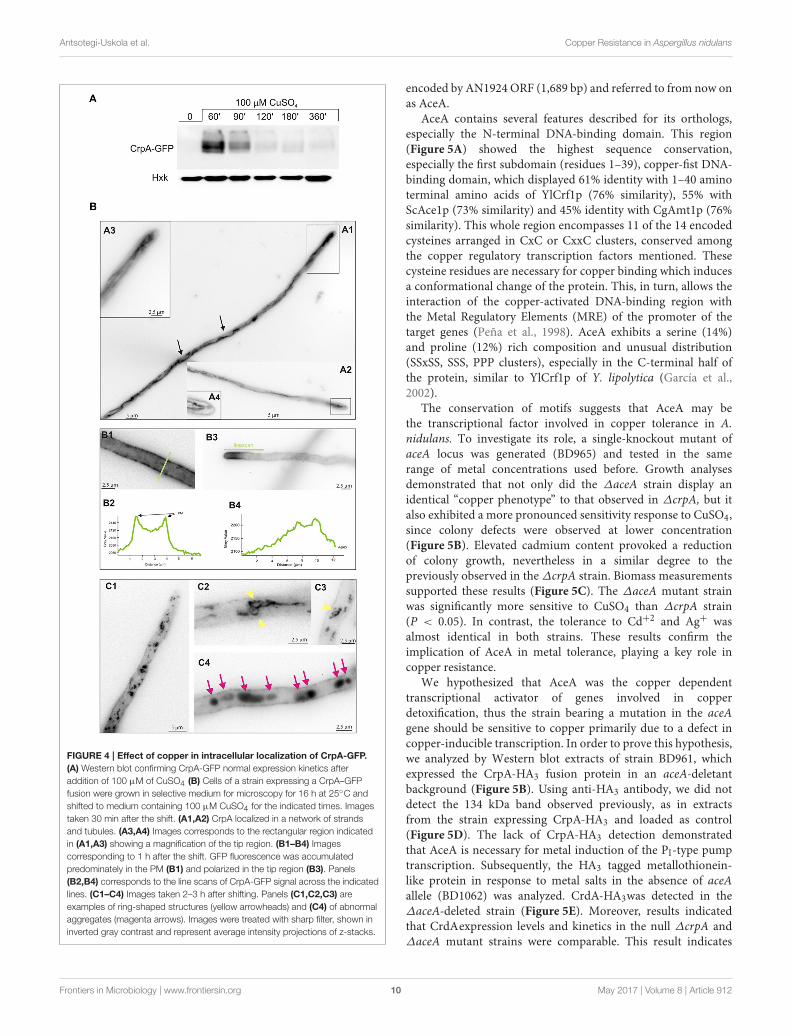
Antsotegi-Uskola et al. Copper Resistance in Aspergillus nidulans
FIGURE 4 | Effect of copper in intracellular localization of CrpA-GFP.
(A) Western blot confirming CrpA-GFP normal expression kinetics after
addition of 100 µM of CuSO4 (B) Cells of a strain expressing a CrpA–GFP
fusion were grown in selective medium for microscopy for 16 h at 25◦C and
shifted to medium containing 100 µM CuSO4 for the indicated times. Images
taken 30 min after the shift. (A1,A2) CrpA localized in a network of strands
and tubules. (A3,A4) Images corresponds to the rectangular region indicated
in (A1,A3) showing a magnification of the tip region. (B1–B4) Images
corresponding to 1 h after the shift. GFP fluorescence was accumulated
predominately in the PM (B1) and polarized in the tip region (B3). Panels
(B2,B4) corresponds to the line scans of CrpA-GFP signal across the indicated
lines. (C1–C4) Images taken 2–3 h after shifting. Panels (C1,C2,C3) are
examples of ring-shaped structures (yellow arrowheads) and (C4) of abnormal
aggregates (magenta arrows). Images were treated with sharp filter, shown in
inverted gray contrast and represent average intensity projections of z-stacks.
encoded by AN1924 ORF (1,689 bp) and referred to from now onas AceA.
AceA contains several features described for its orthologs,especially the N-terminal DNA-binding domain. This region(Figure 5A) showed the highest sequence conservation,especially the first subdomain (residues 1–39), copper-fist DNA-binding domain, which displayed 61% identity with 1–40 aminoterminal amino acids of YlCrf1p (76% similarity), 55% withScAce1p (73% similarity) and 45% identity with CgAmt1p (76%similarity). This whole region encompasses 11 of the 14 encodedcysteines arranged in CxC or CxxC clusters, conserved amongthe copper regulatory transcription factors mentioned. Thesecysteine residues are necessary for copper binding which inducesa conformational change of the protein. This, in turn, allows theinteraction of the copper-activated DNA-binding region withthe Metal Regulatory Elements (MRE) of the promoter of thetarget genes (Peña et al., 1998). AceA exhibits a serine (14%)and proline (12%) rich composition and unusual distribution(SSxSS, SSS, PPP clusters), especially in the C-terminal half ofthe protein, similar to YlCrf1p of Y. lipolytica (García et al.,2002).
The conservation of motifs suggests that AceA may bethe transcriptional factor involved in copper tolerance in A.nidulans. To investigate its role, a single-knockout mutant ofaceA locus was generated (BD965) and tested in the samerange of metal concentrations used before. Growth analysesdemonstrated that not only did the ∆aceA strain display anidentical “copper phenotype” to that observed in ∆crpA, but italso exhibited a more pronounced sensitivity response to CuSO4,since colony defects were observed at lower concentration(Figure 5B). Elevated cadmium content provoked a reductionof colony growth, nevertheless in a similar degree to thepreviously observed in the ∆crpA strain. Biomass measurementssupported these results (Figure 5C). The ∆aceA mutant strainwas significantly more sensitive to CuSO4 than ∆crpA strain(P < 0.05). In contrast, the tolerance to Cd+2 and Ag+ wasalmost identical in both strains. These results confirm theimplication of AceA in metal tolerance, playing a key role incopper resistance.
We hypothesized that AceA was the copper dependenttranscriptional activator of genes involved in copperdetoxification, thus the strain bearing a mutation in the aceAgene should be sensitive to copper primarily due to a defect incopper-inducible transcription. In order to prove this hypothesis,we analyzed by Western blot extracts of strain BD961, whichexpressed the CrpA-HA3 fusion protein in an aceA-deletantbackground (Figure 5B). Using anti-HA3 antibody, we did notdetect the 134 kDa band observed previously, as in extractsfrom the strain expressing CrpA-HA3 and loaded as control(Figure 5D). The lack of CrpA-HA3 detection demonstratedthat AceA is necessary for metal induction of the PI-type pumptranscription. Subsequently, the HA3 tagged metallothionein-like protein in response to metal salts in the absence of aceAallele (BD1062) was analyzed. CrdA-HA3was detected in the∆aceA-deleted strain (Figure 5E). Moreover, results indicatedthat CrdAexpression levels and kinetics in the null ∆crpA and∆aceA mutant strains were comparable. This result indicates
Frontiers in Microbiology | www.frontiersin.org 10 May 2017 | Volume 8 | Article 912
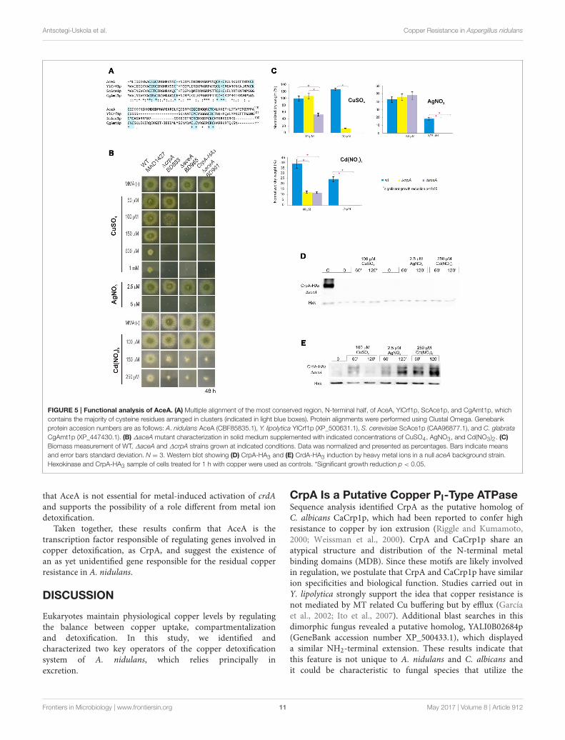
Antsotegi-Uskola et al. Copper Resistance in Aspergillus nidulans
FIGURE 5 | Functional analysis of AceA. (A) Multiple alignment of the most conserved region, N-terminal half, of AceA, YlCrf1p, ScAce1p, and CgAmt1p, which
contains the majority of cysteine residues arranged in clusters (indicated in light blue boxes). Protein alignments were performed using Clustal Omega. Genebank
protein accesion numbers are as follows: A. nidulans AceA (CBF85835.1), Y. lipolytica YlCrf1p (XP_500631.1), S. cerevisiae ScAce1p (CAA96877.1), and C. glabrata
CgAmt1p (XP_447430.1). (B) ∆aceA mutant characterization in solid medium supplemented with indicated concentrations of CuSO4, AgNO3, and Cd(NO3)2. (C)
Biomass measurement of WT, ∆aceA and ∆crpA strains grown at indicated conditions. Data was normalized and presented as percentages. Bars indicate means
and error bars standard deviation. N = 3. Western blot showing (D) CrpA-HA3 and (E) CrdA-HA3 induction by heavy metal ions in a null aceA background strain.
Hexokinase and CrpA-HA3 sample of cells treated for 1 h with copper were used as controls. *Significant growth reduction p < 0.05.
that AceA is not essential for metal-induced activation of crdAand supports the possibility of a role different from metal iondetoxification.
Taken together, these results confirm that AceA is thetranscription factor responsible of regulating genes involved incopper detoxification, as CrpA, and suggest the existence ofan as yet unidentified gene responsible for the residual copperresistance in A. nidulans.
DISCUSSION
Eukaryotes maintain physiological copper levels by regulatingthe balance between copper uptake, compartmentalizationand detoxification. In this study, we identified andcharacterized two key operators of the copper detoxificationsystem of A. nidulans, which relies principally inexcretion.
CrpA Is a Putative Copper PI-Type ATPaseSequence analysis identified CrpA as the putative homolog ofC. albicans CaCrp1p, which had been reported to confer highresistance to copper by ion extrusion (Riggle and Kumamoto,2000; Weissman et al., 2000). CrpA and CaCrp1p share anatypical structure and distribution of the N-terminal metalbinding domains (MDB). Since these motifs are likely involvedin regulation, we postulate that CrpA and CaCrp1p have similarion specificities and biological function. Studies carried out inY. lipolytica strongly support the idea that copper resistance isnot mediated by MT related Cu buffering but by efflux (Garcíaet al., 2002; Ito et al., 2007). Additional blast searches in thisdimorphic fungus revealed a putative homolog, YALI0B02684p(GeneBank accession number XP_500433.1), which displayeda similar NH2-terminal extension. These results indicate thatthis feature is not unique to A. nidulans and C. albicans andit could be characteristic to fungal species that utilize the
Frontiers in Microbiology | www.frontiersin.org 11 May 2017 | Volume 8 | Article 912
Antsotegi-Uskola et al. Copper Resistance in Aspergillus nidulans
described ATPase-based extrusion system as the primary way toovercome high copper loads. It is also important to underscorethe crucial role of copper resistance for virulence of clinicallyimportant fungal pathogens as C. albicans (Mackie et al., 2016),Cryptococcus neoformans (Samanovic et al., 2012; Ding et al.,2013) and A. fumigatus (Dietl et al., 2016; Wiemann et al.,2017).
Copper Detoxification Relies on thePI-Type ATpase CrpAEvidence of the direct participation of CrpA in copper resistancewas obtained by the extreme sensitivity exhibited by CrpA nullmutants. An atypical aspect of the phenotype of the null crpAmutation was the poor growth in the central region of the colonythat overcame toxicity with time. This adaptive behavior couldbe partially explained by the growth pattern described by Valixet al. (2001). As the colony matures, copper may bind to newcellular material locally, resulting in reduction of free copperconcentration, and increased growth.
Besides copper, ∆crpA also exhibited sensitivity to cadmium,however the divergent phenotype reflected either affinity ofmetals for different targets and/or distinct detoxification paths(Mendoza-Cózatl et al., 2010). Neither crpA Cd-inductionnor Cd-protein expression under low Cd concentration wereobserved. In addition, CrpA was detected only in the presence ofhigh cadmium loads. Together, these results support an indirectPI-type ATPase activation taking place, possibly due to thesaturation of the main cadmium detoxification system, a Cd+2
pump (Shiraishi et al., 2000; Thorsen et al., 2009) or glutathione-derived peptides denominated phytochelatins (Mendoza-Cózatlet al., 2005, 2010).
Regarding silver toxicity, our data showed that A. nidulans issensitive to this metal at a very low (2.5–5 µM) concentrationrange. Biomass measurements indicate that CrpA mayparticipate in silver resistance. On the other hand, it wasobserved that silver induced expression of CrpA which matchedcopper kinetics, although to a lesser extent. Based on theseresults, we propose that copper is the principal substrate of the A.nidulans ATPase, however silver can also be transported. Indeed,this is consistent with the fact that orthologs described aboveare also reported Cu+/Ag+ transporters. Although we did notascertain it in this study, CrpA likely exports Cu+ and not Cu+2
since; (a) in the reduced cytosolic environment Cu+2 is foundin Cu+ form and (b) substitution of Cu+ by Ag+ is possiblein proteins with CxxC containing MBDs, but not Cu+2 (Petriset al., 1996).
Long term copper exposure induces an early, strong, andtransient expression followed by a basal maintenance expressionlevel of CrpA. Considering the effect of copper exposure oncrpA expression over time, we demonstrated that crpA inductionis rapid (within 15 min), transiently strong (57-fold increasein 1 h) and sustained throughout the exposure to copper,since mRNA levels were detectable along the whole experiment.Hypothesizing that crpA mRNA levels may reflect intracellularion concentrations, we consider that under long term high copperexposure, cells would firstly synthesize an elevated amount of
Cu-exporter in order to rapidly reduce the initial burst ofcopper incorporated via high affinity transporters. Subsequently,pump levels would be downregulated and then maintained ata certain level during the period in which copper continuesto enter chronically to the cell via low affinity transporters,similar to S. cerevisiae (Yu et al., 1996). Regulation studies areongoing at present in our laboratory to verify whether CrpA post-translational modifications (PTM) are involved in CrpA activitycontrol besides the transcriptional regulation described in thiswork.
CrpA Localizes in the Plasma MembraneCrpA localization close to the cell surface in responseto copper addition sustains the expected copper extrusionrole of this ATPase. Detailed evaluation of the subcellulardistribution indicated that CrpA is under the control of ahighly orchestrated trafficking process that resembles membranetransporter regulation (Pantazopoulou and Diallinas, 2007;Lauwers et al., 2010). We speculate, that in response toincreased cytosolic Cu+, de novo synthesized CrpA traffics fromthe ER to the Golgi compartment and then to the plasmamembrane (PM). Upon a purported decrease in cytosolic copperconcentration, a fraction of PM CrpA would be endocyticallyinternalized and sorted to the multivesicular body (MVB).CrpA could then be recycled back to the PM via the TGNas reported in human cells (Pase et al., 2004), where PMCrpA could be the result of two different protein pools; anewly synthesized and a recycled CrpA pool. Post-traslationalmodification and organelle trafficking mutant studies would berequired to establish a model of CrpA sorting and regulation.Solving the molecular mechanism of the PI-type ATPasetrafficking in A. nidulans could provide novel insights intohow cells with a polarized architecture regulate copper ATPaseactivity.
The Transcription Factor AceA RegulatesMetal-Responsive CrpA ExpressionThe active role of AceA in copper resistance gene regulationwas demonstrated by the extreme copper-sensitive phenotypeof the knock-out mutant and the absence of expression ofCrpA in the ∆aceA strain. In fact, the increased sensitivityof ∆aceA with respect to ∆crpA mutants highlighted twomajor points: firstly, the participation of an additionalelement in the copper detoxification path and secondly,the predominant role of CrpA over the minor participationof the other player in the system. This second participantcould be either a copper-metallothionein (MT) that hasnot been identified or a copper scavenging phytochelatin,like in Schizosaccahoromyces pombe (Clemens et al., 1999).Identification of new metallothioneins, since the primarystructure is highly diverse, could be a laborious task. It may beworthy to attempt a different approach. The characterization ofthe AceA binding metal-response element (MRE) on the CrpApromoter might propose a consensus MRE sequence for A.nidulans copper detoxification genes that could facilitate newsearches.
Frontiers in Microbiology | www.frontiersin.org 12 May 2017 | Volume 8 | Article 912
Antsotegi-Uskola et al. Copper Resistance in Aspergillus nidulans
CrdA, a Putative Metallothionein withUndefined FunctionCrdA expression was induced by copper, silver and cadmiumwithin 1 h, indicating a role in metal homeostasis. In summary,we found that: (a) a ∆crdA strain did not exhibit a metalsensitivity phenotype, (b) a non-additive effect of crpA andcrdA deletion was observed, (c) the deletion of crpA did notincrease crdA transcription, and (d) AceA was not requiredto activate CrdA expression. Taken together, these results donot support the participation of CrdA in CrpA-mediated metaldetoxification. A likely role for this putative metallothioneinmay be as an antioxidant (Palmiter, 1998; Blindauer andLeszczyszyn, 2010). Further studies will confirm whetherthe principal AceA-regulated PI-type ATPase (CrpA) systemis connected with CrdA mechanisms in response to metalstress.
AUTHOR CONTRIBUTIONS
MA conducted the experimental work, analyzed data, andcontributed to the writing of the paper. AM made the concept
and designed the work, analyzed, and interpreted the results,wrote the manuscript, and ensured the accuracy of the projectas whole. UU co-conceived the work, ensured the scientificissue was appropriately investigated, ensured the integrity ofthe work, revised, and approved the final version to bepublished.
FUNDING
This work has been supported by the Basque Governmentthrough the grants IT599-13 and S-PC13UN041 to UU.Publication fees of the work has been co-financed by FrontiersPublishing Grants.
SUPPLEMENTARY MATERIAL
The Supplementary Material for this article can be foundonline at: http://journal.frontiersin.org/article/10.3389/fmicb.2017.00912/full#supplementary-material
Supplementary Figure 1 | crpA rescue phenotype. WT, ∆crpA, and crpA+
complemented mutant strain characterization in solid medium supplemented with
the indicated CuSO4 concentrations.
REFERENCES
Balamurugan, K., and Schaffner, W. (2006). Copper homeostasis ineukaryotes: teetering on a tightrope. Biochim. Biophys. Acta 1763, 737–746.doi: 10.1016/j.bbamcr.2006.05.001
Barry, A. N., Shinde, U., and Lutsenko, S. (2010). Structural organization of humanCu-transporting ATPases: learning from building blocks. J. Biol. Inorg. Chem.
15, 47–59. doi: 10.1007/s00775-009-0595-4Blindauer, C. A., and Leszczyszyn, O. I. (2010). Metallothioneins: unparalleled
diversity in structures and functions for metal ion homeostasis and more. Nat.Prod. Rep. 27, 720–741. doi: 10.1039/b906685n
Bradford, M. M. (1976). A rapid and sensitive method for the quantitationof microgram quantities of protein utilizing the principle of protein-dye binding. Anal. Biochem. 72, 248–254. doi: 10.1016/0003-2697(76)90527-3a
Church, G. M., and Gilbert, W. (1984). Genomic sequencing. Proc. Natl. Acad. Sci.U.S.A. 81, 1991–1995.
Clemens, S., Kim, E. J., Neumann, D., and Schroeder, J. I. (1999). Toleranceto toxic metals by a gene family of phytochelatin synthases fromplants and yeast. EMBO J. 18, 3325–3333. doi: 10.1093/emboj/18.12.3325
Clutterbuck, A. J. (1990). The genetics of conidiophore pigmentation inAspergillusnidulans. J. Gen. Microbiol. 136, 1731–1738. doi: 10.1099/00221287-136-9-1731
Cobbold, C., Ponnambalam, S., Francis, M. J., and Monaco, A. P. (2002). Novelmembrane traffic steps regulate the exocytosis of the Menkes disease ATPase.Hum. Mol. Genet. 11, 2855–2866. doi: 10.1093/hmg/11.23.2855
Culotta, V. C., Howard, W. R., and Liu, X. F. (1994). CRS5 encodes ametallothionein-like protein in Saccharomyces cerevisiae. J. Biol. Chem. 269,25295–25302.
Dameron, C. T., Winge, D. R., George, G. N., Sansone, M., Hu, S., and Hamer, D.(1991). A copper-thiolate polynuclear cluster in the ACE1 transcription factor.Proc. Natl. Acad. Sci. U.S.A. 88, 6127–6131. doi: 10.1073/pnas.88.14.6127
Dietl, A. M., Amich, J., Leal, S., Beckmann, N., Binder, U., Beilhack,A., et al. (2016). Histidine biosynthesis plays a crucial role in metalhomeostasis and virulence of Aspergillus fumigatus. Virulence 7, 465–476.doi: 10.1080/21505594.2016.1146848
Ding, C., Festa, R. A., Chen, Y. L., Espart, A., Palacios, Ò., Espin, J., et al.(2013). Cryptococcus neoformans copper detoxification machinery is critical
for fungal virulence. Cell Host. Microbe 13, 265–276. doi: 10.1016/j.chom.2013.02.002
Drubin, D. G., Miller, K. G., and Botstein, D. (1988). Yeast actin-bindingproteins: evidence for a role in morphogenesis. J. Cell Biol. 107, 2551–2561.doi: 10.1083/jcb.107.6.2551
Ehrensberger, K. M., and Bird, A. J. (2011). Hammering out details:regulating metal levels in eukaryotes. Trends Biochem. Sci. 36, 524–531.doi: 10.1016/j.tibs.2011.07.002
Furst, P., Hu, S., Hackett, R., and Hamer, D. (1988). Copper activatesmetallothionein gene transcription by altering the conformation of a specificDNA binding protein. Cell 55, 705–717. doi: 10.1016/0092-8674(88)90229-2
García, S., Prado, M., Dégano, R., and Domínguez, A. (2002). A copper-responsive transcription factor, CRF1, mediates copper and cadmiumresistance in Yarrowia lipolytica. J. Biol. Chem. 277, 37359–37368.doi: 10.1074/jbc.M201091200
Garzia, A., Etxebeste, O., Rodriguez-Romero, J., Fischer, R., Espeso, E.A., and Ugalde, U. (2013). Transcriptional changes in the transitionfrom vegetative cells to asexual development in the model fungusAspergillus nidulans. Eukaryot. Cell 12, 311–321. doi: 10.1128/EC.00274-12
Graden, J. A., and Winge, D. R. (1997). Copper-mediated repression of theactivation domain in the yeast Mac1p transcription factor. Proc. Natl. Acad.Sci. U.S.A. 94, 5550–5555. doi: 10.1073/pnas.94.11.5550
Halliwell, B., and Gutteridge, J. M. (1990). Role of free radicals and catalyticmetal ions in human disease: an overview. Methods Enzymol. 186, 1–85.doi: 10.1016/0076-6879(90)86093-B
Hervás-Aguilar, A., and Peñalva, M. A. (2010). Endocytic machinery protein SlaBis dispensable for polarity establishment but necessary for polarity maintenancein hyphal tip cells of Aspergillus nidulans. Eukaryot. Cell 9, 1504–1518.doi: 10.1128/EC.00119-10
Inesi, G., Pilankatta, R., and Tadini-Buoninsegni, F. (2014). Biochemicalcharacterization of P-type copper ATPases. Biochem. J. 463, 167–176.doi: 10.1042/BJ20140741
Ito, H., Inouhe, M., Tohoyama, H., and Joho, M. (2007). Characteristicsof copper tolerance in Yarrowia lipolytica. Biometals 20, 773–780.doi: 10.1007/s10534-006-9040-0
Käfer, E. (1965). Origins of translocations in Aspergillus nidulans. Genetics 52,217–232.
Frontiers in Microbiology | www.frontiersin.org 13 May 2017 | Volume 8 | Article 912
Antsotegi-Uskola et al. Copper Resistance in Aspergillus nidulans
Keller, G., Bird, A., and Winge, D. R. (2005). Independent metalloregulationof Ace1 and Mac1 in Saccharomyces cerevisiae. Eukaryot. Cell 4, 1863–1871.doi: 10.1128/EC.4.11.1863-1871.2005
Labbé, S., Zhu, Z., and Thiele, D. J. (1997). Copper-specific transcriptionalrepression of yeast genes encoding critical components in the copper transportpathway. J. Biol. Chem. 272, 15951–15958. doi: 10.1074/jbc.272.25.15951
Ladomersky, E., and Petris, M. J. (2015). Copper tolerance and virulence inbacteria.Metallomics 7, 957–964. doi: 10.1039/C4MT00327F
Lauwers, E., Erpapazoglou, Z., Haguenauer-Tsapis, R., and André, B. (2010). Theubiquitin code of yeast permease trafficking. Trends Cell Biol. 20, 196–204.doi: 10.1016/j.tcb.2010.01.004
Lemire, J. A., Harrison, J. J., and Turner, R. J. (2013). Antimicrobial activity ofmetals: mechanisms, molecular targets and applications. Nat. Rev. Microbiol.
11, 371–384. doi: 10.1038/nrmicro3028Liu, X. D., and Thiele, D. J. (1997). Yeast metallothionein gene expression
in response to metals and oxidative stress. Methods 11, 289–299.doi: 10.1006/meth.1996.0423
Lutsenko, S. (2010). Human copper homeostasis: a network of interconnectedpathways. Curr. Opin. Chem. Biol. 14, 211–217. doi: 10.1016/j.cbpa.2010.01.003
Lutsenko, S., Barnes, N. L., Bartee, M. Y., and Dmitriev, O. Y. (2007). Function andregulation of human copper-transporting ATPases. Physiol Rev. 87, 1011–1046.doi: 10.1152/physrev.00004.2006
Mackie, J., Szabo, E. K., Urgast, D. S., Ballou, E. R., Childers, D. S., MacCallum,D. M., et al. (2016). Host-imposed copper poisoning impacts fungalmicronutrient acquisition during systemic Candida albicans infections. PLoSONE 11:e0158683. doi: 10.1371/journal.pone.0158683
Macomber, L., and Imlay, J. A. (2009). The iron-sulfur clusters of dehydratases areprimary intracellular targets of copper toxicity. Proc. Natl. Acad. Sci. U.S.A. 106,8344–8349. doi: 10.1073/pnas.0812808106
Mandal, A. K., Cheung, W. D., and Arguello, J. M. (2002). Characterization of athermophilic P-type Ag+/Cu+-ATPase from the extremophile Archaeoglobus
fulgidus. J. Biol. Chem. 277, 7201–7208. doi: 10.1074/jbc.M109964200Markina-Iñarrairaegui, A., Etxebeste, O., Herrero-Garcia, E., Araújo-Bazán, L.,
Fernández-Martínez, J., Flores, J. A., et al. (2011). Nuclear transportersin a multinucleated organism: functional and localization analyses inAspergillus nidulans. Mol. Biol. Cell 22, 3874–3886. doi: 10.1091/mbc.E11-03-0262
Markina-Iñarrairaegui, A., Pantazopoulou, A., Espeso, E. A., and Peñalva,M. A. (2013). The Aspergillus nidulans peripheral ER: disorganizationby ER stress and persistence during mitosis. PLoS ONE 8:e67154.doi: 10.1371/journal.pone.0067154
Matts, R. L., Schatz, J. R., Hurst, R., and Kagen, R. (1991). Toxic heavy metalions activate the heme-regulated eukaryotic initiation factor-2 alpha kinase byinhibiting the capacity of hemin-supplemented reticulocyte lysates to reducedisulfide bonds. J. Biol. Chem. 266, 12695–12702.
Mendoza-Cózatl, D. G., Zhai, Z., Jobe, T. O., Akmakjian, G. Z., Song,W. Y., Limbo,O., et al. (2010). Tonoplast-localized Abc2 transporter mediates phytochelatinaccumulation in vacuoles and confers cadmium tolerance. J. Biol. Chem. 285,40416–40426. doi: 10.1074/jbc.M110.155408
Mendoza-Cózatl, D., Loza-Tavera, H., Hernandez-Navarro, A., and Moreno-Sanchez, R. (2005). Sulfur assimilation and glutathione metabolism undercadmium stress in yeast, protists and plants. FEMSMicrobiol. Rev. 29, 653–671.doi: 10.1016/j.femsre.2004.09.004
Migocka, M. (2015). Copper-transporting ATPases: the evolutionarily conservedmachineries for balancing copper in living systems. IUBMB Life 67, 737–745.doi: 10.1002/iub.1437
Nevitt, T., Ohrvik, H., and Thiele, D. J. (2012). Charting the travels of copper ineukaryotes from yeast to mammals. Biochim. Biophys. Acta 1823, 1580–1593.doi: 10.1016/j.bbamcr.2012.02.011
Odermatt, A., Krapf, R., and Solioz, M. (1994). Induction of the putativecopper ATPases, CopA and CopB, of Enterococcus hirae by Ag+ and Cu2+,and Ag+ extrusion by CopB. Biochem. Biophys. Res. Commun. 202, 44–48.doi: 10.1006/bbrc.1994.1891
Odermatt, A., Suter, H., Krapf, R., and Solioz, M. (1993). Primary structure of twoP-type ATPases involved in copper homeostasis in Enterococcus hirae. J. Biol.Chem. 268, 12775–12779.
Palmgren, M. G., and Nissen, P. (2011). P-type ATPases. Annu. Rev. Biophys. 40,243–266. doi: 10.1146/annurev.biophys.093008.131331
Palmiter, R. D. (1998). The elusive function of metallothioneins. Proc. Natl. Acad.Sci. U.S.A. 95, 8428–8430. doi: 10.1073/pnas.95.15.8428
Pantazopoulou, A., and Diallinas, G. (2007). Fungal nucleobase transporters.FEMS Microbiol. Rev. 31, 657–675. doi: 10.1111/j.1574-6976.2007.00083.x
Pantazopoulou, A., Lemuh, N. D., Hatzinikolaou, D. G., Drevet, C., Cecchetto,G., Scazzocchio, C., et al. (2007). Differential physiological and developmentalexpression of the UapA and AzgA purine transporters in Aspergillus nidulans.Fungal Genet. Biol. 44, 627–640. doi: 10.1016/j.fgb.2006.10.003
Pantazopoulou, A., and Peñalva, M. A. (2009). Organization and dynamics of theAspergillus nidulans Golgi during apical extension and mitosis. Mol. Biol. Cell
20, 4335–4347. doi: 10.1091/mbc.E09-03-0254Pase, L., Voskoboinik, I., Greenough, M., and Camakaris, J. (2004). Copper
stimulates trafficking of a distinct pool of the Menkes copper ATPase (ATP7A)to the plasma membrane and diverts it into a rapid recycling pool. Biochem. J.
378, 1031–1037. doi: 10.1042/bj20031181Peña, M. M., Koch, K. A., and Thiele, D. J. (1998). Dynamic regulation of copper
uptake and detoxification genes in Saccharomyces cerevisiae.Mol. Cell Biol. 18,2514–2523. doi: 10.1128/MCB.18.5.2514
Peñalva, M. A. (2005). Tracing the endocytic pathway of Aspergillus nidulans withFM4-64. Fungal. Genet. Biol. 42, 963–975. doi: 10.1016/j.fgb.2005.09.004
Petris, M. J., Mercer, J. F., Culvenor, J. G., Lockhart, P., Gleeson, P. A., andCamakaris, J. (1996). Ligand-regulated transport of the Menkes copper P-typeATPase efflux pump from the Golgi apparatus to the plasmamembrane: a novelmechanism of regulated trafficking. EMBO J. 15, 6084–6095.
Riggle, P. J., and Kumamoto, C. A. (2000). Role of a Candida albicans P1-type ATPase in resistance to copper and silver ion toxicity. J. Bacteriol. 182,4899–4905. doi: 10.1128/JB.182.17.4899-4905.2000
Rosenzweig, A. C., and Argüello, J. M. (2012). Toward a molecular understandingof metal transport by P(1B)-type ATPases. Curr. Top. Membr. 69, 113–136.doi: 10.1016/B978-0-12-394390-3.00005-7
Samanovic, M. I., Ding, C., Thiele, D. J., and Darwin, K. H. (2012). Copperin microbial pathogenesis: meddling with the metal. Cell Host. Microbe 11,106–115. doi: 10.1016/j.chom.2012.01.009
Sambrook, J., Fritsch, E. F., and Maniatis, T. (1989). Molecular Cloning: A
Laboratory Manual. New York, NY: Cold Spring Harbor Laboratory Press.Shiraishi, E., Inouhe, M., Joho, M., and Tohoyama, H. (2000). The cadmium-
resistant gene, CAD2, which is a mutated putative copper-transporter gene(PCA1), controls the intracellular cadmium-level in the yeast S. cerevisiae.Curr. Genet. 37, 79–86. doi: 10.1007/s002940050013
Smith, N., Wei, W., Zhao, M., Qin, X., Seravalli, J., Kim, H., et al. (2016). Cadmiumand secondary structure-dependent function of a degron in the Pca1p cadmiumexporter. J. Biol. Chem. 291, 12420–12431. doi: 10.1074/jbc.M116.724930
Solioz, M., and Odermatt, A. (1995). Copper and silver transport by CopB-ATPasein membrane vesicles of Enterococcus hirae. J. Biol. Chem. 270, 9217–9221.doi: 10.1074/jbc.270.16.9217
Staneviciene, I., Sadauskiene, I., Lesauskaite, V., Ivanoviene, L., Kasauskas, A.,and Ivanov, L. (2008). Subacute effects of cadmium and zinc ions on proteinsynthesis and cell death in mouse liver.Medicina 44, 131–138.
Sutherland, D. E., and Stillman, M. J. (2011). The “magic numbers” ofmetallothionein.Metallomics 3, 444–463. doi: 10.1039/c0mt00102c
Suzuki, M., and Gitlin, J. D. (1999). Intracellular localization of the Menkesand Wilson’s disease proteins and their role in intracellular copper transport.Pediatr. Int. 41, 436–442. doi: 10.1046/j.1442-200x.1999.01090.x
Szewczyk, E., Nayak, T., Oakley, C. E., Edgerton, H., Xiong, Y., Taheri-Talesh, N.,et al. (2006). Fusion PCR and gene targeting inAspergillus nidulans. Nat. Protoc.1, 3111–3120. doi: 10.1038/nprot.2006.405
Thiele, D. J. (1988). ACE1 regulates expression of the Saccharomyces cerevisiae
metallothionein gene.Mol. Cell Biol. 8, 2745–2752. doi: 10.1128/MCB.8.7.2745Thorsen, M., Perrone, G. G., Kristiansson, E., Traini, M., Ye, T., Dawes, I. W.,
et al. (2009). Genetic basis of arsenite and cadmium tolerance in Saccharomyces
cerevisiae. BMC Genomics 10:105. doi: 10.1186/1471-2164-10-105Tilburn, J., Scazzocchio, C., Taylor, G. G., Zabicky-Zissman, J. H., Lockington,
R. A., and Davies, R. W. (1983). Transformation by integration in Aspergillus
nidulans. Gene 26, 205–221. doi: 10.1016/0378-1119(83)90191-9Valix, M., Tang, J. Y., and Malik, R. (2001). Heavy metal tolerance of fungi.Miner.
Eng. 14, 499–505. doi: 10.1016/S0892-6875(01)00037-1Valko, M., Morris, H., and Cronin, M. T. (2005). Metals, toxicity and oxidative
stress. Curr. Med. Chem. 12, 1161–1208. doi: 10.2174/0929867053764635
Frontiers in Microbiology | www.frontiersin.org 14 May 2017 | Volume 8 | Article 912
Antsotegi-Uskola et al. Copper Resistance in Aspergillus nidulans
Weissman, Z., Berdicevsky, I., Cavari, B. Z., and Kornitzer, D. (2000). The highcopper tolerance of Candida albicans is mediated by a P-type ATPase. Proc.Natl. Acad. Sci. U.S.A. 97, 3520–3525. doi: 10.1073/pnas.97.7.3520
Weissman, Z., Shemer, R., and Kornitzer, D. (2002). Deletion of thecopper transporter CaCCC2 reveals two distinct pathways for ironacquisition in Candida albicans. Mol. Microbiol. 44, 1551–1560.doi: 10.1046/j.1365-2958.2002.02976.x
Wiemann, P., Perevitsky, A., Lim, F. Y., Shadkchan, Y., Knox, B. P.,Landero Figueora, J. A., et al. (2017). Aspergillus fumigatus copperexport machinery and reactive oxygen intermediate defense counter hostcopper-mediated oxidative antimicrobial offense. Cell Rep. 19, 1008–1021.doi: 10.1016/j.celrep.2017.04.019
Yu, W., Farrell, R. A., Stillman, D. J., and Winge, D. R. (1996). Identificationof SLF1 as a new copper homeostasis gene involved in copper sulfidemineralization in Saccharomyces cerevisiae. Mol. Cell Biol. 16, 2464–2472.doi: 10.1128/MCB.16.5.2464
Zhou, P., Szczypka, M. S., Sosinowski, T., and Thiele, D. J. (1992). Expression ofa yeast metallothionein gene family is activated by a single metalloregulatorytranscription factor. Mol. Cell Biol. 12, 3766–3775. doi: 10.1128/MCB.12.9.3766
Conflict of Interest Statement: The authors declare that the research wasconducted in the absence of any commercial or financial relationships that couldbe construed as a potential conflict of interest.
Copyright © 2017 Antsotegi-Uskola, Markina-Iñarrairaegui and Ugalde. This is an
open-access article distributed under the terms of the Creative Commons Attribution
License (CC BY). The use, distribution or reproduction in other forums is permitted,
provided the original author(s) or licensor are credited and that the original
publication in this journal is cited, in accordance with accepted academic practice.
No use, distribution or reproduction is permitted which does not comply with these
terms.
Frontiers in Microbiology | www.frontiersin.org 15 May 2017 | Volume 8 | Article 912
REVIEW
New insights into copper homeostasis in filamentous fungi
Martzel Antsotegi-Uskola1 & Ane Markina-Iñarrairaegui1 & Unai Ugalde1
Received: 23 October 2018 /Revised: 15 April 2019 /Accepted: 23 April 2019# The Author(s) 2019
AbstractCopper is a metal ion that is required as a micronutrient for growth and proliferation. However, copper accumulation generatestoxicity by multiple mechanisms, potentially leading to cell death. Due to its toxic nature at high concentrations, differentchemical variants of copper have been extensively used as antifungal agents in agriculture and medicine. Most studies on copperhomeostasis have been carried out in bacteria, yeast, and mammalian organisms. However, knowledge on filamentous fungi isless well documented. This review summarizes the knowledge gathered in the last few years about copper homeostasis in thefilamentous fungi Aspergillus fumigatus and Aspergillus nidulans: The mechanism of action of copper, the uptake and detoxi-fication systems, their regulation at the transcriptional level, and the role of copper homeostasis in fungal pathogenicity arepresented.
Keywords Copper . Homeostasis . Ctr . P-typeATPase . Transcription factor
Introduction
Virtually, all organisms depend on metal ions as catalysts, asstructural elements in proteins, in electron transfer reactions,or as messengers. The first-row transition metals, cobalt (Co),copper (Cu), iron (Fe), manganese (Mn), and nickel (Ni), pos-sess specific redox potential characteristics which are un-avoidably required in key biological processes (Gerwienet al. 2018; Nevitt et al. 2012).
The availability of these metals must be maintained withina narrow range of concentrations, as an excess can easily resultin metal toxicity (Blatzer and Latge 2017). All organisms havedeveloped precise mechanisms to respond to fluctuations inextracellular metal availability enabling homeostatic regula-tion for each metal ion species. This is achieved by accuratesensing of extracellular and intracellular levels, and the acti-vation of stimulus-response pathways that maintain intracel-lular levels within the safety range (Nevitt et al. 2012).
Copper functions as a cofactor in enzymes involved inprocesses such as cellular respiration (cytochrome-c oxidase),free radical detoxification (superoxide dismutase),
pigmentation (tyrosinase), collagen maturation (lysyl oxi-dase), and iron acquisition (Ding et al. 2014; Lutsenko 2010;Nevitt et al. 2012). However, accumulation of copper beyondhomeostatic capacity generates toxicity. An excess of free cy-tosolic copper ions leads to the inactivation of othermetalloenzymes by metal displacement, the inappropriate en-gagement with intracellular metalophilic ligands such as Fe-Sclusters, and the generation of reactive oxygen species (ROS)through Fenton chemistry (Fridovich 1983; Macomber andImlay 2009). The essential, yet toxic, nature of copper de-mands for sensitive control mechanisms over intracellularCu levels.
Most studies in copper homeostasis have been carried outon mammalian, bacteria, and yeast model systems. The modelyeast Saccharomyces cerevisiae has served as an importantreference for other lower eukaryotes (Balamurugan andSchaffner 2006), including pathogens like Cryptococcusneoformans and Candida albicans (Ballou and Wilson2016). In these models, copper is also a recognized virulencefactor (Zhang et al. 2016). A number of recent publicationshave described copper import and export mechanisms inAspergillus fumigatus, a common airborne fungal pathogen(Blatzer and Latge 2017), responsible of severe invasive as-pergillosis in immunocompromised patients (Cai et al. 2017).
In this review, we present an overview of recently pub-lished findings on different aspects of copper homeostasis infilamentous fungi, which constitute a major group of micro-organisms associated with agricultural, food, clinical, and
* Unai [email protected]
1 Microbial Biochemistry Laboratory, Department of AppliedChemistry, Faculty of Chemistry, University of the Basque Country,San Sebastian, Spain
https://doi.org/10.1007/s10123-019-00081-5International Microbiology (2020) 23:65–73
/Published online: 15 May 2019
environmental issues. We will pay special attention toAspergillus fumigatus, the model organism Aspegillusnidulans, and the plant pathogen Botrytis cinerea.
Copper import across membranes
Fungal hyphae must acquire copper from the environment andmaintain intracellular concentration within the micromolarrange. Copper is generally internalized through low- andhigh-affinity uptake systems, depending on extracellular cop-per concentration. Themost studied copper transport system isthat of S. cerevisiae, and studies in other fungi have beenshown to be considerably similar (Balamurugan andSchaffner 2006).
The membrane-associated copper transporting protein(Ctr) family is omnipresent in all eukaryotes and they all sharehigh specificity for copper. Ctr proteins are relatively small(18–30 kDa) and can contain up to three transmembrane do-mains (Petris 2004; Puig et al. 2002). Copper binding or ac-quisition motifs (Mets) are located in the extracellular N-terminal region and are rich in methionine (MxxM or MxM)(Jiang et al. 2005). Another methionine-rich motif MxxxM,located in a transmembrane domain, has been proved to beessential for copper ion (Cu+) transport through the membrane(Puig et al. 2002). Ctr monomers assemble forming a trimer,as Cu+ transport requires Ctr protein multimerization to createa pore that drives the copper ion through the membrane (DeFeo et al. 2009). Cu+ enters the cell by passive transport,facilitated by an extremely low intracellular copper concentra-tion (Balamurugan and Schaffner 2006). Copper is normallypresent in the environment in two oxidation forms: Cu2+ andCu+. However, only Cu+ is recognized as a substrate by thetransporters. The extracellular Cu2+ is reduced by plasmamembrane reductases prior to internalization. These plasmamembrane reductases are equally widespread in eukaryotes(Garcia-Santamarina et al. 2018; Marvin et al. 2004).
The high-affinity copper uptake system in filamentous fun-gi contains all the elements mentioned above. In Aspergillusfumigatus, four Ctr proteins have been identified, AfCtrA1,AfCtrA2, AfCtrB, and AfCtrC. Phylogenetic analysis showedthat these four proteins are closely related to S. cerevisiaeCtr1. AfCtrB showed higher homology to ScCtr2 (Parket al. 2014), a copper transporter that pumps copper ions outof the vacuoles in conditions of copper scarcity (Rees et al.2004). Each protein has at least two transmembrane domains,four in the case of AfCtrB. All of them resemble the charac-teristic N-terminal Mets motifs for copper uptake and theMxxxM motif in one transmembrane domain, responsible ofion translocation (Park et al. 2014). In A. nidulans, three Ctrproteins have been identified, AnCtrA and AnCtrB andAnCtrC. AnCtrB is the putative ortholog of ScCtr2. The othertwo proteins possess the characteristic features of the copper
transporting proteins. However, orthology studies of each Ctrprotein reveal very different results. AnCtrC is related tomanyother Ctr proteins that have already been characterized, forexample, ScCtr3 and the abovementioned AfCtrC. On theother hand, AnCtrA is related to no characterized protein sofar (Antsotegi-Uskola et al., unpublished). Even though theyhave not been characterized, A. nidulans and A. fumigatuspossess several putative plasma membrane reductases, re-sponsible of iron and copper reduction prior to uptake. Twoputative plasma membrane reductases are phylogeneticallyrelated to ScFre1 in A. nidulans and only one in A. fumigatus.In conclusion, both organisms possess all the components ofthe high-affinity copper uptake system described in S.cerevisiae: multiple Ctr proteins and the plasma membranereductases (Antsotegi-Uskola et al., unpublished) (Fig. 1).
Out of all the Ctr proteins identified in each organism,usually, two function as copper uptake proteins. Althoughthe contribution of each protein to the high-affinity copperuptake system varies, functional complementation betweenthe activities is a general trait. In A. fumigatus, only AfctrA2and AfctrC were able to complement the disruption of ctr1 inS. cerevisiae (Park et al. 2014). The same authors claim thatboth proteins are equally functional, since individual deletionmutants of AfctrA2 and AfctrC exhibited a wild type–like phe-notype with 100 μM BCS (Bathocuproinedisulfonic aciddisodium salt, a copper chelator). According to Cai et al.(2017), however, the AfctrC deletion mutant showed defectivecolony morphology with 100 μM BCS, suggesting that thisprotein plays a dominant role in copper homeostasis.
Expression of Ctr proteins is copper-dependent, as expres-sion of AfctrA2 and AfctrC increases under copper deficiencyconditions and progressively decreases as extracellular copperconcentrations rise. According to Park and collaborators (Parket al. 2014), the expression of either AfctrC or AfctrA2 isupregulated when the other is missing. Cai and collaborators(Cai et al. 2017), on the other hand, claim that only AfctrCexpression is upregulated when the counterpart is deleted.Taken together, the data obtained by Cai and collaboratorssuggest that AfCtrC plays a dominant role in copper uptake.In both cases, the double deletion mutant displayed severegrowth defects with 100 μM BCS, suggesting that AfCtrA2and AfCtrC, the two Ctr proteins that work as copper inter-nalization proteins, function in a complementary manner.
In A. nidulans, the AnctrC deletion mutant displayed adefect in pigmentation with 100 μM BCS (copper is requiredfor the formation of conidiospores and colony pigmentation).On the other hand, AnctrA deletion had no visible effect oncolony phenotype. The double deletion mutant showed a veryaggravated growth defect in 100 μM BCS, suggesting a syn-ergistic relationship between AnctrA and AnctrC. The short-term response, from basal condition to 3 h of exposure tocopper and BCS of these proteins, has been studied and theyshow a different expression profile. The AnctrC transcript, as
66 Int Microbiol (2020) 23:65–73
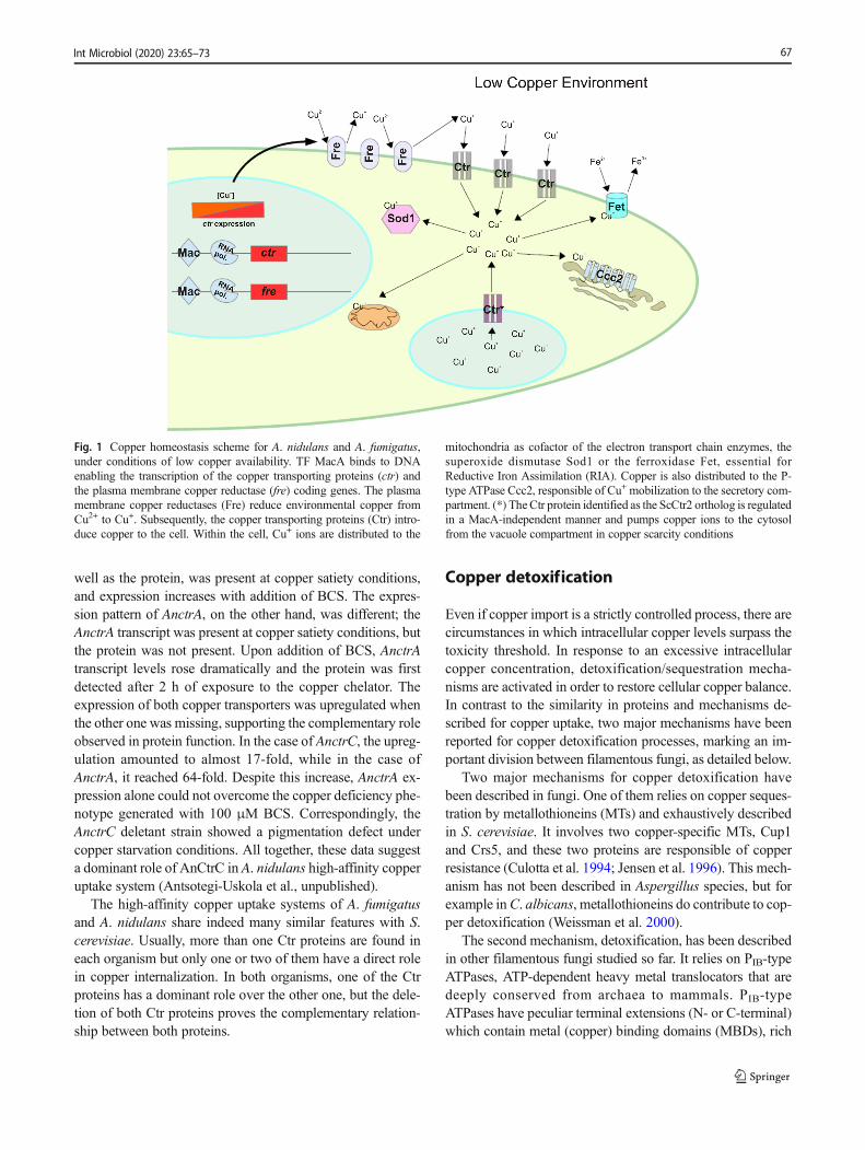
well as the protein, was present at copper satiety conditions,and expression increases with addition of BCS. The expres-sion pattern of AnctrA, on the other hand, was different; theAnctrA transcript was present at copper satiety conditions, butthe protein was not present. Upon addition of BCS, AnctrAtranscript levels rose dramatically and the protein was firstdetected after 2 h of exposure to the copper chelator. Theexpression of both copper transporters was upregulated whenthe other one was missing, supporting the complementary roleobserved in protein function. In the case of AnctrC, the upreg-ulation amounted to almost 17-fold, while in the case ofAnctrA, it reached 64-fold. Despite this increase, AnctrA ex-pression alone could not overcome the copper deficiency phe-notype generated with 100 μM BCS. Correspondingly, theAnctrC deletant strain showed a pigmentation defect undercopper starvation conditions. All together, these data suggesta dominant role of AnCtrC in A. nidulans high-affinity copperuptake system (Antsotegi-Uskola et al., unpublished).
The high-affinity copper uptake systems of A. fumigatusand A. nidulans share indeed many similar features with S.cerevisiae. Usually, more than one Ctr proteins are found ineach organism but only one or two of them have a direct rolein copper internalization. In both organisms, one of the Ctrproteins has a dominant role over the other one, but the dele-tion of both Ctr proteins proves the complementary relation-ship between both proteins.
Copper detoxification
Even if copper import is a strictly controlled process, there arecircumstances in which intracellular copper levels surpass thetoxicity threshold. In response to an excessive intracellularcopper concentration, detoxification/sequestration mecha-nisms are activated in order to restore cellular copper balance.In contrast to the similarity in proteins and mechanisms de-scribed for copper uptake, two major mechanisms have beenreported for copper detoxification processes, marking an im-portant division between filamentous fungi, as detailed below.
Two major mechanisms for copper detoxification havebeen described in fungi. One of them relies on copper seques-tration by metallothioneins (MTs) and exhaustively describedin S. cerevisiae. It involves two copper-specific MTs, Cup1and Crs5, and these two proteins are responsible of copperresistance (Culotta et al. 1994; Jensen et al. 1996). This mech-anism has not been described in Aspergillus species, but forexample inC. albicans, metallothioneins do contribute to cop-per detoxification (Weissman et al. 2000).
The second mechanism, detoxification, has been describedin other filamentous fungi studied so far. It relies on PIB-typeATPases, ATP-dependent heavy metal translocators that aredeeply conserved from archaea to mammals. PIB-typeATPases have peculiar terminal extensions (N- or C-terminal)which contain metal (copper) binding domains (MBDs), rich
Fig. 1 Copper homeostasis scheme for A. nidulans and A. fumigatus,under conditions of low copper availability. TF MacA binds to DNAenabling the transcription of the copper transporting proteins (ctr) andthe plasma membrane copper reductase (fre) coding genes. The plasmamembrane copper reductases (Fre) reduce environmental copper fromCu2+ to Cu+. Subsequently, the copper transporting proteins (Ctr) intro-duce copper to the cell. Within the cell, Cu+ ions are distributed to the
mitochondria as cofactor of the electron transport chain enzymes, thesuperoxide dismutase Sod1 or the ferroxidase Fet, essential forReductive Iron Assimilation (RIA). Copper is also distributed to the P-type ATPase Ccc2, responsible of Cu+ mobilization to the secretory com-partment. (*) TheCtr protein identified as the ScCtr2 ortholog is regulatedin a MacA-independent manner and pumps copper ions to the cytosolfrom the vacuole compartment in copper scarcity conditions
67Int Microbiol (2020) 23:65–73
in Cys and sometimes His. PIB-ATPases possess 6 to 8 trans-membrane domains (TMDs), metal binding sequences in theirTMDs, regulatory MBDs, a structure involved in enzymephosphorilation (P-domain), a nucleotide binding domain(N-domain), and an energy transduction domain (A-domain)(Palmgren and Nissen 2011).
These copper extrusion pumps represent the main detoxi-fication mechanism in bacteria (Ladomersky and Petris 2015),and in eukaryotes, they are also involved in copper compart-mentalization into the secretory network where copper is in-corporated into different Cu-dependent proteins as a cofactor.In humans, two PIB-ATPases ATP7A (Menkes disease pro-tein) and ATP7B (Wilson disease protein) are responsible ofdelivery of copper to the trans-Golgi compartment (TGN)(Lutsenko et al. 2007). In response to Cu toxicity, both trans-porters change their location from the TGN to the cell mem-brane to act as detoxification (export) pumps conferring cop-per resistance to the cell (Suzuki and Gitlin 1999). In thedimorphic fungus C. albicans, each task is fulfilled by a dif-ferent PIB-ATPase; CaCcc2 is involved in copper compart-mentalization into the TGN and CaCrp1p is responsible ofcopper detoxification at the plasma membrane (Riggle andKumamoto 2000; Weissman et al. 2000). It has recently beendescribed that some species of the Aspergillus spp. possesstwo CaCrp1p homologs involved in copper detoxification(Yang et al. 2018).
This divergence in the mechanism of metal detoxificationby different fungal species may be the result of an adaptationto their respective ecological niches. S. cerevisiae has beenisolated from a range of environments (Goddard and Greig2015), many of which are specific, such as fruits and flowers.In contrast, C. albicans, like most filamentous fungi, is an all-rounder found in environments ranging from the digestivetracts of animals to soils, where competition and antagonismare common. In these environments, PIB-ATPase (CaCrp1p)mediated copper export confers superior tolerance with re-spect to MTs, despite the greater cost in energy required toproduce it and operate it. MTs, on the other hand, may allowfor the immobilization of greater amounts of copper, as aresource, in media which may be especially poor in copper,such as flower nectar. In the same medium and growing con-ditions, S. cerevisiae is able to grow in up to 2 mM of CuSO4,whereas C. albicans tolerates 20 mMCuSO4 (Weissman et al.2000).
C. albicans has become a benchmark for recent studies infilamentous fungi, such as the Aspergilli. Copper detoxifica-tion in A. nidulans relies on a PIB-type ATPase termed CrpA(Antsotegi-Uskola et al. 2017). AnCrpA contains all the char-acteristic domains described in a PIB-type ATPase: 8 TMDs,an ion translocation motif (CPC) in the sixth TMD andcysteine-rich MBDs in the cytoplasmic N-terminal domain.Five N-terminal MBDs are present as tandem repeats, andtwo CxxxC motifs are followed by three GMxCxxC classic
heavy metal–associated domains (HMA). Copper detoxifica-tion in A. fumigatus also depends on a PIB-type ATPasetermed AfCrpA (Wiemann et al. 2017).
Deletion of crpA resulted in acute susceptibility to copperin A. nidulans and A. fumigatus. Susceptibility towards othermetals was also observed in the A. nidulans crpA deletantstrain, albeit not comparably with Cu susceptibility.Expression analysis corroborated the copper detoxificationrole of AnCrpA. Exposure to copper had a dramatic effecton AncrpA expression, while other metals barely induced ex-pression. The AncrpA deletant strain manifested reduced re-sistance to Cu compared with the wild type strain in solidmedium (~ 10-fold). With 100 μM CuSO4, the colony exhib-ited morphological defects and at 150 μM CuSO4, an almostcomplete growth inhibition. At 100 μM CuSO4, the colonyexhibited a typical wild type radial growth pattern, but withvery slight cellular density. This cellular morphology wasnamed the Bcopper phenotype.^ The AncrpA deletant strainshowed some sensitivity to Cd and Ag, but far lower than toCu (Antsotegi-Uskola et al. 2017).
AncrpA expression analysis revealed that it is principallyexpressed in the presence of copper (100 μM CuSO4), show-ing strong transient induction shortly upon exposure. mRNAappears 15 min after copper addition and the protein, after30 min. The expression maximum is reached at 60 minfollowed by a gradual repression over time. The pattern ofAnCrpA expressionwithAg and Cd toxicities is similar, albeitseveral times weaker. In the case of Cd, with 250 μMCd(NO3)2, a very faint bandwas visible at 120′ after exposure.It has recently been reported that AfCrpA also contributes toZn resistance in A. fumigatus (Cai et al. 2018). These resultsindicate that even if it might have some role in other heavymetal detoxification, copper is the principal ion transported byCrpA (Antsotegi-Uskola et al. 2017).
AnCrpA is localized at the plasma membrane under copperstress conditions; however, depending on the level of stress,AnCrpA localization and function can change. After half anhour of copper addition to induce AnCrpA expression, sub-cellular localization studies show that fluorescence was local-ized at a reticulated network (highly likely the endoplasmicreticulum), surrounding structures that resemble the nucleusand strands associated to the plasma membrane under non-toxic copper loads. An hour after copper addition, AnCrpAappeared homogeneously dispersed in the cytoplasm and pri-marily distributed at the cell periphery, most likely at the plas-ma membrane. In numerous cells, fluorescence was polarizedto the tip of hyphae. Approximately 2 h after copper addition,the protein mostly remained at the periphery, but fluorescentaggregates gradually appeared throughout the cytosol. After3 h of copper exposure, large aggregates on vesicles resem-bling vacuolar compartments became visible (Antsotegi-Uskola et al. 2017). The authors hypothesized that de novosynthesized AnCrpA was translocated from the Golgi
68 Int Microbiol (2020) 23:65–73
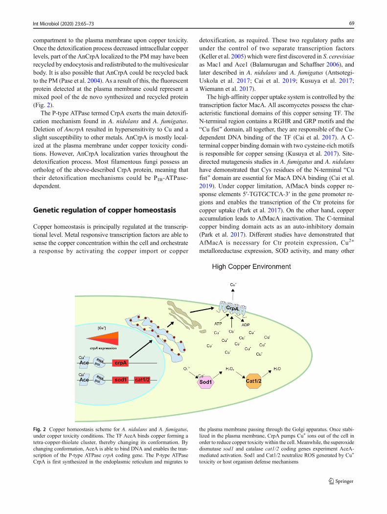
compartment to the plasma membrane upon copper toxicity.Once the detoxification process decreased intracellular copperlevels, part of the AnCrpA localized to the PMmay have beenrecycled by endocytosis and redistributed to themultivesicularbody. It is also possible that AnCrpA could be recycled backto the PM (Pase et al. 2004). As a result of this, the fluorescentprotein detected at the plasma membrane could represent amixed pool of the de novo synthesized and recycled protein(Fig. 2).
The P-type ATPase termed CrpA exerts the main detoxifi-cation mechanism found in A. nidulans and A. fumigatus.Deletion of AncrpA resulted in hypersensitivity to Cu and aslight susceptibility to other metals. AnCrpA is mostly local-ized at the plasma membrane under copper toxicity condi-tions. However, AnCrpA localization varies throughout thedetoxification process. Most filamentous fungi possess anortholog of the above-described CrpA protein, meaning thattheir detoxification mechanisms could be PIB-ATPase-dependent.
Genetic regulation of copper homeostasis
Copper homeostasis is principally regulated at the transcrip-tional level. Metal responsive transcription factors are able tosense the copper concentration within the cell and orchestratea response by activating the copper import or copper
detoxification, as required. These two regulatory paths areunder the control of two separate transcription factors(Keller et al. 2005) which were first discovered in S. cerevisiaeas Mac1 and Ace1 (Balamurugan and Schaffner 2006), andlater described in A. nidulans and A. fumigatus (Antsotegi-Uskola et al. 2017; Cai et al. 2019; Kusuya et al. 2017;Wiemann et al. 2017).
The high-affinity copper uptake system is controlled by thetranscription factor MacA. All ascomycetes possess the char-acteristic functional domains of this copper sensing TF. TheN-terminal region contains a RGHR and GRP motifs and theBCu fist^ domain, all together, they are responsible of the Cu-dependent DNA binding of the TF (Cai et al. 2017). A C-terminal copper binding domain with two cysteine-rich motifsis responsible for copper sensing (Kusuya et al. 2017). Site-directed mutagenesis studies in A. fumigatus and A. nidulanshave demonstrated that Cys residues of the N-terminal BCufist^ domain are essential for MacA DNA binding (Cai et al.2019). Under copper limitation, AfMacA binds copper re-sponse elements 5′-TGTGCTCA-3′ in the gene promoter re-gions and enables the transcription of the Ctr proteins forcopper uptake (Park et al. 2017). On the other hand, copperaccumulation leads to AfMacA inactivation. The C-terminalcopper binding domain acts as an auto-inhibitory domain(Park et al. 2017). Different studies have demonstrated thatAfMacA is necessary for Ctr protein expression, Cu2+
metalloreductase expression, SOD activity, and many other
Fig. 2 Copper homeostasis scheme for A. nidulans and A. fumigatus,under copper toxicity conditions. The TF AceA binds copper forming atetra-copper-thiolate cluster, thereby changing its conformation. Bychanging conformation, AceA is able to bind DNA and enables the tran-scription of the P-type ATPase crpA coding gene. The P-type ATPaseCrpA is first synthesized in the endoplasmic reticulum and migrates to
the plasma membrane passing through the Golgi apparatus. Once stabi-lized in the plasma membrane, CrpA pumps Cu+ ions out of the cell inorder to reduce copper toxicity within the cell. Meanwhile, the superoxidedismutase sod1 and catalase cat1/2 coding genes experiment AceA-mediated activation. Sod1 and Cat1/2 neutralize ROS generated by Cu+
toxicity or host organism defense mechanisms
69Int Microbiol (2020) 23:65–73
copper-dependent cellular processes (Fig. 1). It has recentlybeen reported that AfMacA also regulates iron homeostasisand its localization in the cell varies depending on copper andiron concentration (Park et al. 2018). AfmacA disruption gen-erated a copper starvation phenotype with manifest growthdefects in copper depletion conditions (Cai et al. 2017;Kusuya et al. 2017; Park et al. 2017).
The copper detoxification process is orchestrated by thetranscription factor AceA. In A. fumigatus, AfAceA is alsoinvolved in zinc detoxification (Cai et al. 2018). The charac-teristic domains that identify this TF are the BCu fist^ DNAbinding domain, and the numerous cysteine residues arrangedin CxC-CxxC segments through the protein sequence, identi-fied as necessary for function in S. cerevisiae (Hu et al. 1990).Within the DNA binding domain, two different motifs arefound, a 3 x Cys-His zinc finger and the KGRP motif forDNA binding stabilization. A single cysteine-rich domain islocated downstream of the DNA binding domain. Under ex-cess copper conditions, four Cu+ atoms bind the Cys-richdomain to form a tetra-copper-thiolate cluster (Dameronet al. 1991). Cluster formation leads to a conformationalchange that enables AceA-DNA binding. In this way, AceAenables the transcription of the copper detoxification machin-ery, principally crpA. AfAceA also regulates ROS detoxifica-tion genes sod1, cat1, and cat2 (Wiemann et al. 2017), as wellas the TFs AftA and Yap1 mediating ROS defense (Hagiwaraet al. 2016) (Fig. 2). aceA deletion resulted in a defectivephenotype with severe growth defects under copper excessconditions in A. fumigatus and A. nidulans (Antsotegi-Uskola et al. 2017; Cai et al. 2018).
In summary, even though copper homeostasis is transcrip-tionally regulated by a single transcription factor in some or-ganisms, such as C. neoformans, in most filamentous fungi, itis orchestrated by two transcription factors. The TF-termedMacA is responsible of conducting the copper internalizationprocess and AceA does the same with copper detoxification.The characteristic features of these transcription factors arewell conserved in filamentous fungi.
Copper as a virulence factor
A. fumigatus is one of the most lethal airborne fungal patho-gens responsible of severe invasive aspergillosis (IA), espe-cially in immunocompromised individuals (Cai et al. 2017).Copper is a recognized virulence factor, as it is the cofactor ofmany enzymes that contribute to virulence, such as laccases orSOD. On the other hand, host organisms have developed de-fense strategies against fungal pathogens that target copperavailability. By scavenging all Cu+ in the infection area, cop-per deprivation can be induced in the pathogen. On the con-trary, host innate immune cells, such as macrophages, are ableto mobilize copper to invading fungal tissue as a defense
mechanism (Garcia-Santamarina and Thiele 2015). The gen-eration of Reactive Oxidative Species (ROS) is another de-fense mechanism employed by the innate immune system.The superoxide dismutase (SOD) enzymes are copper-dependent enzymes responsible of neutralizing ROS, so theSOD enzyme is considered as an important virulence factor inorganisms, like C. albicans and C. neoformans (Frohner et al.2009; Narasipura et al. 2005). In conclusion, copper homeo-stasis plays a key role to overcome host barriers and the de-velopment of pathogenesis.
Disruption of any of the abovementioned TFs responsiblefor copper homeostasis has a direct effect on virulence. Recentstudies revealed that AfMacA is necessary for normal viru-lence inA. fumigatus (Cai et al. 2017).AfmacA deletion resultsin delayed growth and pigmentation deficiency by DHN mel-anin synthesis inhibition. Laccases AfAbr1 and AfAbr2 areinvolved in melanin biosynthesis and they are copper-dependent proteins (Upadhyay et al. 2013). When copper up-take is impaired, laccase activity is substantially reduced (Parket al. 2014). Melanin confers a non-immunogenic status to thefungus. Inappropriately melanized conidia therefore becomeimmunoreactive (Pihet et al. 2009). Melanin also provides aprotective layer to the action of host-derived ROS (Jahn et al.2000). A. fumigatus copper transporting protein expression isupregulated in the presence of human neutrophils (Sugui et al.2008). This underscores the importance of copper uptake forpathogen viability within the host.
AfAceA has also been shown to be an important virulencefactor. As detailed above, this TF controls the expression ofthe PIB-type ATPase AfcrpA responsible of copper detoxifica-tion in A. fumigatus. Moreover, AfAceA regulates the expres-sion of catalases such as Afcat1 and Afcat2 involved in ROSneutralization and the TF AfatfA involved in ROS-response(Hagiwara et al. 2014). It has been reported that AfatfA dele-tion renders A. fumigatus avirulent (Pereira et al. 2017).AfaceA deletion represses AfatfA expression considerably, to-gether with Afcat1 and Afcat2, making the fungus more vul-nerable to oxidative stress (Wiemann et al. 2017). Deletion ofAfaceA inhibits the expression AfcrpA, the main copper detox-ification system in A. fumigatus, leaving the fungus exposedto copper mobilization to fungal tissue by host innate immunesystem cells. Thus, the copper detoxification machinery is akey factor in pathogen viability during infection.
Botrytis cinerea is a necrotrophic fungal plant pathogen ofworldwide distribution, capable of infecting a wide range ofhosts. It is probably the best studied necrotrophic plant path-ogen. Copper-dependent proteins play a central role in manyaspects of the B. cinerea, including pathogenesis. The P-typeATPase BcCcc2, an ortholog of the S. cerevisiae Ccc2 coppertransporting P-type ATPase that delivers Cu to the secretorycompartment for subsequent protein modification (Smith et al.2017), is crucial for virulence in B. cinerea (Saitoh et al.2010). The Bcccc2 deletion strain presented melanization
70 Int Microbiol (2020) 23:65–73
and morphogenesis defects. Moreover, the deletion strain wasnot able to penetrate and infect host organisms (Saitoh et al.2010). The most probable reason for this could be that theBcCcc2 targeted proteins could get no copper, and for thisreason, all these cellular processes were impaired (Saitohet al. 2010). B. cinerea is able to break through healthy tissueand secrete enzymes and toxins that generate necrosis (vanKan 2006). Lopez-Cruz and collaborators (Lopez-Cruz et al.2017) revealed the importance of the Cu-Zn SOD in this pro-cess. The host oxidative burst (O−), a plant defense mecha-nism against pathogen organisms, plays an active role inBotrytis infection (Rossi et al. 2017). The O2
−/H2O2 ratio inthe necrotic lesion area is very significant for infection devel-opment. Higher concentration of H2O2 favors infection.BcSod1 actively generates H2O2, which damages plant tissueand reduces plant defense. The absence BcSod1 reduces thecapacity to generate necrotic lesions in plant tissues as theoxidative environment is not appropriate for Botrytis infectiondevelopment. The measured O−/H2O2 ratio in necrotic lesionscaused by aΔBcsod1 strain is too high for infection. Incorrectfunction of BcCcc2 results in a defective BcSod1 function, aswell as other proteins, proving the importance of the copperhomeostasis system for B. cinerea virulence.
In summary, copper homeostasis plays an essential role inA. fumigatus virulence development. The high-affinity copperuptake system and the copper detoxification system are keyfactors in the infection mechanism. The high-affinity copperuptake system enables the maturation of many copper-dependent enzymes for virulence. On the other hand, the de-toxification system confers the organism the necessary resis-tance for survival. In the necrotrophic fungus B. cinerea, thecopper transporting P-type ATPase BcCcc2 is necessary forsuccessful virulence development. Hence, copper homeostasisappears to be required for the development of infection inevery major pathogenic fungus described thus far.
Conclusions
The study of copper homeostasis in filamentous fungi hasunveiled the relevance of this cation in growth, development,and pathogenicity. In the last years, there have been somecontributions in the pathogenic fungus A. fumigatus and themodel organism A. nidulans, but many details remain to beuncovered. Considering the repercussion of copper homeosta-sis on fungal biology, it is a topic worth studying in moredepth, and in a wider range of species. The copper uptakeand detoxification mechanisms and their respective transcrip-tion factors have been the focus of recent publications, iden-tifying the principal participants of each process, but the cop-per buffering and intracellular distribution mechanisms re-main practically unknown. Phylogenetic analyses have re-vealed the presence of proteins that act as copper scavenging
and trafficking vehicles in other organisms (Ding et al. 2014),but so far, those proteins have not been investigated. Up todate, there is no evidence of CrpA homolog–dependent viru-lence in a phytopathogenic fungus. However, the BcCcc2 P-type ATPase, responsible of Cu traffic to the secretory com-partment, in Botrytis cinerea has been proved to be essentialfor virulence. This evidence calls for further investigation cov-ering intracellular copper trafficking and storage in oncomingyears.
Copper has long been used as an antimicrobial agent inmany different areas (Judet-Correia et al. 2011); however, ithas often been applied in doses which cause severe impact onthe environment. This excessive use has also brought aboutincreasing levels of copper resistance in microbial pathogens.The mechanisms involved in fungal copper resistance are nowin need of a thorough assessment in order to find new andeffective methods to complement or replace copper in thecontrol of fungal disease. New details on the mechanism ofcopper toxicity on fungal cells may shed light on the potentialuse of synergists affecting copper homeostasis that could helplower the currently required copper dose. The acquisition ofcopper by fungi in the soil, and its exchange between micro-organisms and plants in symbiotic relationship, is another as-pect which should be considered, to ensure sustainable agri-cultural and environmental conservation programs.
Funding This work has been supported by the Basque Governmentthrough the grants IT599-13 and S-PC13UN041 to UU.
Compliance with ethical standards
Conflict of interest The authors declare that they have no conflict ofinterest.
Open Access This article is distributed under the terms of the CreativeCommons At t r ibut ion 4 .0 In te rna t ional License (h t tp : / /creativecommons.org/licenses/by/4.0/), which permits unrestricted use,distribution, and reproduction in any medium, provided you give appro-priate credit to the original author(s) and the source, provide a link to theCreative Commons license, and indicate if changes were made.
References
Antsotegi-Uskola M, Markina-Inarrairaegui A, Ugalde U (2017) Copperresistance in Aspergillus nidulans relies on the PI-type ATPaseCrpA, regulated by the transcription factor AceA. Front Microbiol8:912
Balamurugan K, Schaffner W (2006) Copper homeostasis in eukaryotes:teetering on a tightrope. Biochim Biophys Acta 1763:737–746
Ballou ER, Wilson D (2016) The roles of zinc and copper sensing infungal pathogenesis. Curr Opin Microbiol 32:128–134
Blatzer M, Latge JP (2017) Metal-homeostasis in the pathobiology of theopportunistic human fungal pathogen Aspergillus fumigatus. CurrOpin Microbiol 40:152–159
71Int Microbiol (2020) 23:65–73
Cai Z, Du W, Liu L, Pan D, Lu L (2019) Molecular characteristics of theconserved Aspergillus nidulans transcription factor Mac1 and itsfunctions in response to copper starvation. mSphere. 4:e00670-18
Cai Z, DuW, Zeng Q, Long N, Dai C, Lu L (2017) Cu-sensing transcrip-tion factor Mac1 coordinates with the Ctr transporter family to reg-ulate Cu acquisition and virulence in Aspergillus fumigatus. FungalGenet Biol 107:31–43
Cai Z, DuW, Zhang Z, Guan L, Zeng Q, Chai Y, Dai C, Lu L (2018) TheAspergillus fumigatus transcription factor AceA is involved not onlyin Cu but also in Zn detoxification through regulating transportersCrpA and ZrcA. Cell Microbiol 20:e12864
Culotta VC, Howard WR, Liu XF (1994) CRS5 encodes ametallothionein-like protein in Saccharomyces cerevisiae. J BiolChem 269:25295–25302
Dameron CT, Winge DR, George GN, Sansone M, Hu S, Hamer D(1991) A copper-thiolate polynuclear cluster in the ACE1 transcrip-tion factor. Proc Natl Acad Sci U S A 88:6127–6131
De Feo CJ, Aller SG, Siluvai GS, Blackburn NJ, Unger VM (2009)Three-dimensional structure of the human copper transporterhCTR1. Proc Natl Acad Sci U S A 106:4237–4242
Ding C, Festa RA, Sun TS,Wang ZY (2014) Iron and copper as virulencemodulators in human fungal pathogens. Mol Microbiol 93:10–23
Fridovich I (1983) Superoxide radical: an endogenous toxicant. AnnuRev Pharmacol Toxicol 23:239–257
Frohner IE, Bourgeois C, Yatsyk K, Majer O, Kuchler K (2009) Candidaalbicans cell surface superoxide dismutases degrade host-derivedreactive oxygen species to escape innate immune surveillance.Mol Microbiol 71:240–252
Garcia-Santamarina S, Festa RA, Smith AD, Yu CH, Probst C, Ding C,Homer CM, Yin J, Noonan JP, Madhani H, Perfect JR, Thiele DJ(2018) Genome-wide analysis of the regulation of Cumetabolism inCryptococcus neoformans. Mol Microbiol 108:473–494
Garcia-Santamarina S, Thiele DJ (2015) Copper at the fungal pathogen-host axis. J Biol Chem 290:18945–18953
Gerwien F, Skrahina V, Kasper L, Hube B, Brunke S (2018) Metals infungal virulence. FEMS Microbiol Rev 42
Goddard MR, Greig D (2015) Saccharomyces cerevisiae: a nomadicyeast with no niche?. FEMS Yeast Res 15
Hagiwara D, Suzuki S, Kamei K, Gonoi T, Kawamoto S (2014) The roleof AtfA and HOG MAPK pathway in stress tolerance in conidia ofAspergillus fumigatus. Fungal Genet Biol 73:138–149
Hagiwara D, Takahashi H, Kusuya Y, Kawamoto S, Kamei K, Gonoi T(2016) Comparative transcriptome analysis revealing dormant co-nidia and germination associated genes in Aspergillus species: anessential role for AtfA in conidial dormancy. BMC Genomics 17:358
Hu S, Furst P, Hamer D (1990) The DNA and Cu binding functions ofACE1 are interdigitated within a single domain. New Biol 2:544–555
Jahn B, Boukhallouk F, Lotz J, Langfelder K, Wanner G, Brakhage AA(2000) Interaction of human phagocytes with pigmentlessAspergillus conidia. Infect Immun 68:3736–3739
Jensen LT, Howard WR, Strain JJ, Winge DR, Culotta VC (1996)Enhanced effectiveness of copper ion buffering by CUP1 metallo-thionein compared with CRS5 metallothionein in Saccharomycescerevisiae. J Biol Chem 271:18514–18519
Jiang J, Nadas IA, KimMA, FranzKJ (2005) AMets motif peptide foundin copper transport proteins selectively binds Cu(I) with methionine-only coordination. Inorg Chem 44:9787–9794
Judet-Correia D, Charpentier C, Bensoussan M, Dantigny P (2011)Modelling the inhibitory effect of copper sulfate on the growth ofPenicillium expansum and Botrytis cinerea. Lett ApplMicrobiol 53:558–564
Keller G, Bird A, Winge DR (2005) Independent metalloregulation ofAce1 and Mac1 in Saccharomyces cerevisiae. Eukaryot Cell 4:1863–1871
Kusuya Y, Hagiwara D, Sakai K, Yaguchi T, Gonoi T, Takahashi H(2017) Transcription factor Afmac1 controls copper import machin-ery in Aspergillus fumigatus. Curr Genet 63:777–789
Ladomersky E, Petris MJ (2015) Copper tolerance and virulence in bac-teria. Metallomics. 7:957–964
Lopez-Cruz J, Oscar CS, Emma FC, Pilar GA, Carmen GB (2017)Absence of Cu-Zn superoxide dismutase BCSOD1 reducesBotrytis cinerea virulence in Arabidopsis and tomato plants, reveal-ing interplay among reactive oxygen species, callose and signallingpathways. Mol Plant Pathol 18:16–31
Lutsenko S (2010) Human copper homeostasis: a network of intercon-nected pathways. Curr Opin Chem Biol 14:211–217
Lutsenko S, Barnes NL, Bartee MY, Dmitriev OY (2007) Function andregulation of human copper-transporting ATPases. Physiol Rev 87:1011–1046
Macomber L, Imlay JA (2009) The iron-sulfur clusters of dehydratasesare primary intracellular targets of copper toxicity. Proc Natl AcadSci U S A 106:8344–8349
Marvin ME, Mason RP, Cashmore AM (2004) The CaCTR1 gene isrequired for high-affinity iron uptake and is transcriptionally con-trolled by a copper-sensing transactivator encoded by CaMAC1.Microbiology 150:2197–2208
Narasipura SD, Chaturvedi V, Chaturvedi S (2005) Characterization ofCryptococcus neoformans variety gattii SOD2 reveals distinct rolesof the two superoxide dismutases in fungal biology and virulence.Mol Microbiol 55:1782–1800
Nevitt T, Ohrvik H, Thiele DJ (2012) Charting the travels of copper ineukaryotes from yeast to mammals. Biochim Biophys Acta 1823:1580–1593
Palmgren MG, Nissen P (2011) P-type ATPases. Annu Rev Biophys 40:243–266
Park YS, Kang S, Seo H, Yun CW (2018) A copper transcription factor,AfMac1, regulates both iron and copper homeostasis in the oppor-tunistic fungal pathogen Aspergillus fumigatus. Biochem J 475:2831–2845
Park YS, Kim TH, Yun CW (2017) Functional characterization of thecopper transcription factor AfMac1 from Aspergillus fumigatus.Biochem J 474:2365–2378
Park YS, Lian H, Chang M, Kang CM, Yun CW (2014) Identification ofhigh-affinity copper transporters in Aspergillus fumigatus. FungalGenet Biol 73:29–38
Pase L, Voskoboinik I, Greenough M, Camakaris J (2004) Copper stim-ulates trafficking of a distinct pool of the Menkes copper ATPase(ATP7A) to the plasma membrane and diverts it into a rapidrecycling pool. Biochem J 378:1031–1037
Pereira SL, Alves de CP, Dos Reis TF, PazianiMH, VonZeska KressMR,Riano-Pachon DM, Hagiwara D, Ries LN, Brown NA, GoldmanGH (2017) Genome-wide transcriptome analysis of Aspergillusfumigatus exposed to osmotic stress reveals regulators of osmoticand cell wall stresses that are SakA(HOG1) and MpkC dependent.Cell Microbiol 19:e12681
Petris MJ (2004) The SLC31 (Ctr) copper transporter family. PflugersArch 447:752–755
Pihet M, Vandeputte P, Tronchin G, Renier G, Saulnier P, Georgeault S,Mallet R, Chabasse D, Symoens F, Bouchara JP (2009) Melanin isan essential component for the integrity of the cell wall ofAspergillus fumigatus conidia. BMC Microbiol 9:177
Puig S, Lee J, LauM, Thiele DJ (2002) Biochemical and genetic analysesof yeast and human high affinity copper transporters suggest a con-served mechanism for copper uptake. J Biol Chem 277:26021–26030
Rees EM, Lee J, Thiele DJ (2004) Mobilization of intracellular copperstores by the ctr2 vacuolar copper transporter. J Biol Chem 279:54221–54229
72 Int Microbiol (2020) 23:65–73
Riggle PJ, Kumamoto CA (2000) Role of a Candida albicans P1-typeATPase in resistance to copper and silver ion toxicity. J Bacteriol182:4899–4905
Rossi FR, Krapp AR, Bisaro F, Maiale SJ, Pieckenstain FL, Carrillo N(2017) Reactive oxygen species generated in chloroplasts contributeto tobacco leaf infection by the necrotrophic fungus Botrytis cinerea.Plant J 92:761–773
Saitoh Y, Izumitsu K, Morita A, Tanaka C (2010) A copper-transportingATPase BcCCC2 is necessary for pathogenicity of Botrytis cinerea.Mol Gen Genomics 284:33–43
Smith AD, Logeman BL, Thiele DJ (2017) Copper acquisition and utili-zation in Fungi. Annu Rev Microbiol 71:597–623
Sugui JA, Kim HS, Zarember KA, Chang YC, Gallin JI, Nierman WC,Kwon-Chung KJ (2008) Genes differentially expressed in conidiaand hyphae of Aspergillus fumigatus upon exposure to human neu-trophils. PLoS One 3:e2655
Suzuki M, Gitlin JD (1999) Intracellular localization of the Menkes andWilson’s disease proteins and their role in intracellular copper trans-port. Pediatr Int 41:436–442
Upadhyay S, Torres G, Lin X (2013) Laccases involved in 1,8-dihydroxynaphthalene melanin biosynthesis in Aspergillusfumigatus are regulated by developmental factors and copper ho-meostasis. Eukaryot Cell 12:1641–1652
van Kan JA (2006) Licensed to kill: the lifestyle of a necrotrophic plantpathogen. Trends Plant Sci 11:247–253
Weissman Z, Berdicevsky I, Cavari BZ, Kornitzer D (2000) The highcopper tolerance of Candida albicans is mediated by a P-typeATPase. Proc Natl Acad Sci U S A 97:3520–3525
Wiemann P, Perevitsky A, Lim FY, Shadkchan Y, Knox BP, LanderoFigueora JA, Choera T, Niu M, Steinberger AJ, Wuthrich M, IdolRA, Klein BS, Dinauer MC, Huttenlocher A, Osherov N, Keller NP(2017) Aspergillus fumigatus copper export machinery and reactiveoxygen intermediate defense counter host copper-mediated oxida-tive antimicrobial offense. Cell Rep 19:2174–2176
Yang K, Shadkchan Y, Tannous J, Landero Figueroa JA, Wiemann P,Osherov N, Wang S, Keller NP (2018) Contribution of ATPasecopper transporters in animal but not plant virulence of the crossoverpathogen Aspergillus flavus. Virulence. 9:1273–1286
Zhang P, Zhang D, Zhao X, Wei D, Wang Y, Zhu X (2016) Effects ofCTR4 deletion on virulence and stress response in Cryptococcusneoformans. Antonie Van Leeuwenhoek 109:1081–1090
Publisher’s note Springer Nature remains neutral with regard tojurisdictional claims in published maps and institutional affiliations.
73Int Microbiol (2020) 23:65–73