lipid profiles and hepatitis c viral markers in hcv-infected thalassemic patients
TRANSCRIPT

ORiginal Article
Gut and Liver, Vol. 5, No. 3, September 2011, pp. 348-355
Lipid Profi les and Hepatitis C Viral Markers in HCV-Infected Thalassemic Patients
Seyed-Moayed Alavian*, Seyyed Mohammad Miri*, Seyed-Vahid Tabatabaei*, Maryam Keshvari†, Bita Behnava*, Pegah Karimi Elizee*, Nastaran Mahboobi*, and Kamran Bagheri Lankarani‡
*Baqiyatallah University of Medical Sciences, Baqiyatallah Research Center for Gastroenterology and Liver Disease, Tehran, †Iranian Blood Transfusion Organization Research Centre, Tehran, ‡Shiraz University of Medical Sciences, Shiraz, Iran
Background/Aims: The distribution of blood lipids, glucose and their determinants in thalassemic patients with chronic hepatitis C virus (HCV) infection has rarely been investigated. Thus, we aimed to investigate the relationship between both liver histologic fi ndings and viral markers and serum lipids in thalassemic patients chronically infected with HCV. Methods: We enrolled 280 polytransfused thalassemic patients with chronic hepatitis C. HCV viral load was determined using the Amplicor test. Genotyping was performed using genotype specifi c primers. Fasting serum lipid, glucose, ferritin and liv-er function enzyme concentrations were measured. A modi-fi ed Knodell scoring system was used to stage liver fi brosis and to grade necroinfl ammatory activity. Perls’ staining was used to assess hepatic siderosis. Results: Just one subject had total cholesterol >200 mg/dL, and 7% had triglycerides >150 mg/dL. The mean high-density lipoprotein cholesterol (HDL-C) and glucose levels were 37 and 104 (97-111) mg/dL, respectively. Viral markers, liver histological fi ndings and aminotransferase activity were not associated with serum lipid levels. Serum triglycerides, total cholesterol and ferritin were independent risk factors for impaired glucose tolerance or diabetes in these patients. Conclusions: The majority of the patients had blood lipid levels (with the exception of HDL) within the defi ned normal range; viral and liver histological factors do not appear to play a significant role in changing the levels of serum lipids or glucose in these patients. (Gut Liver 2011;5:348-355)
Key Words: Triglyceride; Cholesterol; HDL cholesterol; Thalas-semia; Hepatitis C virus; Iran
Correspondence to: Seyed-Moayed AlavianBaqiyatallah Research Center for Gastroenterology and Liver Diseases, Ground floor of Baqiyatallah Hospital, Mollasadra Avenue, Vanak Square, P.O. Box 14155-3651, Tehran, IranTel: +98-21-88067114, Fax: +98-21-88067114, E-mail: [email protected]
Received on July 13, 2010. Accepted on January 7, 2011.pISSN 1976-2283 eISSN 2005-1212 http://dx.doi.org/10.5009/gnl.2011.5.3.348
This is an Open Access article distributed under the terms of the Creative Commons Attribution Non-Commercial License (http://creativecommons.org/licenses/by-nc/3.0) which permits unrestricted non-commercial use, distribution, and reproduction in any medium, provided the original work is properly cited.
INTRODUCTION
Hepatitis C is a major health problem in the world,1 for which thalassemia patients are at a higher risk.2 Metabolic syndrome is a risk factor for progression of liver diseases as well. As the liver is the main determinant of serum lipoprotein synthesis and lipid metabolism, chronic liver diseases are often accompanied with an impaired lipid metabolism.3 The relation between low levels of serum cholesterol, particularly low-density lipoprotein cho-lesterol (LDL-C) and severity of liver disease has previously been described.4 Lower total cholesterol and LDL-C levels were also described in hepatitis C virus (HCV)-infected patients.5,6 Total se-rum cholesterol and LDL-C have even been proposed to be pre-dictors of response to interferon in HCV infected patients.7 Fur-thermore, it was determined that patients with chronic hepatitis C had lower total cholesterol levels in comparison with patients chronically infected with hepatitis B.8 An association between HCV infection and lipid metabolism has been described as well. Bonding of HCV to VLDL or LDL could facilitate its entry via the LDL receptor.9-11 These findings suggest that plasma lipids play an important role in the pathogenesis of HCV infection. Major beta-thalassemic patients are the most affected group of pa-tients chronically infected with HCV. It is well known that beta-thalassemia is associated with changes in plasma lipids. A low total cholesterol levels caused by a significant decrease in both LDL-C and high-density lipoprotein cholesterol (HDL-C) have been described previously in beta-thalassemia,12 but findings for triglycerides were heterogeneous.13,14 In spite of the possible role of serum lipids in pathogenesis of HCV infection and implica-tion of liver disease caused by hypertransfusion in thalassemic patients, data regarding the distribution of blood lipids among thalassemic patients with chronic HCV infection are lacking.

Alavian SM, et al: Lipid Profiles and Hepatitis C Viral Markers in HCV-Infected Thalassemic Patients 349
Therefore, in this study we aimed to investigate the distribution of serum lipids in beta-thalassemic patients with chronic HCV infection, and to determine if there are any correlations between serum lipid levels and viral load, HCV genotype, liver histology and serum iron.
MATERIALS AND METHODS
1. Study design
This study was designed as an observational study to investi-gate lipid and glucose levels and their relation with viral mark-ers and liver histologic findings in 280 HCV infected major-thalassemic patients.
2. Patients
From a total of three hundred thalassemia patients with es-tablished diagnosis of chronic HCV (positive polymerase chain reaction [PCR] for the last 6 months and liver histologic pattern of chronic hepatitis), 280 were enrolled in the present study. Twenty patients refused to participate or went to other centers to start treatment. Among all patients, 269 (96.1%) were major thalassemic patients receiving regular blood transfusions at 2- to 4-week intervals to maintain the level of hemoglobin at 10-13 g/dL along with regular therapy with deferoxamine while 11 (3.9%) had thalassemia-intermedia and received hydroxyurea and blood transfusion at long intervals. Informed consent has been obtained from patients at registration time. Sixty-seven subjects with previous liver biopsies performed more than four years ago refused to undergo another liver biopsy. Thus, their liver histologic findings were considered missing.
3. Laboratory assessment
Serum lipid and glucose concentration were determined after an overnight fast of 12 and 9 hours respectively. Alanine ami-notransferase (ALT), aspartate aminotransferase (AST), alkaline phosphatase (ALP), and alpha-fetoprotein (AFP) were detected using ELISA. Serum ferritin concentration was measured by applying IRMA. Triglyceride, total cholesterol, HDL-C, and LDL-C were measured enzymatically with commercial kits and automated analyzer. LDL-C was calculated using the Friedwald formula: (total cholesterol)-(HDL-C)-1/5(triglycerides).15 The body mass index (BMI) was calculated in accordance with the formula of weight (kg) divided by height2 (m2).16
4. Defi nitions
Subjects with a previously established diagnosis of diabetes, currently taking any form of insulin injections or hypoglyce-mic drugs and/or fasting blood glucose level >126 mg/dL were categorized as diabetes and > 110 mg/dL as significant enough insulin resistance (impaired glucose tolerance). As well as fast-ing blood glucose, the cut off point for triglyceride, total cho-lesterol was set at 150 and 200 mg/dL accordingly, with respect
to World Health Organization (WHO) definition of metabolic syndrome. HDL-C <40 for males and <50 for females were also considered low. It is noteworthy that none of our patients were taking lipid lowering agents. Fasting blood glucose was above 110 mg/dL in all diabetic patients.
5. Histologic evaluation
All subjects underwent percutaneous liver biopsy by Meng-hini needles. Each biopsy specimen was evaluated according to the modified Knodell score grading and staging system. A scale of 0-18 (modified HAI grading) was applied for grading of necroinflammatory activity and a scale of 0-6 (modified stag-ing) was applied for staging of liver fibrosis and architectural disturbances. Then, staging and grading of liver damage were categorized into three levels of mild, moderate and severe. 0-6 for grading and 0-2 for staging were designated as mild, 7-12 and 3-4 as moderate and 13-18 and 5-6 as severe. Perls’ stain-ing with score of 0-4 was applied to assess hepatic siderosis. 0-2 was designated as mild, 3 as moderate and 4 as severe.
6. RNA extraction, cDNA synthesis, and PCR procedure
All PCR procedures and genotyping were performed as de-scribed previously.17
7. Statistical analysis
Continuous variables are presented as mean values with 95% confidence interval. However, qualitative and discrete variables are presented as absolute and relative frequencies in the form of percentage. Chi-square test was applied to assess associations between categorical variables. Because of great sample size and power, comparisons between continuous and categorical vari-ables were performed by Student’s t-test and one-way ANOVA regardless of considering normal distribution or homogeneity of variances. Correlations between lipids levels and age, liver enzymes, ferritin, glucose, viral load, and BMI were evaluated by the calculation of Pearson’s correlation coefficient. All com-putations were carried out using SPSS version 16.0 (SPSS Inc., Chicago, IL, USA) while the graphs were provided by Stata SE version 8.0 (Stata Co., College Station, TX, USA). The probabil-ity value (p) <0.05 was regarded statistically significant.
RESULTS
1. Patients’ demographic and clinical characteristics
Table 1 has summarized our subjects’ demographic and base-line clinical characteristics. The mean age of our patients was 24 years (ranging from 11 to 54) and 59% of them were male. Only 2 patients (0.7%) had received their first transfusion after 1996, the year in which anti-HCV screening was established in Iran. 183 (65%) of subjects were splenectomized with mean age of 13 years at the time of procedure. The most frequent HCV genotypes were genotype 1 (57%) followed by 3 (35%). Fifty-
350 Gut and Liver, Vol. 5, No. 3, September 2011
three out of 280 subjects (24%) had severe liver fibrosis and 9 subjects (4%) had severe necroinflammatory. 21.1% of subjects had normal serum ALT level.
2. Blood lipids and glucose distribution
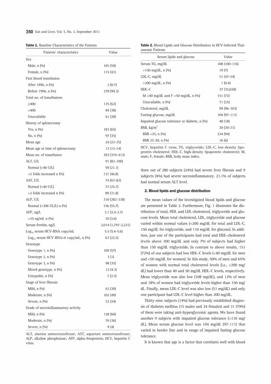
The mean values of the investigated blood lipids and glucose are presented in Table 2. Furthermore, Fig. 1 illustrates the dis-tribution of total, HDL and LDL cholesterol, triglyceride and glu-cose levels. Mean total cholesterol, LDL, triglyceride and glucose varied within normal values (<200 mg/dL for total and LDL-C, 150 mg/dL for triglyceride, and 110 mg/dL for glucose). In addi-tion, just one of the participants had total and HDL-cholesterol levels above 200 mg/dL and only 7% of subjects had higher than 150 mg/dL triglyceride. In contrast to above results, 151 (72%) of our subjects had low HDL-C levels (<40 mg/dL for men and <50 mg/dL for women). In this study, 50% of men and 65% of women with normal total cholesterol levels (i.e., <200 mg/dL) had lower than 40 and 50 mg/dL HDL-C levels, respectively. Mean triglyceride was also low (108 mg/dL), and 12% of men and 18% of women had triglyceride levels higher than 150 mg/dL. Finally, mean LDL-C level was also low (51 mg/dL) and only one participant had LDL-C level higher than 200 mg/dL.
Thirty-nine subjects (14%) had previously established diagno-sis of diabetes mellitus (15 males and 24 females) and 31 (79%) of them were taking anti-hyperglycemic agents. We have found another 9 subjects with impaired glucose tolerance (>110 mg/dL). Mean serum glucose level was 104 mg/dL (97-111) that varied in border line and in range of impaired fasting glucose tolerance.
It is known that age is a factor that correlates well with blood
Table 1. Baseline Characteristics of the Patients
Patients’ characteristics Value
Sex
Male, n (%) 165 (59) Female, n (%) 115 (41) First blood transfusion
After 1996, n (%) 2 (0.7) Before 1996, n (%) 278 (99.3) Total no. of transfusions
≥400 135 (62) <400 84 (38) Unavailable 61 (28) History of splenectomy
Yes, n (%) 183 (65) No, n (%) 97 (35) Mean age 24 (23-25)
Mean age at time of splenectomy 13 (12-14)
Mean no. of transfusion 393 (374-413)
ALT, U/L 91 (82-100)
Normal (<40 U/L) 59 (21.1) >2 folds increased n (%) 131 (46.8) AST, U/L 74 (67-82)
Normal (<40 U/L) 72 (25.7) >2 folds increased n (%) 89 (31.8) ALP, U/L 310 (282-338)
Normal (<306 UL/L) n (%) 156 (55.7) AFP, ng/L 3.1 (2.4-3.7)
>10 ng/mL n (%) 10 (3.6) Serum ferritin, ng/L 2,014 (1,797-2,231)
Log10 serum HCV-RNA copy/mL 5.5 (5.4-5.6)
Log10 serum HCV-RNA>6 copy/mL, n (%) 63 (22.5) Genotype
Genotype 1, n (%) 160 (57) Genotype 2, n (%) 3 (1) Genotype 3, n (%) 98 (35) Mixed genotype, n (%) 12 (4.3) Untypable, n (%) 7 (2.5) Stage of liver fibrosis
Mild, n (%) 63 (30) Moderate, n (%) 102 (49) Severe, n (%) 53 (24) Grade of necroinflammatory activity
Mild, n (%) 128 (60) Moderate, n (%) 76 (36) Severe, n (%) 9 (4)
ALT, alanine aminotransferase; AST, aspartate aminotransferase; ALP, alkaline phosphatase; AFP, alpha-fetoprotein; HCV, hepatitis C virus.
Table 2. Blood Lipids and Glucose Distribution in HCV-Infected Thal-assemic Patients
Serum lipids and glucose Value
Serum TG, mg/dL 108 (100-116)
>150 mg/dL, n (%) 19 (7)
LDL-C, mg/dL 51 (47-54)
>200 mg/dL, n (%) 1 (0.4)
HDL-C 37 (35,038)
M <40 mg/dL and F <50 mg/dL, n (%) 151 (72)
Unavailable, n (%) 71 (25)
Cholesterol, mg/dL 99 (96-103)
Fasting glucose, mg/dL 104 (97-111)
Impaired glucose tolerance or diabetic, n (%) 48 (18)
BMI, kg/m2 20 (20-21)
BMI <25, n (%) 234 (94)
BMI 25-30, n (%) 16 (6)
HCV, hepatitis C virus; TG, triglyceride; LDL-C, low-density lipo-protein cholesterol; HDL-C, high-density lipoprotein cholesterol; M, male; F, female; BMI, body mass index.
Alavian SM, et al: Lipid Profiles and Hepatitis C Viral Markers in HCV-Infected Thalassemic Patients 351
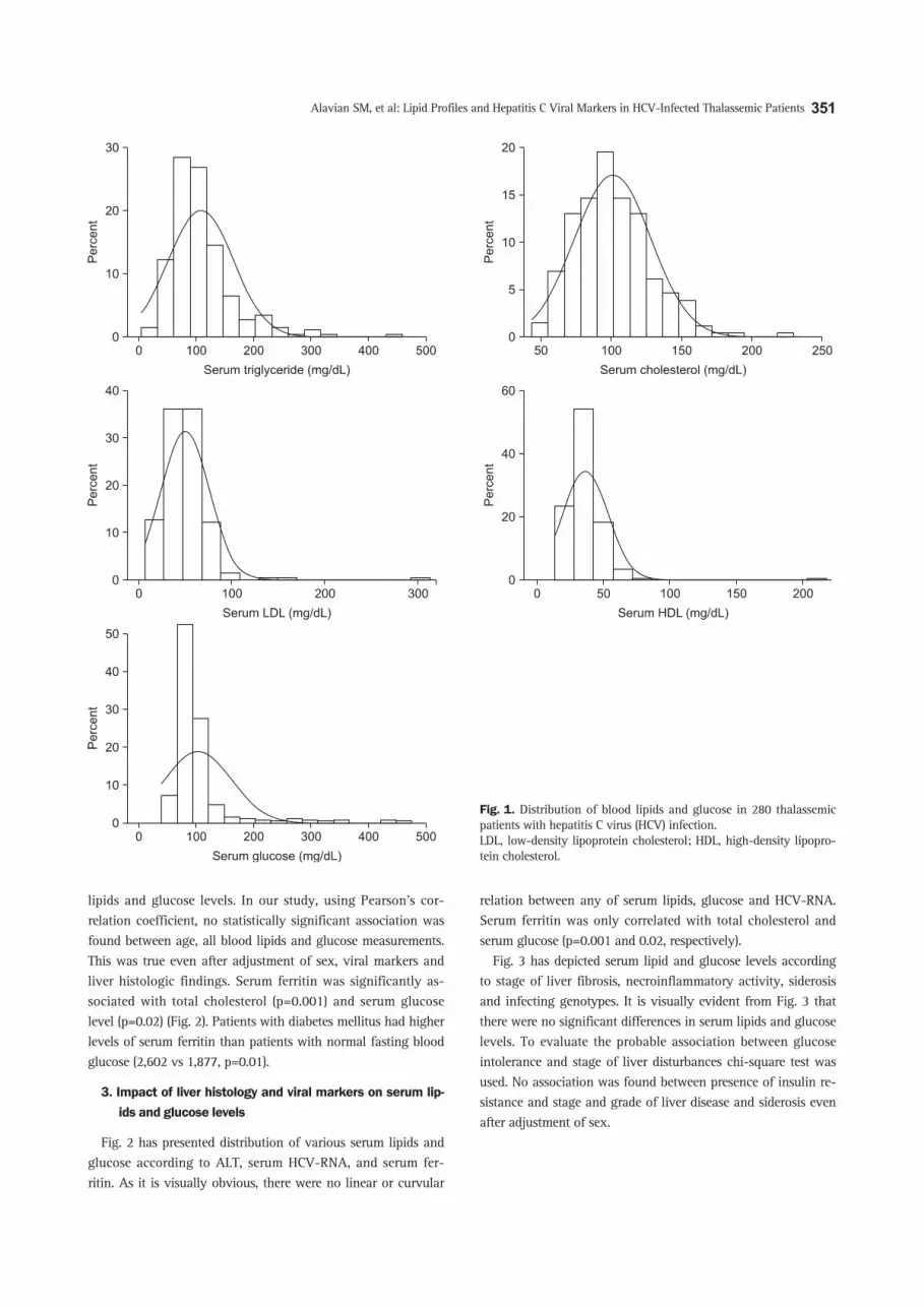
lipids and glucose levels. In our study, using Pearson’s cor-relation coefficient, no statistically significant association was found between age, all blood lipids and glucose measurements. This was true even after adjustment of sex, viral markers and liver histologic findings. Serum ferritin was significantly as-sociated with total cholesterol (p=0.001) and serum glucose level (p=0.02) (Fig. 2). Patients with diabetes mellitus had higher levels of serum ferritin than patients with normal fasting blood glucose (2,602 vs 1,877, p=0.01).
3. Impact of liver histology and viral markers on serum lip-ids and glucose levels
Fig. 2 has presented distribution of various serum lipids and glucose according to ALT, serum HCV-RNA, and serum fer-ritin. As it is visually obvious, there were no linear or curvular
relation between any of serum lipids, glucose and HCV-RNA. Serum ferritin was only correlated with total cholesterol and serum glucose (p=0.001 and 0.02, respectively).
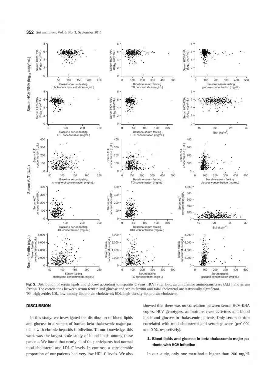
Fig. 3 has depicted serum lipid and glucose levels according to stage of liver fibrosis, necroinflammatory activity, siderosis and infecting genotypes. It is visually evident from Fig. 3 that there were no significant differences in serum lipids and glucose levels. To evaluate the probable association between glucose intolerance and stage of liver disturbances chi-square test was used. No association was found between presence of insulin re-sistance and stage and grade of liver disease and siderosis even after adjustment of sex.
Fig. 1. Distribution of blood lipids and glucose in 280 thalassemic patients with hepatitis C virus (HCV) infection. LDL, low-density lipoprotein cholesterol; HDL, high-density lipopro-tein cholesterol.
352 Gut and Liver, Vol. 5, No. 3, September 2011
DISCUSSION
In this study, we investigated the distribution of blood lipids and glucose in a sample of Iranian beta-thalassemic major pa-tients with chronic hepatitis C infection. To our knowledge, this work was the largest scale study of blood lipids among these patients. We found that nearly all of the participants had normal total cholesterol and LDL-C levels. In contrast, a considerable proportion of our patients had very low HDL-C levels. We also
showed that there was no correlation between serum HCV-RNA copies, HCV genotypes, aminotransferase activities and blood lipids and glucose in thalassemic patients. Only serum ferritin correlated with total cholesterol and serum glucose (p=0.001 and 0.02, respectively).
1. Blood lipids and glucose in beta-thalassemic major pa-tients with HCV infection
In our study, only one man had a higher than 200 mg/dL
Fig. 2. Distribution of serum lipids and glucose according to hepatitis C virus (HCV) viral load, serum alanine aminotransferase (ALT), and serum ferritin. The correlations between serum ferritin and glucose and serum ferritin and total cholesterol are statistically significant. TG, triglyceride; LDL, low-density lipoprotein cholesterol; HDL, high-density lipoprotein cholesterol.

Alavian SM, et al: Lipid Profiles and Hepatitis C Viral Markers in HCV-Infected Thalassemic Patients 353
level of total cholesterol. A recent report from Azizi et al.18 which enrolled healthy children and adolescents from Iran has suggested that 16% of males and females at our patients’ ages had high total cholesterol. According to the previous studies, it is known that patients with beta-thalassemia major have lower total cholesterol levels compared with healthy individuals of the same age.19 Based on the previous findings in thalassemic patients, we also observed low mean LDL-C levels in our sub-jects. It is of interest that just three subjects had higher than 95 mg/dL LDL-C levels. On the contrary, Azizi et al.18 reported that 17% of males and females with ages similar to our patients had above 130 mg/dL levels of LDL-C. Mean triglyceride was also low in our patients. Nineteen (7%) participants had triglyceride
levels of higher than 150 mg/dL. In our study, thalassemic men had higher total cholesterol and LDL-C than thalassemic women (p=0.001 and 0.02). This is reciprocal to Azizi et al.’s findings in healthy children and adolescents18 and others.20 The majority of our participants had very low HDL-C levels. It has previously been described that low HDL-C is the most common type of dyslipidemia in Iranian healthy adults.21 Hypocholesterolemia as well as low serum concentration of other lipids have been ex-plained in various chronic anemic disorders such as thalassemia major, thalassemia intermedia and aplastic anemia.22-24 Several mechanisms including plasma dilution resulting from anemia, increased cholesterol requirement associated with erythroid hy-perplasia, macrophage system activation with cytokine release,
Fig. 3. Distribution of the mean serum lipid and glucose levels according to hepatitis C virus (HCV) genotype, liver fibrosis, necroinflammatory activity, and liver iron deposition. None of the observed differences is statistically significant. TG, triglyceride; LDL, low-density lipoprotein cholesterol; HDL, high-density lipoprotein cholesterol; BMI, body mass index.

354 Gut and Liver, Vol. 5, No. 3, September 2011
increased cholesterol uptake by the reticuloendotial system, and liver injury secondary to iron overload have been proposed.25 In our study, we showed that chronic HCV infection does not alter pattern of serum lipids in thalassemic patients.
Forty-eight subjects (18%) had previous established diagnosis of diabetes mellitus or had impaired glucose tolerance in our study. Ghoddusi et al.26 and Hadaegh et al.27 previously reported the prevalence of diabetes in Iranian general population to be around 11.0%.26 Since we did not have control group, apply-ing binomial test showed glucose intolerance in our study to be significantly higher than what was reported in Iranian general population (p=0.001). Serum total cholesterol and triglyceride were significantly higher in subjects with serum glucose >110 mg/dL (p=0.02 and 0.006, respectively) which was similar to what Ghoddusi et al. have described in diabetic patients.26,27 It is determined that thalassemic patients had significantly higher insulin resistance than healthy controls.28 Dmochowski et al.28 have also explained that decreased hepatic extraction of insulin, but not higher excretion of pancreas beta-cells, is responsible for higher serum insulin level in thalassemic patients. Reduction in capability of liver to extract serum insulin as a result of in-flammation and fibrosis caused by HCV infection and resultant higher serum insulin as well as low age of our subjects can ex-plain our findings. We also indicated that distribution of mean serum fasting blood glucose in our participants is in border line of impaired tolerance. Hence, thalassemic patients with HCV infection may have very high rate of diabetes mellitus in 3rd or 4th decades of their lives. We also showed that serum glucose is significantly correlated with serum iron, and more effective iron chelation therapy in these patients can avert or postpone devel-opment of diabetes mellitus.
2. Implication of chronic HCV infection on serum lipids and glucose of thalassemic patients
In non-thalassemic HCV infected patients, it has previously been explained that there are association among metabolic syndrome, serum level of glucose, cholesterol, triglyceride and ALT levels as well as HCV viral load and genotype 1 and 2.29,30
Petit et al.31 have also described a correlation between hypo-betalipoproteinemia and liver fibrosis and viral load. Maeno et al.32 by applying insulin homeostasis assessment model on 56 non-thalassemic HCV infected subjects revealed that insulin resistance increased parallel with the progression of fibrosis. As presented in Figs. 2 and 3, in spite of enough sample size, we could not find any significant association of serum lipids and glucose with liver histologic findings, aminotransferase activity as well as serum HCV-RNA and HCV genotype in thalassemic patients with HCV infection.32 Siagris et al.3 have also revealed that steatosis in HCV infected thalassemic patients is lower than HCV infected patients.
We showed that in our thalassemic patients, serum glucose is not significantly associated with liver fibrosis, necroinflam-
matory and aminotransferase activities as well as virologic markers such as genotype and HCV-RNA. Genotype 2 had the highest mean blood glucose level. However, this difference was not significant applying one-way ANOVA. Serum glucose level had an increasing trend toward higher degrees of fibrosis and lower degrees of inflammation. This trend was not statistically significant (Fig. 3). Previously, it has been discovered that HCV infection in absence of liver cirrhosis can induce insulin resis-tance and liver iron deposition and TNF-alpha was introduced as culprit mechanisms.33,34 In our study on thalassemic patients, serum glucose was not different between various grades of liver siderosis, but it significantly correlated with serum ferritin. This implies that iron can cause peripheral insulin resistance besides hepatic injury reported in other studies.33,35 To our knowledge, relation of serum glucose and insulin resistance with HCV viral markers is not well understood. In thalassemic patients we could not find any relation either. However, this issue needs more mo-lecular and clinical investigations.
The present study revealed that HCV infected patients with beta-thalassemia major have blood lipid and lipoprotein levels within the normal range. An exception is the observed very low HDL -C levels. Viral markers, liver histologic findings as well as liver enzymes do not seem to play any role in determining serum cholesterol and triglyceride levels in thalassemic patients. Serum triglyceride, total cholesterol and serum iron can be in-dependent risk factors of glucose intolerance in HCV infected thalassemic patients.
CONFLICTS OF INTEREST
No potential conflict of interest relevant to this article was reported.
REFERENCES
1. Alavian SM, Ahmadzad-Asl M, Lankarani KB, Shahbabaie MA,
Bahrami Ahmadi A, Kabir A. Hepatitis C infection in the general
population of Iran: a systematic review. Hepat Mon 2009;9:211-
223.
2. Alavian SM, Tabatabaei SV, Lankarani KB. Epidemiology of HCV
infection among thalassemia patients in eastern Mediterranean
countries: a quantitative review of literature. Iran Red Crescent
Med J 2010;12:365-376.
3. Siagris D, Kouraklis-Symeonidis A, Christofidou M, et al. Serum
lipid profile and hepatic steatosis of adult beta-thalassaemia pa-
tients with chronic HCV infection. Eur J Gastroenterol Hepatol
2005;17:345-350.
4. Cicognani C, Malavolti M, Morselli-Labate AM, Zamboni L, Sama
C, Barbara L. Serum lipid and lipoprotein patterns in patients
with liver cirrhosis and chronic active hepatitis. Arch Intern Med
1997;157:792-796.
5. Maggi G, Bottelli R, Gola D, et al. Serum cholesterol and chronic

Alavian SM, et al: Lipid Profiles and Hepatitis C Viral Markers in HCV-Infected Thalassemic Patients 355
hepatitis C. Ital J Gastroenterol 1996;28:436-440.
6. Biró A, Horváth A, Varga L, et al. Serum anti-cholesterol antibod-
ies in chronic hepatitis-C patients during IFN-alpha-2b treatment.
Immunobiology 2003;207:161-168.
7. Minuk GY, Weinstein S, Kaita KD. Serum cholesterol and low-
density lipoprotein cholesterol levels as predictors of response
to interferon therapy for chronic hepatitis C. Ann Intern Med
2000;132:761-762.
8. Fabris C, Federico E, Soardo G, Falleti E, Pirisi M. Blood lipids of
patients with chronic hepatitis: differences related to viral etiology.
Clin Chim Acta 1997;261:159-165.
9. Agnello V, Abel G, Elfahal M, Knight GB, Zhang QX. Hepatitis C
virus and other flaviviridae viruses enter cells via low density lipo-
protein receptor. Proc Natl Acad Sci U S A 1999;96:12766-12771.
10. Cheng J, Li L. Low density lipoprotein receptor: a receptor re-
lated to hepatitis C virus. Zhonghua Gan Zang Bing Za Zhi
2003;11:190-192.
11. Wünschmann S, Medh JD, Klinzmann D, Schmidt WN, Stapleton
JT. Characterization of hepatitis C virus (HCV) and HCV E2 in-
teractions with CD81 and the low-density lipoprotein receptor. J
Virol 2000;74:10055-10062.
12. Chrysohoou C, Panagiotakos DB, Pitsavos C, et al. Distribution of
serum lipids and lipoproteins in patients with beta thalassaemia
major: an epidemiological study in young adults from Greece.
Lipids Health Dis 2004;3:3.
13. Al-Quobaili FA, Abou Asali IE. Serum levels of lipids and lipopro-
teins in Syrian patients with beta-thalassemia major. Saudi Med J
2004;25:871-875.
14. Rahimi Z, Merat A, Haghshenass M, Madani H, Rezaei M, Nagel
RL. Plasma lipids in Iranians with sickle cell disease: hypocholes-
terolemia in sickle cell anemia and increase of HDL-cholesterol in
sickle cell trait. Clin Chim Acta 2006;365:217-220.
15. Friedewald WT, Levy RI, Fredrickson DS. Estimation of the con-
centration of low-density lipoprotein cholesterol in plasma, with-
out use of the preparative ultracentrifuge. Clin Chem 1972;18:499-
502.
16. Yamaki K, Rimmer JH, Lowry BD, Vogel LC. Prevalence of obe-
sity-related chronic health conditions in overweight adolescents
with disabilities. Res Dev Disabil 2011;32:280-288.
17. Keshvari M, Alavian SM, Behnava B, et al. Distribution of hepati-
tis C virus genotypes in iranian patients with congenital bleeding
disorders. Iran Red Crescent Med J 2010;12:608-614.
18. Azizi F, Rahmani M, Madjid M, et al. Serum lipid levels in an
Iranian population of children and adolescents: Tehran Lipid and
Glucose Study. Eur J Epidemiol 2001;17:281-288.
19. Madani H, Rahimi Z, Manavi-Shad M, et al. Plasma lipids and
lipoproteins in children and young adults with major beta-thal-
assemia from western Iran: influence of genotype. Mol Biol Rep
2011;38:2573-2578.
20. Fesharakinia A, Zarban A, Sharifzadeh GR. Lipid profiles and
prevalence of dyslipidemia in schoolchildren in south Khorasan
Province, eastern Iran. Arch Iran Med 2008;11:598-601.
21. Sharifi F, Mousavinasab SN, Soruri R, Saeini M, Dinmohammadi
M. High prevalence of low high-density lipoprotein cholesterol
concentrations and other dyslipidemic phenotypes in an Iranian
population. Metab Syndr Relat Disord 2008;6:187-195.
22. Papanastasiou DA, Siorokou T, Haliotis FA. Beta-Thalassaemia
and factors affecting the metabolism of lipids and lipoproteins.
Haematologia (Budap) 1996;27:143-153.
23. Hartman C, Tamary H, Tamir A, et al. Hypocholesterolemia in
children and adolescents with beta-thalassemia intermedia. J Pedi-
atr 2002;141:543-547.
24. Yokoyama M, Suto Y, Sato H, et al. Low serum lipids suggest se-
vere bone marrow failure in children with aplastic anemia. Pediatr
Int 2000;42:613-619.
25. Shalev H, Kapelushnik J, Moser A, Knobler H, Tamary H. Hypo-
cholesterolemia in chronic anemias with increased erythropoietic
activity. Am J Hematol 2007;82:199-202.
26. Ghoddusi K, Ameli J, Kachuee H, Pourfarziani V, Saadat A, Kara-
mi Q. Association of diabetes mellitus and dyslipidaemias in the
Tehran population. East Mediterr Health J 2008;14:647-653.
27. Hadaegh F, Bozorgmanesh MR, Ghasemi A, Harati H, Saadat N,
Azizi F. High prevalence of undiagnosed diabetes and abnormal
glucose tolerance in the Iranian urban population: Tehran Lipid
and Glucose Study. BMC Public Health 2008;8:176.
28. Dmochowski K, Finegood DT, Francombe W, Tyler B, Zinman B.
Factors determining glucose tolerance in patients with thalassemia
major. J Clin Endocrinol Metab 1993;77:478-483.
29. Prati D, Shiffman ML, Diago M, et al. Viral and metabolic factors
influencing alanine aminotransferase activity in patients with
chronic hepatitis C. J Hepatol 2006;44:679-685.
30. Hsu CS, Liu CH, Liu CJ, et al. Association of lipid profiles with
hepatitis C viral load in chronic hepatitis C patients with genotype
1 or 2 infection. Am J Gastroenterol 2009;104:598-604.
31. Petit JM, Benichou M, Duvillard L, et al. Hepatitis C virus-associ-
ated hypobetalipoproteinemia is correlated with plasma viral load,
steatosis, and liver fibrosis. Am J Gastroenterol 2003;98:1150-
1154.
32. Maeno T, Okumura A, Ishikawa T, et al. Mechanisms of increased
insulin resistance in non-cirrhotic patients with chronic hepatitis C
virus infection. J Gastroenterol Hepatol 2003;18:1358-1363.
33. Wrede CE, Buettner R, Bollheimer LC, Schölmerich J, Palitzsch KD,
Hellerbrand C. Association between serum ferritin and the insulin
resistance syndrome in a representative population. Eur J Endocri-
nol 2006;154:333-340.
34. Sougleri M, Labropoulou-Karatza C, Paraskevopoulou P, Frago-
panagou H, Alexandrides T. Chronic hepatitis C virus infection
without cirrhosis induces insulin resistance in patients with alpha-
thalassaemia major. Eur J Gastroenterol Hepatol 2001;13:1195-
1199.
35. D’Souza RF, Feakins R, Mears L, Sabin CA, Foster GR. Relationship
between serum ferritin, hepatic iron staining, diabetes mellitus and
fibrosis progression in patients with chronic hepatitis C. Aliment
Pharmacol Ther 2005;21:519-524.