lentiviral vectors interfering with virus-induced cd4 down-modulation potently block human...
TRANSCRIPT
JOURNAL OF VIROLOGY, Dec. 2004, p. 13072–13081 Vol. 78, No. 230022-538X/04/$08.00�0 DOI: 10.1128/JVI.78.23.13072–13081.2004Copyright © 2004, American Society for Microbiology. All Rights Reserved.
Lentiviral Vectors Interfering with Virus-Induced CD4Down-Modulation Potently Block Human Immunodeficiency
Virus Type 1 Replication in Primary LymphocytesHang M. Pham,1 Enrique R. Arganaraz,1 Bettina Groschel,1 Didier Trono,2 and Juan Lama1,3*
Department of Medicine1 and Rebecca and John Moores UCSD Cancer Center,3
University of California at San Diego, School of Medicine, La Jolla, California, and Department ofGenetics and Microbiology, CMU, Faculty of Medicine, University of Geneva, Switzerland2
Received 7 May 2004/Accepted 28 July 2004
CD4 down-modulation is essential for the production of human immunodeficiency virus (HIV) infectiousparticles. Disease progression correlates with enhanced viral induced CD4 down-modulation, and a subset oflong-term nonprogressors carry viruses defective in this function. Despite multiple pieces of evidence high-lighting the importance of this function in viral pathogenesis in vivo, to date, HIV-induced CD4 down-modulation has not been used as a target for intervention. We describe here HIV-based vectors that delivertruncated CD4 molecules resistant to down-modulation by the viral products Nef and Vpu. Infection of cellspreviously transduced with these vectors proceeded normally, and viral particles were released in normalamounts. However, the infectivity of the released virions was reduced 1,000-fold. Lentiviral vectors expressingtruncated CD4 molecules were efficient at blocking HIV-1 infectivity and replication in several cell lines andin CD4-positive primary lymphocytes. The findings presented here provide proof-of-principle that approachestargeting the virus-induced CD4 down-modulation may constitute the basis for novel anti-HIV therapies.
CD4 plays a dual role during human immunodeficiency virus(HIV) infection. CD4 is required for entry of HIV into mostpermissive cells (14). However, during late stages of infection,the viral receptor exerts inhibitory effects on the infectivity ofthe released particles (25). To overcome these effects, HIV hasevolved mechanisms that ensure the removal of the CD4 re-ceptor from the surface of infected cells (reviewed in reference24). Three viral proteins participate in this process: Nef, Vpu,and Env. The effects of Nef (early product) and Vpu/Env (lateproducts) are quantitative and qualitatively distinct. Nef en-hances CD4 internalization from the cell surface and targetsthe receptor for degradation into lysosomes, whereas Env in-terferes with transport of CD4 to the cell surface, and Vputargets CD4 for degradation in proteasomes (16, 33, 34). Un-like Nef, Env and Vpu exert their effect only on newly synthe-sized receptor molecules. Nef acts as a connector, by bridgingtogether the cytoplasmic domain of CD4 with the heterotet-rameric clathrin adaptor protein complexes AP-1, AP-2, andAP-3 (6, 13, 18, 28, 32, 38; reviewed in references 11 and 14).Nef also binds directly to the cytoplasmic domain of the CD4receptor (19, 35, 36). This domain is sufficient to confer Nef-induced down-modulation to heterologous proteins (1, 3). Bybinding to the cytoplasmic domain of CD4 and to componentsof the cellular trafficking machinery, Nef targets the viral re-ceptor to specific trafficking pathways. The interaction betweenNef and AP-2, which is involved in endocytosis from theplasma membrane, appears weak. However, direct interactionsof Nef with AP-1 and AP-3 suggest that in addition to enhanc-ing endocytosis, Nef may also exert its effects by targeting CD4
to the endosomal/lysosomal system and preventing the recy-cling of CD4 to the cell surface (21). Similar to Nef, Vpu alsoacts as a connector between CD4 and the cellular degradationmachinery. Vpu interacts with the cytoplasmic domain of CD4and h-�TrCP, a key connector in the ubiquitin-mediated cel-lular proteolytic machinery (29).
Studies with simian immunodeficiency virus (SIV) Nef havehighlighted the importance of this protein in viral pathogene-sis. SIVs carrying deletions in nef replicate poorly in rhesusmacaques and usually do not cause AIDS (22). However, it hasbeen difficult to determine which of the in vitro functions ofNef contribute to the pathogenesis of HIV or SIV in vivo(reviewed in reference 41). Other findings have highlighted theimportance of CD4 down-modulation in virus pathogenesis invivo. Mutations in SIV Nef that disrupt the ability to down-modulate CD4 strongly reduce viremia in infected monkeysand revert quickly to restore the CD4 down-modulation func-tion (20). Insights into the role of CD4 down-modulation inHIV pathogenesis have also come from analysis of long-termnonprogressors (LTNPs). One study has shown that nef allelesisolated from LTNPs are less efficient in CD4 down-modula-tion than those isolated from progressors or asymptomaticcarriers (40). Another report has shown that nef alleles isolatedduring “early” asymptomatic stages of infection poorly down-modulate CD4. In comparison, “late” nef alleles isolated afterprogression to AIDS show enhanced ability to down-modulatethe HIV receptor (8). Recent findings have directly correlatedthe activity of Nef to down-modulate CD4 with enhanced viralreplication in CD4-positive cells, including primary lympho-cytes (4, 17, 27). Supporting these observations, overexpressionof CD4 in HIV producer cells interferes with Env function andleads to drastic reductions in viral infectivity (25).
The above findings suggest that CD4 down-modulation mayplay an important role in AIDS pathogenesis and imply that
* Corresponding author. Present address: La Jolla Institute for Mo-lecular Medicine, 4570 Executive Dr., Suite 100, San Diego, CA 92121.Phone: (858) 232-7919. Fax: (858) 587-6742. E-mail: [email protected].
13072
on March 29, 2015 by guest
http://jvi.asm.org/
Dow
nloaded from
interfering with this function might delay progression to dis-ease and result in clinical benefits. To date, the HIV-induceddown-modulation of CD4 has not been targeted for interven-tion, and no specific inhibitors of this viral function have beencharacterized. We describe here a strategy to interfere withthis viral function. Our results indicate that lentivirus-mediateddelivery of CD4 molecules resistant to HIV-induced down-modulation potently reduces viral infectivity and replication inCD4-positive cells, including primary lymphocytes. These find-ings demonstrate proof-of-concept that specific inhibition ofthis function may constitute the basis for novel approaches totreat HIV infection.
MATERIALS AND METHODS
Cells. 293T, MAGIC5, and MAGIC5B cells were grown in Dulbecco’s mod-ified Eagle’s medium supplemented with 10% fetal calf serum (FCS). MAGIC-5and MAGIC-5B cells (kindly provided by M. Matsuda, Osaka University) areCD4-positive CCR5-positive derivatives of HeLa cells (39). These cells containan integrated copy of the �-galactosidase gene under the control of the HIV-1long terminal repeat (LTR) promoter. MAGIC5B cells express CD4 receptorlevels 12-fold higher than MAGIC5 cells (4). Transformed T-cell lines (SupT1,C8166, and Jurkat Low-CD4) were maintained in RPMI 1640 medium supple-mented with 10% FCS. Ficoll-purified peripheral blood mononuclear cells(PBMCs) were isolated from healthy donors and cultured in RPMI 1640 mediumsupplemented with 10% human AB serum. PBMCs were activated with 5-�g/mlphytohemagglutinin (PHA) (Sigma) for 2 days prior to transduction with lenti-viral vectors or infection with HIV-1. After PHA stimulation, cells were main-tained in RPMI 1640 medium supplemented with 10% human AB serum con-taining 50 U of interleukin-2 (IL-2)/ml. All culture media used here weresupplemented with 100 U of penicillin/ml, 100 �g of streptomycin/ml, 1 mMsodium pyruvate, and 2 mM glutamine.
Construction of lentiviral vectors expressing modified CD4 molecules. Toengineer lentiviral constructs expressing CD4 variants, we utilized a PCR-basedapproach to amplify either full-length CD4 (CD4WT), or CD4 lacking its cyto-plasmic domain (CD4�cyt). The latter construct expresses a truncated CD4protein in which the transmembrane domain of wild-type CD4 (ending with Valat position 395) is followed by a short 6-amino-acid cytoplasmic domain (se-quence GSWPAS). Similarly, we also engineered a CD4 chimera in which theextracellular and transmembrane domains of CD4 were fused to the HIV-1 MAprotein (CD4-MA). The CD4 variants were PCR amplified and cloned into thelentiviral transfer vectors PPT-PGK-GFP, pHR�-CMV-GFP-WPRE, and pWPI.PPT-PGK-GFP is a lentiviral vector with a chimeric Rous sarcoma virus (RSV)-HIV 5�-LTR and a deletion in the 3�-LTR that renders the vector self-inactivat-ing (SIN) (15). Expression of the transgene is driven by the human phospoglyc-erate kinase (PGK) promoter. pWPI is also a SIN lentiviral vector in which thegene of interest is transcribed from an internal human EF1-alpha promoter. Thetranscribed unit also contains an internal ribosome entry site (IRES) elementfrom encephalomyocarditis virus that allows internal initiation of translation ofgreen fluorescent protein (GFP). In pHR�-CMV-CD4-WPRE, the transgene isexpressed from the internal human cytomegalovirus (CMV) immediate-earlypromoter (30). Unlike PPT-PGK-CD4 and pWPI, pHR�-CMV-CD4-WPRE canbe mobilized upon infection with replication-competent HIV. All lentiviral vec-tors used here contain a posttranscriptional regulatory element from the wood-chuck hepatitis virus (WPRE) that enhances expression of transgenes (44).
Vector particle production. For preparation of HIV-based lentiviral vectorparticles coated with the vesicular stomatitis virus glycoprotein (VSV G) we useda previously described three-plasmid cotransfection procedure (30). 293T cellswere cotransfected with 10 �g of CD4 lentiviral vector (PPT, pWPI, or pHR�based), together with 10 �g of a packaging construct (pCMV�R8.91) (45), and2.5 �g of a plasmid encoding VSV G (pMD.G). Forty-eight hours later, vectorparticles were collected from culture supernatants, filtered through 0.45-�m-pore-size nitrocellulose membranes, aliquoted, and frozen to �80°C until use.All transductions described here were performed with unpurified vector-contain-ing supernatants from 293T cells.
Transduction with lentiviral vectors and viral replication assays. 293T,MAGIC-5B, SUPT-1, C8166, and PBMCs were transduced following a previ-ously described spinoculation procedure (12). Briefly, 0.5 � 106 cells were incu-bated with viral or vector supernatants (200 to 500 ng of p24 antigen) in 24-wellplates in the presence of 4-�g/ml Polybrene and 10 mM HEPES (pH 7.4).
Mixtures of cells and vectors were centrifuged at room temperature for 90 min(2,500 rpm) in a tabletop centrifuge (Sorvall RT6000B). After centrifugation,cells were washed with propagation medium and then incubated in the samemedium for 2 to 3 days at 37°C. Transduced cells were infected with HIV-1 (1 �gof p24 protein) following the above protocol. Viral replication assays in PBMCswere performed by incubating cells with supernatant-containing viruses in theabsence of Polybrene. Routinely, PBMCs were activated for 48 h with 5-�g/mlPHA, transduced with lentiviral vectors, and sorted for GFP expression 2 to 3days later. Three to 4 days after sorting, 106 transduced PBMCs were infectedwith 2,000 50% tissue culture infective doses (without additional PHA treat-ment) in 150 �l of propagation media. After 2 h at 37°C, cells were washed twiceand distributed into 96-well plates (2 � 105/well). Cells were maintained in thepresence of 50 U of IL-2/ml. Replication assays in Jurkat Low-CD4 cells wereperformed by incubating 106 cells with HIV-1 R9 (50 ng of p24).
HIV-1 virus production. Infectious HIV-1 (R9) was produced by transienttransfection of 293T cells with HIV-1 proviral clones. VSV G-pseudotyped R9HIV-1 was generated by cotransfecting proviral DNA (10 to 20 �g) with 2.5 �gof pMD.G plasmid DNA. The CCR5-tropic HIV-1 BaL virus was harvested andpurified from the supernatant of infected monocyte-derived macrophages.
Single-round infectivity assays. Infectivity assays were performed withMAGIC5 cells using equal amounts of input virus (measured as nanograms ofp24 protein) in the presence of 20-�g/ml DEAE dextran. Infection was allowedfor 48 h. Routinely, after 8 h of infection, 1 �M zidovudine (AZT) was added toblock second rounds of virus replication. Infected cells were scored after stainingfor �-galactosidase with X-Gal (5-bromo-4-chloro-3-indolyl-�-D-galactopyrano-side).
Protein analysis. Incorporation of cellular and viral proteins in HIV particleswas determined by Western blot with the following antibodies (NIH AIDSResearch Reference Reagent Program). For HIV-1 Env, a gp120-specific sheepantiserum was used. For CD4, the T4-4 rabbit antiserum was used. gp41 wasprobed with the 2F5 human monoclonal antibody (MAb), and p24 was analyzedwith the 183-H12-5C murine MAb. Loading controls for cell lysates were per-formed by probing with an �-tubulin MAb (Sigma). Virion-associated proteinswere pelleted through a 20% sucrose cushion at 26,000 rpm in a SW28 rotor(Beckman) for 1.5 h at 4°C. Equal amounts of p24 protein were separated ontoa sodium dodecyl sulfate–10%-polyacrylamide gel, transferred to polyvinylidenedifluoride membrane, and then probed with specific antibodies. Antibody bind-ing was detected by the Amersham ECL enhanced chemiluminescence Westernblotting kit, using horseradish peroxidase-conjugated secondary antibodies (Am-ersham Bioscience, Inc.).
Flow cytometry and cell sorting. Surface staining of CD4 was performed witheither the OKT4 MAb, followed by addition of goat anti-mouse Cy5-conjugatedantibody, or with CD4-V4 MAb (phycoerythrin [PE] conjugated; Becton Dick-inson). The epitopes recognized by these MAbs do not overlap with the gp120-binding domain (37). Intracellular staining of HIV-1 Gag was performed with ap24-specific MAb (fluorescein isothiocyanate [FITC]-conjugated KC57 clone;Coulter), using a cell permeabilization kit following the recommendations of themanufacturer (Caltag Lab). PBMCs transduced with lentiviral vectors weresorted for enrichment of double-positive (GFP-positive/CD4-positive) cells. PB-MCs were resuspended at 20 � 106 cells/ml in PBS with 2% FCS and stained witha CD4 MAb (CD4-PE-Cy5 conjugated; DAKO) at room temperature for 15 min.Then the cells were washed twice and resuspended in RPMI supplemented with5% human AB serum at 10 to 25 � 106 cells/ml. Cells were immediately sortedwith a FACSVantage SE system (Becton Dickinson), collected in RPMI supple-mented with 20% human AB serum, centrifuged, and resuspended in propaga-tion media supplemented with 50 U of IL-2/ml.
RESULTS
Lentiviral vectors expressing CD4 molecules resistant tomodulation by HIV-1 Nef and Vpu. Since Nef and Vpu requirethe cytoplasmic domain of CD4 to connect the viral receptorwith cellular degradation pathways, we hypothesized that trun-cated CD4 molecules lacking the cytoplasmic domain would bepoorly down-modulated by HIV-1 and subsequently inhibit theinfectivity of the newly synthesized particles. We designed len-tiviral vectors delivering full-length CD4 (wild type), CD4 lack-ing its cytoplasmic domain (CD4�cyt), or the extracellular andtransmembrane domains of CD4 fused to the full-lengthHIV-1 matrix protein (CD4-MA). Production of these engi-
VOL. 78, 2004 INTERFERENCE WITH CD4 DOWN-MODULATION 13073
on March 29, 2015 by guest
http://jvi.asm.org/
Dow
nloaded from
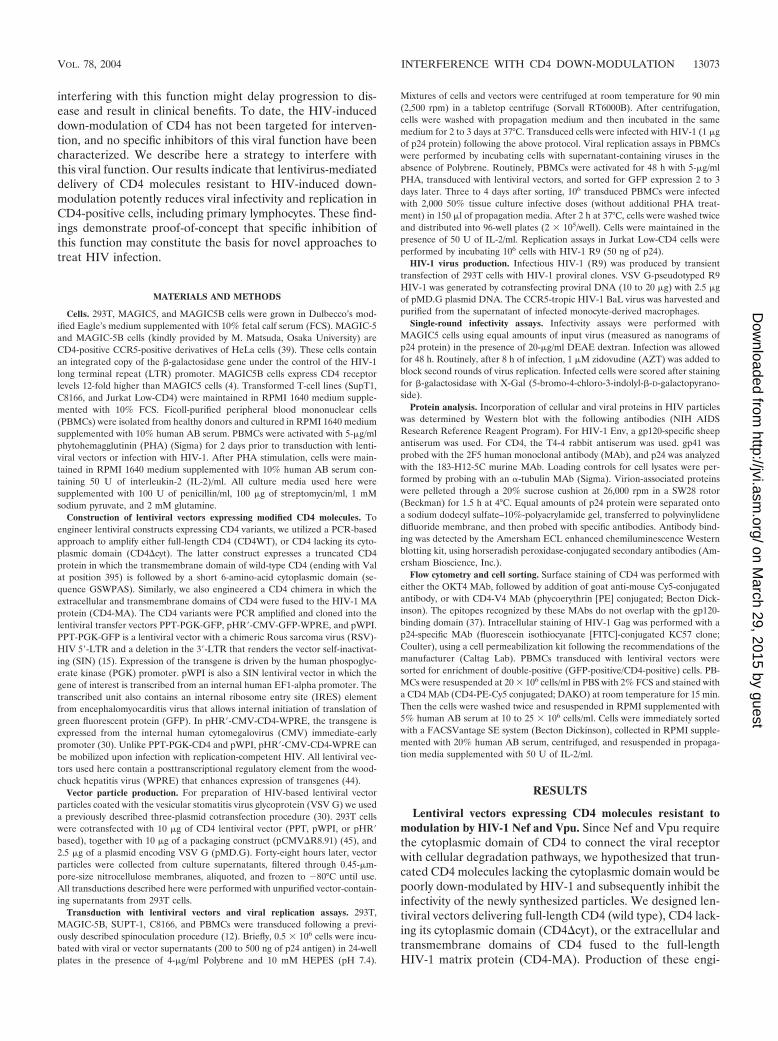
neered proteins was confirmed in 293T cells transduced withthese vectors (Fig. 1A). CD4-MA fusion protein was ex-pressed at lower levels than full-length or truncated CD4.This was more evident in cells transduced with PPT-PGKvectors. The reason for this is unknown. To test the ability ofthe various CD4 proteins to be down-modulated by HIV-1Nef, 293T cells transduced with lentiviral vectors were thentransfected with plasmids expressing either the HIV-1 NA7primary nef allele fused to GFP (Nef-GFP) or GFP alone(Fig. 1B and 1C, respectively). Expression of Nef-GFP de-creased 15-fold the surface levels of full-length CD4, but didnot alter expression of CD4�cyt or CD4-MA, consistentwith the requirement of the CD4 cytoplasmic domain for
efficient Nef-induced down-modulation. Elevated levels ofCD4 have been previously shown to interfere with HIVproduction in an HIV-1 Env-independent manner (5, 12).To test whether expression of CD4 affects vector produc-tion, we estimated the amount of viral p24 protein in cul-tures of 293T producer cells. 293T cells were cotransfectedwith packaging and VSV G plasmids, together with eithercontrol transfer vectors (GFP) or vectors expressing theCD4 variants. As shown in Fig. 1D, no significant reductionin p24 released was observed with any of the vectors (PPT-PGK- or pHR�-CMV-based), suggesting that expression ofthe various CD4 transgenes does not interfere with vectorproduction in 293T cells.
FIG. 1. Lentiviral vectors expressing CD4 molecules resistant to the down-modulatory effect mediated by HIV-1 Nef and Vpu. (A) Schematicdrawing of the lentiviral vectors PPT-PGK-CD4 and pHR�-CMV-CD4. These vectors were used to express GFP alone (GFP), full-length CD4(CD4WT), CD4 lacking its cytoplasmic domain (CD4�cyt), or the extracellular and transmembrane domain of CD4 fused to HIV-1 matrix(CD4-MA). Lysates of 293T cells transduced with these vectors were analyzed by sodium dodecyl sulfate-polyacrylamide gel electrophoresis-Western blot with CD4-specific antibodies. In panels B and C, 293T cells were transduced with pHR�-CMV vectors expressing either wild-typeCD4, CD4-MA, or CD4�cyt and transfected with a plasmid expressing Nef (NA7) fused to GFP (B) or GFP alone (C). Histograms represent CD4levels in GFP-positive cells. Results for an IgG isotypic control are shown as dotted lines. (D) Vector production in 293T cells was analyzed 48 hafter transfection by estimating the levels of HIV-1 p24 antigen in culture supernatants.
13074 PHAM ET AL. J. VIROL.
on March 29, 2015 by guest
http://jvi.asm.org/
Dow
nloaded from
Suppression of HIV-1 infectivity by lentiviral vectors ex-pressing truncated CD4 proteins. Interference with HIV-1infectivity was first analyzed in CD4-negative 293T cells. Thesecells express the CXCR4 coreceptor, and upon transductionwith CD4 lentiviral vectors, they become permissive to infec-tion with HIV-1. This strategy ensured that release of HIV-1particles would occur only from the lentiviral vector-trans-duced cells. We were interested in evaluating the inhibitoryeffect of the vectors under optimal conditions of HIV infection,in which overexpression of Nef, Vpu, and Env could overcome
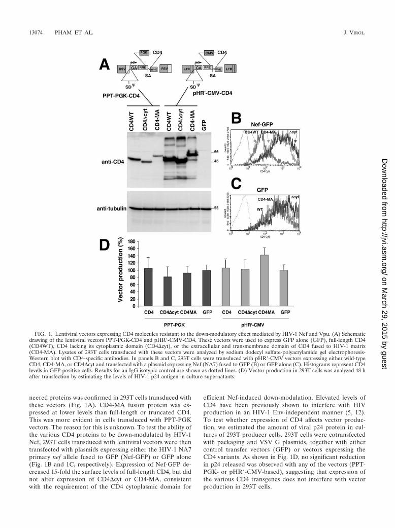
the block imposed by CD4. To achieve this goal, we infectedcells with HIV-1 R9 (1,000 ng of p24) following a spinoculationprocedure (31). We also evaluated whether the above lentiviralvectors could inhibit the infectivity of VSV G-pseudotypedHIV-1 [HIV-1(vsv)], which infects cells to a higher extent thanHIV-1 particles using the natural viral envelope (Env) forentry. Figure 2A shows that more than 90% of the cells becametransduced and expressed CD4, as estimated by surface stain-ing with anti-CD4 antibodies. Levels of surface CD4 weresignificantly lower in the p24-negative cells present in HIV-
FIG. 2. Suppression of HIV-1 infectivity by lentiviral vectors expressing CD4 variants. (A) Twenty-four hours after transduction with pHR�-CMV lentiviral vectors expressing CD4 variants, 293T cells were either mock infected (left columns) or infected with HIV-1 (R9) (middle columns)or HIV-1 R9 pseudotyped with VSV G (right columns). After 48 h of infection, CD4 surface expression and intracellular accumulation of HIVp24 protein were estimated by flow cytometry. Mock-infected cells were used to set up staining levels for p24-negative cells. p24-positiveHIV-infected cells are shown in gray dots. Inlet numbers indicate mean values of CD4 expression in p24-negative, and p24-positive infected cells(upper and lower inlets, respectively). (B) Infectivity of the released particles was estimated in MAGIC5 cells infected with equal amounts of p24input virus. Control infection was performed with virus produced in 293T cells infected with HIV-1 pseudotyped with VSV G. The effect of additionof 1 �M AZT before infection of MAGIC5 cells with HIV-1(vsv) is also shown.
VOL. 78, 2004 INTERFERENCE WITH CD4 DOWN-MODULATION 13075
on March 29, 2015 by guest
http://jvi.asm.org/
Dow
nloaded from
treated cultures than in mock-infected cultures (Fig. 2A, com-pare the values 725 versus 1,049 in CD4-transduced cells). Thisis likely due to the fact that Nef is incorporated into viralparticles. The amount of Nef protein encapsidated into virionsis enough to down-modulate surface CD4 even in the presenceof reverse transcriptase or integrase inhibitors (43; and datanot shown). This fact is more notable in infections with HIV-1(vsv), which resulted in p24-negative cells with very low levelsof CD4. Furthermore, cells expressing higher levels of CD4 aremore susceptible to infection with HIV-1. Upon infection,these cells move out from the p24-negative region, furthercontributing to the impression that CD4 down-modulation oc-curs in p24-negative cells. Upon infection with HIV-1, full-length CD4 was efficiently down-modulated in p24-positivecells, presumably by the combined action of Nef, Vpu, and Envproteins (Fig. 2A, top-middle plot). In contrast, the levels ofreceptor remained elevated in HIV-1-infected cells transducedwith CD4�cyt and CD4-MA, with surface levels 11- and 7-foldhigher, respectively, than those observed in infected cells trans-duced with wild-type CD4.
As expected, pseudotyping HIV-1 with VSV G led to en-hanced infection (4- to 5-fold higher, estimated as percentageof p24-positive cells) and higher levels of p24 expression ininfected cells (52% higher). Consequently, the level of down-modulation of full-length CD4 occurred to higher extents. Un-der these conditions, surface levels of CD4-MA were partiallyreduced, as compared to cells infected with HIV-1. However,CD4�cyt molecules still remained completely resistant todown-modulation by HIV-1(vsv) (Fig. 2A, right-center panel).These results demonstrate the feasibility of a lentiviral vectorsystem to achieve elevated levels of expression of CD4. Ourfindings also demonstrate that expression of Env in HIV-1-infected cells is not sufficient to down-modulate receptor mol-ecules insensitive to the action of Nef and Vpu.
293T cells tranduced with CD4 vectors and then infectedwith HIV-1 did not show significant differences in the releaseof viral particles, estimated by p24 enzyme-linked immunosor-bent assay (ELISA), as compared to mock-tranduced cells(data not shown). The infectivity of HIV-1 particles producedfrom 293T-transduced cells was analyzed in single-round in-fectivity assays in MAGIC5 cells. MAGIC5 cells were chal-lenged with equal amounts of p24, and the number of blue fociwas estimated (Fig. 2B). Lentiviral-based expression ofCD4�cyt reduced the infectivity of HIV-1 by more than 1,000-fold. The extent of inhibition was comparable to that observedby addition to target cells of 1 �M AZT, an inhibitor of theHIV reverse transcriptase (Fig. 2B). These results prove theability of these lentiviral vectors to efficiently block HIV-1infectivity in the presence of a full set of CD4 down-modulatorgenes (nef, vpu, and env). Particles synthesized in cells trans-duced with CD4-MA expressed smaller amounts of the fusionprotein on the surface of infected cells, as compared toCD4�cyt (mean value: 1,604 versus 2,587, respectively), butstill reduced HIV-1 infectivity by 88%. Interestingly, the viralprogeny from cells transduced with CD4�cyt and then infectedwith VSV G-pseudotyped HIV-1 showed severely reduced in-fectivity (100-fold), suggesting that higher levels of viral prod-ucts were not sufficient to overcome the inhibitory effects oftruncated CD4. Expression of full-length CD4 by itself did notaffect HIV-1 infectivity, suggesting that the inhibitory effects
mediated by CD4�cyt and CD4-MA were likely due to theexpression of the transgenes and not to the lentiviral vectors,which might interfere with viral replication at different stages(7, 23).
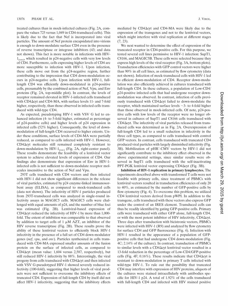
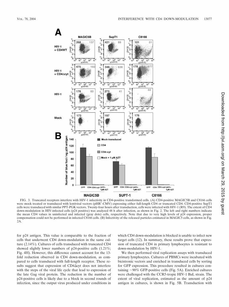
We next wanted to determine the effect of expression of thetruncated receptor in CD4-positive cells. For this purpose, wetested several cell lines permissive to HIV-1 infection: SupT1,C8166, and MAGIC5B. These cells were selected because theyexpress high levels of the viral receptor (Fig. 3A, bottom plots).Transduction efficiencies with GFP control vectors were higherthan 90% in all cell lines, as estimated by flow cytometry (datanot shown). Infection of mock-transduced cells with HIV-1 ledto efficient down-modulation of CD4. Receptor down-modu-lation was also efficiently achieved in cultures transduced withfull-length CD4. In these cultures, a population of Low-CD4/p24-positive infected cells that had undergone receptor down-modulation was observed. In contrast, infection of cells previ-ously transduced with CD4�cyt failed to down-modulate thereceptor, which maintained surface levels 3- to 4-fold higherthan those observed in mock-infected cells. Of note, p24-pos-itive cells with low levels of the receptor were no longer ob-served in cultures of SupT1 and C8166 cells transduced withCD4�cyt. The infectivity of viral particles released from trans-duced cells was determined as in Fig. 2A. Overexpression offull-length CD4 led to a small reduction in infectivity in thethree cell types, as compared to cells transduced with controlGFP vectors. In contrast, cells transduced with truncated CD4produced viral particles with largely diminished infectivity (Fig.3B). Mobilization of pHR�-CMV vectors by HIV-1 did notsignificantly contribute to the inhibitory effect observed in theabove experimental settings, since similar results were ob-served in SupT1 cells transduced with the self-inactivatingPPT-PGK lentiviral vector expressing CD4�cyt (Fig. 3B).
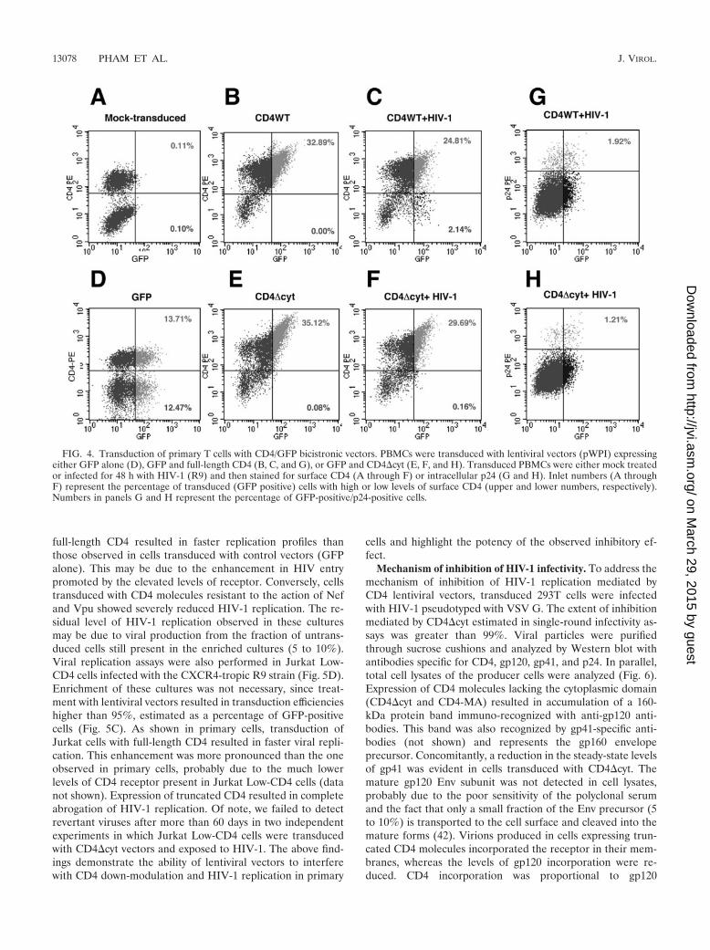
Inhibition of HIV-1 replication in primary lymphocytes. Theexperiments described above with transformed T cells were notfeasible with primary cells, since treatment of PBMCs withlentiviral vectors resulted in transduction efficiencies of only 20to 40%, as estimated by the number of GFP-positive cells byflow cytometry (Fig. 4). To overcome this problem, we utilizedCD4 lentiviral vectors derived from pWPI. In addition to thetransgene, cells transduced with these vectors also express GFPunder the control of an IRES element. Transduced cells canthus be readily identified by gating GFP-positive cells. Primarycells were transduced with either GFP alone, full-length CD4,or with the most potent inhibitor of HIV infectivity, CD4�cyt.Three days after transduction with bicistronic vectors, PBMCswere infected with HIV-1 (R9) and analyzed by flow cytometryfor surface CD4 and GFP fluorescence (Fig. 4). Infection withHIV-1 resulted in the appearance of a population of GFP-positive cells that had undergone CD4 down-modulation (Fig.4C; 2.14% of the culture). In contrast, transduction of PBMCsto similar levels with a CD4�cyt lentiviral vector resulted in a13-fold reduction in the percentage of Low-CD4/GFP-positivecells (Fig. 4F; 0.16%). These results indicate that CD4�cyt isresistant to down-modulation in primary T cells infected withwild-type HIV-1. To rule out the possibility that truncatedCD4 may interfere with expression of HIV proteins, aliquots ofthe cultures were stained intracellularly with antibodies spe-cific for HIV-1 p24. A total of 1.92% of the cells transducedwith full-length CD4 and infected with HIV stained positive
13076 PHAM ET AL. J. VIROL.
on March 29, 2015 by guest
http://jvi.asm.org/
Dow
nloaded from
for p24 antigen. This value is comparable to the fraction ofcells that underwent CD4 down-modulation in the same cul-ture (2.14%). Cultures of cells transduced with truncated CD4showed slightly lower numbers of p24-positive cells (1.21%;Fig. 4H). However, this difference cannot account for the 13-fold reduction observed in CD4 down-modulation, as com-pared to cells transduced with full-length receptor. These re-sults suggest that expression of CD4�cyt does not interferewith the steps of the viral life cycle that lead to expression ofthe late Gag viral protein. The reduction in the number ofp24-positive cells is likely due to a block in second rounds ofinfection, since the output virus produced under conditions in
which CD4 down-modulation is blocked is unable to infect newtarget cells (12). In summary, these results prove that expres-sion of truncated CD4 in primary lymphocytes is resistant todown-modulation by HIV-1.
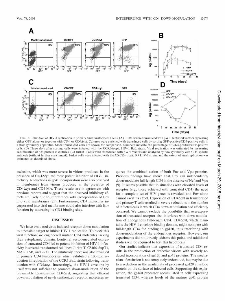
We then performed viral replication assays with transducedprimary lymphocytes. Cultures of PBMCs were incubated withbicistronic vectors and enriched in transduced cells by sortingfor GFP expression. This procedure resulted in cultures con-taining 90% GFP-positive cells (Fig. 5A). Enriched cultureswere challenged with the CCR5-tropic HIV-1 BaL strain. Theextent of viral replication, estimated as the amount of p24antigen in cultures, is shown in Fig. 5B. Transduction with
FIG. 3. Truncated receptors interfere with HIV-1 infectivity in CD4-positive transformed cells. (A) CD4-positive MAGIC5B and C8166 cellswere mock treated or transduced with lentiviral vectors (pHR�-CMV) expressing either full-length CD4 or truncated CD4. CD4-positive SupT1cells were transduced with similar PPT-PGK vectors. Twenty-four hours after transduction, cells were infected with HIV-1 (R9). The extent of CD4down-modulation in HIV-infected cells (p24 positive) was analyzed 48 h after infection, as shown in Fig. 2. The left and right numbers indicatethe mean CD4 values in uninfected and infected (gray dots) cells, respectively. Note that due to very high levels of p24 expression, propercompensation could not be performed in infected C8166 cells. (B) Infectivity of the released particles estimated in MAGIC5 cells, as shown in Fig.2A.
VOL. 78, 2004 INTERFERENCE WITH CD4 DOWN-MODULATION 13077
on March 29, 2015 by guest
http://jvi.asm.org/
Dow
nloaded from
full-length CD4 resulted in faster replication profiles thanthose observed in cells transduced with control vectors (GFPalone). This may be due to the enhancement in HIV entrypromoted by the elevated levels of receptor. Conversely, cellstransduced with CD4 molecules resistant to the action of Nefand Vpu showed severely reduced HIV-1 replication. The re-sidual level of HIV-1 replication observed in these culturesmay be due to viral production from the fraction of untrans-duced cells still present in the enriched cultures (5 to 10%).Viral replication assays were also performed in Jurkat Low-CD4 cells infected with the CXCR4-tropic R9 strain (Fig. 5D).Enrichment of these cultures was not necessary, since treat-ment with lentiviral vectors resulted in transduction efficiencieshigher than 95%, estimated as a percentage of GFP-positivecells (Fig. 5C). As shown in primary cells, transduction ofJurkat cells with full-length CD4 resulted in faster viral repli-cation. This enhancement was more pronounced than the oneobserved in primary cells, probably due to the much lowerlevels of CD4 receptor present in Jurkat Low-CD4 cells (datanot shown). Expression of truncated CD4 resulted in completeabrogation of HIV-1 replication. Of note, we failed to detectrevertant viruses after more than 60 days in two independentexperiments in which Jurkat Low-CD4 cells were transducedwith CD4�cyt vectors and exposed to HIV-1. The above find-ings demonstrate the ability of lentiviral vectors to interferewith CD4 down-modulation and HIV-1 replication in primary
cells and highlight the potency of the observed inhibitory ef-fect.
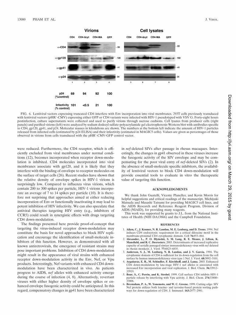
Mechanism of inhibition of HIV-1 infectivity. To address themechanism of inhibition of HIV-1 replication mediated byCD4 lentiviral vectors, transduced 293T cells were infectedwith HIV-1 pseudotyped with VSV G. The extent of inhibitionmediated by CD4�cyt estimated in single-round infectivity as-says was greater than 99%. Viral particles were purifiedthrough sucrose cushions and analyzed by Western blot withantibodies specific for CD4, gp120, gp41, and p24. In parallel,total cell lysates of the producer cells were analyzed (Fig. 6).Expression of CD4 molecules lacking the cytoplasmic domain(CD4�cyt and CD4-MA) resulted in accumulation of a 160-kDa protein band immuno-recognized with anti-gp120 anti-bodies. This band was also recognized by gp41-specific anti-bodies (not shown) and represents the gp160 envelopeprecursor. Concomitantly, a reduction in the steady-state levelsof gp41 was evident in cells transduced with CD4�cyt. Themature gp120 Env subunit was not detected in cell lysates,probably due to the poor sensitivity of the polyclonal serumand the fact that only a small fraction of the Env precursor (5to 10%) is transported to the cell surface and cleaved into themature forms (42). Virions produced in cells expressing trun-cated CD4 molecules incorporated the receptor in their mem-branes, whereas the levels of gp120 incorporation were re-duced. CD4 incorporation was proportional to gp120
FIG. 4. Transduction of primary T cells with CD4/GFP bicistronic vectors. PBMCs were transduced with lentiviral vectors (pWPI) expressingeither GFP alone (D), GFP and full-length CD4 (B, C, and G), or GFP and CD4�cyt (E, F, and H). Transduced PBMCs were either mock treatedor infected for 48 h with HIV-1 (R9) and then stained for surface CD4 (A through F) or intracellular p24 (G and H). Inlet numbers (A throughF) represent the percentage of transduced (GFP positive) cells with high or low levels of surface CD4 (upper and lower numbers, respectively).Numbers in panels G and H represent the percentage of GFP-positive/p24-positive cells.
13078 PHAM ET AL. J. VIROL.
on March 29, 2015 by guest
http://jvi.asm.org/
Dow
nloaded from
exclusion, which was more severe in virions produced in thepresence of CD4�cyt, the most potent inhibitor of HIV-1 in-fectivity. Reductions in gp41 incorporation were also observedin membranes from virions produced in the presence ofCD4�cyt and CD4-MA. These results are in agreement withprevious reports and suggest that the observed inhibitory ef-fects are likely due to interference with incorporation of Envinto viral membranes (25). Furthermore, CD4 molecules in-corporated into viral membranes could also interfere with Envfunction by saturating its CD4 binding sites.
DISCUSSION
We have evaluated virus-induced receptor down-modulationas a possible target to inhibit HIV-1 replication. To block thisviral function, we engineered mutant CD4 molecules lackingtheir cytoplasmic domain. Lentiviral vector-mediated expres-sion of truncated CD4 led to potent inhibition of HIV-1 infec-tivity in several transformed cell lines: Jurkat T, C8166, SupT1,MAGIC5B, and 293T. The inhibitory effect was also revealedin primary CD4 lymphocytes, which exhibited a 100-fold re-duction in replication of the CCR5 BaL strain following trans-duction with CD4�cyt. Interestingly, the HIV-1 envelope byitself was not sufficient to promote down-modulation of thepresumably Env-sensitive CD4�cyt, suggesting that efficientdown-modulation of newly synthesized receptor molecules re-
quires the combined action of both Env and Vpu proteins.Previous findings have shown that Env can independentlydown-modulate full-length CD4 in the absence of Nef and Vpu(9). It seems possible that in situations with elevated levels ofreceptor (e.g., those achieved with truncated CD4) the needfor a complete set of HIV genes is revealed, and Env alonecannot exert its effect. Expression of CD4�cyt in transformedand primary T cells resulted in severe reductions in the numberof infected cells in which CD4 down-modulation had efficientlyoccurred. We cannot exclude the possibility that overexpres-sion of truncated receptor also interferes with down-modula-tion of endogenous full-length CD4. CD4�cyt, which main-tains the HIV-1 envelope binding domain, might compete withfull-length CD4 for binding to gp160, thus interfering withdown-modulation of the endogenous receptor. However, ourexperiments did not directly address this point, and additionalstudies will be required to test this hypothesis.
Our studies indicate that expression of truncated CD4 re-sults in the production of defective virions with severely re-duced incorporation of gp120 and gp41 proteins. The mecha-nism of exclusion is not completely understood, but may be dueto a reduction in the availability of processed gp120 envelopeprotein on the surface of infected cells. Supporting this expla-nation, the gp160 precursor accumulated in cells expressingtruncated CD4, whereas levels of the mature gp41 protein
FIG. 5. Inhibition of HIV-1 replication in primary and transformed T cells. (A) PBMCs were transduced with pWPI lentiviral vectors expressingeither GFP alone, or together with CD4, or CD4�cyt. Cultures were enriched with transduced cells by sorting GFP-positive/CD4-positive cells ina flow cytometry apparatus. Mock-transduced cells are shown for comparison. Numbers indicate the percentage of CD4-positive/GFP-positivecells. (B) Three days after sorting, cells were infected with the CCR5-tropic HIV-1 BaL strain. Viral replication was estimated by measuringaccumulation of p24 protein in cultures. (C) Jurkat T cells were transduced with pWPI vectors and analyzed by flow cytometry with CD4-specificantibody (without further enrichment). Jurkat cells were infected with the CXCR4-tropic R9 HIV-1 strain, and the extent of viral replication wasestimated as described above.
VOL. 78, 2004 INTERFERENCE WITH CD4 DOWN-MODULATION 13079
on March 29, 2015 by guest
http://jvi.asm.org/
Dow
nloaded from
were reduced. Furthermore, the CD4 receptor, which is effi-ciently excluded from viral membranes under normal condi-tions (12), becomes incorporated when receptor down-modu-lation is inhibited. CD4 molecules incorporated into viralmembranes associate with gp120, and it is likely that theyinterfere with the binding of envelope to receptor molecules onthe surface of target cells (26). Recent studies have shown thatthe relative density of envelope spikes in HIV-1 virions issurprisingly low. Compared to influenza virus virions, whichcontain 200 to 300 spikes per particle, HIV-1 virions incorpo-rate an average of 7 to 14 spikes per particle (10). It is there-fore not surprising that approaches aimed at either reducingincorporation of Env or functionally inactivating it may lead topotent inhibition of HIV infectivity. We can also speculate thatantiviral therapies targeting HIV entry (e.g., inhibitors ofCCR5) could result in synergistic effects with drugs targetingCD4 down-modulation.
The findings presented here provide proof-of-concept thattargeting the virus-induced receptor down-modulation mayconstitute the basis for novel approaches to block HIV repli-cation and encourage the identification of small-molecule in-hibitors of this function. However, as demonstrated with allknown antiretrovirals, the emergence of resistant strains maypose important problems. Inhibition of CD4 down-modulationmight result in the appearance of viral strains with enhancedreceptor down-modulation activity in the Env, Nef, or Vpuproducts. Interestingly, Nef variants with enhanced CD4 down-modulation have been characterized in vivo. As patientsprogress to AIDS, nef alleles with enhanced activity emergeduring the course of infection (4, 8). Alternatively, revertantviruses with either higher density of envelope spikes or en-hanced envelope fusogenic activity could be anticipated. In thisregard, compensatory changes in gp41 have been characterized
in nef-deleted SIVs after passage in rhesus macaques. Inter-estingly, the changes in gp41 observed in these viruses increasethe fusogenic activity of the SIV envelope and may be com-pensating for the poor viral entry of nef-deleted SIVs (2). Inthe absence of small-molecule specific inhibitors, the availabil-ity of lentiviral vectors to block CD4 down-modulation willprovide essential tools to evaluate in vitro the therapeuticvalue of targeting this function.
ACKNOWLEDGMENTS
We thank John Guatelli, Vicente Planelles, and Kevin Morris forhelpful suggestions and critical readings of the manuscript, MichiyukiMatsuda and Masashi Tatsumi for providing MAGIC5 cell lines, andthe AIDS Research and Reference Reagent Program, Division ofAIDS (NIAID), for providing many reagents.
This work was supported by grants to J.L. from the National Insti-tutes of Health (NIH DA13866) and the Campbell Foundation.
REFERENCES
1. Aiken, C., J. Konner, N. R. Landau, M. E. Lenburg, and D. Trono. 1994. Nefinduces CD4 endocytosis: requirement for a critical dileucine motif in themembrane-proximal CD4 cytoplasmic domain. Cell 76:853–864.
2. Alexander, L., P. O. Illyinskii, S. M. Lang, R. E. Means, J. Lifson, K.Mansfield, and R. C. Desrosiers. 2003. Determinants of increased replicativecapacity of serially passaged simian immunodeficiency virus with nef deletedin rhesus monkeys. J. Virol. 77:6823–6835.
3. Anderson, S. J., M. Lenburg, N. R. Landau, and J. V. Garcia. 1994. Thecytoplasmic domain of CD4 is sufficient for its down-regulation from the cellsurface by human immunodeficiency virus type 1 Nef. J. Virol. 68:3092–3101.
4. Arganaraz, E. R., M. Schindler, F. Kirchhoff, and J. Lama. 2003. EnhancedCD4 down-modulation by late-stage HIV-1 nef alleles is associated withincreased Env incorporation and viral replication. J. Biol. Chem. 36:33912–33919.
5. Bour, S., C. Perrin, and K. Strebel. 1999. Cell surface CD4 inhibits HIV-1particle release by interfering with Vpu activity. J. Biol. Chem. 274:33800–33806.
6. Bresnahan, P. A., W. Yonemoto, and W. C. Greene. 1999. Cutting edge: SIVNef protein utilizes both leucine- and tyrosine-based protein sorting path-ways for down-regulation of CD4. J. Immunol. 163:2977–2981.
FIG. 6. Lentiviral vectors expressing truncated CD4 interfere with Env incorporation into viral membranes. 293T cells previously transducedwith lentiviral vectors (pHR�-CMV) expressing either GFP or CD4 variants were infected with HIV-1 pseudotyped with VSV G. Forty-eight hourspostinfection, culture supernatants were collected and used to purify virions through sucrose cushions. Cell lysates from producer cells (rightpanels) and purified virions (left) were analyzed by sodium dodecyl sulfate-polyacrylamide gel electrophoresis-Western blot with antibodies specificto CD4, gp120, gp41, and p24. Molecular masses in kilodaltons are shown. The numbers at the bottom left indicate the amount of HIV-1 particlesreleased from infected cells (estimated by p24 ELISA) and their infectivity (estimated in MAGIC5 cells). Values are given as percentages of thoseobserved in virions from cells transduced with the pHR�-CMV-GFP control vector.
13080 PHAM ET AL. J. VIROL.
on March 29, 2015 by guest
http://jvi.asm.org/
Dow
nloaded from
7. Bukovsky, A. A., J.-P. Song, and L. Naldini. 1999. Interaction of humanimmunodeficiency virus-derived vectors with wild-type virus in transducedcells. J. Virol. 73:7087–7092.
8. Carl, S., T. C. Greenough, M. Krumbiegel, M. Greenberg, J. Skowronski,J. L. Sullivan, and F. Kirchhoff. 2001. Modulation of different human im-munodeficiency virus type 1 Nef functions during progression to AIDS.J. Virol. 75:3657–3665.
9. Chen, B. K., R. T. Gandhi, and D. Baltimore. 1996. CD4 down-modulationduring infection of human T cells with human immunodeficiency virus type1 involves independent activities of vpu, env, and nef. J. Virol. 70:6044–6053.
10. Chertova, E., J. W. Bess, Jr., B. J. Crise, R. C. Sowder II, T. M. Schaden,J. M. Hilburn, J. A. Hoxie, R. E. Benveniste, J. D. Lifson, L. E. Henderson,and L. O. Arthur. 2002. Envelope glycoprotein incorporation, not sheddingof surface envelope glycoprotein (gp120/SU), is the primary determinant ofSU content of purified human immunodeficiency virus type 1 and simianimmunodeficiency virus. J. Virol. 76:5315–5325.
11. Coleman, S. H., J. R. Day, and J. Guatelli. 2001. The HIV-1 Nef protein asa target for antiretroviral therapy. Emerg. Ther. Targets 5:1–22.
12. Cortes, M. J., F. Wong-Staal, and J. Lama. 2002. Cell surface CD4 interfereswith the infectivity of HIV-1 particles released from T cells. J. Biol. Chem.277:1770–1779.
13. Craig, H. M., M. W. Pandori, and J. C. Guatelli. 1998. Interaction of HIV-1nef with the cellular dileucine-based sorting pathway is required for CD4down-regulation and optimal viral infectivity. Proc. Natl. Acad. Sci. USA95:11229–11234.
14. Doms, R. D., and D. Trono. 2000. The plasma membrane as a combat zonein the HIV battlefield. Genes Dev. 14:2677–2688.
15. Dull, T., R. Zufferey, M. Kelly, R. J. Mandel, M. Nguyen, D. Trono, and L.Naldini. 1998. A third-generation lentivirus vector with a conditional pack-aging system. J. Virol. 72:8463–8471.
16. Geleziunas, R., S. Bour, and M. A. Wainberg. 1994. Cell surface down-modulation of CD4 after infection by HIV-1. FASEB J. 8:593–600.
17. Glushakova, S., J. Munch, S. Carl, T. C. Greenough, J. L. Sullivan, L.Margolis, and F. Kirchhoff. 2001. CD4 down-modulation by human immu-nodeficiency virus type 1 Nef correlates with the efficiency of viral replicationand with CD4� T-cell depletion in human lymphoid tissue ex vivo. J. Virol.75:10113–10117.
18. Greenberg, M., L. DeTulleo, I. Rapoport, J. Skowronski, and T. Kirch-hausen. 1998. A dileucine motif in HIV-1 Nef is essential for sorting intoclathrin-coated pits and for downregulation of CD4. Curr. Biol. 8:1239–1242.
19. Grzesiek, S., S. J. Stahl, P. T. Wingfield, and A. Bax. 1996. The CD4determinant for downregulation by HIV-1 Nef directly binds to Nef. Map-ping of the Nef binding surface by NMR. Biochemistry 35:10256–10261.
20. Iafrate, A. J., S. Carl, S. Bronson, C. Stahl-Hennig, T. Swigut, J. Skowronski,and F. Kirchhoff. 2000. Disrupting surfaces of Nef required for downregu-lation of CD4 and for enhancement of virion infectivity attenuates simianimmunodeficiency virus replication in vivo. J. Virol. 74:9836–9844.
21. Janvier, K., H. Craig, D. Hitchin, R. Madrid, N. Sol-Foulon, L. Renault,J. Cherfils, D. Cassel, S. Benichou, and J. Guatelli. 2003. HIV-1 Nef stabi-lizes the association of adaptor protein complexes with membranes. J. Biol.Chem. 278:8725–8732.
22. Kestler, H. W., D. J. Ringler, K. Mori, D. L. Panicali, P. K. Sehgal, M. D.Daniel, and R. C. Desrosiers. 1991. Importance of the nef gene for mainte-nance of high virus load and development of AIDS. Cell 65:651–662.
23. Klimatcheva, E., V. Planelles, S. L. Day, F. Fulreader, M. J. Renda, and J.Rosenblatt. 2001. Defective lentiviral vectors are efficiently trafficked byHIV-1 and inhibit its replication. Mol. Ther. 3:928–939.
24. Lama, J. 2003. The physiological relevance of CD4 receptor down-modula-tion during HIV infection. Curr. HIV Res. 1:167–184.
25. Lama, J., A. Mangasarian, and D. Trono. 1999. Cell-surface expression of
CD4 reduces HIV-1 infectivity by blocking Env incorporation in a Nef- andVpu-inhibitable manner. Curr. Biol. 9:622–631.
26. Levesque, K., Y. S. Zhao, and E. A. Cohen. 2003. Vpu exerts a positive effecton HIV-1 infectivity by down-modulating CD4 receptor molecules at thesurface of HIV-1-producing cells. J. Biol. Chem. 278:28346–28353.
27. Lundquist, C. A., M. Tobiume, J. Zhou, D. Unutmaz, and C. Aiken. 2002.Nef-mediated downregulation of CD4 enhances human immunodeficiencyvirus type 1 replication in primary T lymphocytes. J. Virol. 76:4625–4633.
28. Mangasarian, A., M. Foti, C. Aiken, D. Chin, J. L. Carpentier, and D. Trono.1997. The HIV-1 Nef protein acts as a connector with sorting pathways in theGolgi and at the plasma membrane. Immunity 6:67–77.
29. Margottin, F., S. P. Bour, H. Durand, L. Selig, S. Benichou, V. Richard, D.Thomas, K. Strebel, and R. Benarous. 1998. A novel human WD protein,h-beta TrCp, that interacts with HIV-1 Vpu connects CD4 to ER degrada-tion pathway through an F-box motif. Mol. Cell 4:565–574.
30. Naldini, L., U. Blomer, P. Gallay, D. Ory, R. Mulligan, F. H. Gage, I. M.Verma, and D. Trono. 1996. In vivo gene delivery and stable transduction ofnondividing cells by a lentiviral vector. Science 272:263–267.
31. O’Doherty, U., W. J. Swiggard, and M. H. Malim. 2000. Human immuno-deficiency virus type 1 spinoculation enhances infection through virus bind-ing. J. Virol. 74:10074–10080.
32. Piguet, V., Y.-L. Chen, F. Mangasarian, J.-L. Carpentier, and D. Trono.1998. Mechanism of nef-induced CD4 endocytosis: Nef connects CD4 withthe � chain of adaptor complexes. EMBO J. 17:2472–2481.
33. Piguet, V., O. Schwartz, S. Le Gall, and D. Trono. 1999. The downregulationof CD4 and MHC-I by primate lentiviruses: a paradigm for the modulationof cell surface receptors. Immunol. Rev. 168:51–63.
34. Piguet, V., and D. Trono. 1999. The Nef protein of primate lentiviruses. Rev.Med. Virol. 9:111–120.
35. Preusser, A., L. Briese, A. S. Baur, and D. Willbold. 2001. Direct in vitrobinding of full-length human immunodeficiency virus type 1 Nef protein toCD4 cytoplasmic domain. J. Virol. 75:3960–3964.
36. Rossi, F., A. Gallina, and G. Milanesi. 1996. Nef-CD4 physical interactionsensed with the yeast two-hybrid system. Virology 217:397–403.
37. Saggioro, D., C. Sorio, F. Calderazzo, L. Calleagro, M. Panozzo, G. Berton,and L. Chieco-Bianchi. 1993. Mechanism of action of the monogangliosideGM1 as a modulator of CD4 expression. J. Biol. Chem. 268:1368–1375.
38. Schwartz, O., A. Dautry-Varsat, B. Goud, V. Marechal, A. Subtil, J. M.Heard, and O. Danos. 1995. Human immunodeficiency virus type 1 Nefinduces accumulation of CD4 in early endosomes. J. Virol. 69:528–533.
39. Tobiume, M., M. Takahoko, M. Tatsumi, and M. Matsuda. 2001. Establish-ment of a MAGI-derived indicator cell line that detects the Nef enhance-ment of HIV-1 infectivity with high sensitivity. J. Virol. Methods 97:151–158.
40. Tobiume, M., M. Takahoko, T. Yamada, M. Tatsumi, A. Iwamoto, and M.Matsuda. 2002. Inefficient enhancement of viral infectivity and CD4 down-regulation by human immunodeficiency virus type 1 Nef from Japaneselong-term nonprogressors. J. Virol. 76:5959–5965.
41. Wei, B., V. K. Arora, J. L. Foster, D. L. Sodora, and J. V. Garcia. 2003. Invivo analysis of Nef function. Curr. HIV Res. 1:41–50.
42. Willey, R. L., J. S. Bonifacino, B. J. Potts, M. A. Martin, and R. D. Klausner.1988. Biosynthesis, cleavage, and degradation of the human immunodefi-ciency virus 1 envelope glycoprotein gp160. Proc. Natl. Acad. Sci. USA85:9580–9584.
43. Wu, Y., and J. W. Marsh. 2001. Selective transcription and modulation ofresting T cell activity by preintegrated HIV DNA. Science 293:1503–1506.
44. Zufferey, R., J. E. Donello, D. Trono, and T. J. Hope. 1999. Woodchuckhepatitis virus posttranscriptional regulatory element enhances expression oftransgenes delivered by retroviral vectors. J. Virol. 73:2886–2892.
45. Zufferey, R., D. Nagy, R. J. Mandel, L. Naldini, and D. Trono. 1997. Multiplyattenuated lentiviral vector achieves efficient gene delivery in vivo. Nat.Biotechnol. 15:871–875.
VOL. 78, 2004 INTERFERENCE WITH CD4 DOWN-MODULATION 13081
on March 29, 2015 by guest
http://jvi.asm.org/
Dow
nloaded from