leishmania infantum endog is an endo/exo-nuclease essential for parasite survival
TRANSCRIPT
Leishmania infantum EndoG Is an Endo/Exo-NucleaseEssential for Parasite SurvivalEva Rico1., Cristina Oliva1., Kilian Jesus Gutierrez1, Juan Fernando Alzate2, Carlos Mario Genes1,
David Moreno1, Elena Casanova3, Alba Gigante3, Marıa-Jesus Perez-Perez3, Marıa-Jose Camarasa3,
Joachim Clos4, Federico Gago5, Antonio Jimenez-Ruiz1*
1 Departamento de Biologıa de Sistemas-Unidad Asociada al Consejo Superior de Investigaciones Cientıficas (CSIC), Universidad de Alcala, Alcala de Henares, Madrid,
Spain, 2 Departamento de Microbiologıa y Parasitologıa, Facultad de Medicina, Universidad de Antioquia, Medellın, Colombia, 3 Instituto de Quımica Medica - Consejo
Superior de Investigaciones Cientıficas (IQM-CSIC), Madrid, Spain, 4 Bernhard Nocht Institute for Tropical Medicine, Hamburg, Germany, 5 Departamento de Ciencias
Biomedicas - Unidad Asociada al Consejo Superior de Investigaciones Cientıficas (CSIC), Alcala de Henares, Madrid, Spain
Abstract
EndoG, a member of the DNA/RNA non-specific bba-metal family of nucleases, has been demonstrated to be present inmany organisms, including Trypanosomatids. This nuclease participates in the apoptotic program in these parasites bymigrating from the mitochondrion to the nucleus, where it takes part in the degradation of genomic DNA that characterizesthis process. We now demonstrate that Leishmania infantum EndoG (LiEndoG) is an endo-exonuclease that has apreferential 59 exonuclease activity on linear DNA. Regardless of its role during apoptotic cell death, this enzyme seems tobe necessary during normal development of the parasites as indicated by the reduced growth rates observed in LiEndoGhemi-knockouts and their poor infectivity in differentiated THP-1 cells. The pro-life role of this protein is also corroboratedby the higher survival rates of parasites that over-express this protein after treatment with the LiEndoG inhibitor Lei49.Taken together, our results demonstrate that this enzyme plays essential roles in both survival and death of Leishmaniaparasites.
Citation: Rico E, Oliva C, Gutierrez KJ, Alzate JF, Genes CM, et al. (2014) Leishmania infantum EndoG Is an Endo/Exo-Nuclease Essential for Parasite Survival. PLoSONE 9(2): e89526. doi:10.1371/journal.pone.0089526
Editor: Ger van Zandbergen, Federal Institute for Vaccines and Biomedicines, Germany
Received July 26, 2013; Accepted January 21, 2014; Published February 26, 2014
Copyright: � 2014 Rico et al. This is an open-access article distributed under the terms of the Creative Commons Attribution License, which permits unrestricteduse, distribution, and reproduction in any medium, provided the original author and source are credited.
Funding: Sources of funding: Spanish MEC/MICINN Project SAF2009-13914-C02; Comunidad de Madrid Project BIPEDD-2-CM ref S-2010/BMD-2457; Junta deComunidades de Castilla la Mancha Project POII10-0180-7897. The funders had no role in study design, data collection and analysis, decision to publish, orpreparation of the manuscript.
Competing Interests: The authors have declared that no competing interests exist.
* E-mail: [email protected]
. These authors contributed equally to this work.
Introduction
Members of the Endonuclease G (EndoG) family have been
found in all organisms whose genomes have been fully sequenced.
One of the most important roles described for these enzymes is
their participation in the apoptotic process whereby EndoGs
translocate to the nucleus and contribute to the degradation of
genomic DNA into oligonucleosomal fragments [1]. All of these
proteins belong to the superfamily of non-specific bba-metallonu-
cleases [2] and their nuclease activity depends on the presence of
divalent cations such as Mg2+, Mn2+ or Co2+ whereas it is
inhibited by moderate concentrations of monovalent cations such
as K+ or Na+ [2,3,4]. EndoGs are encoded by a nuclear gene and
imported into the mitochondrion as a consequence of the presence
of an N-terminal signal peptide [5]. Mitochondrial localization is
essential because its ectopic expression in the cytosol induces cell
death [2].
Despite the similarities among EndoG family members, their
nuclease activities are not identical: yeast and Neurospora enzymes
contain an intrinsic 59 exonuclease activity that is absent in their
mammalian counterparts [6]. In fact, mammalian mitochondria
also contain EXOG, an endo-exonuclease that is considered to
have originated from the duplication of an ancestral nuclease gene
that generated the paralogous EndoG- and EXOG-protein
subfamilies in higher eukaryotes. This way, the full endo/
exonuclease activities found in mitochondria of lower eukaryotes
were maintained [7].
Mammalian members of this family of nucleases attack the
nucleotide sequence in duplex DNA in a highly nonrandom
fashion and show a particularly strong, but not exclusive,
preference for nicking at positions adjacent to guanines [8]. This
striking preference to attack guanine tracts is the hallmark of the
endonuclease activity of the mammalian members of this family
and the reason why these enzymes were named Endonucleases G.
In contrast, the yeast endo-exonuclease has no preference to nick
specific sequences in double-stranded DNA (dsDNA) [6].
Apart from their involvement in the death process, a pro-life
role for these nucleases has also been suggested in several
organisms. Thus, reduction in the levels of the C. elegans EndoG
ortholog cps-6 by RNA interference (RNAi) delays cellular growth
during development of this worm [1]. Similarly, deletion of the
yeast EndoG ortholog Nuc1p sensitized cells to cell death using
standard culture conditions but, on the contrary, diminished
apoptotic death when mitochondrial respiration was increased.
These findings point to a dual (pro-life and pro-death) role [9].
PLOS ONE | www.plosone.org 1 February 2014 | Volume 9 | Issue 2 | e89526
In this work we show that Leishmania infantum EndoG (LiEndoG)
not only displays endonuclease activity but, similarly to its
counterparts from yeast and Neurospora, also functions as a 59-
exonuclease. In an attempt to evaluate its possible pro-life role, we
used a gene replacement strategy to diminish endogenous
LiEndoG levels. This decrease reduced the growth rate of the
parasites and limited their capacity to infect and survive inside
differentiated THP-1 cells. Moreover, results from assays on
control and LiEndoG-transfected parasites using a recently
described LiEndoG inhibitor support the vital function of this
protein.
Materials and Methods
Cells and culture conditionsL. infantum promastigotes (M/CAN/ES/96/BCN150 MON-1),
kindly provided by Dr. Alonso (CBMSO- Universidad Autonoma
Madrid, Spain), were grown in RPMI-1640 medium (Gibco,
Paisley, UK) supplemented with 10% heat-inactivated fetal calf
serum (FCS), antibiotics, and 25 mM HEPES at 26uC. Parasite
death was induced by addition of edelfosine (Calbiochem) or
Lei49 (2-[(S)-2-Amino-3-methylbutanoyl-oxy]ethyl [(2R,3S,5R)-
5-(thymin-1-yl)-2-(trityloximethyl)tetrahydrofuran-3-yl] succinate)
to the culture medium [10]. Growth curves were plotted as the
product of cell concentration versus time. The graph shows the
log2 values of the parasite concentration. Parasites were kept at the
log phase by daily dilution to 1.16106–1.56106 parasites/mL and
successive dilutions were considered to calculate the number of
parasites.
Sequence cloning and construct designDNA fragments containing the LiEndoG 59UTR were obtained
after amplification of genomic DNA with 59GGGGAATTCT-
GAACATCACAGTGTGGAG39 and 59CTCATGGGTACCT-
GATGAATCGTTTCGCGCA39 primers (underlined sequences
highlight the EcoRI and the KpnI restriction sites respectively).
DNA fragments containing the LiEndoG 39UTR were obtained
after amplification of genomic DNA with 59GGGCTGCAGCT-
TGTCGCGGTTTTGG39 and 59GGGGGATCCTTGGGGT-
TGCCCTCGTTGC39 primers (underlined sequences highlight
the PstI and the BamHI restriction sites respectively). Gene
replacement was achieved by transfection of the parasites with
DNA constructions containing the LiEndoG 59 UTR and 39UTR
ends (cloned using the EcoRI/KpnI and BamHI/PstI restriction
sites, respectively) within the pUC19 vector containing either the
Puromycin N-acetyl-transferase (PAC) (pUC1959UTRLiendoG:pur-
o:39UTRLiendoG) or the neomycin resistance gene (neo) (pU-
C1959UTRLiendoG:neo:39UTRLiendoG). Both pac and neo genes
had been previously inserted into the pUC19 vector using the
KpnI and BamHI restriction sites.
EndoG expression and purificationE. coli BL21(DE3) pLys bacterial cells were transformed with
pRSET-LiendoG. Recombinant LiEndoG (rLiEndoG) expression
was then induced during 30 minutes with 1 mM IPTG at 37uC.
Because most of the expressed protein was found in the non-
soluble fraction, E. coli cells were lysed in 6 M GuHCl and the
cleared lysate was loaded onto a Ni-NTA resin under denaturing
conditions according to the manufacturer’s instructions (Qiagen).
On-column refolding of the protein was achieved by progressive
reduction of the urea concentration from 6 M to 1 M. The protein
was used for monoclonal antibody production according to
standard protocols [11].
Nuclease activity assay1 mg of plasmid DNA (pRSETa) was digested in a final volume
of 20 mL with increasing amounts of rLiEndoG (20–150 ng) for
1 h at 37uC. Digested DNA samples were denatured using 0.1
volumes of NaOH 1 M, heated 2 minutes at 55uC, neutralized
with 0.2 volumes of TrisHCl 1 M, pH 4.0, chilled in ice and
finally diluted in Milli-Q water in order to avoid the effect of the
salts on the migration of the samples during the subsequent
electrophoresis. The DNA samples were finally analyzed on an
agarose gel at 1.2% w/v, stained with ethidium bromide and
visualized under UV light.
Nuclease activity was also tested against a 500-bp PCR product
amplified with a forward primer fluorescently labeled at its 59 end
with 6-carboxyfluorescein (FAM) (Applied Biosystems). The PCR
fragment with a fluorescent 59 end was purified using a spin-
column (Qiagen) in order to remove the primers used for the
amplification. The PCR fragments were digested in a final volume
of 20 mL with increasing amounts of rLiEndoG or DNase I
(Boehringer Mannheim) at 37uC for 1 h. 5 mL of the digested
samples were heat-denatured and diluted 1:10 and analyzed by
capillary electrophoresis using the 3130 Genetic Analyzer (Applied
Biosystems). The results obtained were examined using the Peak
Scanner v1.0 (Applied Biosystems) software. The remaining 15 mL
from each digestion were analyzed by electrophoresis on an
agarose gel at 1.2% w/v, stained with ethidium bromide and
visualized under UV light.
Promastigote transfectionThe parasites were harvested in logarithmic growth phase and
transfected by electroporation as previously described [12]. Stably
transfected strains were selected in RPMI/20%FCS with 20 mg/
mL of puromycin for hemi-KO parasites and with 20 mg/mL of
puromycin and 30 mg/mL of G418 for double-KO parasites.
The pUC1959UTRLiendoG:puro:39UTRLiendoG construct was
linearized using the restriction enzymes HindIII and EcoRI. The
pUC1959UTRLiendoG:neo:39UTRLiendoG construct was linearized
using the restriction enzyme EcoRI. The digestion products were
gel purified (Illustra GFX gel purification kit, General Electric)
prior to transfection. Individual clones were isolated in agar plates
[13]. Cultured cells were diluted 1:5 when the density exceeded
56106 cells/mL and cell densities were determined by triplicates
using cell counting grids (Marienfeld, with a depth of 0.1 mm).
Protein Extraction and Western Blotting406106 parasites were lysed in 100 mL of Laemmli lysis buffer
and boiled for 10 minutes. 20 mL of each sample were used for
SDS-PAGE analysis in 10% acrylamide gels. For Western blot
analysis, proteins were transferred from the gels to a PROTRAN
nitrocellulose membrane (Whatman) in transfer buffer (25 mM
Tris-HCl, 192 mM glycine, 20% methanol, 0.02%SDS, pH 8.3).
The membranes were first incubated for 2 h in a blocking solution
consisting of TBS-T buffer (150 mM NaCl, 10 mM Tris, 0,1%
Tween, pH 8) supplemented with 5% of BSA (bovine serum
albumin) (Sigma) and then with anti-LiEndoG monoclonal
antibody for 16 hours at 4uC with shaking. An anti-mouse
monoclonal antibody HRP-conjugated (Sigma) diluted 1:5000 in
the blocking solution was used as the secondary antibody.
Polyclonal anti-Hsp70 antibody diluted 1:2000 was used as a
loading control. In this case, a polyclonal anti-rabbit antibody
conjugated with HRP (Santa Cruz) diluted 1:5000 was used as the
secondary antibody. Antibodies were recognized with ECL
reagent (Thermo Scientific).
LiEndoG Is Essential for Parasite Survival
PLOS ONE | www.plosone.org 2 February 2014 | Volume 9 | Issue 2 | e89526
THP-1 infectionsTHP-1 cells were seeded at 120.000 cells mL21 in 24-multiwell
plates (Nunc, Roskilde, Denmark) and differentiated to macro-
phages for 24 h in 1 mL RPMI-1640 medium containing 10 ng
mL21 phorbol 12-myristate 13-acetate (PMA, Sigma–Aldrich) at
37uC. Then the culture medium was removed and L. infantum
promastigotes in a ratio 20:1 to THP-1 cells were added in RPMI-
1640 medium. After 24 hours of incubation all medium with non-
infecting promastigotes was removed. Cells were washed three
times with 16phosphate-buffered saline (PBS) and detached with
TrypLE Express (Invitrogen, Leiden, The Netherlands) according
to the manufacturer’s indications. Infection rates were measured
by flow cytometry as the percentage of GFP positive cells. The
number of amastigotes inside the cells was indirectly determined
based on the intensity of the green fluorescence of the infected
cells.
Mitochondrial transmembrane potential determinationLogarithmically growing promastigotes (16106 cells) were
incubated with Mitotracker Red CMXRos (Life Technologies)
to a final concentration of 100 nM and maintained under normal
growing conditions for 45 minutes. As a mitochondrial membrane
depolarization control the protonophore carbonyl cyanide m-
chloro phenyl hydrazone (CCCP) was used to a final concentra-
tion of 100 mM under the same conditions. Subsequently, samples
were analyzed using a FC500 MPL flow cytometer (Beckman-
Coulter).
LiEndoG activityThe nuclease activity of LiEndoG in the presence of Lei49 was
monitored by measuring the increase in fluorescence derived from
the digestion of a piece of dsDNA. 6-Carboxyfluorescein (FAM)-
derived fluorescence is quenched by the proximity of tetra-
methylrhodamine (TAMRA) in the undigested probe constructed
by hybridization of the oligonucleotides FAM-59-CTG TCG CTA
CCT GTG G-39-TAMRA and FAM-59- CCA CAG GTA GCG
ACA G-39-TAMRA. Digestion of any of the two oligonucleotides
causes separation of the fluorophore from the quencher giving rise
to emission of a fluorescent signal. 30 pmol of the double-stranded
probe were digested with 2.5 ng mL21 LiEndoG. Reactions were
monitored in a Victor 1420 Multilabel Counter (Wallac) at an
excitation and emission wavelengths of 492 and 517 nm,
respectively.
Cell viability assayDrug treatment of promastigotes was performed during
logarithmic growth phase at a concentration of 26106 parasites/
mL at 26uC. The percentage of living cells was evaluated by flow
cytometry by the propidium iodide (PI) exclusion method [14].
Briefly, treated parasites were stained for 10 min with 5 mg/mL
PI. The number of PI-negative parasites was determined in a
Beckman Coulter FC500 flow cytometer.
Confocal microscopyAll experiments were done starting with 106106 logarithmically
growing promastigotes and visualized under a LEICA TCS-SP5
confocal microscope. Mito-Tracker Red CMXRos (Life Technol-
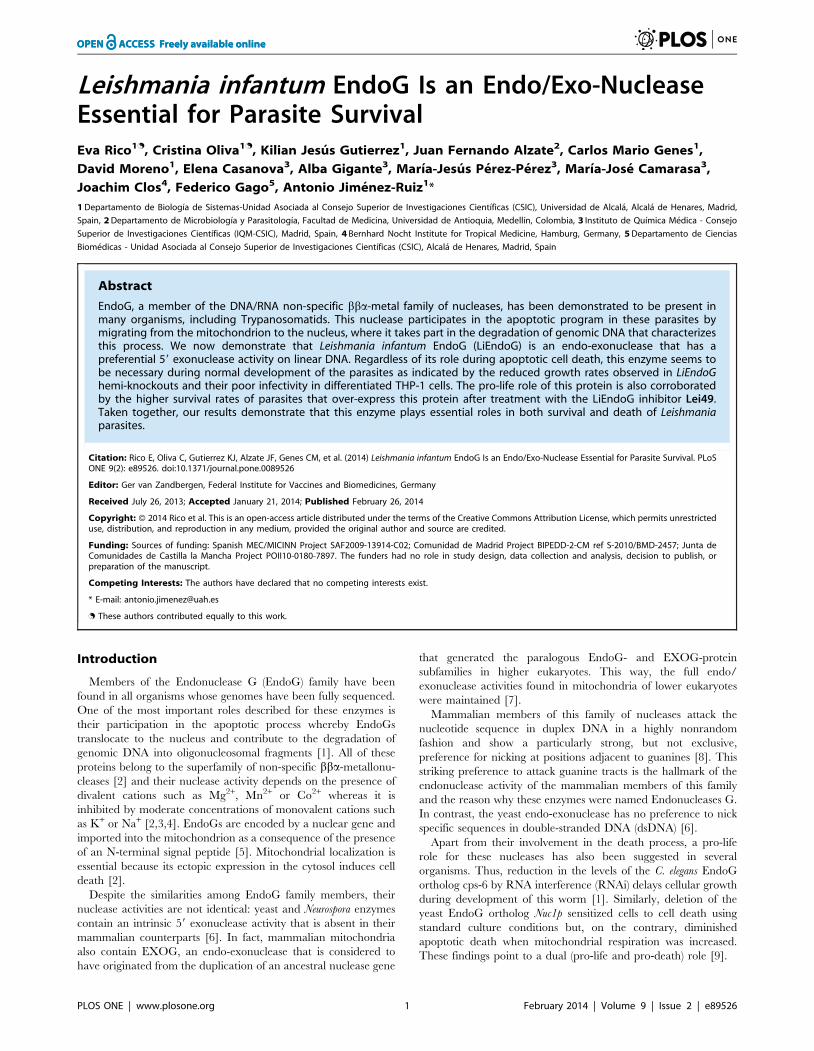
Figure 1. Analysis of rLiEndoG activity on double-stranded DNA. 1 mg of supercoiled plasmid DNA (sc) was digested with increasingamounts of rLiEndoG (0, 20, 40, 80 and 160 ng) and with 160 ng of denatured rLiEndoG protein (160DP) as a negative control. The restriction enzymeBgl II (Bgl II) was used as a control to generate linear DNA (lin). Each sample of digested DNA was split into two aliquots and one of them was heat-denatured (D) while the other one remained non-denatured (ND). All samples were electrophoresed in a 1% agarose gel and visualized under UVlight. The different forms of plasmid DNA are indicated: open circular (oc); linear double-stranded (lin); supercoiled (sc); linear single-stranded (lin ss);circular single-stranded (cir ss).doi:10.1371/journal.pone.0089526.g001
LiEndoG Is Essential for Parasite Survival
PLOS ONE | www.plosone.org 3 February 2014 | Volume 9 | Issue 2 | e89526
ogies) was used for mitochondrial localization at a final concen-
tration of 200 nM according to the manufacturer’s instructions.
Briefly, cell cultures were centrifuged to obtain a cell pellet and
gently suspended in warm (26uC) staining solution containing
200 nM Mito-Tracker Red CMXRos. After 45 minutes at 26uC
the cells were re-pelleted by centrifugation and suspended in fresh
warm medium for 30 minutes. Finally, the cells were re-pelleted
again and washed in PBS. Cells were then fixed in 2%
paraformaldehyde in PBS for 10 minutes at room temperature
and washed with PBS. 1.56106 cells were spread on poly-lysine
pre-coated slides and permeabilized with 0.1% Triton X-100 (TX-
100) in PBS solution for 10 minutes. Preparations were then
incubated with: i) blocking solution (2% BSA, 0.1% TX-100 in
PBS) for 60 minutes at room temperature, ii) primary monoclonal
antibody anti-LiEndoG (diluted 1:2 in blocking solution) for 16 h
at 4uC, and iii) antibody Alexa Fluor 488 anti-mouse (diluted
1:1000 in blocking solution) (Life Technologies). Each incubation
was followed by 3 washes with 16PBS.
Finally, the samples were covered with ProLong Gold Antifade
Reagent with DAPI liquid mountant (Life Techologies) and
visualized under the microscope.
StatisticsAll the analyses were carried out with a minimum of three
independent experiments. The statistical significance of the
differences between treatments was evaluated by a two-tailed
unpaired t-test.
Results
The endonuclease activity of LiEndoG generates single-stranded cuts in the DNA double helix
To determine whether, as described for other endonucleases
[6], LiEndoG is able to produce nicks in dsDNA, we proceeded to
digest supercoiled (sc) plasmid DNA with increasing amounts of
rLiEndoG expressed in E. coli. Each digested sample was divided
into two aliquots, and one of them was denatured to follow the
migration of the distinct forms of released single-stranded DNA.
The restriction enzyme Bgl II was used as a control for the
simultaneous digestion of both strands to generate a linear (lin)
DNA molecule (Figure 1; Bgl II). rLiEndoG is able to digest
supercoiled closed circular DNA, thus demonstrating that this
enzyme displays endonuclease activity. The enzyme primarily
generates open circular (oc) intermediates while only a very faint
band of linear (lin) DNA can be observed. Denaturation of the
samples (D lanes) allowed the detection of the fully-sized linear and
circular single-stranded (ss) DNA molecules that made up the open
circular forms generated after digestion. Because ethidium
bromide has much higher affinity for dsDNA, these ss molecules
are weakly stained compared to the oc DNA. Accordingly,
rLiEndoG displays endonucleolytic activity but digests only one of
the two strands of the double helix in every digestion step.
However, this enzymatic activity may render linear DNA as a
minor digestion product when two independent cuts in the
complementary strands are so close together that base pairing can
no longer maintain the circular structure (Figure 1; lin). The
progressive decrease in the total ethidium-derived fluorescence
reveals a process of DNA degradation that can be explained by the
release of small oligonucleotides from the double helix after
accumulation of single strand breaks. Thus, the enzyme seems to
cut stochastically any of the two strands, as has been described for
Drosophila melanogaster EndoG [15].
rLiEndoG shows exonuclease activityWhile EndoGs from higher eukaryotes display only endonucle-
ase activity [6], Nuc1p from S. cerevisiae behaves both as an
endonuclease and as an exonuclease [16]. Once established that
LiEndoG is an endonuclease, we addressed the possibility that it
might also display exonuclease activity. To this end, genomic
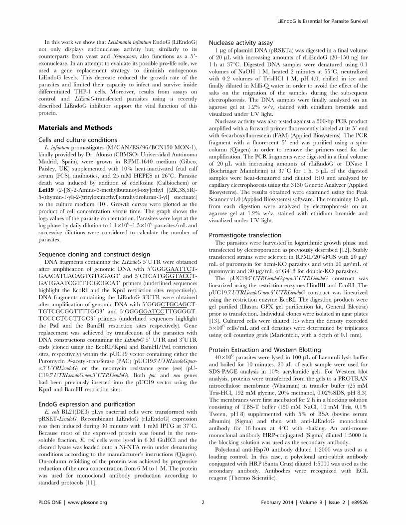
Figure 2. Exonuclease activity of rLiEndoG. 500 ng of a PCRfragment 59-labeled with FAM were digested with rLiEndoG or DNase I.The amount of enzyme used for digestion is indicated. Digestionproducts were processed by both agarose gel (1%) and capillaryelectrophoresis. A) Agarose gel of the DNA fragments generated after1 h of digestion with rLiEndoG or DNase I. B) Capillary electrophoresisof the DNA probe digested for 1 h with 0.1 mg of rLiEndoG. C) Capillaryelectrophoresis results obtained for the DNA probe digested for 1 hourwith 0.01 units of DNase I. Digested DNA was heat-denatured prior tocapillary electrophoresis. Fluorescence intensities (arbitrary units) of thesingle-stranded DNA fragments generated after digestion and dena-turation are shown on the y axis. Sizes of the ssDNA fragments (innucleotides) are shown on the x axis. Fragment sizes were analyzedwith the Peak Scanner (Applied Biosystems) software. Accurate sizescan only be predicted for fragments longer than 50 nucleotides.doi:10.1371/journal.pone.0089526.g002
LiEndoG Is Essential for Parasite Survival
PLOS ONE | www.plosone.org 4 February 2014 | Volume 9 | Issue 2 | e89526
DNA was amplified by PCR using a FAM-labeled forward primer
so as to generate a PCR product with a fluorescent 59 end. The
resulting PCR product was digested with increasing concentrations
of rLiEndoG. DNase I was used as a control for endonuclease
activity [17,18]. The products resulting from the digestion were
processed by both agarose gel electrophoresis (Figure 2A) and
capillary electrophoresis (Figure 2B and 2C). Whereas gel
electrophoresis allows the detection of all the dsDNA fragments
generated after digestion, only ss FAM-labelled fragments
(fragments containing the labelled 59 end) are detected in capillary
electrophoresis.
Digestion with rLiEndoG mostly generated fluorescent low-
molecular-weight ss- DNA fragments, being the major peak the
one closest to the y axis (Figure 2B). This accumulation of small
DNA fragments is not caused by extensive digestion because the
conditions of the reaction were adjusted to allow the detection of a
major peak of undigested DNA (500 bases). By contrast, the peak
pattern observed after digestion with DNase I is much more
dispersed along the x axis and the smallest fragments are not the
most abundant (Figure 2C). Taken these results together, the
nicking activity of rLiEndoG appears to be preferentially directed
against the 59 end of DNA.
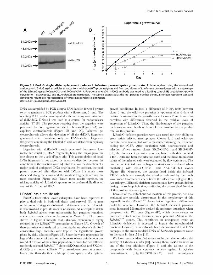
LiEndoG has a pro-life roleEndoGs from other lower eukaryotes have been reported to
play a dual role in both cell death and survival [9]. A gene
replacement strategy was followed to determine whether LiEndoG
is also involved in pro-life roles in Leishmania. All attempts to delete
both LiEndoG alleles were unsuccessful but parasites remained
viable after single allele replacement (LiEndoG2/+). The results
shown in Figure 3 indicate that LiEndoG expression is highly
reduced in the LiEndoG2/+ promastigotes (Figure 3A). Growth of
these parasites was analyzed by counting the number of cells for 6
consecutive days. Parasites were kept in the logarithmic growth
phase by daily dilutions (Figure 3B). Since the y axis represents the
log2 of the number of parasites an increase of one unit denotes one
round of division of the entire population. Results for two different
randomly selected LiEndoG2/+ clones (SKOendoG(2) and SKOen-
doG(6)) are shown. LiEndoG2/+ promastigotes grow at a much
lower rate than do their wild-type counterparts under optimal
growth conditions. In fact, a difference of 9 log2 units between
clone 6 and the wild-type parasites is apparent after 6 days of
culture. Variations in the growth rates of clones 2 and 6 seem to
correlate with differences observed in the residual levels of
expression of LiEndoG. Thus, the disadvantage of the parasites
harboring reduced levels of LiEndoG is consistent with a pro-life
role for this protein.
LiEndoG-deficient parasites were also tested for their ability to
grow inside infected macrophages. Clones 2, 6 and wild-type
parasites were transfected with a plasmid containing the sequence
coding for eGFP. After incubation with nourseothricin and
selection of two random clones (SKO-GFP-2.1 and SKO-GFP-
6.1), the fluorescent parasites were incubated with differentiated
THP-1 cells and both the infection rates and the mean fluorescent
values of the infected cells were evaluated by flow cytometry. The
number of infected macrophages is significantly reduced when
incubating with SKO-GFP-2.1 and SKO-GFP-6.1 clones
(Figure 4B). Moreover, the parasite load inside the infected
THP-1 cells is also strongly decreased as indicated by the much
lower mean fluorescence intensities of the infected cells (Figure 4C).
Accordingly, LiEndoG-deficient parasites also have growth defects
during macrophage infection, confirming the pro-survival function
of this protein in amastigotes.
Because of the mitochondrial location of this protein, we also
evaluated any possible disturbances in the morphology of this
organelle in the LiEndoG2/+ clones but no significant differences
could be observed. However, the LiEndoG-deficient parasites
show increased Mitotracker-derived fluorescence intensities when
compared with WT ones (Figure 4D), which demonstrates an
increased mitochondrial transmembrane potential (Dym) in the
LiEndoG2/+ clones. This constitutes an unexpected result as
LiEndoG deficiency is expected to impair the mitochondrial
function. However, it has already been demonstrated that DNA
damages in the mitochondrial DNA of Leishmania parasites cause
an increase in their Dym [19].
We have recently identified several molecules able to reduce the
activity of LiEndoG in vitro [10]. Among them, Lei49 behaves as
one of the best inhibitors (Figure 5) and also as one of the
compounds with better cytotoxic activity against L. infantum
promastigotes (IC50 = 4.3360.05 mM) and amastigotes
Figure 3. LiEndoG single allele replacement reduces L. infantum promastigotes growth rate. A) Immuno-blot using the monoclonalantibody a-rLiEndoG against cellular extracts from wild-type (WT) promastigotes and from two clones of L. infantum promastigotes with a single copyof the LiEndoG gene: SKOendoG(2) and SKOendoG(6). A Polyclonal a-Hsp70 (1:5000) antibody was used as a loading control. B) Logarithmic growthcurve for WT, SKOendoG(2) and SKOendoG(6) promastigotes. The curve is expressed as the log2 parasite number per mL. Error bars represent standarddeviations; results are representative of three independent experiments.doi:10.1371/journal.pone.0089526.g003
LiEndoG Is Essential for Parasite Survival
PLOS ONE | www.plosone.org 5 February 2014 | Volume 9 | Issue 2 | e89526
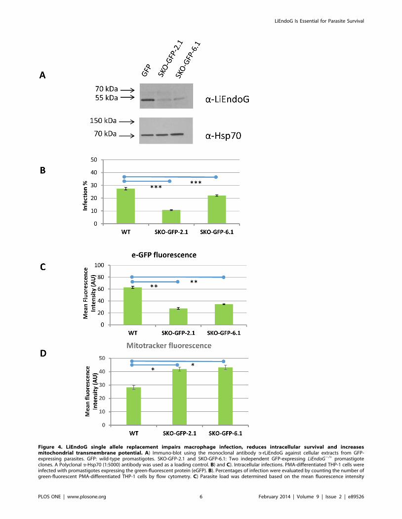
Figure 4. LiEndoG single allele replacement impairs macrophage infection, reduces intracellular survival and increasesmitochondrial transmembrane potential. A) Immuno-blot using the monoclonal antibody a-rLiEndoG against cellular extracts from GFP-expressing parasites. GFP: wild-type promastigotes. SKO-GFP-2.1 and SKO-GFP-6.1: Two independent GFP-expressing LiEndoG2/+ promastigoteclones. A Polyclonal a-Hsp70 (1:5000) antibody was used as a loading control. B) and C). Intracellular infections. PMA-differentiated THP-1 cells wereinfected with promastigotes expressing the green-fluorescent protein (eGFP). B). Percentages of infection were evaluated by counting the number ofgreen-fluorescent PMA-differentiated THP-1 cells by flow cytometry. C) Parasite load was determined based on the mean fluorescence intensity
LiEndoG Is Essential for Parasite Survival
PLOS ONE | www.plosone.org 6 February 2014 | Volume 9 | Issue 2 | e89526
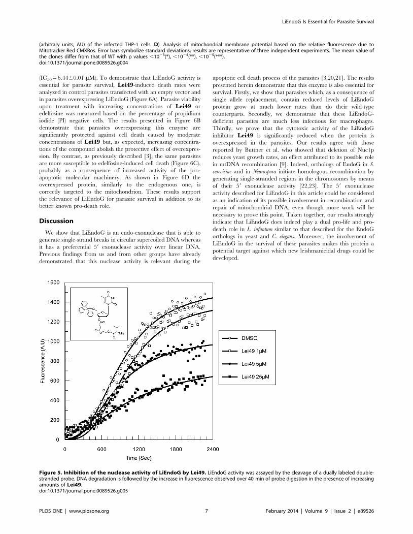
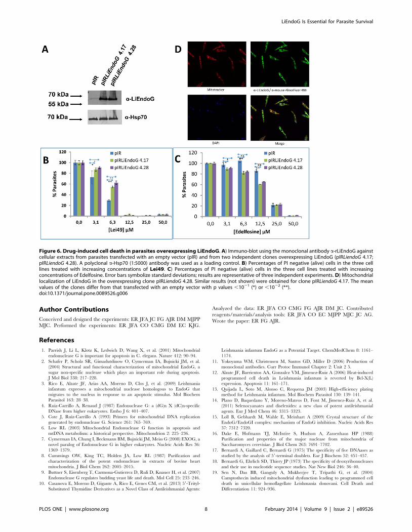
(IC50 = 6.4460.01 mM). To demonstrate that LiEndoG activity is
essential for parasite survival, Lei49-induced death rates were
analyzed in control parasites transfected with an empty vector and
in parasites overexpressing LiEndoG (Figure 6A). Parasite viability
upon treatment with increasing concentrations of Lei49 or
edelfosine was measured based on the percentage of propidium
iodide (PI) negative cells. The results presented in Figure 6B
demonstrate that parasites overexpressing this enzyme are
significantly protected against cell death caused by moderate
concentrations of Lei49 but, as expected, increasing concentra-
tions of the compound abolish the protective effect of overexpres-
sion. By contrast, as previously described [3], the same parasites
are more susceptible to edelfosine-induced cell death (Figure 6C),
probably as a consequence of increased activity of the pro-
apoptotic molecular machinery. As shown in Figure 6D the
overexpressed protein, similarly to the endogenous one, is
correctly targeted to the mitochondrion. These results support
the relevance of LiEndoG for parasite survival in addition to its
better known pro-death role.
Discussion
We show that LiEndoG is an endo-exonuclease that is able to
generate single-strand breaks in circular supercoiled DNA whereas
it has a preferential 59 exonuclease activity over linear DNA.
Previous findings from us and from other groups have already
demonstrated that this nuclease activity is relevant during the
apoptotic cell death process of the parasites [3,20,21]. The results
presented herein demonstrate that this enzyme is also essential for
survival. Firstly, we show that parasites which, as a consequence of
single allele replacement, contain reduced levels of LiEndoG
protein grow at much lower rates than do their wild-type
counterparts. Secondly, we demonstrate that these LiEndoG-
deficient parasites are much less infectious for macrophages.
Thirdly, we prove that the cytotoxic activity of the LiEndoG
inhibitor Lei49 is significantly reduced when the protein is
overexpressed in the parasites. Our results agree with those
reported by Buttner et al. who showed that deletion of Nuc1p
reduces yeast growth rates, an effect attributed to its possible role
in mtDNA recombination [9]. Indeed, orthologs of EndoG in S.
cerevisiae and in Neurospora initiate homologous recombination by
generating single-stranded regions in the chromosomes by means
of their 59 exonuclease activity [22,23]. The 59 exonuclease
activity described for LiEndoG in this article could be considered
as an indication of its possible involvement in recombination and
repair of mitochondrial DNA, even though more work will be
necessary to prove this point. Taken together, our results strongly
indicate that LiEndoG does indeed play a dual pro-life and pro-
death role in L. infantum similar to that described for the EndoG
orthologs in yeast and C. elegans. Moreover, the involvement of
LiEndoG in the survival of these parasites makes this protein a
potential target against which new leishmanicidal drugs could be
developed.
(arbitrary units; AU) of the infected THP-1 cells. D). Analysis of mitochondrial membrane potential based on the relative fluorescence due toMitotracker Red CMXRos. Error bars symbolize standard deviations; results are representative of three independent experiments. The mean value ofthe clones differ from that of WT with p values ,1023(*), ,1024(**), ,1025(***).doi:10.1371/journal.pone.0089526.g004
Figure 5. Inhibition of the nuclease activity of LiEndoG by Lei49. LiEndoG activity was assayed by the cleavage of a dually labeled double-stranded probe. DNA degradation is followed by the increase in fluorescence observed over 40 min of probe digestion in the presence of increasingamounts of Lei49.doi:10.1371/journal.pone.0089526.g005
LiEndoG Is Essential for Parasite Survival
PLOS ONE | www.plosone.org 7 February 2014 | Volume 9 | Issue 2 | e89526
Author Contributions
Conceived and designed the experiments: ER JFA JC FG AJR DM MJPP
MJC. Performed the experiments: ER JFA CO CMG DM EC KJG.
Analyzed the data: ER JFA CO CMG FG AJR DM JC. Contributed
reagents/materials/analysis tools: ER JFA CO EC MJPP MJC JC AG.
Wrote the paper: ER FG AJR.
References
1. Parrish J, Li L, Klotz K, Ledwich D, Wang X, et al. (2001) Mitochondrial
endonuclease G is important for apoptosis in C. elegans. Nature 412: 90–94.
2. Schafer P, Scholz SR, Gimadutdinow O, Cymerman IA, Bujnicki JM, et al.
(2004) Structural and functional characterization of mitochondrial EndoG, a
sugar non-specific nuclease which plays an important role during apoptosis.
J Mol Biol 338: 217–228.
3. Rico E, Alzate JF, Arias AA, Moreno D, Clos J, et al. (2009) Leishmania
infantum expresses a mitochondrial nuclease homologous to EndoG that
migrates to the nucleus in response to an apoptotic stimulus. Mol Biochem
Parasitol 163: 28–38.
4. Ruiz-Carrillo A, Renaud J (1987) Endonuclease G: a (dG)n X (dC)n-specific
DNase from higher eukaryotes. Embo J 6: 401–407.
5. Cote J, Ruiz-Carrillo A (1993) Primers for mitochondrial DNA replication
generated by endonuclease G. Science 261: 765–769.
6. Low RL (2003) Mitochondrial Endonuclease G function in apoptosis and
mtDNA metabolism: a historical perspective. Mitochondrion 2: 225–236.
7. Cymerman IA, Chung I, Beckmann BM, Bujnicki JM, Meiss G (2008) EXOG, a
novel paralog of Endonuclease G in higher eukaryotes. Nucleic Acids Res 36:
1369–1379.
8. Cummings OW, King TC, Holden JA, Low RL (1987) Purification and
characterization of the potent endonuclease in extracts of bovine heart
mitochondria. J Biol Chem 262: 2005–2015.
9. Buttner S, Eisenberg T, Carmona-Gutierrez D, Ruli D, Knauer H, et al. (2007)
Endonuclease G regulates budding yeast life and death. Mol Cell 25: 233–246.
10. Casanova E, Moreno D, Gigante A, Rico E, Genes CM, et al. (2013) 59-Trityl-
Substituted Thymidine Derivatives as a Novel Class of Antileishmanial Agents:
Leishmania infantum EndoG as a Potential Target. ChemMedChem 8: 1161–
1174.
11. Yokoyama WM, Christensen M, Santos GD, Miller D (2006) Production of
monoclonal antibodies. Curr Protoc Immunol Chapter 2: Unit 2 5.
12. Alzate JF, Barrientos AA, Gonzalez VM, Jimenez-Ruiz A (2006) Heat-induced
programmed cell death in Leishmania infantum is reverted by Bcl-X(L)
expression. Apoptosis 11: 161–171.
13. Quijada L, Soto M, Alonso C, Requena JM (2003) High-efficiency plating
method for Leishmania infantum. Mol Biochem Parasitol 130: 139–141.
14. Plano D, Baquedano Y, Moreno-Mateos D, Font M, Jimenez-Ruiz A, et al.
(2011) Selenocyanates and diselenides: a new class of potent antileishmanial
agents. Eur J Med Chem 46: 3315–3323.
15. Loll B, Gebhardt M, Wahle E, Meinhart A (2009) Crystal structure of the
EndoG/EndoGI complex: mechanism of EndoG inhibition. Nucleic Acids Res
37: 7312–7320.
16. Dake E, Hofmann TJ, McIntire S, Hudson A, Zassenhaus HP (1988)
Purification and properties of the major nuclease from mitochondria of
Saccharomyces cerevisiae. J Biol Chem 263: 7691–7702.
17. Bernardi A, Gaillard C, Bernardi G (1975) The specificity of five DNAases as
studied by the analysis of 59-terminal doublets. Eur J Biochem 52: 451–457.
18. Bernardi G, Ehrlich SD, Thiery JP (1973) The specificity of deoxyribonucleases
and their use in nucleotide sequence studies. Nat New Biol 246: 36–40.
19. Sen N, Das BB, Ganguly A, Mukherjee T, Tripathi G, et al. (2004)
Camptothecin induced mitochondrial dysfunction leading to programmed cell
death in unicellular hemoflagellate Leishmania donovani. Cell Death and
Differentiation 11: 924–936.
Figure 6. Drug-induced cell death in parasites overexpressing LiEndoG. A) Immuno-blot using the monoclonal antibody a-rLiEndoG againstcellular extracts from parasites transfected with an empty vector (pIR) and from two independent clones overexpressing LiEndoG (pIRLiendoG 4.17;pIRLiendoG 4.28). A polyclonal a-Hsp70 (1:5000) antibody was used as a loading control. B) Percentages of PI negative (alive) cells in the three celllines treated with increasing concentrations of Lei49. C) Percentages of PI negative (alive) cells in the three cell lines treated with increasingconcentrations of Edelfosine. Error bars symbolize standard deviations; results are representative of three independent experiments. D) Mitochondriallocalization of LiEndoG in the overexpressing clone pIRLiendoG 4.28. Similar results (not shown) were obtained for clone pIRLiendoG 4.17. The meanvalues of the clones differ from that transfected with an empty vector with p values ,1021 (*) or ,1022 (**).doi:10.1371/journal.pone.0089526.g006
LiEndoG Is Essential for Parasite Survival
PLOS ONE | www.plosone.org 8 February 2014 | Volume 9 | Issue 2 | e89526
20. Bosedasgupta S, Das BB, Sengupta S, Ganguly A, Roy A, et al. (2008) The
caspase-independent algorithm of programmed cell death in Leishmaniainduced by baicalein: the role of LdEndoG, LdFEN-1 and LdTatD as a DNA
‘degradesome’. Cell Death Differ.
21. Gannavaram S, Vedvyas C, Debrabant A (2008) Conservation of the pro-apoptotic nuclease activity of endonuclease G in unicellular trypanosomatid
parasites. J Cell Sci 121: 99–109.
22. Zassenhaus HP, Denniger G (1994) Analysis of the role of the NUC1 endo/
exonuclease in yeast mitochondrial DNA recombination. Curr Genet 25: 142–
149.
23. Fraser MJ, Hatahet Z, Huang XT (1989) The actions of Neurospora endo-
exonuclease on double strand DNAs. J Biol Chem 264: 13093–13101.
LiEndoG Is Essential for Parasite Survival
PLOS ONE | www.plosone.org 9 February 2014 | Volume 9 | Issue 2 | e89526