leishmania chagasi: cytotoxic effect of infected macrophages on parenchymal liver cells
TRANSCRIPT
Available online at www.sciencedirect.com
www.elsevier.com/locate/yexpr
Experimental Parasitology 117 (2007) 390–398
Leishmania chagasi: Cytotoxic effect of infected macrophageson parenchymal liver cells
Juliana Dias Costa a,b, Maria de Nazareth Meirelles b, Carlos Eduardo Pereira Velloso b,Renato Porrozzi a,c,*
a Laboratorio de Pesquisas em Leishmaniose, Instituto Oswaldo Cruz, Fiocruz, Avenida Brasil 4365, Rio de Janeiro, RJ 21045-900, Brazilb Laboratorio de Ultra-Estrutura Celular, Instituto Oswaldo Cruz, Fiocruz, Avenida Brasil 4365, Rio de Janeiro, RJ 21045-900, Brazil
c Centro Universitario de Volta Redonda – UniFOA, Avenida Paulo Erlei Alves Abrantes 1325, Tres Pocos, Volta Redonda, RJ 27240-560, Brazil
Received 2 August 2006; received in revised form 7 May 2007; accepted 14 May 2007Available online 20 July 2007
Abstract
Leishmania (Leishmania) chagasi, the ethiological agent of New World visceral leishmaniasis, causes morphological and functionalinjury to the liver. To investigate the role of macrophage-released leishmanicidal factors in hepatocyte damage, we used a co-culturemodel of hepatocytes and L. chagasi promastigote-infected peritoneal macrophages obtained from C57BL/6 or BALB/c mice.C57BL/6 macrophages killed intracellular parasites more efficiently than BALB/c macrophages, leading to higher number of intracellularamastigotes in the BALB/c culture during the entire course of infection. Early TNF-a production led to macrophages activation resultingin parasite growth control. Hepatic transaminases and lactate dehydrogenase were present at high levels in the supernatants of both co-cultures; concurrently, parasites were eliminated from infected macrophages. Nitric oxide production was higher in C57BL/6 co-culturesthan in BALB/c co-cultures. Inhibitors of the oxidative burst and secreted proteinases protected hepatocytes against toxicity, and treat-ment with an inducible nitric oxide synthase inhibitor fully impeded the enzyme release. Our data suggest that the intracellular cytotoxiceffects elicited by macrophages for parasite destruction are directly associated with hepatocyte damage, and that nitric oxide plays a piv-otal role in this phenomenon.� 2007 Elsevier Inc. All rights reserved.
Index Descriptors and Abbreviations: ALT, glutamate pyruvate transaminase; AST, glutamate oxaloacetate transaminase; iNOS, inducible nitric oxidesynthase; IL-10, interleukin 10; Hep, hepatocytes; LDH, lactate dehydrogenase; MF, macrophages; NO, nitric oxide; ROI, reactive oxygen intermediates;RNI, reactive nitrogen intermediates; TNF-a, tumor necrosis factor-alpha; SOD, superoxide dismute; PMSF, phenylmethylsulfonyl fluoride; 1-10 PHEN,1,10-phenanthroline; Leishmania chagasi; Macrophage; Hepatocyte; Co-culture
1. Introduction
Trypanosomatid protozoa of the Leishmania genus areintracellular parasites that cause a large spectrum of clini-cal symptoms in humans, ranging from self-healing cutane-ous infections, to mucocutaneous disfigurement, to visceralleishmaniasis, which can be fatal when not treated. The
0014-4894/$ - see front matter � 2007 Elsevier Inc. All rights reserved.
doi:10.1016/j.exppara.2007.05.015
* Corresponding author. Address: Laboratorio de Pesquisas em Leish-maniose, Instituto Oswaldo Cruz, Fiocruz, Avenida Brasil 4365, Rio deJaneiro, RJ 21045-900, Brazil. Fax: +55 21 22094110.
E-mail address: [email protected] (R. Porrozzi).
outcome of infection in humans depends on both the par-asite species involved and on the host immune response(Goto and Lindoso, 2004).
Natural transmission of Leishmania occurs during theblood meal of phlebotomine sandflies, which introducesmetacyclic infective promastigotes into the vertebratehost dermis. These forms are resistant to complementattack and quickly invade local phagocytes, subsequentlytransforming into amastigotes inside the host cells. Themechanism involved in the dissemination or visceraliza-tion of the parasites is not clear. Infected macrophagesmay migrate from the initial site of infection to the
J. Dias Costa et al. / Experimental Parasitology 117 (2007) 390–398 391
spleen, liver and bone marrow (Murray et al., 2003);alternatively, free promastigotes may directly enter thebloodstream as a result of the pool-feeding behavior ofthe sandfly (Engwerda et al., 2004a). Why Leishmania
species of the L. donovani complex are viscerotropicwhile other species are exclusively cutaneous is a keyquestion that still remains unanswered (Engwerdaet al., 2004b).
In order to survive, intracellular organisms must escapethe anti-microbial mechanisms of macrophages. DuringLeishmania promastigote infection, macrophages respondby producing reactive oxygen intermediates (ROI),together with reactive nitrogen intermediates (RNI), lyso-somal hydrolases, and neutral peptidases (Murray andNathan, 1999). Nitric oxide (NO) is a regulatory moleculethat is important in host protective responses, acting notonly as an anti-microbial effector molecule (Brunet,2001), but also as a potential host-destructive mediator inseveral pathologies, mainly in infectious diseases (Holzmul-ler et al., 2006). In murine models of Leishmania infection,it has been well established that cytokine activation (e.g.IFN-c and TNF-a) of macrophages leads to ROI andRNI production, which is ultimately responsible for leish-manicidal activity (Bogdan et al., 2000). NO productionis the main microbicidal mechanism in murine macro-phages (Dey et al., 2005).
Although ROI, RNI, and lysosomal enzymes are cru-cial for eliminating intracellular parasites, there arenumerous clinical situations in which macrophage-secreted mediators cause tissue injury as a collateraleffect of microbicidal activity (Clemens, 1999). Liver tis-sue damage associated with pathological conditions hasbeen reported in both clinical and experimental visceralleishmaniasis (Gutierrez et al., 1984; McElrath et al.,1988; El Hag et al., 1994). For example, hepatocyte dam-age in experimental Leishmania infection has been associ-ated with an increase in serum transaminases and lipidperoxidation in liver tissue (Kausalya et al., 1996; Viannaet al., 2002), and granuloma formation has been reportedto be the most prevalent lesion found in L. donovani-infected BALB/c mice (Gutierrez et al., 1984). Interest-ingly, experimental data indicate that liver resident mac-rophages (Kupffer cells) fail to produce toxic oxygenintermediates, a key anti-microbicidal mechanism (Kaus-alya et al., 1996), whereas competent blood-recruitedmonocytes and monocyte-derived macrophages are themajor cells involved in killing and degrading ingestedintracellular parasites.
In this study, we adopted an adherent peritoneal macro-phage–hepatocyte co-culture model to study L. chagasi
infection. Peritoneal macrophages from two mouse strainswith different patterns of susceptibility to Leishmania infec-tion were used; these cells generate microbicidal productsvia both oxygen-dependent and -independent pathways.We evaluated hepatocyte damage and the role of microbi-cidal mediators secreted by infected macrophages in thisprocess.
2. Materials and methods
2.1. Mice
C57BL/6 and BALB/c mice weighing about 20 g eachwere used to obtain primary cultures of peritoneal macro-phages. Pregnant mice (18–20 days of gestation) were uti-lized for hepatocyte primary cultures. The animals wereobtained from the Animal Care Facility of the OswaldoCruz Institute (Fiocruz, Rio de Janeiro, Brazil). All proce-dures involving animals in this study were reviewed andapproved by the Oswaldo Cruz Foundation Ethics Com-mittee (CEUA-FIOCRUZ).
2.2. Parasites
Leishmania (Leishmania) chagasi (strain MCAN/BR/2000/CNV-FEROZ) was isolated from a dog in the Stateof Espırito Santo, Brazil, and typed by multilocus enzymeelectrophoresis at the Leishmania Collection at theOswaldo Cruz Institute, RJ, Brazil (CLIOC, WDCM731). The parasites were maintained through inoculationin golden hamsters. Infective forms were obtained fromhamster spleen or liver and cultured at 25 �C in biphaseNNN blood agar medium/Schneider’s Drosophila culturemedium (Sigma–Aldrich St. Louis, MO, USA) containing10% FCS.
2.3. Macrophage isolation and infection
Mouse peritoneal macrophages were obtained fromboth mouse strains as previously described (Araujo-Jorgeand de Souza, 1986). Briefly, the animals were sacrificedusing CO2 and the peritoneal cavity was washed with10 ml of MEM/199 culture medium (Cultilab, Sao Paulo,SP, Brazil) at 4 �C. For each experiment, ten mice fromeach strain were utilized. The cells were pooled andcounted using a Neubauer chamber. The cells were seededin tissue culture flasks (2 · 106 macrophages/flask) or onglass coverslips (2 · 105 macrophages/coverslip) and main-tained in MEM/199 culture medium at 37 �C in a humidi-fied atmosphere containing 5% CO2.
For in vitro infection of the macrophages, the L. chagasi
promastigotes were harvested at the stationary growthphase. Peritoneal macrophages were cultivated for 24 hand then infected at a ratio of 10 parasites per cell withor without 1 lg/ml LPS (Sigma Chemical Co., St Louis,MO, USA). The macrophages and parasites were incu-bated together for 1 h at 34 �C. The cultures were thenwashed three times with MEM/199 culture medium toremove non-adherent or non-internalized parasites.
2.4. Hepatocyte isolation
Hepatocytes from both mouse strains were isolatedaccording to Porrozzi et al., 1997. Briefly, 7–11 mouseembryo livers (1–2 g each) were aseptically removed and
392 J. Dias Costa et al. / Experimental Parasitology 117 (2007) 390–398
washed with Hepes buffer (Sigma–Aldrich, St. Louis, MO,USA). The livers were minced and then incubated at 37 �Cfor 20 min with about 50 ml of Hepes buffer containing0.05% Type II collagenase (Sigma–Aldrich, St. Louis,MO, USA). The cells were dispersed by pipetting and thencollected by centrifugation at 200g. Viable cells were puri-fied by sedimentation for 10 min at room temperature withMEM/199 medium containing 10% fetal calf serum (FCS,Sigma–Aldrich, St. Louis, MO, USA).
2.5. Hepatocyte-infected macrophage co-culture
Hepatocytes were seeded directly onto the infected-mac-rophage plates, as reported previously (Kausalya et al.,1993). Infected peritoneal macrophages were co-cultivatedwith hepatocytes from the same mouse strain at a 10:1 ratio(2 · 106 macrophages/2 · 105 hepatocytes in 3 ml ofMEM/199 medium plus 10% FCS in tissue culture flasksor 2 · 105 macrophages/2 · 104 hepatocytes in 300 ll ofMEM/199 medium plus 10% FCS on glass coverslips).These co-cultures were maintained in culture medium at37 �C in a 5% CO2 atmosphere. Hepatocytes were co-cul-tured with uninfected macrophages as controls. The culturemedium was maintained without changing during theentire experimentation period. After the interaction ofinfected macrophages and hepatocytes, the coverslips werewashed three times in culture medium, fixed in Bouin’ssolution, and stained with Giemsa (Merck). The percentageof infected macrophages and the mean number of intracel-lular parasites per infected macrophage were determined bycounting at least 100 cells. These experiments wererepeated three times to confirm that the experimental datawere reproducible. All experiments were also performed inmacrophage cultures alone for comparison with the co-cul-tures. The co-culture supernatant was collected and exam-ined by light microscopy for detached or dead cells.
2.6. Detection of cytokines in the co-culture supernatant
Aliquots of culture supernatant were collected from con-trol and infected co-cultures after 24, 48, and 72 h. Tumornecrosis factor (TNF-a) and interleukin-10 (IL-10) weredetected using a commercial ELISA kit (DuoSET ELISA;R&D Systems, Minneapolis, MN, USA).
2.7. Release of hepatocyte enzymes
Glutamate oxaloacetate transaminase (AST) and gluta-mate pyruvate transaminase (ALT) activities were assayedwith commercial kits (Labtest, Lagoa Santa, MG, Brazil).Lactate dehydrogenase (LDH) activity was measuredusing a Merck activity assay kit (Merck, Darmstadt,Germany). Enzymatic activity assays were performedaccording to the manufacturer’s instructions and adaptedfor detecting hepatocyte damage markers. Analyses wereperformed with both the cell lysate and the co-culturesupernatant, using one Petri dish for each time point. Cell
lysate was obtained by scraping the culture monolayersand then subjecting the scraped cells to sonication. Mea-surements of enzymes released from infected macrophagecultures were carried out in parallel. The results wereexpressed as enzyme release percentage and calculated asfollows:
ES
ðESþ ELÞ � 100 ¼ % ER
ES, enzyme measured in the supernatant; EL, enzymemeasured in the lysates; ER, enzyme release.
2.8. Ultrastructural examination of co-cultured cells
The co-cultured cells were fixed for 1 h in a solutioncontaining 2.5% glutaraldehyde in 0.1 M cacodylate buffersupplemented with 3.5% sucrose (Sigma–Aldrich, St.Louis, MO, USA). The cells were then washed with0.1 M cacodylate buffer and post-fixed with 1% OsO4 inthe same buffer. The samples were then washed twicein buffer, dehydrated in graded acetone, and embedded inEpon. Samples were observed using a Zeiss EM10C trans-mission electron microscope.
2.9. Treatment of co-cultures with microbicidal inhibitors
Infected and uninfected co-cultures were incubated withdifferent microbicidal mediator inhibitors: superoxidedismutase (SOD, 30 U); mannitol (20 lM); NG-nitro-L-arginine-methyl ester (L-NAME, 4 mM); phenylmethyl-sulfonyl fluoride (PMSF, 1 mM); 1,10-phenantroline(1-10 PHEN, 10 mM); and cystatin C (4 mM). Untreatedinfected co-cultures were treated with the same inhibitorsas controls. Samples of supernatant from C57BL/6 andBALB/c hepatocyte–macrophage co-cultures were col-lected after 24 and 48 h of treatment, respectively. The inhi-bition results indicate the difference between infected anduninfected treated co-cultures. Samples were then analyzedfor hepatic transaminases and LDH as described above.
2.10. Nitrite (NO2�) accumulation
The production of NO in the co-cultures was evaluatedby measuring nitrite, a stable breakdown product of NO, inthe culture supernatant. Cells were cultured for up to 72 hon culture plates, and then culture supernatant was col-lected. A 100 lL aliquot was mixed with an equal volumeof Griess reagent (50 lL of 1% sulfanilamide plus 50 lLof 0.1% N-1-naphthylethylenediamine in 5% phosphoricacid solution), and absorbance was measured at 550 nm.Nitrite concentration was calculated using a standardcurve.
2.11. Statistical analysis
All experiments were carried out in triplicate, andrepeated at least three times. The mean values and standard
J. Dias Costa et al. / Experimental Parasitology 117 (2007) 390–398 393
deviations (SD) for all numerical data were calculated. Thedata were analyzed with the Student’s t-test. Simple linearregression analysis was performed to calculate correlations.Differences with a P value <0.05 were considered statisti-cally significant.
3. Results
3.1. Infection of co-cultured cells with L. chagasi
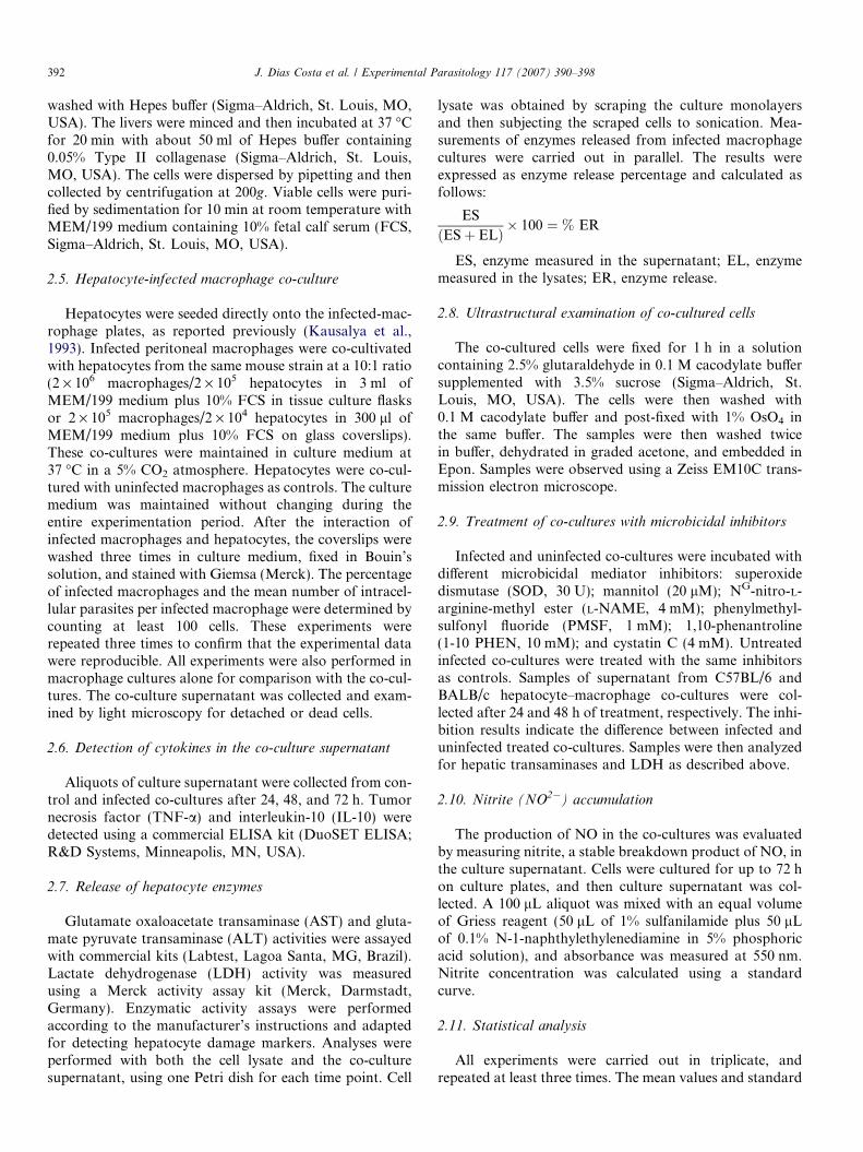
The C57BL/6 and BALB/c macrophage–hepatocyte co-cultured cells grew in a distinct morphological pattern:groups of closely associated cells that appeared to be differ-entiated hepatocytes were surrounded by spread-out, sepa-rated macrophages (Fig. 1A and B). The progression of L.
chagasi infection in co-cultures was monitored by countingthe number of amastigotes and infected macrophages usinglight microscopy (Fig. 1C). Hepatocytes were not infected,so that amastigotes were not observed inside these cells inany of the co-cultures. The course of L. chagasi infectionin the C57BL/6 co-cultures exhibited a similar pattern tothat in the BALB/c co-cultures, but had different kinetics.In cultures of infected macrophages from both mouse lin-eages, infection followed a similar course as in the co-cul-tures (data not shown). During the course of infection,
Fig. 1. L. chagasi infection in macrophage (MF)-hepatocyte (Hep) co-cultures.infection (hpi). The amastigote forms of L. chagasi (arrows) were located onlymacrophages is represented as open bars (C57BL/6) or shaded bars (BALB/cstraight or dashed lines, respectively. Bars indicate mean ± standard deviationinfected BALB/c and C57BL/6 macrophages at 48 hpi. **Statistically significBALB/c and C57BL/6 macrophages. (A) P < 0.05 for the number of intracellulP < 0.05 for the number of intracellular parasites inside BALB/c macrophage
C57BL/6 co-cultures had fewer parasites inside the macro-phages than in the BALB/c co-cultures. In addition, themacrophages from C57BL/6 mice reduced the number ofintracellular amastigotes faster than the BALB/c cells(Fig. 1C). The number of infected BALB/c mouse macro-phages increased up to 48 h post-infection (hpi), accompa-nied by intracellular parasite multiplication, while thenumber of infected C57BL/6 mouse macrophages and par-asites had already declined at this time point (Fig. 1C).However, despite the reduction in the number of parasit-ized cells at 72 hpi, neither BALB/c nor C57BL/6 co-cul-tures were able to control the infection completely(Fig. 1C). The co-cultures showed no significant differencesin the total number of cells during the course of the exper-iment. We also counted cells in the supernatant of the co-cultures and very few cells were found (data not shown).
3.2. Cytokine production
Cytokine production by the L. chagasi-infected co-cul-tures was assessed by ELISA. Following the infection,the C57BL/6 and BALB/c hepatocyte–macrophage co-cul-tures displayed differences in the level and kinetics of cyto-kine production. Production of the cytokine TNF-a wasobserved for both mouse strain co-cultures, demonstrating
A and B show C57BL/6 and BALB/c co-cultures, respectively, at 48 h postinside macrophages. Scale bar = 10 lm. In (C), the percentage of infected), and intracellular parasites in C57BL/6 and BALB/c cells are shown as. *Statistically significant difference (P < 0.05) between the percentage of
ant difference (P < 0.05) between the number of amastigotes at 48 hpi inar parasites inside C57BL/6 macrophages at 24 hpi compared to 72 hpi. (B)s at 48 hpi compared to 72 hpi.
394 J. Dias Costa et al. / Experimental Parasitology 117 (2007) 390–398
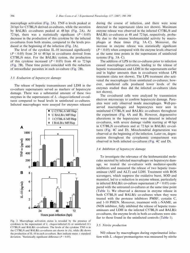
macrophage activation (Fig. 2A). TNF-a levels peaked at24 hpi for C57BL/6 derived co-cultures, while the secretionby BALB/c co-cultures peaked at 48 hpi (Fig. 2A). At72 hpi, there was a statistically significant (P < 0.05)decrease in the production of this cytokine by the infectedco-cultures from both strains, compared to the levels pro-duced at the beginning of the infection (Fig. 2A).
The level of the cytokine IL-10 increased significantly(P < 0.05) from 24 to 48 hpi in co-cultures derived fromC57BL/6 mice. For the BALB/c system, the productionof this cytokine increased (P < 0.05) from 48 to 72 hpi(Fig. 2B). These time points coincided with the reductionof intracellular parasites in each co-culture (Fig. 2B).
3.3. Evaluation of hepatocyte damage
The release of hepatic transaminases and LDH in theco-culture supernatants served as markers of hepatocytedamage. There was a substantial amount of these twoenzymes in the supernatants of L. chagasi-infected co-cul-tures compared to basal levels in uninfected co-cultures.Infected macrophages were assayed for enzymes released
Fig. 2. Macrophage activation status is revealed by the presence ofcytokines in the supernatant of L. chagasi-infected (I) or uninfected (U)C57BL/6 and BALB/c co-cultures. The levels of the cytokine TNF-a inthe C57BL/6 and BALB/c co-cultures are shown in (A), while (B) showsthe production of IL-10 in each co-culture. Bars indicate mean ± standarddeviation. *Statistically significant difference (P < 0.05).
during the course of infection, and there were nonedetected in the supernatant (data not shown). Maximumenzyme release was observed in the infected C57BL/6 andBALB/c co-cultures at 48 and 72 hpi, respectively, proba-bly due to the intense leishmanicidal activity by macro-phages at these times (Fig. 3A–C). Moreover, theincrease in enzyme release was statistically significant(P < 0.05) when compared with the enzyme levels observedat the same time points in the supernatant of uninfectedsystems (Fig. 3A–C).
The addition of LPS to the co-cultures prior to infectioncaused macrophage activation, leading to the release ofhepatic transaminases and LDH in the supernatants earlierand in higher amounts than in co-cultures without LPStreatment (data not shown). The LPS treatment also acti-vated the macrophages from uninfected co-cultures; how-ever, uninfected cells produced lower levels of theenzymes studied than did the infected co-cultures (datanot shown).
The co-cultured cells were analyzed by transmissionelectron microscopy to confirm hepatocyte damage. Para-sites were only observed inside macrophages. Well-pre-served macrophages and hepatocytes were seen inuninfected C57BL/6 and BALB/c co-cultures throughoutthe experiment (Fig. 4A and B). However, degenerativealterations in the hepatocytes were detected in infectedco-cultures, with severe damage visible starting at 48 hpiin C57BL/6 co-cultures and at 72 hpi in BALB/c co-cul-tures (Fig. 4C and D). Mitochondrial degeneration wasobserved at the beginning of the infection. Later on, degen-eration throughout the cytoplasmic compartment wasobserved in both infected co-cultures (Fig. 4C and D).
3.4. Inhibition of hepatocyte damage
To investigate the relevance of the leishmanicidal mole-cules secreted by infected macrophages on hepatocyte dam-age, we treated the co-cultures with mediator-specificinhibitors and measured the release of two hepatic trans-aminase (AST and ALT) and LDH. Treatment with ROSscavengers, which suppress the oxidative burst, SOD andmannitol, led to a reduction in enzyme release, particularlyin infected BALB/c co-culture supernatant (P < 0.05) com-pared with the untreated co-cultures at the same time point(Table 1). We observed a decrease in enzyme release inboth C57BL/6 and BALB/c co-cultures when they weretreated with the protease inhibitors PMSF, cystatin C,and 1-10 PHEN. Moreover, treatment with L-NAME, aniNOS inhibitor, fully inhibited the release of hepatic trans-aminases and LDH in the infected C57BL/6 and BALB/cco-cultures, the enzyme levels in both co-cultures were sim-ilar to those found in the uninfected controls (Table 1).
3.5. Nitrite production
NO release by macrophages during experimental infec-tion with L. chagasi promastigotes was measured by nitrite
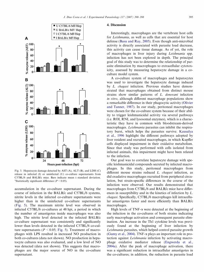
Fig. 3. Hepatocyte damage detected by AST (A), ALT (B), and LDH (C)release in infected (I) or uninfected (U) co-culture supernatants fromC57BL/6 and BALB/c mice. Bars indicate mean ± standard deviation.*Statistically significant difference (P < 0.05).
J. Dias Costa et al. / Experimental Parasitology 117 (2007) 390–398 395
accumulation in the co-culture supernatant. During thecourse of infection in the BALB/c and C57BL/6 systems,nitrite levels in the infected co-culture supernatants werehigher than in the uninfected co-culture supernatants(Fig. 5). The maximum nitrite level was observed ininfected C57BL/6 co-cultures at 48 hpi, a period in whichthe number of amastigotes inside macrophages was alsohigh. The nitrite level detected in the infected BALB/cco-culture supernatant was consistently and significantlylower than levels detected in the infected C57BL/6 co-cul-ture supernatants (P < 0.05; Fig. 5). Treatment of macro-phages with LPS resulted in increased NO production inboth co-cultures (data not shown). NO production in hepa-tocyte cultures was also evaluated, and a low level of NOwas detected (data not shown). This suggests that macro-phages are the major source of NO in the co-culturesupernatant.
4. Discussion
Interestingly, macrophages are the vertebrate host cellsfor Leishmania, as well as cells that are essential for hostdefense (Basu and Ray, 2005). Even though anti-microbialactivity is directly associated with parasite load decrease,this activity can cause tissue damage. As of yet, the roleof macrophages in liver injury during Leishmania spp.infection has not been explored in depth. The principalgoal of this study was to determine the relationship of par-asite elimination by macrophages to extracellular cytotox-icity, assessed by measuring hepatocyte damage in a co-culture model system.
A co-culture system of macrophages and hepatocyteswas used to investigate the hepatocyte damage inducedby L. chagasi infection. Previous studies have demon-strated that macrophages obtained from distinct mousestrains show similar patterns of L. donovani infectionin vitro, although different macrophage populations showa remarkable difference in their phagocytic activity (Olivierand Tanner, 1987). In our study, peritoneal macrophageswere chosen for the co-culture system because of their abil-ity to trigger leishmanicidal activity via several pathways(i.e. ROI, RNI, and lysosomal enzymes), which is a charac-teristic they have in common with bloodstream-derivedmacrophages. Leishmania parasites can inhibit the respira-tory burst, which helps the parasites survive. Kausalyaet al., 1996 highlight the different pathways adopted byliver resident and recruited macrophages, in which Kupffercells displayed impairment in their oxidative metabolism.Since that study was performed with cells isolated frominfected animals, this impairment might have been relatedto the infection.
Our goal was to correlate hepatocyte damage with spe-cific leishmanicidal compounds secreted by infected macro-phages. In this study, peritoneal macrophages fromdifferent mouse strains reduced L. chagasi infection, asdid exudative macrophages recruited from peripheral circu-lation, but strain-specific differences in the course of theinfection were observed. Our results demonstrated thatmacrophages from C57BL/6 and BALB/c mice have differ-ences in susceptibility and in the kinetics of infection by L.
chagasi. Specifically, C57BL/6 macrophages kill intracellu-lar amastigotes faster and more efficiently than BALB/cmacrophages.
High levels of TNF-a were detected at the beginning ofthe infection in the co-cultures of both strains indicatingearly macrophage activation and consequent parasite elim-ination. An increase in the Th1 cytokine levels was previ-ously found at the beginning of liver infection byLeishmania parasites, which helped control parasite growth(Gumy et al., 2004). TNF-a plays an important role in pro-tection against Leishmania infection by activating macro-phage oxidative mediator release (Engwerda et al.,2004a). After the peak of macrophage activation, therewas a significant reduction in intracellular amastigotes inthe co-cultures; in addition, the reduction in parasite load
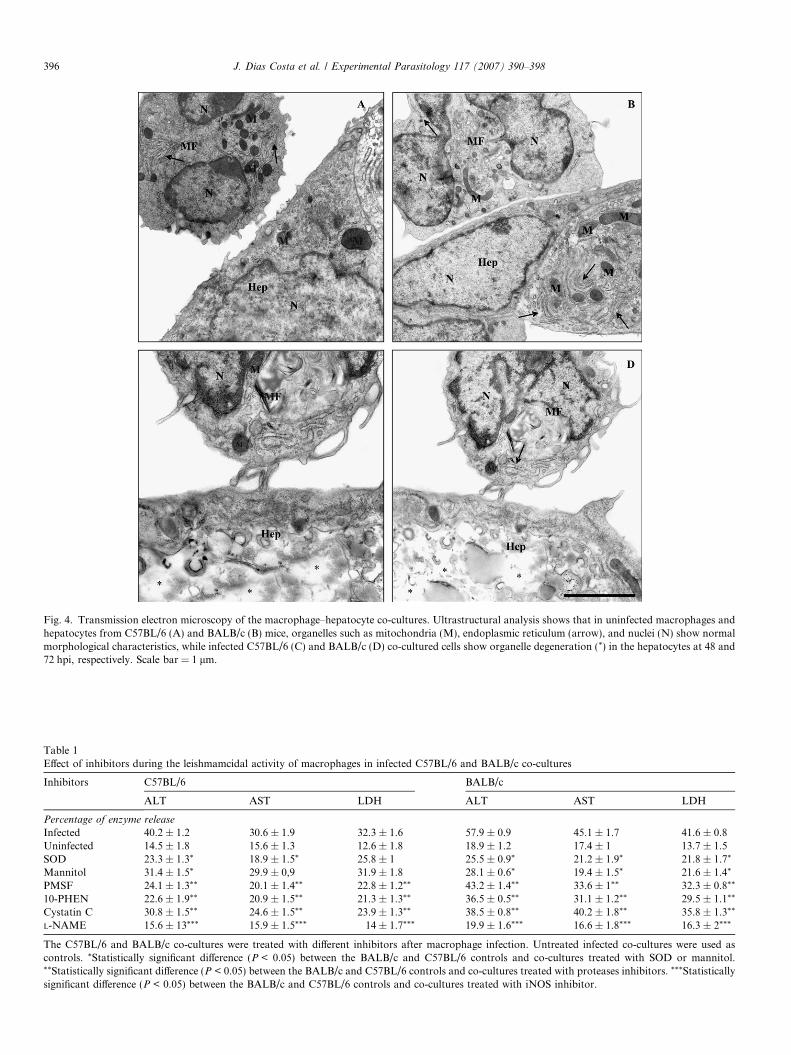
Fig. 4. Transmission electron microscopy of the macrophage–hepatocyte co-cultures. Ultrastructural analysis shows that in uninfected macrophages andhepatocytes from C57BL/6 (A) and BALB/c (B) mice, organelles such as mitochondria (M), endoplasmic reticulum (arrow), and nuclei (N) show normalmorphological characteristics, while infected C57BL/6 (C) and BALB/c (D) co-cultured cells show organelle degeneration (*) in the hepatocytes at 48 and72 hpi, respectively. Scale bar = 1 lm.
Table 1Effect of inhibitors during the leishmamcidal activity of macrophages in infected C57BL/6 and BALB/c co-cultures
Inhibitors C57BL/6 BALB/c
ALT AST LDH ALT AST LDH
Percentage of enzyme release
Infected 40.2 ± 1.2 30.6 ± 1.9 32.3 ± 1.6 57.9 ± 0.9 45.1 ± 1.7 41.6 ± 0.8Uninfected 14.5 ± 1.8 15.6 ± 1.3 12.6 ± 1.8 18.9 ± 1.2 17.4 ± 1 13.7 ± 1.5SOD 23.3 ± 1.3* 18.9 ± 1.5* 25.8 ± 1 25.5 ± 0.9* 21.2 ± 1.9* 21.8 ± 1.7*
Mannitol 31.4 ± 1.5* 29.9 ± 0,9 31.9 ± 1.8 28.1 ± 0.6* 19.4 ± 1.5* 21.6 ± 1.4*
PMSF 24.1 ± 1.3** 20.1 ± 1.4** 22.8 ± 1.2** 43.2 ± 1.4** 33.6 ± 1** 32.3 ± 0.8**
10-PHEN 22.6 ± 1.9** 20.9 ± 1.5** 21.3 ± 1.3** 36.5 ± 0.5** 31.1 ± 1.2** 29.5 ± 1.1**
Cystatin C 30.8 ± 1.5** 24.6 ± 1.5** 23.9 ± 1.3** 38.5 ± 0.8** 40.2 ± 1.8** 35.8 ± 1.3**
L-NAME 15.6 ± 13*** 15.9 ± 1.5*** 14 ± 1.7*** 19.9 ± 1.6*** 16.6 ± 1.8*** 16.3 ± 2***
The C57BL/6 and BALB/c co-cultures were treated with different inhibitors after macrophage infection. Untreated infected co-cultures were used ascontrols. *Statistically significant difference (P < 0.05) between the BALB/c and C57BL/6 controls and co-cultures treated with SOD or mannitol.**Statistically significant difference (P < 0.05) between the BALB/c and C57BL/6 controls and co-cultures treated with proteases inhibitors. ***Statisticallysignificant difference (P < 0.05) between the BALB/c and C57BL/6 controls and co-cultures treated with iNOS inhibitor.
396 J. Dias Costa et al. / Experimental Parasitology 117 (2007) 390–398
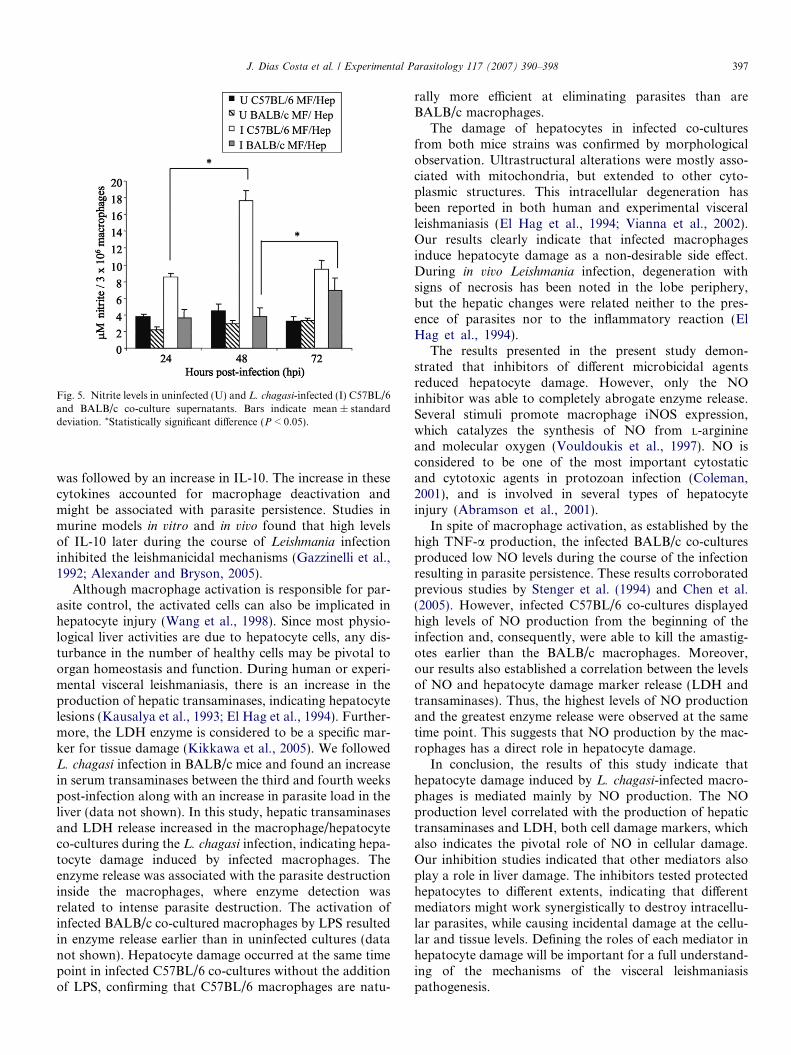
Fig. 5. Nitrite levels in uninfected (U) and L. chagasi-infected (I) C57BL/6and BALB/c co-culture supernatants. Bars indicate mean ± standarddeviation. *Statistically significant difference (P < 0.05).
J. Dias Costa et al. / Experimental Parasitology 117 (2007) 390–398 397
was followed by an increase in IL-10. The increase in thesecytokines accounted for macrophage deactivation andmight be associated with parasite persistence. Studies inmurine models in vitro and in vivo found that high levelsof IL-10 later during the course of Leishmania infectioninhibited the leishmanicidal mechanisms (Gazzinelli et al.,1992; Alexander and Bryson, 2005).
Although macrophage activation is responsible for par-asite control, the activated cells can also be implicated inhepatocyte injury (Wang et al., 1998). Since most physio-logical liver activities are due to hepatocyte cells, any dis-turbance in the number of healthy cells may be pivotal toorgan homeostasis and function. During human or experi-mental visceral leishmaniasis, there is an increase in theproduction of hepatic transaminases, indicating hepatocytelesions (Kausalya et al., 1993; El Hag et al., 1994). Further-more, the LDH enzyme is considered to be a specific mar-ker for tissue damage (Kikkawa et al., 2005). We followedL. chagasi infection in BALB/c mice and found an increasein serum transaminases between the third and fourth weekspost-infection along with an increase in parasite load in theliver (data not shown). In this study, hepatic transaminasesand LDH release increased in the macrophage/hepatocyteco-cultures during the L. chagasi infection, indicating hepa-tocyte damage induced by infected macrophages. Theenzyme release was associated with the parasite destructioninside the macrophages, where enzyme detection wasrelated to intense parasite destruction. The activation ofinfected BALB/c co-cultured macrophages by LPS resultedin enzyme release earlier than in uninfected cultures (datanot shown). Hepatocyte damage occurred at the same timepoint in infected C57BL/6 co-cultures without the additionof LPS, confirming that C57BL/6 macrophages are natu-
rally more efficient at eliminating parasites than areBALB/c macrophages.
The damage of hepatocytes in infected co-culturesfrom both mice strains was confirmed by morphologicalobservation. Ultrastructural alterations were mostly asso-ciated with mitochondria, but extended to other cyto-plasmic structures. This intracellular degeneration hasbeen reported in both human and experimental visceralleishmaniasis (El Hag et al., 1994; Vianna et al., 2002).Our results clearly indicate that infected macrophagesinduce hepatocyte damage as a non-desirable side effect.During in vivo Leishmania infection, degeneration withsigns of necrosis has been noted in the lobe periphery,but the hepatic changes were related neither to the pres-ence of parasites nor to the inflammatory reaction (ElHag et al., 1994).
The results presented in the present study demon-strated that inhibitors of different microbicidal agentsreduced hepatocyte damage. However, only the NOinhibitor was able to completely abrogate enzyme release.Several stimuli promote macrophage iNOS expression,which catalyzes the synthesis of NO from L-arginineand molecular oxygen (Vouldoukis et al., 1997). NO isconsidered to be one of the most important cytostaticand cytotoxic agents in protozoan infection (Coleman,2001), and is involved in several types of hepatocyteinjury (Abramson et al., 2001).
In spite of macrophage activation, as established by thehigh TNF-a production, the infected BALB/c co-culturesproduced low NO levels during the course of the infectionresulting in parasite persistence. These results corroboratedprevious studies by Stenger et al. (1994) and Chen et al.(2005). However, infected C57BL/6 co-cultures displayedhigh levels of NO production from the beginning of theinfection and, consequently, were able to kill the amastig-otes earlier than the BALB/c macrophages. Moreover,our results also established a correlation between the levelsof NO and hepatocyte damage marker release (LDH andtransaminases). Thus, the highest levels of NO productionand the greatest enzyme release were observed at the sametime point. This suggests that NO production by the mac-rophages has a direct role in hepatocyte damage.
In conclusion, the results of this study indicate thathepatocyte damage induced by L. chagasi-infected macro-phages is mediated mainly by NO production. The NOproduction level correlated with the production of hepatictransaminases and LDH, both cell damage markers, whichalso indicates the pivotal role of NO in cellular damage.Our inhibition studies indicated that other mediators alsoplay a role in liver damage. The inhibitors tested protectedhepatocytes to different extents, indicating that differentmediators might work synergistically to destroy intracellu-lar parasites, while causing incidental damage at the cellu-lar and tissue levels. Defining the roles of each mediator inhepatocyte damage will be important for a full understand-ing of the mechanisms of the visceral leishmaniasispathogenesis.
398 J. Dias Costa et al. / Experimental Parasitology 117 (2007) 390–398
Acknowledgments
The authors thank Dr. Maurılio Jose Soares and Dr.Elisa Cupolillo for critical review of the manuscript, BrunoAvila for computer imaging support. This work was sup-ported by grants from Fundacao Oswaldo Cruz (Fiocruz),Fundacao de Amparo a Pesquisa do Estado do Rio de Ja-neiro (FAPERJ), and Conselho Nacional de Desenvolvi-mento Cientıfico e Tecnologico (CNPq).
References
Abramson, S.B., Amim, R.A., Clancy, R.M., Attur, M., 2001. The role ofnitric oxide in tissue destruction. Best Practice and Research ClinicalRheumatology 15, 831–845.
Alexander, J., Bryson, K., 2005. T helper (h)1/Th2 and Leishmania:paradox rather than paradigm. Immunology Letter 99, 17–23.
Araujo-Jorge, T.C., de Souza, W., 1986. Interaction of Trypanosoma cruzi
with macrophages: effect of previous incubation of the parasites or thehost cells with lectins. Zeitschrift fur Parasitenkunde 72, 153–171.
Basu, M.K., Ray, M., 2005. Macrophage and Leishmania: an unaccept-able coexistence. Critical Review Microbiology 31, 145–154.
Bogdan, C., Rollinghoff, M., Dienfenbach, A., 2000. Reactive Oxygen andreactive nitrogen intermediates in innate and specific immunity.Current Opinion in Immunology 12, 64–66.
Brunet, L.R., 2001. Nitric oxide in parasitic infections. InternationalImmunopharmacology 1, 1457–1467.
Clemens, M.G., 1999. Nitric oxide in liver injury. Hepatology 30, 1–5.Coleman, J.W., 2001. Nitric oxide in immunity and inflammation.
International Immunopharmacology 1, 1397–1406.Chen, X., Oppenheim, J.J., Howard, O.M., 2005. BALB/c mice has more
CD4CD25 T regulatory cells and show greater susceptibility tosuppression of their CD24CD25 responder T cells than C57BL/6mice. Journal of Leukocyte Biology 78, 114–121.
Dey, R., Sarkar, A., Majumder, N., Majumbar, S.B., Roychoudhury, K.,Brattacharya, S., Roy, S., Majumdar, S., 2005. Regulation of impairedprotein kinase C signaling by chemokines in murine macrophagesduring visceral leishmaniasis. Infection Immunity 73, 8334–8344.
El Hag, I.A., Hashim, F.A., El Tourm, I.A., Homeida, M., El Kalifa, M.,El Hassan, A.M., 1994. Liver morphology and function in visceralleishmaniasis (Kala-azar). Journal of Clinical Pathology 47, 547–551.
Engwerda, C.R., Ato, M., Stager, S., Alexander, C.E., Stanley, A.C.,Kaye, P.M., 2004a. Distinct roles for lymphotoxin-a and tumornecrosis factor in the control of Leishmania donovani infection.American Journal of Pathology 165, 2123–2133.
Engwerda, C.R., Ato, M., Kayer, P.M., 2004b. Macrophages, pathologyand parasite persistence in experimental visceral leishmaniasis. Trendsin Parasitology 20, 524–530.
Gazzinelli, R.T., Oswald, I.P., James, S., Sher, A., 1992. IL-10 inhibitsparasite killing and nitrogen oxide production by IFN-(-activatedmacrophages. Journal of Immunology 148, 1792–1796.
Goto, H., Lindoso, J.A., 2004. Immunity and immunosuppression inexperimental visceral leishmaniasis. Brazil Journal of Medical BiologyResearch 37, 615–623.
Gumy, A., Louis, J.A., Launois, P., 2004. The murine model of infectionwith Leishmania major and its importance for the deciphering of
mechanisms underlying differences in Th cell differenciation in micefrom different genetic backgrounds. International Journal for Parasi-tology 34, 433–444.
Gutierrez, Y., Maksem, J.A., Reiner, N.E., 1984. Pathologic changes inmurine leishmaniasis (Leishmania donovani) with special reference tothe dynamics of granuloma formation in the liver. American Journalof Pathology 114 (2), 222–230.
Holzmuller, P., Hide, M., Sereno, D., Lemestre, J.L., 2006. Leishmania
infantum amastigotes resistant to nitric oxide cytotoxicity: impact onthe in vitro parasite developmental cycle and metabolic enzymeactivities. Infection, Genetics and Evolution 6, 187–197.
Kausalya, S., Malla, N., Ganguly, N.K., Mahajan, R.C., 1993. Leish-
mania donovani: in vitro evidence of hepatocyte damage by Kupffercells and immigrant macrophages in a murine model. ExperimentalParasitology 77, 326–333.
Kausalya, S., Kaur, S., Malla, N., Ganguly, N.K., Mahajan, R.C., 1996.Microbicidal mechanisms of liver macrophages in experimentalvisceral leishmaniasis. Acta Pathological, Microbiologica et Imuno-logica Scadinavica 104, 171–175.
Kikkawa, R., Yamamoto, T., Fukushima, T., Yamada, H., Horii, I., 2005.Investigation of a hepatotoxicity screening system in primary cellcultures ‘‘what biomarkers would need to be addressed to estimatetoxicity in conventional and new approaches?’’. Journal of ToxicologyScience 30, 61–72.
McElrath, M.J., Murray, H.W., Cohn, Z.A., 1988. The dynamics ofgranuloma formation in experimental visceral leishmaniasis. Journalof Experimental Medicine 167, 1927–1937.
Murray, H.W., Nathan, C.F., 1999. Macrophage microbicidal mecha-nisms in vivo: reactive nitrogen vs. oxygen intermediates in the killingof intracellular visceral Leishmania donovani. Journal of ExperimentalMedicine 189, 741–746.
Murray, H.W., Lu, C.M., Brooks, E.B., Fichtl, R.E., DeVecchio, J.L.,Heinzel, F.P., 2003. Modulation of T-cell costimulation as immuno-therapy or immunochemotherapy in experimental visceral leishmani-asis. Infection Immunology 71, 6453–6462.
Olivier, M., Tanner, C.E., 1987. Susceptibilities of macrophage popu-lation to infection in vitro by L. donovani. Infection Immunology 55,467–471.
Porrozzi, R., Soares, R., Meuser, M., Guguen-Guillouzo, C., Meirelles,M.N., 1997. Invasion and development of Trypanosoma cruzi inprimary cultures of mouse embryo hepatocytes. Memorias do InstitutoOswaldo Cruz 92, 117–120.
Stenger, S., Thuring, H., Rollinghoff, M., Bogdan, C., 1994. Tissueexpression of inducible nitric oxide synthase is closely associated withresistance to Leishmania major. Journal of Experimental Medicine 180,783–793.
Vianna, V.L., Takiya, C.M., de Brito-Gitirana, L., 2002. Histopathologicanalysis of hamster hepatocytes submitted to experimental infectionwith Leishmania donovani. Parasitology Research 88, 829–836.
Vouldoukis, I., Riveros-Moreno, V., Dugas, B., Quaaz, F., Becherel, P.,Debre, P., Moncada, S., Mossalayi, M.D., 1997. The killing ofLeishmania major by human macrophages is mediated by nitric oxideinduced after ligation of FCRII/CD23 surface antigen. Proceedings ofthe National Academy of Sciences of the United States of America 92,7804–7808.
Wang, J.H., Redmond, H.P., Wu, Q.D., Bouchier-Hayes, D., 1998. Nitricoxide mediates hepatocytes injury. American Journal of Physiology275, 1117–1126.