survey of domestic cattle for anti-leishmania antibodies and leishmania dna in a visceral...
TRANSCRIPT
Survey of domestic cattle for anti-Leishmaniaantibodies and Leishmania DNA in a visceralleishmaniasis endemic area of BangladeshAlam et al.
Alam et al. BMC Veterinary Research 2011, 7:27http://www.biomedcentral.com/1746-6148/7/27 (8 June 2011)
RESEARCH ARTICLE Open Access
Survey of domestic cattle for anti-Leishmaniaantibodies and Leishmania DNA in a visceralleishmaniasis endemic area of BangladeshMohammad Shafiul Alam1*, Debashis Ghosh1, Md Gulam Musawwir Khan1, Mohammad Faizul Islam1,Dinesh Mondal1, Makoto Itoh2, Md Nurul Islam3 and Rashidul Haque1
Abstract
Background: Visceral leishmaniasis (VL), caused by an intracellular parasite Leishmania donovani in the Indiansubcontinent, is considered to be anthroponotic. The role of domestic animals in its transmission is still unclear.Although cattle are the preferred blood host for Phlebotomus argentipes, the sandfly vector of VL in the Indiansubcontinent, very little information is available for their role in the disease transmission. In this study, weexamined domestic cattle for serological and molecular evidence of Leishmania infection in a VL-endemic area inBangladesh. Blood samples from 138 domestic cattle were collected from houses with active or recently-treated VLand post-kala-azar dermal leishmaniasis patients. The presence of anti-leishmanial antibodies in serum wasinvestigated using enzyme-linked immunosorbent assay (ELISA) and then with direct agglutination tests (DAT).Nested PCR (Ln PCR) was performed to amplify the ssu-rRNA gene using the DNA extracted from Buffy coat.Recently-developed molecular assay loop-mediated isothermal amplification (LAMP) was also performed for furthersensitive detection of parasite DNA.
Results: In this study, 9.4% (n = 13) of the cattle were found to be positive by ELISA. Of the 13 ELISA-positivecattle, only four (30.8%) were positive in DAT. Parasite DNA was not detected in either of the molecular assays(Ln PCR and LAMP).
Conclusions: The study confirmed the presence of antibodies against Leishmania parasite in cattle. However, theabsence of Leishmania DNA in the cattle indicates clearly that the cattle do not play a role as reservoir host. Similarstudy needs to be undertaken in the Indian subcontinent to determine the role of other domestic animals onwhich sandflies feed.
BackgroundEighty-eight countries of the world are endemic witheither of the two major forms of leishmaniasis: cuta-neous leishmaniasis (CL), a disfiguring and stigmatizingdisease, and visceral leishmaniasis (VL) or kala-azar,which is fatal if remains untreated [1]. One hundredfifty million people are living with the risk of VL in theIndian subcontinent (India, Nepal, and Bangladesh) [2].VL leads to a loss of about 400,000 disability-adjustedlife-years (DALYs) every year in this region [3]. VL isbelieved to be anthroponotic in the subcontinent.
Results of several studies have shown that Phlebotomusargentipes, the only known vector for Leishmania dono-vani in the Indian subcontinent, prefer to feed on bothbovine and human blood [4-8]. Being a preferable hostfor P. argentipes, cattle was shown to play an undecidedrole in several epidemiological studies in the Indian sub-continent [9]. For example, Ownership of cattle inNepal and its density in Bangladesh were found to beprotective [10,11]. Whereas, increased risk of VL wasfound to be associated with the density of cattle or itsownership in India [12,13]. Serological evidences of anti-L. donovani antibodies in different domestic animalsincluding cattle were reported in Sudan [14]. In a recentstudy in Nepal, Leishmania DNA was detected in sev-eral domestic animals including cattle from an endemic
* Correspondence: [email protected], B, 68 Shaheed Tajuddin Ahmed Sarani, Mohakhali, Dhaka 1212,BangladeshFull list of author information is available at the end of the article
Alam et al. BMC Veterinary Research 2011, 7:27http://www.biomedcentral.com/1746-6148/7/27
© 2011 Alam et al; licensee BioMed Central Ltd. This is an Open Access article distributed under the terms of the Creative CommonsAttribution License (http://creativecommons.org/licenses/by/2.0), which permits unrestricted use, distribution, and reproduction inany medium, provided the original work is properly cited.
area [15]. However, to date, no study has been con-ducted in Bangladesh to investigate the role of anydomestic animal in VL transmission.This study was aimed to investigate the evidence of
anti-leishmanial antibodies in blood of domestic cattlefrom VL-endemic villages of Mymensingh district inBangladesh. Molecular diagnostic tests were also per-formed to detect circulating parasite DNA in bloodthrough polymerase chain reaction (PCR) and loop-mediated isothermal amplification (LAMP).
MethodsStudy areaThe study was conducted in Trishal upazila (subdistrict)of Mymensingh district in Bangladesh. Trishal has aland area of 339 sq km, with a population of 3.7 million.The annual incidence of kala-azar in Trishal rangesfrom 21 to 26 per 10,000 people per year [16].
Sample-sizeResults of a previous study with domestic and wild ani-mals in Sudan showed that 21.4% had seropositivityagainst anti-L. donovani antibodies in cows [14]. How-ever, in the absence of a similar study in the Indian sub-continent, we assumed that cattle might show 10% ofseropositivity in our study. Based on this assumption,we calculated that 138 cattle would be required for ourstudy [precession 5% and 95% confidence interval (CI)].Sample-size was calculated using Windows® version ofthe Epi Info 3.2.2 software.
Sample collection from cattleBlood samples were collected from cattle duringAugust-September 2008. At the beginning, past (withinlast 3 months) and active (treatment ongoing or await-ing for treatment) VL and post-kala-azar dermal leish-maniasis (PKDL) patients were identified from therecords of Trishal Upazila Health Complex, Mymen-singh. A research team, consisting of an experiencedveterinary doctor or a veterinary assistant, visited thestudy houses, enumerated domestic cattle (Bos indicus),and collected five mL of blood from the jugular veinfrom randomly-selected one from each study house.After collection, three mL of blood was transferred toan EDTA containing tube for Buffy coat separation, andthe remaining two mL was transferred to a sterile testtube for serum separation. Relevant information on age,sex, body condition score, etc. and any changes in beha-viour within the past six months of the selected cattlewere recorded. Their physical condition was scored inthe scale of 1-5 (with 0.5 fractions between 2 scores)which represent worst to best physical condition on thebasis of bony prominence and deposition of subcuta-neous fat [17]. The blood samples were transported to
the nearby field office for Buffy coat and serum separa-tion. The processed samples were then preserved at-20°C before transferring these to the ParasitologyLaboratory of ICDDR, B in Dhaka for further serologicaland molecular investigations.
Ethical considerationThe study was approved by the Research Review Com-mittee and the Animal Experimentation Ethics Commit-tee of ICDDR, B. Consent was obtained from theowners of the cattle before the collection of bloodsample.
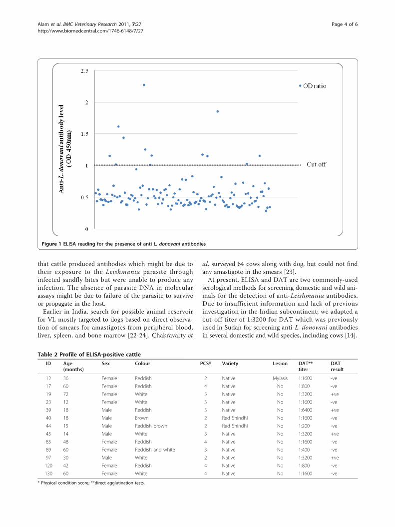
Laboratory investigationEnzyme-linked immunosorbent assayFor the qualitative detection of antibodies (IgG, IgM,and IgA) against L. donovani in the serum samples, theRecombiLISA Leishmania Ab Test (CTK Biotech Inc.,San Siego, USA) was performed. The RecombiLISALeishmania Ab Test comprises two key components: (a)solid microwells pre-coated with recombinant L. dono-vani antigens and (b) liquid conjugates composed ofrecombinant L. donovani antigens conjugated with horsereddish peroxidase (HRP-Leish Ag conjugates). The testwas performed by incubating serum and HRP-Leish Agconjugate in wells of the plate. Antibodies (IgG, IgM, orIgA) to L. donovani in the specimens would react to theL. donovani antigens coated onto the plate well by mak-ing complex with the HRP-Leish Ag conjugates.Unbounded conjugates were then removed by washing.After adding substrate and stop solution, the change incolor to yellow indicates Ab-positive, and then theabsorbance was measured by spectrophotometer at 450nm absorbance. Duplicate wells were run for eachsample.Interpretation of ELISA resultsThe ELISA results were interpreted according to theinstructions of the manufacturer. Briefly, a cut-off valuewas obtained from the mean optical density (OD) valueof the negative controls + 0.1. Then an OD ratio wascalculated for each sample by dividing the mean ODvalue by the cut-off value. Sample with OD ratio of ≥1.00 was considered to be positive, and the OD ratio of< 1.00 was considered to be negative. Serum samplescollected from four cattle from a non-endemic areawere used as negative controls in each test. Repeatedtests were performed with serum samples with a positiveresult along with an equal number of randomly-selectednegative serum samples and negative controls to make aprecise conclusion. However, no discrepancy was foundin the repeat tests.Direct agglutination testThe DAT was performed with the serum samples whichwere found to be positive in the ELISA test, along with
Alam et al. BMC Veterinary Research 2011, 7:27http://www.biomedcentral.com/1746-6148/7/27
Page 2 of 6
a similar number of ELISA-negative samples. Commer-cially-available DAT antigen (Koninklijk Instituut voorde Tropen, Amsterdam, the Netherlands) was used fol-lowing the protocol described by Harith et al. [18].Duplicate wells were run for each of the serum dilution.Heat-inactive fetal bovine serum was used along withphysiologic saline and DAT antigen as negative control[19]. An agglutination titer of 1:3200 was consideredpositive in this study [14].PCRDNA from Buffy coat was extracted using QIAamp®
DNA blood mini kit (QIAGEN, Düsseldorf, Germany).Nested PCR (Ln PCR) was performed to amplify thessu-rRNA gene with some modifications of the protocolpreviously described by Cruz et al. [20]. The first PCRwas specific for the order Kinetoplastida using the pri-mer R221 and R332 with a product size of 603 basepair. The second PCR was specific for Leishmania genususing the diluted (1:50) first PCR product using the pri-mers R223 and R333, with the product size of 358 basepair. Briefly, in the first reaction, 2 μL of the extractedDNA, 15 pmol of primers (Kinetoplastida-specific) R221and R332 were used in 25 μL PCR mix containing 0.2mM dNTP, 2 mM MgCl2, 5 mM KCl, 75 mM Tris-HCl(PH 9.0), 0.001% bovine serum albumin, 2.0 mM (NH4)
2SO4 and 2.5 U of Taq polymerase. For the second reac-tion, 1 μL of a 1/50 dilution of the first PCR productwas used as a template in the presence of 15 pmol ofprimer R223 and 15 pmol of primer R333 which areLeishmania genus-specific. Reaction conditions are thesame as the first PCR. Amplification products werevisualized on 1.5% agarose gel using a 100 bp DNA lad-der after staining with ethidium bromide (0.1 mg/mL).DNA from in-house-cultured L. donovani was used as apositive control in the PCR assay.Loop-mediated isothermal amplificationLAMP was performed followed the protocol describedby Takagi et al. [21]. In brief, the LAMP reaction wasperformed in 25 μL of reaction mixture containing 40pmol each of FIP and BIP primers, 5 pmol each of F3and B3c primers, 1.4 mM each of deoxynucleoside tri-phosphate, 0.8 M betaine, 20 mM Tris-HCl, pH 8.8, 10mM KCl, 10 mM (NH 4)2 SO4, 8 mM MgSO4, 0.1%TritonX-100, 8 units of Bst DNA polymerase large frag-ment (New England Biolabs, Ipswich, MA), and 2 μL ofsample DNA. The mixture was incubated at 65°C for 50minutes in a heat block. After incubation, the turbiditywas examined visually.Data analysisData were computed in the Microsoft Excel software.Frequencies and the association between different vari-ables (chi-square test with Yate’s correction and Fisher’sexact test) were analyzed using the SPSS software (ver-sion 11.5). A p value of < 0.05 was considered significant.
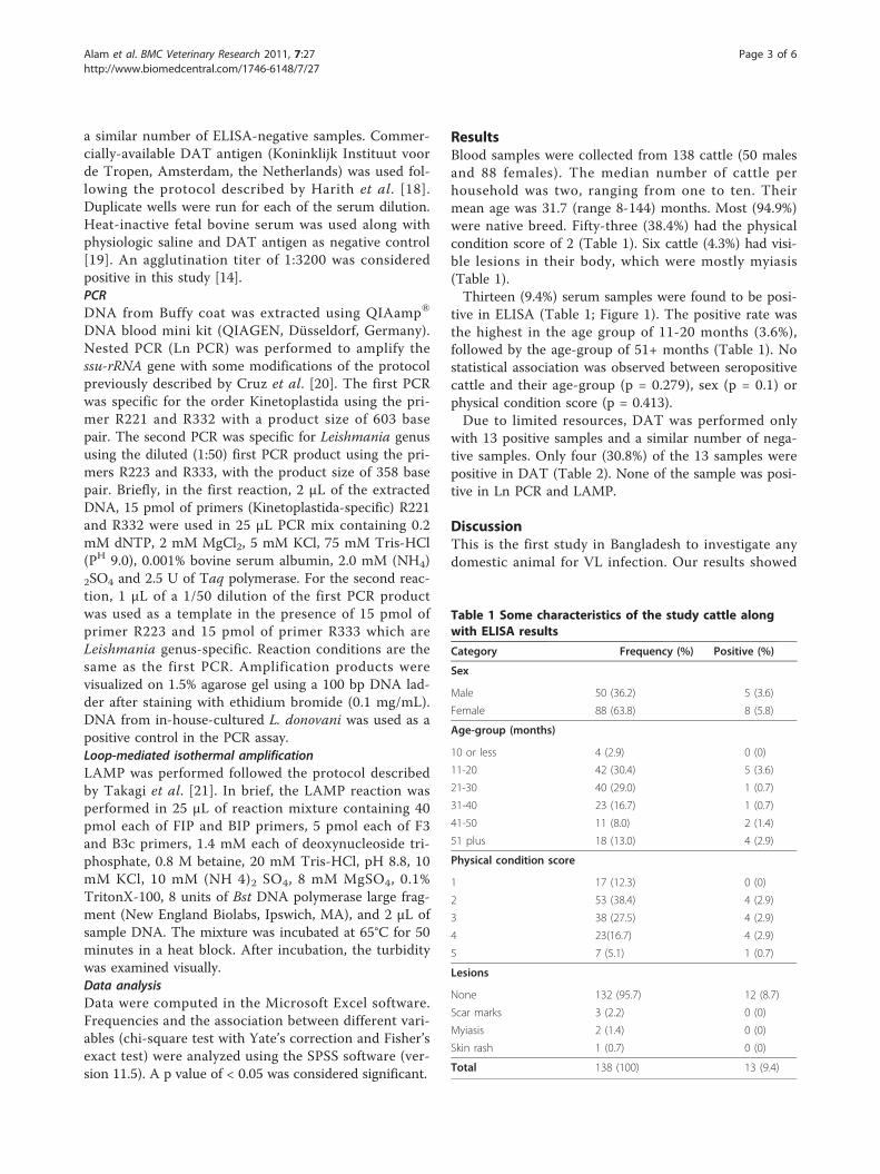
ResultsBlood samples were collected from 138 cattle (50 malesand 88 females). The median number of cattle perhousehold was two, ranging from one to ten. Theirmean age was 31.7 (range 8-144) months. Most (94.9%)were native breed. Fifty-three (38.4%) had the physicalcondition score of 2 (Table 1). Six cattle (4.3%) had visi-ble lesions in their body, which were mostly myiasis(Table 1).Thirteen (9.4%) serum samples were found to be posi-
tive in ELISA (Table 1; Figure 1). The positive rate wasthe highest in the age group of 11-20 months (3.6%),followed by the age-group of 51+ months (Table 1). Nostatistical association was observed between seropositivecattle and their age-group (p = 0.279), sex (p = 0.1) orphysical condition score (p = 0.413).Due to limited resources, DAT was performed only
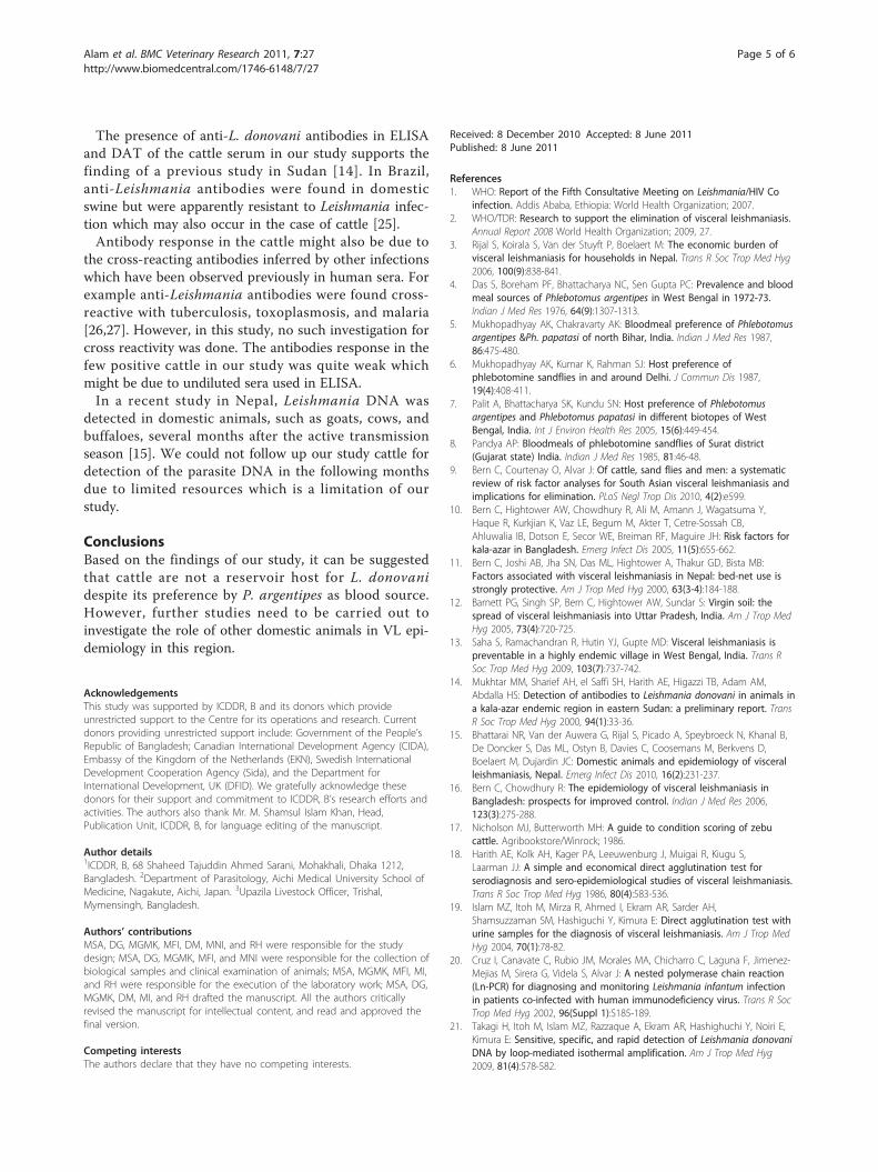
with 13 positive samples and a similar number of nega-tive samples. Only four (30.8%) of the 13 samples werepositive in DAT (Table 2). None of the sample was posi-tive in Ln PCR and LAMP.
DiscussionThis is the first study in Bangladesh to investigate anydomestic animal for VL infection. Our results showed
Table 1 Some characteristics of the study cattle alongwith ELISA results
Category Frequency (%) Positive (%)
Sex
Male 50 (36.2) 5 (3.6)
Female 88 (63.8) 8 (5.8)
Age-group (months)
10 or less 4 (2.9) 0 (0)
11-20 42 (30.4) 5 (3.6)
21-30 40 (29.0) 1 (0.7)
31-40 23 (16.7) 1 (0.7)
41-50 11 (8.0) 2 (1.4)
51 plus 18 (13.0) 4 (2.9)
Physical condition score
1 17 (12.3) 0 (0)
2 53 (38.4) 4 (2.9)
3 38 (27.5) 4 (2.9)
4 23(16.7) 4 (2.9)
5 7 (5.1) 1 (0.7)
Lesions
None 132 (95.7) 12 (8.7)
Scar marks 3 (2.2) 0 (0)
Myiasis 2 (1.4) 0 (0)
Skin rash 1 (0.7) 0 (0)
Total 138 (100) 13 (9.4)
Alam et al. BMC Veterinary Research 2011, 7:27http://www.biomedcentral.com/1746-6148/7/27
Page 3 of 6
that cattle produced antibodies which might be due totheir exposure to the Leishmania parasite throughinfected sandfly bites but were unable to produce anyinfection. The absence of parasite DNA in molecularassays might be due to failure of the parasite to surviveor propagate in the host.Earlier in India, search for possible animal reservoir
for VL mostly targeted to dogs based on direct observa-tion of smears for amastigotes from peripheral blood,liver, spleen, and bone marrow [22-24]. Chakravarty et
al. surveyed 64 cows along with dog, but could not findany amastigote in the smears [23].At present, ELISA and DAT are two commonly-used
serological methods for screening domestic and wild ani-mals for the detection of anti-Leishmania antibodies.Due to insufficient information and lack of previousinvestigation in the Indian subcontinent; we adapted acut-off titer of 1:3200 for DAT which was previouslyused in Sudan for screening anti-L. donovani antibodiesin several domestic and wild species, including cows [14].
Cut off
Figure 1 ELISA reading for the presence of anti L. donovani antibodies
Table 2 Profile of ELISA-positive cattle
ID Age(months)
Sex Colour PCS* Variety Lesion DAT**titer
DATresult
12 36 Female Reddish 2 Native Myiasis 1:1600 -ve
17 60 Female Reddish 4 Native No 1:800 -ve
19 72 Female White 5 Native No 1:3200 +ve
23 12 Female White 3 Native No 1:1600 -ve
39 18 Male Reddish 3 Native No 1:6400 +ve
40 18 Male Brown 2 Red Shindhi No 1:1600 -ve
44 15 Male Reddish brown 2 Red Shindhi No 1:200 -ve
45 14 Male White 3 Native No 1:3200 +ve
85 48 Female Reddish 4 Native No 1:1600 -ve
89 60 Female Reddish and white 3 Native No 1:400 -ve
97 30 Male White 2 Native No 1:3200 +ve
120 42 Female Reddish 4 Native No 1:800 -ve
130 60 Female White 4 Native No 1:1600 -ve
* Physical condition score; **direct agglutination tests.
Alam et al. BMC Veterinary Research 2011, 7:27http://www.biomedcentral.com/1746-6148/7/27
Page 4 of 6
The presence of anti-L. donovani antibodies in ELISAand DAT of the cattle serum in our study supports thefinding of a previous study in Sudan [14]. In Brazil,anti-Leishmania antibodies were found in domesticswine but were apparently resistant to Leishmania infec-tion which may also occur in the case of cattle [25].Antibody response in the cattle might also be due to
the cross-reacting antibodies inferred by other infectionswhich have been observed previously in human sera. Forexample anti-Leishmania antibodies were found cross-reactive with tuberculosis, toxoplasmosis, and malaria[26,27]. However, in this study, no such investigation forcross reactivity was done. The antibodies response in thefew positive cattle in our study was quite weak whichmight be due to undiluted sera used in ELISA.In a recent study in Nepal, Leishmania DNA was
detected in domestic animals, such as goats, cows, andbuffaloes, several months after the active transmissionseason [15]. We could not follow up our study cattle fordetection of the parasite DNA in the following monthsdue to limited resources which is a limitation of ourstudy.
ConclusionsBased on the findings of our study, it can be suggestedthat cattle are not a reservoir host for L. donovanidespite its preference by P. argentipes as blood source.However, further studies need to be carried out toinvestigate the role of other domestic animals in VL epi-demiology in this region.
AcknowledgementsThis study was supported by ICDDR, B and its donors which provideunrestricted support to the Centre for its operations and research. Currentdonors providing unrestricted support include: Government of the People’sRepublic of Bangladesh; Canadian International Development Agency (CIDA),Embassy of the Kingdom of the Netherlands (EKN), Swedish InternationalDevelopment Cooperation Agency (Sida), and the Department forInternational Development, UK (DFID). We gratefully acknowledge thesedonors for their support and commitment to ICDDR, B’s research efforts andactivities. The authors also thank Mr. M. Shamsul Islam Khan, Head,Publication Unit, ICDDR, B, for language editing of the manuscript.
Author details1ICDDR, B, 68 Shaheed Tajuddin Ahmed Sarani, Mohakhali, Dhaka 1212,Bangladesh. 2Department of Parasitology, Aichi Medical University School ofMedicine, Nagakute, Aichi, Japan. 3Upazila Livestock Officer, Trishal,Mymensingh, Bangladesh.
Authors’ contributionsMSA, DG, MGMK, MFI, DM, MNI, and RH were responsible for the studydesign; MSA, DG, MGMK, MFI, and MNI were responsible for the collection ofbiological samples and clinical examination of animals; MSA, MGMK, MFI, MI,and RH were responsible for the execution of the laboratory work; MSA, DG,MGMK, DM, MI, and RH drafted the manuscript. All the authors criticallyrevised the manuscript for intellectual content, and read and approved thefinal version.
Competing interestsThe authors declare that they have no competing interests.
Received: 8 December 2010 Accepted: 8 June 2011Published: 8 June 2011
References1. WHO: Report of the Fifth Consultative Meeting on Leishmania/HIV Co
infection. Addis Ababa, Ethiopia: World Health Organization; 2007.2. WHO/TDR: Research to support the elimination of visceral leishmaniasis.
Annual Report 2008 World Health Organization; 2009, 27.3. Rijal S, Koirala S, Van der Stuyft P, Boelaert M: The economic burden of
visceral leishmaniasis for households in Nepal. Trans R Soc Trop Med Hyg2006, 100(9):838-841.
4. Das S, Boreham PF, Bhattacharya NC, Sen Gupta PC: Prevalence and bloodmeal sources of Phlebotomus argentipes in West Bengal in 1972-73.Indian J Med Res 1976, 64(9):1307-1313.
5. Mukhopadhyay AK, Chakravarty AK: Bloodmeal preference of Phlebotomusargentipes &Ph. papatasi of north Bihar, India. Indian J Med Res 1987,86:475-480.
6. Mukhopadhyay AK, Kumar K, Rahman SJ: Host preference ofphlebotomine sandflies in and around Delhi. J Commun Dis 1987,19(4):408-411.
7. Palit A, Bhattacharya SK, Kundu SN: Host preference of Phlebotomusargentipes and Phlebotomus papatasi in different biotopes of WestBengal, India. Int J Environ Health Res 2005, 15(6):449-454.
8. Pandya AP: Bloodmeals of phlebotomine sandflies of Surat district(Gujarat state) India. Indian J Med Res 1985, 81:46-48.
9. Bern C, Courtenay O, Alvar J: Of cattle, sand flies and men: a systematicreview of risk factor analyses for South Asian visceral leishmaniasis andimplications for elimination. PLoS Negl Trop Dis 2010, 4(2):e599.
10. Bern C, Hightower AW, Chowdhury R, Ali M, Amann J, Wagatsuma Y,Haque R, Kurkjian K, Vaz LE, Begum M, Akter T, Cetre-Sossah CB,Ahluwalia IB, Dotson E, Secor WE, Breiman RF, Maguire JH: Risk factors forkala-azar in Bangladesh. Emerg Infect Dis 2005, 11(5):655-662.
11. Bern C, Joshi AB, Jha SN, Das ML, Hightower A, Thakur GD, Bista MB:Factors associated with visceral leishmaniasis in Nepal: bed-net use isstrongly protective. Am J Trop Med Hyg 2000, 63(3-4):184-188.
12. Barnett PG, Singh SP, Bern C, Hightower AW, Sundar S: Virgin soil: thespread of visceral leishmaniasis into Uttar Pradesh, India. Am J Trop MedHyg 2005, 73(4):720-725.
13. Saha S, Ramachandran R, Hutin YJ, Gupte MD: Visceral leishmaniasis ispreventable in a highly endemic village in West Bengal, India. Trans RSoc Trop Med Hyg 2009, 103(7):737-742.
14. Mukhtar MM, Sharief AH, el Saffi SH, Harith AE, Higazzi TB, Adam AM,Abdalla HS: Detection of antibodies to Leishmania donovani in animals ina kala-azar endemic region in eastern Sudan: a preliminary report. TransR Soc Trop Med Hyg 2000, 94(1):33-36.
15. Bhattarai NR, Van der Auwera G, Rijal S, Picado A, Speybroeck N, Khanal B,De Doncker S, Das ML, Ostyn B, Davies C, Coosemans M, Berkvens D,Boelaert M, Dujardin JC: Domestic animals and epidemiology of visceralleishmaniasis, Nepal. Emerg Infect Dis 2010, 16(2):231-237.
16. Bern C, Chowdhury R: The epidemiology of visceral leishmaniasis inBangladesh: prospects for improved control. Indian J Med Res 2006,123(3):275-288.
17. Nicholson MJ, Butterworth MH: A guide to condition scoring of zebucattle. Agribookstore/Winrock; 1986.
18. Harith AE, Kolk AH, Kager PA, Leeuwenburg J, Muigai R, Kiugu S,Laarman JJ: A simple and economical direct agglutination test forserodiagnosis and sero-epidemiological studies of visceral leishmaniasis.Trans R Soc Trop Med Hyg 1986, 80(4):583-536.
19. Islam MZ, Itoh M, Mirza R, Ahmed I, Ekram AR, Sarder AH,Shamsuzzaman SM, Hashiguchi Y, Kimura E: Direct agglutination test withurine samples for the diagnosis of visceral leishmaniasis. Am J Trop MedHyg 2004, 70(1):78-82.
20. Cruz I, Canavate C, Rubio JM, Morales MA, Chicharro C, Laguna F, Jimenez-Mejias M, Sirera G, Videla S, Alvar J: A nested polymerase chain reaction(Ln-PCR) for diagnosing and monitoring Leishmania infantum infectionin patients co-infected with human immunodeficiency virus. Trans R SocTrop Med Hyg 2002, 96(Suppl 1):S185-189.
21. Takagi H, Itoh M, Islam MZ, Razzaque A, Ekram AR, Hashighuchi Y, Noiri E,Kimura E: Sensitive, specific, and rapid detection of Leishmania donovaniDNA by loop-mediated isothermal amplification. Am J Trop Med Hyg2009, 81(4):578-582.
Alam et al. BMC Veterinary Research 2011, 7:27http://www.biomedcentral.com/1746-6148/7/27
Page 5 of 6
22. Bhattacharya A, Ghosh TN: A search for Leishmania in vertebrates fromkala-azar-affected areas of Bihar, India. Trans R Soc Trop Med Hyg 1983,77(6):874-875.
23. Chakravarty AK, Sanyat RK, Suri JC: Zoonotic reservoir in Indian kala-azar. JCommun Dis 1979, 11:219-220.
24. Zahar AR: Studies Leishmaniasis, Vectors/Reservoirs and their control inthe old world; Part IV. Asia and Pacific. World Health Organization;198087.
25. Moraes-Silva E, Antunes FR, Rodrigues MS, da Silva Juliao F, Dias-Lima AG,Lemos-de-Sousa V, de Alcantara AC, Reis EA, Nakatani M, Badaro R, Reis MG,Pontes-de-Carvalho L, Franke CR: Domestic swine in a visceralleishmaniasis endemic area produce antibodies against multipleLeishmania infantum antigens but apparently resist to L. infantuminfection. Acta Trop 2006, 98(2):176-182.
26. Badaro R, Eulalio MC, Benson D, Freire M, Miranda JC, Pedral-Sampaio D,Burns JM, David JR, Johnson WD, Reed SG: Sensitivity and specificity of arecombinant Leishmania chagasi antigen in the serodiagnosis of visceralleishmaniasis. Arch Inst Pasteur Tunis 1993, 70(3-4):331-332.
27. Burns JM, Shreffler WG, Benson DR, Ghalib HW, Badaro R, Reed SG:Molecular characterization of a kinesin-related antigen of Leishmaniachagasi that detects specific antibody in African and American visceralleishmaniasis. Proc Natl Acad Sci USA 1993, 90(2):775-779.
doi:10.1186/1746-6148-7-27Cite this article as: Alam et al.: Survey of domestic cattle for anti-Leishmania antibodies and Leishmania DNA in a visceral leishmaniasisendemic area of Bangladesh. BMC Veterinary Research 2011 7:27.
Submit your next manuscript to BioMed Centraland take full advantage of:
• Convenient online submission
• Thorough peer review
• No space constraints or color figure charges
• Immediate publication on acceptance
• Inclusion in PubMed, CAS, Scopus and Google Scholar
• Research which is freely available for redistribution
Submit your manuscript at www.biomedcentral.com/submit
Alam et al. BMC Veterinary Research 2011, 7:27http://www.biomedcentral.com/1746-6148/7/27
Page 6 of 6