lateral scattered light used to study laser light propagation in turbid media phantoms
TRANSCRIPT
Lateral scattered light used to study laser light propagation in turbid media phantoms
Claudia Valdes*, Efrain Solarte
Quantum Optics Group, Universidad del Valle, A.A. 25360, Cali, Colombia
ABSTRACT
Understanding laser light propagation in soft tissue is important because of the growing biomedical applications of lasers and the need to optically characterize the biological media. Following previous developments of our group, we have developed low cost models, Phantoms, of soft tissue. The process was developed in a clean room to avoid the medium contamination. Each model was characterized by measuring its refractive index, and spectral reflectance and transmittance. To study the laser light propagation, each phantom was illuminated with a clean beam of laser light, using sources such as He-Ne (632nm) and DPSSL (473 nm). Laterally scattered light was imaged and these images were digitally processed. We analyzed the intensity distribution of the scattered radiation in order to obtain details of the beam evolution in the medium. Line profiles taken from the intensity distribution surface allow measurement of the beam spread, and expressions for the longitudinal (along the beam incident direction) and transverse (across the beam incident direction) intensities distributions were found. From these behaviors, the radiation penetration depth and the total coefficient of extinction have been determined. The multiple scattering effects were remarkable, especially for the low wavelength laser beam.
Keywords: Tissue Optics, Laser scattering, Spectroscopy, Low Cost Phantom, DPSSL
1. INTRODUCTION Studies of light propagation in materials or tissues with strongly scattering characteristics are very necessary to understand the behavior and the possible applications of light in biological systems1. Modeling these systems is usually done using materials different from the very tissue, but with similar optical properties. As our main interest is to make a low cost scattering media, following previous developments2, 3, we have developed low cost models, or phantoms, that can be used to study the soft tissue optical properties2. These models were made using a rich collagen gel, which exhibits high scattering, an adequate refraction index, that the gel can be used as a matrix for other substances which will provide other tissue properties.
Monochromatic light beam propagation in highly scattering media is a complex problem, which can be handled in different ways1, 4. In the simplest model, uniformly distributed absorbers and scatters cause a loss in the beam intensity because the light is elastically scattered or absorbed in the medium. This intensity loss along an infinitesimal path dz, is proportional to the incoming beam intensity. These considerations lead to the well known Beer-Lambert law for the light intensity I(z) at certain point z along the light beam path:
)exp()( 0 zIzI μ−= (1)
I0 is the incident light intensity, corrected by reflections at the surfaces, and μ is the total attenuation coefficient, which is given by the sum of the scattering coefficient μs and the absorption coefficient μa. This simple model underlies the main idea of a single photon interaction process in which photons are removed from the laser beam, as the beam propagates on a straight path. The removed photons are completely annihilated or they continue moving in all other directions different from the direction of the original beam. In a more realistic model for turbid media, multiple scattering of photons must be taken into account. So, the light intensity in certain point r in a highly scattering and absorbing medium must be constructed by considering the remaining beam intensity within a small volume around this point, which includes the losses by single scattering, but also the contributions from all other points inside the medium, from which photons can be scattered into a given direction in such a way that the photon density in the considered volume will be increased.
*[email protected]; phone 57 2 339-4610; fax 57 2 339-3237
Design and Performance Validation of Phantoms Used in Conjunction with Optical Measurementof Tissue II, edited by Robert J. Nordstrom, Proc. of SPIE Vol. 7567, 75670C
© 2010 SPIE · CCC code: 1605-7422/10/$18 · doi: 10.1117/12.843101
Proc. of SPIE Vol. 7567 75670C-1

For the case of a continuous beam propagating in a highly scattering medium, the stationary radiative transfer theory5 [RTT] is usually applied to determine the photon density variation in an elemental volume. It is then possible to establish that the time diminution of the photon density in the volume is given by the radiation flux on the surface surrounding this volume, the absorption and the scattering that remove photons from this volume, and increases because of the photons arriving to the volume due to the scattering, from direction s’ into the direction s, in other points of the medium. The stationary condition is achieved considering that the density remains constant along the time, so that the time derivative of the photons density is null, which gives the RRT equation. The most basic form of this equation could be written for the radiance I [W cm-2sr-1], in the following6 way:
')'ˆ,()'ˆ,(4
)ˆ,()ˆ,(
4
Ω+−=∂
∂∫ dsrpsrIsrI
ssrI s rrr
r
ππμ
μ (2)
Where p(s, s’) is the phase function, giving the photon probability of scattering from direction s’ into direction s; ds is the infinitesimal path length, and dΩ’ the solid angle element. In this approach, by considering laser beam propagation, the radiance can be separated into two terms: the coherent radiance which follows the Beer-Lambert law, and will thus be characterized by an exponential decay in a definite direction, and the diffuse radiance coming from multiple scattering, considering photons which do not follow a straight determined path. In this work we present the main experimental results concerning the optical properties of our designed phantoms, as well as the intensity distributions of light in the media, along and across the propagation direction of the laser beam, and the attenuation coefficients obtained from image data processing of laser illuminated phantoms.
2. EXPERIMENTAL METHODS 2.1 Phantom preparation
In order to prepare a low cost phantom, commercial unflavored gelatin powder was used, because it is a rich source of collagen, which it is assumed that it would result in a high similarity with human tissue. For the phantoms used in this study we used 60 g of unflavored gelatin powder, 350 ml of distilled water and 250 ml of physiological saline solution. These phantoms were prepared at the Universidad del Valle Pharmacology Group cleanroom to prevent external contamination and to assure a longer useful life of the phantoms. In addition, all the containers were gas sterilized prior to the preparation.
First, the powder was placed in container and the distilled water, previously heated, was added. This mixture was stirred until there were no lumps. Then, the saline solution was stirred in and this mixture was sterilized again. Finally, this prepared gelatin was poured in a 10x10x10 cm3 cubic glass container, covered with a sterilized aluminum foil and set aside to solidify into a gel at room temperature in a laminar air flow chamber. Following this preparation method, two different brand gelatin models were prepared, which were labeled R and F.
2.2 Experimental procedure
Figure 1. Scheme of the experimental setup used for laser beam propagation studies in high scattering media. The photodetector (PD) marked with dotted line is removed out of the beam path when taking the pictures.
Proc. of SPIE Vol. 7567 75670C-2

For the purpose of studying laser light propagation within the finished phantoms, each phantom was illuminated using a clean laser beam in a set up that is shown in Figure 1. The laser beam was first passed through an attenuation optical system, where the laser intensity was set. Next, the beam was directed to a 90° total reflecting mirror, which allows positioning the input laser beam normally to the phantom surface. Before and after passing through the phantom, the laser beam power was measured with a standard Si photometer. The laterally scattered light was collected using a digital camera. The laser sources used for this study were a stabilized CW He-Ne laser (632.8 nm) and a Nd:YVO4 DPSSL (473 nm), CW. For each phantom, several laser powers were applied ranging from 0.1 mW to 18 mW approximately every 2 mW. Lateral photographs of the illuminated phantoms were taken using a Kodak Z740 digital camera with a resolution of 2576 x 1932 pixels. The shooting conditions (aperture, exposition time) for every picture were kept invariant. Because it is difficult to guarantee the same distance between the camera and the medium, a calibration image was taken every time a light source or a phantom was changed.
After these procedures, spectral reflectance and transmittance of the medium were measured using a halogen lamp as light source and a USB4000 ® Ocean Optics spectrometer using a reflectance probe to illuminate the glass-free face of the phantom. A lens attached to an optical fiber was used to collect the transmitted light in the incoming beam direction. Collimated transmittance was measured by directly recording the laser beam intensity before and after passing through the phantom. The photodetectors were placed as shown in Figure 1. The indices of refraction of the phantom media were measured using a standard Abbe refractometer. To perform these measurements, it was necessary to cut thin slices of each phantom.
2.3 Image processing
Because we were interested in studying the light propagation behavior using the digital images, it was necessary to apply a digital image process to prepare the images. First, a luminance plane extraction was done. This procedure gives a gray scale image from which intensity information could be obtained. Second, a 7x7 Gaussian smoothing process was applied to reduce random intensity variations produced by speckles and by the camera detector. Finally, a rotation correction was made to align the laser beam in all the pictures. These digital procedures were done using the ImaQ Vision Builder® software. From these processed images, the required information has been obtained by means of line profiles conveniently traced over the image, which provide light intensity behavior along that line. Seeing that all the obtained data are given in terms of pixel position, a calibration coefficient must be found to convert these pixel positions to real distance units. To obtain this coefficient, a calibration image, corresponding to each set of pictures, was used and a calibration procedure was performed3. Once the distances on the pictures were in real distance units, the set of line profiles give information about the light intensities in the lateral faces of the phantom. These are scattered light distributions carrying information about the phantom optical properties. Other image processing utilities have been used to produce binary maps and 3D representations, very useful for comparison between pictures and to present intensity distribution effects.
3. EXPERIMENTAL RESULTS
3.1 Phantom optical properties
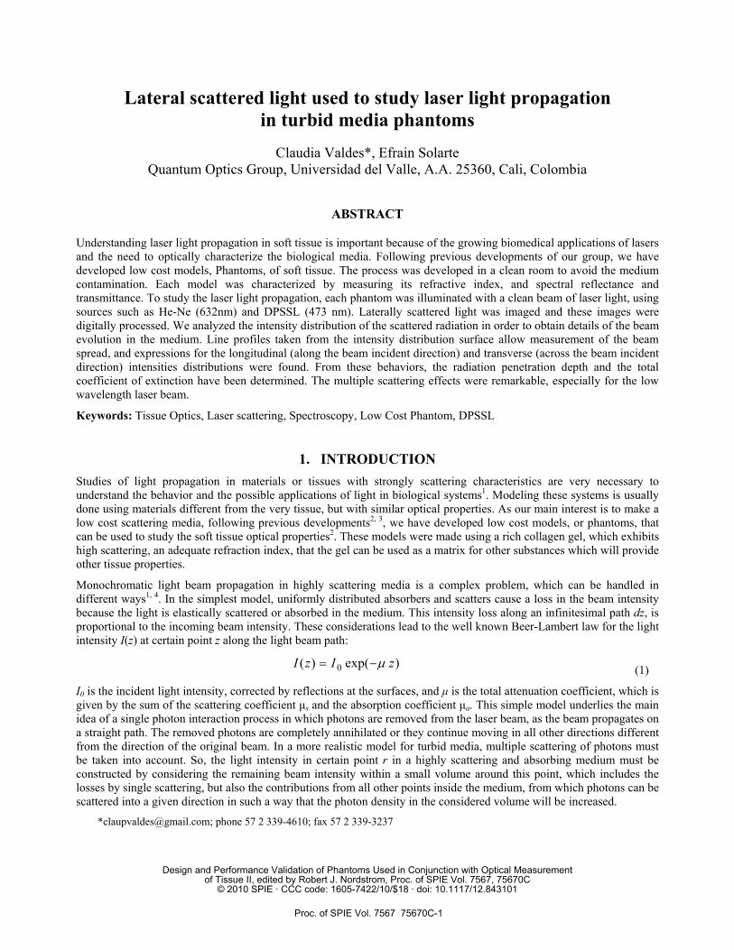
The refractive indices of these models were 1.350 for phantom R and 1.352 for phantom F. Collimated Transmittance (CT) measurements are shown in Figure 2. HeNe laser light (633 nm) transmittance is about ten times larger as this for the Nd:YVO4 blue laser (473nm), and the phantom F has about 2 times the transmittance of phantom R.
The HeNe, 633 nm, average CT values are found to be (9.14 ± 0.005)% for the R phantom, and (22.49 ± 0.008)% for the F; whereas the blue laser line (473 nm) presents a (0.32 ± 0.001)% for the R phantom and a (2.39 ±0.002)% transmittance value for the F phantom. These results indicate the better scattering properties of phantom R, and also the well known property relating scattering and wavelength, showing that large wavelengths beams are less scattered as the short wavelengths ones.
Typical spectral measurements exhibit an almost flat spectrum which falls at the violet and NIR regions as shown in Figure 3. From these measurements it is not possible to distinguish absorption bands in the visible range.
Proc. of SPIE Vol. 7567 75670C-3
-2 0 2 4 6 8 10 12 14 16 18 20 22 240
2
4
6
8
10
12
14
16
18
20
22
24
Ave
rage
Col
limat
ed T
rans
mitt
ance
[%]
Input Laser Power [mW]
HeNe 633 nm Phantom F Phantom R
Nd:YVO 473 nm Phantom F Phantom R
Figure 2. Collimated Transmittance as a function of the input laser power. Measurements are shown for the two studied phantoms.
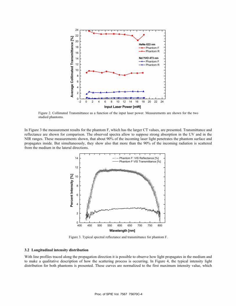
In Figure 3 the measurement results for the phantom F, which has the larger CT values, are presented. Transmittance and reflectance are shown for comparison. The observed spectra allow to suppose strong absorption in the UV and in the NIR ranges. These measurements shown, that about 90% of the incoming laser light penetrates the phantom surface and propagates inside. But simultaneously, they show also that more than the 90% of the incoming radiation is scattered from the medium in the lateral directions.
400 450 500 550 600 650 700 750 8000
2
4
6
8
10
12
14
Perc
ent I
nten
sity
[%]
Wavelength [nm]
Phantom F: VIS Reflectance [%] Phantom F VIS Transmittance [%]
Figure 3. Typical spectral reflectance and transmittance for phantom F.
3.2 Longitudinal intensity distribution
With line profiles traced along the propagation direction it is possible to observe how light propagates in the medium and to make a qualitative description of how the scattering process is occurring. In Figure 4, the typical intensity light distribution for both phantoms is presented. These curves are normalized to the first maximum intensity value, which
Proc. of SPIE Vol. 7567 75670C-4
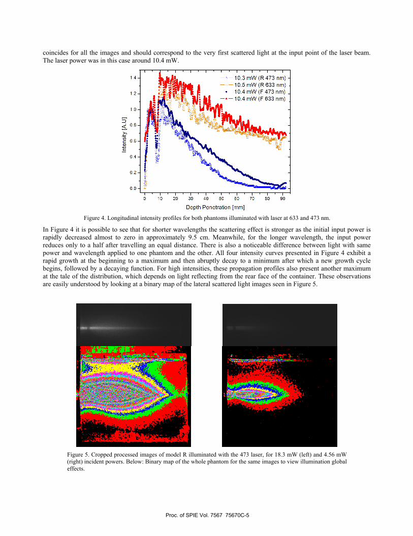
coincides for all the images and should correspond to the very first scattered light at the input point of the laser beam. The laser power was in this case around 10.4 mW.
Figure 4. Longitudinal intensity profiles for both phantoms illuminated with laser at 633 and 473 nm.
In Figure 4 it is possible to see that for shorter wavelengths the scattering effect is stronger as the initial input power is rapidly decreased almost to zero in approximately 9.5 cm. Meanwhile, for the longer wavelength, the input power reduces only to a half after travelling an equal distance. There is also a noticeable difference between light with same power and wavelength applied to one phantom and the other. All four intensity curves presented in Figure 4 exhibit a rapid growth at the beginning to a maximum and then abruptly decay to a minimum after which a new growth cycle begins, followed by a decaying function. For high intensities, these propagation profiles also present another maximum at the tale of the distribution, which depends on light reflecting from the rear face of the container. These observations are easily understood by looking at a binary map of the lateral scattered light images seen in Figure 5.
Figure 5. Cropped processed images of model R illuminated with the 473 laser, for 18.3 mW (left) and 4.56 mW (right) incident powers. Below: Binary map of the whole phantom for the same images to view illumination global effects.
Proc. of SPIE Vol. 7567 75670C-5
The images in Figure 5 show blue light propagating from the left, passing through phantom R, with two different incident powers. In the upper view, the processed images are shown. Below each is the binary map corresponding to the processed image. These maps provide pixel detailed information about what is happening with light in the whole medium. Comparing these images with the intensity profiles in Figure 4, it is seen that the first and the tail maxima of the distributions are due to the reflections on the faces of the glass mold. The minimum correspond to a waist that is better seen in the right map for a lower power. This is caused because at the beginning of the propagation in the medium the scattering is mainly in the forward direction and the intensity decays as expected, but after some distance, the multiple scattering begins to have an important contribution in all directions which increases the intensity again. The binary maps also show a layer distribution of the light showing regions of equal intensity in the medium.
3.3 Transverse intensity distributions
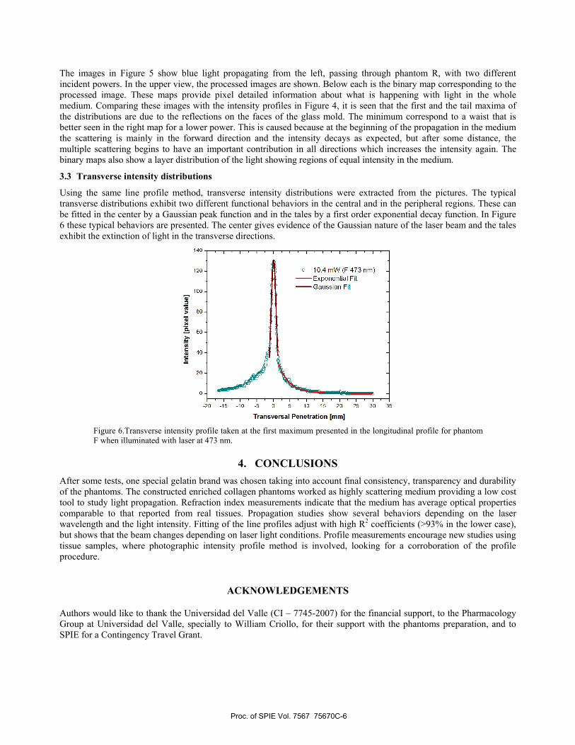
Using the same line profile method, transverse intensity distributions were extracted from the pictures. The typical transverse distributions exhibit two different functional behaviors in the central and in the peripheral regions. These can be fitted in the center by a Gaussian peak function and in the tales by a first order exponential decay function. In Figure 6 these typical behaviors are presented. The center gives evidence of the Gaussian nature of the laser beam and the tales exhibit the extinction of light in the transverse directions.
Figure 6.Transverse intensity profile taken at the first maximum presented in the longitudinal profile for phantom F when illuminated with laser at 473 nm.
4. CONCLUSIONS After some tests, one special gelatin brand was chosen taking into account final consistency, transparency and durability of the phantoms. The constructed enriched collagen phantoms worked as highly scattering medium providing a low cost tool to study light propagation. Refraction index measurements indicate that the medium has average optical properties comparable to that reported from real tissues. Propagation studies show several behaviors depending on the laser wavelength and the light intensity. Fitting of the line profiles adjust with high R2 coefficients (>93% in the lower case), but shows that the beam changes depending on laser light conditions. Profile measurements encourage new studies using tissue samples, where photographic intensity profile method is involved, looking for a corroboration of the profile procedure.
ACKNOWLEDGEMENTS
Authors would like to thank the Universidad del Valle (CI – 7745-2007) for the financial support, to the Pharmacology Group at Universidad del Valle, specially to William Criollo, for their support with the phantoms preparation, and to SPIE for a Contingency Travel Grant.
Proc. of SPIE Vol. 7567 75670C-6
REFERENCES
[1] Tuchin V. V., [Tissue Optics: Light Scattering Methods and Instruments for Medical Diagnosis], SPIE Press, Bellingham, 3-17 (2007).
[2] Solarte, E, Valdes, C. P., Banguero Y., and Cabrera B., "Estudio Experimental y Reconstrucción de la Propagación de un Haz Laser en un Modelo de Tejido Blando," Rev. Col. Fís., 41(1), 138-141 (2009).
[3] Valdes, C. and Solarte E., "Coherence measurements of a Nd-YAG laser by image processing of Michelson interferometer fringe patterns," Proc. SPIE 6028, 60281F (2005).
[4] Ren, K., Bal, G., and Hielscher, A. H., “Transport- and diffusion-based optical tomography in small domains: a comparative study,” Applied Optics 46(27), 6669-6679 (2007).
[5] Chandrasekhar, S., [Radiative Transfer], Dover Publications Inc., New York, 393 (1960). [6] Niemz, M. H., [Laser-Tissue Interactions], Second Edition, Springer, Berlin-Heidelberg, 9-44 (2007).
Proc. of SPIE Vol. 7567 75670C-7