lateral hypothalamic involvement in feeding elicited from the ventral pallidum
TRANSCRIPT

Lateral hypothalamic involvement in feeding elicited fromthe ventral pallidum
Thomas R. Stratford and David WirtshafterLaboratory of Integrative Neuroscience, Department of Psychology (m/c 285), University of Illinois at Chicago, Chicago, IL,USA
Keywords: bicuculline, food intake, GABA, hypothalamus, rats
Abstract
Intense feeding can be elicited by injections of the GABAA receptor antagonist bicuculline into the medial ventral pallidum(VPm), a basal forebrain structure anatomically interposed between two other feeding-related brain regions, the nucleus ac-cumbens shell and the lateral hypothalamus (LH). To determine whether the VPm effects changes in feeding behaviorthrough actions on the LH, we examined feeding following unilateral injections of bicuculline into the VPm made either ipsilat-eral or contralateral to a unilateral excitotoxic lesion of the LH in nondeprived rats. We found that lesions of the LH signifi-cantly attenuated feeding induced from the ipsilateral VPm, as compared to sham-operated controls. In striking contrast,unilateral LH lesions significantly potentiated the feeding response elicited by injections of bicuculline into the contralateralVPm. The ‘ipsilateral–contralateral disruption’ design we used makes it extremely unlikely that our findings could haveresulted from nonspecific effects of the lesions. These results suggest that the LH is causally involved in mediating theingestive effects produced by activation of the VPm, and provide an important insight into the functional circuitry by whichbasal forebrain structures control food intake in mammals.
Introduction
The medial ventral pallidum (VPm) has been shown to exert amajor influence on food intake. Injections of either GABAA
antagonists or l-opioid agonists into the VPm induce intensefeeding in nondeprived rats (Stratford et al., 1999; Smith & Ber-ridge, 2005). Conversely, lesions within the VPm reduce foodintake and body weight (Cromwell & Berridge, 1993). It hasbeen proposed that the VPm is an integral component of a basalforebrain feeding circuit incorporating the nucleus accumbensshell (AcbSh) and the lateral hypothalamus (LH; Stratford et al.,1999; Stratford, 2007). This suggestion is based on the fact thatthe VPm is anatomically interposed between these two feeding-related brain regions, receiving a dense GABAergic projectionfrom the AcbSh (Nauta et al., 1978; Churchill et al., 1990;Zahm & Heimer, 1990; Heimer et al., 1991) and sending a sub-stantial projection to the LH (Haber et al., 1985; Groenewegenet al., 1993). Importantly, it has been shown recently thatlesions of either the VPm or the LH disrupt the feeding behav-ior evoked by injections of muscimol into the AcbSh (Stratford& Wirtshafter, 2012), a result supporting the hypothesis that theVPm may act to relay information from the AcbSh to the LH.However, no data which directly address the possibility that theVPm may control food through an effect on the LH are cur-rently available. One prediction of this hypothesis is that
destruction of the LH should attenuate the feeding induced bystimulation of the VPm.Most studies examining the role of the LH in ingestive
responses elicited from other structures have examined theeffects of bilateral LH lesions or inactivation (e.g., Maldonado-Irizarry et al., 1995; Stratford & Kelley, 1999; Stratford & Wir-tshafter, 2000). This approach suffers from the potential pitfallthat LH manipulations could suppress feeding indirectly byinducing some effect that interferes with the feeding response,such as sedation, motor problems or alterations in gustatory pro-cessing. It is possible to render these alternative explanationsmuch less likely through the use of what we have referred to asan ‘ipsilateral–contralateral disruption‘design (Stratford & Wirtsh-after, 2012). In this approach, experimental subjects receive uni-lateral lesions of the LH and are then tested followingmanipulations of the target structure on either the ipsilateral orthe contralateral side of the brain. If the effects of the unilateralLH lesions were due to nonspecific or generalized effects, onewould expect that they would suppress feeding to the sameextent whether it was elicited from the same or the oppositeside of the brain. In contrast, if the LH were an essential com-ponent of a lateralized circuit underlying the elicited feeding,one would expect that ipsilateral lesions would produce a greatersuppression than contralateral ones. The use of this designrequires that connections between the target structure and theLH be predominantly ipsilateral, a condition which is fulfilled inthe case of connections between the VPm and the LH (Haberet al., 1985; Groenewegen et al., 1993).
Correspondence: T. R. Stratford, as above.E-mail: [email protected]
Received 20 September 2012, revised 29 October 2012, accepted 1 November 2012
© 2012 Federation of European Neuroscience Societies and Blackwell Publishing Ltd
European Journal of Neuroscience, pp. 1–6, 2012 doi:10.1111/ejn.12077
European Journal of Neuroscience
Materials and methods
Subjects
Seventeen male Sprague–Dawley rats (Charles Rivers), weighingbetween 340 and 360 g at the time of surgery, served as subjects.The rats were housed individually in plastic cages on a 12-hlight : 12-h dark cycle at a constant room temperature (~21 °C) withfood (Harlan Teklad) and tap water available ad libitum, except asnoted below. All experiments conformed to the NIH Guide for theCare and Use of Laboratory Animals and were approved by theInstitutional Animal Care and Use Committee.
Surgery
Surgery was performed using standard, aseptic, flat-skull stereotaxictechniques under sodium pentobarbital (60 mg/kg) anesthesia. Inexperimental subjects (n = 9), a 28-gauge stainless steel injectorwas lowered into the LH at the following coordinates: AP, �2.4;LM, 1.8; DV, �9.1 (mm from bregma). Fifteen micrograms of ib-otenic acid (10 lg/lL) was then infused into the LH at a rate of0.33 lL/min after which the injector was allowed to remain in placefor 5 min to minimize diffusion up the injector path. In order thatmechanical damage dorsal to the LH would be equivalent on thetwo sides of the brain, an injector was then lowered to a height ofDV �7.1 on the opposite side of the brain but no infusions weremade at this site. Identical procedures were employed in control ani-mals (n = 8) except that the phosphate buffer vehicle, rather than ib-otenic acid, was infused unilaterally. Immediately after the LHprocedures had been performed, bilateral 22-gauge stainless steelguide cannulae (Plastics One, Roanoke, VA, USA), aimed so as toterminate 2.0 mm dorsal to the VPm, were implanted using the fol-lowing coordinates: AP, 0.2; LM, � 1.8; DV, �6.8. The guidecannulae were held in place using stainless steel screws and denturelining material and a stainless steel obturator was inserted into thelumen of each cannula to help maintain patency. After surgery, therats received an injection of carprofen (5 mg/kg, s.c.) to help allevi-ate postoperative pain. Each rat was allowed to recover for at least7 days before the start of behavioral testing.
Test apparatus
Test chambers consisted of plastic shoebox cages (43 9 22 cm)equipped with automated dispensers (Med-Associates, St Albans,VT, USA) that delivered a single 45-mg pellet of food (PrecisionDustless pellets; Bio-Serve, Frenchtown, NJ, USA) whenever theprevious pellet was removed from the hopper. In order to acclimatethe rats to eating in the test chambers, they were placed in them onfive consecutive days prior to the start of drug testing. The rats weremildly food-deprived for the first of these trials and nondeprived forthe remainder.
Intracerebral injection technique
During the intracerebral injections the rats were restrained gently,the obturators removed, and 28-gauge injection cannulae, extending2.0 mm beyond the ventral tip of the guide, were inserted into eachguide cannula. Injections were made at a rate of 0.33 lL/min usingmotor-driven microsyringe pumps connected to the injection cannu-lae with fluid-filled polyethylene tubing. After the infusion, theinjection cannulae were left in place for an additional 60 s in orderto minimize leakage up the cannula track. The obturators were thenreplaced and the rats were placed in the test cages for 120 min. On
the last of the acclimation days described above, each rat receivedbilateral 0.25-lL intracerebral injections of sterile 0.15 M saline.Test sessions began 48 h later; on test days, each rat received simul-taneous bilateral 0.5-lL injections at a rate of 0.33 lL/min. At least48 h were allowed between injections.
Experimental protocol
Each rat first received a series of three injections which were admin-istered in a counterbalanced order. The three conditions were: (i)bilateral saline injections; (ii) bicuculline (1(S),9(R)-(�)-bicucullinemethbromide; 50 ng; Sigma) ipsilateral and saline contralateral tothe lesion; and (iii) bicuculline contralateral and saline ipsilateral.Following these treatments, animals received two further experimen-tal manipulations consisting of (i) bilateral bicuculline injections;and (ii) bilateral saline injections, which were again administered ina counterbalanced order. Subjects were then prepared for histologi-cal examination as described below.
Perfusion and immunohistochemistry
When behavioral testing was completed, each of the rats was deeplyanesthetized using sodium pentobarbital and perfused transcardiallywith 50 mL of a 0.15-M saline solution followed immediately by200 mL of a 10% buffered formalin solution at pH 6.5, then300 mL of a 10% buffered formalin solution at pH 9.0. The brainswere removed and stored in PBS with 20% sucrose for at least2 days, after which they were frozen and 35-lm-thick coronal sec-tions were taken throughout the extent of the AcbSh and the lesionsites. The sections then were processed for the immunohistochemicaldetection of neuronal nuclear protein (NeuN), a sensitive marker ofundamaged neurons that is extremely valuable in evaluating excito-toxic lesions (Jongen-Relo & Feldon, 2002). Briefly, the sectionswere rinsed in PBS and incubated on a rotary shaker table for 24 hat 4 °C in a monoclonal mouse anti-NeuN primary antibody (Onco-gene Research Products) diluted 1 : 20 000 with 0.01 M PBS con-taining 4% normal horse serum and 0.2% Triton X-100. Thesections were rinsed again in PBS and incubated in biotinylatedhorse anti-mouse secondary antibody (diluted 1 : 200 with PBS con-taining 4% normal horse serum) for 60 min at room temperature.Sections were then rinsed again in PBS (3 9 10 min) and incubatedin the avidin–biotin complex solution (Vectastain Elite ABC kit;Vector Laboratories, Burlingame, CA, USA) for 60 min. Followinganother series of rinses in PBS, the peroxidase was visualized byincubating the tissue for 5 min in the nickel-enhanced chromogensolution from a Vector 3,3′-diaminobenzidine tetrahydrochloride per-oxidase substrate kit. The sections were then mounted on chrome–alum-coated slides, air-dried, cleared in xylene and coverslippedwith Eukitt mounting medium. The lesion boundaries were mappedand the VPm injection sites were examined for placement accuracyand evidence of excessive damage. In five animals with iboteniclesions of the LH, analogous methods were used to stain sectionsthrough the striatum for tyrosine hydroxylase (TH)-like immunore-activity using a monoclonal mouse anti-TH serum (TE 101; EugeneTech International, Allendale, NJ, USA).
Results
Histology
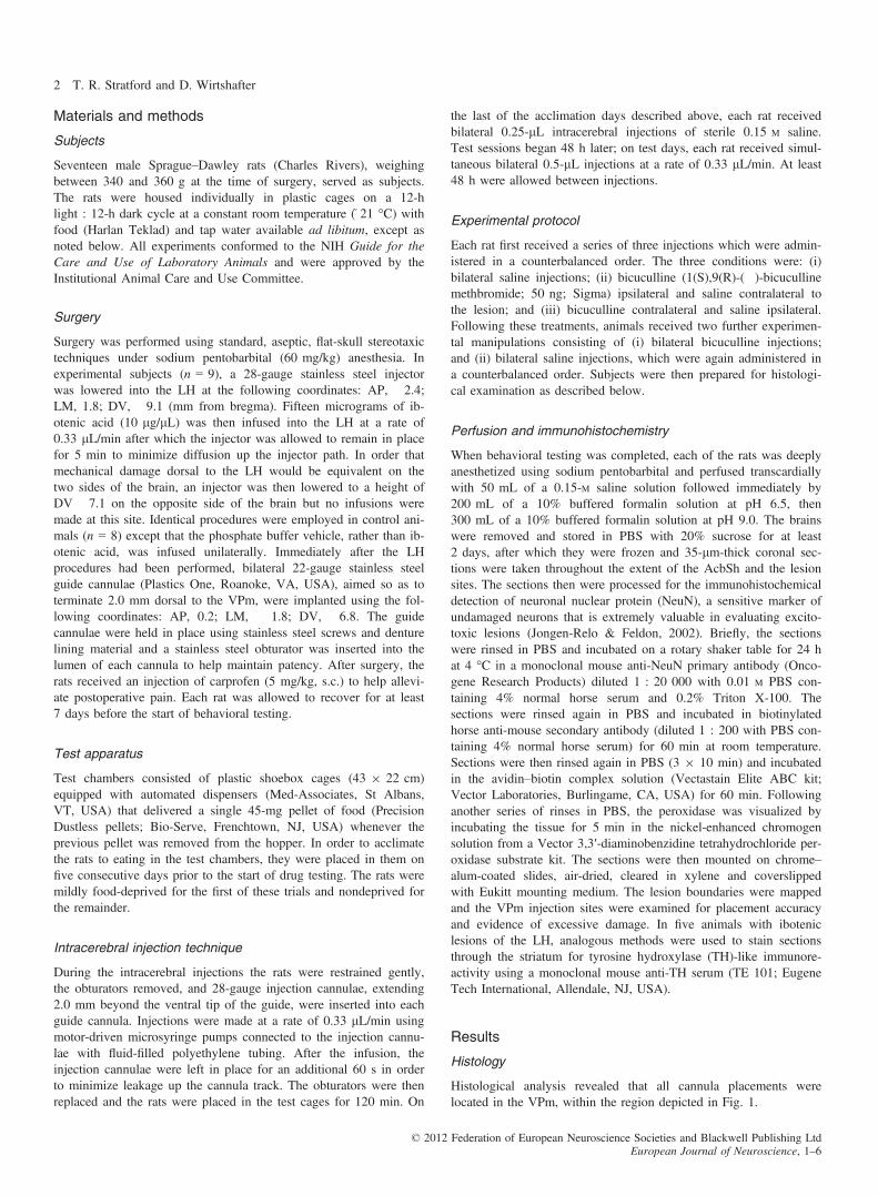
Histological analysis revealed that all cannula placements werelocated in the VPm, within the region depicted in Fig. 1.
© 2012 Federation of European Neuroscience Societies and Blackwell Publishing LtdEuropean Journal of Neuroscience, 1–6
2 T. R. Stratford and D. Wirtshafter
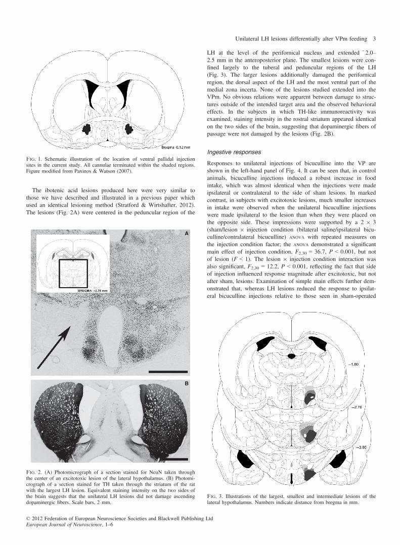
The ibotenic acid lesions produced here were very similar tothose we have described and illustrated in a previous paper whichused an identical lesioning method (Stratford & Wirtshafter, 2012).The lesions (Fig. 2A) were centered in the peduncular region of the
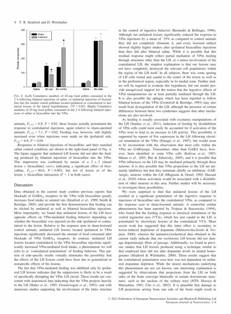
LH at the level of the perifornical nucleus and extended ~2.0–2.5 mm in the anteroposterior plane. The smallest lesions were con-fined largely to the tuberal and peduncular regions of the LH(Fig. 3). The larger lesions additionally damaged the perifornicalregion, the dorsal aspect of the LH and the most ventral part of themedial zona incerta. None of the lesions studied extended into theVPm. No obvious relations were apparent between damage to struc-tures outside of the intended target area and the observed behavioraleffects. In the subjects in which TH-like immunoreactivity wasexamined, staining intensity in the rostral striatum appeared identicalon the two sides of the brain, suggesting that dopaminergic fibers ofpassage were not damaged by the lesions (Fig. 2B).
Ingestive responses
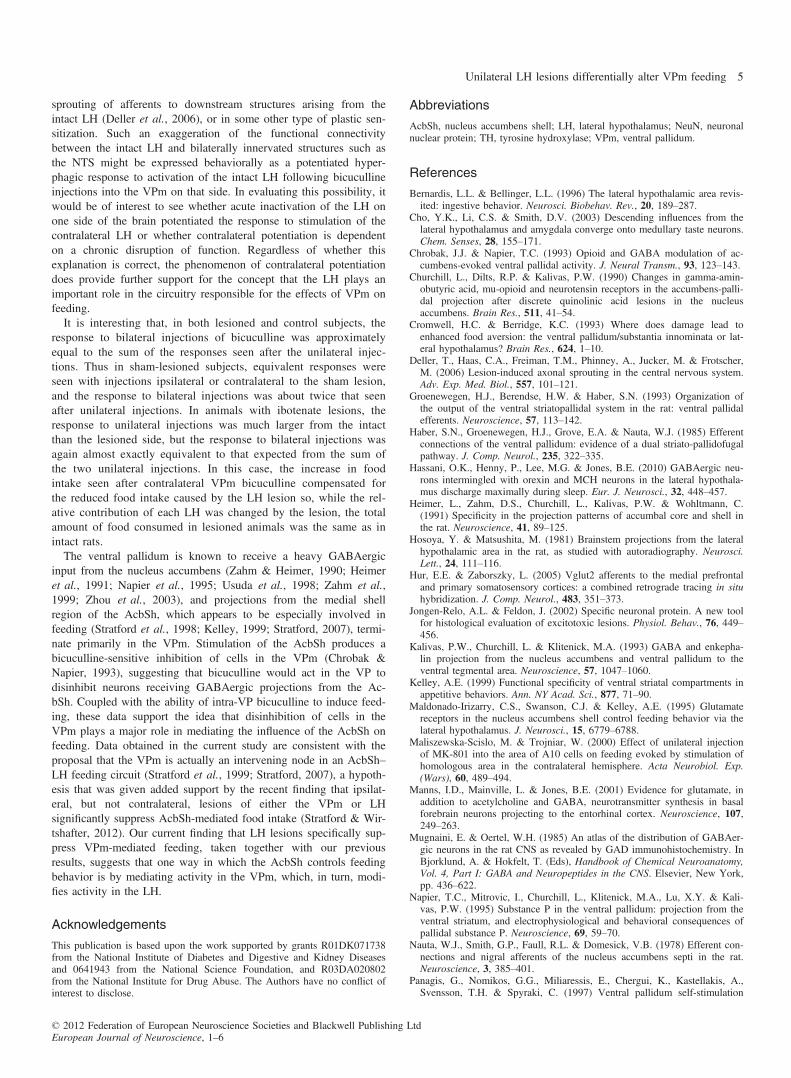
Responses to unilateral injections of bicuculline into the VP areshown in the left-hand panel of Fig. 4. It can be seen that, in controlanimals, bicuculline injections induced a robust increase in foodintake, which was almost identical when the injections were madeipsilateral or contralateral to the side of sham lesions. In markedcontrast, in subjects with excitotoxic lesions, much smaller increasesin intake were observed when the unilateral bicuculline injectionswere made ipsilateral to the lesion than when they were placed onthe opposite side. These impressions were supported by a 2 9 3(sham/lesion 9 injection condition (bilateral saline/ipsilateral bicu-culline/contralateral bicuculline) ANOVA with repeated measures onthe injection condition factor; the ANOVA demonstrated a significantmain effect of injection condition, F2,30 = 36.7, P < 0.001, but notof lesion (F < 1). The lesion 9 injection condition interaction wasalso significant, F2,30 = 12.2, P < 0.001, reflecting the fact that sideof injection influenced response magnitude after excitotoxic, but notafter sham, lesions. Examination of simple main effects further dem-onstrated that, whereas LH lesions reduced the response to ipsilat-eral bicuculline injections relative to those seen in sham-operated
Fig. 1. Schematic illustration of the location of ventral pallidal injectionsites in the current study. All cannulae terminated within the shaded regions.Figure modified from Paxinos & Watson (2007).
A
B
Fig. 2. (A) Photomicrograph of a section stained for NeuN taken throughthe center of an excitotoxic lesion of the lateral hypothalamus. (B) Photomi-crograph of a section stained for TH taken through the striatum of the ratwith the largest LH lesion. Equivalent staining intensity on the two sides ofthe brain suggests that the unilateral LH lesions did not damage ascendingdopaminergic fibers. Scale bars, 2 mm.
Fig. 3. Illustrations of the largest, smallest and intermediate lesions of thelateral hypothalamus. Numbers indicate distance from bregma in mm.
© 2012 Federation of European Neuroscience Societies and Blackwell Publishing LtdEuropean Journal of Neuroscience, 1–6
Unilateral LH lesions differentially alter VPm feeding 3
animals, F1,15 = 6.8, P < 0.02, these lesions actually potentiated theresponse to contralateral injections, again relative to sham-operatedanimals, F1,15 = 7.1, P < 0.02. Feeding was, however, still slightlyincreased even when injections were made on the ipsilateral side,F1,8 = 6.8, P < 0.05.Responses to bilateral injections of bicuculline, and their matched
saline control condition, are shown in the right-hand panel of Fig. 4.The figure suggests that unilateral LH lesions did not alter the feed-ing produced by bilateral injections of bicuculline into the VPm.This impression was confirmed by means of a 2 9 2 (sham/lesion 9 bicuculline) ANOVA indicating a significant effect of bicu-culline, F1,15 = 80.6, P < 0.001, but not of lesion or of thelesion 9 bicuculline interaction (F < 1 in both cases).
Discussion
Data obtained in the current study confirm previous reports thatblockade of GABAA receptors in the VPm with bicuculline greatlyincreases food intake in satiated rats (Stratford et al., 1999; Smith &Berridge, 2005), and provide the first demonstration that feeding canbe elicited by unilateral as well as bilateral bicuculline injections.More importantly, we found that unilateral lesions of the LH haveopposite effects on VPm-mediated feeding behavior depending onwhether the bicuculline was injected on the side ipsilateral or contra-lateral to the lesion. Relative to the response seen in sham-lesionedcontrol animals, unilateral LH lesions located ipsilateral to VPminjections significantly decreased the amount of food consumed afterblockade of VPm GABAA receptors. In contrast, unilateral LHlesions located contralateral to the VPm bicuculline injections signif-icantly increased VPm-mediated food intake, a phenomenon we willrefer to as ‘contralateral potentiation’ of feeding behavior. This pat-tern of side-specific results virtually eliminates the possibility thatthe effects of the LH lesions could have been due to generalized ornonspecific effects of the lesions.The fact that VPm-mediated feeding was inhibited only by ipsilat-
eral LH lesions indicates that the suppression is likely to be a resultof specifically disrupting the VPm–LH circuit. These results are con-sistent with anatomical data indicating that the VPm projects heavilyto the LH (Haber et al., 1985; Groenewegen et al., 1993), and withnumerous studies supporting the involvement of the latter structure
in the control of ingestive behavior (Bernardis & Bellinger, 1996).Although our unilateral lesions significantly reduced the response toVPm injections by a mean of~55% as compared to control animalsthey did not completely eliminate it, and every lesioned subjectshowed slightly higher intakes after ipsilateral bicuculline injectionsthan they did after bilateral saline. While it is possible that thisresidual response might reflect partial mediation of VPm feedingthrough structures other than the LH, or a minor involvement of thecontralateral LH, the simplest explanation is that our lesions maynot have completely destroyed the relevant cell populations withinthe region of the LH itself. In all subjects, there was some sparingof LH cells rostral and caudal to the center of the lesion as well asin the perifornical region, especially in its medial zone. Further stud-ies will be required to evaluate this hypothesis, but our results pro-vide unequivocal support for the notion that the ingestive effects ofVPm manipulations are at least partially mediated through the LH.It is also possible the aphagia which has been reported to followbilateral lesions of the VPm (Cromwell & Berridge, 1993) may alsoresult from dysregulation of the LH, although the presence of certaindifferences between these two syndromes suggests that other mecha-nisms are also involved.As feeding is usually associated with excitatory manipulations of
the LH (Stanley et al., 2011), induction of feeding by disinhibitionof VPm cells could most easily be accounted for if activation of theVPm were to lead to an increase in LH activity. This possibility issupported by reports of Fos expression in the LH following electri-cal stimulation of the VPm (Panagis et al., 1997), but would seemto be inconsistent with the observation that most cells within theVPm are GABAergic. Transmitters other than GABA have, how-ever, been identified in some VPm cells (Kalivas et al., 1993;Manns et al., 2001; Hur & Zaborszky, 2005), and it is possible thatVPm influences on the LH may be mediated primarily through theseneurons. It is also possible that VPm projections to the LH are pri-marily inhibitory but that they terminate chiefly on inhibitory, GAB-Aergic, neurons within the LH (Mugnaini & Oertel, 1985; Hassaniet al., 2010) whose activation would be associated with a disinhibi-tion of other neurons in this area. Further studies will be necessaryto investigate these possibilities.We were surprised to find that unilateral lesions of the LH
resulted in a significant potentiation of the ingestive response toinjections of bicuculline into the contralateral VPm, as compared tothe response seen in sham-lesioned animals. A somewhat similarphenomenon has been reported by Trojniar & Staszewska (1994),who found that the feeding response to electrical stimulation of theventral tegmental area (VTA), which lies just caudal to the LH, ispotentiated by electrolytic lesions of the contralateral VTA. Morerecent work has suggested that this latter effect is secondary tolesion-induced depletions of dopamine (Maliszewska-Scislo & Tro-jniar, 2000), whereas the immunocytochemical data obtained in thecurrent study indicate that our excitotoxic LH lesions did not dam-age dopaminergic fibers of passage. Additionally, we found in previ-ous studies that LH lesions produced using a technique similar tothat employed here did not alter dopamine levels in striatal homo-genates (Stratford & Wirtshafter, 2000). These results suggest thatthe contralateral potentiation seen here was not dependent on unilat-eral dopamine depletion. While the neural mechanisms underlyingthis phenomenon are not yet known, one interesting explanation issuggested by observations that projections from the LH on bothsides of the brain converge bilaterally in certain downstream struc-tures, such as the nucleus of the solitary tract (NTS; Hosoya &Matsushita, 1981; Cho et al., 2003). It is plausible that damage toLH projections arising from one side of the brain might result in
Fig. 4. (Left) Cumulative numbers of 45-mg food pellets consumed in the2 h following bilateral injections of saline, or unilateral injections of bicucul-line into the medial ventral pallidum located ipsilateral or contralateral to uni-lateral lesions of the lateral hypothalamus. (*P < 0.05). (Right) Cumulativenumbers of 45-mg food pellets consumed in the 2 h following bilateral injec-tions of saline or bicuculline into the VPm.
© 2012 Federation of European Neuroscience Societies and Blackwell Publishing LtdEuropean Journal of Neuroscience, 1–6
4 T. R. Stratford and D. Wirtshafter
sprouting of afferents to downstream structures arising from theintact LH (Deller et al., 2006), or in some other type of plastic sen-sitization. Such an exaggeration of the functional connectivitybetween the intact LH and bilaterally innervated structures such asthe NTS might be expressed behaviorally as a potentiated hyper-phagic response to activation of the intact LH following bicucullineinjections into the VPm on that side. In evaluating this possibility, itwould be of interest to see whether acute inactivation of the LH onone side of the brain potentiated the response to stimulation of thecontralateral LH or whether contralateral potentiation is dependenton a chronic disruption of function. Regardless of whether thisexplanation is correct, the phenomenon of contralateral potentiationdoes provide further support for the concept that the LH plays animportant role in the circuitry responsible for the effects of VPm onfeeding.It is interesting that, in both lesioned and control subjects, the
response to bilateral injections of bicuculline was approximatelyequal to the sum of the responses seen after the unilateral injec-tions. Thus in sham-lesioned subjects, equivalent responses wereseen with injections ipsilateral or contralateral to the sham lesion,and the response to bilateral injections was about twice that seenafter unilateral injections. In animals with ibotenate lesions, theresponse to unilateral injections was much larger from the intactthan the lesioned side, but the response to bilateral injections wasagain almost exactly equivalent to that expected from the sum ofthe two unilateral injections. In this case, the increase in foodintake seen after contralateral VPm bicuculline compensated forthe reduced food intake caused by the LH lesion so, while the rel-ative contribution of each LH was changed by the lesion, the totalamount of food consumed in lesioned animals was the same as inintact rats.The ventral pallidum is known to receive a heavy GABAergic
input from the nucleus accumbens (Zahm & Heimer, 1990; Heimeret al., 1991; Napier et al., 1995; Usuda et al., 1998; Zahm et al.,1999; Zhou et al., 2003), and projections from the medial shellregion of the AcbSh, which appears to be especially involved infeeding (Stratford et al., 1998; Kelley, 1999; Stratford, 2007), termi-nate primarily in the VPm. Stimulation of the AcbSh produces abicuculline-sensitive inhibition of cells in the VPm (Chrobak &Napier, 1993), suggesting that bicuculline would act in the VP todisinhibit neurons receiving GABAergic projections from the Ac-bSh. Coupled with the ability of intra-VP bicuculline to induce feed-ing, these data support the idea that disinhibition of cells in theVPm plays a major role in mediating the influence of the AcbSh onfeeding. Data obtained in the current study are consistent with theproposal that the VPm is actually an intervening node in an AcbSh–LH feeding circuit (Stratford et al., 1999; Stratford, 2007), a hypoth-esis that was given added support by the recent finding that ipsilat-eral, but not contralateral, lesions of either the VPm or LHsignificantly suppress AcbSh-mediated food intake (Stratford & Wir-tshafter, 2012). Our current finding that LH lesions specifically sup-press VPm-mediated feeding, taken together with our previousresults, suggests that one way in which the AcbSh controls feedingbehavior is by mediating activity in the VPm, which, in turn, modi-fies activity in the LH.
Acknowledgements
This publication is based upon the work supported by grants R01DK071738from the National Institute of Diabetes and Digestive and Kidney Diseasesand 0641943 from the National Science Foundation, and R03DA020802from the National Institute for Drug Abuse. The Authors have no conflict ofinterest to disclose.
Abbreviations
AcbSh, nucleus accumbens shell; LH, lateral hypothalamus; NeuN, neuronalnuclear protein; TH, tyrosine hydroxylase; VPm, ventral pallidum.
References
Bernardis, L.L. & Bellinger, L.L. (1996) The lateral hypothalamic area revis-ited: ingestive behavior. Neurosci. Biobehav. Rev., 20, 189–287.
Cho, Y.K., Li, C.S. & Smith, D.V. (2003) Descending influences from thelateral hypothalamus and amygdala converge onto medullary taste neurons.Chem. Senses, 28, 155–171.
Chrobak, J.J. & Napier, T.C. (1993) Opioid and GABA modulation of ac-cumbens-evoked ventral pallidal activity. J. Neural Transm., 93, 123–143.
Churchill, L., Dilts, R.P. & Kalivas, P.W. (1990) Changes in gamma-amin-obutyric acid, mu-opioid and neurotensin receptors in the accumbens-palli-dal projection after discrete quinolinic acid lesions in the nucleusaccumbens. Brain Res., 511, 41–54.
Cromwell, H.C. & Berridge, K.C. (1993) Where does damage lead toenhanced food aversion: the ventral pallidum/substantia innominata or lat-eral hypothalamus? Brain Res., 624, 1–10.
Deller, T., Haas, C.A., Freiman, T.M., Phinney, A., Jucker, M. & Frotscher,M. (2006) Lesion-induced axonal sprouting in the central nervous system.Adv. Exp. Med. Biol., 557, 101–121.
Groenewegen, H.J., Berendse, H.W. & Haber, S.N. (1993) Organization ofthe output of the ventral striatopallidal system in the rat: ventral pallidalefferents. Neuroscience, 57, 113–142.
Haber, S.N., Groenewegen, H.J., Grove, E.A. & Nauta, W.J. (1985) Efferentconnections of the ventral pallidum: evidence of a dual striato-pallidofugalpathway. J. Comp. Neurol., 235, 322–335.
Hassani, O.K., Henny, P., Lee, M.G. & Jones, B.E. (2010) GABAergic neu-rons intermingled with orexin and MCH neurons in the lateral hypothala-mus discharge maximally during sleep. Eur. J. Neurosci., 32, 448–457.
Heimer, L., Zahm, D.S., Churchill, L., Kalivas, P.W. & Wohltmann, C.(1991) Specificity in the projection patterns of accumbal core and shell inthe rat. Neuroscience, 41, 89–125.
Hosoya, Y. & Matsushita, M. (1981) Brainstem projections from the lateralhypothalamic area in the rat, as studied with autoradiography. Neurosci.Lett., 24, 111–116.
Hur, E.E. & Zaborszky, L. (2005) Vglut2 afferents to the medial prefrontaland primary somatosensory cortices: a combined retrograde tracing in situhybridization. J. Comp. Neurol., 483, 351–373.
Jongen-Relo, A.L. & Feldon, J. (2002) Specific neuronal protein. A new toolfor histological evaluation of excitotoxic lesions. Physiol. Behav., 76, 449–456.
Kalivas, P.W., Churchill, L. & Klitenick, M.A. (1993) GABA and enkepha-lin projection from the nucleus accumbens and ventral pallidum to theventral tegmental area. Neuroscience, 57, 1047–1060.
Kelley, A.E. (1999) Functional specificity of ventral striatal compartments inappetitive behaviors. Ann. NY Acad. Sci., 877, 71–90.
Maldonado-Irizarry, C.S., Swanson, C.J. & Kelley, A.E. (1995) Glutamatereceptors in the nucleus accumbens shell control feeding behavior via thelateral hypothalamus. J. Neurosci., 15, 6779–6788.
Maliszewska-Scislo, M. & Trojniar, W. (2000) Effect of unilateral injectionof MK-801 into the area of A10 cells on feeding evoked by stimulation ofhomologous area in the contralateral hemisphere. Acta Neurobiol. Exp.(Wars), 60, 489–494.
Manns, I.D., Mainville, L. & Jones, B.E. (2001) Evidence for glutamate, inaddition to acetylcholine and GABA, neurotransmitter synthesis in basalforebrain neurons projecting to the entorhinal cortex. Neuroscience, 107,249–263.
Mugnaini, E. & Oertel, W.H. (1985) An atlas of the distribution of GABAer-gic neurons in the rat CNS as revealed by GAD immunohistochemistry. InBjorklund, A. & Hokfelt, T. (Eds), Handbook of Chemical Neuroanatomy,Vol. 4, Part I: GABA and Neuropeptides in the CNS. Elsevier, New York,pp. 436–622.
Napier, T.C., Mitrovic, I., Churchill, L., Klitenick, M.A., Lu, X.Y. & Kali-vas, P.W. (1995) Substance P in the ventral pallidum: projection from theventral striatum, and electrophysiological and behavioral consequences ofpallidal substance P. Neuroscience, 69, 59–70.
Nauta, W.J., Smith, G.P., Faull, R.L. & Domesick, V.B. (1978) Efferent con-nections and nigral afferents of the nucleus accumbens septi in the rat.Neuroscience, 3, 385–401.
Panagis, G., Nomikos, G.G., Miliaressis, E., Chergui, K., Kastellakis, A.,Svensson, T.H. & Spyraki, C. (1997) Ventral pallidum self-stimulation
© 2012 Federation of European Neuroscience Societies and Blackwell Publishing LtdEuropean Journal of Neuroscience, 1–6
Unilateral LH lesions differentially alter VPm feeding 5
induces stimulus dependent increase in c-fos expression in reward-relatedbrain regions. Neuroscience, 77, 175–186.
Paxinos, G. & Watson, C (2007) The Rat Brain in Stereotaxic Coordinates.Academic Press, San Diego.
Smith, K.S. & Berridge, K.C. (2005) The ventral pallidum and hedonicreward: neurochemical maps of sucrose “liking” and food intake. J. Neuro-sci., 25, 8637–8649.
Stanley, B.G., Urstadt, K.R., Charles, J.R. & Kee, T. (2011) Glutamate andGABA in lateral hypothalamic mechanisms controlling food intake. Phys-iol. Behav., 104, 40–46.
Stratford, T.R. (2007) The nucleus accumbens shell as a model of inte-grative subcortical forebrain systems regulating food intake. In Kirk-ham, T.C. & Cooper, S.J. (Eds), Appetite and Body Weight:Integrative Systems and the Development of Anti-Obesity Drugs. Else-vier, London, pp. 27–65.
Stratford, T.R. & Kelley, A.E. (1999) Evidence of a functional relation-ship between the nucleus accumbens shell and lateral hypothalamussubserving the control of feeding behavior. J. Neurosci., 19, 11040–11048.
Stratford, T.R. & Wirtshafter, D. (2000) Forebrain lesions differentiallyaffect drinking elicited by dipsogenic challenges and injections ofmuscimol into the median raphe nucleus. Behav. Neurosci., 114,760–771.
Stratford, T.R. & Wirtshafter, D. (2012) Evidence that the nucleus accum-bens shell, ventral pallidum, and lateral hypothalamus are components of alateralized feeding circuit. Behav. Brain Res., 226, 548–554.
Stratford, T.R., Swanson, C.J. & Kelley, A.E. (1998) Specific changes infood intake elicited by blockade or activation of glutamate receptors in thenucleus accumbens shell. Behav. Brain Res., 93, 43–50.
Stratford, T.R., Kelley, A.E. & Simansky, K.J. (1999) Blockade of GABAA
receptors in the medial ventral pallidum elicits feeding in satiated rats.Brain Res., 825, 199–203.
Trojniar, W. & Staszewska, M. (1994) Unilateral damage to the ventral teg-mental area facilitates feeding induced by stimulation of the contralateralventral tegmental area. Brain Res., 641, 333–340.
Usuda, I., Tanaka, K. & Chiba, T. (1998) Efferent projections of the nucleusaccumbens in the rat with special reference to subdivision of the nucleus:biotinylated dextran amine study. Brain Res., 797, 73–93.
Zahm, D.S. & Heimer, L. (1990) Two transpallidal pathways originating inthe rat nucleus accumbens. J. Comp. Neurol., 302, 437–446.
Zahm, D.S., Jensen, S.L., Williams, E.S. & Martin, J.R. (1999) Direct com-parison of projections from the central amygdaloid region and nucleus ac-cumbens shell. Eur. J. Neurosci., 11, 1119–1126.
Zhou, L., Furuta, T. & Kaneko, T. (2003) Chemical organization of projec-tion neurons in the rat accumbens nucleus and olfactory tubercle. Neuro-science, 120, 783–798.
© 2012 Federation of European Neuroscience Societies and Blackwell Publishing LtdEuropean Journal of Neuroscience, 1–6
6 T. R. Stratford and D. Wirtshafter