lactobacillus casei administration reduces lung injuries in a streptococcus pneumoniae infection in...
TRANSCRIPT
Microbes and Infection 8 (2006) 2359e2366www.elsevier.com/locate/micinf
Original article
Lactobacillus casei administration reduces lung injuries in aStreptococcus pneumoniae infection in mice
Silvia Racedo a, Julio Villena a,b, Marcela Medina a,c, Graciela Aguero b,Virginia Rodrıguez a, Susana Alvarez a,b,*
a Laboratorio de Bioquımica Clınica Experimental, Centro de Referencia para Lactobacilos (CERELA-CONICET),Chacabuco 145, 4000 San Miguel de Tucuman, Tucuman, Argentina
b Instituto de Bioquımica Aplicada, Facultad de Bioquımica, Quımica y Farmacia, Universidad Nacional de Tucuman,
San Miguel de Tucuman, Tucuman, Argentinac Instituto de Microbiologıa, Facultad de Bioquımica, Quımica y Farmacia, Universidad Nacional de Tucuman,
San Miguel de Tucuman, Tucuman, Argentina
Received 23 November 2005; accepted 3 April 2006
Available online 7 July 2006
Abstract
The effect of the oral administration of Lactobacillus casei on the prevention of a Streptococcus pneumoniae lung infection in a mouseexperimental model was studied, analyzing the innate and specific immune response. Adult Swiss albino mice were treated with L. casei(109 CFU/day) for 2, 5 and 7 d. Mice were infected intranasally with S. pneumoniae (106 CFU/mouse) after each treatment and the microbio-logical, histopathological and host responses were determined for 15 d after infection. Feeding L. casei for 2 d induced a faster clearance ofS. pneumoniae, with a lower number of pneumococci in lung and a shorter period of septicemia than in the control group. L. casei administrationinduced activation of phagocytes as evidenced by the strong myeloperoxidase activity and the nitro blue tetrazolium assay in lung. Mice givenL. casei for 2 d showed higher levels of anti-pneumococcal serum IgG and bronchoalveolar lavage IgA than the control mice. The group fedL. casei for 2 d could beneficially regulate the balance between tumor necrosis factor alpha and interleukin 10, allowing a more effective immuneresponse against infection and modulating the inflammatory response, with less damage to the lung.� 2006 Elsevier SAS. All rights reserved.
Keywords: Lactobacillus casei; Lung infection; Streptococcus pneumoniae
1. Introduction
Streptococcus pneumoniae causes pneumonia, septicemia,otitis media and meningitis. Acute respiratory infection by
Abbreviations: BAL, bronchoalveolar lavages; BALT, bronchus-associated
lymphoid tissue; FICT, fluorescein isothiocyanate; LAB, lactic acid bacteria;
Lc2d (Lc5d, Lc7d), mice fed for 2 (5,7) days with Lactobacillus casei;
MPO, myeloperoxidase; NBT, nitro blue tetrazolium; NFM, non-fat milk; PMN,
polymorphonuclear neutrophils; sIgA, secretory immunoglobulin A.
* Corresponding author. Laboratorio de Bioquımica Clınica Experimental,
Centro de Referencia para Lactobacilos (CERELA-CONICET), Chacabuco
145, 4000 San Miguel de Tucuman, Tucuman, Argentina. Tel.: þ54 381 431
0465; fax: þ54 381 400 5600.
E-mail address: [email protected] (S. Alvarez).
1286-4579/$ - see front matter � 2006 Elsevier SAS. All rights reserved.
doi:10.1016/j.micinf.2006.04.022
pneumococci results yearly in more than one million deathsworldwide despite the widespread use of antibiotics [1]. Thereis growing evidence that certain aspects of the immune responsegreatly contribute to the high mortality rate: while immunosup-pressed patients die as a consequence of poor host response,immunocompetent hosts face overwhelming inflammatoryreactions that may lead to tissue injury, shock and, eventually,death [2,3]. Pneumonia is the most common clinical presenta-tion of pneumococcal disease; in fact, 95% of cases appear aspneumonia or meningitis and S. pneumoniae continues to causeconsiderable morbidity and mortality throughout the world [4].Consequently, it is necessary to develop more effective methodsfor the prevention of pneumococcal infections, especially in de-veloping countries, where the incidence is high [4].
2360 S. Racedo et al. / Microbes and Infection 8 (2006) 2359e2366
The normal host defense response against lung pathogens isperformed mainly by the immune system and includes boththe innate and the specific immune response. The fact that cer-tain lactic acid bacteria (LAB) activate and modulate the im-mune system suggests that these microorganisms can bebeneficially used as immune modulators [5]. The oral ad-ministration of certain LAB induces the activation of perito-neal macrophages, which may be important effector cellsin specific and non-specific host defense [6]. Moreover, vari-ous strains of probiotic lactobacilli have been well character-ized in terms of their ability to induce cytokine productionfollowing contact with mononuclear phagocytes or accessorycells [7].
Although most research on probiotic-mediated enhancedimmune protection is focused on the gastrointestinal tract,a few recent studies consider the possibility that probioticsmight sufficiently stimulate the common mucosal immunesystem so as to provide increased protection to other mucosalsites [8], including the upper respiratory and urogenital tracts.
We demonstrated previously that Lactobacillus casei in-duces protective immunity in the gut [9] and that the oraladministration of different species of Lactobacillus enhancesbronchial immunity by increasing the number of lymphocytesin the bronchus-associated lymphoid tissue (BALT) [10].Moreover, we showed that the oral administration of L. caseito young mice enhances lung Pseudomonas aeruginosa clear-ance and improves the phagocytic activity of alveolar macro-phages with a dose-dependent effect [11]. On the basis ofthese previous results, the aim of the present research was tostudy the effect of the oral administration of L. casei CRL431 on the prevention of S. pneumoniae lung infection inmice, analyzing the innate and specific immune response.
2. Materials and methods
2.1. Animals and microorganisms
Adult 8-week-old Swiss albino mice (22e28 g) were ob-tained from CERELA and housed individually during theexperiments. Each parameter studied was carried out in 5e6mice for each time point.
Lactobacillus casei CRL 431 (CERELA culture collection)was cultured for 8 h at 37 �C (final log phase) in Man-Rogosa-Sharpe broth (Oxoid), harvested and washed with sterile0.01 M phosphate buffer saline (PBS), pH 7.2. CapsulatedS. pneumoniae serotype 14 (ANLIS, Argentina), one of theten most frequent serotypes isolated in Argentina [12], was ob-tained from the respiratory tract of a patient from the Child-ren’s Hospital (Tucuman-Argentina).
2.2. Feeding procedures
L. casei was administered to different groups of mice for 2,5 or 7 consecutive days at a dose of 109 CFU/mouse/day(groups Lc2d, Lc5d and Lc7d respectively). L. casei was sus-pended in 5 ml of sterile 10% non-fat milk (NFM) and addedto the drinking water (20% v/v). The control group received
sterile NFM in the same conditions as the test groups. Allmice were fed a conventional balanced diet ad libitum.
2.3. Experimental infection
S. pneumoniae was grown on blood agar for 18 h. Colonieswere suspended in Todd Hewitt broth (Oxoid), incubated over-night at 37 �C, harvested and washed with sterile PBS. Celldensity was adjusted to 4 � 107 CFU/ml. Challenge withpneumococci was performed on the day after the end ofeach L. casei treatment (on the 3rd, 6th or 8th day). Treatedand control mice were challenged intranasally with the patho-gen by dripping 25 ml of an inoculum containing 106 CFU (logphase) in PBS into each nostril.
2.4. Bacterial cell counts in lung and blood
Treated and control mice were sacrificed before infection(d 0) and on d 1, 2, 3, 5, 7, 10 and 15 postinfection. Lungswere excised, weighed and homogenized in sterile peptonewater. Homogenates were diluted appropriately, plated induplicate on blood agar and incubated for 18 h at 37 �C.S. pneumoniae was identified by standard techniques [13] andthe results were expressed as log of CFU/g of organ. Bacter-emia was monitored by blood samples obtained by cardiacpuncture which were plated on blood agar. Results werereported as negative or positive hemocultures.
2.5. Total and differential blood and broncho-alveolarlavage (BAL) leukocyte counts
Blood samples were obtained as described above. BALsamples were obtained according to Bergeron et al. [14] mod-ified as follows: the trachea was exposed and intubated witha catheter and 2 sequential lavages were performed in eachmouse with 0.5 ml of sterile PBS. The recovered fluid wascentrifuged for 10 min at 900 � g; the pellet was used to countBAL leukocytes and the fluid was frozen at �70 �C for anti-body analyses. The total number of leukocytes was determinedwith a hemocytometer. Differential cell counts were per-formed by counting 200 cells in blood or BAL smears stainedwith May Grunwald-Giemsa.
2.6. Serum and BAL anti-pneumococcal antibodies
A previously developed ELISA technique was used to de-termine anti-pneumococcal IgA and IgG in serum and BAL[15]. Briefly, plates were coated with a heat killed S. pneumo-niae-sodium carbonate-bicarbonate buffer (1:100) suspension,pH 9.6. Non-specific protein binding sites were blocked withPBS containing 5% NFM. Samples were diluted (serum1:20; BAL 1:2) with PBS containing 0.05% (v/v) Tween 20(PBS-T). Peroxidase-conjugated goat anti-mouse IgA or IgG(Fc specific, Sigma) was diluted (1:500) in PBS-T. Antibodieswere revealed with a substrate solution [3-30, 5-50-tetramethyl-benzidine (Sigma)] in citrate-phosphate buffer (pH 5, contain-ing 0.05% H2O2) and the reaction was stopped by addition of
2361S. Racedo et al. / Microbes and Infection 8 (2006) 2359e2366
H2SO4 1 M. Readings were carried out at 493 nm. Antibodyconcentration was expressed as pg/mL determined from a stan-dard curve made with commercial mouse IgA or IgG (Sigma).
2.7. Phagocyte activation
2.7.1. Washburn testMeasurement of myeloperoxidase activity of blood neutro-
phils was carried out using a cytochemical method [15]. Cellswere graded as negative or weakly, moderately or stronglypositive and used to calculate the score.
2.7.2. Nitro blue tetrazolium (NBT) testThe bactericidal activity of macrophages and neutrophils
was measured in the pellet of BAL using the NBT reductiontest (Sigma) [15]. A hundred cells were counted and the per-centage of NBT positive (þ) cells was determined.
2.8. Myeloperoxidase (MPO) assay in lung
Neutrophil infiltration in lung tissue was quantified by mea-surement of MPO. Lungs were cleared of blood, removed andhomogenized in 50 mM acetate buffer, pH 5.4 (MPO-assaybuffer). Homogenates were frozen at �70 �C for 15 min,thawed, sonicated for 60 s and centrifuged at 3600 � g for15 min at 4 �C. MPO was evaluated by adding 200 ml of anappropriate dilution of the lysate to 20 mM 3,30,5,50-tetrame-thylbenzidine in dimethylphormamide and 30 ml of 2.7 mMof hydrogen peroxide in MPO-assay buffer. The reaction mix-ture was incubated for 3 min at 37 �C and stopped with ice-cold 200 mM sodium acetate buffer (pH 3) [16]. Absorbancewas read at 655 nm against a standard curve made with com-mercial MPO (Sigma). The results were expressed as specificactivity of MPO (MPO units/mg of total proteins in lunghomogenate). Total protein concentration was determined inthe cellular lysates by Bradford’s method.
2.9. Pro and anti-inflammatory cytokines
Tumor necrosis factor alpha (TNF-a) and interleukin-10(IL-10) were measured in serum and lung homogenates. Com-mercially available ELISA kits were used according to themanufacturer’s recommendations (R&D systems, TNF-alphaMTA00; IL-10 M1000, Minn., USA). Lungs were homoge-nized in PBS, centrifuged at 900 � g and supernatants werealiquoted and stored at �70 �C.
2.10. Histopathological examination and IgAþ cellidentification in lung
To evaluate tissue damage caused by infection, lungs wereaseptically removed, fixed in 4% formalin and embedded inhistowax (Leica Microsystems). Histopathological assessmentwas performed on five-micron tissue sections stained with he-matoxylin-eosin. At least four tissue sections from variousareas of the lung of each mouse in all experimental groupswere examined. The number of IgAþ cells in lung was
determined by an immunofluorescence technique accordingto Perdigon et al. [10]. The results were expressed as the num-ber of positive fluorescent cells per 10 fields.
2.11. Statistical analysis
Experiments were performed in triplicate and results wereexpressed as mean � standard deviation (S.D.). After verifica-tion of a normal distribution of data, 2-way ANOVA was used.Tukey’s test (for pairwise comparisons of the means) was usedto test for differences between the groups. Differences wereconsidered significant at P < 0.05.
3. Results
3.1. Bacterial cell counts in lung and blood
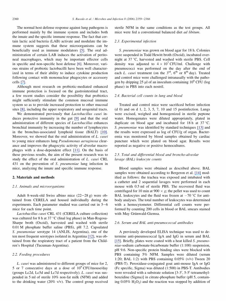
The pathogen was detected in lung and blood samples ofcontrol mice throughout the period assayed. Mice treated pre-ventively with L. casei for 2 days (Lc2d) showed lower lungbacterial counts than control mice until d 10 postinfection;from then on, bacterial counts were negative (Fig. 1). In addi-tion, hemocultures from the Lc2d group were negative till thed 2 postinfection (data not shown). Lung bacterial counts weresignificantly lower in Lc5d mice than in the control groupuntil d 5 postinfection; from then on, there was no differencebetween the two groups (Fig. 1).
3.2. Total and differential blood leukocyte counts
A significant increase in blood leukocytes was observed incontrol mice from h 24 till d 5 after challenge followed by
Fig. 1. Kinetics of S. pneumoniae clearance from lungs. Mice were challenged
intranasally with 106 S. pneumoniae cells after treatment with L. casei for 2, 5
or 7 d (Lc2d, Lc5d, Lc7d, respectively). For evaluation of CFU pathogen
counts in individual lung homogenates, mice were sacrificed on d 1, 2, 3, 5,
7, 10 and 15 postinfection. Results are expressed as mean � S.D. from 6
mice per group at the indicated time point. *Significantly different from the
control group at the same time point (P < 0.005).
2362 S. Racedo et al. / Microbes and Infection 8 (2006) 2359e2366
Table 1
Blood leukocyte (BL) and PMN counts (109 cells/liter) of mice fed L. casei
Time post-challenge (d) Experimental groups
Control Lc2d Lc5d Lc7d
BL PMNs BL PMNs BL PMNs BL PMNs
0 6.9 � 0.2 0.9 � 0.05 6.3 � 0.7 0.9 � 0.07 5.9 � 0.3 0.8 � 0,03 6.2 � 0.6 0.7 � 0.06
1 8.9 � 0.1 1.1 � 0.07 11.0 � 0.4* 3.3 � 0.03* 8.1 � 0.3 1.9 � 0.04 8.9 � 0.3 0.9 � 0.05
5 9.7 � 0.9 2.5 � 0.07 11.5 � 0.7* 3.2 � 0.06* 9.4 � 0.4 2.6 � 0.03 9.5 � 0.7 2.5 � 0.05
10 8.5 � 0.4 2.3 � 0.04 10.2 � 0.5* 2.9 � 0.06 9.7 � 0.5 2.7 � 0.02 8.5 � 0.5 2.6 � 0.05
15 7.2 � 0.7 2.1 � 0.01 8.9 � 0.4 2.8 � 0.08 8.5 � 0.2 2.6 � 0.04 7.5 � 0.5 2.3 � 0.03
Mice were fed L. casei for 2, 5 or 7 d (Lc2d, Lc5d, Lc7d, respectively) before (d 0) and after challenge (d 1, 5, 10, 15) with S. pneumoniae (1 � 106 CFU per
mouse). Control mice were challenged with the pathogen without previous treatment. Results represent mean � S.D. for six mice per group at each time point.
*Significantly different from the control at the same time point (P < 0.05).
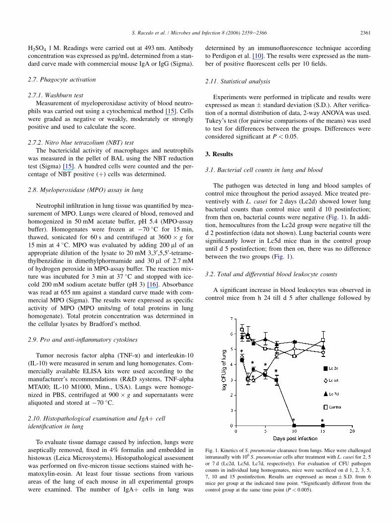
a gradual decrease and a return to normal values on d 15 post-infection (Table 1). The changes in the number of leukocyteswere mediated principally by the polymorph nuclear neutro-phil (PMN) population (Table 1).
Lc2d mice showed a significant increase in the total numberof leukocytes and in PMN cells compared to controls, and thenumbers remained high until d 10 postinfection.
3.3. Serum and BAL anti-pneumococcal antibodies
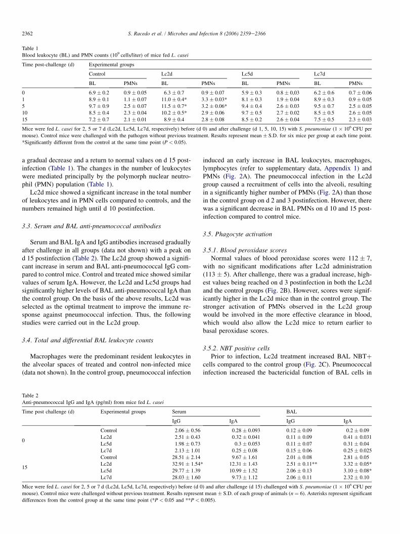
Serum and BAL IgA and IgG antibodies increased graduallyafter challenge in all groups (data not shown) with a peak ond 15 postinfection (Table 2). The Lc2d group showed a signifi-cant increase in serum and BAL anti-pneumococcal IgG com-pared to control mice. Control and treated mice showed similarvalues of serum IgA. However, the Lc2d and Lc5d groups hadsignificantly higher levels of BAL anti-pneumococcal IgA thanthe control group. On the basis of the above results, Lc2d wasselected as the optimal treatment to improve the immune re-sponse against pneumococcal infection. Thus, the followingstudies were carried out in the Lc2d group.
3.4. Total and differential BAL leukocyte counts
Macrophages were the predominant resident leukocytes inthe alveolar spaces of treated and control non-infected mice(data not shown). In the control group, pneumococcal infection
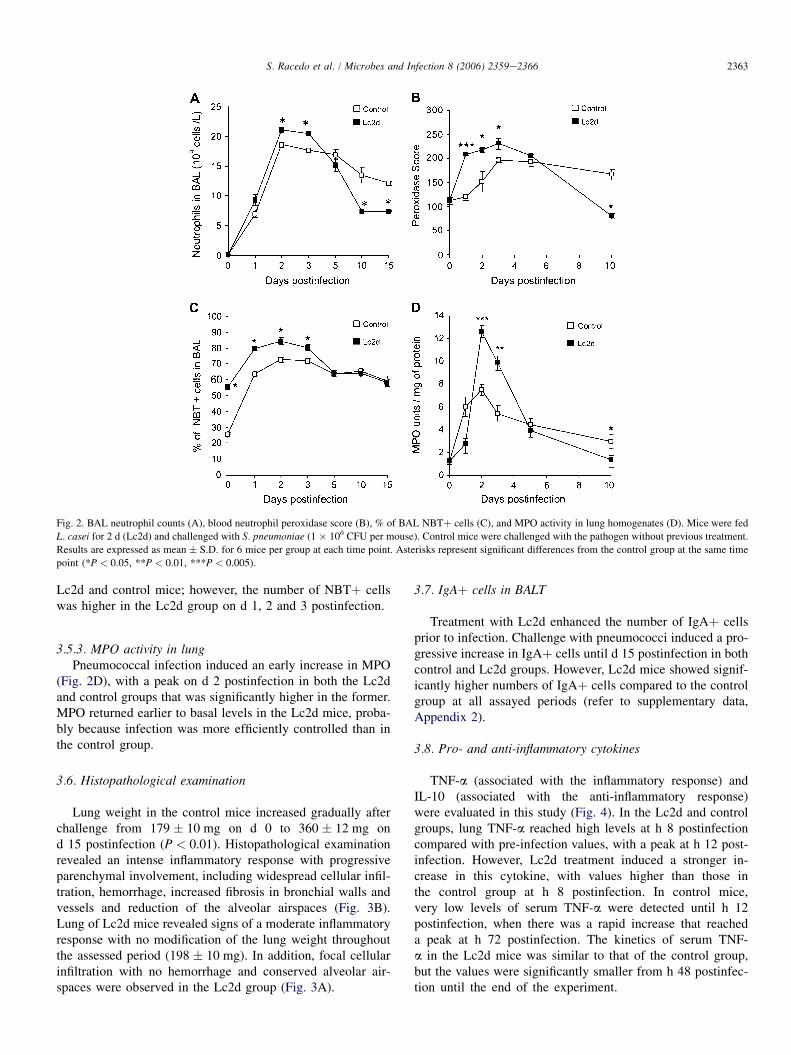
induced an early increase in BAL leukocytes, macrophages,lymphocytes (refer to supplementary data, Appendix 1) andPMNs (Fig. 2A). The pneumococcal infection in the Lc2dgroup caused a recruitment of cells into the alveoli, resultingin a significantly higher number of PMNs (Fig. 2A) than thosein the control group on d 2 and 3 postinfection. However, therewas a significant decrease in BAL PMNs on d 10 and 15 post-infection compared to control mice.
3.5. Phagocyte activation
3.5.1. Blood peroxidase scoresNormal values of blood peroxidase scores were 112 � 7,
with no significant modifications after Lc2d administration(113 � 5). After challenge, there was a gradual increase, high-est values being reached on d 3 postinfection in both the Lc2dand the control groups (Fig. 2B). However, scores were signif-icantly higher in the Lc2d mice than in the control group. Thestronger activation of PMNs observed in the Lc2d groupwould be involved in the more effective clearance in blood,which would also allow the Lc2d mice to return earlier tobasal peroxidase scores.
3.5.2. NBT positive cellsPrior to infection, Lc2d treatment increased BAL NBTþ
cells compared to the control group (Fig. 2C). Pneumococcalinfection increased the bactericidal function of BAL cells in
Table 2
Anti-pneumococcal IgG and IgA (pg/ml) from mice fed L. casei
Time post challenge (d) Experimental groups Serum BAL
IgG IgA IgG IgA
0
Control 2.06 � 0.56 0.28 � 0.093 0.12 � 0.09 0.2 � 0.09
Lc2d 2.51 � 0.43 0.32 � 0.041 0.11 � 0.09 0.41 � 0.031
Lc5d 1.98 � 0.73 0.3 � 0.053 0.11 � 0.07 0.31 � 0.04
Lc7d 2.13 � 1.01 0.25 � 0.08 0.15 � 0.06 0.25 � 0.025
15
Control 28.51 � 2.14 9.67 � 1.61 2.01 � 0.08 2.81 � 0.05
Lc2d 32.91 � 1.54* 12.31 � 1.43 2.51 � 0.11** 3.32 � 0.05*
Lc5d 29.77 � 1.39 10.99 � 1.52 2.06 � 0.13 3.10 � 0.08*
Lc7d 28.03 � 1.60 9.73 � 1.12 2.06 � 0.11 2.32 � 0.10
Mice were fed L. casei for 2, 5 or 7 d (Lc2d, Lc5d, Lc7d, respectively) before (d 0) and after challenge (d 15) challenged with S. pneumoniae (1 � 106 CFU per
mouse). Control mice were challenged without previous treatment. Results represent mean � S.D. of each group of animals (n ¼ 6). Asterisks represent significant
differences from the control group at the same time point (*P < 0.05 and **P < 0.005).
2363S. Racedo et al. / Microbes and Infection 8 (2006) 2359e2366
Fig. 2. BAL neutrophil counts (A), blood neutrophil peroxidase score (B), % of BAL NBTþ cells (C), and MPO activity in lung homogenates (D). Mice were fed
L. casei for 2 d (Lc2d) and challenged with S. pneumoniae (1 � 106 CFU per mouse). Control mice were challenged with the pathogen without previous treatment.
Results are expressed as mean � S.D. for 6 mice per group at each time point. Asterisks represent significant differences from the control group at the same time
point (*P < 0.05, **P < 0.01, ***P < 0.005).
Lc2d and control mice; however, the number of NBTþ cellswas higher in the Lc2d group on d 1, 2 and 3 postinfection.
3.5.3. MPO activity in lungPneumococcal infection induced an early increase in MPO
(Fig. 2D), with a peak on d 2 postinfection in both the Lc2dand control groups that was significantly higher in the former.MPO returned earlier to basal levels in the Lc2d mice, proba-bly because infection was more efficiently controlled than inthe control group.
3.6. Histopathological examination
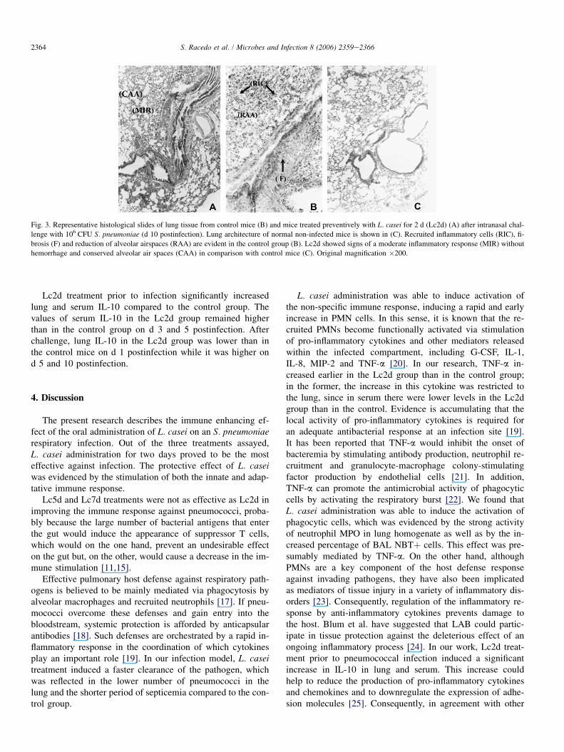
Lung weight in the control mice increased gradually afterchallenge from 179 � 10 mg on d 0 to 360 � 12 mg ond 15 postinfection (P < 0.01). Histopathological examinationrevealed an intense inflammatory response with progressiveparenchymal involvement, including widespread cellular infil-tration, hemorrhage, increased fibrosis in bronchial walls andvessels and reduction of the alveolar airspaces (Fig. 3B).Lung of Lc2d mice revealed signs of a moderate inflammatoryresponse with no modification of the lung weight throughoutthe assessed period (198 � 10 mg). In addition, focal cellularinfiltration with no hemorrhage and conserved alveolar air-spaces were observed in the Lc2d group (Fig. 3A).
3.7. IgAþ cells in BALT
Treatment with Lc2d enhanced the number of IgAþ cellsprior to infection. Challenge with pneumococci induced a pro-gressive increase in IgAþ cells until d 15 postinfection in bothcontrol and Lc2d groups. However, Lc2d mice showed signif-icantly higher numbers of IgAþ cells compared to the controlgroup at all assayed periods (refer to supplementary data,Appendix 2).
3.8. Pro- and anti-inflammatory cytokines
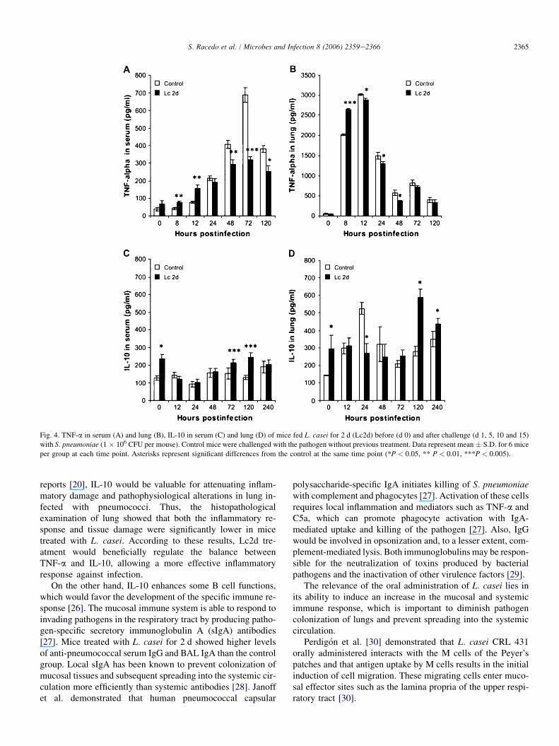
TNF-a (associated with the inflammatory response) andIL-10 (associated with the anti-inflammatory response)were evaluated in this study (Fig. 4). In the Lc2d and controlgroups, lung TNF-a reached high levels at h 8 postinfectioncompared with pre-infection values, with a peak at h 12 post-infection. However, Lc2d treatment induced a stronger in-crease in this cytokine, with values higher than those inthe control group at h 8 postinfection. In control mice,very low levels of serum TNF-a were detected until h 12postinfection, when there was a rapid increase that reacheda peak at h 72 postinfection. The kinetics of serum TNF-a in the Lc2d mice was similar to that of the control group,but the values were significantly smaller from h 48 postinfec-tion until the end of the experiment.
2364 S. Racedo et al. / Microbes and Infection 8 (2006) 2359e2366
Fig. 3. Representative histological slides of lung tissue from control mice (B) and mice treated preventively with L. casei for 2 d (Lc2d) (A) after intranasal chal-
lenge with 106 CFU S. pneumoniae (d 10 postinfection). Lung architecture of normal non-infected mice is shown in (C). Recruited inflammatory cells (RIC), fi-
brosis (F) and reduction of alveolar airspaces (RAA) are evident in the control group (B). Lc2d showed signs of a moderate inflammatory response (MIR) without
hemorrhage and conserved alveolar air spaces (CAA) in comparison with control mice (C). Original magnification �200.
Lc2d treatment prior to infection significantly increasedlung and serum IL-10 compared to the control group. Thevalues of serum IL-10 in the Lc2d group remained higherthan in the control group on d 3 and 5 postinfection. Afterchallenge, lung IL-10 in the Lc2d group was lower than inthe control mice on d 1 postinfection while it was higher ond 5 and 10 postinfection.
4. Discussion
The present research describes the immune enhancing ef-fect of the oral administration of L. casei on an S. pneumoniaerespiratory infection. Out of the three treatments assayed,L. casei administration for two days proved to be the mosteffective against infection. The protective effect of L. caseiwas evidenced by the stimulation of both the innate and adap-tative immune response.
Lc5d and Lc7d treatments were not as effective as Lc2d inimproving the immune response against pneumococci, proba-bly because the large number of bacterial antigens that enterthe gut would induce the appearance of suppressor T cells,which would on the one hand, prevent an undesirable effecton the gut but, on the other, would cause a decrease in the im-mune stimulation [11,15].
Effective pulmonary host defense against respiratory path-ogens is believed to be mainly mediated via phagocytosis byalveolar macrophages and recruited neutrophils [17]. If pneu-mococci overcome these defenses and gain entry into thebloodstream, systemic protection is afforded by anticapsularantibodies [18]. Such defenses are orchestrated by a rapid in-flammatory response in the coordination of which cytokinesplay an important role [19]. In our infection model, L. caseitreatment induced a faster clearance of the pathogen, whichwas reflected in the lower number of pneumococci in thelung and the shorter period of septicemia compared to the con-trol group.
L. casei administration was able to induce activation ofthe non-specific immune response, inducing a rapid and earlyincrease in PMN cells. In this sense, it is known that the re-cruited PMNs become functionally activated via stimulationof pro-inflammatory cytokines and other mediators releasedwithin the infected compartment, including G-CSF, IL-1,IL-8, MIP-2 and TNF-a [20]. In our research, TNF-a in-creased earlier in the Lc2d group than in the control group;in the former, the increase in this cytokine was restricted tothe lung, since in serum there were lower levels in the Lc2dgroup than in the control. Evidence is accumulating that thelocal activity of pro-inflammatory cytokines is required foran adequate antibacterial response at an infection site [19].It has been reported that TNF-a would inhibit the onset ofbacteremia by stimulating antibody production, neutrophil re-cruitment and granulocyte-macrophage colony-stimulatingfactor production by endothelial cells [21]. In addition,TNF-a can promote the antimicrobial activity of phagocyticcells by activating the respiratory burst [22]. We found thatL. casei administration was able to induce the activation ofphagocytic cells, which was evidenced by the strong activityof neutrophil MPO in lung homogenate as well as by the in-creased percentage of BAL NBTþ cells. This effect was pre-sumably mediated by TNF-a. On the other hand, althoughPMNs are a key component of the host defense responseagainst invading pathogens, they have also been implicatedas mediators of tissue injury in a variety of inflammatory dis-orders [23]. Consequently, regulation of the inflammatory re-sponse by anti-inflammatory cytokines prevents damage tothe host. Blum et al. have suggested that LAB could partic-ipate in tissue protection against the deleterious effect of anongoing inflammatory process [24]. In our work, Lc2d treat-ment prior to pneumococcal infection induced a significantincrease in IL-10 in lung and serum. This increase couldhelp to reduce the production of pro-inflammatory cytokinesand chemokines and to downregulate the expression of adhe-sion molecules [25]. Consequently, in agreement with other
2365S. Racedo et al. / Microbes and Infection 8 (2006) 2359e2366
Fig. 4. TNF-a in serum (A) and lung (B), IL-10 in serum (C) and lung (D) of mice fed L. casei for 2 d (Lc2d) before (d 0) and after challenge (d 1, 5, 10 and 15)
with S. pneumoniae (1 � 106 CFU per mouse). Control mice were challenged with the pathogen without previous treatment. Data represent mean � S.D. for 6 mice
per group at each time point. Asterisks represent significant differences from the control at the same time point (*P < 0.05, ** P < 0.01, ***P < 0.005).
reports [20], IL-10 would be valuable for attenuating inflam-matory damage and pathophysiological alterations in lung in-fected with pneumococci. Thus, the histopathologicalexamination of lung showed that both the inflammatory re-sponse and tissue damage were significantly lower in micetreated with L. casei. According to these results, Lc2d tre-atment would beneficially regulate the balance betweenTNF-a and IL-10, allowing a more effective inflammatoryresponse against infection.
On the other hand, IL-10 enhances some B cell functions,which would favor the development of the specific immune re-sponse [26]. The mucosal immune system is able to respond toinvading pathogens in the respiratory tract by producing patho-gen-specific secretory immunoglobulin A (sIgA) antibodies[27]. Mice treated with L. casei for 2 d showed higher levelsof anti-pneumococcal serum IgG and BAL IgA than the controlgroup. Local sIgA has been known to prevent colonization ofmucosal tissues and subsequent spreading into the systemic cir-culation more efficiently than systemic antibodies [28]. Janoffet al. demonstrated that human pneumococcal capsular
polysaccharide-specific IgA initiates killing of S. pneumoniaewith complement and phagocytes [27]. Activation of these cellsrequires local inflammation and mediators such as TNF-a andC5a, which can promote phagocyte activation with IgA-mediated uptake and killing of the pathogen [27]. Also, IgGwould be involved in opsonization and, to a lesser extent, com-plement-mediated lysis. Both immunoglobulins may be respon-sible for the neutralization of toxins produced by bacterialpathogens and the inactivation of other virulence factors [29].
The relevance of the oral administration of L. casei lies inits ability to induce an increase in the mucosal and systemicimmune response, which is important to diminish pathogencolonization of lungs and prevent spreading into the systemiccirculation.
Perdigon et al. [30] demonstrated that L. casei CRL 431orally administered interacts with the M cells of the Peyer’spatches and that antigen uptake by M cells results in the initialinduction of cell migration. These migrating cells enter muco-sal effector sites such as the lamina propria of the upper respi-ratory tract [30].
2366 S. Racedo et al. / Microbes and Infection 8 (2006) 2359e2366
On the basis of these previous findings and our own results,we propose a possible mechanism to explain the role ofL. casei in the improved resistance to pneumococcal infection.L. casei would stimulate immune cell migration from thePeyer’s patches in the small intestine to BALT. In addition,L. casei would affect cytokine expression in a specific ornon-specific manner by stimulating distant immune cells.This cell migration as well as the activation of cells in distantsites would allow a faster immune response, both innate andspecific, against pneumococcal infection, thus favoring clear-ance of the pathogen and modulating the inflammatory im-mune response, with less damage to lung tissue. The resultsobtained in this study may lead to new insights concerningthe use of L. casei as an oral adjuvant or as an oral vaccinevector for a wide range of infectious lung diseases, whichhave a high incidence in Argentina.
Acknowledgements
Supported by grants from FONCyT PICT-99 N�5-7206,CIUNT-26D/202 and CIUNT-D/305.
Appendix. Supplementary information
Supplementary information for this manuscript can bedownloaded at doi:10.1016/j.micinf.2006.04.022.
References
[1] G.R. Siber, Pneumococcal disease: prospects for a new generation of vac-
cines, Science 265 (1994) 1385e1387.
[2] R.H. Simon, R. Paine, Participation of pulmonary alveolar epithelial cells
in lung inflammation, J. Lab. Clin. Med. 126 (1995) 108e118.
[3] E.I. Tuomanen, R. Austrian, H.R. Masure, Pathogenesis of pneumococ-
cal infection, N. Engl. J. Med. 332 (1995) 1280e1284.
[4] R. Ruvinsky, A. Gentile, M. Regueira, A. Corso, Infecciones invasivas por
Streptococcus pneumoniae: estudio epidemiologico e importancia del desar-
rollo de un sistema de vigilancia, Arch. Argent. Pediatr. 100 (2002) 31e43.
[5] K.L. Erickson, N.E. Hubbard, Probiotic immunomodulation in health and
disease, J. Nutr. 130 (2000) 403Se409S.
[6] G. Perdigon, M. Nader de Macias, S. Alvarez, G. Oliver, A. Pesce de
Ruiz Holgado, Effect of oral administration of lactobacilli on macro-
phage activation in mice, Infect. Immun. 53 (1986) 404e410.
[7] M.L. Cross, A. Ganner, D. Teilab, M.F. Linley, Patterns of cytokine in-
duction by gram-positive and gram-negative probiotic bacteria, FEMS
Immunol. Med. Microbiol. 42 (2004) 173e180.
[8] M.L. Cross, Microbes versus microbes: immune signals generated by
probiotic lactobacilli and their role in protection against microbial path-
ogens, FEMS Immunol. Med. Microbiol. 34 (2002) 245e253.
[9] S. Alvarez, N. Gobbato, E. Bru, A. Ruiz Holgado, G. Perdigon, Specific
immunity induction at mucosal level by viable Lactobacillus casei. Per-
spective for oral vaccine development, Food Agric. Immunol. 10 (1998)
79e87.
[10] G. Perdigon, S. Alvarez, M. Medina, E. Vintini, E. Roux, Influence of the
oral administration of lactic acid bacteria on IgA producing cells associ-
ated to bronchus, Int. J. Immunopathol. Pharmacol. 12 (1999) 97e102.
[11] S. Alvarez, C. Herrero, E. Bru, G. Perdigon, Effect of Lactobacillus casei
and yogurt administration on prevention of Pseudomonas aeruginosa in-
fection in young mice, J. Food Protect. 64 (2001) 1768e1774.
[12] H. Zemlickova, M.I. Crisostomo, M.C. Brandileone, T. Camou,
E. Castaneda, A. Corso, G. Echaniz-Aviles, M. Pasztor, A. Tomasz,
Serotypes and clonal types of penicillin-susceptible Streptococcus pneu-moniae causing invasive disease in children in five Latin American coun-
tries, Microb. Drug Resist. 11 (2005) 195e204.
[13] R. Facklam, J.A. Washington II, Streptococcus and related catalase neg-
ative gram positive cocci, in: A. Balows, W.J. Hausler Jr., L.H. Kenneth,
H.D. Isenberg, J. Shadomy (Eds.), Manual of Clinical Microbiology,
fifth ed. American Society for Microbiology, Washington D.C, 1992,
pp. 238e257.
[14] Y. Bergeron, N. Oullet, A. Deslauriers, M. Simard, M. Olivier,
M.G. Bergeron, Cytokine kinetics and other host factors in response to
pneumococcal pulmonary infection in mice, Infect. Immun. 66 (1998)
912e922.
[15] J. Villena, S. Racedo, G. Aguero, E. Bru, M. Medina, S. Alvarez, Lacto-
bacillus casei improves resistance to pneumococcal respiratory infection
in malnourished mice, J. Nutr. 135 (2005) 1462e1469.
[16] P.M. Bozeman, D.B. Learn, E.L. Thomas, Assay of the human leukocyte
enzymes myeloperoxidase and eosinophil peroxidase, J. Immunol.
Methods 126 (1990) 125e133.
[17] S.B. Gordon, G.R. Irving, R.A. Lawson, M.E. Lee, R.C. Read, Intracel-
lular trafficking and killing of Streptococcus pneumoniae by human alve-
olar macrophages are influenced by opsonins, Infect. Immun. 68 (2000)
2286e2293.
[18] M. Anttila, M.J. Voutilainen, V.J. Eskola, H. Kayhty, Contribution of
serotype-specific IgG concentration, IgG subclasses and relative antibody
avidity to opsonophagocytic activity against Streptococcus pneumoniae,
Clin. Exp. Immunol. 118 (1999) 402e407.
[19] S. Nelson, W.R. Summer, Innate immunity cytokines and pulmonary host
defense, Infect. Dis. Clin. N. Am. 12 (1998) 555e567.
[20] A.R. Kerr, J.J. Irvine, J.J. Search, N.A. Gingles, A. Kadioglu,
P.W. Andrew, W.L. McPheat, C.G. Booth, T.J. Mitchell, Role of inflam-
matory mediators in resistance and susceptibility to pneumococcal
infection, Infect. Immun. 70 (2002) 1547e1557.
[21] K. Takashima, K. Tateda, T. Matsumoto, Y. Iizawa, M. Nakao,
K. Yamaguchi, Role of tumor necrosis factor alpha in pathogenesis of
pneumococcal pneumonia in mice, Infect. Immun. 65 (1997) 257e260.
[22] S. Dusi, V.D. Bianca, M. Donini, K.A. Nadalini, F. Rossi, Mechanisms of
stimulation of the respiratory burst by TNF in non-adherent neutropphils:
its independence of lipidic transmembrane signaling and dependence on
protein tyrosine phosphorylation and cytoskeleton, J. Immunol. 157
(1996) 4615e4623.
[23] P. Zhang, W.R. Summer, G.J. Bagby, S. Nelson, Innate immunity and
pulmonary host defense, Immunol. Rev. 173 (2000) 39e51.
[24] S. Blum, Y. Delneste, S. Alvarez, D. Haller, P.F. Perez, C. Bode,
W.P. Hammes, A.M.A. Pfeifer, E.J. Schiffrin, Interactions between com-
mensal bacteria and mucosal immunocompetent cells, Internat. Dairy J. 9
(1999) 63e68.
[25] F. Willems, A. Marchant, J.P. Delville, C. Gerard, A. Delvaux, T. Velu,
M. de Boer, M. Goldman, Interleukin-10 inhibits B7 and intercellular
adhesion molecule-1 expression on human monocytes, Eur. J. Immunol.
24 (4) (1994) 1007e1009.
[26] A.J. Husband, K.W. Beagley, J.R. McGhee, Mucosal cytokines, in:
L.O. Pearay, M.E. Lamm, J. Biewenstock, R. Jerry (Eds.), Mucosal Immu-
nology, second ed. Academic Press, San Diego, CA, 1999, pp. 541e557.
[27] E.N. Janoff, C. Fasching, J.M. Oresnstein, J.B. Rubins, N.L. Osptad,
A.P. Dalmasso, Killing of Streptococcus pneumoniae by capsular
polysaccharide-specific polymeric IgA, complement, and phagocytes,
J. Clin. Invest. 104 (1999) 1139e1147.
[28] J.M. Woof, J. Mestecky, Mucosal immunoglobulins, Immunol. Rev. 206
(2005) 64e82.
[29] V.E. Garcıa, M.M. Gherardi, M.F. Iglesias, M.C. Cerquetti, D.O. Sordelli,
Local and systemic immunity against Streptococcus pneumoniae: hu-
moral responses against a non-capsulated temperature-sensitive mutant,
FEMS Microbiol. Lett. 108 (1993) 163e168.
[30] G. Perdigon, R. Fuller, M. Medina, The influence of the lactic acid
bacteria and other resident microflora on the immune system of the
growing animal, in: W. Holzapfel, P. Naughton (Eds.), Microbiological
Ecology of Growing Animals, Biology of Growing Animals Series,
first ed. Elsevier, UK, 2005, pp. 351e375.