l-dopa-treatment in primates disrupts the expression of a2a adenosine-cb1 cannabinoid-d2 dopamine...
TRANSCRIPT

lable at ScienceDirect
Neuropharmacology 79 (2014) 90e100
Contents lists avai
Neuropharmacology
journal homepage: www.elsevier .com/locate/neuropharm
L-DOPA-treatment in primates disrupts the expression of A2AadenosineeCB1 cannabinoideD2 dopamine receptor heteromers inthe caudate nucleus
Jordi Bonaventura a,b,1, Alberto J. Rico b,c,1, Estefanía Moreno a,b, Salvador Sierra b,c,Marta Sánchez a,b, Natasha Luquin b,c, Daniel Farré a,b, Christa E. Müller d,Eva Martínez-Pinilla c, Antoni Cortés a,b, Josefa Mallol a,b, Marie-Therese Armentero e,Annalisa Pinna f, Enric I. Canela a,b, Carme Lluís a,b, Peter J. McCormick a,b,José L. Lanciego b,c, Vicent Casadó a,b,**,1, Rafael Franco a,c,*,1
aDepartment of Biochemistry and Molecular Biology, Faculty of Biology, University of Barcelona, 08028 Barcelona, SpainbCentro de Investigación Biomédica en Red sobre Enfermedades Neurodegenerativas, SpaincCentro de Investigación Médica Aplicada, Universidad de Navarra, 31008 Pamplona, Spaind PharmaCenter Bonn, Pharmaceutical Institute, Pharmaceutical Chemistry I, University of Bonn, Germanye Laboratory of Functional Neurochemistry, C. Mondino National Neurological Institute, via Mondino 2, Pavia, ItalyfCNR Institute of Neuroscience, Cagliari, Italy
a r t i c l e i n f o
Article history:Received 25 April 2013Received in revised form21 October 2013Accepted 23 October 2013
Keywords:CaudatePutamenL-DopaDopamineParkinson’s diseaseReceptor heteromerStriatum
* Corresponding author. CIMA, University of NavarraSpain. Tel.: þ34 948194700; fax: þ34 948194715.** Corresponding author. Department of BiochemiFaculty of Biology, University of Barcelona, 08028 Bar
E-mail addresses: [email protected] (V. Casadó), rgmail.com (R. Franco).
1 These authors contributed equally to this work.
0028-3908/$ e see front matter � 2013 Elsevier Ltd.http://dx.doi.org/10.1016/j.neuropharm.2013.10.036
a b s t r a c t
The molecular basis of priming for L-DOPA-induced dyskinesias in Parkinson’s disease (PD), which de-pends on the indirect pathway of motor control, is not known. In rodents, the indirect pathway containsstriatopallidal GABAergic neurons that express heterotrimers composed of A2A adenosine, CB1 canna-binoid and D2 dopamine receptors that regulate dopaminergic neurotransmission. The present study wasdesigned to investigate the expression of these heteromers in the striatum of a primate model of Par-kinson’s disease and to determine whether their expression and pharmacological properties are alteredupon L-DOPA treatment. By using the recently developed in situ proximity ligation assay and by iden-tification of a biochemical fingerprint, we discovered a regional distribution of A2A/CB1/D2 receptorheteromers that predicts differential D2-mediated neurotransmission in the caudateeputamen of Macacafascicularis. Whereas heteromers were abundant in the caudate nucleus of both naïve and MPTP-treatedmonkeys, L-DOPA treatment blunted the biochemical fingerprint and led to weak heteromer expression.These findings constitute the first evidence of altered receptor heteromer expression in pathologicalconditions and suggest that drugs targeting A2AeCB1eD2 receptor heteromers may be successful toeither normalize basal ganglia output or prevent L-DOPA-induced side effects.
� 2013 Elsevier Ltd. All rights reserved.
1. Introduction
The current model of basal ganglia function (Albin et al., 1989;DeLong, 1990) postulates that stimulation of the direct and indi-rect striatal efferent pathways provokes motor activation and
, Pio XII 55, 31008 Pamplona,
stry and Molecular Biology,celona, [email protected], rfranco123@
All rights reserved.
motor inhibition, respectively, and that smoothmotor performanceis the result of the balanced influence of these pathways on theneural activity of the basal ganglia output structures (Kravitz et al.,2010). In Parkinson’s disease (PD), the equilibrium of the basalganglia circuits is lost due to the depletion in striatal dopaminecaused by degeneration of nigrostriatal dopaminergic neurons(Lewis et al., 2003; Obeso et al., 2008). As the degenerative processprogresses, dopamine replacement therapy becomes necessary toalleviate motor dysfunction. The L-3,4-dihydroxyphenylalanine (L-DOPA) treatment is the most effective means of improving PDmotor symptoms as this compound is converted to dopamineleading to activation of D1-like and D2-like dopamine receptors (see
J. Bonaventura et al. / Neuropharmacology 79 (2014) 90e100 91
Koller, 2000; Hornykiewicz, 2010; Rascol et al., 2011 for review).However, its chronic use is associated with the development ofmotor complications, including on-off fluctuations and dyskinesias(Bezard et al., 2001). Although the mechanisms underlying thesemotor complications are not completely understood, L-DOPA-induced dyskinesias (LIDs) are thought to result from the combinedeffects of nigrostriatal dopaminergic damage and pulsatile L-DOPAtreatment (Obeso et al., 2000; Cenci, 2007). Accordingly, muchresearch has focused on identifying compounds that alleviate theseabnormal involuntary movements, and the development of non-dopaminergic compounds to be used as adjuvants to L-DOPAtherapy in PD has attracted much interest in recent years(Schwarzschild et al., 2006; Fox et al., 2006; Armentero et al., 2011;Gottwald and Aminoff, 2011).
Among several new classes of drugs, A2A adenosine receptor an-tagonists have emerged as the most promising candidates. We, andothers, have described antagonistic interactions between A2A and D2or D3 receptors within heteromers (Hillion et al., 2002; Canals et al.,2003; Ferré et al., 2004; Torvinen et al., 2005; Fuxe et al., 2007;Schiffmann et al., 2007). On the one hand, A2A receptor antagonistsmay prevent the endogenous adenosine-mediated decrease in D2
receptor agonist affinity (Ferré et al., 1991; Ferré and Fuxe, 1992;Ferré et al., 2004; Muller and Ferré, 2007; Pinna, 2009). Thus, A2A
receptor antagonists may enhance the D2 receptor-mediated effectsby attenuating the tonic adenosine-mediated inhibition of D2 re-ceptors in A2AeD2 receptor heteromers (Schwarzschild et al., 2006).On theotherhand, blockadeofA2A receptors also indirectlyenhancesthe effects of D1 receptor activation in striatonigral-projecting neu-rons (Pinna et al., 1996; Pollack and Fink, 1996), suggesting that A2Areceptor antagonists exert a facilitatory effect on dopamine trans-mission that promotes motor activation, and pointing to a potentialbeneficial effect of these compounds onparkinsonianmotor deficits.
Cannabinoid receptors also play a key role in the motor alter-ations associated with PD. CB1 receptors are downregulated in theearly stages of PD (Sagredo et al., 2007), which may render stria-tonigral neurons more vulnerable to the cytotoxic stimuli associ-ated with this disease (Sagredo et al., 2007). By contrast, thecannabinoid system becomes overactive in the advanced stages ofthe disease, when the major PD symptoms appear. This has beendemonstrated in humans by analyzing the number and function ofCB1 receptors in post-mortem basal ganglia tissue (Lastres-Beckeret al., 2001) and by measuring endocannabinoid levels in the ce-rebrospinal fluid of PD patients (Pisani et al., 2005). Similar varia-tions in CB1 receptor levels accompanying the progression of PDhave been described in animal models (Garcia-Arencibia et al.,2009). The reversal of these responses with L-DOPA treatmentsupports the view that overactivation of cannabinoid signaling isinversely correlated with striatal dopaminergic denervation(Romero et al., 2000; Lastres-Becker et al., 2001), strongly sug-gesting that blocking CB1 receptors could protect against parkin-sonian bradykinesia (Garcia-Arencibia et al., 2009; Orgado et al.,2009). Using a resonance energy transfer based technique(Carriba et al., 2008), we demonstrated previously that CB1 re-ceptors can form heteromers with A2A adenosine and D2 dopaminereceptors. We described the expression of A2AeCB1eD2 receptorheterotrimers in transfected cells and in the mouse striatum(Navarro et al., 2010). Moreover, we defined the negative modula-tion of A2A and CB1 receptor agonists upon dopamine binding to D2
receptors as a biochemical fingerprint of this heteromer. Anothercharacteristic of this complex is its selective coupling to themitogen-activated protein kinase pathway (Navarro et al., 2010;Higley and Sabatini, 2010; Marcellino et al., 2008). In the presentstudy, we have analyzed the expression and function of A2AeCB1eD2 receptor heteromers in the striatum of a monkey model of PDand used in situ proximity ligation assay (PLA) to directly detect
molecular proteineprotein interactions. Accordingly, we demon-strate that A2AeCB1, A2AeD2 and CB1eD2 receptor heteromerizationoccurs in the caudate nucleus (CN), and to a lesser extent in theputamen (Put). These results were confirmed by analyzing thebiochemical fingerprint of the A2AeCB1eD2 receptor heteromers atthe ligand binding level. The same techniques were used todemonstrate the absence of heteromers in L-DOPA-treatedparkinsonian monkeys exhibiting involuntary movements. Takentogether, our results demonstrate that such heteromultimers areassociated with PD and constitute the first evidence of changes inG-protein-coupled receptor heteromerization during the course ofa neurological disease.
2. Materials and methods
2.1. Generation of parkinsonian monkeys and L-DOPA treatment
Animals were supplied by Harlan Laboratories. All animals were naïve beforeany treatment. For radioligand binding experiments, fresh tissue from a total of 21young adult male Macaca fascicularis primates (between 4 and 5.5 years old) wereused. Proximity ligation assays were performed in samples from 6 additionalmonkeys (see details in 2.2). Animal handling was conducted in accordance with theEuropean Council Directive 86/609/EEC as well as in agreement with the Society forNeuroscience Policy on the Use of Animals in Neuroscience Research. The experi-mental design was approved by the Ethical Committee for Animal Testing of theUniversity of Navarra (ref: 018/2008) as well as by the Department of Health fromthe Government of Navarra (ref: NA-UNAV-04-08). Of the 21 animals devoted toperform binding assays in freshly isolated tissue, 11 were naïve and 10 monkeyswere treated with systemic delivery of the dopaminergic neurotoxin MPTP (1-Methyl-4-phenyl-1,2,3,6-tetrahydropyridine from Sigma, Madrid, Spain) to inducea bilateral parkinsonian syndrome (Rico et al., 2010). Animals received a weeklyinjection of MPTP (0.2 mg/kg i/v; accumulated doses ranging from 5 to 7 mg/kg)until reaching a non-reversible parkinsonian syndrome. The severity of the MPTP-induced parkinsonism was evaluated using a clinical rating scale (Kurlan et al.,1991). This scale rates parkinsonian motor symptoms such as facial expression(0e3), resting tremor (0e3), action or intention tremor (0e3), posture (0e2), bra-dykinesia (0e4), balance coordination (0e3), gait (0e3), gross motor skills of theupper limb (0e3) and lower limb (0e3), and defense reaction (0e2) in an accu-mulating scalewhere themaximum score (i.e., highest severity) is 29. Once primatesreached minimum score of 21 points and above, the MPTP treatment was dis-continued for a wash-out period of 2 months to ensure that the parkinsonian syn-dromewas fully stabilized. At the end of the stabilization period, the PD scores werebetween 21 and 24 for all primates treated with MPTP. To 3 monkeys with stabilizedparkinsonian syndrome (scoring 21, 22 and 24 points in the Kurlan scale), L-DOPAwas orally administered (in orange juice) at a dose of 25 mg/kg daily (Madopar� L-DOPA/benserazide, 200:50, Roche). Untreated animals were also administered or-ange juice. The changes in the score of Kurlan et al. (1991) from the “off” to the “on”state were monitored and the duration of the “on” response recorded (Lanciegoet al., 2008). LIDs were rated as 1 (mild), when presented only occasionally understress; 2 (moderate) for LIDs present during most of the ‘on’ period without inter-fering with voluntary movements; and 3 (severe) when LIDs were continuous,generalized and they perturbed motor behavior. This scoring system is in keepingwith the scale included in the Core Assessment Program for Intracerebral Trans-plantation for Parkinson’s disease (Langston et al., 1992), subsequently modified andvalidated for the assessment of dyskinesias in patients (Goetz et al., 1994). By thetime of sacrifice, all 3 monkeys treated with L-DOPA showed severe dyskinesia.These primates entered in the “on” state 30 min post-L-DOPA oral delivery and theduration of the “on” period was maintained for 2.5e3 h. Animals were treated dailywith L-DOPA until they showed an overt dyskinetic syndrome; a mild dyskineticsyndrome was displayed by the end of the first month of treatment and the overtdyskinetic symptoms appeared one month later and remained stable until sacrifice.In all cases, primates were sacrificed by decapitation after an intravenous overdoseof T61 (terminal anesthesia, 0.3 mg/kg). L-DOPA-treated animals were killed in theon state, when they showed peak-of-dose dyskinesia. The skull was opened, and thebrain extracted, blocked on ice, and the areas of interest (CN and the pre- and post-commissural Put) were removed from the brain under a dissection microscope,stored in Eppendorf vials, quickly frozen on dry ice and preserved at �80� C untiluse. Basal ganglia-related structures were selected according to the atlas of Lanciegoand Vazquez (2012). Blocks containing the substantia nigra pars compacta (SNc) werefixed by immersion and finally stained using antibodies against tyrosine hydroxylaseto assess the degree of dopaminergic neuronal death induced by the MPTP treat-ment. The extent of the induced nigrostriatal lesion was very similar for all MPTP-treated monkeys and a representative image is shown in Supplementary Fig. 1.
2.2. In situ proximity ligation assays (PLA)
PLA experiments were carried out by using paraformaldehyde-fixed, sagittalsections (40 mm-thick) available in our monkey brain bank. Briefly, sagittal sections

J. Bonaventura et al. / Neuropharmacology 79 (2014) 90e10092
through the caudate nucleus and putamen from 6 additional monkeys (2 control, 2parkinsonian and 2 dyskinetic), with similar features as those displayed by theanimals used in binding experiments, were used. Samples weremounted on gelatin-coated slides and further processed for the PLA technique. Briefly, A2AeD2, CB1eD2,and A2AeCB1 heteromers were detected using the Duolink II in situ PLA detection Kit(OLink; Bioscience, Uppsala, Sweden). Slices were thawed at 4 �C, washed in 50 mMTriseHCl, 0.9% NaCl pH 7.8 buffer (TBS), permeabilized with TBS containing 0.1%Triton X-100 for 10 min and successively washed with TBS. After 1 h incubation at37 �C with the blocking solution in a pre-heated humidity chamber (37 �C), sliceswere incubated overnight in the antibody diluent medium with a mixture of equalamounts of mouse monoclonal anti-A2A receptor antibody (1:200, ref. 05-717;Millipore, Billerica, MA, USA; Rosin et al., 1998) and rabbit polyclonal anti-CB1 re-ceptor antibody (1:200, ref. PA 1-745, Thermo Scientific, Rockford, USA) to detectA2AeCB1 receptor heteromers or the rabbit polyclonal anti-D2 receptor antibody(1:200, ref. AB5084P; Millipore) and monoclonal mouse anti-A2A receptor antibodyto detect A2AeD2 receptor heteromers. Slices were washed as indicated by thesupplier and incubated for 2 h in a pre-heated humidity chamber at 37 �C with PLAprobes detecting mouse or rabbit antibodies, Duolink II PLA probe anti-mouse plusand Duolink II PLA probe rabbit minus (prepared following the instructions of thesupplier) diluted in the antibody diluent to a concentration of 1:5. To detect CB1eD2
receptor heteromers, slices were incubated with a mixture of equal amounts of thepolyclonal rabbit anti-CB1 receptor antibody (1:100) and the polyclonal rabbit anti-D2 receptor antibody (1:100) directly coupled to DNA chain plus or DNA chainminus, respectively, following the instructions of the supplier. After washing at roomtemperature, slices were incubated in a pre-heated humidity chamber for 30 min at37 �C, with the ligation solution (Duolink II Ligation stock 1:5 and Duolink II Ligase1:40). Detection of the amplified probe was done with the Duolink II DetectionReagents Red Kit. After exhaustively washing at room temperature as indicated inthe kit, slices were mounted using a DAPI-containing mounting medium. Thesamples were observed in a Leica SP2 confocal microscope (Leica Microsystems,Mannheim, Germany) equipped with an apochromatic 63X oil-immersion objective(N.A. 1.4), and a 405 nm and a 561 nm laser lines. For each field of view a stack of twochannels (one per staining) and 9 to 15 Z stacks with a step size of 1 mm were ac-quired. A quantification of cells containing one or more red spots versus total cells(blue nucleus) and, in cells containing spots, the ratio r (number of red spots/cell),were determined considering a total 300e400 cells from six different fields withinstriatum from two different animals per group using the Fiji package (http://pacific.mpi-cbg.de/). Nuclei and red spots were counted on the maximum projections ofeach image stack. After getting the projection each channel was processed indi-vidually. The nuclei were segmented by filtering with amedian filter, subtracting thebackground, enhancing the contrast with the Contrast Limited Adaptive HistogramEqualization (CLAHE) plug-in and finally applying a threshold to obtain the binaryimage and the regions of interest (ROI) around each nucleus. Red spots images werealso filtered and thresholded to obtain the binary images. Red spots were counted in
Atotal bound ¼�KDA2Aþ 2A2 þ KDA2AB= KDAB
�RT= KDA1KDA2 þ KDA2Aþ A2 þ KDA2AB=KDAB þ KDA1KDA2B=KDB1 þ KDA1KDA2B
2=�
KDB1KDB2ð ÞÞ þ Anon�specific bound (1)
each of the ROIs obtained in the nuclei images. Two-way ANOVA followed byBonferroni’s post hocmultiple comparison test was used to compare the values (% ofpositive cells or re spots/cell) obtained for each pair of receptors in different diseasestates or in different striatal regions.
2.3. Brain striatal membranes preparation and protein determination
Monkey cerebral tissue was disrupted with a Polytron homogenizer (PTA 7 rotor,setting 5; Kinematica, Basel, Switzerland) for three 5 s-periods in 10 volumes of50 mM TriseHCl buffer, pH 7.4, containing a protease inhibitor cocktail (Sigma, 1/1000). After eliminating cell debris by centrifugation at 1000 g for 10 min, mem-branes were obtained by centrifugation at 105,000 g (40 min, 4 �C). Membraneswere washed in the same medium and the pellet was obtained by centrifugationunder the same conditions and stored at �80 �C until use (Casadó et al., 1990).Membranes were thawed, washed once more and resuspended in 50 mM TriseHClbuffer for immediate use. Protein was quantified by the bicinchoninic acid method
Atotal bound ¼ 4KDA1Aþ2A2þ4KDA1AB=KDAB
�RT=
�4K2
DA1þ4KDA1A�
þAnon�specific bound
(Pierce Chemical Co., Rockford, IL, USA) using bovine serum albumin dilutions tobuild up the standard curve.
2.4. Radioligand binding assays
Binding experiments were performed incubating monkey cerebral membranesuspensions (0.2 mg of protein/ml) for 2 h at 25 �C in 50 mM TriseHCl buffer, pH 7.4,containing 10 mM MgCl2 and 0.2 I.U./ml (1 mg/ml) of ADA (EC 3.5.4.4; Roche, Basel,Switzerland). For competition experiments, this membrane suspensions wereincubated with a constant free concentration of [3H]ZM 241385 (4 nM; 50.0 Ci/mmol, American Radiolabeled Chemicals Inc., St. Louis, MO, USA), [3H]YM-09151-2(0.5 nM; 84.4 Ci/mmol, PerkinElmer, Boston, MA, USA) or [3H]CP 55940 (6 nM;144 Ci/mmol, PerkinElmer) and increasing concentrations of the competitors ZM241385 (Tocris, Bristol, UK), dopamine (Sigma) or CP 55940 (Tocris), for the deter-mination of A2A, D2 or CB1 receptors, respectively. All incubates with [3H]CP 55940were performed in low binding tubes (Eppendorf, Hamburg, Germany) containing1 mg/ml fatty-acid-free BSA (Sigma). When indicated, competition experimentswere developed in the presence of a constant concentration of a ligand acting asmodulator, 250 nM CGS 21680 (Sigma) or 100 nM CP 55940. Nonspecific bindingwas determined in the presence of 50 mM ZM 241385, YM-09151-2 (Tocris) or CP55940 added prior to the radioligand. Free and membrane-bound ligand wereseparated by rapid filtration of 500 ml aliquots in a cell harvester (Brandel, Gai-thersburg, MD, USA) through Whatman GF/C filters embedded in 0.3% poly-ethylenimine (Sigma) that were subsequently washed for 5 s with 5 ml of ice-coldTriseHCl buffer. All incubates with [3H]CP 55940 were filtered through filtersembedded in 50 mM TriseHCl buffer containing 1 mg/ml fatty-acid-free BSA,without polyethylenimine, and washed twice with 4 ml of this same buffer. In allcases, the filters were incubated with 10 ml of Ecoscint H scintillation cocktail(National Diagnostics, Atlanta, GA, USA) overnight at room temperature andradioactivity counts were determined using a Tri-Carb 1600 scintillation counter(PerkinElmer) with an efficiency of 62%.
2.5. Binding data analysis
Radioligand competition curves were analyzed by nonlinear regression usingthe commercial Grafit curve-fitting software (Erithacus Software, Surrey, UK), byfitting the binding data to the mechanistic two-state dimer receptor model (Casadóet al., 2007, 2009). Since there is now abundant evidence for GPCR homomerizationat the membrane level, including A2A, D2 and CB1 receptors (Ferré et al., 2009b), thismodel considers a homodimer as the minimal structural unit of the receptor. Here,we consider the possibility of a homodimer as the minimal structural unit of a re-ceptor forming homomers or forming heteromers with another receptor. To calcu-late the macroscopic equilibrium dissociation constants the following equation for acompetition binding experiment deduced previously (Franco et al., 2006) wasconsidered:
where A represents free radioligand (A2A, D2 or CB1 receptor ligands: [3H]ZM241385, [3H]YM-09151-2 or [3H]CP 55940, respectively) concentration, RT is the totalamount of receptor dimers and KDA1 and KDA2 are the macroscopic equilibriumdissociation constants describing the binding of the first and the second radioligandmolecule (A) to the dimeric receptor; B represents the assayed competing com-pound concentration, and KDB1 and KDB2 are, respectively, the macroscopic equi-librium dissociation constants for the binding of the first competitor molecule (B) toa dimer and for the binding of the second competitor molecule (B) to the semi-occupied dimer; KDAB is the hybrid equilibrium radioligand/competitor dissocia-tion constant, which is the dissociation constant of B binding to a receptor dimersemi-occupied by A.
Since the radioligand A ([3H]ZM 241385, [3H]YM-09151-2 or [3H]CP 55940)shows non-cooperative behavior, eq. (1) can be simplified to eq. (2) due to the factthat KDA2 ¼ 4KDA1 (Casadó et al., 2007, 2009); and, therefore, KDA1 is enough tocharacterize the binding of the radioligand A:
þA2þ4KDA1AB=KDABþ4K2DA1B=KDB1þ4K2
DA1B2= KDB1KDB2ð Þ
�
(2)

J. Bonaventura et al. / Neuropharmacology 79 (2014) 90e100 93
2.6. Binding data e statistical analysis
Goodness of fit was tested according to reduced chi-squared value given by theGrafit program. The test of significance for two different model population varianceswas based upon the F-distribution. Using this F-test, a probability greater than 95%(p < 0.05) was considered the criterion to select a more complex model (coopera-tivity) over the simplest one (non-cooperativity). Competition curves for each ani-mal were performed in triplicates to obtain accurate parameter values. Data in tablesare the mean � SEM of parameters obtained using samples from 11 control animals,7 MPTP-treated animals or 3 dyskinetic animals. Differences were analyzed forsignificance by two-way ANOVA followed by Bonferroni’s post hoc multiple com-parison test considering preincubationwith vehicle or different ligands as one factorand different disease states or different striatal regions as another factor. For one-factor comparisons, one-way ANOVA followed by a Dunnett’s post hoc multiplecomparison test was applied.
3. Results
3.1. Expression of A2AeCB1eD2 receptor heteromers in the monkeystriatum
Having previously demonstrated the presence of A2AeCB1eD2receptor heteromers in transfected cells and in the mouse brain(Navarro et al., 2010), in the present study, we explored theexpression of this heteromer in the striatum of M. fascicularis,taking advantage of the fact that the regions of interest within thestriatum are more clearly delineated in the brains of primates thanin mice. To identify the precise locations of these heteromer
Fig. 1. A2AeCB1eD2 receptor heteromer expression in the monkey striatum. In situ proximityslices of monkey CN (A to C) or Put (D to F), and primary antibodies for A2A and CB1 (A and Dare shown (superimposed sections) in which heteromers appear as red clusters in A, B andnumber of cells containing one or more red spots is expressed as the percentage of the totawere counted only in cells that contained red spots and are shown above each bar. Data (% oFrom data taken from 12 fields, two-way ANOVA analysis showed significant between-regioshowed a statistically significant decrease of cells expressing the three pairs of heteromers
complexes, the CN was separated from the Put and the interactionsbetween A2A, CB1 and D2 receptors were analyzed by in situ PLA.This technique permits the direct detection of molecular in-teractions between two proteins. Labeling heterodimers by PLArequires both receptors to be sufficiently close to allow the twoantibody-DNA probes to form double stranded segments (<17 nm),a signal that is further amplified in the presence of fluorescentnucleotides (Soderberg et al., 2008; Trifilieff et al., 2011). Thus, thedetection of a punctate fluorescent signal by confocal microscopy isdependent on the close proximity of the receptors. In these ex-periments, specific primary antibodies directed against each of thethree receptors were used: a rabbit anti-CB1 receptor antibodywhose specificity has been described previously (Callen et al.,2012), as well as a mouse anti-A2A antibody and a rabbit anti-D2receptor antibody that have been characterized previously for PLA(Trifilieff et al., 2011). By incubating monkey CN slices with theseantibodies we observed the formation of A2AeCB1 (Fig. 1A), A2AeD2(Fig. 1B) and CB1eD2 (Fig. 1C) heteromers, visible as red spots inneurons with DAPI-stained nuclei. In all cases, staining wasobserved in a relatively high percentage of cells (approximately60%), each having 2.3e2.7 red spots/cell (Fig. 1G). Although PLA canonly assess interaction of two proteins and cannot directlydemonstrate the presence of heterotrimers, the fact that the per-centage of positive cells and the number of red spots/cell weresimilar (p > 0.05) for the three pairs of receptors (A2AeCB1, A2AeD2
ligation assays (PLA) were performed as described in the Materials and Methods using), A2A and D2 (B and E), or CB1 and D2 (C and F) receptors. Confocal microscopy imagesC only. Scale bars ¼ 20 mm. In all cases, cell nuclei were stained with DAPI (blue). Thel number of cells (blue nucleus) in the CN (G) and Put (H). r (number of red spots/cell)f positive cells or r) are the mean � SEM of counts in 12 different fields (see Methods).n differences on positive cell or spots/cell values (p < 0.0001). Bonferroni’s post hoc testin Put versus CN (p < 0.001 in all cases).

J. Bonaventura et al. / Neuropharmacology 79 (2014) 90e10094
and CB1eD2) in the CN and in the Put, suggests that A2AeCB1eD2receptor heterotrimers are expressed in the striatum. Very impor-tantly, the number of positive cells or r (red spots/cell) values cor-responding to A2AeCB1 (Fig. 1D), A2AeD2 (Fig. 1E) and CB1eD2(Fig. 1F) heteromers was significantly reduced (p < 0.001 in allcases) in Put slices compared with the CN (Fig. 1H), indicating alower expression of the heteromer in the Put. No red spots wereobserved in slices treated with only one primary antibody and thesecondary antibody-DNA probes (Supplementary Fig. 2).
A common characteristic of receptor heteromers is that ligandbinding to one receptor can modulate ligand binding to the partnerreceptor in the heteromer (Ferré et al., 2009a; Gonzalez et al.,2012). This biochemical cross-talk constitutes a fingerprint of theheteromer and has been described for A2AeD2 receptor heteromersin which agonist binding to A2A receptors modulates the agonistaffinity of D2 receptors (Ferré et al., 1991, 2007). Thus, we sought toidentify cross-talk between A2A, CB1 and D2 receptors at the ligandbinding level in monkey CN and Put membranes. Competition ex-periments were performed on monkey CN membranes to measurethe binding of 0.5 nM radiolabeled D2 receptor antagonist [3H]YM-09151-2 at increasing dopamine concentrations (0.01 nMe30 mM)in the presence or absence of the A2A receptor agonist CGS 21680
Fig. 2. Competition curves of the D2 receptor antagonist [3H]YM-09151-2 bindingversus increasing concentrations of unlabeled dopamine. Competition experimentswere performed as described in the Materials and Methods using 0.2 mg/ml protein ofmonkey CN membranes, 0.5 nM [3H]YM-09151-2 and increasing concentrations ofdopamine (0.01 nMe30 mM) in the presence (red curves) or absence (black curves) of250 nM CGS 21680 (A), 100 nM CP 55940 (B), or both (C). Values represent themean � SEM from a representative experiment (n ¼ 3) performed in triplicate. (Forinterpretation of the references to color in this figure legend, the reader is referred tothe web version of this article.)
(250 nM, Fig. 2A), the CB1 receptor agonist CP 55940 (100 nM,Fig. 2B), or both (Fig. 2C). Biphasic competition curves were ob-tained in all conditions and the affinity constants for dopaminebinding to the D2 receptor were calculated by fitting the bindingdata to equation (2). Binding of both the A2A receptor agonist andthe CB1 receptor agonist induced a decrease in dopamine affinityfor D2 receptors (Table 1). This effect was almost completelyreversed when A2A and CB1 receptor agonists were added simul-taneously. Competition experiments under the same conditionswere also performed using membranes from the pre- and post-commissural Put. Again, biphasic competition curves were ob-tained, and affinity constants for dopamine binding to the D2 re-ceptor were calculated by fitting the binding data to equation (2)(Table 1). No change in dopamine affinity for D2 receptors wasobserved following agonist binding to the A2A and/or CB1 receptorin both the pre- and post-commissural Put. These results areconsistent with the above-described PLA results. By statisticalanalysis it is shown that dopamine binding to D2 receptors in theCN, but not in the Put, is negatively modulated by the activation ofCB1 or A2A receptors in the heteromer, an effect blocked by co-activation of A2A and CB1 receptors (Table 1). Importantly, thequalitative changes in receptor heteromerization observed in thedifferent regions were not due to differences in receptor expres-sion. Indeed, the relative expression of A2A and D2 receptors wasvery similar in both the CN and Put (Fig. 3), while CB1 receptorexpression was higher in the Put, particularly in the post-commissural Put, with respect to the CN (Fig. 3). Considering theheteromer, binding results may be interpreted as an interprotomermolecular interaction, i.e. an allosteric effect reflecting the confor-mational change caused by a given agonist on its receptor andtransmitted to partner receptors in the heteromer (Gracia et al.,2013). The change in dopamine affinity for D2 receptors serves todetect the heteromer fingerprint (Ferré et al., 2009a,b) and thedisappearance of the allosteric effect on dopamine binding uponsimultaneous incubation with A2A and CB1 receptor agonists sug-gests differential changes upon co-activation, perhaps a morerestricted (“frizzed”) conformation of the macromolecularcomplex.
Table 1Effect of A2A and CB1 receptor agonist on dopamine affinity for D2 receptors instriatal membranes. Pharmacological parameters obtained from competitionexperiments.
Competition experiments[3H]YM-09151-02 vs dopamine
Parameters
KDB1 (nM) KDB2 (nM)
Caudate nucleusVehicle 2 � 1 230 � 80þ CGS 21680 (250 nM) 8 � 2** ss 580 � 80þ CP 55940 (100 nM) 6 � 2* s 250 � 130þ CGS 21680 þ CP 55940 2 � 1 380 � 180
Pre-commissural putVehicle 2 � 1 280 � 100þ CGS 21680 (250 nM) 5 � 2 490 � 130þ CP 55940 (100 nM) 2 � 1 400 � 190þ CGS 21680 þ CP 55940 4 � 2 700 � 400
Post-commissural putVehicle 3 � 1 110 � 80þ CGS 21680 (250 nM) 2 � 1 140 � 20þ CP 55940 (100 nM) 1.5 � 0.6 90 � 20þ CGS 21680 þ CP 55940 3 � 1 120 � 20
KDB1 and KDB2 are, respectively, the equilibrium dissociation constants of the firstand the second dopamine binding to the receptor. Two-way ANOVA analysisshowed significant between-region differences (p < 0.05) on KDB1 values and not onKDB2 values.*p< 0.05, **p< 0.01 (respect to vehicle) and s p< 0.05, ss p< 0.01 (respect to CGS21680 þ CP 55940) after Bonferroni’s post hoc test.

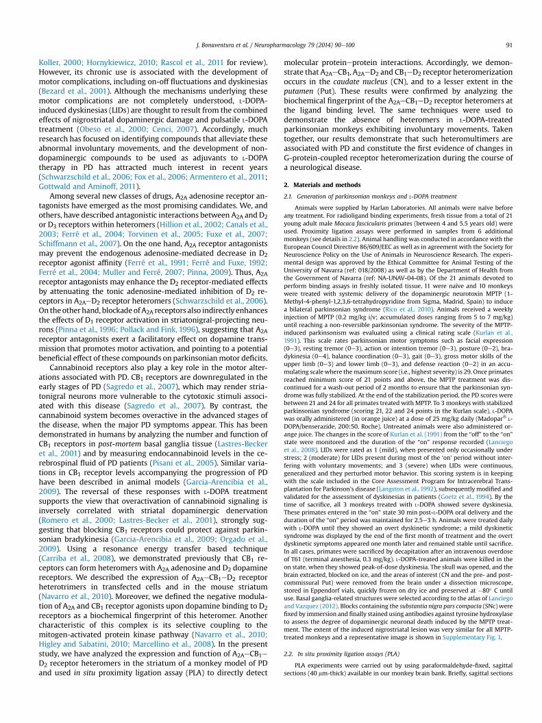
Fig. 3. A2A, CB1 and D2 receptor expression in monkey striatum. Maximum bindingwas calculated in competitive binding experiments using 0.2 mg/ml protein of monkeyCN, pre- or post-commissural Put membranes, and an A2A receptor antagonist [3H]ZM241385 (4 nM, A), a D2 receptor antagonist [3H]YM-09151-2 (0.5 nM, B) or a CB1 re-ceptor agonist [3H]CP 55940 (6 nM, C) vs. 50 mM of the same non-radiolabeled ligands,applying the following affinity constants: 1 nM for ZM 241385, 0.6 nM for YM-09151-2and 4 nM for CP 55940. Values represent the mean � SEM of a representativeexperiment performed in triplicate. Statistical significance was calculated by one-wayANOVA followed by a Dunnett’s post-hoc multiple comparison test (*p < 0.05,**p < 0.01 with respect to the CN).
J. Bonaventura et al. / Neuropharmacology 79 (2014) 90e100 95
3.2. Expression of A2AeCB1eD2 receptor heteromers in the striatumof MPTP-treated monkeys
As A2AeCB1eD2 heteromers aremainly expressed in themonkeyCN, we hypothesized that this complex might play a role in thephysiopathology of PD and thus, it may represent a potential ther-apeutic target. Whenwe first used PLA to analyze the expression ofA2AeCB1eD2 heteromers in CN slices fromMPTP-lesionedmonkeys,A2AeCB1 (Fig. 4A), A2AeD2 (Fig. 4B) and CB1eD2 (Fig. 4C) heteromerswere visualized as red spots in several DAPI-stained neurons(Fig. 4D) at levels similar to those found in control monkeys(p> 0.05, two-way ANOVA from Figs.1G and 4D data). These resultsstrongly suggest the presence of A2AeCB1eD2 receptor heteromersin the CN of parkinsonian animals. We next investigated the effectsof A2A and CB1 receptor agonists on dopamine affinity for D2 re-ceptors in the CN of MPTP-lesioned monkeys. Competition experi-ments were performed to measure the binding of 0.5 nMradiolabeled D2 receptor antagonist [3H]YM-09151-2 at increasingdopamine concentrations (0.01 nMe30 mM) in the presence orabsence of 250 nM CGS 21680, 100 nM CP 55940, or both. Biphasiccompetition curves were obtained in all cases and the affinity
constants for dopamine binding to D2 receptor were calculated byfitting the binding data to the equation (2) (Table 2).When the sameexperiment was performed using pre- and post-commissural Putmembranes fromPDmonkeys, the affinity constants obtained for D2receptors were similar to those found in naïve animals (Table 2). Asseen in non-lesioned monkeys, both A2A and CB1 receptor agonistsdecreased the affinity of dopamine for CND2 receptors, an effect thatwas reversed when A2A and CB1 receptor agonists were addedsimultaneously. The fact that similar ligand binding behavior wasobserved for A2AeCB1eD2 receptor heteromers in control and PDmonkeys suggests that MPTP lesions had no effect on either het-eromer expression or intra-heteromer cross-talk.
3.3. Loss of A2AeCB1eD2 receptor heteromers in the CN of L-DOPA-treated monkeys
While dopamine replacement therapy with L-DOPA remains themost effective way to treat parkinsonian hypokinesia, its chronicuse is associatedwithmotor complications that include dyskinesias(Obeso et al., 2000). We investigated the changes that induce achronic L-DOPA treatment to A2AeCB1eD2 receptor heteromerfunction by analyzing A2AeCB1eD2 heteromer expression by PLA inCN slices from monkeys treated with L-DOPA. We investigatedwhether chronic L-DOPA treatment altered A2AeCB1eD2 receptorheteromer function. First, we examined whether there werechanges in A2AeCB1eD2 heteromer expression by PLA in CN slicesfrom monkeys treated with L-DOPA. The expression of A2AeCB1(Fig. 5A), A2AeD2 (Fig. 5B) and CB1eD2 (Fig. 5C) heteromers wasdramatically reduced in CN slices from L-DOPA treated monkeys(Fig. 5D) as compared with control and PD monkeys (p < 0.0001,two-way ANOVA from Figs. 1G, 4D and 5D data), suggesting ablocking of heterotrimer formation. Second, we investigated re-ceptor cross-talk at the ligand binding level using CN membranesfrom MPTP- and L-DOPA-treated monkeys. In competitive bindingassays similar to those described above to measure ligand binding,biphasic competition curves were obtained in all cases and theaffinity constants for dopamine binding to D2 receptor werecalculated by fitting the binding data to the equation (2) (Table 3).In accordance with the decreased heteromer expression seen in thePut (see Fig. 1), dopamine affinity for D2 receptors was unchangedfollowing agonist binding to A2A or CB1 receptors in the pre- andpost-commissural Put of treated animals (Table 3). Interestingly,the dopamine affinity for CN D2 receptorswas unchanged followingagonist binding to A2A and/or CB1 receptors, indicating a loss offunctional cross-talk between receptors, consistent with the loss ofheteromer formation observed in the PLA experiments. Two-wayANOVA showed a significant between-group differences(p < 0.0001) effect of disease state on KDB1 values e not on KDB2values e (data from Tables 1e3) and on percentage of positive cellsand spots/cell (r) in CN e not in Put e (data from Figs. 1G, 4D and5D). The changes in receptor heteromerization in L-DOPA-treatedmonkeys were not due to decreased receptor expression (Fig. 6).Taken together, these results indicate a loss of heteromer-mediatedfunctional cross-talk following chronic L-DOPA treatment.
4. Discussion
The ‘‘receptor heteromer’’ concept postulates that receptorsfrom the same and different gene families combine with oneanother to generate complexes with unique biochemical andfunctional characteristics. This theory is becoming widely acceptedfor G-protein-coupled receptors and it constitutes an emergingarea of interest in the field of receptor signaling (Ferré et al., 2009a).Through heteromerization, receptors generate unique functionalentities and novel potential therapeutic targets (Ferré et al., 2007,

Fig. 4. A2AeCB1eD2 receptor heteromer expression in the CN of MPTP-lesioned monkeys. In situ proximity ligation assays (PLA) were performed as described in the Materials andMethods using CN slices from MPTP-lesioned monkeys and primary antibodies for A2A and CB1 (A), A2A and D2 (B), or CB1 and D2 (C) receptors. Confocal microscopy images(superimposed sections) are shown in which heteromers appear as red clusters. Scale bars ¼ 20 mm. In all cases, cell nuclei were stained with DAPI (blue). (D) The number of cellscontaining one or more red spots is expressed as the percentage of the total number of cells (blue nucleus). r values (number of red spots/cell) were counted only in cells thatcontained red spots and are shown above each bar. Data (% of positive cells or r) are the mean � SEM of counts in 12 different fields (see Methods). From data taken from 12 fields,two-way ANOVA analysis did not show significant differences on positive cell or spots/cell values (p > 0.05) respect to naïve control monkeys (Fig. 1).
Table 2Effect of A2A and CB1 receptor agonist on dopamine affinity for D2 receptors instriatal membranes from PD monkeys. Pharmacological parameters obtained fromcompetition experiments.
Competition experiments[3H]YM-09151-02 vs. dopamine
Parameters
KDB1 (nM) KDB2 (nM)
Caudate nucleusVehicle 6 � 2 1300 � 500þ CGS 21680 (250 nM) 19 � 2*** ss 3000 � 1000þ CP 55940 (100 nM) 18 � 3*** ss 2500 � 800þ CGS 21680 þ CP 55940 9 � 4 1300 � 600
Pre-commissural putVehicle 3 � 1 120 � 40þ CGS 21680 (250 nM) 5 � 1 600 � 200þ CP 55940 (100 nM) 3 � 1 140 � 60þ CGS 21680 þ CP 55940 5 � 2 500 � 100
Post-commissural putVehicle 3 � 1 60 � 20þ CGS 21680 (250 nM) 1 � 1 200 � 60þ CP 55940 (100 nM) 3 � 1 300 � 100þ CGS 21680 þ CP 55940 2 � 1 70 � 20
KDB1 and KDB2 are, respectively, the equilibrium dissociation constants of the firstand the second dopamine binding to the receptor. Two-way ANOVA analysisshowed significant between-region differences (p < 0.001) on KDB1 values and noton KDB2 values.***p< 0.001 (respect to vehicle) andss p< 0.01 (respect to CGS 21680þ CP 55940)after Bonferroni’s post hoc test.
J. Bonaventura et al. / Neuropharmacology 79 (2014) 90e10096
2009a; Pin et al. 2007; Dalrymple et al., 2008; Casadó et al., 2009).A large number of heteromers composed of two different receptorshave been identified to date (Pin et al. 2007; Dalrymple et al., 2008).We have presented evidence supporting the existence of receptorheteromultimers (i.e., heteromers composed of more than twodifferent receptors), containing A2A adenosine, CB1 cannabinoidand D2 dopamine receptors (Carriba et al., 2008; Navarro et al.,2010). The use of mutants in which the quaternary structure ofthe A2AeCB1eD2 heteromer is altered has revealed a keybiochemical characteristic of this complex: activation of adenosineor cannabinoid receptors within the heteromer negatively modu-lates D2 receptor function (Navarro et al., 2010). Although powerfultools are available to identify heteromers in heterologous systems,it remains a challenge to detect heteromer expression in naturaltissue. We identified A2AeCB1eD2 heteromers in a natural sourceusing PLA and radioligand binding techniques. As PLA is limited tothe direct screening of pairs of receptors, we analyzed all possiblecombinations for the A2AeCB1eD2 heterotrimeric complex. The PLAdata and the cross-talk revealed in binding assays, points to theexpression of this complex in the CN of M. fascicularis. The occur-rence of A2AeCB1, CB1eD2 and A2AeD2 heteromers cannot be ruledout and there is not any tool available to know the proportion ofeach of the heteromeric species that might coexist in the sample.These findings demonstrate however that some current (dopaminereceptor agonists) and experimental (A2A receptor antagonists;

Fig. 5. A2AeCB1eD2 receptor heteromer expression in the CN of L-DOPA-treated monkeys. In situ proximity ligation assays (PLA) were performed as described in the Materials andMethods using CN slices from monkeys treated with L-DOPA, and primary antibodies for A2A and CB1 (A), A2A and D2 (B), or CB1 and D2 (C) receptors. Confocal microscopy images(superimposed sections) are shown in which heteromers appear as red clusters. Scale bars ¼ 20 mm. In all cases, cell nuclei were stained with DAPI (blue). (D) The number of cellscontaining one or more red spots is expressed as the percentage of the total number of cells (blue nucleus). r values (number of red spots/cell) were counted only in cells thatcontained red spots and are shown above each bar. Data (% of positive cells or r) are the mean � SEM of counts in 12 different fields (see Methods). From data taken from 12 fields,two-way ANOVA analysis showed significant differences on positive cell or spots/cell values (p < 0.001) respect to naïve control (Fig. 1) or PD monkeys (Fig. 4).
J. Bonaventura et al. / Neuropharmacology 79 (2014) 90e100 97
phase II and III clinical trials) anti-parkinsonian therapies target D2or A2A receptors that in the CN are expressed as heteromers withthemselves and/or with CB1 receptors. It has been demonstrated(Orru et al., 2011) that different A2A receptor antagonists may differ
Table 3Effect of A2A and CB1 receptor agonist on dopamine affinity for D2 receptors instriatal membranes from L-DOPA-treated monkeys. Pharmacological parametersobtained from competition experiments.
Competition experiments[3H]YM-09151-02 vs. dopamine
Parameters
KDB1 (nM) KDB2 (nM)
Caudate nucleusVehicle 3 � 2 200 � 100þ CGS 21680 (250 nM) 4 � 2 600 � 300þ CP 55940 (100 nM) 3 � 1 500 � 200þ CGS 21680 þ CP 55940 3 � 1 360 � 90
Pre-commissural putVehicle 4 � 1 230 � 70þ CGS 21680 (250 nM) 8 � 3 280 � 140þ CP 55940 (100 nM) 6 � 2 170 � 70þ CGS 21680 þ CP 55940 8 � 5 430 � 130
Post-commissural putVehicle 7 � 1 120 � 50þ CGS 21680 (250 nM) 10 � 4 260 � 50þ CP 55940 (100 nM) 7 � 1 200 � 70þ CGS 21680 þ CP 55940 7 � 5 220 � 100
KDB1 and KDB2 are, respectively, the equilibrium dissociation constants of the firstand the second dopamine binding to the receptor. Two-way ANOVA analysisshowed no significant between-region differences (p> 0.05) on KDB1 or KDB2 values.
in the action exerted depending on a preferential pre- versuspostsynaptic location of A2A receptor-containing heteromers.Whereas in striatum A1eA2A receptor heteromers are presynapti-cally located in glutamatergic terminals of cortical neurons, A2Ae
CB1eD2 receptor heteromers are postsynaptically located in thespines of GABAergic enkephalinergic neurons. Targeting pre-versus postsynaptic A2A receptors, or vice versa, may represent auseful approach to differentially combat disorders affecting thebasal ganglia.
Neurons can differentially express heteromers to achieve a di-versity of neurotransmitter-mediated actions. According to thebasal ganglia model (Albin et al., 1989; DeLong, 1990), the CN andthe Put form part of distinct but parallel cortico-basal ganglia-thalamocortical loops (Alexander and Crutcher, 1990). Accordingly,the post-commissural Put primarily mediates motor-related infor-mation processing, while the CN forms part of the associative andlimbic loops. Results from recent high-resolution functional mag-netic resonance imaging (fMRI) studies, performed during areward- and punishment-based probabilistic associative learningtask, have revealed functional specialization within the striatum(ventral Put vs. dorsal CN: Mattfeld et al., 2011). As the fingerprintof D2eA2AeCB1 receptor heteromer was identified in the CN but notin the Put, differential D2-mediated dopaminergic neurotransmis-sion may occur in different striatal areas in primates. Indeed, theA2AeCB1eD2 receptor heteromer becomes an instrument foradenosine or cannabinoid modulation of dopamine binding to D2
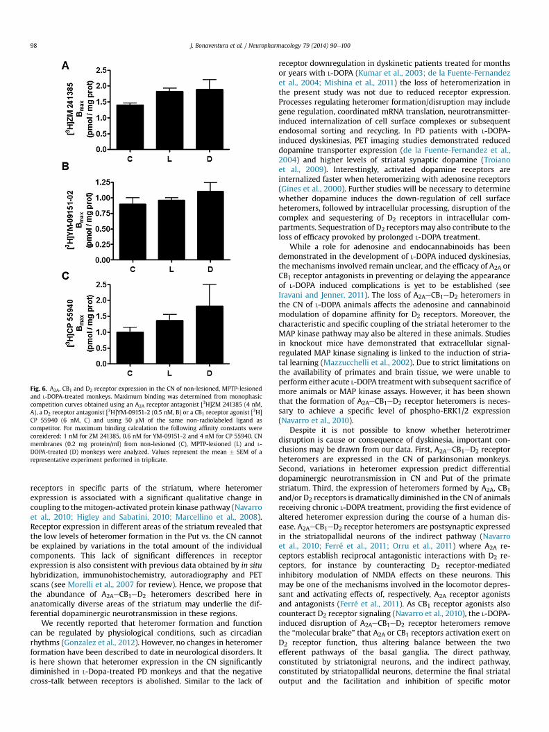
Fig. 6. A2A, CB1 and D2 receptor expression in the CN of non-lesioned, MPTP-lesionedand L-DOPA-treated monkeys. Maximum binding was determined from monophasiccompetition curves obtained using an A2A receptor antagonist [3H]ZM 241385 (4 nM,A), a D2 receptor antagonist [3H]YM-09151-2 (0.5 nM, B) or a CB1 receptor agonist [3H]CP 55940 (6 nM, C) and using 50 mM of the same non-radiolabeled ligand ascompetitor. For maximum binding calculation the following affinity constants wereconsidered: 1 nM for ZM 241385, 0.6 nM for YM-09151-2 and 4 nM for CP 55940. CNmembranes (0.2 mg protein/ml) from non-lesioned (C), MPTP-lesioned (L) and L-DOPA-treated (D) monkeys were analyzed. Values represent the mean � SEM of arepresentative experiment performed in triplicate.
J. Bonaventura et al. / Neuropharmacology 79 (2014) 90e10098
receptors in specific parts of the striatum, where heteromerexpression is associated with a significant qualitative change incoupling to the mitogen-activated protein kinase pathway (Navarroet al., 2010; Higley and Sabatini, 2010; Marcellino et al., 2008).Receptor expression in different areas of the striatum revealed thatthe low levels of heteromer formation in the Put vs. the CN cannotbe explained by variations in the total amount of the individualcomponents. This lack of significant differences in receptorexpression is also consistent with previous data obtained by in situhybridization, immunohistochemistry, autoradiography and PETscans (see Morelli et al., 2007 for review). Hence, we propose thatthe abundance of A2AeCB1eD2 heteromers described here inanatomically diverse areas of the striatum may underlie the dif-ferential dopaminergic neurotransmission in these regions.
We recently reported that heteromer formation and functioncan be regulated by physiological conditions, such as circadianrhythms (Gonzalez et al., 2012). However, no changes in heteromerformation have been described to date in neurological disorders. Itis here shown that heteromer expression in the CN significantlydiminished in L-Dopa-treated PD monkeys and that the negativecross-talk between receptors is abolished. Similar to the lack of
receptor downregulation in dyskinetic patients treated for monthsor years with L-DOPA (Kumar et al., 2003; de la Fuente-Fernandezet al., 2004; Mishina et al., 2011) the loss of heteromerization inthe present study was not due to reduced receptor expression.Processes regulating heteromer formation/disruption may includegene regulation, coordinated mRNA translation, neurotransmitter-induced internalization of cell surface complexes or subsequentendosomal sorting and recycling. In PD patients with L-DOPA-induced dyskinesias, PET imaging studies demonstrated reduceddopamine transporter expression (de la Fuente-Fernandez et al.,2004) and higher levels of striatal synaptic dopamine (Troianoet al., 2009). Interestingly, activated dopamine receptors areinternalized faster when heteromerizing with adenosine receptors(Gines et al., 2000). Further studies will be necessary to determinewhether dopamine induces the down-regulation of cell surfaceheteromers, followed by intracellular processing, disruption of thecomplex and sequestering of D2 receptors in intracellular com-partments. Sequestration of D2 receptors may also contribute to theloss of efficacy provoked by prolonged L-DOPA treatment.
While a role for adenosine and endocannabinoids has beendemonstrated in the development of L-DOPA induced dyskinesias,the mechanisms involved remain unclear, and the efficacy of A2A orCB1 receptor antagonists in preventing or delaying the appearanceof L-DOPA induced complications is yet to be established (seeIravani and Jenner, 2011). The loss of A2AeCB1eD2 heteromers inthe CN of L-DOPA animals affects the adenosine and cannabinoidmodulation of dopamine affinity for D2 receptors. Moreover, thecharacteristic and specific coupling of the striatal heteromer to theMAP kinase pathway may also be altered in these animals. Studiesin knockout mice have demonstrated that extracellular signal-regulated MAP kinase signaling is linked to the induction of stria-tal learning (Mazzucchelli et al., 2002). Due to strict limitations onthe availability of primates and brain tissue, we were unable toperform either acute L-DOPA treatment with subsequent sacrifice ofmore animals or MAP kinase assays. However, it has been shownthat the formation of A2AeCB1eD2 receptor heteromers is neces-sary to achieve a specific level of phospho-ERK1/2 expression(Navarro et al., 2010).
Despite it is not possible to know whether heterotrimerdisruption is cause or consequence of dyskinesia, important con-clusions may be drawn from our data. First, A2AeCB1eD2 receptorheteromers are expressed in the CN of parkinsonian monkeys.Second, variations in heteromer expression predict differentialdopaminergic neurotransmission in CN and Put of the primatestriatum. Third, the expression of heteromers formed by A2A, CB1and/or D2 receptors is dramatically diminished in the CN of animalsreceiving chronic L-DOPA treatment, providing the first evidence ofaltered heteromer expression during the course of a human dis-ease. A2AeCB1eD2 receptor heteromers are postsynaptic expressedin the striatopallidal neurons of the indirect pathway (Navarroet al., 2010; Ferré et al., 2011; Orru et al., 2011) where A2A re-ceptors establish reciprocal antagonistic interactions with D2 re-ceptors, for instance by counteracting D2 receptor-mediatedinhibitory modulation of NMDA effects on these neurons. Thismay be one of the mechanisms involved in the locomotor depres-sant and activating effects of, respectively, A2A receptor agonistsand antagonists (Ferré et al., 2011). As CB1 receptor agonists alsocounteract D2 receptor signaling (Navarro et al., 2010), the L-DOPA-induced disruption of A2AeCB1eD2 receptor heteromers removethe “molecular brake” that A2A or CB1 receptors activation exert onD2 receptor function, thus altering balance between the twoefferent pathways of the basal ganglia. The direct pathway,constituted by striatonigral neurons, and the indirect pathway,constituted by striatopallidal neurons, determine the final striataloutput and the facilitation and inhibition of specific motor

J. Bonaventura et al. / Neuropharmacology 79 (2014) 90e100 99
responses. An imbalance in the indirect pathway due to the lack ofthe A2AeCB1eD2-receptor-heteromer functional component, maypromote, at least in part, motor alterations associated to dyskinesia.Taken together, these findings provide a better understanding ofthe role of heteromers containing dopamine and adenosine re-ceptors in the physiopathology of PD and L-DOPA-induced com-plications, and may contribute to the improvement of currenttherapeutic strategies to combat PD.
Conflict of interest
The authors declare no conflict of interest.
Acknowledgments
We thank Jasmina Jiménez (Molecular Neurobiology laboratory,Barcelona University) for technical assistance. This study wassupported by Grants from Spanish Ministerio de Ciencia y Tecno-logía (SAF2012-39875-C02-01, SAF2010-18472 and SAF2008-03118-E, within the framework of the Era-NET Neuron program)and a grant for collaborative projects (PI2011/02-7) from the Centrode Investigación Biomédica en Red sobre Enfermedades Neuro-degenerativas (CIBERNED). PJM is a Ramón y Cajal Fellow.
Appendix A. Supplementary data
Supplementary data related to this article can be found at http://dx.doi.org/10.1016/j.neuropharm.2013.10.036.
References
Albin, R.L., Young, A.B., Penney, J.B., 1989. The functional anatomy of basal gangliadisorders. Trends Neurosci. 12 (10), 366e375.
Alexander, G.E., Crutcher, M.D., 1990. Functional architecture of basal ganglia cir-cuits: neural substrates of parallel processing. Trends Neurosci. 13 (7), 266e271.
Armentero, M.T., Pinna, A., Ferré, S., Lanciego, J.L., Muller, C.E., Franco, R., 2011. Past,present and future of A(2A) adenosine receptor antagonists in the therapy ofParkinson’s disease. Pharmacol. Ther. 132 (3), 280e299.
Bezard, E., Dovero, S., Prunier, C., Ravenscroft, P., Chalon, S., Guilloteau, D., et al.,2001. Relationship between the appearance of symptoms and the level ofnigrostriatal degeneration in a progressive 1-methyl-4-phenyl-1,2,3,6-tetrahydropyridine-lesioned macaque model of Parkinson’s disease.J. Neurosci. 21 (17), 6853e6861.
Callen, L., Moreno, E., Barroso-Chinea, P., Moreno-Delgado, D., Cortes, A., Mallol, J.,et al., 2012. Cannabinoid receptors CB1 and CB2 form functional heteromers inbrain. J. Biol. Chem. 287 (25), 20851e20865.
Canals, M., Marcellino, D., Fanelli, F., Ciruela, F., de Benedetti, P., Goldberg, S.R., et al.,2003. Adenosine A2A-dopamine D2 receptor-receptor heteromerization: quali-tative and quantitative assessment by fluorescence and bioluminescence en-ergy transfer. J. Biol. Chem. 278 (47), 46741e46749.
Carriba, P., Navarro, G., Ciruela, F., Ferré, S., Casadó, V., Agnati, L., et al., 2008.Detection of heteromerization of more than two proteins by sequential BRET-FRET. Nat. Methods 5 (8), 727e733.
Casadó, V., Canti, C., Mallol, J., Canela, E.I., Lluis, C., Franco, R., 1990. Solubilization ofA1 adenosine receptor from pig brain: characterization and evidence of the roleof the cell membrane on the coexistence of high- and low-affinity states.J. Neurosci. Res. 26 (4), 461e473.
Casadó, V., Cortes, A., Ciruela, F., Mallol, J., Ferré, S., Lluis, C., et al., 2007. Old and newways to calculate the affinity of agonists and antagonists interacting with G-protein-coupled monomeric and dimeric receptors: the receptor-dimer coop-erativity index. Pharmacol. Ther. 116 (3), 343e354.
Casadó, V., Ferrada, C., Bonaventura, J., Gracia, E., Mallol, J., Canela, E.I., et al., 2009.Useful pharmacological parameters for G-protein-coupled receptor homo-dimers obtained from competition experiments. Agonist-antagonist bindingmodulation. Biochem. Pharmacol. 78 (12), 1456e1463.
Cenci, M.A., 2007. L-DOPA-induced dyskinesia: cellular mechanisms and ap-proaches to treatment. Parkinsonism Relat. Disord. 13 (Suppl. 3), S263eS267.
Dalrymple, M.B., Pfleger, K.D., Eidne, K.A., 2008. G protein-coupled receptor dimers:functional consequences, disease states and drug targets. Pharmacol. Ther. 118(3), 359e371.
de la Fuente-Fernandez, R., Sossi, V., Huang, Z., Furtado, S., Lu, J.Q., Calne, D.B., et al.,2004. Levodopa-induced changes in synaptic dopamine levels increase withprogression of Parkinson’s disease: implications for dyskinesias. Brain 127 (Pt12), 2747e2754.
DeLong, M.R., 1990. Primate models of movement disorders of basal ganglia origin.Trends Neurosci. 13 (7), 281e285.
Ferré, S., von Euler, G., Johansson, B., Fredholm, B.B., Fuxe, K., 1991. Stimulation ofhigh-affinity adenosine A2 receptors decreases the affinity of dopamine D2receptors in rat striatal membranes. Proc. Natl. Acad. Sci. U. S. A. 88 (16),7238e7241.
Ferré, S., Fuxe, K., 1992. Dopamine denervation leads to an increase in the intra-membrane interaction between adenosine A2 and dopamine D2 receptors inthe neostriatum. Brain Res. 594 (1), 124e130.
Ferré, S., Ciruela, F., Canals, M., Marcellino, D., Burgueno, J., Casadó, V., et al., 2004.Adenosine A2A-dopamine D2 receptor-receptor heteromers. Targets for neuro-psychiatric disorders. Parkinsonism Relat. Disord. 10 (5), 265e271.
Ferré, S., Ciruela, F., Woods, A.S., Lluis, C., Franco, R., 2007. Functional relevance ofneurotransmitter receptor heteromers in the central nervous system. TrendsNeurosci. 30 (9), 440e446.
Ferré, S., Baler, R., Bouvier, M., Caron, M.G., Devi, L.A., Durroux, T., et al., 2009a.Building a new conceptual framework for receptor heteromers. Nat. Chem. Biol.5 (3), 131e134.
Ferré, S., Goldberg, S.R., Lluis, C., Franco, R., 2009b. Looking for the role of canna-binoid receptor heteromers in striatal function. Neuropharmacology 56(Suppl. 1), 226e234.
Ferré, S., Quiroz, C., Orru, M., Guitart, X., Navarro, G., Cortés, A., et al., 2011. Aden-osine A(2A) receptors and A(2A) receptor heteromers as key players in striatalfunction. Front Neuroanat. 5, 36.
Fox, S.H., Lang, A.E., Brotchie, J.M., 2006. Translation of nondopaminergic treat-ments for levodopa-induced dyskinesia from MPTP-lesioned nonhuman pri-mates to phase IIa clinical studies: keys to success and roads to failure. Mov.Disord. 21 (10), 1578e1594.
Franco, R., Casadó, V., Mallol, J., Ferrada, C., Ferré, S., Fuxe, K., et al., 2006. The two-state dimer receptor model: a general model for receptor dimers. Mol. Phar-macol. 69 (6), 1905e1912.
Fuxe, K., Ferré, S., Genedani, S., Franco, R., Agnati, L.F., 2007. Adenosine receptor-dopamine receptor interactions in the basal ganglia and their relevance forbrain function. Physiol. Behav. 92 (1e2), 210e217.
Garcia-Arencibia, M., Garcia, C., Fernandez-Ruiz, J., 2009. Cannabinoids and Par-kinson’s disease. CNS Neurol. Disord. Drug Targets 8 (6), 432e439.
Gines, S., Hillion, J., Torvinen, M., Le Crom, S., Casadó, V., Canela, E.I., et al., 2000.Dopamine D1 and adenosine A1 receptors form functionally interacting het-eromeric complexes. Proc. Natl. Acad. Sci. U. S. A. 97 (15), 8606e8611.
Goetz, C.G., Stebbins, G.T., Shale, H.M., Lang, A.E., Chernik, D.A., Chmura, T.A., et al.,1994. Utility of an objective dyskinesia rating scale for Parkinson’s disease:inter- and intrarater reliability assessment. Mov. Disord. 9 (4), 390e394.
Gonzalez, S., Moreno-Delgado, D., Moreno, E., Perez-Capote, K., Franco, R., Mallol, J.,et al., 2012. Circadian-related heteromerization of adrenergic and dopamineD(4) receptors modulates melatonin synthesis and release in the pineal gland.PLoS Biol. 10 (6), e1001347.
Gottwald, M.D., Aminoff, M.J., 2011. Therapies for dopaminergic-induced dyskine-sias in Parkinson disease. Ann. Neurol. 69 (6), 919e927.
Gracia, E., Moreno, E., Cortés, A., Lluís, C., Mallol, J., McCormick, P.J., et al., 2013.Homodimerization of adenosine A1 receptors in brain cortex explains thebiphasic effect of caffeine. Neuropharmacology 71, 56e69.
Higley, M.J., Sabatini, B.L., 2010. Competitive regulation of synaptic Ca2þ influx by D2dopamine and A2A adenosine receptors. Nat. Neurosci. 13 (8), 958e966.
Hillion, J., Canals, M., Torvinen, M., Casadó, V., Scott, R., Terasmaa, A., et al., 2002.Coaggregation, cointernalization, and codesensitization of adenosine A2A re-ceptors and dopamine D2 receptors. J. Biol. Chem. 277 (20), 18091e18097.
Hornykiewicz, O., 2010. A brief history of levodopa. J. Neurol. 257 (Suppl. 2), S249eS252.
Iravani, M.M., Jenner, P., 2011. Mechanisms underlying the onset and expression oflevodopa-induced dyskinesia and their pharmacological manipulation. J. NeuralTransm. 118 (12), 1661e1690.
Koller, W.C., 2000. Levodopa in the treatment of Parkinson’s disease. Neurology 55(11 Suppl. 4), S2eS7 discussion S8e12.
Kravitz, A.V., Freeze, B.S., Parker, P.R., Kay, K., Thwin, M.T., Deisseroth, K., et al., 2010.Regulation of parkinsonian motor behaviours by optogenetic control of basalganglia circuitry. Nature 466 (7306), 622e626.
Kumar, A., Mann, S., Sossi, V., Ruth, T.J., Stoessl, A.J., Schulzer, M., et al., 2003. [11C]DTBZ-PET correlates of levodopa responses in asymmetric Parkinson’s disease.Brain 126 (Pt 12), 2648e2655.
Kurlan, R., Kim, M.H., Gash, D.M., 1991. Oral levodopa dose-response study in MPTP-induced hemiparkinsonian monkeys: assessment with a new rating scale formonkey parkinsonism. Mov. Disord. 6 (2), 111e118.
Lanciego, J.L., Rodriguez-Oroz, M.C., Blesa, F.J., Alvarez-Erviti, L., Guridi, J., Barroso-Chinea, P., et al., 2008. Lesion of the centromedian thalamic nucleus in MPTP-treated monkeys. Mov. Disord. 23 (5), 708e715.
Lanciego, J.L., Vazquez, A., 2012. The basal ganglia and thalamus of the long-tailedmacaque in stereotaxic coordinates. A template atlas based on coronal, sagittaland horizontal brain sections. Brain Struct. Funct. 217 (2), 613e666.
Langston, J.W., Widner, H., Goetz, C.G., Brooks, D., Fahn, S., Freeman, T., et al., 1992.Core assessment program for intracerebral transplantations (CAPIT). Mov.Disord. 7 (1), 2e13.
Lastres-Becker, I., Cebeira, M., de Ceballos, M.L., Zeng, B.Y., Jenner, P., Ramos, J.A.,et al., 2001. Increased cannabinoid CB1 receptor binding and activation of GTP-binding proteins in the basal ganglia of patients with Parkinson’s syndrome andof MPTP-treated marmosets. Eur. J. Neurosci. 14 (11), 1827e1832.

J. Bonaventura et al. / Neuropharmacology 79 (2014) 90e100100
Lewis, S.J., Caldwell, M.A., Barker, R.A., 2003. Modern therapeutic approaches inParkinson’s disease. Expert Rev. Mol. Med. 5 (10), 1e20.
Marcellino, D., Carriba, P., Filip, M., Borgkvist, A., Frankowska, M., Bellido, I., et al.,2008. Antagonistic cannabinoid CB1/dopamine D2 receptor interactions instriatal CB1/D2 heteromers. A combined neurochemical and behavioral analysis.Neuropharmacology 54 (5), 815e823.
Mattfeld, A.T., Gluck, M.A., Stark, C.E., 2011. Functional specialization within thestriatum along both the dorsal/ventral and anterior/posterior axes duringassociative learning via reward and punishment. Learn Mem. 18 (11), 703e711.
Mazzucchelli, C., Vantaggiato, C., Ciamei, A., Fasano, S., Pakhotin, P., Krezel, W., et al.,2002. Knockout of ERK1MAP kinase enhances synaptic plasticity in the striatumand facilitates striatal-mediated learning and memory. Neuron 34 (5), 807e820.
Mishina, M., Ishiwata, K., Naganawa, M., Kimura, Y., Kitamura, S., Suzuki, M., et al.,2011. Adenosine A(2A) receptors measured with [C]TMSX PET in the striata ofParkinson’s disease patients. PLoS One 6 (2), e17338.
Morelli, M., Di Paolo, T., Wardas, J., Calon, F., Xiao, D., Schwarzschild, M.A., 2007. Roleof adenosine A2A receptors in parkinsonian motor impairment and l-DOPA-induced motor complications. Prog. Neurobiol. 83 (5), 293e309.
Muller, C.E., Ferré, S., 2007. Blocking striatal adenosine A2A receptors: a newstrategy for basal ganglia disorders. Recent Pat. CNS Drug Discov. 2 (1), 1e21.
Navarro,G., Ferré, S., Cordomi, A.,Moreno,E.,Mallol, J., Casadó, V., et al., 2010. Interactionsbetween intracellular domains as key determinants of the quaternary structure andfunction of receptor heteromers. J. Biol. Chem. 285 (35), 27346e27359.
Obeso, J.A., Rodriguez-Oroz, M.C., Rodriguez, M., Lanciego, J.L., Artieda, J.,Gonzalo, N., et al., 2000. Pathophysiology of the basal ganglia in Parkinson’sdisease. Trends Neurosci. 23 (10 Suppl.), S8e19.
Obeso, J.A., Rodriguez-Oroz, M.C., Benitez-Temino, B., Blesa, F.J., Guridi, J., Marin, C.,et al., 2008. Functional organization of the basal ganglia: therapeutic implica-tions for Parkinson’s disease. Mov. Disord. 23 (Suppl. 3), S548eS559.
Orgado, J.M., Fernandez-Ruiz, J., Romero, J., 2009. The endocannabinoid system inneuropathological states. Int. Rev. Psychiatry 21 (2), 172e180.
Orru, M., Bake�sová, J., Brugarolas, M., Quiroz, C., Beaumont, V., Goldberg, S.R.,Lluís, C., Cortés, A., Franco, R., Casadó, V., Canela, E.I., Ferré, S., 2011 Jan 11.Striatal pre- and postsynaptic profile of adenosine A(2A) receptor antagonists.PLoS One 6 (1).
Pin, J.P., Neubig, R., Bouvier, M., Devi, L., Filizola, M., Javitch, J.A., et al., 2007. In-ternational Union of Basic and Clinical Pharmacology. LXVII. Recommendationsfor the recognition and nomenclature of G protein-coupled receptor hetero-multimers. Pharmacol. Rev. 59 (1), 5e13.
Pinna, A., di Chiara, G., Wardas, J., Morelli, M., 1996. Blockade of A2A adenosinereceptors positively modulates turning behaviour and c-Fos expression inducedby D1 agonists in dopamine-denervated rats. Eur. J. Neurosci. 8 (6), 1176e1181.
Pinna, A., 2009. Novel investigational adenosine A2A receptor antagonists for Par-kinson’s disease. Expert Opin. Investig. Drugs 18 (11), 1619e1631.
Pisani, A., Fezza, F., Galati, S., Battista, N., Napolitano, S., Finazzi-Agro, A., et al., 2005.High endogenous cannabinoid levels in the cerebrospinal fluid of untreatedParkinson’s disease patients. Ann. Neurol. 57 (5), 777e779.
Pollack, A.E., Fink, J.S., 1996. Synergistic interaction between an adenosineantagonist and a D1 dopamine agonist on rotational behavior and striatal c-Fos induction in 6-hydroxydopamine-lesioned rats. Brain Res. 743 (1e2),124e130.
Rascol, O., Lozano, A., Stern, M., Poewe, W., 2011. Milestones in Parkinson’s diseasetherapeutics. Mov. Disord. 26 (6), 1072e1082.
Rico, A.J., Barroso-Chinea, P., Conte-Perales, L., Roda, E., Gomez-Bautista, V.,Gendive, M., et al., 2010. A direct projection from the subthalamic nucleus to theventral thalamus in monkeys. Neurobiol. Dis. 39 (3), 381e392.
Romero, J., Berrendero, F., Perez-Rosado, A., Manzanares, J., Rojo, A., Fernandez-Ruiz, J.J., et al., 2000. Unilateral 6-hydroxydopamine lesions of nigrostriataldopaminergic neurons increased CB1 receptor mRNA levels in the caudate-pu-tamen. Life Sci. 66 (6), 485e494.
Rosin, D.L., Robeva, A., Woodard, R.L., Guyenet, P.G., Linden, J., 1998. Immunohis-tochemical localization of adenosine A2A receptors in the rat central nervoussystem. J. Comp. Neurol. 401 (2), 163e186.
Sagredo, O., Garcia-Arencibia, M., de Lago, E., Finetti, S., Decio, A., Fernandez-Ruiz, J.,2007. Cannabinoids and neuroprotection in basal ganglia disorders. Mol. Neu-robiol. 36 (1), 82e91.
Schiffmann, S.N., Fisone, G., Moresco, R., Cunha, R.A., Ferré, S., 2007. Adenosine A2Areceptors and basal ganglia physiology. Prog. Neurobiol. 83 (5), 277e292.
Schwarzschild,M.A., Agnati, L., Fuxe,K., Chen, J.F.,Morelli,M., 2006. TargetingadenosineA2A receptors in Parkinson’s disease. Trends Neurosci. 29 (11), 647e654.
Soderberg, O., Leuchowius, K.J., Gullberg, M., Jarvius, M., Weibrecht, I., Larsson, L.G.,et al., 2008. Characterizing proteins and their interactions in cells and tissuesusing the in situ proximity ligation assay. Methods 45 (3), 227e232.
Torvinen, M., Marcellino, D., Canals, M., Agnati, L.F., Lluis, C., Franco, R., et al.,2005. Adenosine A2A receptor and dopamine D3 receptor interactions: evi-dence of functional A2A/D3 heteromeric complexes. Mol. Pharmacol. 67 (2),400e407.
Trifilieff, P., Rives, M.L., Urizar, E., Piskorowski, R.A., Vishwasrao, H.D., Castrillon, J.,et al., 2011. Detection of antigen interactions ex vivo by proximity ligationassay: endogenous dopamine D2-adenosine A2A receptor complexes in thestriatum. Biotechniques 51 (2), 111e118.
Troiano, A.R., de la Fuente-Fernandez, R., Sossi, V., Schulzer, M., Mak, E., Ruth, T.J.,et al., 2009. PET demonstrates reduced dopamine transporter expression in PDwith dyskinesias. Neurology 72 (14), 1211e1216.