arousal effect of caffeine depends on adenosine a2a receptors in the shell of the nucleus accumbens
TRANSCRIPT
Arousal effect of caffeine depends on adenosine A2A receptors inthe shell of the nucleus accumbens
Michael Lazarus1,*, Hai-Ying Shen2,3,*, Yoan Cherasse1,*, Wei-Min Qu4, Zhi-Li Huang1,4,Caroline E. Bass5, Raphaelle Winsky-Sommerer6, Kazue Semba7, Bertil B. Fredholm8,Detlev Boison3, Osamu Hayaishi1, Yoshihiro Urade1, and Jiang-Fan Chen2
1Department of Molecular Behavioral Biology, Osaka Bioscience Institute, 6-2-4 Furedai, Suita,Osaka 565-0874, Japan2Department of Neurology, Boston University School of Medicine, 715 Albany Street, Boston, MA02118, USA3R.S. Dow Neurobiology Laboratories, Legacy Research, 1225 NE 2nd Avenue, Portland, OR97232, USA4Department of Pharmacology, Fudan University Shanghai Medical College, 138 Yixueyuan RoadShanghai 200032, China5Department of Physiology and Pharmacology, Wake Forest University School of Medicine,Medical Center Boulevard, Winston-Salem, NC 27157, USA6Faculty of Health and Medical Sciences, University of Surrey, Guildford, Surrey GU2 7XH,United Kingdom7Department of Anatomy and Neurobiology, Faculty of Medicine, Sir Charles Tupper MedicalBuilding, Dalhousie University, Halifax, Nova Scotia B3H 1X5, Canada8Department of Physiology and Pharmacology, Karolinska Institutet, Stockholm SE-171 77,Sweden
AbstractCaffeine, the most widely used psychoactive compound, is an adenosine receptor antagonist. Itpromotes wakefulness by blocking adenosine A2A receptors (A2ARs) in the brain, but the specificneurons on which caffeine acts to produce arousal have not been identified. Using selective genedeletion strategies based on the Cre/loxP technology in mice and focal RNA interference to silencethe expression of A2ARs in rats by local infection with adeno-associated virus carrying short-hairpin RNA, we report that the A2ARs in the shell region of the nucleus accumbens (NAc) areresponsible for the effect of caffeine on wakefulness. Caffeine-induced arousal was not affected inrats when A2ARs were focally removed from the NAc core or other A2AR-positive areas of thebasal ganglia. Our observations suggest that caffeine promotes arousal by activating pathways thattraditionally have been associated with motivational and motor responses in the brain.
Correspondence should be either addressed to Yoshihiro Urade, Department of Molecular Behavioral Biology, Osaka BioscienceInstitute, 6-2-4 Furuedai, Suita, Osaka 565-0874, Japan. Phone: 81-6-6872-4851; Fax: 81-6-6872-2841; [email protected] or Jiang-Fan Chen, Department of Neurology, Boston University School of Medicine, 715 Albany Street, C329, Boston, MA 02118, USA.Phone: 617-414-1249; Fax: 617-638-5354; [email protected].*Contributed equally to this work.Conflict of interest: No conflict of interest exists.
NIH Public AccessAuthor ManuscriptJ Neurosci. Author manuscript; available in PMC 2012 January 6.
Published in final edited form as:J Neurosci. 2011 July 6; 31(27): 10067–10075. doi:10.1523/JNEUROSCI.6730-10.2011.
NIH
-PA Author Manuscript
NIH
-PA Author Manuscript
NIH
-PA Author Manuscript

Keywordsadeno-associated virus; Cre/loxP mice; EEG; focal RNAi; locomotion; wakefulness
IntroductionCaffeine is the world’s most consumed psychoactive compound. It is readily availablethrough dietary products such as coffee, tea, soft drinks and chocolate treats, but is alsoadded to non-prescription medications like pain-relievers and cold remedies. Irrespective ofthe source, the worldwide average caffeine consumption has been estimated to be just under80 mg/day, although the levels of intake in countries such as Sweden and Finland are in therange of 400 mg of caffeine per day (Fredholm et al., 1999).
Caffeine is widely used to promote wakefulness and to counteract fatigue. Caffeine bindswith very similar affinity to adenosine A1 (A1Rs) and A2A receptors (A2ARs) and at dosescommonly consumed by humans, adenosine actions at both receptors are antagonized.Adenosine is an inhibitory neuromodulator involved in sleep-wake regulation (Porkka-Heiskanen et al., 1997; Huang et al., 2011). Using global genetic knockouts of A1Rs andA2ARs, in which the receptor is deleted from the entire animal, we previously demonstratedthat the A2AR, but not the A1R, mediates the arousal effect of caffeine (Huang et al., 2005).However, the neurons with A2ARs on which caffeine acts to produce wakefulness have notyet been identified.
A2ARs are densely expressed on striatopallidal neurons in the indirect pathway of the basalganglia (BG), where dopamine D2 receptors (D2R) are co-expressed with the A2ARs andcontribute to the control of locomotor activity, motivation, and addiction, all activities thatrequire wakefulness (Rosin et al., 1998; Svenningsson et al., 1999a). The striatopallidalneurons also facilitate movement by operating in parallel with dopamine D1 receptor (D1R)-bearing striatonigral neurons in the direct pathway of the BG. Abilities to maintain arousalare compromised under low dopamine conditions such as Parkinson’s disease (Arnulf et al.,2002; Qu et al., 2010), but the extent to which A2ARs in the BG contribute to the regulationof wakefulness is not known and the role of A2ARs in other brain regions is unclear.
In the present study, we used site-specific gene deletion strategies based on the Cre/loxtechnology in mice and also silenced focally the expression of A2ARs in rats by usingstereotaxic microinjections of adeno-associated virus (AAV) carrying short-hairpin RNA.We found that the A2ARs in the shell region of the nucleus accumbens (NAc) areresponsible for the effect of caffeine on wakefulness.
Materials and MethodsGenetic mouse models
Animals were handled according to the NIH Guide for the Care and Use of LaboratoryAnimals and in accordance with the protocols approved by the IACUC at the BostonUniversity School of Medicine, the Legacy Research IACUC, and the Animal ResearchCommittee at the Osaka Bioscience Institute. All mice (weighing 24–28 g, 11–13 weeks old)and male Sprague–Dawley rats (weighing 150–180 g, 6 weeks old; Shizuoka LaboratoryAnimal Center, Shizuoka, Japan) used in the present study were housed at a constanttemperature (24 ± 0.5°C) with a relative humidity of 60 ± 2% on an automatically controlled12:12 light/dark cycle (light on at 7 a.m.). Three genetic mouse lines on a C57BL/6background were used in the present study: (i) global A2AR knockout mice (A2AR KO)(Chen et al., 1999), (ii) basal ganglia-A2AR knockout mice (BG-A2AR KO), exclusively
Lazarus et al. Page 2
J Neurosci. Author manuscript; available in PMC 2012 January 6.
NIH
-PA Author Manuscript
NIH
-PA Author Manuscript
NIH
-PA Author Manuscript
lacking BG A2ARs (Shen et al., 2008), and (iii) a mouse line with a loxP-site-inserted A2ARgene that is amenable to conditional disruption by the injection of Cre recombinase-expressing AAV.
Vigilance state assessment using EEG/EMG/LMA recordingsAssessment of vigilance states was performed on adult male conditional A2AR KO mice,and their respective WT-littermates (n = 4–5, per genotype and drug dose). Under anesthesiausing 1.5% isoflurane in N2O/O2 (2:1), mice were implanted with electroencephalogram(EEG) and electromyogram (EMG) electrodes for polysomnographic recordings. To monitorEEG signals, 2 stainless steel EEG recording screws (Plastics One, Roanoke, VA, USA)were implanted epidurally over the frontal cortical area (1 mm anterior to bregma, 1.5 mmlateral to the midline) and over the parietal area (2 mm posterior to bregma, 3 mm lateral tomidline) of the right hemisphere. EMG activity was monitored by stainless steel, Teflon-coated wires (0.2 mm in diameter, Plastics One, Roanoke, VA, USA) bilaterally placed intoboth trapezius muscles. Finally, the electrode assembly was anchored and fixed to the skullwith Super6-Bond (Sun Medical Co., Shiga, Japan) and dental cement. After a 10-dayrecovery period, the mice were placed in experimental cages for a 4-day habituation/acclimatization period with connection of counterbalanced recording leads.
All mice that were subjected to EEG recordings received vehicle and drug treatment on 2consecutive days. On day 1, mice were treated with vehicle (saline, i.p.) at 9 a.m., and the24-h recordings performed on day 1 were used as baseline data. On day 2, mice were treatedwith caffeine (i.p., in a volume of 10 ml/kg body weight) and EEG/EMG signals wererecorded for 24 h. The EEG/EMG signals were amplified and filtered (EEG: 0.5–30 Hz,EMG: 20–200 Hz), then digitized at a sampling rate of 128 Hz, and recorded usingSLEEPSIGN software (Kohtoh et al., 2008). In addition, locomotor activity (LMA) wasrecorded with an infrared photocell sensor (Biotex, Kyoto, Japan). The vigilance states werescored offline by 10-s epochs into 3 stages, including waking, rapid-eye movement (REM)sleep, and non-REM (NREM) sleep, according to standard criteria (Mizoguchi et al., 2001).As a final step, defined vigilance stages were examined visually, and corrected whennecessary.
Assessment of activity and inactivityAssessment of LMA and inactivity was performed in adult male A2AR KO, BG-A2AR KOmice and their respective WT-littermates (n = 8, per genotype and drug dose). At 9 a.m. allanimals received an i.p. injection of either vehicle or caffeine at one of the following doses:2, 10 or 30 mg/kg. Inactivity/activity was used to assess sleep and wakefulness based on aprevious report (Pack et al., 2007) following caffeine treatment. LMA was recorded instandard polypropylene cages with 7 infrared photocell beams (San Diego Instrument, CA,USA) in 50-s bins during the 2 experimental days. Inactivity and activity were defined basedon LMA as follows: time spent in inactivity (no beam break/50-seconds) was used as anapproximation of sleep; each period of activity was subdivided into high (≥2 beam breaks/50 s, assessing ambulation) versus low (1 beam break/50 s, assessing rest activity with finemovements) LMA. These three levels of activity were used to analyze the motor stimulanteffects of caffeine, as opposed to the arousal effects of caffeine which were assessed usingpolygraphic recordings as described above.
Generation of AAV vectorsFor the generation of the AAV-shRNA-mCherry vector plasmids, a U6-shA2AR cassettewas amplified by PCR from the psiSTRIKE-hMGFP plasmid (Promega) containing the ratA2A receptor shRNA (target sequence 1913) (Chen et al., 2004) or a control shRNAsequence (GTCAGGCTATCGCGTATCG), and was inserted into the MluI site of the
Lazarus et al. Page 3
J Neurosci. Author manuscript; available in PMC 2012 January 6.
NIH
-PA Author Manuscript
NIH
-PA Author Manuscript
NIH
-PA Author Manuscript

pAAV-hrGFP plasmid (Stratagene). Subsequently, the hrGFP gene was replaced by the geneencoding mCherry. For the generation of the AAV-Cre plasmid, the hrGFP in the pAAV-hrGFP plasmid was replaced by the Cre recombinase coding sequence derived by PCR fromthe pBS185 plasmid (Sauer and Henderson, 1990). The AAVs of serotype rh10 weregenerated by tripartite transfection (AAV-rep2/caprh10 expression plasmid, adenovirushelper plasmid, and AAV-vector plasmid) into 293A cells and purified by iodixanol densitystep-gradient centrifugation, as previously described (Zolotukhin et al., 1999). The virusdistributed in the 40% density step was concentrated and dialyzed against phosphate-buffered saline (PBS) with a centrifugal concentrator (MWCO 100 kDa, Sartorius) and thentitered by qPCR.
Stereotaxic AAV injection and placement of EEG/EMG electrodesSurgeries for AAV injections were conducted under pentobarbital anesthesia (50 mg/kg,i.p.). Using aseptic techniques, 6-week-old rats were injected stereotaxically into the NAcand other BG nuclei with recombinant AAV-shA2AR or AAV-shCTRL (250 nl perinjection, 6×10¹² particles/ml) with a glass micropipette and an air pressure injector system(Chamberlin et al., 1998). Also, 8–10 week-old conditional A2AR KO mice were injectedwith AAV-Cre or AAV-mCherry. Table 1 summarizes coordinates used for bilateralinjections into selected BG nuclei of rats or conditional A2AR KO mice, according to theatlases of Paxinos and Watson (Paxinos and Franklin, 2001; Paxinos and Watson, 2007). At3 weeks after the AAV injection, rats underwent surgery for implantation of electrodes forEEG and EMG recordings as described previously (Matsumura et al., 1994), whereas EEG/EMG electrodes in the conditional KO mice were implanted as described above.Postoperatively, animals were housed individually for 8–10 days. The caffeine treatmentwas performed as described above; i.p. injections were made at 9 (mice) or 10 a.m. (rats). Inaddition, at least 1 week after the caffeine treatment, each animal received an injection ofvehicle or modafinil on a 2-day schedule as described above. Modafinil (Sigma-Aldrich)was dissolved in saline containing 10% DMSO and 2% (w/v) cremophor immediatelybefore use and administered i.p. at 9 a.m. on the experimental day at a dose of 45 mg/kg.
ImmunohistochemistryFollowing all of the above procedures, animals were deeply anesthetized with an overdoseof chloral hydrate (500 mg/kg, i.p.) and perfused through the left ventricle of the heart withsaline followed by neutral buffered 10% formalin. Brains were removed and placed in 10%sucrose in PBS overnight at 4°C to reduce freezing artifacts. The brains were then frozen ondry ice and sectioned at 30 µm (mice) or 40 µm (rats) on a freezing microtome.Immunohistochemistry was performed on free-floating sections as described previously(Estabrooke et al., 2001). In brief, sections were rinsed in PBS, incubated in 3% hydrogenperoxide in PBS for 30 min at room temperature, and then sequentially at room temperaturein 3% normal donkey serum and 0.25% Triton X-100 in PBS (PBT) for 1 h and primaryantibody diluted in PBT with 0.02% sodium azide overnight. Primary antibodies includedrabbit anti-Cre (1:10000; EMD Biosciences, Darmstadt, Germany), rabbit anti-mCherry(1:10000; Clontech, Mountain View, CA), mouse anti-A2AR (1:2000; Millipore, Bellerica,MA), goat anti-A2AR (1:1000; Santa Cruz Biotechnology, Santa Cruz, CA), chicken anti-β-gal (1:4000; Abcam, Cambridge, MA), and mouse anti-NeuN (1:2000; Millipore). Afterincubation with the primary antisera overnight, sections were rinsed and incubated for 2 h inbiotinylated anti-rabbit, anti-goat, anti-chicken or anti-mouse secondary antiserum (JacksonImmunoResearch, West Grove, PA) at a dilution of 1:1000. Immunoreactions for D2R witha rabbit anti-D2R antibody (1:500; Millipore) were conducted over 2 nights at 4°C and 1night at room temperature. All tissue sections were then treated with avidin–biotin complex(1:1000; Vectastain ABC Elite kit, Vector Laboratories) for 1 h, and immunoreactive cellswere visualized by reaction with 3,3'-diaminobenzidine and 0.1% hydrogen peroxide. Tissue
Lazarus et al. Page 4
J Neurosci. Author manuscript; available in PMC 2012 January 6.
NIH
-PA Author Manuscript
NIH
-PA Author Manuscript
NIH
-PA Author Manuscript

sections mounted on glass slides were scanned with Aperio ScanScope (Vista, CA), anddigital photomicrographs were analyzed with Aperio ImageScope software v10. The regionof A2AR knockdown by shA2AR was identified by the expression of mCherry and confirmedby the absence of A2AR immunoreactivity, whereupon the area of loss of A2ARs in the NAcwas quantified on photomicrographs of sections containing the rostral, central, and caudalNAc. Digital photomicrographs were adjusted for optimal display for the output levels of thecontained color values and then imported into ImageJ 1.42 software for area measurementsof mCherry expression in the NAc versus total nucleus extension. Doubleimmunofluorescence staining for β-gal and choline acetyltransferase (ChAT) was alsocarried out. Sections were incubated overnight at room temperature in a mixture of anti-β-gal and goat anti-ChAT (1:200; Millipore) primary antibodies in PBT with donkey normalserum. On the next day, sections were incubated for 2h in a mixture of biotinylated anti-chicken and Alexa-594-conjugated anti-goat secondary antibodies (Invitrogen, Carlsbad,CA) at a dilution of 1:500. After several washes, sections were incubated for 1h inAlexa-488-conjugated streptavidin (Invitrogen) at a dilution of 1:500. Fluorescencemicroscopy with tissue sections mounted on glass slides was performed with a Zeiss laserscanning confocal microscope (Oberkochen, Germany).
Statistical analysisThe data were presented as the mean ± standard error of the mean (SEM). Statisticalcomparisons between 2 groups were performed by using the unpaired Student’s t-test.Comparisons among combined multiple parameters (genotype, experimental conditions, and>2 groups) were performed by one-way ANOVA followed by Bonferroni post-hoccomparisons.
ResultsDeletion of A2ARs in the basal ganglia of mice abolishes the arousal effect of caffeine
We first examined sleep-wake profiles in our previously developed BG-specific A2ARknockout (BG-A2AR KO) mice based on the Cre/loxP technology (Shen et al., 2008). Theexistence of A2ARs in arousal-related cell groups surrounding the striatum like the nucleusof the horizontal limb of the diagonal band of Broca (HDB), the substantia inominata (SI) orthe ventrolateral preoptic area (VLPO) remains elusive (Svenningsson et al., 1997; Rosin etal., 1998). However, to assess Cre-dependent knockout of A2ARs in these adjacent areas, wecross-bred the Dlx5/6-Cre transgenic mouse, which was used to create the BG-A2AR KOmouse, with a Rosa26 reporter line (Soriano, 1999) expressing β-galactosidase (β-gal) onlyin the presence of Cre recombinase (Rosa26-Dlx5/6-Cre). The limit of this method mayhowever hinge on the barely existing expression of A2ARs in various regions of the brain.As shown in Figure 1A, the striatum including the olfactory tubercle (OLT), caudate-putamen (CPu), and nucleus accumbens (NAc) showed robust β-gal staining. The vastmajority of neurons in the striatum are GABAergic medium-sized spiny output neurons, butdouble immunofluorescence staining for β-gal and ChAT (Figure 1B) revealed that theA2AR knockout in the striatum occurred also in the cholinergic interneurons. Outside of thestriatum, sparse β-gal expression was detected in the HDB, whereas β-gal staining wasabsent in the SI and the VLPO (Figure 1C). In addition, only scattered cells with β-galimmunostaining were observed in the septum, cerebral cortex, thalamic nuclei, andhippocampus (Figure 1A or data not shown). Brain sections from Cre-negative mice of theRosa26-Dlx5/6-Cre line did not show any β-gal staining (data not shown).
We then recorded EEG and EMG for 2 consecutive days in the BG-A2AR KO mice and theirwild-type littermates (Figure 1D and E). On day 1, the mice were treated with vehicle (i.p.)at 9 a.m. in the early phase of the light (inactive) period, and the recordings made on that
Lazarus et al. Page 5
J Neurosci. Author manuscript; available in PMC 2012 January 6.
NIH
-PA Author Manuscript
NIH
-PA Author Manuscript
NIH
-PA Author Manuscript

day served as the baseline data. The animals were then treated with caffeine 24 h later(either 2 or 30 mg/kg, i.p.). The vigilance states were classified offline into 3 stages: waking,REM sleep, and NREM sleep. Caffeine dose-dependently increased wakefulness in controlWT mice 2- and 3.2-fold following the 2 and 30 mg/kg dose, respectively (Figure 1D andE). This arousal effect of caffeine was almost completely eliminated in the BG-A2AR KOmice.
Deletion of A2ARs in the basal ganglia of mice abolishes caffeine-induced locomotoractivity
We measured activity in global A2AR KO (Chen et al., 1999) and BG-A2AR KO mice todetermine whether caffeine induces in these mice the motor pattern that is typical for ananimal during caffeine-induced wakefulness. We injected intraperitonally (i.p.) male A2ARKO and BG-A2AR KO mice with vehicle (saline) or caffeine in a physiologically relevantrange of 2 to 30 mg/kg at 9 a.m. and monitored the behavior of the mice in a field of infraredphotocell beams to assess inactivity versus low and high levels of activity. Caffeine dose-dependently decreased time spent in inactivity in the control WT mice but not in A2AR KOand BG-A2AR KO mice (Figure 2A and B). Interestingly, while the time spent in low levelsof locomotor activity (fine-movement or quiet wakefulness) was not altered by caffeine inthe A2AR KO and BG-A2AR KO mice (data not shown), the waking period characterized byhigh LMA (ambulation) was dose-dependently increased by caffeine only in the control WTmice of both A2AR KO genotypes (global versus BG; Figure 2C and D). Thus, caffeine actsat A2ARs in the BG to promote wakefulness and associated locomotion.
Selective deletion of A2AR in the NAc of mice eliminates the arousal effect of caffeineThe A2AR agonist CGS21680, which induces sleep, also produces c-Fos expression in theNAc (Scammell et al., 2001). We therefore tested if the arousal effect of caffeine dependedon A2ARs in the NAc by using a mouse strain with a loxP-modified A2AR geneconditionally deletable by Cre recombinase. An AAV vector that contained the gene for Crerecombinase under the control of the cytomegalovirus (CMV) promoter was stereotaxicallyinjected bilaterally into the NAc of loxP-modified A2AR mice to generate NAc-A2AR KO.At 3 weeks after the injection of AAV-Cre, immunohistochemistry for A2AR confirmed theloss of A2ARs in the NAc (Figure 3A, right photomicrograph); whereas A2AR expressionwas unchanged in the control group of loxP-modified WT mice injected with the redfluorescent protein mCherry-expressing AAV (Figure 3A, left photomicrograph).Microinjections of AAV vectors do not induce inflammation at the injection site, and tissueinjury is minimal, as after the injection of saline (Lazarus et al., 2007). A2ARs are known tobe co-expressed with D2Rs on neurons of the NAc (Svenningsson et al., 1997; Durieux etal., 2009), and the unaltered D2R-staining confirmed the integrity of the NAc of AAV-mCherry- and AAV-Cre-injected mice except for the absence of the A2ARs (Figure 3A andB).
Next, we injected both mouse groups with caffeine (15 mg/kg, i.p.) and recorded their EEGand EMG (Figure 3C and D). Typically, the effect of caffeine on wakefulness was stronglyattenuated in the NAc-A2AR KO mice generated by the AAV-Cre injection as comparedwith the control group injected with AAV-mCherry (Figure 3C). The time spent in wakingin the control mice was increased 2-fold during a 3-h period following the 15 mg/kg dose ofcaffeine, but was indistinguishable from the vehicle injection in mice with a deletion ofA2AR in the NAc (Figure 3D, left). In addition, modafinil (45 mg/kg, i.p.), a wakefulness-inducing compound that primarily requires D2R (Qu et al., 2008), induced strong arousal inthe NAc-A2AR KO and control mice, causing almost complete insomnia during a 3-h periodafter injection (Figure 3D, right). Typical examples of EEG, EMG, LMA and hypnogramsare shown in Figure 3 (E and F) after the administration of caffeine (15 mg/kg, i.p., upper
Lazarus et al. Page 6
J Neurosci. Author manuscript; available in PMC 2012 January 6.
NIH
-PA Author Manuscript
NIH
-PA Author Manuscript
NIH
-PA Author Manuscript

panels) or modafinil (45 mg/kg, i.p., lower panels) in a WT and NAc-A2AR KO mouse. Thevehicle control is only shown for the caffeine administration (middle panels), as the vehicleresponse in the modafinil and caffeine experiment was similar. Caffeine increasedwakefulness in the WT mouse, but not in the mouse with a deletion of A2AR in the NAc(Figure 3F, upper panel), whereas modafinil induced long-lasting suppression of sleep inboth the NAc-A2AR KO and control mice. Since the possibility of a knockout of A2ARs inthe HDB of the BG-A2AR KO mice cannot be entirely excluded (cf. Figure 1C), we alsoinjected AAV-Cre bilaterally into the basal forebrain region, including the HDB and SI, ofloxP-modified A2AR mice and found that caffeine (15 mg/kg, i.p.) induced wakefulness(155±7 min/3h, n=4) at similar levels as in WT mice (153±3 min/3h, cf. Figure 1D). Theseexperiments indicate that A2ARs in the NAc were specifically required for the caffeine-induced arousal and that dopamine D2R functions of these neurons were not affected by thedeletion of the A2ARs in the same neurons.
Site-specific knockdown of A2AR in the NAc of rats blocks caffeine-induced arousalWe next aimed to validate our findings by using a knockdown of A2ARs and to define theextent to which the A2AR-positive neurons in the core and shell regions of the NAc wererequired for the arousal effect of caffeine. Because rats are more suitable for anatomicalwork than mice, we used rats to dissect the contribution of A2AR-positive neurons in thecore and shell of the NAc as well as in several other BG regions (the OLT, CPu, and globuspallidus) by stereotaxically injecting AAV vectors that contained short-hairpin interferingRNA specific for A2AR (shA2AR) and the reporter gene mCherry (Figure 4A and B). TheA2AR shRNA sequence was derived from a previously validated small-interfering RNAtarget for A2AR (Chen et al., 2004). At 4 weeks after the injection, A2ARs were completelyeliminated at the site of injection (Figure 4D). A2AR expression was not attenuated wheninjections were made with control short-hairpin RNA (shCTRL) with no homology to anyknown sequences in the rat genome (Figure 4C). Neuronal nuclei (NeuN)-stainingconfirmed the absence of neuronal toxicity at the injection site in the NAc of both AAV-shCTRL- and shA2AR-injected rats (Figure 4E and F).
We then examined the effects of an i.p. injection of caffeine (15 mg/kg) in rats that had beenbilaterally injected with AAV-shCTRL or AAV-shA2AR into the NAc (Figure 5). EEG/EMG recordings made during a 3-h period after caffeine and vehicle injections wereanalyzed to obtain the total duration of wakefulness after each injection, and the differencein total amount of wakefulness after caffeine versus vehicle injection was defined ascaffeine-induced wakefulness. We also measured the area of reporter expression as anindicator for the loss of A2A receptors, using tissue sections of the rostral, central, andcaudal NAc that were immunostained for the reporter. A Pearson correlation of r = 0.8 (P <0.01) between the reporter immunoreactivity and the caffeine-induced wakefulness (Figure5A) showed that the disruption of A2ARs in the NAc was proportional to the loss of thearousal effect of caffeine. In contrast, the caffeine-induced arousal was not affected in ratswhen AAV-shA2AR was bilaterally injected into other A2AR-positive areas of the BG,including the caudate putamen (CPu), the olfactory tubercle (OLT), and the globus pallidus(GP); or in AAV-shCTRL-treated or AAV-untreated rats (Figure 5B).
All injections into the NAc were aimed at the border area between the core and shell of theNAc (Figure 5D), but occasionally injections fell more laterally. In several such cases, onlyneurons of the NAc core but not those of the shell portion were infected with AAV-shA2AR(Figure 5C) and those rats showed a normal response to caffeine (Figure 5B, green column).Figure 5 (C and D) shows typical examples of EEG, EMG, LMA and hypnograms after theadministration of caffeine at a dose of 15 mg/kg (upper polysomnographic displays) orvehicle (lower polysomnographic displays) in two rats with AAV-shA2AR infections of theshell (Figure 5D) or the core (Figure 5C) of the NAc. The A2AR depletion in the NAc shell
Lazarus et al. Page 7
J Neurosci. Author manuscript; available in PMC 2012 January 6.
NIH
-PA Author Manuscript
NIH
-PA Author Manuscript
NIH
-PA Author Manuscript

attenuated the effect of caffeine on wakefulness (Figure 5D, top polysomnographic display),whereas the rat with a loss of A2ARs only in the core portion of the NAc showed a normalresponse to caffeine (Figure 5C, top polysomnographic display). These results indicate thatA2AR-positive neurons in the shell of the NAc were crucial for caffeine to inducewakefulness (Figure 5B, red column).
DiscussionOur results, using a combination of different gene ablation strategies based on the Cre/loxtechnology and RNA interference in two different species, clearly demonstrate that theexpression of A2ARs by neurons in the shell of the NAc is essential for the caffeine-inducedarousal. For caffeine to be effective as an A2AR antagonist, excitatory A2ARs on NAc shellneurons must be tonically activated by endogenous adenosine. Such tonic activation likelyoccurs, because A2ARs are abundantly expressed in the NAc shell and, even under the mostbasal conditions, a finite level of adenosine is detected in the extracellular space(Svenningsson et al., 1999b; Svenningsson et al., 1999a). Thus, adenosine activates A2ARson medium spiny projection neurons in the NAc shell and contributes to restrain the arousalsystem. As a consequence, caffeine clearly overrides the ‘adenosine brake’ and promoteswakefulness. Therefore, based on a similarity between mouse and man, the area of thehuman brain in which caffeine acts to counteract fatigue, the shell of the NAc, is just aboutthe astonishingly small size of a pea.
The depletion of A2ARs in the NAc shell diminished the caffeine-induced wakefulness butdid not change the amount of wakefulness after the vehicle injection (Figure 3C, D),indicating that the inhibition of A2AR in the NAc shell is crucial for caffeine-induced, butnot the basal, wakefulness. Therefore, the adenosine A2AR system in the NAc shell isconsidered to function as an accessory nucleus for regulation of the main sleep center in theVLPO, which is activated by prostaglandin D2 (Scammell et al., 1998) and adenosine actingvia A2AR (Scammell et al., 2001). The neural network accounting for the arousal effect ofcaffeine on A2AR-expressing neurons in the NAc shell is summarized in Figure 6, in whichblockade of the massive GABAergic output of NAc shell neurons activates classical arousalcenters, such as the lateral hypothalamus (LHA), the tuberomammillary hypothalamicnucleus (TMN) and the locus coeruleus (LC), via direct or indirect projections from the NAcshell. Those arousal centers are reciprocally regulated by the primary sleep-promotingneurons in the VLPO via GABAergic inhibitory projections (Saper et al., 2005; Saper et al.,2010).
The inability of caffeine (15 mg/kg) to induce any arousal effect in mice with A2AR genedeletions in the NAc shell (Figures 3 and 5) indicate that the blockade of A2ARs in the restof the brain is not sufficient at this dose to promote arousal. However, this observation doesnot rule out the possibility that A2ARs in other brain regions may also contribute to caffeine-induced wakefulness. Selective reinsertion of A2ARs in the NAc shell of A2AR KO micewould be necessary to show that A2ARs in the NAc shell are sufficient to produce caffeinearousal. In fact, caffeine at 30 mg/kg decreased inactivity and increased LMA as comparedwith the vehicle treatment for the BG-A2AR KO mice but not for the global A2AR KO mice(Figure 2), suggesting that caffeine at high concentrations has psychomotor effects at A2ARsoutside the BG. This minor wake-promoting effect in BG-A2AR KO mice with 30 mg/kgcaffeine (Figure 1D, E) may be due to the blockade of A2AR in the leptomeninges near theVLPO, because these receptors are responsible for activation of sleep-promoting neurons inthe VLPO (Scammell et al., 2001).
Adenosine clearly acts as an endogenous somnogen that regulates the homeostatic sleepdrive (Urade and Hayaishi, 2010). There are multiple pathways through which sleep and
Lazarus et al. Page 8
J Neurosci. Author manuscript; available in PMC 2012 January 6.
NIH
-PA Author Manuscript
NIH
-PA Author Manuscript
NIH
-PA Author Manuscript

wakefulness can be regulated. Because A2ARs on medium spiny neurons of the NAc are co-localized with D2Rs, which are essential in the maintenance of wakefulness (Qu et al., 2008;Qu et al., 2010), caffeine acting on neurons in the shell of the NAc may modulate neuralsubstrates through which dopamine produces arousal. Adenosine acting via A1R has alsobeen shown to induce sleep by inhibiting the cholinergic region of the basal forebrain (BF)(Basheer et al., 2004). For example, the unilateral infusion of the BF with an A1R-selectiveantagonist increased waking and decreased sleep (Strecker et al., 2000). Single unitrecording of BF neurons in conjunction with in vivo microdialysis of an A1R-selectiveagonist decreased, and an A1R antagonist, increased the discharge activity of the neurons inthe BF (Alam et al., 1999). In addition, the activation of A1Rs expressed in the TMNinhibits the histaminergic system and promotes NREM sleep (Oishi et al., 2008). Althoughcaffeine is an antagonist for both A1R and A2AR, it increased wakefulness in A1R KO miceand in WT mice, but not in A2AR KO mice (Huang et al., 2005) and therefore, A1Rs areclearly not required for the effect of caffeine on wakefulness.
Instead of acting at the classical sleep/wake-regulatory neurons, such as the cholinergic BFneurons and the sleep-promoting preoptic neurons, caffeine appears to induce arousal byactivating, at least initially, many neuronal pathways that have traditionally been associatedwith locomotion and motivational behaviors. The NAc shell has long been thought toactivate, mainly through indirect pathways via the ventral pallidum and substantiainnominata, midbrain-pontine areas that are involved in exploratory locomotion (Mogensonet al., 1983; Groenewegen and Trimble, 2007). In addition, reciprocal connections betweenthe NAc shell and the ventral tegmental area (Zahm and Heimer, 1993), a site of dopamineneurons involved in motivation, reward, and motor control, promote arousal driven bymotivation (Sesack and Grace, 2010).
The NAc shell is also well-positioned to recruit the cortex, in particular the medial prefrontalcortex (mPFC), into sleep-regulatory circuits. The mPFC is a key executive interfacebetween cognition and emotion but is also uniquely sensitive to sleep and sleep need (Muzuret al., 2002) and might promote sleep (Koenigs et al., 2010). In addition to its modulatory re-entrant projections to the NAc shell, the mPFC could provide a top-down modulationthrough its direct descending projections to sleep-wake regulatory systems in thehypothalamus (e.g., the TMN containing histamine and the LHA containing orexins) and thebrainstem including the LC containing noradrenaline (Hurley et al., 1991; Saper et al.,2005). Many of these cortical and subcortical areas also directly or indirectly receive NAcshell outputs (Zahm and Heimer, 1993; Yoshida et al., 2006; Sano and Yokoi, 2007; Sesackand Grace, 2010), and produce strong c-Fos expression, a marker for neuronal activation, inresponse to systemic caffeine (Deurveilher et al., 2006). The critical role of the NAc shelland its A2ARs in caffeine-induced arousal suggests that this unique transition area betweenthe striatum and the stress and anxiety systems within the extended amygdala may play aregulatory role for sleep mechanisms.
Different from amphetamine, A2AR antagonists (including caffeine) enhance motor andarousal activities but have minimal addictive potential (Fredholm et al., 1999). Thisdifference is probably due to the unique cellular localization of the A2AR in the D2R-bearingneurons of the indirect pathway, but not in the D1R-bearing striatonigral neurons, where thepsychostimulants amphetamine and cocaine predominantly act, and which constitute themajor therapeutic target site implicated in drug addiction and dependence. Caffeine may,however, influence the intake or actions of dependence-producing drugs, because theblockade of A2AR can synergize with agents that activate D1R pathways (Le Moine et al.,1997).
Lazarus et al. Page 9
J Neurosci. Author manuscript; available in PMC 2012 January 6.
NIH
-PA Author Manuscript
NIH
-PA Author Manuscript
NIH
-PA Author Manuscript
AcknowledgmentsWe thank N. Matsumoto, E. Ko-Mitamura, M. Masaki, M. Nakamura and X.-H. Xu for their technical assistance.This work was supported by grants from the Japan Society for the Promotion of Science (JSPS, 21300133), JapanScience and Technology Agency (JST), Takeda Science Foundation, Sankyo Foundation, the Program of Basic andApplied Researches for Innovations in Bio-oriented Industry of Japan, Osaka City, the National Natural ScienceFoundation of China (30625021, 30821002), the Ministry of Science and Technology (2009CB5220004,2009ZX09303-006), and the Shanghai Leading Academic Discipline Project (B119), as well as by NIH grants(NS048995, DA019362, DA024763 and MH083973) and a Department of Defense grant (W81XWH-07-0012).
ReferencesAlam MN, Szymusiak R, Gong H, King J, McGinty D. Adenosinergic modulation of rat basal
forebrain neurons during sleep and waking: neuronal recording with microdialysis. J Physiol. 1999;521:679–690. [PubMed: 10601498]
Arnulf I, Konofal E, Merino-Andreu M, Houeto JL, Mesnage V, Welter ML, Lacomblez L, GolmardJL, Derenne JP, Agid Y. Parkinson's disease and sleepiness: an integral part of PD. Neurology.2002; 58:1019–1024. [PubMed: 11940685]
Basheer R, Strecker RE, Thakkar MM, McCarley RW. Adenosine and sleep-wake regulation. ProgNeurobiol. 2004; 73:379–396. [PubMed: 15313333]
Chamberlin NL, Du B, de Lacalle S, Saper CB. Recombinant adeno-associated virus vector: use fortransgene expression and anterograde tract tracing in the CNS. Brain Res. 1998; 793:169–175.[PubMed: 9630611]
Chen JF, Huang Z, Ma J, Zhu J, Moratalla R, Standaert D, Moskowitz MA, Fink JS, SchwarzschildMA. A2A adenosine receptor deficiency attenuates brain injury induced by transient focal ischemiain mice. J Neurosci. 1999; 19:9192–9200. [PubMed: 10531422]
Chen Y, Epperson S, Makhsudova L, Ito B, Suarez J, Dillmann W, Villarreal F. Functional effects ofenhancing or silencing adenosine A2B receptors in cardiac fibroblasts. Am J Physiol Heart CircPhysiol. 2004; 287:H2478–H2486. Epub 2004 Jul 2429. [PubMed: 15284071]
Deurveilher S, Lo H, Murphy JA, Burns J, Semba K. Differential c-Fos immunoreactivity in arousal-promoting cell groups following systemic administration of caffeine in rats. J Comp Neurol. 2006;498:667–689. [PubMed: 16917819]
Durieux PF, Bearzatto B, Guiducci S, Buch T, Waisman A, Zoli M, Schiffmann SN, de Kerchoved'Exaerde A. D2R striatopallidal neurons inhibit both locomotor and drug reward processes. NatNeurosci. 2009; 12:393–395. Epub 2009 Mar 2008. [PubMed: 19270687]
Estabrooke IV, McCarthy MT, Ko E, Chou TC, Chemelli RM, Yanagisawa M, Saper CB, ScammellTE. Fos expression in orexin neurons varies with behavioral state. J Neurosci. 2001; 21:1656–1662.[PubMed: 11222656]
Fredholm BB, Battig K, Holmen J, Nehlig A, Zvartau EE. Actions of caffeine in the brain with specialreference to factors that contribute to its widespread use. Pharmacol Rev. 1999; 51:83–133.[PubMed: 10049999]
Groenewegen HJ, Trimble M. The ventral striatum as an interface between the limbic and motorsystems. CNS Spectr. 2007; 12:887–892. [PubMed: 18163034]
Huang Z-L, Qu W-M, Eguchi N, Chen J-F, Schwarzschild MA, Fredholm BB, Urade Y, Hayaishi O.Adenosine A2A, but not A1, receptors mediate the arousal effect of caffeine. Nat Neurosci. 2005;8:858–859. [PubMed: 15965471]
Huang Z-L, Urade Y, Hayaishi O. The role of adenosine in the regulation of sleep. Curr Top MedChem. 2011 Mar 14.11 [Epub ahead of print].
Hurley KM, Herbert H, Moga MM, Saper CB. Efferent projections of the infralimbic cortex of the rat.J CompNeurol. 1991; 308:249–276.
Koenigs M, Holliday J, Solomon J, Grafman J. Left Dorsomedial Frontal Brain Damage Is Associatedwith Insomnia. J Neurosci. 2010; 30:16041–16043. [PubMed: 21106842]
Kohtoh S, Taguchi Y, Matsumoto N, Wada M, Huang Z-L, Urade Y. Algorithm for sleep scoring inexperimental animals based on fast Fourier transform power spectrum analysis of theelectroencephalogram. Sleep Biol Rhythms. 2008; 6:163–171.
Lazarus et al. Page 10
J Neurosci. Author manuscript; available in PMC 2012 January 6.
NIH
-PA Author Manuscript
NIH
-PA Author Manuscript
NIH
-PA Author Manuscript

Lazarus M, Yoshida K, Coppari R, Bass CE, Mochizuki T, Lowell BB, Saper CB. EP3 prostaglandinreceptors in the median preoptic nucleus are critical for fever responses. Nat Neurosci. 2007;10:1131–1133. Epub 2007 Aug 1135. [PubMed: 17676060]
Le Moine C, Svenningsson P, Fredholm BB, Bloch B. Dopamine-adenosine interactions in thestriatum and the globus pallidus: inhibition of striatopallidal neurons through either D2 or A2Areceptors enhances D1 receptor-mediated effects on c-fos expression. J Neurosci. 1997; 17:8038–8048. [PubMed: 9315922]
Matsumura H, Nakajima T, Osaka T, Satoh S, Kawase K, Kubo E, Kantha S, Kasahara K, Hayaishi O.Prostaglandin D2-Sensitive, Sleep-Promoting Zone Defined in the Ventral Surface of the RostralBasal Forebrain. Proc Natl Acad Sci USA. 1994; 91:11998–12002. [PubMed: 7991572]
Mizoguchi A, Eguchi N, Kimura K, Kiyohara Y, Qu WM, Huang ZL, Mochizuki T, Lazarus M,Kobayashi T, Kaneko T, Narumiya S, Urade Y, Hayaishi O. Dominant localization ofprostaglandin D receptors on arachnoid trabecular cells in mouse basal forebrain and theirinvolvement in the regulation of non-rapid eye movement sleep. Proc Natl Acad Sci USA. 2001;98:11674–11679. [PubMed: 11562489]
Mogenson GJ, Swanson LW, Wu M. Neural projections from nucleus accumbens to globus pallidus,substantia innominata, and lateral preoptic-lateral hypothalamic area: an anatomical andelectrophysiological investigation in the rat. J Neurosci. 1983; 3:189–202. [PubMed: 6822855]
Muzur A, Pace-Schott EF, Hobson JA. The prefrontal cortex in sleep. Trends Cogn Sci. 2002; 6:475–481. [PubMed: 12457899]
Oishi Y, Huang ZL, Fredholm BB, Urade Y, Hayaishi O. Adenosine in the tuberomammillary nucleusinhibits the histaminergic system via A1 receptors and promotes non-rapid eye movement sleep.Proc Natl Acad Sci U S A. 2008; 105:19992–19997. Epub 12008 Dec 19999. [PubMed:19066225]
Pack AI, Galante RJ, Maislin G, Cater J, Metaxas D, Lu S, Zhang L, Von Smith R, Kay T, Lian J,Svenson K, Peters LL. Novel method for high-throughput phenotyping of sleep in mice. PhysiolGenomics. 2007; 28:232–238. [PubMed: 16985007]
Paxinos, G.; Franklin, KBJ. The mouse brain in stereotaxic coordinates. San Diego: Academic; 2001.Paxinos, G.; Watson, C. The rat brain in stereotaxic coordinates. Amsterdam; Boston: Elsevier; 2007.Porkka-Heiskanen T, Strecker RE, Thakkar M, Bjorkum AA, Greene RW, McCarley RW. Adenosine:
a mediator of the sleep-inducing effects of prolonged wakefulness. Science. 1997; 276:1265–1268.[PubMed: 9157887]
Qu WM, Huang ZL, Xu XH, Matsumoto N, Urade Y. Dopaminergic D1 and D2 receptors are essentialfor the arousal effect of modafinil. J Neurosci. 2008; 28:8462–8469. [PubMed: 18716204]
Qu WM, Xu XH, Yan MM, Wang YQ, Urade Y, Huang ZL. Essential role of dopamine D2 receptor inthe maintenance of wakefulness, but not in homeostatic regulation of sleep, in mice. J Neurosci.2010; 30:4382–4389. [PubMed: 20335474]
Rosin DL, Robeva A, Woodard RL, Guyenet PG, Linden J. Immunohistochemical localization ofadenosine A2A receptors in the rat central nervous system. J Comp Neurol. 1998; 401:163–186.[PubMed: 9822147]
Sano H, Yokoi M. Striatal medium spiny neurons terminate in a distinct region in the lateralhypothalamic area and do not directly innervate orexin/hypocretin- or melanin-concentratinghormone-containing neurons. J Neurosci. 2007; 27:6948–6955. [PubMed: 17596443]
Saper CB, Scammell TE, Lu J. Hypothalamic regulation of sleep and circadian rhythms. Nature. 2005;437:1257–1263. [PubMed: 16251950]
Saper CB, Fuller PM, Pedersen NP, Lu J, Scammell TE. Sleep state switching. Neuron. 2010;68:1023–1042. [PubMed: 21172606]
Sauer B, Henderson N. Targeted insertion of exogenous DNA into the eukaryotic genome by the Crerecombinase. New Biol. 1990; 2:441–449. [PubMed: 2288914]
Scammell T, Gerashchenko D, Urade Y, Onoe H, Saper C, Hayaishi O. Activation of ventrolateralpreoptic neurons by the somnogen prostaglandin D2. Proc Natl Acad Sci USA. 1998; 95:7754–7759. [PubMed: 9636223]
Lazarus et al. Page 11
J Neurosci. Author manuscript; available in PMC 2012 January 6.
NIH
-PA Author Manuscript
NIH
-PA Author Manuscript
NIH
-PA Author Manuscript

Scammell TE, Gerashchenko DY, Mochizuki T, McCarthy MT, Estabrooke IV, Sears CA, Saper CB,Urade Y, Hayaishi O. An adenosine A2A agonist increases sleep and induces Fos in ventrolateralpreoptic neurons. Neuroscience. 2001; 107:653–663. [PubMed: 11720788]
Sesack SR, Grace AA. Cortico-Basal Ganglia reward network: microcircuitry.Neuropsychopharmacology. 2010; 35:27–47. [PubMed: 19675534]
Shen HY, Coelho JE, Ohtsuka N, Canas PM, Day YJ, Huang QY, Rebola N, Yu L, Boison D, CunhaRA, Linden J, Tsien JZ, Chen JF. A critical role of the adenosine A2A receptor in extrastriatalneurons in modulating psychomotor activity as revealed by opposite phenotypes of striatum andforebrain A2A receptor knock-outs. J Neurosci. 2008; 28:2970–2975. [PubMed: 18354001]
Soriano P. Generalized lacZ expression with the ROSA26 Cre reporter strain. Nat Genet. 1999; 21:70–71. [PubMed: 9916792]
Strecker RE, Morairty S, Thakkar MM, Porkka-Heiskanen T, Basheer R, Dauphin LJ, Rainnie DG,Portas CM, Greene RW, McCarley RW. Adenosinergic modulation of basal forebrain andpreoptic/anterior hypothalamic neuronal activity in the control of behavioral state. Beh Brain Res.2000; 115:183–204.
Svenningsson P, Le Moine C, Kull B, Sunahara R, Bloch B, Fredholm BB. Cellular expression ofadenosine A2A receptor messenger RNA in the rat central nervous system with special referenceto dopamine innervated areas. Neuroscience. 1997; 80:1171–1185. [PubMed: 9284069]
Svenningsson P, Le Moine C, Fisone G, Fredholm BB. Distribution, biochemistry and function ofstriatal adenosine A2A receptors. Prog Neurobiol. 1999a; 59:355–396. [PubMed: 10501634]
Svenningsson P, Fourreau L, Bloch B, Fredholm BB, Gonon F, Le Moine C. Opposite tonicmodulation of dopamine and adenosine on c-fos gene expression in striatopallidal neurons.Neuroscience. 1999b; 89:827–837. [PubMed: 10199616]
Urade Y. Prostaglandin D2 and adenosine as endogenous somnogens. Sleep Biol Rhythms. 2011;9:10–17.
Urade Y, Hayaishi O. Crucial role of prostaglandin D2 and adenosine in sleep regulation: experimentalevidence from pharmacological approaches to gene-knockout mice. Future Neurol. 2010; 5:363–376.
Yoshida K, McCormack S, Espana RA, Crocker A, Scammell TE. Afferents to the orexin neurons ofthe rat brain. J Comp Neurol. 2006; 494:845–861. [PubMed: 16374809]
Zahm DS, Heimer L. Specificity in the efferent projections of the nucleus accumbens in the rat:comparison of the rostral pole projection patterns with those of the core and shell. J Comp Neurol.1993; 327:220–232. [PubMed: 8425943]
Zolotukhin S, Byrne BJ, Mason E, Zolotukhin I, Potter M, Chesnut K, Summerford C, Samulski RJ,Muzyczka N. Recombinant adeno-associated virus purification using novel methods improvesinfectious titer and yield. Gene Ther. 1999; 6:973–985. [PubMed: 10455399]
Lazarus et al. Page 12
J Neurosci. Author manuscript; available in PMC 2012 January 6.
NIH
-PA Author Manuscript
NIH
-PA Author Manuscript
NIH
-PA Author Manuscript

Figure 1. Arousal effect of caffeine was abolished in BG-A2AR KO mice(A–C) Typical sections from the Rosa26-Dlx5/6-Cre reporter mouse were stained withmouse polyclonal antibodies against β-gal to visualize Cre-expressing neurons indirectly.(A) Robust expression of β-gal is seen in the striatum of the reporter mouse. (B) At a single-cell level, double immunofluorescence for β-gal (green) and ChAT (magenta) on an adjacentsection to (A) shows that cholinergic interneurons in the striatum also express β-gal. Thearrows in the upper and lower panels of (B) indicate neurons with dual immunolabeling forβ-gal and ChAT. (C) In the classical arousal/sleep-related cell groups of the basal forebrainand anterior hypothalamus, i.e., the nucleus of the horizontal limb of the diagonal band ofBroca (HDB), the substantia inominata (SI) or the ventrolateral preoptic area (VLPO), onlymoderate immunoreactivity for β-gal is detected in the HDB. β-Gal immunolabeling isabsent in neurons of the SI and VLPO. Scale bars: 500 µm in (A), 20 µm in (B), and 250 µmin (C). (D and E) The BG-A2AR KO mice and WT littermates were treated with vehicle orcaffeine (2 or 30 mg/kg, i.p.). Time course (D) and total time (E) of wakefulness during thefirst 3 h after caffeine injection (arrows) were assessed with EEG/EMG recordings. Data arepresented as the mean ± S.E.M. (n=4–5). * P<0.05; ** P<0.01; compared with vehicle
Lazarus et al. Page 13
J Neurosci. Author manuscript; available in PMC 2012 January 6.
NIH
-PA Author Manuscript
NIH
-PA Author Manuscript
NIH
-PA Author Manuscript

treatment within corresponding genotype. ## P<0.01; compared with corresponding WTlittermates. †† P<0.01; compared between caffeine doses.
Lazarus et al. Page 14
J Neurosci. Author manuscript; available in PMC 2012 January 6.
NIH
-PA Author Manuscript
NIH
-PA Author Manuscript
NIH
-PA Author Manuscript
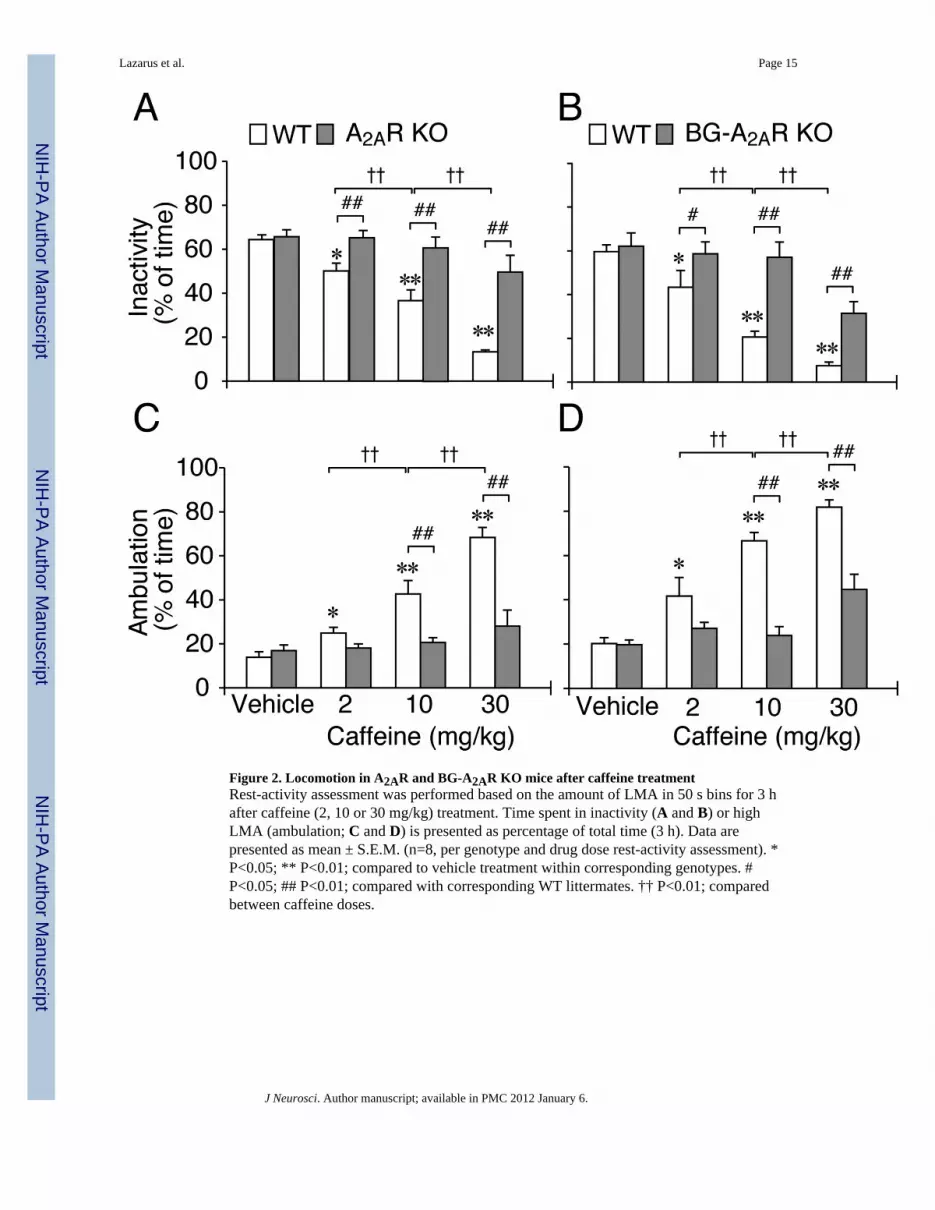
Figure 2. Locomotion in A2AR and BG-A2AR KO mice after caffeine treatmentRest-activity assessment was performed based on the amount of LMA in 50 s bins for 3 hafter caffeine (2, 10 or 30 mg/kg) treatment. Time spent in inactivity (A and B) or highLMA (ambulation; C and D) is presented as percentage of total time (3 h). Data arepresented as mean ± S.E.M. (n=8, per genotype and drug dose rest-activity assessment). *P<0.05; ** P<0.01; compared to vehicle treatment within corresponding genotypes. #P<0.05; ## P<0.01; compared with corresponding WT littermates. †† P<0.01; comparedbetween caffeine doses.
Lazarus et al. Page 15
J Neurosci. Author manuscript; available in PMC 2012 January 6.
NIH
-PA Author Manuscript
NIH
-PA Author Manuscript
NIH
-PA Author Manuscript

Figure 3. Arousal effect of caffeine was abolished in NAc-A2AR KO mice(A and B) Typical sections of conditional A2AR KO mice after injection with mCherry-expressing AAVs (WT; left photomicrograph) and AAV carrying Cre recombinase (NAc-A2AR KO; right photomicrograph) were stained with a goat polyclonal antibody againstA2AR (Santa Cruz) to visualize the presence (black circle) or loss (red dashed circle) ofA2ARs in the NAc. Immunostaining with a rabbit polyclonal antibody against D2R(Millipore) confirms the integrity of the NAc (black and blue circles) in the WT and NAc-A2AR KO mice (B). (C and D) The NAc-A2AR KO and WT mice were treated with caffeine(15 mg/kg, i.p.) or modafinil (45 mg/kg, i.p.). The time course (C) and total time (D) ofwakefulness for the first 3 h after caffeine treatment were assessed from EEG/EMGrecordings, as was modafinil-induced wakefulness for 3 h (right panel in D). Arrows in (C)indicate the time of caffeine injection. (E and F) Typical examples of EEG, EMG, LMA andhypnograms after administration of caffeine (15 mg/kg, i.p., upper panels), vehicle forcaffeine administration (middle panels) or modafinil (45 mg/kg, i.p., lower panels) in a WT(E) and NAc-A2AR KO (F) mouse. Arrows in (C, E and F) indicate the time of injection.Data are presented as the mean ± S.E.M. (n=4–5). * P<0.05; ** P<0.01; compared withvehicle treatment within corresponding AAV injection. ## P<0.01; compared with AAVtreatment. Scale bars: 500 µm in (A) and (B).
Lazarus et al. Page 16
J Neurosci. Author manuscript; available in PMC 2012 January 6.
NIH
-PA Author Manuscript
NIH
-PA Author Manuscript
NIH
-PA Author Manuscript

Figure 4. Site-specific deletion of A2ARs in rats by focal RNA interference(A) Generation of AAV vectors that contained short-hairpin RNA specific for the A2AR(shA2AR) and the red fluorescent protein mCherry as a reporter gene. (B) AAV-shA2ARvectors were stereotaxically injected into the A2AR-positive core and shell of the nucleusaccumbens (NAc) as well as into the olfactory tubercle (OLT), caudate-putamen (CPu), andglobus pallidus (GP). (C and D) Typical sections from rats injected bilaterally with AAVcarrying control shRNA (shCTRL) or shA2AR into the NAc were stained with mousemonoclonal antibody against A2AR. Immunoreactivity for A2AR is depleted selectively inthe NAc of the AAV-shA2AR-treated rats (D, dashed red circle), but it is unaffected in theAAV-shCTRL-treated rats (C, red circle). (E and F) NeuN-staining confirms the integrity ofNAc neurons (green circles) in AAV-shCTRL (E) and AAV-shA2AR injected rats (F). Scalebars: 500 µm in (C) and (E); also apply to (D) and (F), respectively.
Lazarus et al. Page 17
J Neurosci. Author manuscript; available in PMC 2012 January 6.
NIH
-PA Author Manuscript
NIH
-PA Author Manuscript
NIH
-PA Author Manuscript
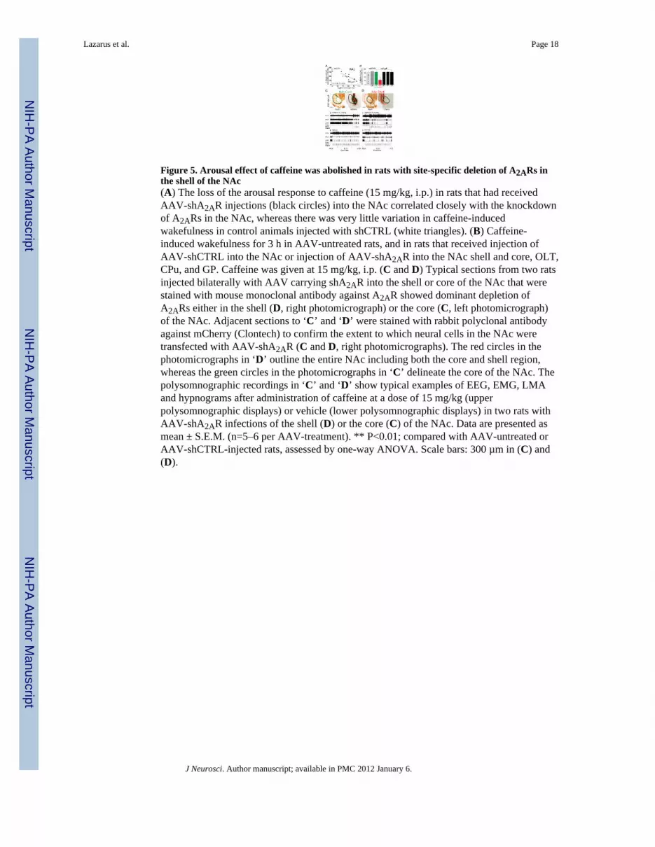
Figure 5. Arousal effect of caffeine was abolished in rats with site-specific deletion of A2ARs inthe shell of the NAc(A) The loss of the arousal response to caffeine (15 mg/kg, i.p.) in rats that had receivedAAV-shA2AR injections (black circles) into the NAc correlated closely with the knockdownof A2ARs in the NAc, whereas there was very little variation in caffeine-inducedwakefulness in control animals injected with shCTRL (white triangles). (B) Caffeine-induced wakefulness for 3 h in AAV-untreated rats, and in rats that received injection ofAAV-shCTRL into the NAc or injection of AAV-shA2AR into the NAc shell and core, OLT,CPu, and GP. Caffeine was given at 15 mg/kg, i.p. (C and D) Typical sections from two ratsinjected bilaterally with AAV carrying shA2AR into the shell or core of the NAc that werestained with mouse monoclonal antibody against A2AR showed dominant depletion ofA2ARs either in the shell (D, right photomicrograph) or the core (C, left photomicrograph)of the NAc. Adjacent sections to ‘C’ and ‘D’ were stained with rabbit polyclonal antibodyagainst mCherry (Clontech) to confirm the extent to which neural cells in the NAc weretransfected with AAV-shA2AR (C and D, right photomicrographs). The red circles in thephotomicrographs in ‘D’ outline the entire NAc including both the core and shell region,whereas the green circles in the photomicrographs in ‘C’ delineate the core of the NAc. Thepolysomnographic recordings in ‘C’ and ‘D’ show typical examples of EEG, EMG, LMAand hypnograms after administration of caffeine at a dose of 15 mg/kg (upperpolysomnographic displays) or vehicle (lower polysomnographic displays) in two rats withAAV-shA2AR infections of the shell (D) or the core (C) of the NAc. Data are presented asmean ± S.E.M. (n=5–6 per AAV-treatment). ** P<0.01; compared with AAV-untreated orAAV-shCTRL-injected rats, assessed by one-way ANOVA. Scale bars: 300 µm in (C) and(D).
Lazarus et al. Page 18
J Neurosci. Author manuscript; available in PMC 2012 January 6.
NIH
-PA Author Manuscript
NIH
-PA Author Manuscript
NIH
-PA Author Manuscript

Figure 6. A proposed wake-regulatory role of the A2AR-expressing neurons in the shell of thenucleus accumbens (NAc) that accounts for caffeine’s wake-promoting effectEndogenous somnogens such as adenosine and prostaglandin D2 (PGD2) promote sleep byactivating sleep-promoting neurons of the ventrolateral preoptic area (VLPO) (Saper et al.,2005; Saper et al., 2010; Urade, 2011) which, in a putative flip-flop arrangement, inhibit thearousal-promoting regions, including the lateral hypothalamus (LHA), thetuberomammillary hypothalamic nucleus (TMN), and the locus coeruleus (LC) in thebrainstem. Adenosine acting at A2ARs on medium spiny neurons in the shell of the NAc ishypothesized to exert inhibitory effects on the arousal systems via indirect (GABAergic andglutamatergic) pathways. Caffeine blocks the A2ARs in the NAc shell, thereby removing therestraint on the arousal systems to promote wakefulness.
Lazarus et al. Page 19
J Neurosci. Author manuscript; available in PMC 2012 January 6.
NIH
-PA Author Manuscript
NIH
-PA Author Manuscript
NIH
-PA Author Manuscript

NIH
-PA Author Manuscript
NIH
-PA Author Manuscript
NIH
-PA Author Manuscript
Lazarus et al. Page 20
Table 1
Coordinates for bilateral injections of AAV-Cre or AAV-mCherry in rats and mice according to the atlases ofPaxinos and Watson (Paxinos and Franklin, 2001; Paxinos and Watson, 2007).
Species1 Area2 Coordinates (mm)
Anterior to bregma Lateral to midline Below dural surface
Rat OLT 1.8 1.5 8
1.3 1.3 8
0.8 1.3 8
NAc 1.8 1.5 7
1.3 1.2 7
0.8 1 6.8
CPu 1.8 2.0 4.5
1.3 2.5 4
0.8 2.5 4.5
GP −0.4 2.2 7
−0.9 2.8 6
Mouse NAc 1 0.75 4.2
1.4 0.8 3.9
SI/HDB 0 1.4 4.6
1Each animal received 2 or 3 bilateral injections using different sets of coordinates.
2A2AR+ areas in the BG: CPu, caudate putamen; NAc, nucleus accumbens; OLT, olfactory tubercle and GP, globus pallidus. Arousal related areas
in the basal forebrain: SI, substantia inominata; HDB, horizontal limb of the diagonal band of Broca.
J Neurosci. Author manuscript; available in PMC 2012 January 6.