arousal regulation in affective disorders
TRANSCRIPT

Chapter 12
Arousal Regulation in AffectiveDisorders
Ulrich Hegerl1,2, Christian Sander1,2, Tilman Hensch11Department of Psychiatry and Psychotherapy, University of Leipzig, Leipzig, Germany;
2Research Centre of the German Depression Foundation, Leipzig, Germany
INTRODUCTION
Arousal, specifically brain arousal, fundamentally impacts all human behav-
iors (NIMH, 2012; Pfaff, Ribeiro, Matthews, & Kow, 2008). The long tradition
of analyzing the role of general arousal in normal and abnormal behavior and
cognition (Eysenck, 1990; Yerkes & Dodson, 1908; Zuckerman, 1979) has
been renewed by the Research Domain Criteria project of the National Insti-
tute of Mental Health, which implemented arousal as a basic dimension of
mental diseases (Cuthbert & Insel, 2013). In daily life, brain arousal has to be
precisely regulated to fulfill situational requirements. For example, brain
arousal must be heightened in case of potential danger or maintained during
cognitive tasks and reduced at bedtime.
In this chapter a new concept will be introduced that links affective dis-
orders and other psychiatric conditions to a disturbed regulation of brain
arousal. After a short overview on terminological difficulties, theoretical
models, and common means of assessments an electroencephalography
(EEG)-based assessment approach, the Vigilance Algorithm Leipzig (VIG-
ALL), will be described, facilitating research on brain arousal regulation.
Afterward, the arousal model of affective disorders will be described with
respect to depression, mania, and attention deficit/hyperactivity disorder
(ADHD).
DISTURBED AROUSAL REGULATION IN AFFECTIVEDISORDERS
Studies on disturbance of brain arousal in affective disorders have previously
focused on disturbed sleep. Although sleep disturbances are a very common
complaint in many psychiatric disorders, they are of special prominence in
affective disorders. Most patients suffering from depression experience some
Systems Neuroscience in Depression. http://dx.doi.org/10.1016/B978-0-12-802456-0.00012-1
Copyright © 2016 Elsevier Inc. All rights reserved. 341

kind of sleep disorder. Insomnia is typical for cases of unipolar depression,
where pathological sleep patterns with prolonged sleep latencies (Armitage,
2007; Kayumov et al., 2000; Tsuno, Besset, & Ritchie, 2005), disturbed sleep
continuity, and early awakenings are often seen, paralleled by an altered sleep
architecture with decreased slow-wave sleep and increased rapid eye move-
ment density in the first sleep cycle (Riemann, Berger, & Voderholzer, 2001;
Wichniak, Wierzbicka, & Jernajczyk, 2012). Hypersomnia, however, is a
symptom of atypical depression. In the past, sleep disorders have been
considered to be a mere symptom of depression, but evidence now suggests
that disrupted sleep plays a more central role in the pathophysiology of
depression (Baglioni et al., 2011; Riemann & Voderholzer, 2003) and should
be considered as another core symptom of depression (Nutt, Wilson, &
Paterson, 2008).
Furthermore, an association between sleep duration and mood has been
observed: prolonged time in bed and long sleep are associated with a decline in
mood, whereas short sleep duration and sleep deprivation have
mood-enhancing properties and in vulnerable persons may even trigger manic
episodes (Bauer et al., 2006; Wehr, 1989). This effect is further illustrated by
the effectiveness of therapeutic sleep deprivation, which in about 60% of
patients quickly reduces depressive symptoms (Giedke & Schwarzler, 2002).
However, this antidepressive effect often only lasts until the next sleep episode
is initiated, after which the depressive symptomatology resurfaces (Riemann,
Wiegand, Lauer, & Berger, 1993). Some evidence suggests that chronically
restricting sleep or time in bed might improve mood and depressive symptoms
(Dirksen & Epstein, 2008; Manber et al., 2008).
It would be unjustified, however, to focus on disrupted sleep alone, as the
wake state comprises the largest part of the day, and subjects suffering from
sleep problems may also exhibit dysregulation of arousal during wakefulness.
Tiredness and feelings of fatigue or weariness are typically reported by
depressed patients (Shen et al., 2011) and, especially when severe sleep
problems are experienced at night, patients consider themselves in grave need
of sleep. However, in contrast to their subjective feeling of exhaustion, patients
with typical depression do not show increased daytime sleepiness as assessed
by sleep onset latencies during the day. Their sleep onset latencies are pro-
longed, and patients report difficulties in relaxing (Kayumov et al., 2000;
Reynolds, Coble, Kupfer, & Holzer, 1982). Furthermore, they often carry signs
of higher noradrenergic and hypothalamicepituitaryeadrenal (HPA) axis ac-
tivity (Pariante & Lightman, 2008; Wong et al., 2000) and subjectively report
high inner tension. Hypersomnia, although often reported subjectively by
patients, is in most cases not verifiable with objective assessments (Dauvilliers,
Lopez, Ohayon, & Bayard, 2013; Nofzinger et al., 1991).
This points to an important terminological problem (Hegerl, 2014).
Expressions such as tiredness and fatigue are, in many cases, used to describe
completely distinct phenomena (Hegerl et al., 2013; Neu et al., 2008):
342 Systems Neuroscience in Depression

(a) tiredness/fatigue in the sense of sleepiness, i.e., increased tendency to get
drowsy or fall asleep and (b) tiredness/fatigue in the sense of exhaustion with a
tonically high inner tension and physiological arousal. It is the latter syndrome
that is typically found in patients with unipolar depression. Patients are
convinced that they could improve their condition by extended bed rest, yet in
many cases this only aggravates the underlying problem (as will be described
below).
Terminological blurs are also one reason for the lesser acknowledgment of
the wake stage differentiation within the research community. Different levels
of wakefulness have been conceptualized independently within several
research fields, e.g., psychophysiology, psychology, or cognitive neuroscience,
so that several terms and concepts exist, which are in parts synonymous,
contradictory, or simply referring to specific aspects of wakefulness. Some
terms are used to describe what is going on within the organism (physiological
phenomena) and others to describe patterns of nonverbal and verbal behavior
(behavioral phenomena). Brain arousal describes different states of physio-
logical activation, and a generalized central nervous system arousal is
considered to underlie all motivated behavior (Pfaff et al., 2008). Alertness
describes different behavioral patterns, especially different states of respon-
siveness and watchfulness to mostly external stimuli. The term vigilance itself
is used both at the behavioral and physiological levels (Oken, Salinsky, &
Elsas, 2006). Originally it was coined to describe a state of maximal physi-
ologic efficiency (Head, 1923) and later to describe the brain arousal levels
assessable with EEG during the transition period between sleep and wake-
fulness (vigilance levels; see below). Within cognitive psychology, however,
vigilance is frequently used to describe behavioral patterns, especially the
ability to maintain attention and alertness over long durations, and has become
a synonym for sustained attention in psychology. Accordingly, different as-
sessments have been put forward (see below).
In the context of this chapter, the term vigilance is used to describe
different states of global brain function, indicating different brain arousal
states. It is common knowledge that during sleep distinct sleep stages can be
separated using EEG. However, in sleep research the wake state is often
described simply as the non-sleeping state and is not further considered. Still,
the wake state can also be subdivided into several vigilance stages. Just as
sleep stages, these vigilance stages can best be classified using EEG
(see below). This knowledge has existed since the 1960s, when these sub-
stages were first described (Bente, 1964; Roth, 1961); however, it has
received much less research attention as compared to the sleep stages. Yet,
sleep and vigilance stages are both global functional physiological states of
the organism. Within these different levels an organism is more or less
sensitive to internal or external stimuli, and therefore performance on the
behavioral level differs according to the global functional state. Besides the
different vigilance stages the regulation of vigilance (or arousal respectively)
Arousal Regulation in Affective Disorders Chapter j 12 343
is of special importance. Life is a constant interaction with the environment,
and an organism needs to adapt to the environmental needs and challenges.
On one hand, it is important to adapt the degree of brain arousal to the
specific needs and challenges of the current environment to achieve goals and
avoid harm and potential death (e.g., avoiding or coping with dangerous
situations); on the other hand, it is important to actively shape or seek an
environment fitting to the current arousal level (e.g., find a safe place to
sleep). If the internal arousal regulation is disturbed, health, functioning, and
well-being are severely threatened.
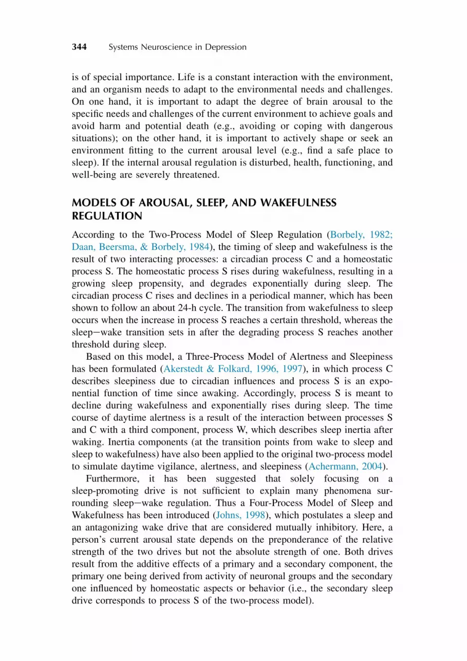
MODELS OF AROUSAL, SLEEP, AND WAKEFULNESSREGULATION
According to the Two-Process Model of Sleep Regulation (Borbely, 1982;
Daan, Beersma, & Borbely, 1984), the timing of sleep and wakefulness is the
result of two interacting processes: a circadian process C and a homeostatic
process S. The homeostatic process S rises during wakefulness, resulting in a
growing sleep propensity, and degrades exponentially during sleep. The
circadian process C rises and declines in a periodical manner, which has been
shown to follow an about 24-h cycle. The transition from wakefulness to sleep
occurs when the increase in process S reaches a certain threshold, whereas the
sleepewake transition sets in after the degrading process S reaches another
threshold during sleep.
Based on this model, a Three-Process Model of Alertness and Sleepiness
has been formulated (Akerstedt & Folkard, 1996, 1997), in which process C
describes sleepiness due to circadian influences and process S is an expo-
nential function of time since awaking. Accordingly, process S is meant to
decline during wakefulness and exponentially rises during sleep. The time
course of daytime alertness is a result of the interaction between processes S
and C with a third component, process W, which describes sleep inertia after
waking. Inertia components (at the transition points from wake to sleep and
sleep to wakefulness) have also been applied to the original two-process model
to simulate daytime vigilance, alertness, and sleepiness (Achermann, 2004).
Furthermore, it has been suggested that solely focusing on a
sleep-promoting drive is not sufficient to explain many phenomena sur-
rounding sleepewake regulation. Thus a Four-Process Model of Sleep and
Wakefulness has been introduced (Johns, 1998), which postulates a sleep and
an antagonizing wake drive that are considered mutually inhibitory. Here, a
person’s current arousal state depends on the preponderance of the relative
strength of the two drives but not the absolute strength of one. Both drives
result from the additive effects of a primary and a secondary component, the
primary one being derived from activity of neuronal groups and the secondary
one influenced by homeostatic aspects or behavior (i.e., the secondary sleep
drive corresponds to process S of the two-process model).
344 Systems Neuroscience in Depression
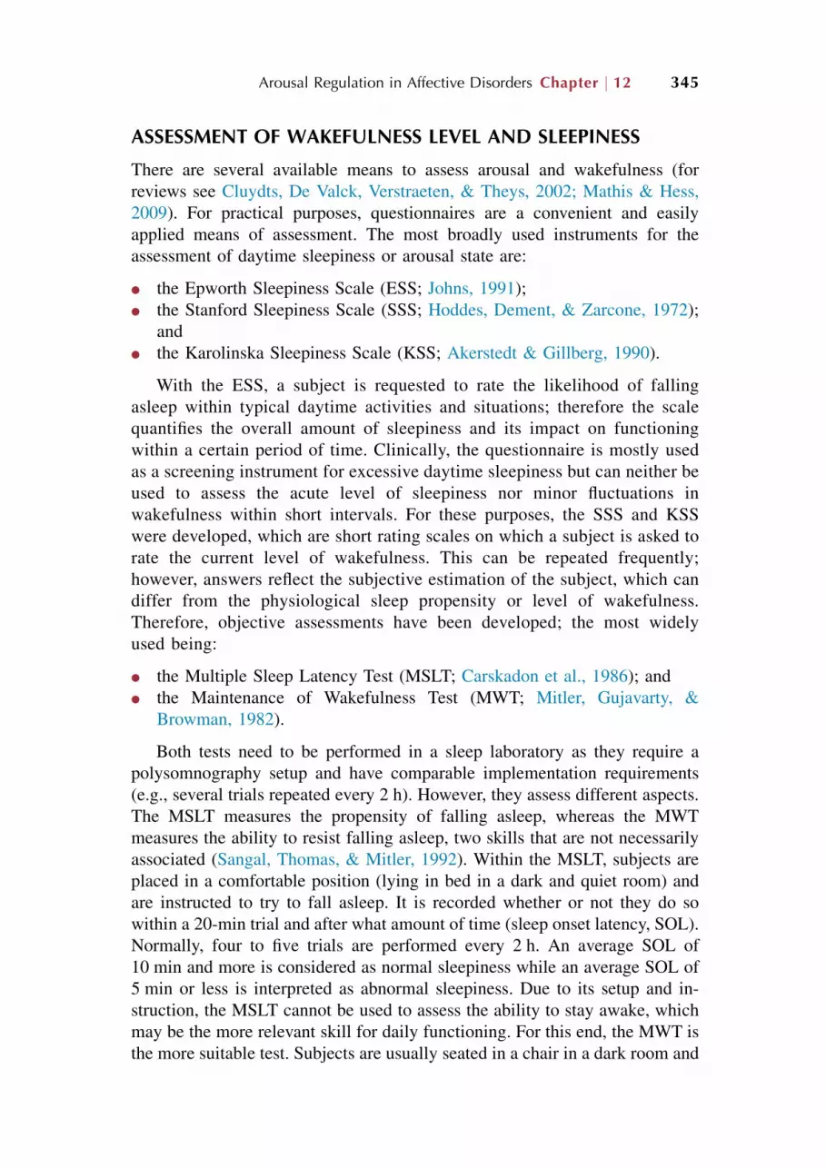
ASSESSMENT OF WAKEFULNESS LEVEL AND SLEEPINESS
There are several available means to assess arousal and wakefulness (for
reviews see Cluydts, De Valck, Verstraeten, & Theys, 2002; Mathis & Hess,
2009). For practical purposes, questionnaires are a convenient and easily
applied means of assessment. The most broadly used instruments for the
assessment of daytime sleepiness or arousal state are:
l the Epworth Sleepiness Scale (ESS; Johns, 1991);
l the Stanford Sleepiness Scale (SSS; Hoddes, Dement, & Zarcone, 1972);
and
l the Karolinska Sleepiness Scale (KSS; Akerstedt & Gillberg, 1990).
With the ESS, a subject is requested to rate the likelihood of falling
asleep within typical daytime activities and situations; therefore the scale
quantifies the overall amount of sleepiness and its impact on functioning
within a certain period of time. Clinically, the questionnaire is mostly used
as a screening instrument for excessive daytime sleepiness but can neither be
used to assess the acute level of sleepiness nor minor fluctuations in
wakefulness within short intervals. For these purposes, the SSS and KSS
were developed, which are short rating scales on which a subject is asked to
rate the current level of wakefulness. This can be repeated frequently;
however, answers reflect the subjective estimation of the subject, which can
differ from the physiological sleep propensity or level of wakefulness.
Therefore, objective assessments have been developed; the most widely
used being:
l the Multiple Sleep Latency Test (MSLT; Carskadon et al., 1986); and
l the Maintenance of Wakefulness Test (MWT; Mitler, Gujavarty, &
Browman, 1982).
Both tests need to be performed in a sleep laboratory as they require a
polysomnography setup and have comparable implementation requirements
(e.g., several trials repeated every 2 h). However, they assess different aspects.
The MSLT measures the propensity of falling asleep, whereas the MWT
measures the ability to resist falling asleep, two skills that are not necessarily
associated (Sangal, Thomas, & Mitler, 1992). Within the MSLT, subjects are
placed in a comfortable position (lying in bed in a dark and quiet room) and
are instructed to try to fall asleep. It is recorded whether or not they do so
within a 20-min trial and after what amount of time (sleep onset latency, SOL).
Normally, four to five trials are performed every 2 h. An average SOL of
10 min and more is considered as normal sleepiness while an average SOL of
5 min or less is interpreted as abnormal sleepiness. Due to its setup and in-
struction, the MSLT cannot be used to assess the ability to stay awake, which
may be the more relevant skill for daily functioning. For this end, the MWT is
the more suitable test. Subjects are usually seated in a chair in a dark room and
Arousal Regulation in Affective Disorders Chapter j 12 345

are instructed to stay awake during a 20- or 40-min trial, which are also
repeated (normally four trials every 2 h).
Apart from the MSLT and MWT, other objective but less
resource-demanding assessments of wakefulness and sleepiness are available.
One class is composed of performance tests, such as the Psychomotor Vigi-
lance Task (Dinges & Powell, 1985). In these tests, subjects are required to
perform an easy and monotonous task for an extended period of time, and it is
recorded whether or not they can successfully carry out that task. The per-
formance decrement is used as an indicator of sleepiness. Other tests are based
on electrophysiological measures, such as the Karolinska drowsiness test
(Akerstedt & Gillberg, 1990) or the alpha attenuation test (Stampi, Stone, &
Michimori, 1995). These tests estimate the current level of wakefulness by
comparing EEG activity between eyes open and eyes closed conditions. This
takes into account the distinct changes in EEG activity in both conditions with
increasing sleepiness (increase of alpha activity in eyes open condition versus
decreased alpha activity with eyes closed). Such assessment can be repeated
several times but are still not useful to continuously monitor fluctuations in
arousal. One approach to overcome this limitation has been the pupillographic
sleepiness test (Wilhelm et al., 2001), where the diameter of the pupil is
continuously monitored. Pupil diameter is inversely related to sleepiness, and
its variability over time is used as an indicator for arousal changes. Still, EEG
recordings provide the best temporal resolution and therefore remain the gold
standard to objectively assess sleep stages. They should also be the method of
choice to assess wakefulness fluctuations.
EEG VIGILANCE AS A MARKER OF BRAIN AROUSAL
As described above, different EEG-vigilance stages can be discerned not only
during sleep but also during wakefulness. According to original conceptions
from the 1960s (Bente, 1964; Roth, 1961), the following EEG-vigilance stages
can be observed during the transition from high alertness to relaxed wake-
fulness to drowsiness and finally sleep onset:
l Stage 0 is characterized by a desynchronized nonalpha EEG in the absence
of slow horizontal eye movements. This stage is typically seen during
activated states (e.g., reflecting mental effort).
l Stage A (with substages A1, A2, and A3) is characterized by dominant
alpha activity in the EEG trace and corresponds to relaxed wakefulness.
With decreasing vigilance there is a slight slowing of alpha activity and a
shift from occipital to more anterior regions.
l Stage B1 is again characterized by desynchronized non-alpha EEG with
low amplitude (similar spectral composition as stage 0) but often (yet not
necessarily) in the presence of slow horizontal eye movements. This stage
corresponds to drowsiness.
346 Systems Neuroscience in Depression

l Stage B2/3 is characterized by a non-alpha EEG with predominant theta/
delta activity and occasional occurrence of vertex waves. It reflects a state
of more severe drowsiness and marks the transition to sleep onset.
l Stage C is reached when sleep spindles or K-complexes are seen, which are
signs of sleep onset.
Studies on changes of EEG activity during the transition from active
wakefulness to sleep onset endorse these classifications (Benca et al., 1999;
Cantero, Atienza, & Salas, 2002; Corsi-Cabrera, Guevara, Del Rio-Portilla,
Arce, & Villanueva-Hernandez, 2000; De Gennaro, Ferrara, & Bertini, 2001;
De Gennaro, Ferrara, Curcio, et al., 2001, 2004; Kaida et al., 2006; Marzano
et al., 2007; Strijkstra, Beersma, Drayer, Halbesma, & Daan, 2003; Tsuno
et al., 2002). Research on wakefulness regulation has been hindered by the
absence of explicit scoring rules, which have long been established for the
scoring of sleep stages (Iber, Ancoli-Israel, Chessonn, & Quan, 2007;
Rechtschaffen & Kales, 1968). Furthermore, changes in wakefulness are not as
uniform as the typical changes in sleep stages, as subjects go back and forth
between vigilance stages with sometimes very short-lasting switches. There-
fore, a segmentation of the resting EEG into 30-s epochs, as is the consensus in
sleep medicine, is not feasible for scoring vigilance changes in a resting EEG,
where much shorter periods have to be considered. Visual classification of
vigilance stages in a resting EEG has therefore been an arduous and
time-consuming task, and the problem of inter- and intrarater reliability has
always been a crucial issue. Therefore, the development of computer-assisted
scoring algorithms has been essential for rejuvenating research interest in
wakefulness regulation. Several algorithms have been put forward (Khushaba,
Kodagoda, Lal, & Dissanayake, 2011; Sauvet et al., 2014; Shi, Duan, & Lu,
2013), in most cases with the aim of detecting drowsiness/sleep lapses during
task performance, e.g., for driver safety. A new algorithm (Vigilance Algorithm
Leipzig, VIGALL) has been introduced, which was developed with the specific
aim of facilitating research on brain arousal in psychiatric conditions and will
be described in detail in the next paragraph. In parallel to the development of
the VIGALL algorithm a pathogenetic concept has been formulated, linking
affective disorders to disturbances of brain arousal regulation. This brain
arousal model of affective disorders will be presented and discussed below.
THE VIGALL
The VIGALL (for manual and download go to http://research.uni-leipzig.de/
vigall/) is an EEG- and EOG-based tool that allows for objectively classi-
fying vigilance levels within multichannel EEG recordings by automatically
attributing one of the above-mentioned vigilance stages to a certain EEG
segment of preferably 1 s duration (Hegerl et al., 2008; Olbrich et al., 2009,
2011). The VIGALL algorithm takes into account EEG activity in different
Arousal Regulation in Affective Disorders Chapter j 12 347

frequency bands (delta/theta, alpha) and the cortical distribution of EEG
activity using EEG source localization approaches (Pascual-Marqui, Esslen,
Kochi, & Lehmann, 2002; Pascual-Marqui, Michel, & Lehmann, 1994).
However, EEG activity is characterized by high intraindividual stability and
large interindividual variability; therefore, VIGALL has adaptive features
concerning individual alpha peaks and amplitude levels (see Figure 1).
Before the vigilance classification is performed, VIGALL automatically
detects the individual alpha frequency and power from a representative epoch
of alpha activity. Some of the parameters (e.g., upper and lower border of the
alpha band) and decision criteria of the VIGALL (e.g., absolute alpha power
cutoff to classify an A-stage) are then adapted accordingly. It should be taken
into account that VIGALL should not be applied in cases of alpha variant
rhythms, major modifications due to drugs (e.g., anticholinergic drugs), or
diseases (e.g., severe Alzheimer disease). EEGs of children under the age of
10 (or older in the case of delayed maturation) should also be assessed with
caution.
FIGURE 1 Operational sequence and decision criteria of the VIGALL 2.0 algorithm. VIGALL
first screens the EEG trace for a 10-s epoch with prominent alpha activity (default range
7.5e12.5 Hz). For the respective epoch, the Alpha center of gravity frequency (ACF) and mean
alpha power at occipital sites (OAP) are calculated. ACF is then used to set the individual alpha
range (ACF "2 Hz); the delta/theta range is fixed to 2e7 Hz. Afterward, VIGALL calculates
spectral power (alpha versus delta/theta) in four regions of interest (ROI, i.e., the frontal, parietal,
temporal, and occipital lobe) using LORETA. For classification of vigilance stages, the OAP is used
to determine an individual alpha threshold, which is then used as a cutoff value in the classification
of A- and B2/3-stages. Segments not classified as A- or B2/3 stages are classified as B1- or 0 stages,
according to the presence of slow horizontal eye movements. If graphoelements indicating sleep
onset (sleep spindles or K-complexes) are present, segments are classified as stage C.
348 Systems Neuroscience in Depression

The very high temporal resolution of 1 s allows investigations on brain
arousal regulation by assessing the time course of vigilance levels during the
recording period. This regulation shows considerable interindividual differ-
ences (Huang et al., 2015). During eyes closed resting conditions of
15e20 min duration, most subjects show progressive declines to lower vigi-
lance stages (adaptive vigilance regulation). However, whereas some subjects
steadily remain in stages of high vigilance, others exhibit rapid declines within
only a few seconds. These patterns are called hyperstable or unstable vigilance
regulation, respectively (see Figure 2). This regulation of EEG vigilance has
been found to be intraindividually stable (Huang et al., 2015) with, at the same
0
(a)
(b)
A1
A2
A3
B1
B2/3
C
Recording Minute
Hyperstable Vigilance Regulation
0 1 2 3 4 5 6 7 8 9 10 11 12 13 14 15
0
A1
A2
A3
B1
B2/3
C
0
A1
A2
A3
B1
B2/3
C
Recording Minute
Unstable Vigilance Regulation
0 1 2 3 4 5 6 7 8 9 10 11 12 13 14 15
0
A1
A2
A3
B1
B2/3
C
FIGURE 2 Examples for vigilance time courses within 15 min of resting EEG with eyes closed.
(a) A subject with a hyperstable EEG-vigilance pattern, i.e., remaining continuously in A1-stages.
(b) A subject with an unstable EEG-vigilance pattern, i.e., immediate decline to drowsiness (B2/
3-stages) and sleep onset (stage C) within the seventh minute of recording. Red dots indicate the
respective vigilance stage that was assigned to each segment (resolution 1 s); gray vertical lines
mark segments containing artifacts.
Arousal Regulation in Affective Disorders Chapter j 12 349

time, considerable interindividual differences. This trait is modulated by many
individual and environmental factors such as sleep deficits, arousal-enhancing
substances, effort, motivation, and disease-related factors.
VIGALL 2.0 improves upon earlier versions of the algorithm, which have
been validated performing simultaneous EEGefunctional magnetic reso-
nance imaging (fMRI; Olbrich et al., 2009) as well as simultaneous EEGe
positron emission tomography (PET) studies (Guenther et al., 2011). These
studies showed that decreases in EEG-vigilance levels are associated with
increased metabolic activity in cortical but decreased activity in subcortical
areas. A further study relating vigilance stages to autonomous functions
found evidence that during lower vigilance stages heart rates and skin
conductance levels also decline (Olbrich et al., 2011). Further studies related
the vigilance stages to different behavioral parameters (Bekhtereva et al.,
2014; Minkwitz et al., 2011). Finally, the influence of different vigilance
stages on evoked potentials and reaction times has been demonstrated in an
oddball task (Huang, Spada, Sander, Hegerl, & Hensch, 2014). These basic
research studies also imply clinical relevance given the importance of
cognitive tests, MRI, and PET in diagnostic procedures, where VIGALL
might contribute to improve diagnostic accuracy by assessing arousal-
induced error variance.
THE AROUSAL REGULATION MODEL OF AFFECTIVEDISORDERS
The arousal regulation model of affective disorders (Hegerl & Hensch, 2014)
suggests that the level and the regulation of brain arousal are not only affected
by the environment but also that humans can create a more or less “arousing”
environment by their own behavior. In an autoregulatory manner, a more or
less stimulating environment can be actively created in order to reduce or
increase brain arousal levels. An everyday life example for such an autor-
egulatory attempt to increase arousal by enhancing external stimulation may
be overtired children, who often develop hyperactive, sensation seeking, and
talkative behavior. In contrast, states of tonic hyperarousal are often associated
with the tendency to withdraw and to avoid external stimulations, such as loud
music or social interactions.
The arousal regulation model builds on earlier concepts of the autor-
egulatory function of behavioral syndromes (Bente, 1964; Ulrich, 1994);
related concepts have also been suggested for ADHD (Weinberg & Brumback,
1990; Zentall & Zentall, 1983). In biological personality research, traits such
as extraversion (Eysenck, 1990) and sensation seeking (Zuckerman, 1979)
were interpreted as autoregulatory behavior in order to achieve an optimal
level of arousal. These personality traits were also suggested to reflect some
vulnerability to affective disorders and ADHD (Hensch, Herold, & Brocke,
2007; White, 1999).
350 Systems Neuroscience in Depression
The arousal regulation model assumes that in vulnerable subjects such
autoregulatory mechanisms can result in clinically relevant behavioral syn-
dromes. Several lines of arguments indicate that this is the case for manic and
depressed states as well as for ADHD.
Arousal Regulation in Mania
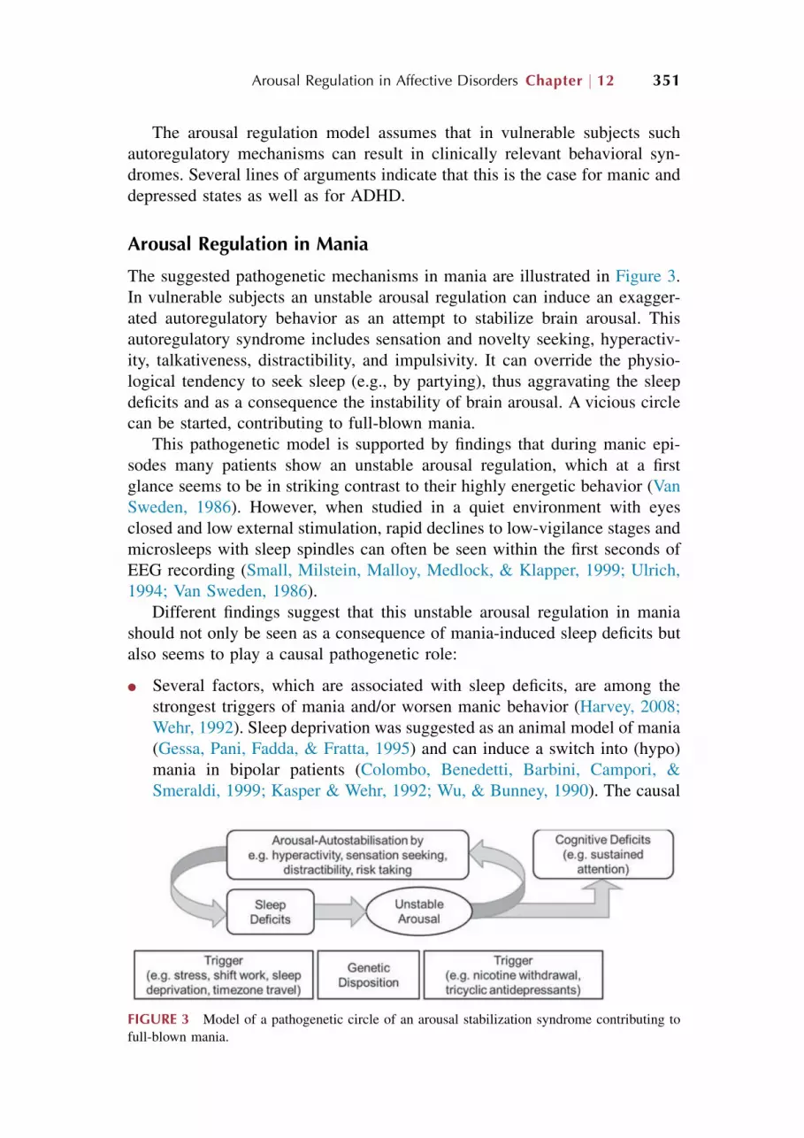
The suggested pathogenetic mechanisms in mania are illustrated in Figure 3.
In vulnerable subjects an unstable arousal regulation can induce an exagger-
ated autoregulatory behavior as an attempt to stabilize brain arousal. This
autoregulatory syndrome includes sensation and novelty seeking, hyperactiv-
ity, talkativeness, distractibility, and impulsivity. It can override the physio-
logical tendency to seek sleep (e.g., by partying), thus aggravating the sleep
deficits and as a consequence the instability of brain arousal. A vicious circle
can be started, contributing to full-blown mania.
This pathogenetic model is supported by findings that during manic epi-
sodes many patients show an unstable arousal regulation, which at a first
glance seems to be in striking contrast to their highly energetic behavior (Van
Sweden, 1986). However, when studied in a quiet environment with eyes
closed and low external stimulation, rapid declines to low-vigilance stages and
microsleeps with sleep spindles can often be seen within the first seconds of
EEG recording (Small, Milstein, Malloy, Medlock, & Klapper, 1999; Ulrich,
1994; Van Sweden, 1986).
Different findings suggest that this unstable arousal regulation in mania
should not only be seen as a consequence of mania-induced sleep deficits but
also seems to play a causal pathogenetic role:
l Several factors, which are associated with sleep deficits, are among the
strongest triggers of mania and/or worsen manic behavior (Harvey, 2008;
Wehr, 1992). Sleep deprivation was suggested as an animal model of mania
(Gessa, Pani, Fadda, & Fratta, 1995) and can induce a switch into (hypo)
mania in bipolar patients (Colombo, Benedetti, Barbini, Campori, &
Smeraldi, 1999; Kasper & Wehr, 1992; Wu, & Bunney, 1990). The causal
FIGURE 3 Model of a pathogenetic circle of an arousal stabilization syndrome contributing to
full-blown mania.
Arousal Regulation in Affective Disorders Chapter j 12 351

relevance of sleep reduction (e.g., as a consequence of obstructive sleep
apnea, bereavement, newborn infants, travel, or shift work) for triggering
mania is reviewed in Plante and Winkelman (2008). Furthermore, sleep
disturbances are by far the most robust early symptom of mania (median
prevalence of 77%; Jackson, Cavanagh, & Scott, 2003), and it has been
found that life events disturbing sleepewake rhythms can trigger or
aggravate (hypo)manic syndromes (Barbini, Bertelli, Colombo, & Smeraldi,
1996; Plante & Winkelman, 2008; Wehr, 1991).
l Stabilization of sleepewake rhythms is an established and important
element in behavioral therapies for bipolar affective disorders (Frank et al.,
2005; Leibenluft & Suppes, 1999; Riemann, Voderholzer, & Berger, 2002).
Additionally, extended bed rest and darkness as an add-on to the usual
treatment of acute mania resulted in a faster decrease of manic symptoms
in those patients with a recent (within 2 weeks) onset of mania (Barbini
et al., 2005; for similar results see also Nowlin-Finch, Altshuler, Szuba, &
Mintz, 1994; Wehr et al., 1998). These interventions can be expected to
stop the pathogenetic circle described in Figure 3 by stabilizing arousal
regulation.
l All standard antidepressants reduce the firing rate of locus coeruleus
(LC; see Arousal Regulation in Depression) and are often associated
with drowsiness as a side effect (Hensch et al., 2015). It might be that
this arousal reduction contributes to the antidepressants’ potential to
induce manic episodes. It is interesting that this switch risk has been
found, in some studies, to be higher in sedating antidepressants with
anticholinergic and antihistaminic properties such as tricyclic antide-
pressants than in less drowsiness-inducing selective serotonin reuptake
inhibitors (Gijsman, Geddes, Rendell, Nolen, & Goodwin, 2004; Peet,
1994).
l Arousal-enhancing psychostimulants are considered to be contraindicated
in mania by many clinicians. However, following the model presented
here, brain arousal-stabilizing drugs could be able to stop the manic vicious
circle. Indeed, when reviewing the literature, there is a lack of empirical
evidence for detrimental effects of psychostimulants in mania (Hegerl,
Sander, Olbrich, & Schoenknecht, 2009). If psychostimulants had a high
risk to induce or worsen mania, then the broad description of stimulants in
ADHD would result in considerable problems due to the high comorbidity
between ADHD and bipolar affective disorders (Singh, DelBello, Kowatch,
& Strakowski, 2006). Given the differential diagnostic difficulties in dis-
tinguishing both diseases, especially in children, one can assume that many
unrecognized or misdiagnosed pediatric manic patients have already
received stimulants. Therefore, a reanalysis of randomized trials with
stimulants in ADHD was carried out by the Food and Drug Administration,
demonstrating that psychotic or maniclike reactions occurred rarely (in
about 1 of 400 treated patients), and in the majority of cases (55 of 60), the
352 Systems Neuroscience in Depression

symptoms resolved within 2 days (Gelperin & Phelan, 2006; Mosholder,
2006; Phelan, 2006a, 2006b; Ross, 2006). Additionally, in a controlled
trial in children with ADHD and severe mood dysregulation, an
improvement in manic symptoms was observed under methylphenidate
treatment (Waxmonsky et al., 2008). Stimulants have already been
prescribed to bipolar patients as an add-on to mood stabilizers. In children
and adolescents, open trials (Kowatch, Sethuraman, Hume, Kromelis, &
Weinberg, 2003; Kummer & Teixeira, 2008) and controlled trials (Findling
et al., 2007; Scheffer, Kowatch, Carmody, & Rush, 2005; Zeni,
Tramontina, Ketzer, Pheula, & Rohde, 2009) showed that adding a psy-
chostimulant did not worsen but often improved manic symptomatology.
Mirroring these findings, in adults neither uncontrolled studies (Carlson,
Merlock, & Suppes, 2004; El-Mallakh, 2000; Fernandes & Petty, 2003;
Lydon & El-Mallakh, 2006; Nasr, Wendt, & Steiner, 2006) nor controlled
trials (Calabrese, Frye, Yang, & Ketter, 2014; Calabrese et al., 2010; Frye
et al., 2007; Ketter, Yang, & Frye, 2015) could detect a greater risk for
(hypo)manic symptoms in bipolar depressed patients treated with stimu-
lants as an add-on to mood stabilizers.
l In conclusion, stimulants in bipolar disorder seem to be relatively safe, and
there are even several case reports suggesting rapid antimanic effects of
psychostimulants (Beckmann & Heinemann, 1976; Garvey, Hwang,
Teubner-Rhodes, Zander, & Rhem, 1987; Max, Richards, & Hamdanallen,
1995). In a study by Bschor, Muller-Oerlinghausen, and Ulrich (2001),
improvement of manic symptoms occurred about 2 h after oral intake of
methylphenidate in a manic patient with signs of unstable EEG-vigilance
regulation. Three months later, when the patient was admitted anew, a
rapid antimanic effect was again shown after re-exposition to methylphe-
nidate. In contrast, no improvement was found in another manic patient
without this EEG pattern. Schoenknecht, Olbrich, Sander, Spindler, and
Hegerl (2010) reported a rapid response of an acutely manic patient to
monotherapy with the arousal-stabilizing drug modafinil. After 5 days the
patient had clearly improved and after stopping modafinil, treatment was
continued with lithium. Clinical improvement went along with a stabili-
zation of arousal regulation. Based on these findings, an international
randomized placebo-controlled clinical trial was started, analyzing the
effect of acute treatment with methylphenidate in mania (Kluge et al.,
2013; NCT01541605).
The arousal regulation model can also explain symptomatology and effects
of stimulants in ADHD. At the symptom level, manic episodes show
remarkable similarities to ADHD (Hegerl, Himmerich, Engmann, & Hensch,
2010), in line with the high comorbidity of bipolar disorder and ADHD.
Therefore, in the following ADHD will be discussed in the context of the
arousal regulation model.
Arousal Regulation in Affective Disorders Chapter j 12 353

Based on studies with skin conductance level and quantitative EEG, a
chronic hypoarousal had been postulated in ADHD for many years (reviewed
in Geissler, Romanos, Hegerl, & Hensch, 2014). Furthermore, unstable arousal
regulation has also been found in ADHD using MSLT (Geissler et al., 2014)
and VIGALL (Sander, Arns, Olbrich, & Hegerl, 2010). Additionally, a higher
subjective sleepiness was reported in ADHD and associated with symptom
severity (Cortese, Faraone, Konofal, & Lecendreux, 2009; Gamble, May,
Besing, Tankersly, & Fargason, 2013; Yoon, Jain, & Shapiro, 2012).
An unstable arousal regulation provides an explanation for the attention
deficits in ADHD, especially the well-documented deficits in continuous
performance tasks (Nichols & Waschbusch, 2004), and can also explain the
ADHD presentation specifiers according to the Diagnostic and Statistical
Manual of Mental Disorders, fifth edition (DSM-V) (and the ADHD subtypes
as their predecessors in the DSM-IV-TR). In the predominantly inattentive
presentation (formerly named predominantly inattentive subtype), the deficits
are explained by the instability of the arousal regulation. In the combined
presentation (subtype) with attention deficits and hyperactivity, additional
autoregulatory aspects come into play with hyperactivity, sensation, and
novelty seeking as an attempt to stabilize brain arousal. The arousal regulation
model is also able to explain why studies reported low prevalence rates for the
predominantly hyperactive-impulsive subtype or even called into question the
general validity of this subtype (Hurtig et al., 2007; Willcutt et al., 2012): The
model suggests that the unstable brain arousal is a core pathogenetic factor in
ADHD, which results in attention deficits. Hyperactivity, in contrast, does not
represent a primary disorder per se but rather an autoregulatory response,
which may or may not be present. Thus, a “pure” hyperactive subgroup should
not exist (for a more detailed discussion see Geissler et al., 2014).
In accordance with the model proposed for mania, the well-established
therapeutic effects of stimulants in ADHD can be explained by their
arousal-stabilizing effects, which interrupt the autoregulatory hyperactivity
and sensation-seeking behavior. Stimulants reduce attention deficits,
sensation-seeking behavior, and hyperactivity in patients with ADHD
(Pietrzak, Mollica, Maruff, & Snyder, 2006; Riccio, Waldrop, Reynolds, &
Lowe, 2001; Spencer et al., 2005), and symptom improvement is usually rapid
(Greydanus, Pratt, & Patel, 2007), similar to the quick antimanic effects
observed in case reports.
Arousal Regulation in Depression
While the behavioral syndrome in mania and ADHD is suggested to stabilize
an unstable brain arousal by creating a stimulating environment, the opposite
is suggested to be the case in depression. Depressed patients are characterized
by withdrawal from all interactions and sensation avoidance, possibly as a
reaction to a hyperstable arousal regulation. Furthermore, other symptoms of
354 Systems Neuroscience in Depression
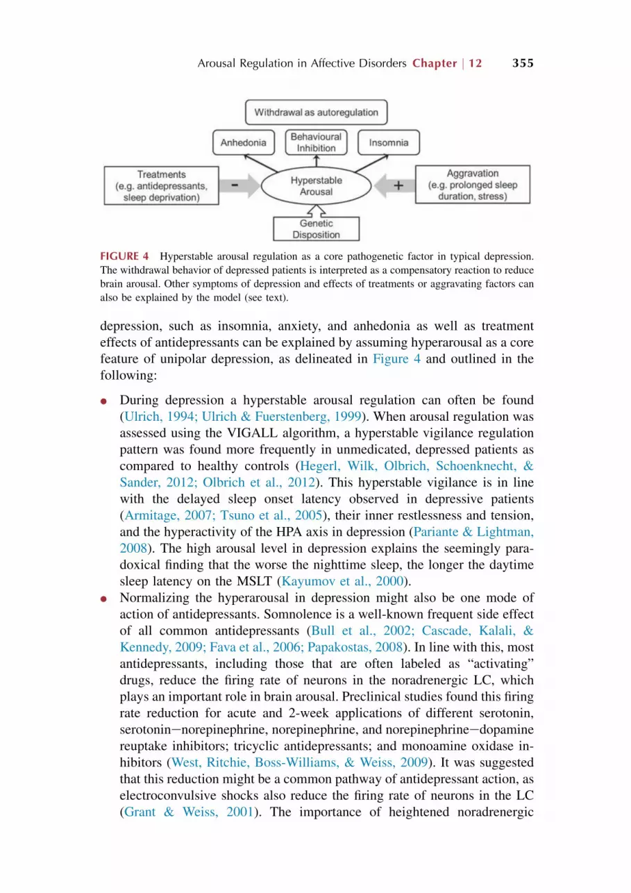
depression, such as insomnia, anxiety, and anhedonia as well as treatment
effects of antidepressants can be explained by assuming hyperarousal as a core
feature of unipolar depression, as delineated in Figure 4 and outlined in the
following:
l During depression a hyperstable arousal regulation can often be found
(Ulrich, 1994; Ulrich & Fuerstenberg, 1999). When arousal regulation was
assessed using the VIGALL algorithm, a hyperstable vigilance regulation
pattern was found more frequently in unmedicated, depressed patients as
compared to healthy controls (Hegerl, Wilk, Olbrich, Schoenknecht, &
Sander, 2012; Olbrich et al., 2012). This hyperstable vigilance is in line
with the delayed sleep onset latency observed in depressive patients
(Armitage, 2007; Tsuno et al., 2005), their inner restlessness and tension,
and the hyperactivity of the HPA axis in depression (Pariante & Lightman,
2008). The high arousal level in depression explains the seemingly para-
doxical finding that the worse the nighttime sleep, the longer the daytime
sleep latency on the MSLT (Kayumov et al., 2000).
l Normalizing the hyperarousal in depression might also be one mode of
action of antidepressants. Somnolence is a well-known frequent side effect
of all common antidepressants (Bull et al., 2002; Cascade, Kalali, &
Kennedy, 2009; Fava et al., 2006; Papakostas, 2008). In line with this, most
antidepressants, including those that are often labeled as “activating”
drugs, reduce the firing rate of neurons in the noradrenergic LC, which
plays an important role in brain arousal. Preclinical studies found this firing
rate reduction for acute and 2-week applications of different serotonin,
serotoninenorepinephrine, norepinephrine, and norepinephrineedopamine
reuptake inhibitors; tricyclic antidepressants; and monoamine oxidase in-
hibitors (West, Ritchie, Boss-Williams, & Weiss, 2009). It was suggested
that this reduction might be a common pathway of antidepressant action, as
electroconvulsive shocks also reduce the firing rate of neurons in the LC
(Grant & Weiss, 2001). The importance of heightened noradrenergic
FIGURE 4 Hyperstable arousal regulation as a core pathogenetic factor in typical depression.
The withdrawal behavior of depressed patients is interpreted as a compensatory reaction to reduce
brain arousal. Other symptoms of depression and effects of treatments or aggravating factors can
also be explained by the model (see text).
Arousal Regulation in Affective Disorders Chapter j 12 355
activity for depressive symptomatology is further supported by research,
suggesting symptoms of anhedonia and behavioral inhibition as a
consequence of noradrenergic hyperactivity (Stone, Lin, Sarfraz, &
Quartermain, 2011; West & Weiss, 2011).
l The antidepressant or depressiogenic effects of other drugs might also be
partly explained by their arousal-modulating effectsdfor example, the
depressiogenic effects of cholinesterase inhibitors in manic patients and
healthy subjects (Burt, Sachs, & Demopulos, 1999; Dagyte, Den Boer, &
Trentani, 2011; Janowsky, el-Yousef, Davis, & Sekerke, 1972) as well as
the antidepressant and promanic effects of anticholinergics (Drevets &
Furey, 2010; Fleischhacker et al., 1987; Furey & Drevets, 2006; Knable,
1989) or ketamine (Coyle & Laws, 2015). For more details see Hegerl and
Hensch (2014).
Sleep deprivation is another well-established treatment in depressive
episodes. Sleep deprivation in the second half of the night results in a pro-
nounced reduction of the depressive symptoms in more than half of the
patients (Benedetti & Colombo, 2011). Unfortunately, even a short recovery
nap can be followed by the immediate recurrence of depressive symptom-
atology (Berger, van Calker, & Riemann, 2003). The arousal model of af-
fective disorders provides a straightforward explanation for these effects:
sleep deprivation might increase sleep propensity and reduce the hyperstable
arousal regulation found in depressed patients and by that reduce the
autoregulatory behavior with withdrawal and sensation avoidance. Further
tentative support for this hypothesis comes from studies showing that patients
with higher subjective arousal ratings or higher sustained attention benefit
more from sleep deprivation (Bouhuys, Vandenburg, & Vandenhoofdakker,
1995; Wu et al., 1992).
l While sleep deprivation has antidepressant effects and can trigger mania,
sleep can have depressiogenic effects in vulnerable subjects. During
depressive episodes within major depressive and bipolar disorder, many
patients clearly describe their depression as most severe in the morning,
becoming less severe during the course of the day and the late evening.
Sleep may reduce sleep propensity and aggravate the hyperstable arousal
regulation, whereas being awake may increase sleep propensity and by that
reduce this arousal dysregulation during the course of the day. In line with
this reasoning, switches from mania to depression tend to occur during the
second half of the night, whereas switches from depression to mania tend
to occur during the afternoon and evening (Feldman-Naim, Turner, &
Leibenluft, 1997; Wilk & Hegerl, 2010).
l The relationship between changes in sleep and changes in mood has been
longitudinally analyzed in patients with bipolar affective disorder (Bauer
et al., 2006). Cross-correlations between self-reported sleep or bed rest and
mood demonstrated that in the majority of patients with a significant
356 Systems Neuroscience in Depression
cross-correlation, an increase in sleep or bed rest was followed by an in-
crease in depression, whereas a reduction of sleep or bed rest was followed
by hypomania or mania. Relationships between reduced sleep and (hypo)
mania as well as increased sleep and depressive symptoms have also been
reported by other research groups (Leibenluft, Albert, Rosenthal, & Wehr,
1996; Wehr, Goodwin, Wirz-Justice, Breitmaier, & Craig, 1982).
l Whereas the pathogenetic arousal model outlined here suggests stimulants
as a possible acute antimanic treatment, it would in consequence not
suggest stimulants in typical depression. Nonetheless, due to the energizing
properties of psychostimulants seen in healthy subjects, stimulants have
been tried as antidepressants in numerous studies with limited success.
This failure can be explained, because depressive symptoms only super-
ficially suggest sleepiness and a lack of drive, symptoms that might
respond to psychostimulants. As mentioned before, many patients with
typical depression do not suffer from sleepiness (tendency to fall asleep)
but rather from insomnia and decreased sleep drive (prolonged sleep la-
tencies) despite feelings of exhaustion and weariness. They also do not
suffer from lack of drive but rather from inhibition of drive (retardation)
combined with high inner tension. Thus, given the increased brain arousal
during depressive episodes, psychostimulants are unlikely to be helpful in
general. In line with this assumption, evidence for an antidepressant effect
of stimulants in patients with typical major depressive disorder is indeed
lacking. A Cochrane review (Candy, Jones, Williams, Tookman, & King,
2008) analyzed randomized, controlled trials from the past six decades,
testing antidepressant effects of stimulants as monotherapy or an add-on in
depression. With respect to clinical response, no significant effects could
be shown. Concerning the second outcome variable (reduction in depres-
sion symptoms), only one of the subanalyses by Candy et al. (2008)
showed a significant effect based on three studies. However, two of the
trials were on patients with serious comorbid diseases (HIV with hyper-
somnia and traumatic brain injury). The third trial included 20 outpatients
with “moderate depression” and “apathy, fatigue, [and] lack of energy”
(Elizur, Wintner, & Davidson, 1979). No diagnostic details are specified,
but considering the symptom of apathy, one might suspect atypical
depression in this group. In the years following the meta-analysis by Candy
et al. (2008) several studies on stimulants as an add-on to antidepressants
or mood stabilizers were published. In unipolar depression, an add-on of
methylphenidate (Ravindran et al., 2008), atomoxetine (Michelson et al.,
2007), or modafinil (Beck et al., 2010; Dunlop et al., 2007) failed, but two
further studies showed promising augmentation effects: an underpowered,
short-term trial with a modafinil add-on (Abolfazli et al., 2011; N ¼ 46;
6 weeks) and a study with lisdexamfetamine augmentation in partial
escitalopram responders with residual symptoms (Trivedi et al., 2013;
N ¼ 129; p¼ 0.09, significant at prespecified alpha level of 0.1). However,
Arousal Regulation in Affective Disorders Chapter j 12 357

two phase 3 studies with a lisdexamfetamine add-on again failed to meet
the primary efficacy end point, and the manufacturer accordingly stopped
the clinical development program (Shire, 2014). In bipolar depression,
three trials showed some add-on effect of (ar)modafinil (Calabrese et al.,
201, 2010; Frye et al., 2007), whereas two following multicenter studies
failed. Again, due to efficacy concerning the primary end point, the
manufacturer announced that the company would not proceed with regu-
latory filings for armodafinil for depression in bipolar I patients (Teva,
2013). Two other studies with lisdexamfetamine augmentation in bipolar
disorder (NCT01093963, NCT01131559) were also stopped by the
sponsor.
l To conclude, in uni- and bipolar depression there is no evidence for specific
antidepressant effects of stimulants as a monotherapy or add-on. However,
depression is a heterogeneous condition (Baumeister & Parker, 2012), and
the arousal regulation model may help to identify subgroups, who none-
theless might respond to stimulants. Uncontrolled studies point to a
possible antidepressant effect of stimulants in secondary depression
(Masand, Pickett, & Murray, 1991). Such secondary depressive syndromes
may be characterized by sleepiness and a lack of drive. Similarly, in
atypical depression, which is likely characterized by unstable arousal
regulation, stimulants might show some possible benefit.
CONCLUSION
Sufficient brain arousal is an important prerequisite for higher cognitive
functions, and a successful regulation of brain arousal according to environ-
mental and physiological needs is crucial for any organism. Disturbances in
brain arousal regulation have been linked to psychiatric conditions, especially
affective disorders. The outlined arousal regulation model gives an explanation
for different clinical phenomena, such as response to psychostimulants in
ADHD (established) and mania (currently being tested in an international
controlled trial; Kluge et al., 2013; NCT01541605), the antidepressant but
potentially mania-triggering effects of sleep deprivation, and the antimanic but
depressiogenic effects of increased sleep duration. The model allows for
deriving testable hypotheses concerning treatment response, thereby possibly
contributing to personalized treatment. The biomarker EEG-vigilance regu-
lation is suggested to stratify the highly heterogeneous category of major
depressive disorder into biologically more homogenous subgroups.
Clinicians should differentiate between exhaustion and weariness in the
context of chronic hyperarousal (as typically observed during a depressive
episode) and actual sleepiness. According to the arousal regulation model, it is
not surprising that current empirical studies have consistently shown that
treatments with stimulants are not effective in depression, as they would
358 Systems Neuroscience in Depression

further increase the hyperarousal. However, first evidence exists that stimu-
lants could be effective in mania or secondary depression where they would
improve the unstable brain arousal. In contrast, depressed patients who show
no signs of increased sleepiness when assessed with VIGALL might not only
profit from antidepressants, which reduce noradrenergic activity and thereby
arousal, but also from monitoring their sleeping behavior. Many depressed
patients have long bedtimes, trying to relax and to get as much sleep as
possible. However, in the case of a chronic hyperarousal, this behavior will
perpetuate symptomatology. Instead, patients without objectively confirmed
increased sleepiness should carefully monitor their bedtimes, reduce their
sleep duration, and avoid additional daytime sleep in order to counter their
hyperstable arousal regulation.
REFERENCES
Abolfazli, R., Hosseini, M., Ghanizadeh, A., Ghaleiha, A., Tabrizi, M.,
Raznahan, M.,…Akhondzadeh, S. (2011). Double-blind randomized parallel-group clinical trial
of efficacy of the combination fluoxetine plus modafinil versus fluoxetine plus placebo in the
treatment of major depression. Depression and Anxiety, 28(4), 297e302. http://dx.doi.org/
10.1002/da.20801.
Achermann, P. (2004). The two-process model of sleep regulation revisited. Aviation Space and
Environmental Medicine, 75(3 Suppl.), A37eA43.
Akerstedt, T., & Folkard, S. (1996). Predicting duration of sleep from the three process model of
regulation of alertness. Occupational and Environmental Medicine, 53(2), 136e141.
Akerstedt, T., & Folkard, S. (1997). The three-process model of alertness and its extension
to performance, sleep latency, and sleep length. Chronobiology International, 14(2),
115e123.
Akerstedt, T., & Gillberg, M. (1990). Subjective and objective sleepiness in the active individual.
International Journal of Neuroscience, 52(1e2), 29e37.
Armitage, R. (2007). Sleep and circadian rhythms in mood disorders. Acta Psychiatrica Scandi-
navica, 115(Suppl. 433), 104e115. http://dx.doi.org/10.1111/j.1600-0447.2007.00968.x.
Baglioni, C., Battagliese, G., Feige, B., Spiegelhalder, K., Nissen, C., Voderholzer, U.,…Riemann, D.
(2011). Insomnia as a predictor of depression: a meta-analytic evaluation of longitudinal epide-
miological studies. Journal of Affective Disorders, 135(1e3), 10e19. http://dx.doi.org/10.1016/
j.jad.2011.01.011.
Barbini, B., Benedetti, F., Colombo, C., Dotoli, D., Bernasconi, A., Cigala-Fulgosi,M.,…Smeraldi, E.
(2005). Dark therapy for mania: a pilot study. Bipolar Disorders, 7(1), 98e101. http://dx.doi.org/
10.1111/j.1399-5618.2004.00166.x.
Barbini, B., Bertelli, S., Colombo, C., & Smeraldi, E. (1996). Sleep loss, a possible factor in
augmenting manic episode. Psychiatry Research, 65(2), 121e125. http://dx.doi.org/10.1016/
S0165-1781(96)02909-5.
Bauer, M., Grof, P., Rasgon, N., Bschor, T., Glenn, T., & Whybrow, P. C. (2006). Temporal relation
between sleep and mood in patients with bipolar disorder. Bipolar Disorders, 8(2), 160e167.
http://dx.doi.org/10.1111/j.1399-5618.2006.00294.x.
Baumeister, H., & Parker, G. (2012). Meta-review of depressive subtyping models. Journal of
Affective Disorders, 139(2), 126e140. http://dx.doi.org/10.1016/j.jad.2011.07.015.
Arousal Regulation in Affective Disorders Chapter j 12 359
Beck, J., Hemmeter, U., Brand, S., Muheim, F., Hatzinger, M., & Holsboer-Trachsler, E. (2010).
Modafinil reduces microsleep during partial sleep deprivation in depressed patients. Journal of
Psychiatric Research, 44(13), 853e864. http://dx.doi.org/10.1016/j.jpsychires.2010.01.008.
pii:S0022-3956(10)00017-8.
Beckmann, H., & Heinemann, H. (1976). D-Amphetamine in manic syndrome. Arznelmittel-
Forschung, 26(6), 1185e1186.
Bekhtereva, V., Sander, C., Forschack, N., Olbrich, S., Hegerl, U., & Muller, M. M. (2014). Effects
of EEG-vigilance regulation patterns on early perceptual processes in human visual cortex.
Clinical Neurophysiology, 125(1), 98e107. http://dx.doi.org/10.1016/j.clinph.2013.06.019.
Benca, R. M., Obermeyer, W. H., Larson, C. L., Yun, B., Dolski, I.,
Kleist, K. D.,…Davidson, R. J. (1999). EEG alpha power and alpha power asymmetry in
sleep and wakefulness. Psychophysiology, 36(4), 430e436.
Benedetti, F., & Colombo, C. (2011). Sleep deprivation in mood disorders. Neuropsychobiology,
64(3), 141e151. http://dx.doi.org/10.1159/000328947.
Bente, D. (1964). Vigilanz, dissoziative Vigilanzverschiebung und Insuffizienz des Vigilitatstonus.
In H. Kranz, & K. Heinrich (Eds.), Begleitwirkungen und Mißerfolge der psychiatrischen
Pharmakotherapie. Stuttgart: Georg Thieme Verlag. pp. 13e28.
Berger, M., van Calker, D., & Riemann, D. (2003). Sleep and manipulations of the sleep-wake
rhythm in depression. Acta Psychiatrica Scandinavica Supplementum, 418, 83e91.
Borbely, A. A. (1982). A two process model of sleep regulation.Human Neurobiology, 1(3), 195e204.
Bouhuys, A. L., Vandenburg, W., & Vandenhoofdakker, R. H. (1995). The relationship between
tiredness prior to sleep-deprivation and the antidepressant response to sleep-deprivation in
depression. Biological Psychiatry, 37(7), 457e461.
Bschor, T., Muller-Oerlinghausen, B., & Ulrich, G. (2001). Decreased level of EEG-vigilance in
acute mania as a possible predictor for a rapid effect of methylphenidate: a case study. Clinical
Electroencephalography, 32(1), 36e39.
Bull, S. A., Hunkeler, E. M., Lee, J. Y., Rowland, C. R., Williamson, T. E., Schwab, J. R., &
Hurt, S. W. (2002). Discontinuing or switching selective serotonin-reuptake inhibitors. Annals
of Pharmacotherapy, 36(4), 578e584.
Burt, T., Sachs, G. S., & Demopulos, C. (1999). Donepezil in treatment-resistant bipolar disorder.
Biological Psychiatry, 45(8), 959e964. pii:S0006-3223(98)00320-5.
Calabrese, J. R., Frye, M. A., Yang, R., & Ketter, T. A. (2014). Efficacy and safety of adjunctive
armodafinil in adults with major depressive episodes associated with bipolar I disorder: a
randomized, double-blind, placebo-controlled, multicenter trial. Journal of Clinical Psychi-
atry, 75(10), 1054e1061. http://dx.doi.org/10.4088/JCP.13m08951.
Calabrese, J. R., Ketter, T. A., Youakim, J. M., Tiller, J. M., Yang, R., & Frye, M. A. (2010).
Adjunctive armodafinil for major depressive episodes associated with bipolar I disorder: a
randomized, multicenter, double-blind, placebo-controlled, proof-of-concept study. Journal of
Clinical Psychiatry, 71(10), 1363e1370. http://dx.doi.org/10.4088/JCP.09m05900gry.
Candy, M., Jones, L., Williams, R., Tookman, A., & King, M. (2008). Psychostimulants for
depression. Cochrane Database of Systematic Reviews, 2, CD006722. http://dx.doi.org/10.
1002/14651858.CD006722.pub2.
Cantero, J. L., Atienza, M., & Salas, R. M. (2002). Human alpha oscillations in wakefulness,
drowsiness period, and REM sleep: different electroencephalographic phenomena within the
alpha band. Neurophysiologie Clinique, 32(1), 54e71.
Carlson, P. J., Merlock, M. C., & Suppes, T. (2004). Adjunctive stimulant use in patients with
bipolar disorder: treatment of residual depression and sedation. Bipolar Disorders, 6(5),
416e420. http://dx.doi.org/10.1111/j.1399-5618.2004.00132.x.
360 Systems Neuroscience in Depression

Carskadon, M. A., Dement, W. C., Mitler, M. M., Roth, T., Westbrook, P. R., & Keenan, S. (1986).
Guidelines for the multiple sleep latency test (MSLT): a standard measure of sleepiness. Sleep,
9(4), 519e524.
Cascade, E., Kalali, A. H., & Kennedy, S. H. (2009). Real-world data on SSRI antidepressant side
effects. Psychiatry (Edgmont), 6(2), 16e18.
Cluydts, R., De Valck, E., Verstraeten, E., & Theys, P. (2002). Daytime sleepiness and its eval-
uation. Sleep Medicine Reviews, 6(2), 83e96.
Colombo, C., Benedetti, F., Barbini, B., Campori, E., & Smeraldi, E. (1999). Rate of switch from
depression into mania after therapeutic sleep deprivation in bipolar depression. Psychiatry
Research, 86(3), 267e270. pii:S0165-1781(99)00036-0.
Corsi-Cabrera, M., Guevara, M. A., Del Rio-Portilla, Y., Arce, C., & Villanueva-Hernandez, Y.
(2000). EEG bands during wakefulness, slow-wave and paradoxical sleep as a result of
principal component analysis in man. Sleep, 23(6), 738e744.
Cortese, S., Faraone, S. V., Konofal, E., & Lecendreux, M. (2009). Sleep in children
with attention-deficit/hyperactivity disorder: meta-analysis of subjective and objective
studies. Journal of the American Academy of Child and Adolescent Psychiatry, 48(9),
894e908.
Coyle, C. M., & Laws, K. R. (2015). The use of ketamine as an antidepressant: a systematic review
and meta-analysis. Human Psychopharmacology, 30(3), 152e163. http://dx.doi.org/10.1002/
hup.2475.
Cuthbert, B. N., & Insel, T. R. (2013). Toward the future of psychiatric diagnosis: the seven pillars
of RDoC. BMC Medicine, 11, 126. http://dx.doi.org/10.1186/1741-7015-11-126.
Daan, S., Beersma, D. G., & Borbely, A. A. (1984). Timing of human sleep: recovery process gated
by a circadian pacemaker. American Journal of Physiology, 246(2 Pt 2), R161eR183.
Dagyte, G., Den Boer, J. A., & Trentani, A. (2011). The cholinergic system and depression.
Behavioural Brain Research, 221(2), 574e582. http://dx.doi.org/10.1016/j.bbr.2010.02.023.
pii:S0166-4328(10)00123-3.
Dauvilliers, Y., Lopez, R., Ohayon, M., & Bayard, S. (2013). Hypersomnia and depressive
symptoms: methodological and clinical aspects. BMC Medicine, 11, 78. http://dx.doi.org/10.
1186/1741-7015-11-78.
De Gennaro, L., Ferrara, M., & Bertini, M. (2001). The boundary between wakefulness and sleep:
quantitative electroencephalographic changes during the sleep onset period. Neuroscience,
107(1), 1e11.
De Gennaro, L., Ferrara, M., Curcio, G., & Cristiani, R. (2001). Antero-posterior EEG changes
during the wakefulness-sleep transition. Clinical Neurophysiology, 112(10), 1901e1911.
De Gennaro, L., Vecchio, F., Ferrara, M., Curcio, G., Rossini, P. M., & Babiloni, C. (2004).
Changes in fronto-posterior functional coupling at sleep onset in humans. Journal of Sleep
Research, 13(3), 209e217. http://dx.doi.org/10.1111/j.1365-2869.2004.00406.x.
Dinges, D. F., & Powell, J. W. (1985). Microcomputer analyses of performance on a portable,
simple visual RT task during sustained operations. Behavior Research Methods Instruments
and Computers, 17(6), 652e655.
Dirksen, S. R., & Epstein, D. R. (2008). Efficacy of an insomnia intervention on fatigue, mood and
quality of life in breast cancer survivors. Journal of Advanced Nursing, 61(6), 664e675. http://
dx.doi.org/10.1111/j.1365-2648.2007.04560.x.
Drevets, W. C., & Furey, M. L. (2010). Replication of scopolamine’s antidepressant efficacy
in major depressive disorder: a randomized, placebo-controlled clinical trial. Biological
Psychiatry, 67(5), 432e438. http://dx.doi.org/10.1016/j.biopsych.2009.11.021. pii:S0006-
3223(09)01414-0.
Arousal Regulation in Affective Disorders Chapter j 12 361

Dunlop, B. W., Crits-Christoph, P., Evans, D. L., Hirschowitz, J., Solvason, H. B.,
Rickels, K.,…Ninan, P. T. (2007). Coadministration of modafinil and a selective serotonin
reuptake inhibitor from the initiation of treatment of major depressive disorder with fatigue
and sleepiness: a double-blind, placebo-controlled study. Journal of Clinical Psychophar-
macology, 27(6), 614e619. http://dx.doi.org/10.1097/jcp.0b013e31815abefb.
El-Mallakh, R. S. (2000). An open study of methylphenidate in bipolar depression. Bipolar Dis-
orders, 2(1), 56e59.
Elizur, A., Wintner, I., & Davidson, S. (1979). The clinical and psychological effects of
pemoline in depressed patientsea controlled study. International Pharmacopsychiatry,
14(3), 127e134.
Eysenck, H. J. (1990). Biological dimensions of personality. In L. A. Pervin (Ed.), Handbook of
personality: Theory and research (pp. 244e276). New York: Guilford Press (Reprinted from:
Not in File).
Fava, M., Graves, L. M., Benazzi, F., Scalia, M. J., Iosifescu, D. V., Alpert, J. E., &
Papakostas, G. I. (2006). A cross-sectional study of the prevalence of cognitive and physical
symptoms during long-term antidepressant treatment. Journal of Clinical Psychiatry, 67(11),
1754e1759.
Feldman-Naim, S., Turner, E. H., & Leibenluft, E. (1997). Diurnal variation in the direction of
mood switches in patients with rapid-cycling bipolar disorder. Journal of Clinical Psychiatry,
58(2), 79e84.
Fernandes, P. P., & Petty, F. (2003). Modafinil for remitted bipolar depression with hypersomnia.
Annals of Pharmacotherapy, 37(12), 1807e1809. http://dx.doi.org/10.1345/aph.1D226.
Findling, R. L., Short, E. J., McNamara, N. K., Demeter, C. A., Stansbrey, R. J.,
Gracious, B. L.,…Calabrese, J. R. (2007). Methylphenidate in the treatment of children and
adolescents with bipolar disorder and attention-deficit/hyperactivity disorder. Journal of the
American Academy of Child and Adolescent Psychiatry, 46(11), 1445e1453. http://dx.doi.org/
10.1097/chi.0b013e31814b8d3b.
Fleischhacker, W. W., Barnas, C., Gunther, V., Meise, U., Stuppack, C., & Unterweger, B. (1987).
Mood-altering effects of biperiden in healthy volunteers. Journal of Affective Disorders, 12(2),
153e157.
Frank, E., Kupfer, D. J., Thase, M. E., Mallinger, A. G., Swartz, H. A.,
Fagiolini, A. M.,…Monk, T. (2005). Two-year outcomes for interpersonal and social rhythm
therapy in individuals with bipolar I disorder. Archives of General Psychiatry, 62(9),
996e1004. http://dx.doi.org/10.1001/archpsyc.62.9.996.
Frye, M. A., Grunze, H., Suppes, T., McElroy, S. L., Keck, P. E., Jr., Walden, J.,…Post, R. M.
(2007). A placebo-controlled evaluation of adjunctive modafinil in the treatment of bipolar
depression. American Journal of Psychiatry, 164(8), 1242e1249. http://dx.doi.org/10.1176/
appi.ajp.2007.06060981.
Furey, M. L., & Drevets, W. C. (2006). Antidepressant efficacy of the antimuscarinic drug
scopolamine: a randomized, placebo-controlled clinical trial. Archives of General Psychiatry,
63(10), 1121e1129. http://dx.doi.org/10.1001/archpsyc.63.10.1121.
Gamble, K. L., May, R. S., Besing, R. C., Tankersly, A. P., & Fargason, R. E. (2013). Delayed sleep
timing and symptoms in adults with attention-deficit/hyperactivity disorder: a controlled
actigraphy study. Chronobiology International, 30(4), 598e606. http://dx.doi.org/10.3109/
07420528.2012.754454.
Garvey, M. J., Hwang, S., Teubner-Rhodes, D., Zander, J., & Rhem, C. (1987). Dextroamphet-
amine treatment of mania. Journal of Clinical Psychiatry, 48(10), 412e413.
362 Systems Neuroscience in Depression

Geissler, J., Romanos, M., Hegerl, U., & Hensch, T. (2014). Hyperactivity and sensation
seeking as autoregulatory attempts to stabilize brain arousal in ADHD and mania? Attention
Deficit and Hyperactive Disorders, 6(3), 159e173. http://dx.doi.org/10.1007/s12402-014-
0144-z.
Gelperin, K., & Phelan, K. (2006). Psychiatric adverse events associated with drug treatment of
ADHD: Review of postmarketing safety data. FDA Report PID D050243. US Food and Drug
Administration. March 3, 2006. Available at www.fda.gov/ohrms/dockets/ac/06/briefing/2006-
4210b_11_01_AdverseEvents.pdf.
Gessa, G. L., Pani, L., Fadda, P., & Fratta, W. (1995). Sleep deprivation in the rat: an animal model
of mania. European Neuropsychopharmacology, 5(Suppl), 89e93. 10.1016/0924-977X(95)
00023-I.
Giedke, H., & Schwarzler, F. (2002). Therapeutic use of sleep deprivation in depression. Sleep
Medicine Reviews, 6(5), 361e377.
Gijsman, H. J., Geddes, J. R., Rendell, J. M., Nolen, W. A., & Goodwin, G. M. (2004). Anti-
depressants for bipolar depression: a systematic review of randomized, controlled trials.
American Journal of Psychiatry, 161(9), 1537e1547. http://dx.doi.org/10.1176/appi.ajp.
161.9.1537.
Grant, M. M., & Weiss, J. M. (2001). Effects of chronic antidepressant drug administration and
electroconvulsive shock on locus coeruleus electrophysiologic activity. Biological Psychiatry,
49(2), 117e129. pii:S0006-3223(00)00936-7.
Greydanus, D. E., Pratt, H. D., & Patel, D. R. (2007). Attention deficit hyperactivity disorder
across the lifespan: the child, adolescent, and adult. Disease-a-month, 53(2), 70e131.
Guenther, T., Schonknecht, P., Becker, G., Olbrich, S., Sander, C., Hesse, S.,… Sabri, O. (2011).
Impact of EEG-vigilance on brain glucose uptake measured with [(18)F]FDG and PET in
patients with depressive episode or mild cognitive impairment. NeuroImage, 56(1), 93e101.
http://dx.doi.org/10.1016/j.neuroimage.2011.01.059. pii:S1053-8119(11)00095-4.
Harvey, A. G. (2008). Sleep and circadian rhythms in bipolar disorder: seeking synchrony, har-
mony, and regulation. American Journal of Psychiatry, 165(7), 820e829. http://dx.doi.org/10.
1176/appi.ajp.2008.08010098.
Head, H. (1923). The conception of nervous and mental energy: II. vigilance: a physiological state
of the nervous system. British Journal of Psychology, 14, 126e147.
Hegerl, U. (2014). Largely unnoticed flaws in the fundamentals of depression diagnosis: the
semantics of core symptoms. Australian and New Zealand Journal of Psychiatry, 48(12),
1166. http://dx.doi.org/10.1177/0004867414559550.
Hegerl, U., & Hensch, T. (2014). The vigilance regulation model of affective disorders and ADHD.
Neuroscience and Biobehavioral Reviews, 44, 45e57. http://dx.doi.org/10.1016/j.neubiorev.
2012.10.008.
Hegerl, U., Himmerich, H., Engmann, B., & Hensch, T. (2010). Mania and attention-deficit/
hyperactivity disorder: common symptomatology, common pathophysiology and common
treatment? Current Opinion in Psychiatry, 23(1), 1e7. http://dx.doi.org/10.1097/YCO.
0b013e328331f694.
Hegerl, U., Lam, R. W., Malhi, G. S., McIntyre, R. S., Demyttenaere, K., Mergl, R., & Gorwood, P.
(2013). Conceptualising the neurobiology of fatigue. Australian and New Zealand Journal of
Psychiatry, 47(4), 312e316. http://dx.doi.org/10.1177/0004867413481505.
Hegerl, U., Sander, C., Olbrich, S., & Schoenknecht, P. (2009). Are psychostimulants a treatment
option in mania? Pharmacopsychiatry, 42(5), 169e174. http://dx.doi.org/10.1055/s-0029-
1220888.
Arousal Regulation in Affective Disorders Chapter j 12 363

Hegerl, U., Stein, M., Mulert, C., Mergl, R., Olbrich, S., Dichgans, E.,… Pogarell, O.
(2008). EEG-vigilance differences between patients with borderline personality disor-
der, patients with obsessive-compulsive disorder and healthy controls. European Ar-
chives of Psychiatry and Clinical Neuroscience, 258(3), 137e143. http://dx.doi.org/10.
1007/s00406-007-0765-8.
Hegerl, U., Wilk, K., Olbrich, S., Schoenknecht, P., & Sander, C. (2012). Hyperstable regulation of
vigilance in patients with major depressive disorder. World Journal of Biological Psychiatry,
13(6), 436e446.
Hensch, T., Blume, A., Bottger, D., Sander, C., Niedermeier, N., & Hegerl, U. (2015). Yawning in
depression: Worth looking into. Pharmacopsychiatry, 48(3), 118e120. http://dx.doi.org/10.
1055/s-0035-1545332.
Hensch, T., Herold, U., & Brocke, B. (2007). An electrophysiological endophenotype of
hypomanic and hyperthymic personality. Journal of Affective Disorders, 101(1e3), 13e26.
http://dx.doi.org/10.1016/j.jad.2006.11.018. pii:S0165-0327(06)00500-3.
Hoddes, E., Dement, W., & Zarcone, V. (1972). The development and use of the Stanford
sleepiness scale (SSS). Psychophysiology, 9, 150e151.
Huang, J., Sander, C., Jawinski, P., Ulke, C., Spada, J., Hegerl, U., & Hensch, T. (2015). Stability
of brain arousal regulation as assessed with EEG vigilance. Neuropsychiatric Electrophysi-
ology, 1, 13. http://dx.doi.org/10.1186/s40810-015-0013-9.
Huang, J., Spada, J., Sander, C., Hegerl, U., & Hensch, T. (2014). Auditory event related potentials
during different vigilance stages. Paper presented at the Leipzig Research Festival for Life
Sciences 2014, Leipzig.
Hurtig, T., Ebeling, H., Taanila, A., Miettunen, J., Smalley, S. L., McGough, J. J.,…Moilanen, I. K.
(2007). ADHD symptoms and subtypes: relationship between childhood and adolescent symp-
toms. Journal of American Academy of Child and Adolescent Psychiatry, 46(12), 1605e1613.
http://dx.doi.org/10.1097/chi.0b013e318157517a. pii:S0890-8567(09)61872-4.
Iber, C., Ancoli-Israel, S., Chessonn, A., & Quan, S. F. (2007). The AASM manual for the scoring
of sleep and associated events: rules, terminology and technical specifications (1st ed.).
Westchester, IL: American Academy of Sleep Medicine.
Jackson, A., Cavanagh, J., & Scott, J. (2003). A systematic review of manic and depressive pro-
dromes. Journal of Affective Disorders, 74(3), 209e217.
Janowsky, D. S., el-Yousef, M. K., Davis, J. M., & Sekerke, H. J. (1972). A cholinergic-adrenergic
hypothesis of mania and depression. Lancet, 2(7778), 632e635.
Johns, M. W. (1991). A new method for measuring daytime sleepiness: the Epworth sleepiness
scale. Sleep, 14(6), 540e545.
Johns, M. (1998). Rethinking the assessment of sleepiness. Sleep Medicine Reviews, 2(1), 3e15.
Kaida, K., Takahashi, M., Akerstedt, T., Nakata, A., Otsuka, Y., Haratani, T., & Fukasawa, K.
(2006). Validation of the Karolinska sleepiness scale against performance and EEG variables.
Clinical Neurophysiology, 117(7), 1574e1581.
Kasper, S., & Wehr, T. A. (1992). The role of sleep and wakefulness in the genesis of depression
and mania. L’Encephale, 18(Spec No 1), 45e50.
Kayumov, L., Rotenberg, V., Buttoo, K., Auch, C., Pandi-Perumal, S. R., & Shapiro, C. M.
(2000). Interrelationships between nocturnal sleep, daytime alertness, and sleepiness: two
types of alertness proposed. Journal of Neuropsychiatry and Clinical Neuroscience, 12(1),
86e90.
Ketter, T. A., Yang, R., & Frye, M. A. (2015). Adjunctive armodafinil for major depressive epi-
sodes associated with bipolar I disorder. Journal of Affective Disorders, 181, 87e91. http://dx.
doi.org/10.1016/j.jad.2015.04.012.
364 Systems Neuroscience in Depression

Khushaba, R. N., Kodagoda, S., Lal, S., & Dissanayake, G. (2011). Driver drowsiness classi-
fication using fuzzy wavelet-packet-based feature-extraction algorithm. IEEE Transactions
on Biomedical Engineering, 58(1), 121e131. http://dx.doi.org/10.1109/tbme.2010.
2077291.
Kluge, M., Hegerl, U., Sander, C., Dietzel, J., Mergl, R., Bitter, I.,…Lopez-Garcia, P. (2013).
Methylphenidate in mania project (MEMAP): study protocol of an international randomised
double-blind placebo-controlled study on the initial treatment of acute mania with methyl-
phenidate. BMC Psychiatry, 13, 71. http://dx.doi.org/10.1186/1471-244x-13-71.
Knable, M. B. (1989). Euphorigenic properties of anticholinergics. Journal of Clinical Psychiatry,
50(5), 186.
Kowatch, R. A., Sethuraman, G., Hume, J. H., Kromelis, M., & Weinberg, W. A. (2003). Com-
bination pharmacotherapy in children and adolescents with bipolar disorder. Biological Psy-
chiatry, 53(11), 978e984. pii:S0006322303000672.
Kummer, A., & Teixeira, A. (2008). Methylphenidate in attention deficit hyperactivity disorder and
bipolar disorder. Australasian Psychiatry, 16(6), 458e459.
Leibenluft, E., Albert, P. S., Rosenthal, N. E., & Wehr, T. A. (1996). Relationship between sleep
and mood in patients with rapid-cycling bipolar disorder. Psychiatry Research, 63(2e3),
161e168.
Leibenluft, E., & Suppes, T. (1999). Treating bipolar illness: focus on treatment algorithms and
management of the sleep-wake cycle. American Journal of Psychiatry, 156(12),
1976e1981.
Lydon, E., & El-Mallakh, R. S. (2006). Naturalistic long-term use of methylphenidate in bipolar
disorder. Journal of Clinical Psychopharmacology, 26(5), 516e518. http://dx.doi.org/10.1097/
01.jcp.0000236655.62920.dc.
Manber, R., Edinger, J. D., Gress, J. L., San Pedro-Salcedo, M. G., Kuo, T. F., & Kalista, T. (2008).
Cognitive behavioral therapy for insomnia enhances depression outcome in patients with
comorbid major depressive disorder and insomnia. Sleep, 31(4), 489e495.
Marzano, C., Fratello, F., Moroni, F., Pellicciari, M. C., Curcio, G., Ferrara, M.,…De Gennaro, L.
(2007). Slow eye movements and subjective estimates of sleepiness predict EEG power
changes during sleep deprivation. Sleep, 30(5), 610e616.
Masand, P., Pickett, P., & Murray, G. B. (1991). Psychostimulants for secondary depression in
medical illness. Psychosomatics, 32(2), 203e208.
Mathis, J., & Hess, C. W. (2009). Sleepiness and vigilance tests. Swiss Medical Weekly,
139(15e16), 214e219.
Max, J. E., Richards, L., & Hamdanallen, G. (1995). Antimanic effectiveness of dextroamphet-
amine in a brain-injured adolescent. Journal of the American Academy of Child and
Adolescent Psychiatry, 34(4), 472e476.
Michelson, D., Adler, L. A., Amsterdam, J. D., Dunner, D. L., Nierenberg, A. A.,
Reimherr, F. W.,…Williams, D. W. (2007). Addition of atomoxetine for depression incom-
pletely responsive to sertraline: a randomized, double-blind, placebo-controlled study. Journal
of Clinical Psychiatry, 68(4), 582e587.
Minkwitz, J., Trenner, M. U., Sander, C., Olbrich, S., Sheldrick, A. J.,
Schoenknecht, P.,…Himmerich, H. (2011). Prestimulus vigilance predicts response speed in
an easy visual discrimination task. Behavioral and Brain Functions, 7(1), 31. http://dx.doi.org/
10.1186/1744-9081-7-31.
Mitler, M. M., Gujavarty, K. S., & Browman, C. P. (1982). Maintenance of wakefulness test: a
polysomnographic technique for evaluation treatment efficacy in patients with excessive
somnolence. Electroencephalography and Clinical Neurophysiology, 53(6), 658e661.
Arousal Regulation in Affective Disorders Chapter j 12 365

Mosholder, A. (2006). Psychiatric adverse events in clinical trials of drugs for ADHD.
FDA Report PID D060163. US Food and Drug Administration. March 3, 2006. Available at
http://www.fda.gov/ohrms/dockets/ac/06/briefing/2006-4210b_10_01_Mosholder.pdf.
Nasr, S., Wendt, B., & Steiner, K. (2006). Absence of mood switch with and tolerance to modafinil:
a replication study from a large private practice. Journal of Affective Disorders, 95(1e3),
111e114. http://dx.doi.org/10.1016/j.jad.2006.01.010. pii:S0165-0327(06)00011-5.
Neu, D., Hoffmann, G., Moutrier, R., Verbanck, P., Linkowski, P., & Le Bon, O. (2008). Are
patients with chronic fatigue syndrome just ’tired’ or also ’sleepy’? Journal of Sleep Research,
17(4), 427e431. http://dx.doi.org/10.1111/j.1365-2869.2008.00679.x.
Nichols, S. L., & Waschbusch, D. A. (2004). A review of the validity of laboratory cognitive tasks
used to assess symptoms of ADHD. Child Psychiatry Hum.Dev, 34(4), 297e315. http://dx.doi.
org/10.1023/B:CHUD.0000020681.06865.97.
NIMH. (2012). Arousal and regulatory systems: Workshop proceedings. Retrieved 18.04.15, from:
http://www.nimh.nih.gov/research-priorities/rdoc/arousal-and-regulatory-systems-workshop-
proceedings.shtml.
Nofzinger, E. A., Thase, M. E., Reynolds, C. F., 3rd, Himmelhoch, J. M., Mallinger, A., Houck, P.,
& Kupfer, D. J. (1991). Hypersomnia in bipolar depression: a comparison with narcolepsy
using the multiple sleep latency test. American Journal of Psychiatry, 148(9), 1177e1181.
Nowlin-Finch, N. L., Altshuler, L. L., Szuba, M. P., & Mintz, J. (1994). Rapid resolution of first
episodes of mania: sleep related? Journal of Clinical Psychiatry, 55(1), 26e29.
Nutt, D., Wilson, S., & Paterson, L. (2008). Sleep disorders as core symptoms of depression.
Dialogues in Clinical Neurosciences, 10(3), 329e336.
Oken, B. S., Salinsky, M. C., & Elsas, S. M. (2006). Vigilance, alertness, or sustained attention:
physiological basis and measurement. Clinical Neurophysiology, 117(9), 1885e1901. http://
dx.doi.org/10.1016/j.clinph.2006.01.017. pii:S1388-2457(06)00049-6.
Olbrich, S., Mulert, C., Karch, S., Trenner, M., Leicht, G., Pogarell, O., & Hegerl, U. (2009).
EEG-vigilance and BOLD effect during simultaneous EEG/fMRI measurement. Neuro-
Image, 45(2), 319e332. http://dx.doi.org/10.1016/j.neuroimage.2008.11.014. pii:S1053-
8119(08)01213-5.
Olbrich, S., Sander, C., Matschinger, H., Mergl, R., Trenner, M., Schoenknecht, P., & Hegerl, U.
(2011). Brain and body: associations between EEG-vigilance and the autonomous nervous
system activity during rest. Journal of Psychophysiology, 25(4), 190e200. http://dx.doi.org/
10.1027/0269-8803/a000061.
Olbrich, S., Sander, C., Minkwitz, J., Chittka, T., Mergl, R., Hegerl, U., & Himmerich, H. (2012).
EEG vigilance regulation patterns and their discriminative power to separate patients with
major depression from healthy controls. Neuropsychobiology, 65(4), 188e194. http://dx.doi.
org/10.1159/000337000.
Papakostas, G. I. (2008). Tolerability of modern antidepressants. Journal of Clinical Psychiatry,
69(Suppl. E1), 8e13.
Pariante, C. M., & Lightman, S. L. (2008). The HPA axis in major depression: classical theories
and new developments. Trends in Neurosciences, 31(9), 464e468. http://dx.doi.org/10.1016/j.
tins.2008.06.006. pii:S0166-2236(08)00164-1.
Pascual-Marqui, R. D., Esslen, M., Kochi, K., & Lehmann, D. (2002). Functional imaging with
low-resolution brain electromagnetic tomography (LORETA): a review.Methods and Findings
in Experimental and Clinical Pharmacology, 24(Suppl. C), 91e95.
Pascual-Marqui, R. D., Michel, C. M., & Lehmann, D. (1994). Low resolution electromagnetic
tomography: a new method for localizing electrical activity in the brain. International Journal
of Psychophysiology, 18(1), 49e65.
366 Systems Neuroscience in Depression

Peet, M. (1994). Induction of mania with selective serotonin re-uptake inhibitors and tricyclic
antidepressants. British Journal of Psychiatry, 164(4), 549e550.
Pfaff, D., Ribeiro, A., Matthews, J., & Kow, L. M. (2008). Concepts and mechanisms of gener-
alized central nervous system arousal. Annals of the New York Academy of Sciences, 1129,
11e25. http://dx.doi.org/10.1196/annals.1417.019.
Phelan, K. (2006a).One year post-pediatric exclusivity postmarketing adverse event review: Adderall
XR. FDA Report PID D040761. US Food and Drug Administration. January 5, 2006. Available at
http://www.fda.gov/ohrms/dockets/ac/06/briefing/2006-4210b_05_02_AdderallSafetyReview.pdf.
Phelan, K. (2006b). Summary of psychiatric and neurological adverse events from June 2005
1-year post pediatric exclusivity reviews of Concerta and other methylphenidate products. US
Food and Drug Administration. Available at http://www.fda.gov/ohrms/dockets/ac/06/briefing/
2006-4210b_09_01_Methsummary.pdf.
Pietrzak, R. H., Mollica, C. M., Maruff, P., & Snyder, P. J. (2006). Cognitive effects of
immediate-release methylphenidate in children with attention-deficit/hyperactivity disorder.
Neuroscience and Biobehavioral Reviews, 30(8), 1225e1245.
Plante, D. T., & Winkelman, J. W. (2008). Sleep disturbance in bipolar disorder: therapeutic
implications. American Journal of Psychiatry, 165(7), 830e843. http://dx.doi.org/10.1176/
appi.ajp.2008.08010077.
Ravindran, A. V., Kennedy, S. H., O’Donovan, M. C., Fallu, A., Camacho, F., & Binder, C. E. (2008).
Osmotic-release oral system methylphenidate augmentation of antidepressant monotherapy in
major depressive disorder: results of a double-blind, randomized, placebo-controlled trial.
Journal of Clinical Psychiatry, 69(1), 87e94.
Rechtschaffen, A., & Kales, A. (1968). A manual of standardized terminology techniques and
scoring systems for sleep of human subjects. Washington DC: US Government Printing
Office.
Reynolds, C. F., 3rd, Coble, P. A., Kupfer, D. J., & Holzer, B. C. (1982). Application of the
multiple sleep latency test in disorders of excessive sleepiness. Electroencephalography and
Clinical Neurophysiology, 53(4), 443e452.
Riccio, C. A., Waldrop, J. J. M., Reynolds, C. R., & Lowe, P. (2001). Effects of stimulants on the
continuous performance test (CPT): implications for CPT use and interpretation. Journal of
Neuropsychiatry and Clinical Neurosciences, 13(3), 326e335.
Riemann, D., Berger, M., & Voderholzer, U. (2001). Sleep and depressioneresults from psycho-
biological studies: an overview. Biological Psychology, 57(1e3), 67e103.
Riemann, D., & Voderholzer, U. (2003). Primary insomnia: a risk factor to develop depression?
Journal of Affective Disorders, 76(1e3), 255e259.
Riemann, D., Voderholzer, U., & Berger, M. (2002). Sleep and sleep-wake manipulations in bi-
polar depression. Neuropsychobiology, 45(Suppl. 1), 7e12.
Riemann, D., Wiegand, M., Lauer, C. J., & Berger, M. (1993). Naps after total sleep deprivation in
depressed patients: are they depressiogenic? Psychiatry Research, 49(2), 109e120.
Ross, R. G. (2006). Psychotic and manic-like symptoms during stimulant treatment of attention
deficit hyperactivity disorder. American Journal of Psychiatry, 163(7), 1149e1152.
Roth, B. (1961). The clinical and theoretical importance of EEG rhythms corresponding to
states of lowered vigilance. Electroencephalography and Clinical Neurophysiology, 13,
395e399.
Sander, C., Arns, M., Olbrich, S., & Hegerl, U. (2010). EEG-vigilance and response to
stimulants in paediatric patients with attention deficit/hyperactivity disorder. Clinical
Neurophysiology, 121(9), 1511e1518. http://dx.doi.org/10.1016/j.clinph.2010.03.021.
pii:S1388-2457(10)00309-3.
Arousal Regulation in Affective Disorders Chapter j 12 367

Sangal, R. B., Thomas, L., & Mitler, M. M. (1992). Maintenance of wakefulness test and multiple
sleep latency test. Measurement of different abilities in patients with sleep disorders. Chest,
101(4), 898e902.
Sauvet, F., Bougard, C., Coroenne, M., Lely, L., Van Beers, P., Elbaz, M.,…Chennaoui, M.
(2014). In-flight automatic detection of vigilance states using a single EEG channel. IEEE
Transactions on Biomedical Engineering, 61(12), 2840e2847. http://dx.doi.org/10.1109/tbme.
2014.2331189.
Scheffer, R. E., Kowatch, R. A., Carmody, T., & Rush, A. J. (2005). Randomized,
placebo-controlled trial of mixed amphetamine salts for symptoms of comorbid ADHD in
pediatric bipolar disorder after mood stabilization with divalproex sodium. American Journal
of Psychiatry, 162(1), 58e64.
Schoenknecht, P., Olbrich, S., Sander, C., Spindler, P., & Hegerl, U. (2010). Treatment of acute
mania with modafinil monotherapy. Biological Psychiatry, 67(11), e55ee57. http://dx.doi.org/
10.1016/j.biopsych.2009.12.021. pii:S0006-3223(09)01482-6.
Shen, J., Hossain, N., Streiner, D. L., Ravindran, A. V., Wang, X., Deb, P.,…Shapiro, C. M.
(2011). Excessive daytime sleepiness and fatigue in depressed patients and therapeutic
response of a sedating antidepressant. Journal of Affective Disorders, 134(1e3), 421e426.
http://dx.doi.org/10.1016/j.jad.2011.04.047.
Shi, L. C., Duan, R. N., & Lu, B. L. (2013). A robust principal component analysis algorithm for
EEG-based vigilance estimation. Conference Proceedings: IEEE Engineering in Medicine and
Biology Society, 2013, 6623e6626. http://dx.doi.org/10.1109/embc.2013.6611074.
Shire. (2014). Shire reports top-line results from two phase 3 studies for Vyvanse� (lisdex-
amfetamine dimesylate) capsules (CII) as an adjunctive treatment for adults with major
depressive disorder [Press release]. Retrieved from: http://www.shire.com/shireplc/uploads/
press/MDDPhase3release06Feb2014.pdf.
Singh, M. K., DelBello, M. P., Kowatch, R. A., & Strakowski, S. M. (2006). Co-occurrence of
bipolar and attention-deficit hyperactivity disorders in children. Bipolar Disorders, 8(6),
710e720.
Small, J. G., Milstein, V., Malloy, F. W., Medlock, C. E., & Klapper, M. H. (1999). Clinical and
quantitative EEG studies of mania. Journal of Affective Disorders, 53(3), 217e224.
Spencer, T., Biederman, J., Wilens, T., Doyle, R., Surman, C., Prince, J.,… Faraone, S. (2005).
A large, double-blind, randomized clinical trial of methylphenidate in the treatment of
adults with attention-deficit/hyperactivity disorder. Biological Psychiatry, 57(5),
456e463.
Stampi, C., Stone, P., & Michimori, A. (1995). A new quantitative method for assessing sleepiness.
Work Stress, 9, 368e376.
Stone, E. A., Lin, Y., Sarfraz, Y., & Quartermain, D. (2011). The role of the central noradrenergic
system in behavioral inhibition. Brain Research Reviews, 67(1e2), 193e208. http://dx.doi.
org/10.1016/j.brainresrev.2011.02.002. pii:S0165-0173(11)00015-4.
Strijkstra, A. M., Beersma, D. G., Drayer, B., Halbesma, N., & Daan, S. (2003). Subjective
sleepiness correlates negatively with global alpha (8e12 Hz) and positively with central
frontal theta (4e8 Hz) frequencies in the human resting awake electroencephalogram.
Neuroscience Letters, 340(1), 17e20.
Teva. (2013). Teva reports top-line results from final phase III study of armodafinil (NUVIGIL�) in
patients with major depression associated with bipolar I disorder [Press release]. Retrieved
from: http://ir.tevapharm.com/phoenix.zhtml?c¼73925&p¼irol-newsArticle&ID¼1851019.
Trivedi, M. H., Cutler, A. J., Richards, C., Lasser, R., Geibel, B. B., Gao, J.,…Patkar, A. A.
(2013). A randomized controlled trial of the efficacy and safety of lisdexamfetamine
368 Systems Neuroscience in Depression

dimesylate as augmentation therapy in adults with residual symptoms of major depressive
disorder after treatment with escitalopram. Journal of Clinical Psychiatry, 74(8), 802e809.
http://dx.doi.org/10.4088/JCP.13m08360.
Tsuno, N., Besset, A., & Ritchie, K. (2005). Sleep and depression. Journal of Clinical Psychiatry,
66(10), 1254e1269.
Tsuno, N., Shigeta, M., Hyoki, K., Kinoshita, T., Ushijima, S., Faber, P. L., & Lehmann, D. (2002).
Spatial organization of EEG activity from alertness to sleep stage 2 in old and younger
subjects. Journal of Sleep Research, 11(1), 43e51.
Ulrich, G. (1994). Psychiatrische Elektroenzephalographie. Jena: Gustav Fischer Verlag.
Ulrich, G., & Fuerstenberg, U. (1999). Quantitative assessment of dynamic electroencephalogram
(EEG) organization as a tool for subtyping depressive syndromes. European Psychiatry, 14,
217e229.
Van Sweden, B. (1986). Disturbed vigilance in mania. Biological Psychiatry, 21(3), 311e313.
Waxmonsky, J., Pelham, W. E., Gnagy, E., Cummings, M. R., O’Connor, B.,
Majumdar, A.,…Robb, J. A. (2008). The efficacy and tolerability of methylphenidate and
behavior modification in children with attention-deficit/hyperactivity disorder and severe
mood dysregulation. Journal of Child and Adolescent Psychopharmacology, 18(6), 573e588.
Wehr, T. A. (1989). Sleep loss: a preventable cause of mania and other excited states. Journal of
Clinical Psychiatry, 50(Suppl.), 8e16. discussion 45e17.
Wehr, T. A. (1991). Sleep-loss as a possible mediator of diverse causes of mania. British Journal of
Psychiatry, 159, 576e578.
Wehr, T. A. (1992). Improvement of depression and triggering of mania by sleep deprivation.
Journal of American Medical Association, 267(4), 548e551.
Wehr, T. A., Goodwin, F. K., Wirz-Justice, A., Breitmaier, J., & Craig, C. (1982). 48-hour
sleep-wake cycles in manic-depressive illness: naturalistic observations and sleep depriva-
tion experiments. Archives of General Psychiatry, 39(5), 559e565.
Wehr, T. A., Turner, E. H., Shimada, J. M., Lowe, C. H., Barker, C., & Leibenluft, E. (1998).
Treatment of rapidly cycling bipolar patient by using extended bed rest and darkness to sta-
bilize the timing and duration of sleep. Biological Psychiatry, 43(11), 822e828.
pii:S0006322397005428.
Weinberg, W. A., & Brumback, R. A. (1990). Primary disorder of vigilance: a novel explanation of
inattentiveness, daydreaming, boredom, restlessness, and sleepiness. Journal of Pediatrics,
116(5), 720e725.
West, C. H., Ritchie, J. C., Boss-Williams, K. A., & Weiss, J. M. (2009). Antidepressant drugs with
differing pharmacological actions decrease activity of locus coeruleus neurons. International
Journal of Neuropsychopharmacology, 12(5), 627e641. http://dx.doi.org/10.1017/
S1461145708009474. pii:S1461145708009474.
West, C. H., & Weiss, J. M. (2011). Effects of chronic antidepressant drug administration and
electroconvulsive shock on activity of dopaminergic neurons in the ventral tegmentum.
International Journal of Neuropsychopharmacology, 14(2), 201e210. http://dx.doi.org/10.
1017/s1461145710000489.
White, J. D. (1999). Personality, temperament and ADHD: a review of the literature. Personality
and Individual Differences, 27, 589e598.
Wichniak, A., Wierzbicka, A., & Jernajczyk, W. (2012). Sleep and antidepressant treatment.
Current Pharmaceutical Design, 18(36), 5802e5817.
Wilhelm, B., Giedke, H., Ludtke, H., Bittner, E., Hofmann, A., & Wilhelm, H. (2001). Daytime
variations in central nervous system activation measured by a pupillographic sleepiness test.
Journal of Sleep Research, 10(1), 1e7.
Arousal Regulation in Affective Disorders Chapter j 12 369

Wilk, K., & Hegerl, U. (2010). Time of mood switches in ultra-rapid cycling disorder: a brief
review. Psychiatry Research, 180(1), 1e4. http://dx.doi.org/10.1016/j.psychres.2009.08.011.
pii:S0165-1781(09)00318-7.
Willcutt, E. G., Nigg, J. T., Pennington, B. F., Solanto, M. V., Rohde, L. A.,
Tannock, R.,…Lahey, B. B. (2012). Validity of DSM-IV attention deficit/hyperactivity dis-
order symptom dimensions and subtypes. Journal of Abnormal Psychology, 121(4),
991e1010. http://dx.doi.org/10.1037/a0027347.
Wong, M. L., Kling, M. A., Munson, P. J., Listwak, S., Licinio, J., Prolo, P.,…Gold, P. W. (2000).
Pronounced and sustained central hypernoradrenergic function in major depression with
melancholic features: relation to hypercortisolism and corticotropin-releasing hormone. Pro-
ceedings of the National Academy of Sciences of the United States of America, 97(1),
325e330.
Wu, J. C., & Bunney, W. E. (1990). The biological basis of an antidepressant response to sleep
deprivation and relapse: review and hypothesis. American Journal of Psychiatry, 147(1),
14e21.
Wu, J. C., Gillin, J. C., Buchsbaum, M. S., Hershey, T., Johnson, J. C., & Bunney, W. E., Jr. (1992).
Effect of sleep deprivation on brain metabolism of depressed patients. American Journal of
Psychiatry, 149(4), 538e543.
Yerkes, R. M., & Dodson, J. D. (1908). The relation of strength of stimulus to rapidity of
habit-formation. Journal of Comparative Neurology and Psychology, 18, 459e482. http://dx.
doi.org/10.1002/cne.920180503.
Yoon, S. Y., Jain, U., & Shapiro, C. (2012). Sleep in attention-deficit/hyperactivity disorder in
children and adults: past, present, and future. Sleep Medicine Reviews, 16(4), 371e388. http://
dx.doi.org/10.1016/j.smrv.2011.07.001.
Zeni, C. P., Tramontina, S., Ketzer, C. R., Pheula, G. F., & Rohde, L. A. (2009). Methylphenidate
combined with aripiprazole in children and adolescents with bipolar disorder and
attention-deficit/hyperactivity disorder: a randomized crossover trial. Journal of Child and
Adolescent Psychopharmacology, 19(5), 553e561. http://dx.doi.org/10.1089/cap.2009.0037.
Zentall, S. S., & Zentall, T. R. (1983). Optimal stimulation: a model of disordered activity and
performance in normal and deviant children. Psychological Bulletin, 94(3), 446e471.
Zuckerman, M. (1979). Sensation-seeking: Beyond the optimal level of arousal. Hillsdale, NJ:
Erlbaum.
370 Systems Neuroscience in Depression