kainate receptor-mediated apoptosis in primary cultures of cerebellar granule cells is attenuated by...
TRANSCRIPT
Kainate receptor-mediated apoptosis in primary cultures ofcerebellar granule cells is attenuated by mitogen-activated proteinand cyclin-dependent kinase inhibitors
1Sarah F. Giardina & *,1Philip M. Beart
1Department of Pharmacology, Monash University, Victoria, 3800, Australia
1 Previous studies have suggested that neuronal apoptosis is the result of an abortive attempt to re-enter the cell cycle, and more recently the cyclin-dependent (CDKs) and the mitogen-activatedprotein (MAP) kinases, two superfamilies of kinases that in¯uence and control cell cycle progression,have been implicated in neuronal apoptosis.
2 Here, to examine whether CDK/MAPK related pathways are involved in excitotoxicity, westudied the actions of various kinase inhibitors on apoptosis induced by the ionotropic glutamate(Glu) receptor agonist, kainate (KA), in primary cultures of murine cerebellar granule cells (CGCs).
3 KA-mediated neurotoxicity was concentration-dependent, as determined by a cell viability assaymonitoring the reduction of 3-(4,5-dimethylthiazole-2-yl)-2,5-diphenyltetrazolium bromide (MTT),and largely apoptotic in nature, as shown by morphological examination and labelling of DNAfragmentation in situ using terminal deoxynucleotidyl transferase (TdT)-mediated dUTP digoxigeninnick-end labelling (TUNEL).
4 KA-mediated neurotoxicity and apoptosis was completely attenuated by the mixed CDK andMAP kinase inhibitor, olomoucine, in a concentration-dependent manner (50 ± 600 mM), andpartially by roscovitine (1 ± 100 mM), a more selective CDK inihibitor.
5 The p38 MAP kinase inhibitor, SB203580 (1 ± 100 mM), partially attenuated KA receptor-mediated apoptosis, as did the MAP kinase kinase inhibitors PD98509 (1 ± 100 mM) and U0126 (1 ±100 mM).
6 These ®ndings provide new evidence for a complex network of interacting pathways involvingCDK/MAPK that control apoptosis downstream of KA receptor activation in excitotoxic neuronalcell death.British Journal of Pharmacology (2002) 135, 1733 ± 1742
Keywords: Apoptosis; cerebellar granule cells; cyclin-dependent kinases; excitotoxicity; kainate; MAP kinases; MEK; p38MAP kinase
Abbreviations: CDK, cyclin-dependent kinase; CGCs, cerebellar granule cells; CNQX, 6-cyano-7-nitroquinoxaline-2,3-dione;div, days in vitro. Glu, glutamate; KA, kainate, MAP kinase, mitogen-activated protein kinase; MEK, mitogen-activated protein kinase kinase; MTT, 3-94,5-dimethylthiazole-2-yl)-2,5-diphenyltetrazolium bromide; TUNEL,terminal deoxynucleotidyl transferase (TdT)-mediated dUTP digoxigenin nick-end labelling
Introduction
Neuronal injury mediated by overstimulation of receptorsfor the major excitatory transmitter, L-glutamate (Glu),
termed `excitotoxicity', is well documented, and has beenimplicated in a variety of neurodegenerative conditions(Leist & Nicotera, 1998; Lipton & Rosenberg, 1994).
Excitotoxic neuronal injury can occur quickly, resulting inlesions in the intact nervous system, or neurones candegenerate more slowly by an apoptotic mechanism,
dependent upon the intensity of the insult (Cheung et al.,1998a; Ankarcrona et al., 1995). Exposure to neurotoxicconcentrations of Glu leads to necrosis via the N-methyl-D-aspartate (NMDA) receptors (Choi et al., 1988), while
overstimulation of the non-NMDA receptors, kainate (KA)and a-amino-3-hydroxy-5-methyl-4-isoxazolepropionic acid(AMPA), commonly produces a pattern of cell death
characteristic of apoptosis (Cheung et al., 1998b; Larm etal., 1997; Portera-Cailliau et al., 1997). Apoptosis, or
programmed cell death, is morphologically distinct fromnecrosis, with neurones losing cellular shape and appearshrunken, while the neurites break down (known as neurite
blebbing). Examination of the DNA from apoptotic cellsreveals oligosomal fragmentation, or DNA laddering, wherethe DNA is digested into fragments of approximately 180 bp
(Walker & Sikorska, 1994). Biochemically, apoptosis isdependent upon macromolecular and RNA synthesis,suggesting apoptosis requires the activation of various `deathgenes' to bring about the demise of the cell (Leist &
Nicotera, 1998; Oppenheim et al., 1990)Recently, neuronal apoptosis has been suggested to result
from a failed attempt to re-enter the cell cycle (Ross, 1996).
Various genes and/or proteins associated with the cell cyclecan promote apoptosis or demonstrate altered expressionduring neuronal apoptosis, the most well studied being p53
British Journal of Pharmacology (2002) 135, 1733 ± 1742 ã 2002 Nature Publishing Group All rights reserved 0007 ± 1188/02 $25.00
www.nature.com/bjp
*Author for correspondence; E-mail: [email protected]
(Hughes et al., 1999). Other cell cycle genes have morerecently been implicated in neuronal death, namely thecyclins and their catalytic subunits, the cyclin-dependent
kinases (CDK). Freeman et al. (1994) demonstrated aselective activation of cyclin D1 in sympathetic neuronesdeprived of nerve growth factor. Studies from our laboratoryhave also found a marked increase in the expression of cyclin
D1 after KA-receptor mediated apoptosis in culturedcerebellar granule cells (CGCs) (Giardina et al., 1998),although, loss of cyclin D1 expression has also been reported
in neurones undergoing apoptosis mediated by staurosporine(Small et al., 1999). Overexpressing endogenous inhibitors ofCDKs can attenuate apoptosis mediated by DNA damage in
primary neuronal cultures (Park et al., 1998). Pharmacologi-cal evidence to suggest neuronal apoptosis is an abortiveattempt to re-enter the cell cycle have come from agents that
inhibit various stages of the cell cycle, including CDKinhibitors. Inhibition of the G1/S transition can attenuateapoptosis in PC12 cells and sympathetic neurones, whereasagents that block the later stages of the cell cycle are
ine�ective (Park et al., 1997; 1996; Kranenburg et al., 1996).Mitogen-activated protein (MAP) kinases, in addition to
their already established role in di�erentiation and prolifera-
tion (Fukunaga & Miyamoto, 1998), have recently beenimplicated in neuronal apoptosis (Maas et al., 1998; Waltonet al., 1998). While evidence is confounding, the extracellular
receptor kinases (ERKs) are generally thought to mediateanti-apoptotic signalling, while the stress-activated proteinkinases (SAPKs; including the c-Jun NH2-terminal kinases
(JNKs) and the p38 MAP kinases), are believed to mediatea pro-apoptotic signal (Xia et al., 1995). While survival ofsympathetic neurones is not dependent upon ERK1 orERK2 activation (Virdee & Tolkovsky, 1995), ERK
inhibition may be essential for the execution of apoptosis(Xia et al., 1995). While c-jun has been implicated in variousapoptotic paradigms (Behrens et al., 1999; Cheung et al.,
1998b; Araki et al., 1998; Beer et al., 1998; Guegan et al.,1997), and c-jun dominant-negative mutants attenuateapoptosis in sympathetic neurones (Ham et al., 1995), the
involvement of JNK in neuronal apoptosis is less clear.While an increase in JNK activity has been reported ingrowth-factor mediated apoptosis in sympathetic neurones,suppression of JNK activity is not su�cient to rescue
apoptotic neurones (Virdee et al., 1997). The mostconvincing evidence to suggest JNK is implicated inexcitotoxic neuronal death has come from studies utilizing
JNK3 knockout mice, where kainate (KA)-mediated seizuresin vivo failed to cause apoptosis in hippocampal neurones,coincident with the reduction of c-jun phosphorylation
(Yang et al., 1997).Activation of the NMDA receptor stimulates JNK and
p38 MAP kinases in cultured CGCs (Kawasaki et al., 1997),
and in hippocampal neurones AMPA and KA receptorsstimulate ERKs, JNK and p38 kinase (Mukherjee et al.,1999). This mosaic of results imply the MAP kinases play animportant role in mediating Glu receptor responses, possibly
involving the normal physiology of Glu and associatedpathophysiology. Recently MAP kinases have been asso-ciated with other neuropathological models, likely to involve
excitotoxic injury. Pharmacological inhibition of p38 kinasecan reduce the number of dying cells in axotomized retinalganglion cells (Kikuchi et al., 2000; Castagne & Clarke,
1999) and neuronal apoptosis occurring in in vitro models ofseizure activity is prevented by ERK kinase inhibitors(Murray et al., 1998). KA and/or AMPA receptor
stimulation results in the marked activation of the ERKkinases in oligodendrocytes (Liu et al., 1999) and striatalslices (Fuller et al., 2001; Cruise et al., 2000).
Apoptosis in cultured CGCs has previously been reported
to be independent of p38 kinase and JNK (Gunnmoore &Tavare, 1998), although other studies have reported con-founding results (Ikeuchi et al., 1998). The ERK pathway
mediates the neuroprotective role of pituitary adenylatecyclase-activating polypeptides (PACAP) in potassium-de-prived CGCs (Villalba et al., 1997), whereas low potassium-
induced c-jun phosphorylation seems to be governed by p38MAPK (Yamagishi et al., 2001). These di�ering observationssuggest that in CGCs the recruitment of MAPKs is stimulus-
dependent and, here we have examined the e�ects of CDK,p38 and MAP kinase kinase (MEK) 1/2 inhibitors in theseprimary neuronal cultures undergoing KA receptor-mediatedapoptosis. CGCs provide a unique model system as they are
a homogenous preparation with a negligible glial population,when cultured under de®ned conditions in a chemicallyde®ned medium (Giardina et al., 1998), and have permitted
us to examine whether the cell cycle and the stress kinasecascades contribute to KA-induced neuronal injury. Thismodel system contains no functional AMPA receptors
(Giardina & Beart, 2001), and has allowed us to describefor the ®rst time the involvement of MAPKs downstream ofKA receptor-mediated apoptosis.
Methods
Materials
KA was purchased from Tocris Cookson (Bristol, U.K.).
NeurobasalTM medium (NBM), B27 nutrients, N2 supple-ments and Ca2+-free-Hank's balanced salt solution (HBSS)were purchased from GibcoBRL Life Technologies (Mel-
bourne, Australia). All other reagents were purchased fromSigma or Boehringer Mannheim (Sydney, Australia) andwere of cell culture or molecular biology grade. Olomoucine,iso-olomoucine, SB203580, PD98059, and roscovitine were
purchased from Alexis Biochemicals (CA, U.S.A.) orCalbiochem (Sydney, Australia), and U0126 was purchasedfrom Calbiochem (Sydney, Australia).
Experiments were performed in accordance with the ethicalcode of the National Health and Medical Research Council(Australia) with permission from the Standing Committee for
Ethics in Animal Experimentation (Monash University).
Cell culture
CGCs were prepared from 7-day-old Swiss-White mice andcultured as previously described (Cheung et al., 1998b;Giardina et al., 1998). CGCs were grown in NBM containing
B27 components (Brewer et al., 1993), 25.4 mM K+, 500 mML-glutamine and 100 u ml71 penicillin ± streptomycin andexposed to 10% dialysed foetal calf serum for the ®rst 24 h
and left in serum-free conditions from day 1 in vitro (div).Cells were seeded at a cell density of 0.36106 cells cm72 in24-well NUNCTM plates (Denmark) precoated with poly-D-
British Journal of Pharmacology vol 135 (7)
Kainate receptor-mediated apoptosis, MAP kinases and CDKSS.F. Giardina & P.M. Beart1734
lysine (50 mg ml71). Aphidicolin (2 mg ml71) was added to themedium 18 ± 24 h after plating to inhibit non-neuronal cellproliferation (Giardina et al., 1998; Miller & Johnson, 1996).
Immunocytochemistry previously established that 495% ofthe cells were neurones (Cheung et al., 1998b) and expressKA receptors (Giardina & Beart, 2001).
Agonist exposure and cell viability assays
Initial investigations were carried out to examine the e�ects
of the kinase inhibitors themselves on the viability of thecultures. A range of concentrations were used, according tothose used in previous studies (Maas et al., 1998; Kawasaki et
al., 1997; Park et al., 1996) to ensure no change in cellviability was evident. Optimal survival of the cultures, withno evidence of loss of cell viability, was determined to be a
4 h exposure time and therefore KA and the kinase inhibitorswere incubated for 4 h before being left in drug-free mediaovernight. Studies in our laboratory have previouslydemonstrated that KA (10 ± 1000 mM) induces apoptosis,
coincident with the activation of c-jun (Cheung et al.,1998b) and cyclin D1 (Giardina et al., 1998).Cultures were exposed to KA (10 ± 1000 mM) alone or in
the presence of olomoucine (50 ± 600 mM), iso-olomoucine(50 ± 600 mM), roscovitine (1 ± 100 mM), SB203580 (1 ±100 mM), PD98059 (1 ± 100 mM) or U0126 (1 ± 100 mM) for
4 h at 8 div in N2 supplemented NBM containing 100 u ml71
penicillin ± streptomycin, 0.25% BSA, 83 mM D(+) galactose,16 mM ethanolamine, 6 mM L-carnitine, 0.4 mM biotin and
25.4 mM K+ (Bottenstein & Sato, 1979). On the basis ofprevious experiments (Giardina et al., 1998) this injury timewould to produce a maximal injury representing approxi-mately a 50% reduction in cellular viability. After 4 h the
drug containing medium was aspirated and cultures were leftin fresh, drug-free N2 media overnight (for approximately20 h). Cellular viability was determined at 24 h by the
reduction of 3-(4,5-dimethylthiazole-2-yl)-2,5-diphenyltetrazo-lium bromide (MTT) (Cheung et al., 1998b). MTT wasincubated with the cells for 30 min at 378C and the reduced
formazan product was lysed from the cells in 20% sodiumdodecyl sulphate and 40% dimethylformamide and absor-bance was subsequently measured at 590 nm (Ceres UV900cmicroplate reader; Biotek Instruments, U.S.A.). Cultures
grown in 5.4 mM K+ from div 1 were taken as 100%apoptotic cell death and the results were expressed aspercentage of control determined by the following formula:
(cellular viability 7100% apoptotic cell death)/(averagecontrol treatments 7100% apoptotic cell death) 6100.Vehicle controls were conducted for all agents and included
the media they were dissolved in, plus direct exposure to theinhibitors themselves to identify any inherent neuroprotectiveor neurotoxic actions.
After drug exposure (18 ± 24 h) cultures were examined byphase contrast microscopy for morphological changesconsistent with apoptosis (cellular shrinkage, neurite bleb-bing), necrosis (loss of cellular density and the presence of
cellular debris) or neuroprotection (relative to controlcultures with the preservation of neurites and cellular shape).All morphological changes induced by KA and the
pharmacological inhibitors were visualized by phase contrastmicroscopy with an Olympus inverted microscope (Olympus,IMT-2).
In situ labelling of nuclear DNA fragments
Apoptosis was analysed by terminal deoxynucleotidyl
transferase (TdT)-mediated dUTP digoxigenin nick-endlabelling (TUNEL) as previously described (Cheung et al.,1998b). After the above treatments, CGCs were ®xedovernight in 4% paraformaldehyde and permeabilized with
2% Triton X-100 (TX-100) in Tris bu�ered saline (TBS;50 mM Tris, 0.9% NaCl; pH 7.6). Cultures were subsequentlywashed in TBS and blocked overnight at 48C in solution with
10% normal goat serum and 0.1% TX-100 in TBS and thenincubated with TdT reaction mixture as previously described(Cheung et al., 1998b) for 3 h at 378C. Digoxigenin labelled
dUTP was detected by anti-DIG alkaline phosphatase (AP;1 : 1000 dilution) in solution with 2% normal goat serum and0.1% TX-100 and TBS. TUNEL-positive cells were detected
using AP substrate solution (0.4-mg ml71 5-bromo-4-chloro-3-indolyl-phosphate; 0.6 mg ml71 nitroblue tetrazolium chlor-ide, 100 mM Tris-HCl, 0.5 mM MgCl2; pH 9). Controlcultures included the above treatment with the omission of
TDT. Cells were visualized under bright ®eld microscopy andrandom and representative ®elds photographed. Random cellcounts were taken from 3 ± 6 ®elds of view and unstandarized
data were expressed as percentage of the total number ofcells.
Data analyses
Data are given as mean+s.e.mean from at least quadrupli-
cate experiments across 4 ± 6 independent cultures andconcentration ± response curves were generated by non-linearregression using computer-assisted curve ®tting (GraphPadPrismTM). Statistical signi®cance (P50.05) of data was
examined by two-way ANOVA with a Bonferroni post hoctest to compare individual treatments.
Results
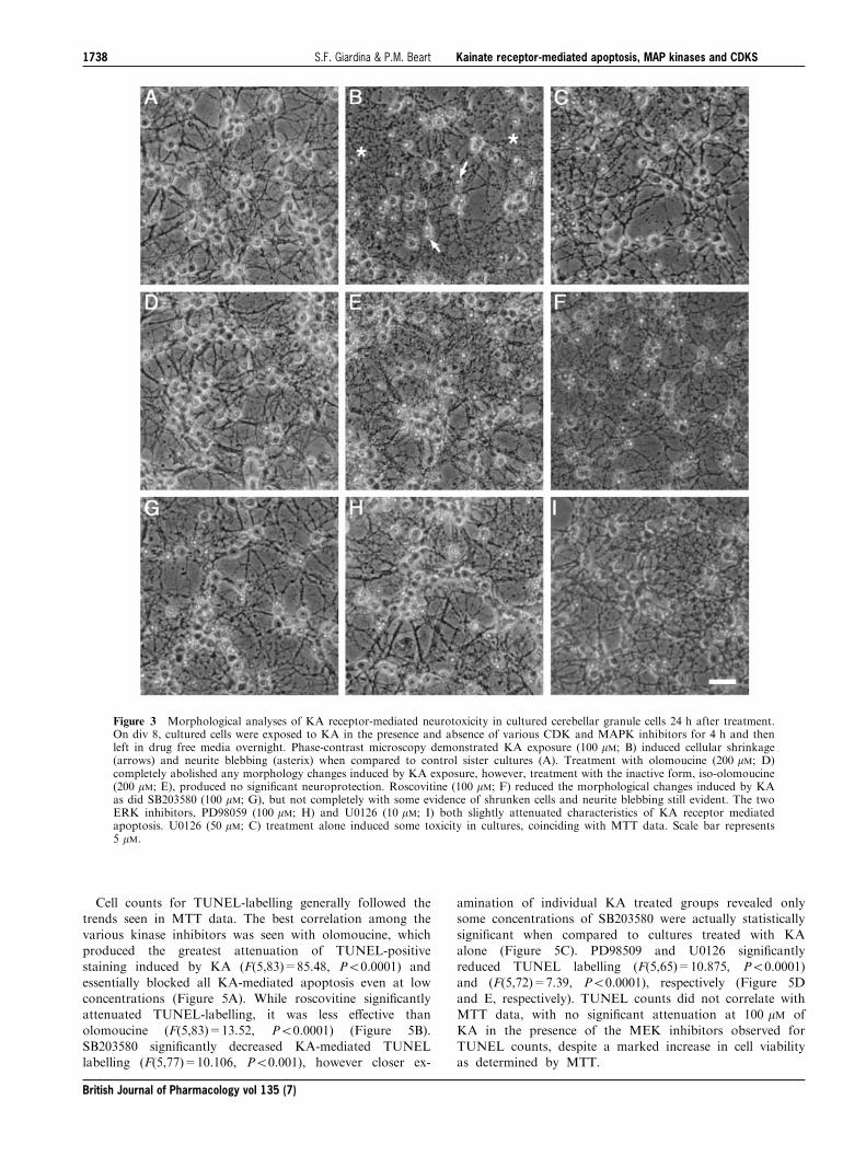
Kainate neurotoxicity: preliminary observations
KA exposure resulted in a reduction in cellular viability,with characteristics of apoptosis (see below), and was
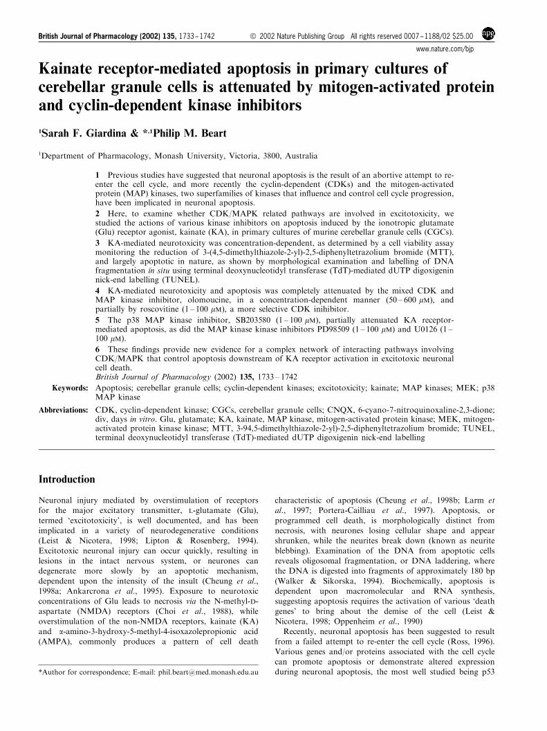
concentration-dependent (F(4,67)=9.18, P50.05). KA-mediated toxicity (EC50=43 mM) was completely attenuatedby the non-NMDA receptor antagonist 6-cyano-7-nitroqui-
noxaline-2,3-dione (CNQX; 50 mM, Figure 1) suggesting aKA-receptor mediated mechanism as AMPA receptor-mediated toxicity is absent in CGCs when cultured under
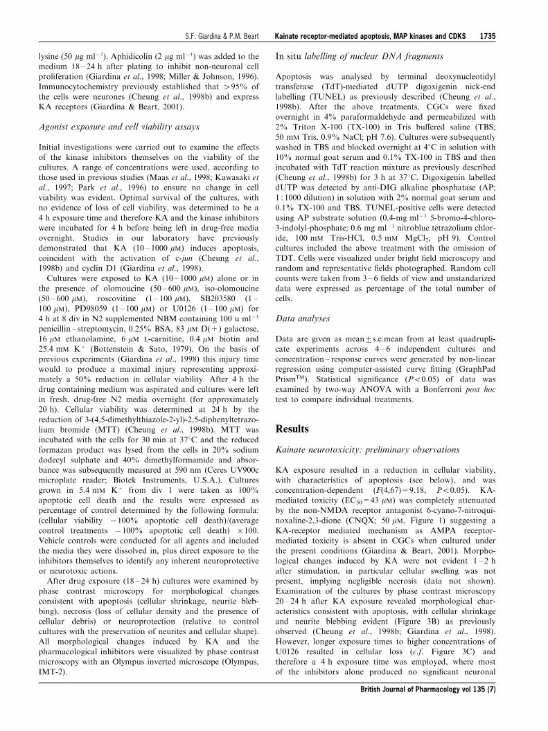
the present conditions (Giardina & Beart, 2001). Morpho-logical changes induced by KA were not evident 1 ± 2 hafter stimulation, in particular cellular swelling was not
present, implying negligible necrosis (data not shown).Examination of the cultures by phase contrast microscopy20 ± 24 h after KA exposure revealed morphological char-acteristics consistent with apoptosis, with cellular shrinkage
and neurite blebbing evident (Figure 3B) as previouslyobserved (Cheung et al., 1998b; Giardina et al., 1998).However, longer exposure times to higher concentrations of
U0126 resulted in cellular loss (c.f. Figure 3C) andtherefore a 4 h exposure time was employed, where mostof the inhibitors alone produced no signi®cant neuronal
British Journal of Pharmacology vol 135 (7)
Kainate receptor-mediated apoptosis, MAP kinases and CDKSS.F. Giardina & P.M. Beart 1735
loss (data not shown). Therefore for routine experimenta-tion, cultures were exposed to KA for 4 h in the absenceand presence of kinase inhibitors, before the medium
containing the drugs was completely aspirated and left indrug free medium overnight.
Kinase inhibitors and kainate neurotoxicity: cell viabilitystudies
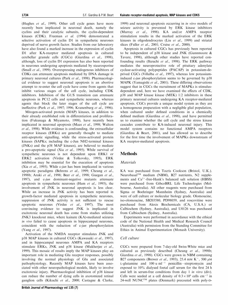
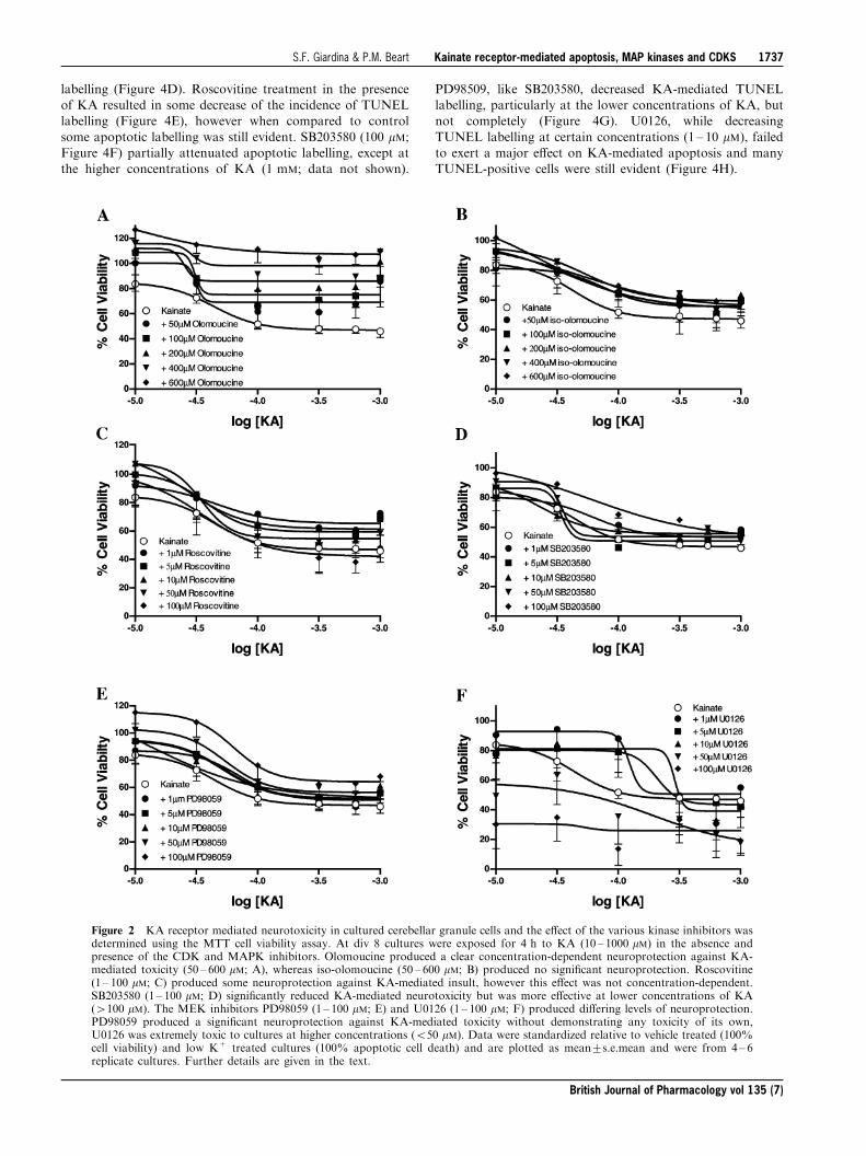
In biochemical investigations olomoucine produced a trend
towards increasing cellular viability in cells not treated withKA, although this action was not statistically signi®cant(F(5,43)=0.165, P40.05). Olomoucine (200 ± 600 mM; Figure
2A) exerted a concentration-dependent neuroprotectionagainst KA-mediated neurotoxicity (F(5,246)=27.56,P50.0001), with cellular viability returning to 100% and
higher. Iso-olomoucine (50 ± 600 mM; Figure 2B), the inactiveform of olomoucine (Havlicek et al., 1997; Park et al., 1996;1997), had no signi®cant e�ect on KA-mediated toxicity(F(5,128)=4.10, P=0.539), con®rming the selective actions of
olomoucine. Roscovitine partially, but signi®cantly, attenu-ated KA-induced neurotoxicity (F(5,426)=4.21, P50.05),but not as e�ectively as olomoucine, and was completely
ine�ective at 100 mM (Figure 2C). SB203580 signi®cantlyattenuated KA receptor-mediated neurotoxicity, particularlyat lower concentrations of KA ((5,276)=5.85, P50.0001)
(Figure 2D). The MEK inhibitor PD98059 e�ectivelyattenuated KA receptor-mediated toxicity (F(5,276)=7.24,P50.0001), although its inhibitory action was not concentra-
tion-dependent, with most attenuation occurring at lowerconcentrations of KA (Figure 2E). The pharmacologicalpro®le of U0126 was the most complex and cellular viabilityshowed more variability when compared to those of the other
inhibitors. U0126 was e�ective at attenuating KA-receptormediated neurotoxicity at concentrations 1, 5 and 10 mM(F(5,206)=17.1, P50.0001; Figure 2F). Treatment with 50
and 100 mM U0126 resulted in a marked reduction in cellularviability, exacerbating KA-induced toxicity (Figure 2F).While previous studies have demonstrated neuroprotection
against a variety of apoptotic insults using the cell cycleinhibitors mimosine, silymarin, ciclopirox and desferrioxa-mine (Park et al., 1997; Farinelli & Greene, 1996), the present
study did not ®nd any signi®cant neuroprotection againstKA-mediated neurotoxicity using these agents (data notshown).
Kinase inhibitors and kainate neurotoxicity:morphological observations
Morphological changes induced by KA were consistentwith apoptosis, where cells demonstrated shrunken cellbodies and the degeneration of neurites (Figure 3B).
Treatment with olomoucine (50 ± 600 mM; Figure 3D)completely attenuated, in an apparently concentration ±dependent manner, all morphological changes induced by
KA (Figure 3B). Iso-olomoucine (50 ± 600 mM; Figure 3E),the inactive isomer used widely as a control treatment(Havlicek et al., 1997; Park et al., 1996; 1997), producedno changes when compared to cultures treated with KA
alone. The other compounds demonstrated more complexactions on the cultures and observations. Roscovitine,surprisingly did not attenuate KA-induced neurotoxicity to
the same extent as olomoucine, observations in aggreementwith the biochemical ®ndings. Roscovitine has previouslybeen shown to potently attenuate neuronal apoptosis
(Maas et al., 1998). While the present study showed thatthe neurones treated with roscovitine demonstrated lessmorphological changes indicative of apoptosis, unlike sister
cultures treated with olomoucine, some cellular damagewas still evident (Figure 3F). The p38 MAP kinaseinhibitor, SB203580 (1 ± 100 mM), slightly improved thecellular viability of the cultures, particularly at higher
concentrations (50 ± 100 mM) and in cultures treated with4100 mM KA (Figure 3G), with some preservation ofneurites and cellular shape. Interestingly, the MEK
inhibitors PD98059 and U0126, di�ered in their abilityto attenuate KA-mediated neurotoxicity. While PD98059e�ectively attenuated morphological changes induced by
KA (10 ± 100 mM), without inducing toxicity of its own(Figure 3H), concentrations of U0126 (450 mM) inducedcellular loss (Figure 3C). Lower concentrations of U0126,did attenuate cellular shrinkage and neurite blebbing
associated with KA receptor-mediated cell death (Figure3I). Exposure of CGCs to the vehicles, either 1% DMSOor 2% ethanol, had no e�ect on the morphological
integrity of the cultures (Figure 3A; 1% DMSO).
Kinase inhibitors and kainate receptor-mediated apoptosis
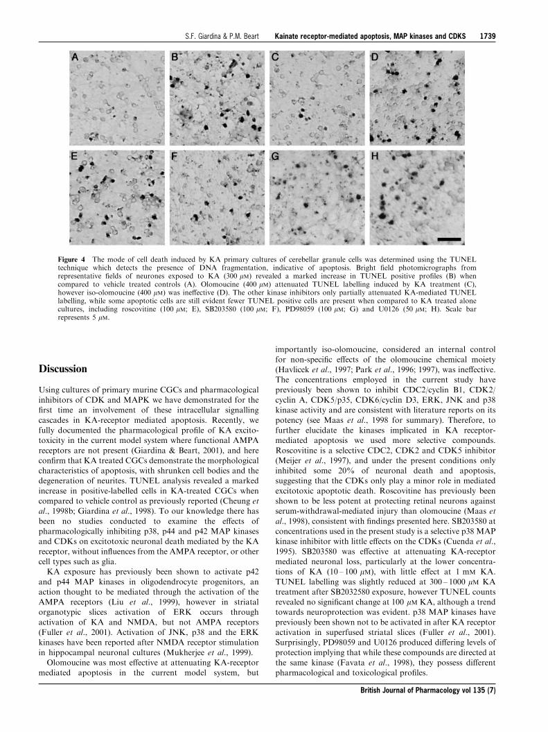
KA receptor-mediated neurotoxicity under the presentexperimental conditions occurs almost exclusively by apop-
tosis (Cheung et al., 1998b; Giardina et al., 1998) and todetermine whether the protective e�ects of the kinaseinhibitors were re¯ected by attenuation of apoptosis TUNELlabelling was employed. TUNEL labelling revealed extensive
TUNEL-positive cells in KA treated cells (300 mM; Figure4B) when compared to vehicle treated cultures (Figure 4A),consistent with observations made under phase-contrast
microscopy. Treatment with olomoucine (400 mM; Figure4C) completely attenuated apoptotic labelling, while theinactive form iso-olomoucine did not reduce TUNEL
Figure 1 KA receptor mediated neurotoxicity in cultured cerebellargranule cells was determined using the MTT cell viability assay. Atdiv 8 cultures were exposed for 4 h to KA (10 ± 1000 mM; closedsquares) and subsequently left in drug-free medium for a further18 h. Cellular injury was concentration-dependent and completelyattenuated by CNQX (50 mM; closed circles). Data were standardizedrelative to vehicle treated (100% cell viability) and low K+ treatedcultures (100% apoptotic cell death) and are plotted as mean+s.e.mean and were from 4 ± 6 replicate cultures. Further details aregiven in the text.
British Journal of Pharmacology vol 135 (7)
Kainate receptor-mediated apoptosis, MAP kinases and CDKSS.F. Giardina & P.M. Beart1736
labelling (Figure 4D). Roscovitine treatment in the presenceof KA resulted in some decrease of the incidence of TUNELlabelling (Figure 4E), however when compared to control
some apoptotic labelling was still evident. SB203580 (100 mM;Figure 4F) partially attenuated apoptotic labelling, except atthe higher concentrations of KA (1 mM; data not shown).
PD98509, like SB203580, decreased KA-mediated TUNELlabelling, particularly at the lower concentrations of KA, butnot completely (Figure 4G). U0126, while decreasing
TUNEL labelling at certain concentrations (1 ± 10 mM), failedto exert a major e�ect on KA-mediated apoptosis and manyTUNEL-positive cells were still evident (Figure 4H).
Figure 2 KA receptor mediated neurotoxicity in cultured cerebellar granule cells and the e�ect of the various kinase inhibitors wasdetermined using the MTT cell viability assay. At div 8 cultures were exposed for 4 h to KA (10 ± 1000 mM) in the absence andpresence of the CDK and MAPK inhibitors. Olomoucine produced a clear concentration-dependent neuroprotection against KA-mediated toxicity (50 ± 600 mM; A), whereas iso-olomoucine (50 ± 600 mM; B) produced no signi®cant neuroprotection. Roscovitine(1 ± 100 mM; C) produced some neuroprotection against KA-mediated insult, however this e�ect was not concentration-dependent.SB203580 (1 ± 100 mM; D) signi®cantly reduced KA-mediated neurotoxicity but was more e�ective at lower concentrations of KA(4100 mM). The MEK inhibitors PD98059 (1 ± 100 mM; E) and U0126 (1 ± 100 mM; F) produced di�ering levels of neuroprotection.PD98059 produced a signi®cant neuroprotection against KA-mediated toxicity without demonstrating any toxicity of its own,U0126 was extremely toxic to cultures at higher concentrations (550 mM). Data were standardized relative to vehicle treated (100%cell viability) and low K+ treated cultures (100% apoptotic cell death) and are plotted as mean+s.e.mean and were from 4 ± 6replicate cultures. Further details are given in the text.
British Journal of Pharmacology vol 135 (7)
Kainate receptor-mediated apoptosis, MAP kinases and CDKSS.F. Giardina & P.M. Beart 1737
Cell counts for TUNEL-labelling generally followed thetrends seen in MTT data. The best correlation among the
various kinase inhibitors was seen with olomoucine, whichproduced the greatest attenuation of TUNEL-positivestaining induced by KA (F(5,83)=85.48, P50.0001) and
essentially blocked all KA-mediated apoptosis even at lowconcentrations (Figure 5A). While roscovitine signi®cantlyattenuated TUNEL-labelling, it was less e�ective than
olomoucine (F(5,83)=13.52, P50.0001) (Figure 5B).SB203580 signi®cantly decreased KA-mediated TUNELlabelling (F(5,77)=10.106, P50.001), however closer ex-
amination of individual KA treated groups revealed onlysome concentrations of SB203580 were actually statistically
signi®cant when compared to cultures treated with KAalone (Figure 5C). PD98509 and U0126 signi®cantlyreduced TUNEL labelling (F(5,65)=10.875, P50.0001)
and (F(5,72)=7.39, P50.0001), respectively (Figure 5Dand E, respectively). TUNEL counts did not correlate withMTT data, with no signi®cant attenuation at 100 mM of
KA in the presence of the MEK inhibitors observed forTUNEL counts, despite a marked increase in cell viabilityas determined by MTT.
Figure 3 Morphological analyses of KA receptor-mediated neurotoxicity in cultured cerebellar granule cells 24 h after treatment.On div 8, cultured cells were exposed to KA in the presence and absence of various CDK and MAPK inhibitors for 4 h and thenleft in drug free media overnight. Phase-contrast microscopy demonstrated KA exposure (100 mM; B) induced cellular shrinkage(arrows) and neurite blebbing (asterix) when compared to control sister cultures (A). Treatment with olomoucine (200 mM; D)completely abolished any morphology changes induced by KA exposure, however, treatment with the inactive form, iso-olomoucine(200 mM; E), produced no signi®cant neuroprotection. Roscovitine (100 mM; F) reduced the morphological changes induced by KAas did SB203580 (100 mM; G), but not completely with some evidence of shrunken cells and neurite blebbing still evident. The twoERK inhibitors, PD98059 (100 mM; H) and U0126 (10 mM; I) both slightly attenuated characteristics of KA receptor mediatedapoptosis. U0126 (50 mM; C) treatment alone induced some toxicity in cultures, coinciding with MTT data. Scale bar represents5 mM.
British Journal of Pharmacology vol 135 (7)
Kainate receptor-mediated apoptosis, MAP kinases and CDKSS.F. Giardina & P.M. Beart1738
Discussion
Using cultures of primary murine CGCs and pharmacologicalinhibitors of CDK and MAPK we have demonstrated for the®rst time an involvement of these intracellular signalling
cascades in KA-receptor mediated apoptosis. Recently, wefully documented the pharmacological pro®le of KA excito-toxicity in the current model system where functional AMPA
receptors are not present (Giardina & Beart, 2001), and herecon®rm that KA treated CGCs demonstrate the morphologicalcharacteristics of apoptosis, with shrunken cell bodies and thedegeneration of neurites. TUNEL analysis revealed a marked
increase in positive-labelled cells in KA-treated CGCs whencompared to vehicle control as previously reported (Cheung etal., 1998b; Giardina et al., 1998). To our knowledge there has
been no studies conducted to examine the e�ects ofpharmacologically inhibiting p38, p44 and p42 MAP kinasesand CDKs on excitotoxic neuronal death mediated by the KA
receptor, without in¯uences from the AMPA receptor, or othercell types such as glia.KA exposure has previously been shown to activate p42
and p44 MAP kinases in oligodendrocyte progenitors, anaction thought to be mediated through the activation of theAMPA receptors (Liu et al., 1999), however in striatalorganotypic slices activation of ERK occurs through
activation of KA and NMDA, but not AMPA receptors(Fuller et al., 2001). Activation of JNK, p38 and the ERKkinases have been reported after NMDA receptor stimulation
in hippocampal neuronal cultures (Mukherjee et al., 1999).Olomoucine was most e�ective at attenuating KA-receptor
mediated apoptosis in the current model system, but
importantly iso-olomoucine, considered an internal control
for non-speci®c e�ects of the olomoucine chemical moiety(Havlicek et al., 1997; Park et al., 1996; 1997), was ine�ective.The concentrations employed in the current study have
previously been shown to inhibit CDC2/cyclin B1, CDK2/cyclin A, CDK5/p35, CDK6/cyclin D3, ERK, JNK and p38kinase activity and are consistent with literature reports on its
potency (see Maas et al., 1998 for summary). Therefore, tofurther elucidate the kinases implicated in KA receptor-mediated apoptosis we used more selective compounds.
Roscovitine is a selective CDC2, CDK2 and CDK5 inhibitor(Meijer et al., 1997), and under the present conditions onlyinhibited some 20% of neuronal death and apoptosis,suggesting that the CDKs only play a minor role in mediated
excitotoxic apoptotic death. Roscovitine has previously beenshown to be less potent at protecting retinal neurons againstserum-withdrawal-mediated injury than olomoucine (Maas et
al., 1998), consistent with ®ndings presented here. SB203580 atconcentrations used in the present study is a selective p38 MAPkinase inhibitor with little e�ects on the CDKs (Cuenda et al.,
1995). SB203580 was e�ective at attenuating KA-receptormediated neuronal loss, particularly at the lower concentra-tions of KA (10 ± 100 mM), with little e�ect at 1 mM KA.
TUNEL labelling was slightly reduced at 300 ± 1000 mM KAtreatment after SB2032580 exposure, however TUNEL countsrevealed no signi®cant change at 100 mM KA, although a trendtowards neuroprotection was evident. p38 MAP kinases have
previously been shown not to be activated in after KA receptoractivation in superfused striatal slices (Fuller et al., 2001).Surprisingly, PD98059 and U0126 produced di�ering levels of
protection implying that while these compounds are directed atthe same kinase (Favata et al., 1998), they possess di�erentpharmacological and toxicological pro®les.
Figure 4 The mode of cell death induced by KA primary cultures of cerebellar granule cells was determined using the TUNELtechnique which detects the presence of DNA fragmentation, indicative of apoptosis. Bright ®eld photomicrographs fromrepresentative ®elds of neurones exposed to KA (300 mM) revealed a marked increase in TUNEL positive pro®les (B) whencompared to vehicle treated controls (A). Olomoucine (400 mM) attenuated TUNEL labelling induced by KA treatment (C),however iso-olomoucine (400 mM) was ine�ective (D). The other kinase inhibitors only partially attenuated KA-mediated TUNELlabelling, while some apoptotic cells are still evident fewer TUNEL positive cells are present when compared to KA treated alonecultures, including roscovitine (100 mM; E), SB203580 (100 mM; F), PD98059 (100 mM; G) and U0126 (50 mM; H). Scale barrepresents 5 mM.
British Journal of Pharmacology vol 135 (7)
Kainate receptor-mediated apoptosis, MAP kinases and CDKSS.F. Giardina & P.M. Beart 1739
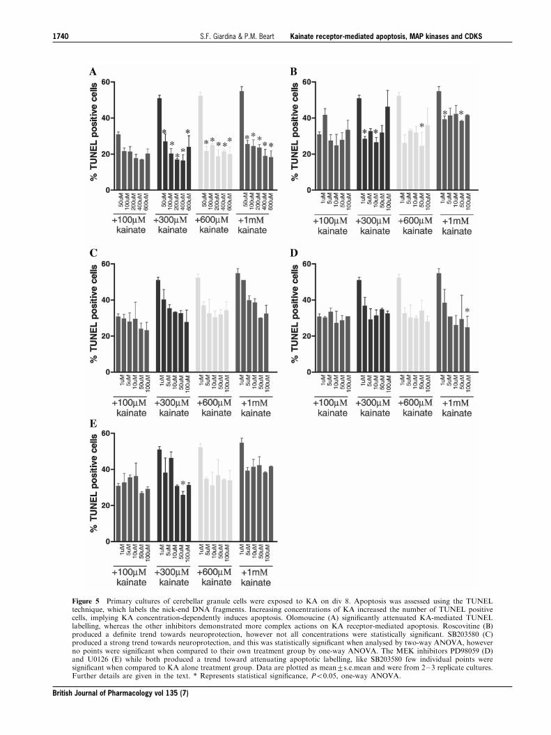
Figure 5 Primary cultures of cerebellar granule cells were exposed to KA on div 8. Apoptosis was assessed using the TUNELtechnique, which labels the nick-end DNA fragments. Increasing concentrations of KA increased the number of TUNEL positivecells, implying KA concentration-dependently induces apoptosis. Olomoucine (A) signi®cantly attenuated KA-mediated TUNELlabelling, whereas the other inhibitors demonstrated more complex actions on KA receptor-mediated apoptosis. Roscovitine (B)produced a de®nite trend towards neuroprotection, however not all concentrations were statistically signi®cant. SB203580 (C)produced a strong trend towards neuroprotection, and this was statistically signi®cant when analysed by two-way ANOVA, howeverno points were signi®cant when compared to their own treatment group by one-way ANOVA. The MEK inhibitors PD98059 (D)and U0126 (E) while both produced a trend toward attenuating apoptotic labelling, like SB203580 few individual points weresigni®cant when compared to KA alone treatment group. Data are plotted as mean+s.e.mean and were from 2 ± 3 replicate cultures.Further details are given in the text. * Represents statistical signi®cance, P50.05, one-way ANOVA.
British Journal of Pharmacology vol 135 (7)
Kainate receptor-mediated apoptosis, MAP kinases and CDKSS.F. Giardina & P.M. Beart1740

While MTT data and TUNEL cell counts generally followedthe same pattern, less neuroprotection was noted for the kinaseinhibitors as re¯ected by the number of TUNEL positive cells
than was expected from theMTT data. This di�erencemay havebeen attributable to technical considerations or it is possiblewhile these inhibitors are increasing cell viability, they are notattenuating, but rather delaying apoptosis, resulting in a relative
increase inMTTreductionbutonly a slight reductionofTUNELlabelling.Alternatively, since cellular viabilitymeasuredbyMTTreduction re¯ects mitochondrial function, the diminution seen
here may re¯ect upstream changes that are not totally predictiveof the extent of ®nal downstream DNA fragmentation (Budd etal., 2000). The delay in apoptosis may explain the good
correlation between theMTTandTUNELdata for olomoucine,as this inhibitor blocks both the CDK and MAP kinases,indicating the therapeutic bene®ts of inhibiting concurrently
these twomechanisms for the executionof apoptosismediatedbythe KA receptor. By inhibiting one speci®c kinase the apoptoticpathwaymayproceeddownanalternative pathway still allowingcell death. Excitotoxicity has previously been shown to involve
the cell cycle (Giardina et al., 1998; Uberti et al., 2000) andMAPkinases (Murray et al., 1998;Yang et al., 1997), and it is thereforelikely that not just one pathway is essential for the execution of
apoptosis. While many intracellular pathways are intertwined,the activation of either the cell cycle or the MAP kinases may infact result inneuronal death, through the activationof a common
downstream pathway, perhaps involving the Bcl-2 family andcaspases. However, if both pathways are inhibited, as demon-strated by olomoucine, the apoptotic pathways are completely
blocked and apoptosis cannot occur.
While the exact mechanism by which these kinases mediateneuronal apoptosis and/or neuronal survival is still to beelucidated, our data add to the growing body of evidence to
suggest they play a central role in neuronal apoptosis. Inaddition, the MAP kinases and CDKs seem to be central to acomplex mosaic of pathways, in¯uencing various intracellularmediators including nuclear factor kb, AKT (protein kinase B),
and the Bcl-2 family, which have all been implicated in neuronalapoptosis (Thomson et al., 1999; Fukunaga&Miyamoto, 1998).Unfortunately, no kinase inhibitors commercially available are
speci®c for one kinase, and it is not presently possible to elucidatewhether one kinase per se is responsible for the execution ofapoptosis. As the activities of these kinase inhibitors directly on
the KA receptor itself has not, to our knowledge, beendetermined, we can not rule out the possibility these kinases actdirectlyon theKAreceptor, or oneof its domains, rather thanact
downstream of receptor activation. We however believe this isunlikely, as no neuroprotection is evident in the inactive isoform,iso-olomoucine. In conclusion, our results support the growingbody of evidence involving the cell cycle and MAP kinases in
excitotoxicity, and provide the ®rst demonstration of theinvolvement of MAPKs in KA receptor-mediated neuronalinjury.
Supported by the National Health and Medical Research Council(Australia), of which P.M. Beart is a Senior Principal ResearchFellow, and by grants from the Ramaciotti, Rebecca Cooper andWilliam Buckland Foundations, and Perpetual Trustees.
References
ANKARCRONA,M.,DYPBUKT, J.M., BONFOCO,E., ZHIVOTOVSKY,B.,
ORRENIUS, S., LIPTON, S.A. & NICOTERA, P. (1995). Glutamate-induced neuronal death: a succession of necrosis or apoptosisdepending on mitochondrial function. Neuron, 15, 961 ± 973.
ARAKI, T., ENOKIDO, Y., INAMURA, N., AIZAWA, S., REED, J.C. &
HATANAKA, H. (1998). Changes in c-Jun but not Bcl-2 familyproteins in p53-dependent apoptosis of mouse cerebellar granuleneurons induced by DNA damaging agent bleomycin. Brain Res.,794, 239 ± 247.
BEER, J., MIELKE, K., ZIPP, M., ZIMMERMANN, M. & HERDEGEN,
T. (1998). Expression of c-jun, junB, c-fos, fra-1 and fra-2 mRNAin the rat brain following seizure activity and axotomy. BrainRes., 794, 255 ± 266.
BEHRENS, A., SIBILIA, M. & WAGNER, E.F. (1999). Amino-terminalphosphorylation of c-Jun regulates stress-induced apoptosis andcellular proliferation. Nature Genetics, 21, 326 ± 329.
BOTTENSTEIN, J.E. & SATO, G.H. (1979). Growth of a ratneuroblastoma cell line in serum free supplemented medium.Proc. Natl. Acad. Sci., USA, 76, 514 ± 517.
BREWER, G.J., TORRICELLI, J.R., EVERGE, E.K. & PRICE, P.J.
(1993). Optimised survival of hippocampal neurons in B27-supplemented Neurobasal, a new serum-free medium combina-tion. J. Neurosci. Res., 35, 567 ± 576.
BUDD, S.L., TENNERTI, L., LISHNAK, T. & LIPTON, S.A. (2000).Mitochondrial and extramitochondrial apoptotic signaling path-ways in cerebrocortical neurons. Proc. Natl. Acad. Sci., USA, 97,6161 ± 6166.
CASTAGNE, V. & CLARKE, P.G.H. (1999). Inhibitors of mitogen-activated protein kinases protect axotomized developing neu-rons. Brain Res., 842, 215 ± 219.
CHEUNG, N.S., PASCOE, C.J., GIARDINA, S.F., JOHN, C.A. & BEART,
P.M. (1998a). Micromolar L-glutamate induces extensive apop-tosis in an apoptotic-necrotic continuum of insult-dependent,excitotoxic injury in cultured cortical neurones. Neuropharma-cology, 37, 1419 ± 1429.
CHEUNG, N.S., CARROLL, F.Y., LARM, J.A., BEART, P.M. &
GIARDINA, S.F. (1998b). Kainate-induced apoptosis correlateswith c-Jun activation in cultured cerebellar granule cells. J.Neurosci. Res., 52, 69 ± 82.
CHOI, D.W., KOH, J.Y. & PETERS, S. (1988). Pharmacology ofglutamate neurotoxicity in cortical cell culture: attenuation byNMDA antagonists. J. Neurosci., 8, 185 ± 196.
CRUISE, L., HO, L.K., VEITCH, K., FULLER, G. & MORRIS, B.J.
(2000). Kainate receptors activate NF-kb via MAP kinase instriatal neurones. Neuroreport, 11, 395 ± 398.
CUENDA, A., ROUSE, J., DOZA, Y.N., MEIER, R., COHEN, P.,
GALLAGHER, T.F., YOUNG, P.R. & LEE, J.C. (1995). SB 203580is a speci®c inhibitor of a MAP kinase homologue which isstimulated by cellular stresses and interleukin-1. FEBS Lett., 364,229 ± 233.
FARINELLI, S.E. & GREENE, L.A. (1996). Cell cycle blockersmimosine, ciclopirox, and deferoxamine prevent the death ofPC12 cells and postmitotic sympathetic neurons after removal oftrophic support. J. Neurosci., 16, 1150 ± 1162.
FAVATA, M.F., HORIUCHI, K.Y., MANOS, E.J., DAULERIO, A.J.,
STRADLEY, D.A., FEESER, W.S., VAN DYK, D.E., PITTS, W.J.,
EARL, R.A., HOBBS, F., COPELAND, R.A., MAGOLDA, R.L.,
SCHERLE, P.A. & TRZASKOS, J.M. (1998). Identi®cation of anovel inhibitor of mitogen-activated protein kinase kinase. J.Biol. Chem., 273, 18623 ± 18632.
FREEMAN, R.S., ESTUS, S. & JOHNSON JR, E.M. (1994). Analysis ofcell cycle-related gene expression in postmitotic neurones:selective induction of cyclin D1 during programmed cell death.Neuron., 12, 343 ± 355.
FUKUNAGA, K. & MIYAMOTO, E. (1998). Role of MAP kinase inneurons. Mol. Neurobiol., 16, 79 ± 95.
FULLER, G., VEITCH, K., HO, L.K., CRUISE, L. & MORRIS, B.J.
(2001). Activation of p44/p42 MAP kinase in striatal neuronsvia kainate receptors and PI3 kinase. Mol. Brain Res., 89, 126 ±132.
British Journal of Pharmacology vol 135 (7)
Kainate receptor-mediated apoptosis, MAP kinases and CDKSS.F. Giardina & P.M. Beart 1741
GIARDINA, S.F., CHEUNG, N.S., REID, M.T. & BEART, P.M. (1998).Kainate-induced apoptosis in cultured murine cerebellar granulecells elevates expression of the cell cycle gene cyclin D1. J.Neurochem., 71, 1325 ± 1328.
GIARDINA, S.F. & BEART, P.M. (2001). Excitotoxic pro®les of novel,low-a�nity kainate receptor agonists in primary cultures of murinecerebellar granule cells. Neuropharmacology, 41, 421 ± 432.
GUEGAN, C., LEVY, V., DAVID, J.P., AJCHENBAUM-CYMBALISTA,
F. & SOLA, B. (1997). c-Jun and cyclin D1 proteins as mediators ofneuronal death after a focal ischaemic insult. Neuroreport, 8,1003 ± 1007.
GUNNMOORE, F.J. & TAVARE, J.M. (1998). Apoptosis of CerebellarGranule Cells Induced By SerumWithdrawal, Glutamate or Beta-Amyloid, Is Independent of Jun Kinase or P38 Mitogen ActivatedProtein Kinase Activation. Neurosci. Lett., 250, 53 ± 56.
HAM, J., BABIJ, C., WHITFIELD, J., PFARR, C.M., LALLEMAND, D.,
YANIV, M. & RUBIN, L.L. (1995). A c-jun dominant-negativemutant protects sympathetic neurons against programmed celldeath. Neuron, 14, 927 ± 939.
HAVLICEK, L., HANUS, J., VESELY, J., LECLERC, S., MEIJER, L.,
SHAW, G. & STRNAD, M. (1997). Cytokinin-derived cyclin-dependent kinase inhibitors: synthesis and cdc2 inhibitoryactivity of olomoucine and related compounds. J. Med. Chem.,40, 408 ± 412.
HUGHES, P.E., ALEXI, T., WALTON, M., WILLIAMS, C.E., DRAGU-
NOW, M., CLARK, R.G. & GLUCKMAN, P.D. (1999). Activity andinjury-dependent expression of inducible transcription factors,growth factors and apoptosis-related genes within the centralnervous system. Prog. Neurobiol., 57, 421 ± 450.
IKEUCHI, T., SHIMOKE, K., KUBO, T., YAMADA, M. & HATANAKA,
H. (1998). Apoptosis-inducing and -preventing signal transduc-tion pathways in cultured cerebellar granule neurons. HumanCell, 11, 125 ± 140.
KAWASAKI, H., MOROOKA, T., SHIMOHAMA, S., KIMURA, J.,
HIRANO, T., GOTOH, Y. & NISHIDA, E. (1997). Activation andinvolvement of p38 mitogen-activated protein kinase in gluta-mate-induced apoptosis in rat cerebellar granule cells. J. Biol.Chem., 272, 18518 ± 18521.
KIKUCHI, M., TENNETI, L. & LIPTON, S.A. (2000). Role of p38mitogen-activated protein kinase in axotomy-induced apoptosisof rat retinal ganglion cells. J. Neurosci., 13, 5037 ± 5044.
KRANENBURG, O., VAN DER EB, A.J. & ZANTEMA, A. (1996).Cyclin D1 is an essential mediator of apoptotic neuronal celldeath. EMBO J., 15, 46 ± 54.
LARM, J.A., CHEUNG, N.S. & BEART, P.M. (1997). Apoptosis inducedvia AMPA-selective glutamate receptors in cultured murinecortical neurons. J. Neurochem., 69, 617 ± 622.
LEIST, M. & NICOTERA, P. (1998). Apoptosis, excitotoxicity, andneuropathology. Experimental Cell Research, 239, 183 ± 201.
LIPTON, S.A. & ROSENBERG, P.A. (1994). Excitatory amino acids asa ®nal common pathway for neurologic disorders. New Engl. J.Med., 330, 613 ± 622.
LIU, H.N., LAROCCA, J.N. & ALMAZAN, G. (1999). Molecularpathways mediating activation by kainate of mitogen-activatedprotein kinase in oligodendrocyte progenitors. Mol. Brain Res.,66, 50 ± 61.
MAAS, J.W., HORSTMANN, S., BORASIO, G.D., ANNESER, J.M.H.,
SHOOTER, E.M. & KAHLE, P.J. (1998). Apoptosis of central andperipheral neurons can be prevented with cyclin-dependentkinase mitogen-activated protein kinase inhibitors. J. Neuro-chem., 70, 1401 ± 1410.
MEIJER, L., BORGNE, A., MULNER, O., CHONG, J.P., BLOW, J.J.,
INAGAKI, N., INAGAKI, M., DELCROS, J.G. & MOULINOUX, J.P.
(1997). Biochemical and cellular e�ects of roscovitine, a potentand selective inhibitor of the cyclin-dependent kinases cdc2, cdk2and cdk5. Eur. J. Biochem., 243, 527 ± 536.
MILLER, T.M. & JOHNSON JR., E.M. (1996). Metabolic and geneticanalyses of apoptosis in potassium/serum-deprived rat cerebellargranule cells. J. Neurosci., 16, 7487 ± 7495.
MUKHERJEE, P.K., DECOSTER, M.A., CAMPBELL, F.Z., DAVIS, R.J.
& BAZAN, N.G. (1999). Glutamate receptor signaling interplaymodulates stress-sensitive mitogen-activated protein kinases andneuronal cell death. J. Biol. Chem., 274, 6493 ± 6498.
MURRAY, B., ALESSANDRINI, A., COLE, A.J., YEE, A.G. &
FURSHPAN, E.J. (1998). Inhibition of the p44/42 MAP kinasepathway protects hippocampal neurons in a cell-culture model ofseizure activity. Proc. Natl. Acad. Sci., USA, 95, 11975 ± 11980.
OPPENHEIM, R.W., PREVETTE, D., TYTELL, M. & HOMMA, S.
(1990). Naturally occuring and induced neuronal death in thechick embryo in vivo requires protein and RNA synthesis:evidence for the role of cell death genes. Dev. Biol., 138, 104 ± 113.
PARK, D.S., FARINELLI, S.E. & GREENE, L.A. (1996). Inhibitors ofcyclin-dependent kinases promote survival of post-mitoticneuronally di�erentiated PC12 cells and sympathetic neurons.J. Biol. Chem., 271, 8161 ± 8169.
PARK, D.S., MORRIS, E.J., GREENE, L.A. & GELLER, H.M. (1997).G1/S cell cycle blockers and inhibitors of cyclin-dependentkinases suppress camptothecin-induced neuronal apoptosis. J.Neurosci., 17, 1256 ± 1270.
PARK, D.S., MORRIS, E.J., PADMANABHAN, J., SHELANSKI, M.L.,
GELLER, H.M. & GREENE, L.A. (1998). Cyclin-dependent kinasesparticipate in death of neurons evoked by DNA-damagingagents. J. Cell Biol., 143, 457 ± 467.
PORTERA-CAILLIAU, C., PRICE, D.L. & MARTIN, L.J. (1997). Non-NMDA and NMDA receptor-mediated excitotoxic neuronaldeaths in adult brain are morphologically distinct: furtherevidence for an apoptosis-necrosis continuum. J. Comp. Neurol.,378, 88 ± 104.
ROSS, M.E. (1996). Cell division and the nervous system: regulatingthe cycle from neural di�erentiation to death. Trends Neurosci.,19, 62 ± 68.
SMALL, D.L., MONETTE, R., COMAS, T., FOURIER, M.C. &MORLEY,
P. (1999). Loss of cyclin D1 in necrotic and apoptotic models ofcortical neuronal degeneration. Brain Res., 842, 376 ± 383.
THOMSON, S., MAHADEVAN, L.C. & CLAYTON, A.L. (1999). MAPkinase-mediated signalling to nucleosomes and immediate-earlygene induction. Sem. Cell Dev. Biol., 10, 205 ± 214.
UBERTI, D., GRILLI, M. & MEMO, M. (2000). Contribution of NF-kappaB and p53 in the glutamate-induced apoptosis. Int. J. Dev.Neurosci., 18, 447 ± 454.
VILLALBA, M., BOCKAERT, J. & JOURNOT, L. (1997). Pituitaryadenylate cyclase-activating polypeptide (PACAP-38) protectscerebellar granule neurons from apoptosis by activating themitogen-activated protein kinase (MAP kinase) pathway. J.Neurosci., 17, 83 ± 90.
VIRDEE, K., BANNISTER, A.J., HUNT, S.P. & TOLKOVSKY, A.M.
(1997). Comparison between the timing of JNK activation, c-Junphosphorylation, and onset of death commitment in sympatheticneurones. J. Neurochem., 69, 550 ± 561.
VIRDEE, K. & TOLKOVSKY, A.M. (1995). Activation of p44 and p42MAP kinases is not essential for the survival of rat sympatheticneurons. Eur. J. Neurosci., 7, 2159 ± 2169.
WALKER, P.R. & SIKORSKA, M. (1994). Endoplasmic activities,chromatin structure and DNA degradation in apoptosis.Biochem. Cell Biol., 264, 9000 ± 9003.
WALTON, K.M., DIROCCO, R., BARTLETT, B.A., KOURY, E.,
MARCY, V.R., JARVIS, B., SCHAEFER, E.M. & BHAT, R.V.
(1998). Activation of p38MAPK in microglia after ischemia. J.Neurochem., 70, 1764 ± 1767.
XIA, Z., DICKENS, M., RAINGEAUD, J., DAVIS, R.J. & GREENBERG,
M.E. (1995). Opposing e�ects of ERK and JNK-p38MAP kinaseson apoptosis. Science, 270, 1326 ± 1331.
YAMAGISHI, S., YAMADA, M., ISHIKAWA, Y., MATSUMOTO, T.,
IKEUCHI, T. & HATANAKA, H. (2001). p38 mitogen-activatedprotein kinase regulates low potassium-induced c-jun phosphor-ylation and apoptosis in cultured cerebellar granule cells. J. Biol.Chem., 276, 5129 ± 5133.
YANG, D.D., KUAN, C.Y., WHITMARSH, A.J., RINCON, M., ZHENG,
T.S., DAVIS, R.J., RAKIC, P. & FLAVELL, R.A. (1997). Absence ofexcitotoxicity-induced apoptosis in the hippocampus of micelacking the Jnk3 gene. Nature, 389, 865 ± 870.
(Received October 8, 2001Revised January 16, 2002
Accepted January 25, 2002)
British Journal of Pharmacology vol 135 (7)
Kainate receptor-mediated apoptosis, MAP kinases and CDKSS.F. Giardina & P.M. Beart1742