involvement of uracil nucleotides in protection of cardiomyocytes from hypoxic stress
TRANSCRIPT

Involvement of UTP in protection of cardiomyocytes fromhypoxic stress1
Asher Shainberg2,Faculty of Life Sciences, Bar-Ilan University, Ramat-Gan, 52900, Israel
Smadar Yitzhaki,Faculty of Life Sciences, Bar-Ilan University, Ramat-Gan, 52900, Israel
Or Golan,Faculty of Life Sciences, Bar-Ilan University, Ramat-Gan, 52900, Israel
Kenneth A. Jacobson, andLaboratory of Bioorganic Chemistry, NIDDK, NIH, Bethesda, Maryland, USA
Edith HochhauserFMRC, Rabin Medical Center, Tel Aviv University, Tel Aviv, Israel
AbstractMassive amounts of nucleotides are released during ischemia in the cardiovascular system.Although the effect of the purine nucleotide ATP has been intensively studied in myocardialinfarction, the cardioprotective role of the pyrimidine nucleotide UTP is still unclear, especially inthe cardiovascular system. The purpose of our study was to elucidate the protective effects of UTPreceptor activation and describe the downstream cascade for the cardioprotective effect. Culturedcardiomyocytes and left anterior descending (LAD)-ligated rat hearts were pretreated with UTPand exposed to hypoxia–ischemia. In vitro experiments revealed that UTP reduced cardiomyocytedeath induced by hypoxia, an effect that was diminished by suramin. UTP caused several effectsthat could trigger a cardioprotective response: a transient increase of [Ca2+]i, an effect that wasabolished by PPADS or RB2; phosphorylation of the kinases ERK and Akt, which was abolishedby U0126 and LY294002, respectively; and reduced mitochondrial calcium elevation afterhypoxia. In vivo experiments revealed that UTP maintained ATP levels, improved mitochondrialactivity, and reduced infarct size. In conclusion, UTP administrated before ischemia reducedinfarct size and improved myocardial function. Reduction of mitochondrial calcium overload canpartially explain the protective effect of UTP after hypoxic–ischemic injury.
Keywordscalcium; cardioprotection; cardiac cell culture; heart; hypoxia; ischemia; nucleotides;preconditioning; pyrimidines
IntroductionDespite intensive research and progress in medicine, myocardial infarction and heart failureremain the leading causes of death in the Western world. Myocyte loss, both necrotic and
1This article is one of a selection of papers from the NATO Advanced Research Workshop on Translational Knowledge for HeartHealth (published in part 2 of a 2-part Special Issue).2Corresponding author ([email protected]).
NIH Public AccessAuthor ManuscriptCan J Physiol Pharmacol. Author manuscript; available in PMC 2012 August 09.
Published in final edited form as:Can J Physiol Pharmacol. 2009 April ; 87(4): 287–299. doi:10.1139/Y09-010.
NIH
-PA Author Manuscript
NIH
-PA Author Manuscript
NIH
-PA Author Manuscript
apoptotic, is a feature of many heart pathologies (Kajstura et al. 1996). Due to the fact thatadult cardiomyocytes possess minimal capacity to reenter the cell cycle (Anversa andKajstura 1998), the reduction of myocyte loss through suppression of cell death pathwaysrepresents a beneficial strategy to prevent myocardial infarction and heart failure.
More than 20 years have passed since the initial description of the phenomenon of ischemicpreconditioning, whereby episodes of intermittent sublethal ischemia and re-perfusionconfer resistance against a subsequent lethal episode of myocardial ischemia (Murry et al.1986). Many experiments have since been done to elucidate the mechanism by which a shortepisode of ischemia makes the heart tolerant to prolonged ischemia. It was found thatadenosine and other internal and external agonists can mimic ischemic preconditioning andprotect the heart from prolonged ischemia and cell injury via the activation of specific cellsurface receptors (Sommerschild and Kirkeboen 2000). This process is called pharmacologicpreconditioning.
Purines and pyrimidines have widespread and specific extracellular signaling actions in theregulation of a variety of functions. In 1978 Burnstock proposed that specific extracellularnucleoside (P1) or nucleotide (P2) receptors mediate the physiologic effects of adenosineand ATP, respectively (Burnstock 1978). Receptors that mediate the extracellular actions ofpurines and pyrimidines are divided into 2 major families, one family of ligand-gatedreceptors (P2X) and another family of G protein-coupled receptors (P2Y), comprising 15subtypes in all. In contrast to P2X receptors, P2Y receptors are activated not only by ATP orits derivatives, but also by other naturally occurring nucleotides. The P2Y2, P2Y4, and P2Y6receptor subtypes have high affinity for uracil nucleotides such as UDP and UTP, whichmediate signaling actions through these unique pyrimidine receptor (Jacobson et al. 2009;von Kügelgen and Wetter 2000).
Massive amounts of nucleotides are released during ischemia and hypoxia in thecardiovascular system (Erlinge et al. 2005). Whereas the effect of purine nucleotides (e.g.,ATP) in myocardial infarction has been intensively studied (Abbracchio and Burnstock1998), the role of pyrimidine nucleotides under hypoxic conditions has not been explored asintensively. In this study we characterize and elucidate the protective effects of pretreatmentwith UTP against detrimental factors of hypoxia–ischemia in 2 systems: an in vitro model,in which the cells were exposed to 120 min of hypoxia, and an in vivo model, in which theleft anterior descending (LAD) artery in rat hearts was ligated. The findings revealed thatUTP pretreatment remarkably reduced lactate dehydrogenase (LDH) release, reduced infarctsize, maintained morphologic structure, maintained ATP levels, and improved mitochondrialfunction.
Materials and methodsCell culture
Rat hearts (2–3 days old) were removed under sterile conditions and washed 3 times inphosphate-buffered saline (PBS) to remove excess blood cells. The hearts were minced andthen gently agitated in RDB, a solution of proteolytic enzymes (Life Sciences ResearchInstitute, Ness-Ziona, Israel) prepared from a fig tree extract (Shneyvays et al. 1998). TheRDB was diluted 1:100 in Ca2+- and Mg2+-free PBS at 25 °C for several cycles of 10 mineach, as described previously (Safran et al. 2001). Dulbecco’s modified Eagle’s medium,supplemented with inactivated 10% horse serum (Biological Industries, Kibbutz BeitHaemek, Israel) and 0.5% chick embryo extract, was added to supernatant suspensionscontaining dissociated cells. The mixture was centrifuged at 300g for 5 min. The supernatantphase was discarded, and the cells were resuspended in the same medium. The suspension ofthe cells was diluted to 1.0 × 10⁶ cells/mL, and 1.5 mL of the suspension was placed in 35-
Shainberg et al. Page 2
Can J Physiol Pharmacol. Author manuscript; available in PMC 2012 August 09.
NIH
-PA Author Manuscript
NIH
-PA Author Manuscript
NIH
-PA Author Manuscript

millimetre plastic culture dishes on collagen/gelatin-coated coverglasses. The cultures wereincubated in a humidified atmosphere of 5% CO2, 95% air at 37 °C. Confluent monolayersexhibiting spontaneous contractions were developed in culture within 2 days. Allexperiments were performed between days 5 and 7 in culture.
[Ca2+]i measurementsIntracellular free calcium ion concentration ([Ca2+]i) was estimated from indo-1fluorescence with the ratio method (the SAMPLE program) described elsewhere (Fixler etal. 2002; Shneyvays et al. 2001).
Hypoxic conditionsMyocyte cultures 5–7 days old were washed from the medium with glucose-free PBScontaining 5 mmol/L Hepes at pH 7.4 before exposing the myocytes to the variousconditions at 37 °C. The hypoxic condition consisted of 120 min in a hypoxic incubator inwhich the atmosphere was replaced by the inert gas argon (100%) in glucose-free media(Safran et al. 2001; Shneyvays et al. 2000). The hypoxic damage was characterized at theend of the hypoxic period by morphologic and biochemical evaluation. Continuousmonitoring of [Ca2+]i during hypoxia was realized in a special barrier well, in which cellswere protected from oxygen by a laminar counterflowing layer of argon gas as previouslydescribed (Yitzhaki et al. 2005). The coverglasses with cultured cells were placed at thebottom of the well. This chamber was mounted on a specially modified Zeiss invertedepifluorescence microscope.
Assays of intracellular ATP levelAfter hypoxia, control and experimental cells were harvested in 1 mL cold 5%trichloroacetic acid. The cell extract was used for the measurement of ATP content with aCL SII luciferin–luciferase bioluminescence kit (Boehringer) following the manufacturer’sprotocol. The values were expressed as nanomoles per milligram of protein (nmol/mg)(Yitzhaki et al. 2005).
Experiments with purinergic and pyrimidinergic ligandsUTP at concentrations of 3–50 μmol/L was applied to the cell cultures for 15 min after 15min preincubation with various antagonists (Sigma). The inhibitors LY294002 (30 μmol/L)and 5-hydroxydecanoate (5HD) (300 μmol/L), the Ca2+ chelator 1,2-bis(o-aminophenoxy)ethane N,N,N′,N′-tetraacetic acid (BAPTA) (5 μmol/L), and the P2Yantagonists suramin (300 μmol/L), pyridoxal-5′-phosphate-6-azophenyl-2′,4′-disulfonate(PPADS) (10 μmol/L), and reactive blue 2 (RB2) (50 μmol/L) were dissolved in PBS.U0126 (10 μmol/L) was dissolved in DMSO.
Release of LDHProtein content and LDH activity were determined according to El Ani et al. (1994). Briefly,25 μL supernatant was transferred into a 96-well dish, and the LDH activities weredetermined with an LDH-L kit (Sigma), as described by the manufacturer. The product ofthe enzyme was measured spectrometrically at 30 °C at a wavelength of 340 nm asdescribed previously (Shneyvays et al. 2000). The results were expressed relative to thecontrol (fold change) in the same experiment. Each experiment was done in triplicate andwas repeated at least 3 times.
Western blot analysisAfter treatment with UTP, cardiomyocytes were washed with ice-cold PBS and harvestedinto an Eppendorf tube with ice-cold radioimmunoprecipitation assay (RIPA) buffer (50
Shainberg et al. Page 3
Can J Physiol Pharmacol. Author manuscript; available in PMC 2012 August 09.
NIH
-PA Author Manuscript
NIH
-PA Author Manuscript
NIH
-PA Author Manuscript
mmol/L Tris–HCl, pH 7.4, 1% NP-40 (nonyl phenoxyl-polyethoxylethanol), 0.25% sodiumdeoxycholate, 150 mmol/L NaCl, 1 mmol/L EDTA, 0.1% SDS). The harvested outcome wasthen sonicated (3 × 5 s) in ice, and clarified by centrifugation (6 min at 12 000 rpm (10000g)) in an Eppendorf microcentrifuge. Next, 100 μL of the cell lysate was removed andstored at –20 °C until required. Protein concentration was determined by using the Bradfordprotein assay, with bovine serum albumin as the standard. Aliquots of the cell lysate (50 μg)in RIPA buffer were diluted with sample buffer (2:1, respectively) and heated at 100 °C for5 min. The sample protein was separated by SDS–PAGE (10% acrylamide gel) using a Bio-Rad Mini-Protean II system (2 h at 100 V). Proteins were transferred into nitrocellulosemembranes with a Bio-Rad Trans-Blot system (2 h at 200 V). After transfer, thenitrocellulose membrane was routinely monitored by transiently staining the membraneswith Ponceau S stain before application of the primary antibody. Then, the membrane waswashed (3 × 5 min) with Tris-buffered saline (TBS) and blocked for 1 h at room temperaturein blocking buffer (TBS, 5% (w/v) skimmed milk powder, 0.05% Tween 20). Blot was thenincubated in blocking buffer overnight at 4 °C with the appropriate primary antibody againstphosphorylated extracellular signal-regulated kinase (p-ERK) (Tyr204) or against p-Akt(Thr308) (1:1000 dilution). The primary antibody was removed and the blot was extensivelywashed 3 times for 5 min in TBS–Tween 20. Blot was then incubated for 1 h at roomtemperature with the suitable secondary antibody coupled to horseradish peroxidase (1:10000 dilutions) in blocking buffer. After removal of the secondary antibody by extensivelywash, blot was developed using an enhanced chemiluminescence detection system andquantified by densitometry using ImageJ software. In these experiments, the uniformity ofprotein loading was confirmed by measuring the total amount of protein, with first antibodyagainst desmin (1:1000 dilutions) as described above.
Western blotting of P2Y receptors was performed by using the following protocol. Aliquots(70 μg) from each protein sample were loaded on 11% SDS polyacrylamide gels and blottedonto nitrocellulose membranes. Filters were saturated with TBS in 10% milk for 1 h at roomtemperature to avoid nonspecific binding of the primary antibody, and then incubatedovernight at 4 °C with anti-P2Y2 (1:200) or anti-P2Y4 (1:300) antibodies. Each P2Yreceptor was also tested in the presence of neutralizing peptides (ratio 1:1 between peptideand antiserum). Blots were then washed in TBS (1 mmol/L Tris–HCl, pH 8.0, 15 mmol/LNaCl, 0.1% Tween 20, final concentration) and incubated for 1 h with anti-rabbitimmunoglobulin (Ig)G coupled to fluorescein isothiocyanate (FITC). Immunoreactions wereanalyzed with enhanced chemiluminescence.
Mitochondrial [Ca2+] measurementMitochondrial [Ca2+] was estimated from rhod-2 fluorescence with the ratio method (theSAMPLE program) described elsewhere (Yitzhaki et al. 2006). Cardiac cells grown oncoverslips were exposed to rhod-2 AM dye (10 μmol/L) dissolved in PBS at 4 °C for 120min, and they were then washed and transferred to 37 °C for an additional 120 min. Rhod-2fluorescence, representing mitochondrial calcium, was elicited by excitation at 540 nm, andemission was measured at a wavelength of 590 nm (Jo et al. 2006).
Preparation of heart slices for histochemical stainingHearts of male Wistar rats (250–300 g) aged 2–3 months were used. All animals in thisstudy received humane care according to the guidelines set forth in The Principles ofLaboratory Animal Care formulated by the US National Society for Medical Research andthe National Institutes of Health Guide for the Care and Use of Laboratory Animals. TheLAD artery in rat hearts was occluded as previously described (Yitzhaki et al. 2006).Twenty-four hours after ligation, hearts were excised and immediately cross-sectioned fromthe apex to the atrioventricular groove into 4 specimens of 0.8 mm thickness using a
Shainberg et al. Page 4
Can J Physiol Pharmacol. Author manuscript; available in PMC 2012 August 09.
NIH
-PA Author Manuscript
NIH
-PA Author Manuscript
NIH
-PA Author Manuscript

stereoscope. Heart slices from the left ventricular papillary muscle area were washed withNaCl 0.85% and put between 2 coverglasses. The pieces were frozen in a 2800 Frigocut Ncryostat (Reichert-Jung) and then put on a disk, using Tissue-Tek, 15 min before cutting.Slices of 5 μm were cut from the frozen pieces using Feather 35S microtome blades. Theslices were then pasted and kept on SuperFrost Plus glass slides at 20 °C for histochemicalstaining.
LAD artery ligationRats were anesthetized (mixture of ketamine (8 mg/100 g body weight) and xylazine (5 mg/100 g)), intubated, and ventilated with a Harvard rodent ventilator model 683 (respiratoryrate 50 breaths/min, respiratory volume 2.5 mL). A rectal thermocouple was used tocontinuously monitor body temperature, which was maintained at 37 °C by using a heating-pad. A left thoracotomy in the third intercostal space was performed to expose the heart. Thelocation of the left descending coronary artery was identified and then occluded with a 6-0silk suture. Occlusion was confirmed by monitoring the pallor of the region at risk, and anelectrocardiogram was used to observe changes such as widening of QRS and ST–Tsegment elevation. Sham-operated rats underwent similar surgery without occlusion of thecoronary artery. The thorax was closed immediately after surgery and rats were returned totheir cages. They were given water and standard chow and housed in a climate-controlledenvironment on a 12 h light: 12 h dark cycle (Morgan et al. 2004). No death occurred inresponse to LAD occlusion or drug injection.
Experimental protocolFour main groups were tested: (i) sham-operated without LAD ligation (n = 15), (ii) LADligation (n = 15), (iii) injected with UTP (0.44 μg/kg i.v.) 30 min before myocardialinfarction (MI) (n = 25), (iv) UTP injection (4.4 μg/kg, i.v.) 24 h before MI (n = 25).Several different concentrations of UTP were tested (0.044–44.4 μg/kg). We chose the mosteffective concentration for our experimental protocol.
StainingThe propidium iodide assay is based on vital binding of propidium iodide to the nuclei ofcells whose plasma membranes have become permeable because of cell damage. The assaywas performed according to Nieminen et al. (1992). For counterstaining we used Hoechst33342 (10 μmol/L), which stains the nuclei of all cells.
Lysosome staining was performed with neutral red, a widely used marker of membrane-bound compartments with an acidic lumen (lysosomes and several other acidiccompartments). Living cardiomyocytes were incubated for 30 min in PBS containing 4 μg/mL neutral red (Molecular Probes).
Cytochrome c oxidaseHeart slices were fixed with 0.5% formaldehyde solution in PBS for 5 min, washed in PBS,and placed in an incubation medium containing PBS with 1 mg/mL diaminobenzidine(DAB), 1 mg/mL cytochrome c (type III), and 85 mg/mL sucrose (Shneyvays et al. 2003).The slices were incubated in the dark at 37 °C for 1–1.5 h. They were then rinsed withdistilled water and coverslipped with glycerol for microscopy and detection of cytochrome coxidase (COX).
NADH:ubiquinone oxidoreductase and succinate dehydrogenaseNADH:ubiquinone oxidoreductase and succinate dehydrogenase (SDH) staining wasperformed using a cytochemical method, based on the reduction of nitroblue tetrazolium to
Shainberg et al. Page 5
Can J Physiol Pharmacol. Author manuscript; available in PMC 2012 August 09.
NIH
-PA Author Manuscript
NIH
-PA Author Manuscript
NIH
-PA Author Manuscript

diformazan. Cell culture plates were washed in PBS and incubated with PBS containing 67.5mg/mL succinate and 1 mg/mL nitroblue tetrazolium for 2 h at 37 °C (Shneyvays et al.2003). Cardiomyocytes were fixed with 4% formaldehyde for 10 min and coverslipped withglycerol for microscopy.
StatisticsResults were expressed as means ± SD. Data were analyzed by analysis of variance withapplication of a post hoc Tukey–Kramer test. Statistical significance was determined as p <0.05.
ResultsResults in cell culture
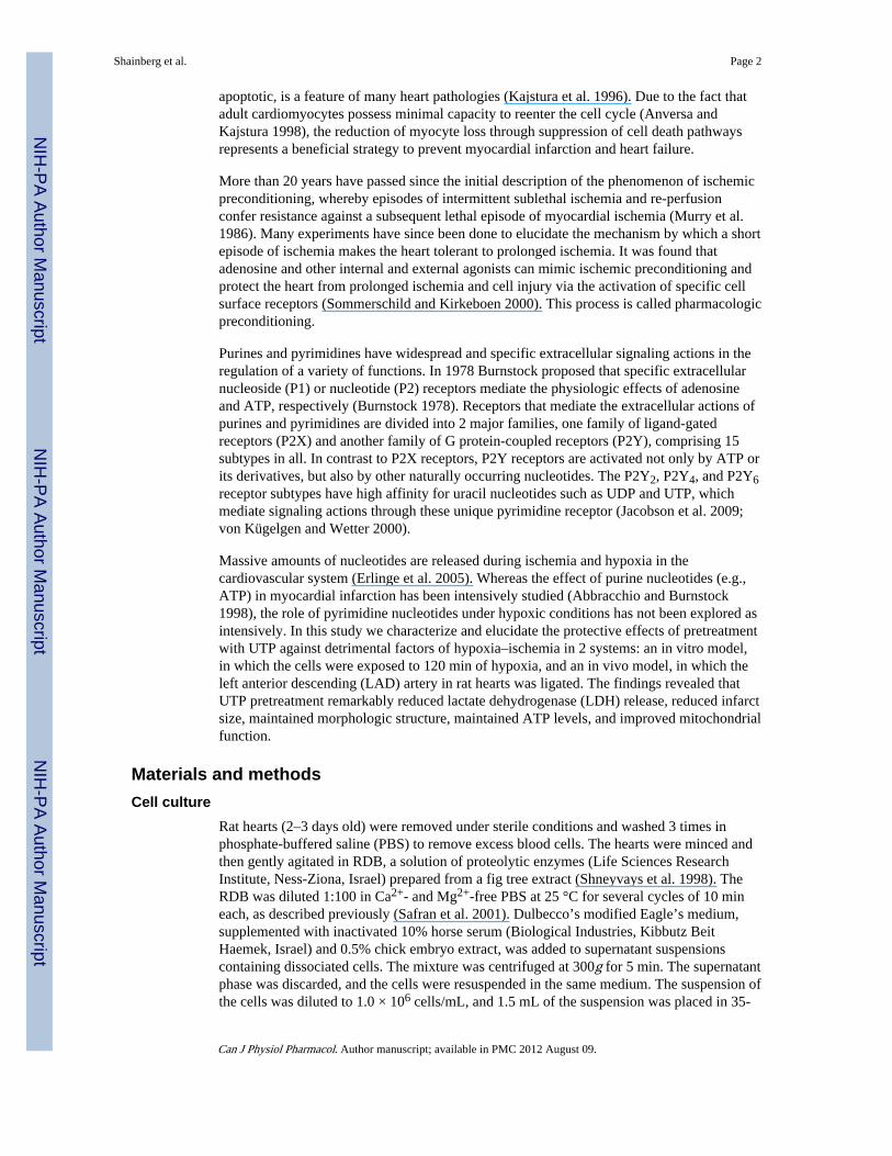
Effect of UTP on cardiomyocytes after hypoxia—To investigate the protectiveeffects of UTP in the attenuation of myocyte injury during prolonged (120 min) hypoxia,cultured cardiomyocytes were incubated with various concentrations of UTP (3–50 μmol/L)for 15 min before hypoxia. As shown in Fig. 1A, the level of LDH release from thecardiomyocytes to the medium was elevated as a result of 120 min of hypoxia (25.55% ±2.84%) compared with that of control cells (4.16% ± 0.48%). Treatment with 50 μmol/LUTP, which was found to be the most efficacious lower dose, reduced LDH elevation(6.06% ± 0.61%). UTP in normoxic conditions did not alter the LDH released to themedium (data not shown). On the other hand, the related derivatives uridine and UDP (50μmol/L) did not exhibit a protective effect against hypoxia in treated cardiomyocytescompared with control cells (data not shown). This ruled out the P2Y6 receptor as a site ofaction of protective uracil nucleotides. These results suggest that UTP pretreatmentdiminishes acute hypoxia-induced cardiomyocyte injury.
To investigate the time-dependence of UTP in protecting cardiomyocytes, the cells werepretreated with 50 μmol/L UTP for 1 h and then exposed to 120 min of hypoxia afterincubation in full medium for 15 min to 72 h. As revealed by LDH release (Fig. 1B), UTPprotected the cells from hypoxia compared with hypoxic cells (55.25% ± 16.29%), evenwhen treated 48 h before hypoxia (33.02% ± 3.43%), but not when treated 72 h before(58.81% ± 2.85%). There was no change in the protective effect between the cells that werewashed after 1 h treatment or incubated with UTP during the entire prolonged treatment (48and 72 h) (data not shown). These results suggest that UTP activates a long, persistentprotective mechanism (a second window of protection).
Involvement of P2 receptors in UTP-induced cardioprotection—To verifywhether the protective effect caused by UTP is mediated via P2 receptors, cardiomyocyteswere pretreated for 15 min with P2 receptor antagonists (RB2, PPADS, or suramin) beforeUTP treatment and hypoxia. Pretreatment with suramin or PPADS, but not with RB2,inhibited the protective effect of UTP, as determined by LDH released to the medium (Table1), indicating the involvement of P2Y2 receptors and (or) P2Y4 receptors in UTP-inducedprotection of cultured cardiomyocytes.
To confirm the presence of P2Y2 and P2Y4 receptors on neonatal cardiomyocytemembranes, immunostaining was performed with specific antibodies. We found that bothreceptors were present in significant amounts on the cell membrane (data not shown).
The protective effect of UTP was also demonstrated through cardiomyocyte morphology.After hypoxia the cells were fixed and stained with hematoxylin and eosin, which showedtypical irreversible necrotic damage of the cardiomyocytes: vacuoles and disorder of the
Shainberg et al. Page 6
Can J Physiol Pharmacol. Author manuscript; available in PMC 2012 August 09.
NIH
-PA Author Manuscript
NIH
-PA Author Manuscript
NIH
-PA Author Manuscript

myofilaments, pyknotic nuclei, and edematous areas in the cytoplasm and around thenucleus. In addition, hypoxic conditions increased the accumulation of the lysosomotropicvital dye neutral red in cytoplasmic granules and around the nucleus and increased the sizeof the lysosomes and their color intensity (Fig. 2). The damage to cultured cells treated withUTP before hypoxia was greatly reduced. Cell loss (percentage of cell death) was presentedas the number of dead cells (propidium iodide-stained) as a percentage of the total numberof cells (Hoechst 33342-stained). Cell loss was 40% ± 5% after 120 min of hypoxia.Activation of the UTP receptors attenuated cell loss caused by hypoxia to 18% ± 2%;control untreated cells had 3.1% ± 0.2% cell loss.
Effect of UTP on cytosolic ATP levels after hypoxia—To evaluate the protectiveeffect produced by extracellular UTP, we measured the total cytosolic ATP content afterhypoxia. The results showed that the ATP level decreased dramatically after 2 h exposure tohypoxia compared with that of normoxic cultures (2.92 ± 0.17 vs. 20.12 ± 2.31 nmol/mg)(Fig. 3). Pretreatment of cardiomyocytes with 50 μmol/L UTP prevented the dramaticdecrease of ATP levels (16.62 ± 4.62 nmol/mg) in cultures subjected to hypoxia. Protectionagainst ATP depletion did not occur when cells were pretreated with the P2 receptorantagonist PPADS (1.84 ± 1.31 nmol/mg) or suramin (2.56 ± 0.87 nmol/mg) beforeexposure to UTP, whereas treatment with RB2 did not prevent the protective effect of UTP(15.88 ± 1.9 nmol/mg) (Fig. 3), indicating that the protective effect of UTP is mediated viaP2 receptors.
Effect of UTP on [Ca2+]i levels during hypoxia—To investigate the effect of UTPpretreatment on [Ca2+]i levels during hypoxic conditions, we continuously monitored the[Ca2+]i level during hypoxia with a specially designed chamber in which oxygen wasexcluded from the cells by a laminar counterflowing layer of the inert gas argon. The levelof [Ca2+]i began to elevate, along with the cessation of beating activity, approximately 10–12 min after the initiation of hypoxia (Fig. 4A). When the cardiomyocytes were pretreatedwith UTP (50 μmol/L) for 15 min and then subjected to hypoxia 30 min later, a delay in[Ca2+]i elevation was observed, and spontaneous contractions lasted for 30–40 min after theinitiation of hypoxia (Fig. 4B). Similar pretreatment with UTP occurring 24 h before thehypoxia maintained spontaneous contractility even after 40 min of hypoxia (Fig. 4C). Theseresults demonstrate that UTP pretreatment is able to prevent an increase in [Ca2+]i duringhypoxia.
Next, we examined cardiomyocyte mitochondrial calcium using the mitochondrial calciumprobe rhod-2 (Jo et al. 2006). The level of mitochondrial calcium observed incardiomyocytes after 2 h hypoxia was high compared with that of controls (Fig. 5). Incardiomyocytes that were treated with 50 μmol/L UTP 30 min or 24 h before hypoxia,however, mitochondrial calcium increase under hypoxic conditions was prevented.
Effect of UTP receptor activation on [Ca2+]i—To investigate the signal transductionpathway of the cardioprotection against hypoxic stress produced by UTP, we examinedseveral effects that might trigger a cardioprotective response. First, we measured the [Ca2+]ilevels by using indo-1 as a fluorescent probe. As shown in Fig. 6A, treatment with 50 μmol/L UTP induced transient acceleration of the beating rate and a transient elevation of thebaseline [Ca2+]i, which lasted 30–40 s before normal beating activity was restored.Application of PPADS (10 μmol/L) or RB2 (50 μmol/L) prevented [Ca2+]i elevation (Figs.6B and 6C). Suramin (300 μmol/L), which acts as an antagonist of P2Y receptors amongother P2 sub-types, did not prevent [Ca2+]i elevation after activation of the UTP receptors(Fig. 6D).
Shainberg et al. Page 7
Can J Physiol Pharmacol. Author manuscript; available in PMC 2012 August 09.
NIH
-PA Author Manuscript
NIH
-PA Author Manuscript
NIH
-PA Author Manuscript

A possible pathway of UTP receptor signaling leading to an increase in [Ca2+]i is activationof phospholipase C (PLC), which mediates inositol 1,4,5-trisphosphate (IP3) production andcalcium release from IP3 receptors (Puceat and Vassort 1996). Figures 6E and 6F show thatapplication of 2 μmol/L U73122 (an inhibitor of PLC activity) or 50 μmol/L 2-aminoethoxydiphenyl borate (2APB) (an IP3 receptor inhibitor) abolished Ca2+ elevationafter activation of UTP receptors.
To correlate the [Ca2+]i elevation after UTP treatment to the cardioprotective effect, wetreated the cells with 2 μmol/L U73122 or with 50 μmol/L 2APB before UTP and hypoxia.LDH levels were measured after the hypoxia and revealed that neither of the 2 inhibitorsdiminished the protective effects produced by UTP. Other evidence to support the exclusionof [Ca2+]i elevation involvement in these protective effects is that UDP and uridine alsocaused [Ca2+]i elevation but not cardioprotection (measured by LDH) (data not shown).
Activation of mitogen-activated protein kinases after UTP treatment—It is wellestablished that activation of various survival kinases can protect cardiomyocytes againststress conditions after pharmacologic preconditioning (Hausenloy and Yellon 2006; Zauggand Schaub 2003). It was previously reported that UTP activates Akt and ERK cascades inneural cells (Arthur et al. 2006) and Akt in renal mesangial cells (Huwiler et al. 2002),which may promote cell survival.
To investigate the signal transduction pathway of UTP that protected cardiomyocytesagainst hypoxic stress, we examined whether UTP treatment before the hypoxic conditionsinduces a change in mitogen-activated protein kinase (MAPK) activation (i.e., Akt andERK1/2). Therefore, we treated the cells with 50 μmol/L UTP for 1–40 min. MAPKsactivation occur via phosphorylation of specific amino acids, which were detected byWestern blotting using specific antibodies. As shown in Fig. 7A, UTP caused an immediateand significant increase in the ERK phosphorylation level. UTP increased ERKphosphorylation in a time-dependent manner, starting after 1 min and lasting over 40 min,with peak activation at 5 min (680% relative to control level). Upon treatment with 10 μmol/L U0126 (a highly selective inhibitor of both MEK1 and MEK2), which blocks the ERKpathway, the UTP-induced ERK phosphorylation was completely abolished (2% comparedwith control level) (Fig. 7B).
We also found that UTP induced Akt phosphorylation in a time-dependent manner, whichstarted after 2 min and lasted over 20 min, with peak activation occurring at 5 min. Blockingthis phosphorylation with LY294002 (a specific phosphatidylinositol 3 (PI3)-kinaseinhibitor, 30 μmol/L) abolished UTP-induced Akt phosphorylation (data not shown).
Next, we examined whether UTP-induced ERK and Akt phosphorylation is related to thecardioprotection against hypoxic stress produced by UTP. Therefore, we treated culturedcardiomyocytes with 10 μmol/L U0126 and 30 μmol/L LY294002 for 15 min, and included50 μmol/L UTP for another 15 min before hypoxia. As shown in Fig. 8, U0126 andLY294002 treatment did not abolish the protective effect induced by UTP (5.5% and 7.6%vs. hypoxia alone). In addition, U0126 and LY294002 in hypoxic conditions had noprotective effect like that induced by UTP, as revealed by LDH release compared withhypoxic conditions alone. These data indicate that ERK and Akt activation induced by UTPis not related to the cardioprotection.
In vivo resultsPretreatment with UTP reduces ischemic damage—To investigate the effect ofUTP on the infarcted hearts in an in vivo model, rats were treated with UTP (0.44 μg/kgi.v.) 30 min or 24 h before MI. The area at risk and the infarcted area at risk in the left
Shainberg et al. Page 8
Can J Physiol Pharmacol. Author manuscript; available in PMC 2012 August 09.
NIH
-PA Author Manuscript
NIH
-PA Author Manuscript
NIH
-PA Author Manuscript

ventricle of rat hearts were measured 24 h after MI by using Evans blue and 2,3,5-triphenyltetrazolium chloride (TTC) staining, respectively. TTC solution stains functionalmitochondria in red, while nonactivated mitochondria appear as white areas. As revealed inthe results, the area at risk in the infarcted animals was similar in all groups, that is,untreated infarcted hearts and the UTP-injected group. The infarcted area (TTC unstained)within the area at risk, however, was markedly reduced compared with that of the control(30.4% ± 2.6%) after UTP treatment 30 min or 24 h before MI (10.9% ± 1.3% and 11.1% ±3.1%, respectively, p < 0.001) (Fig. 9).
UTP preserves ATP levels in LAD-ligated hearts—To evaluate the cytosolic ATPlevels in the LAD-ligated hearts, we harvested the risky myocardial area. The cell extractwas used to measure ATP content with the luciferin–luciferase bioluminescence kit. The invivo experiments were consistent with results obtained in the cultured cardiomyocytes (Fig.3), demonstrating that pretreatment with UTP partially prevented the dramatic decrease inintracellular ATP. The ATP level in the infarcted area of the rat heart post-MI wassignificantly lower than that in sham animals (0.53 ± 0.43 vs. 9.62 ± 2.41 nmol/mg). In theUTP-treated group, the ATP level in the damaged area was higher than that in the untreatedMI rats (2.86 ± 1.01 nmol/mg) (Fig. 10)
UTP preserves mitochondrial complex activity in LAD-occluded rat hearts—Toinvestigate the effects of UTP on mitochondrial complex activity in LAD-occluded rathearts, sections of 10 μm from infarcted hearts, which were pretreated with UTP 30 minbefore the LAD occlusion, were frozen and mitochondrial complexes were stained forNADH:ubiquinone oxidoreductase (complex I), SDH (complex II), or COX (complex IV).The microscopic observation of mitochondria in the infarcted area showed significantdamage at the mitochondrial I and II complexes, which was visualized as white unstainedareas (Fig. 11) (staining results for complex I are not shown). However, pretreatment withUTP decreased the injury dramatically by maintaining their normal activity (as indicated byareas of blue staining). In addition, the efficacy of UTP treatment in mitochondrialprotection in infarcted hearts was observed using COX staining. In slices of the infarctedhearts, large unstained areas were seen, but sham-operated animals and UTP-treated heartsexhibited normal COX activity, which appeared as a deep yellow color, indicating animprovement in mitochondrial respiratory complex IV activity (Fig. 11B). For comparison,the protective effect of UTP on SDH activity in cultured cardiomyocytes subjected tohypoxic conditions is demonstrated (Fig. 11A).
DiscussionIn this work we have demonstrated that UTP protected and significantly reduced cell deathin cultured rat cardiomyocytes and in LAD-ligated hearts that displayed hypoxic–ischemicdamage. This effect of UTP has been demonstrated by biochemical and morphologiccriteria. The difference between UTP-treated and untreated cells that were exposed tohypoxic conditions was remarkable. In untreated cultured cardiomyocytes, loss of striationsin myofibrils, high lysosomal activity, pyknotic nuclei, and edematous areas in thecytoplasm and around the nucleus were observed. In the in vivo model, untreated LAD-ligated hearts showed nonactive mitochondria as white areas by TTC staining. On the otherhand, cells treated with UTP, even when treated 24 h before the hypoxic conditions,exhibited strands of well-organized striated myofibrils running in various directions, and themyocytes possessed 1 or 2 nuclei, which were very similar to those observed with normoxicconditions. The in vivo model showed partially maintained ATP levels, improvedmitochondrial activity, and reduced infarct size in UTP-treated animals.
Shainberg et al. Page 9
Can J Physiol Pharmacol. Author manuscript; available in PMC 2012 August 09.
NIH
-PA Author Manuscript
NIH
-PA Author Manuscript
NIH
-PA Author Manuscript

One of the initial stages during our exploration of the protective effect of UTP was thedetection of UTP receptors on rat cultured cardiomyocytes. By using specific antibodies toP2Y2 and P2Y4 receptors we were able to show that these receptors are highly expressed inthese cells. It is worth mentioning that P2Y4 receptors could not be detected in adultmyocytes (Hou et al. 1999).
Several researchers have reported that the P2Y2 receptor is preferentially inhibited bysuramin, whereas RB2 and PPADS are antagonists for P2Y4 receptors (Ralevic andBurnstock 1998). Our results imply that despite its central physiologic role in cardiac cells,intracellular Ca2+ elevation is not one of the critical elements in the cell signaling pathwayrequired for UTP-induced cardioprotection. It is possible that activation of at least 2 distinctreceptors elicits different downstream signaling pathways. We conclude that the stimulationof the P2Y2 receptor and possibly the P2Y4 receptor by UTP in heart initiates signalingpathways that delay irreversible cardiomyocyte injury and protect myocytes against hypoxicdamage, whereas stimulation of the P2Y4 receptor causes [Ca2+]i elevation, which isprobably irrelevant to classic cardioprotection.
The mechanism by which UTP induces cardioprotection is not known. Yet UTP has beenshown to activate P2Y receptors (i.e., P2Y2 and P2Y4) and this by itself provides anindication of its mode of action. Recent studies on UTP signaling have shown that thesereceptors are coupled through a Gq/11 protein to the IP3 pathway, which causes Ca2+
elevation (Podrasky et al. 1997). Using Ca2+-free medium, or the L-type Ca2+ channelblocker verapamil, we were able to show that in normoxic conditions, UTP elevated the[Ca2+]i from internal sources. Nevertheless, this pathway of Ca2+ elevation is probably notinvolved in myocyte protection from hypoxia, since using different inhibitors of Ca2+
elevation (U73122, 2APB) did not hamper the protective effect. Another piece of evidenceis that other uridine derivatives (UDP, uridine), although causing a transient elevation of[Ca2+]i, did not protect the cells from hypoxia. Moreover, suramin, which reduced thecardioprotective effect of UTP, had no influence on [Ca2+]i elevation.
During ischemia, massive amounts of adenosine, UTP, and other nucleotides are released inthe cardiovascular system. The adenosine receptors are thought to be the dominant playersresponsible for the induction of pharmacologic preconditioning, which enhances theresistance of the heart to ischemic conditions. One can suggest that both UTP and adenosinemay act in a similar mode to promote cardiomyocyte survival under ischemic conditions. Ifthis is the case, UTP may also contribute to the preconditioning effect. Indeed, pretreatmentwith UTP 24 h before exposing to hypoxic conditions led to reduced myocytes damage andpreserved cell contractility. Like adenosine, the cardioprotective effect induced by UTP maybe associated with protection of the mitochondrial respiratory chain, keeping ATPproduction level high by maintaining membrane potential and activating mitochondrialKATP channels. We also found that cardiomyocytes that were treated with 50 μmol/L UTP30 min or 24 h before hypoxia demonstrated a prevention of mitochondrial calcium levelincrease under hypoxic conditions. This reduction may provide an excellent explanation forkeeping ATP production level high by maintaining mitochondrial membrane potential(Yitzhaki et al. 2007).
It is well established that activation of different survival kinases can protect cardiomyocytesagainst stress conditions after pharmacologic preconditioning (Hausenloy and Yellon 2006;Zaugg and Schaub 2003). We found that UTP pre-treatment induced ERK and Aktphosphorylation. Blocking these pathways by U0126 and LY294002, respectively, had noeffect on this protective effect. We will continue to investigate other survival kinases thatmay be activated by UTP pretreatment and may contribute in a cooperative way to thecardioprotection.
Shainberg et al. Page 10
Can J Physiol Pharmacol. Author manuscript; available in PMC 2012 August 09.
NIH
-PA Author Manuscript
NIH
-PA Author Manuscript
NIH
-PA Author Manuscript
Finally, when thinking about therapeutic applications, targeting pyrimidinergic receptors forprotection against ischemic myocardial damage has an advantage compared with targetingpurinergic receptors. Unlike adenosine, the breakdown product of UTP, uridine, has limitedancillary pharmacologic effects (Vassort 2001). There is a general belief that ATP is thepreferred agonist of P2 purinoceptors. At the same time, ATP is degraded to adeninenucleotides and nucleosides, so that the response of cardiomyocytes would be an integrationof the various effects of these multiple compounds generated by the different surroundingtissues (Vassort 2001). UTP therefore may be used in preference to ATP as a P2Y2 andP2Y4 receptor agonist because it does not form cardiovascular active metabolites such asadenosine. Mazzola et al. (2008) confirmed this assumption by demonstrating differenteffects of these nucleotides on cardiomyocyte viability. Whereas exposure of the HL-1cardiomyocytes to ATP induced apoptosis and necrosis, UTP counteracted the deleteriouseffects of ATP (Mazzola et al. 2008). Arthur et al. (2006) have shown that ATP causes arobust activation of phospholipase C via activation of Gq in newborn rat cardiomyocytes,and it would be expected to cause hypertrophic responses like other factors that active Gq-coupled receptors, such as α-adrenergic agonists, endothelin, and angiotensin II during long-term exposure (Pham et al. 2003). However, ATP was reported to be inhibitory tohypertrophic responses (Zheng et al. 1996). On the contrary, UTP, an alternative agonist toATP at P2Y2 receptors, was as effective as ATP in stimulating PLC and caused hypertrophicresponses in rat cardiomyocytes (Pham et al. 2003). As we have shown in this study, shorttime exposure to UTP stimulates both a ‘classic’ and a persistent long-term mechanism ofprotection (second window of protection). The involvement of UTP receptors in mediatingcardioprotection reflects the biological redundancy in life-saving signals of P2Y2 and P2Y4purinoceptors, although the absence of selective antagonists restricts our understanding ofthese signal transduction pathways and the elucidation of the importance of the individualreceptors.
In conclusion, therapeutic targeting of pyrimidinergic receptors for protection againstischemic myocardial damage may be more effective than targeting the purinergic receptors.The possible therapeutic application of such a potent and sustained effect in patients at riskof myocardial ischemic damage is therefore very promising and justifies further exploration.
AcknowledgmentsThis research study was conducted through the generous support of the Adar Program for the Advancement ofResearch in Heart Function and the Horowitz Foundation at Bar-Ilan University. This research was also supportedin part by the Intramural Research Program of the NIH National Institute of Diabetes and Digestive and KidneyDiseases (K.A.J.).
ReferencesAbbracchio MP, Burnstock G. Purinergic signalling: pathophysiological roles. Jpn J Pharmacol. 1998;
78:113–145.10.1254/jjp.78.113 [PubMed: 9829617]
Anversa P, Kajstura J. Ventricular myocytes are not terminally differentiated in the adult mammalianheart. Circ Res. 1998; 83:1–14. [PubMed: 9670913]
Arthur DB, Georgi S, Akassoglou K, Insel PA. Inhibition of apoptosis by P2Y2 receptor activation:novel pathways for neuronal survival. J Neurosci. 2006; 26:3798–3804.10.1523/JNEUROSCI.5338-05.2006 [PubMed: 16597733]
Burnstock, G. A basis for distinguishing two types of purinergic receptor. In: Bolis, L.; Straub, RW.,editors. Cell membrane receptors for drugs and hormones: a multidisciplinary approach. RavenPress; New York: 1978. p. 107-118.
El Ani D, Jacobson KA, Shainberg A. Characterization of adenosine receptors in intact cultured heartcells. Biochem Pharmacol. 1994; 48:727–735.10.1016/0006-2952(94) 90050-7 [PubMed: 8080445]
Shainberg et al. Page 11
Can J Physiol Pharmacol. Author manuscript; available in PMC 2012 August 09.
NIH
-PA Author Manuscript
NIH
-PA Author Manuscript
NIH
-PA Author Manuscript

Erlinge D, Harnek J, van Heusden C, Olivecrona G, Jern S, Lazarowski E. Uridine triphosphate (UTP)is released during cardiac ischemia. Int J Cardiol. 2005; 100:427–433.10.1016/j.ijcard.2004.10.005[PubMed: 15837087]
Fixler D, Tirosh R, Zinman T, Shainberg A, Deutsch M. Differential aspects in ratio measurements of[Ca2+]i relaxation in cardiomyocyte contraction following various drug treatments. Cell Calcium.2002; 31:279–287.10.1016/S0143-4160(02)00056-8 [PubMed: 12098217]
Hausenloy DJ, Yellon DM. Survival kinases in ischemic preconditioning and postconditioning.Cardiovasc Res. 2006; 70:240–253.10.1016/j.cardiores.2006.01.017 [PubMed: 16545352]
Hou M, Malmsjo M, Moller S, Pantev E, Bergdahl A, Zhao XH, et al. Increase in cardiac P2X1-andP2Y2-receptor mRNA levels in congestive heart failure. Life Sci. 1999; 65:1195–1206.10.1016/S0024-3205(99)00353-7 [PubMed: 10503935]
Huwiler A, Rolz W, Dorsch S, Ren S, Pfeilschifter J. Extracellular ATP and UTP activate the proteinkinase B/Akt cascade via the P2Y(2) purinoceptor in renal mesangial cells. Br J Pharmacol. 2002;136:520–529.10.1038/sj.bjp. 0704748 [PubMed: 12055130]
Jacobson KA, Ivanov AA, de Castro S, Harden TK, Ko H. Development of selective agonists andantagonists of P2Y receptors. Purinergic Signal. 2009; 5:75–89.10.1007/s11302-008-9106-2[PubMed: 18600475]
Jo H, Noma A, Matsuoka S. Calcium-mediated coupling between mitochondrial substratedehydrogenation and cardiac workload in single guinea-pig ventricular myocytes. J Mol CellCardiol. 2006; 40:394–404.10.1016/j.yjmcc.2005.12.012 [PubMed: 16480740]
Kajstura J, Cheng W, Reiss K, Clark WA, Sonnenblick EH, Krajewski S, et al. Apoptotic and necroticmyocyte cell deaths are independent contributing variables of infarct size in rats. Lab Invest. 1996;74:86–107. [PubMed: 8569201]
Mazzola A, Amoruso E, Beltrami E, Lecca D, Ferrario S, Cosentino S, et al. Opposite effects of uraciland adenine nucleotides on the survival of murine cardiomyocytes. J Cell Mol Med. 2008; 12:522–536.10.1111/j.1582-4934.2007.00133.x [PubMed: 18419595]
Morgan EE, Faulx MD, McElfresh TA, Kung TA, Zawaneh MS, Stanley WC, et al. Validation ofechocardiographic methods for assessing left ventricular dysfunction in rats with myocardialinfarction. Am J Physiol Heart Circ Physiol. 2004; 287:H2049–H2053.10.1152/ajpheart.00393.2004 [PubMed: 15475530]
Murry CE, Jennings RB, Reimer KA. Preconditioning with ischemia: a delay of lethal cell injury inischemic myocardium. Circulation. 1986; 74:1124–1136. [PubMed: 3769170]
Nieminen AL, Gores GJ, Bond JM, Imberti R, Herman B, Lemasters JJ. A novel cytotoxicityscreening assay using a multiwell fluorescence scanner. Toxicol Appl Pharmacol. 1992; 115:147–155.10.1016/0041-008X(92)90317-L [PubMed: 1641848]
Pham TM, Morris JB, Arthur JF, Post GR, Brown JH, Woodcock EA. UTP but not ATP causeshypertrophic growth in neonatal rat cardiomyocytes. J Mol Cell Cardiol. 2003; 35:287–292.10.1016/S0022-2828(03)00009-9 [PubMed: 12676543]
Podrasky E, Xu D, Liang BT. A novel phospholipase C- and cAMP-independent positive inotropicmechanism via a P2 purinoceptor. Am J Physiol. 1997; 273:H2380–H2387. [PubMed: 9374775]
Puceat M, Vassort G. Purinergic stimulation of rat cardiomyocytes induces tyrosine phosphorylationand membrane association of phospholipase C gamma: a major mechanism for InsP3 generation.Biochem J. 1996; 318:723–728. [PubMed: 8809068]
Ralevic V, Burnstock G. Receptors for purines and pyrimidines. Pharmacol Rev. 1998; 50:413–492.[PubMed: 9755289]
Safran N, Shneyvays V, Balas N, Jacobson KA, Shainberg A. Cardioprotective effects of adenosineA1 and A3 receptor activation during hypoxia in isolated rat cardiac myocytes. Mol Cell Biochem.2001; 217:143–152.10.1023/A:1007209321969 [PubMed: 11269659]
Shneyvays V, Nawrath H, Jacobson KA, Shainberg A. Induction of apoptosis in cardiac myocytes byan A3 adenosine receptor agonist. Exp Cell Res. 1998; 243:383–397.10. 1006/excr.1998.4134[PubMed: 9743598]
Shneyvays V, Safran N, Halili-Rutman I, Shainberg A. Insights into adenosine A1 and A3 receptorsfunction: cardiotoxicity and cardioprotection. Drug Dev Res. 2000; 50:324–337.10.1002/1098-2299(200007/08)50:3/4<324::AID-DDR16>3. 0.CO;2-B
Shainberg et al. Page 12
Can J Physiol Pharmacol. Author manuscript; available in PMC 2012 August 09.
NIH
-PA Author Manuscript
NIH
-PA Author Manuscript
NIH
-PA Author Manuscript

Shneyvays V, Mamedova L, Zinman T, Jacobson K, Shainberg A. Activation of A3 adenosine receptorprotects against doxorubicin-induced cardiotoxicity. J Mol Cell Cardiol. 2001; 33:1249–1261.10.1006/jmcc.2001.1387 [PubMed: 11444927]
Shneyvays, V.; Leshem, D.; Mamedova, LK.; Shainberg, A. Activation of adenosine A1 and A3receptors protects mitochondria during hypoxia in cardiomyocytes by distinct mechanisms. In:Dhalla, NS.; Takeda, N.; Singh, M.; Lukas, A., editors. Myocardial ischemia and preconditioning.Kluwer Academic Publishers; Boston: 2003. p. 347-364.
Sommerschild HT, Kirkeboen KA. Adenosine and cardioprotection during ischaemia and reperfusion:an overview. Acta Anaesthesiol Scand. 2000; 44:1038–1055.10.1034/j.1399-6576.2000.440903.x[PubMed: 11028722]
Vassort G. Adenosine 5′-triphosphate: a P2-purinergic agonist in the myocardium. Physiol Rev. 2001;81:767–806. [PubMed: 11274344]
von Kügelgen I, Wetter A. Molecular pharmacology of P2Y-receptors. Naunyn Schmiedebergs ArchPharmacol. 2000; 362:310–323.10.1007/s002100000310 [PubMed: 11111826]
Yitzhaki S, Shneyvays V, Jacobson KA, Shainberg A. Involvement of uracil nucleotides in protectionof cardiomyocytes from hypoxic stress. Biochem Pharmacol. 2005; 69:1215–1223.10.1016/j.bcp.2005.01.018 [PubMed: 15794942]
Yitzhaki S, Shainberg A, Cheporko Y, Vidne BA, Sagie A, Jacobson KA, Hochhauser E. Uridine-5′-triphosphate (UTP) reduces infarct size and improves rat heart function after myocardial infarct.Biochem Pharmacol. 2006; 72:949–955.10.1016/j.bcp.2006.07.019 [PubMed: 16939682]
Yitzhaki S, Hochhauser E, Porat E, Shainberg A. Uridine-5′-triphosphate (UTP) maintains cardiacmitochondrial function following chemical and hypoxic stress. J Mol Cell Cardiol. 2007; 43:653–662.10.1016/j.yjmcc.2007.07.060 [PubMed: 17880998]
Zaugg M, Schaub MC. Signaling and cellular mechanisms in cardiac protection by ischemic andpharmacological preconditioning. J Muscle Res Cell Motil. 2003; 24:219–249.10.1023/A:1026021430091 [PubMed: 14609033]
Zheng JS, Boluyt MO, Long X, O’Neill L, Lakatta EG, Crow MT. Extracellular ATP inhibitsadrenergic agonist-induced hypertrophy of neonatal cardiac myocytes. Circ Res. 1996; 78:525–535. [PubMed: 8635209]
Shainberg et al. Page 13
Can J Physiol Pharmacol. Author manuscript; available in PMC 2012 August 09.
NIH
-PA Author Manuscript
NIH
-PA Author Manuscript
NIH
-PA Author Manuscript
Fig. 1.Concentration- and time-dependent effects of UTP on cultured cardiomyocytes subjected tohypoxia. (A) Six-day-old rat cardiomyocytes were treated for 15 min with variousconcentrations of UTP (3–50 μmol/L). The cells were then washed twice and subjected tohypoxia for 2 h in glucose-free PBS at 37 °C. The amount of lactate dehydrogenase (LDH)released to the medium was determined and compared with the total activity of controlhomogenate (100%). (B) Cardiomyocytes were treated for 15 min or 24 h with 50 μmol/LUTP before hypoxia. Other groups of cells were incubated for 1 h with 50 μmol/L UTP,washed and replaced in normal medium, and then subjected to hypoxia 24, 48, or 72 hsubsequent to the treatment with UTP. Data are means of at least 3 replicates in 5 separateexperiments ± SD. *, Significant at p < 0.05, **, p < 0.01 compared with hypoxia.
Shainberg et al. Page 14
Can J Physiol Pharmacol. Author manuscript; available in PMC 2012 August 09.
NIH
-PA Author Manuscript
NIH
-PA Author Manuscript
NIH
-PA Author Manuscript
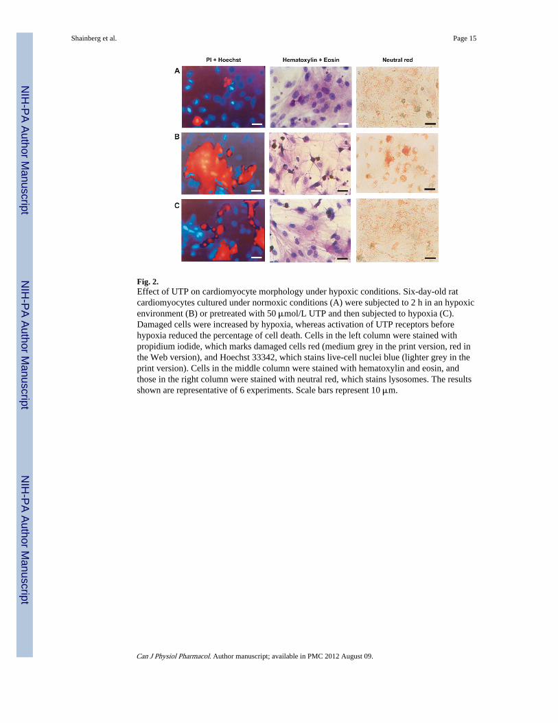
Fig. 2.Effect of UTP on cardiomyocyte morphology under hypoxic conditions. Six-day-old ratcardiomyocytes cultured under normoxic conditions (A) were subjected to 2 h in an hypoxicenvironment (B) or pretreated with 50 μmol/L UTP and then subjected to hypoxia (C).Damaged cells were increased by hypoxia, whereas activation of UTP receptors beforehypoxia reduced the percentage of cell death. Cells in the left column were stained withpropidium iodide, which marks damaged cells red (medium grey in the print version, red inthe Web version), and Hoechst 33342, which stains live-cell nuclei blue (lighter grey in theprint version). Cells in the middle column were stained with hematoxylin and eosin, andthose in the right column were stained with neutral red, which stains lysosomes. The resultsshown are representative of 6 experiments. Scale bars represent 10 μm.
Shainberg et al. Page 15
Can J Physiol Pharmacol. Author manuscript; available in PMC 2012 August 09.
NIH
-PA Author Manuscript
NIH
-PA Author Manuscript
NIH
-PA Author Manuscript

Fig. 3.Effect of P2 receptor agonists and antagonists on intracellular ATP levels after hypoxia invitro. Six-day-old rat cardiomyocytes were treated for 15 min with the P2 antagonistsPPADS (10 μmol/L), RB2 (50 μmol/L), or suramin (300 μmol/L) before application ofUTP (50 μmol/L) for 15 min. Intracellular ATP levels were measured after 2 h of hypoxia.Data are means of at least 3 replicates in 6 separate experiments ± SD. *, Significant at p <0.01 compared with hypoxia; #, p < 0.001 compared with normoxia.
Shainberg et al. Page 16
Can J Physiol Pharmacol. Author manuscript; available in PMC 2012 August 09.
NIH
-PA Author Manuscript
NIH
-PA Author Manuscript
NIH
-PA Author Manuscript

Fig. 4.Effect of UTP on [Ca2+]i accumulation during hypoxia. Effect of hypoxia alone (A) and 30min (B) or 24 h (C) after UTP treatment for 15 min in 6-day-old rat cardiomyocytes in vitro.Calcium was monitored during hypoxia with fluorescent dye indo-1. The fluorescence ratioof 410/490 nm, which is proportional to changes in Ca2+ levels, is demonstrated. Eachrecording was made for 10 s at times 0, 20, 40, and 60 min after the initiation of hypoxia.Each experiment shown is representative of 5 separate experiments. (D) Basal levels of[Ca2+]i of the 3 groups and a normoxic control as a function of time. Values are means ±SD. *, Significant at p < 0.05 UTP-treated (30 min) compared with hypoxia.
Shainberg et al. Page 17
Can J Physiol Pharmacol. Author manuscript; available in PMC 2012 August 09.
NIH
-PA Author Manuscript
NIH
-PA Author Manuscript
NIH
-PA Author Manuscript

Fig. 5.Effect of pretreatment with UTP on mitochondrial Ca2+ overload in cardiomyocytessubjected to hypoxia. Six-day-old rat cardiomyocytes were treated with 50 μmol/L UTP 30min or 24 h before 2 h of hypoxia. During hypoxia, cardiomyocytes were loaded with rhod-2AM (warm after cold incubation), and Ca2+ level was determined. Values are means ± SD.*, Significant at p < 0.001 compared with control; #, p < 0.001 compared with hypoxia.
Shainberg et al. Page 18
Can J Physiol Pharmacol. Author manuscript; available in PMC 2012 August 09.
NIH
-PA Author Manuscript
NIH
-PA Author Manuscript
NIH
-PA Author Manuscript

Fig. 6.Effect of P2 antagonists on [Ca2+]i response to UTP. Indo-1-loaded 6-day-old ratcardiomyocytes were treated with 50 μmol/L UTP alone (A) or pretreated with the P2antagonists PPADS (B), RB2 (C), or suramin (D) 15 min before UTP application. Othercells were pretreated before UTP with (E) IP3 receptor inhibitor 2APB (50 μmol/L) or (F)PLC inhibitor U73122 (2 μmol/L). The fluorescence ratio of 410/490 nm, which isproportional to changes in Ca2+ levels, is demonstrated. Each experiment shown isrepresentative of 7 separate experiments. IP3, inositol 1,4,5-trisphosphate; 2APB, 2-aminoethoxydiphenyl borate; PLC, phospholipase C.
Shainberg et al. Page 19
Can J Physiol Pharmacol. Author manuscript; available in PMC 2012 August 09.
NIH
-PA Author Manuscript
NIH
-PA Author Manuscript
NIH
-PA Author Manuscript
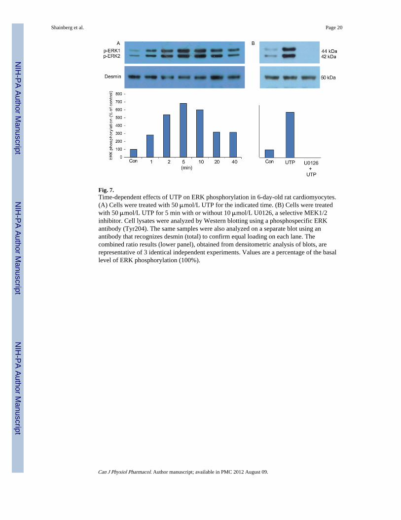
Fig. 7.Time-dependent effects of UTP on ERK phosphorylation in 6-day-old rat cardiomyocytes.(A) Cells were treated with 50 μmol/L UTP for the indicated time. (B) Cells were treatedwith 50 μmol/L UTP for 5 min with or without 10 μmol/L U0126, a selective MEK1/2inhibitor. Cell lysates were analyzed by Western blotting using a phosphospecific ERKantibody (Tyr204). The same samples were also analyzed on a separate blot using anantibody that recognizes desmin (total) to confirm equal loading on each lane. Thecombined ratio results (lower panel), obtained from densitometric analysis of blots, arerepresentative of 3 identical independent experiments. Values are a percentage of the basallevel of ERK phosphorylation (100%).
Shainberg et al. Page 20
Can J Physiol Pharmacol. Author manuscript; available in PMC 2012 August 09.
NIH
-PA Author Manuscript
NIH
-PA Author Manuscript
NIH
-PA Author Manuscript

Fig. 8.Effect of ERK and Akt inhibitors on UTP-induced cardioprotection. Six-day-old ratcardiomyocytes were treated for 15 min with 10 μmol/L U0126 (a selective inhibitor ofMEK1/2) and 30 μmol/L LY294002 (a selective inhibitor of PI3-kinase) before 50 μmol/LUTP. The cells were then washed twice and subjected to hypoxia (Hyp) for 2 h in glucose-free PBS at 37 °C. The amount of LDH released to the medium was determined. The datashown are representative of one of 3 identical independent experiments ± SD. *, Significantat p < 0.01 compared with control; #, p < 0.01 compared with hypoxia. PI3-kinase,phosphatidylinositol 3-kinase.
Shainberg et al. Page 21
Can J Physiol Pharmacol. Author manuscript; available in PMC 2012 August 09.
NIH
-PA Author Manuscript
NIH
-PA Author Manuscript
NIH
-PA Author Manuscript
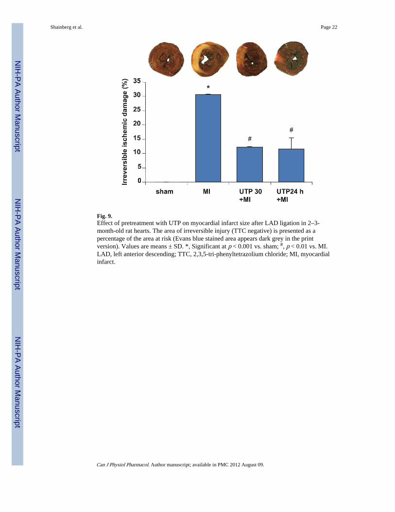
Fig. 9.Effect of pretreatment with UTP on myocardial infarct size after LAD ligation in 2–3-month-old rat hearts. The area of irreversible injury (TTC negative) is presented as apercentage of the area at risk (Evans blue stained area appears dark grey in the printversion). Values are means ± SD. *, Significant at p < 0.001 vs. sham; #, p < 0.01 vs. MI.LAD, left anterior descending; TTC, 2,3,5-tri-phenyltetrazolium chloride; MI, myocardialinfarct.
Shainberg et al. Page 22
Can J Physiol Pharmacol. Author manuscript; available in PMC 2012 August 09.
NIH
-PA Author Manuscript
NIH
-PA Author Manuscript
NIH
-PA Author Manuscript

Fig. 10.Effect of UTP treatment on ATP levels in LAD-ligated hearts of 2–3-month-old rats. TheATP level in the infarct area is shown after the animals were injected with 0.44 μg/kg UTP30 min before MI. Values are means ± SD. *, Significant at p < 0.001 vs. sham; #, p < 0.01vs. MI.
Shainberg et al. Page 23
Can J Physiol Pharmacol. Author manuscript; available in PMC 2012 August 09.
NIH
-PA Author Manuscript
NIH
-PA Author Manuscript
NIH
-PA Author Manuscript
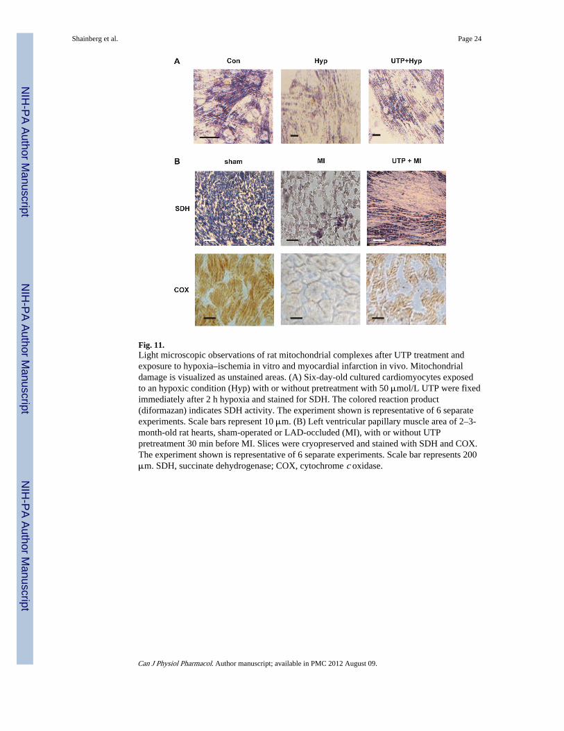
Fig. 11.Light microscopic observations of rat mitochondrial complexes after UTP treatment andexposure to hypoxia–ischemia in vitro and myocardial infarction in vivo. Mitochondrialdamage is visualized as unstained areas. (A) Six-day-old cultured cardiomyocytes exposedto an hypoxic condition (Hyp) with or without pretreatment with 50 μmol/L UTP were fixedimmediately after 2 h hypoxia and stained for SDH. The colored reaction product(diformazan) indicates SDH activity. The experiment shown is representative of 6 separateexperiments. Scale bars represent 10 μm. (B) Left ventricular papillary muscle area of 2–3-month-old rat hearts, sham-operated or LAD-occluded (MI), with or without UTPpretreatment 30 min before MI. Slices were cryopreserved and stained with SDH and COX.The experiment shown is representative of 6 separate experiments. Scale bar represents 200μm. SDH, succinate dehydrogenase; COX, cytochrome c oxidase.
Shainberg et al. Page 24
Can J Physiol Pharmacol. Author manuscript; available in PMC 2012 August 09.
NIH
-PA Author Manuscript
NIH
-PA Author Manuscript
NIH
-PA Author Manuscript

NIH
-PA Author Manuscript
NIH
-PA Author Manuscript
NIH
-PA Author Manuscript
Shainberg et al. Page 25
Table 1
Effect of uracil derivatives or P2 receptor antagonists on cardiomyocytes subjected to hypoxia.
Treatment LDH release, %
Normoxia 4.2±0.6
Hypoxia 40±2.4
Hypoxia + UTP 10±1.6*
Hypoxia + UTP + PPADS 32±3.6
Hypoxia + UTP + suramin 25±3.5
Hypoxia + UTP + RB2 11±0.8*
Note: Six-day-old rat cardiomyocytes were pretreated for 15 min with the P2 receptor antagonists PPADS (pyridoxal-5′-phosphate-6-azophenyl-2′,4′-disulfonate) (10 μmol/L), reactive blue 2 (RB2) (50 μmol/L), or suramin (300 μmol/L) before UTP treatment for 15 min. Thecells were then washed twice and subjected to hypoxia for 2 h in glucose-free PBS at 37 °C. The amount of lactate dehydrogenase (LDH) releasedto the medium was determined and compared with the total activity of control homogenate (100%). Data are means of at least 3 replicates in 3separate experiments ± SD.
*Significant at p < 0.01 compared with hypoxia.
Can J Physiol Pharmacol. Author manuscript; available in PMC 2012 August 09.