in vitro assessment of ros on motility of epididymal sperm of male rat exposed to intraperitoneal...
TRANSCRIPT
169
Document heading doi:
In vitro assessment of ROS on motility of epididymal sperm of male rat exposed to intraperitoneal administration of nonylphenolAnsoumane Kourouma1*, Duan Peng1, Hady Keita2, Aidogie Osamuyimen3, Qi Suqin1, Quan Chao1, Yu Tingting1, Yang Kedi1* 1 MOE Key Lab of Environmental and Health, Department of Occupational and Environmental Health, School of Public Health, Tongji Medical College, Huazhong University of Science and Technology, 13 Hangkong Road, Wuhan 430030, PR China2Universidad Tecnológica del Valle de Toluca. Carretera del Departamento del D.F. km 7.5. Santa María Atarasquillo Lerma, México3School of Medicine and Health Management, Huazhong University of Science and Technology, Wuhan, Hubei, 430030, China
ARTICLE INFO ABSTRACT
Article history:Received 26 December 2014Received in revised form 20 May 2015Accepted 26 May 2015Available online 20 September 2015
Keywords:NonylphenolEpididymisSpermsAntioxidantsCASAROS
*Corresponding author: Dr. Yang Kedi, Corresponding author at: Department of Occupational and Environmental Health, School of Public Health, Tongji Medical College, Huazhong University of Science and Technology, 13 Hangkong Road, Wuhan 430030, PR China. Tel/Fax: +862783692333. E-mail: [email protected] Ansoumane Kourouma, PhD, MOE Key Lab of Environmental and Health, Department of Occupational and Environmental Health, School of Public Health, Tongji Medical College, Huazhong University of Science and Technology, 13 Hangkong Road, Wuhan 430030, PR China. E-mail: [email protected] Funding project: This work was supported by the by grants from the National Natural Science Foundation of China (30972436 and 81172623).
1. Introduction
Male infertility has become a major medical problem as well
as social stigma. The increasing rate of testicular cancer and the
incidences of abnormalities in male and female reproductive tracts[1]
during the last decades have been reported. Alkylphenol especially
nonylphenol (NP) is widely used as lubricating oil additives,
plasticizers and surface-active agents[2]. NP is a final metabolite
of Nonylphenol polyetholate (NPE) and NP is more stable and
persistent than NPE[3]. (NPE) is a non-ionic surfactant widely used
as component of detergent, paints, herbicides, and many other
synthetic products[4].
NP is one of the chemicals believed to cause endocrine disruption
and affect sperm quality in mammals and to play biologically
active for a longer period of time in the body than endogenous
estrogens. However, with the developments of industry, large
amount of NP have been discharged into water[5]. Hence it’s
distributed into aquatic environments such as rivers, lakes and seas
through sewage treatment plants[6, 7]. Its ability to accumulate in
the organs of aquatic species is considered a potential hazard for
the reproductive system in humans and animals exposed to the food
chain. Therefore, cytotoxicity effect of NP carried out in diverse
organs, including those in the reproductive system has been reported
in many studies[8-10]. The disruption of endocrine function by
Objective: To explore the mechanism by which nonylphenol (NP) interferes with male infertility through
evaluation of its effects on epididymal sperm of adult male rats. Methods: Twenty four Sprague-Dawley
(SD) rats were used as epididymal sperm donors. Previously rats were administrated with NP (0, 2, 10 and
50 mg/kg) body weight respectively in corn oil every forty-eight hours by intraperitoneal injection for 30
days. Computer assisted sperm analysis (CASA) was used to determine parameters of sperm. The sperm
morphology examination was conducted with a high resolution microscope. Results: Results indicated that
exposure to NP has no effect on body weight, while testes weights were significantly decreased. Computer
assisted sperm analysis (CASA) showed significant decline in the percentage of motile spermatozoa
(P<0.001), STR and LIN (P<0.01), significant increase in ALH (P<0.001), while significant decline in
BCF (P<0.001) respectively. Plasma LDH was significantly increased while; plasma毭-GT activity was
significantly decreased. H2O2 production and malondialdehyde (MDA) were significantly increased. The
Plasma CAT, GSH-Px and SOD activities were significantly decreased. Conclusions: This concludes that
NP leads oxidative stress in the epididymal sperm of rats. Moreover, NP can disrupt sperm motility and
alterations in the sperm morphology.
Asian Pacific Journal of Reproduction 2015; 4(3): 169-177
Asian Pacific Journal of Reproduction
Journal homepage: www.apjr.net
170 Ansoumane Kourouma et al./ Asian Pacific Journal of Reproduction (2015)169-177
inhibiting between estrogen and the estrogen receptor[10, 11] is due
to its characteristic weak estrogenic activity[12, 13]. Previous studies
collectively suggest that these estrogenic compounds might cause
a variety of negative reproductive outcomes, especially declines in
sperm counts in men, remain extremely controversial[14].
Several environmental contaminants such NP can induce
oxidative stress by generating ROS such as hydrogen peroxide
(H2O2) and superoxide anion (O2-), resulting in great damage to
the male reproductive system, including altered steroidogenesis,
disturbed testicular structure and decreased sperm number
in the epididymis[15]. Moreover, it has been reported that NP
administration increased ROS level and depressed the activity of
antioxidant enzymes in rat epididymal sperms[16], testis[17] and
testicular Sertoli cells[18, 19]. However, the cellular or biochemical
mechanism by which NP causes reproductive toxicity has not
been fully elucidated[8]. When produced in excessive amounts,
the ROS stimulate DNA fragmentation and loss of sperm function
associated with peroxidative damage to the mitochondrial and
plasma membrane. Moreover, sperm plasma membrane, being rich
in polyunsaturated fatty acids, is highly susceptible to ROS attack.
At the same time, epididymis being its transit through caput to cauda
region of epididymis and facilitates their maturation process[20].
The epididymis is known to play an important role in providing the
microenvironment for sperm maturation and storage of sperm[21].
In the last decade the effect of ROS on the semen quality has
been studied but not well established with excess production of
ROS including motility and abnormal spermatozoa. Although
semen analysis is routinely employed in the evaluation of the male
infertility to predict the cause behind impaired fertility. Suitable
sperm motility is precondition for fertilization of the ovum to take
place. Thus an alteration in this time can provoke problems in such
maturation as well as alter the number of gametes available for
ejaculation[22]. Oxidative damage to spermatozoa is well known
to be a potential cause of fertilization failure[23]. This study was
designed to show if estrogens affect sperm function in vitro there
could be important consequences for fertility in vivo. In our present
study, we hypothesized that NP exposure in adult male rats would
lead to oxidative stress by generating ROS that induce reproductive
abnormalities. Thus, the purpose of this study was to evaluate the
effects of NP on sperm motility parameters, morphological and
biochemical marker changes related to oxidative stress in the serum
plasma, and then propose a molecular pathway in order to explain
the deleterious effects of this chemical on epididymal sperm quality.
2. Materials and methods
2.1. Chemicals and reagents
Nonylphenol (NP) was purchased from (DR Co., Augsburg,
Germany, purity: 98%). Corn oil was obtained from (Sigma-Aldrich,
St. Louis, MO, USA). Sigma Chemical Co. (St Louis, MO) USA,
Collagenase, Trypsin–EDTA were obtained from GIBCO (Grand
Island, NY, USA), Sodium lauryl sulphate from SRL, Eosin stain,
Hematoxylin stain, Orange G stain from HiMedia (Mumbai). LDH,
毭-GT CAT, GSH-Px, H2O2, MDA and SOD assay kit (Jiancheng
Bioengineering Ltd., Nanjing, China). All other chemicals are of
analytical grade.
2.2. Animals and treatments
Twenty four healthy male Sprague-Dawley rats (50-days olds) were
used as epididymal sperm donors purchased from the Tongji Medical
College Animal Laboratory (Wuhan, China). Previously rats were
gavaged with NP (0, 2, 10, 50 mg/kg) body weight respectively in corn
oil every forty-eight hours by intra-peritoneal injection for 30 days.
The rats were housed in accordance with the policies of the Ethical
clearance for the use of animals in the study was obtained from the
Institutional Animal Ethics Committee prior to the initiation of the
study, and the experiments were performed in accordance with the
guidelines for the Care and Use of Laboratory Animals published by
Ministry of Health of People’s Republic of China (Approval
ID: 2011-s2456). Rats were housed in conventional rat cages at
(24±3) 曟, humidity (50 ± 5) % in a controlled light environment
(12 h light: 12 h dark) and provided free access to water and
standard rodent chow, and the weight of each animal was recorded
every forty-eight hours for 30 days and any gross abnormality was
noted.
2.3. Dose selection and preparation
The doses and time used for the present study were derived from
published data[24, 25] and the results of our preliminary experiment.
4-NP was dissolved in corn oil to obtain the desired concentration
of NP dose range, i.e., 0, 2, 10 and 5 mg/kg. An additional control
group that had received only corn oil. Dose formulations were
mixed well and stored in crystal bottles at 37 曟 overnight and
were subsequently kept at room temperature throughout the study.
Solutions were mixed thoroughly before use.
2.4. Necropsy and organ collection
2.4.1. Preparation of serum Twenty four hours after the last dose, the animals were sacrificed.
The blood samples were collected from the retro-orbital sinus in
heparinized tubes. Samples were centrifuged at 980 for 15 minutes and
supernatant plasma was separated from the clot and stored at -80 曟
until analysis.
2.4.2. Organ and sperm collection All organs were removed cleaned from adhering fat and connective
tissues and rapidly weighed and frozen until analyzed. The testes
were quickly frozen at -80 曟 for later use for biochemical assays,
while epididymal sperm from individual rat were kept separately and
171Ansoumane Kourouma et al./ Asian Pacific Journal of Reproduction (2015)169-177
immediately to use for sperm analysis (CASA).
2.5. Assessment of oxidative stress
The CAT, GSH-Px, SOD, H2O2 and MDA Assay Kits were used
(Jiancheng Bioengineering Ltd., Nanjing, China). All operation was
done at 4 曟. Protein concentrations were determined using a BCA
kit (Beyotime Biotech Inc., China) that employed serum albumin as
a standard.
CAT can decompose H2O2, the reaction solution with the
absorbance at 240 nm decreased reaction time, CAT activity was
calculated according to the rate of change in absorbance. One unit is
defined as the degradation of 1 nmol H2O2 in the reaction system per
mg protein per minute.
GSH-Px activities were assayed by quantifying the rate of
oxidation of the reduced glutathione to the oxidized glutathione
by H2O2. One unit of GSH-Px was defined as the amount that
reduced the level of GSH by 1 µM in 1 min/mg protein; at 412 nm
absorbance.
SOD activity in supernatant was determined by determining the
reduction of nitro blue tetrazolium (NBT) by O2- produced from the
xanthine-xanthineoxiase system. One unit of SOD was defined as the
amount protein inhibits the rate of NBT reduction by 50%. Results
were defined as U/mg protein.
H2O2 and the titanium dioxide over sulfuric acid to form a yellow
complex, which has a characteristic absorption in 415 nm. Results
were defined as µmol/mg protein.
MDA level were assessed to determinate the concentration of
MDA, measuring thiobarbituric-acid (TBA) reacting substances
at 53 nm. The level of MDA was expressed as nmol MDA per
milligram protein.
Biochemical markers including plasma lactate dehydrogenase
(LDH) activity was measured using a-ketovaleric acid as the
substrate[26] and 毭-Glutamyltransferase (毭-GT) was measured by
the method of[27].
2.6. Sperm count and motility using CASA system (CFT-9200)
Semen quality analysis was performed simultaneously using the
CASA system (CFT-9200 computer-aided sperm and microorganism
test and analysis system). The caudal epididymides sperm (from both
sides) were dissected out, minced with surgical scissors in 2 mL of
0.9% physiological saline and then incubated at 37 曟 and kept for 20
minutes to allow spermatozoa to leave the epididymal tubules.
A 10 µL aliquot was pipetted and placed on slide to evaluate
parameters of sperm. The percentage of motile spermatozoa,
the percentage of progressive motility, DAP (distance average
path, µm), DCL (distance curved line, µm) DSL (distance
straight line, µm), VAP (velocity average path, µm/s), VCL
(velocity curved line, µm/s), VSL (velocity straight line, µm/s),
STR (straightness, VSL/VAP, %), LIN (linearity, VSL/VCL,
%), WOB (wobble, VAP/VCL, %), ALH (amplitude of lateral
head displacement, µm), and BCF (beat cross frequency, Hz)
were evaluated according to the manufacturer’s instructions.
2.7. Morphology and sperm normality criterion
A small amount of sperm suspension was smeared on to a slide
using a pipette and fixed with methanol; after drying for 10 minutes,
it was stained with 2% eosin for 1hour. Each of the stained slides
was analyzed. The images were captured by a color light microscopy
(Olympus IX-71, Tokyo, Japan) for high quality image production.
Morphological evaluation was accomplished on a monitor
screen and the total calculated magnification was (伊400). The
mains abnormalities, subjectively assessed, have been previously
reported[28], we considered the sperm head, neck, midpiece and tail
must be normal. The head should be oval in shape. Stained sperm
samples were counted between 100 and 200 spermatozoa/animals
and the percentage of normal sperm cells was calculated. It showed
normal looking hook-shaped heads and the shape and thickness of
the tail was thin uniform. Abnormal sperm cells included headless
and hook less cells; amorphous shapes and forms; folded, short and
double Y tail and other aberrations.
2.8. Statistical analysis
Results are presented as the (mean ± SD, n= 6) were compared by
one way analysis of variance (ANOVA) followed by the Turkey-
Kramer multiple comparison test using SPSS statistical package 17.0
(SPSS Inc, Chicago, IL, USA) and GraphPad PrismTM software
version 5.0 (San Diego, USA). Pearson’s correlation was used to
evaluate the correlation among sperm motility parameters with
plasma biomarkers using the same software. Semen quality analysis
was performed simultaneously using the CASA system (CFT-9200
computer-aided sperm and microorganism test and analysis system).
A P value less than 0.05 was taken as a criterion for statistically
significant difference.
3. Results
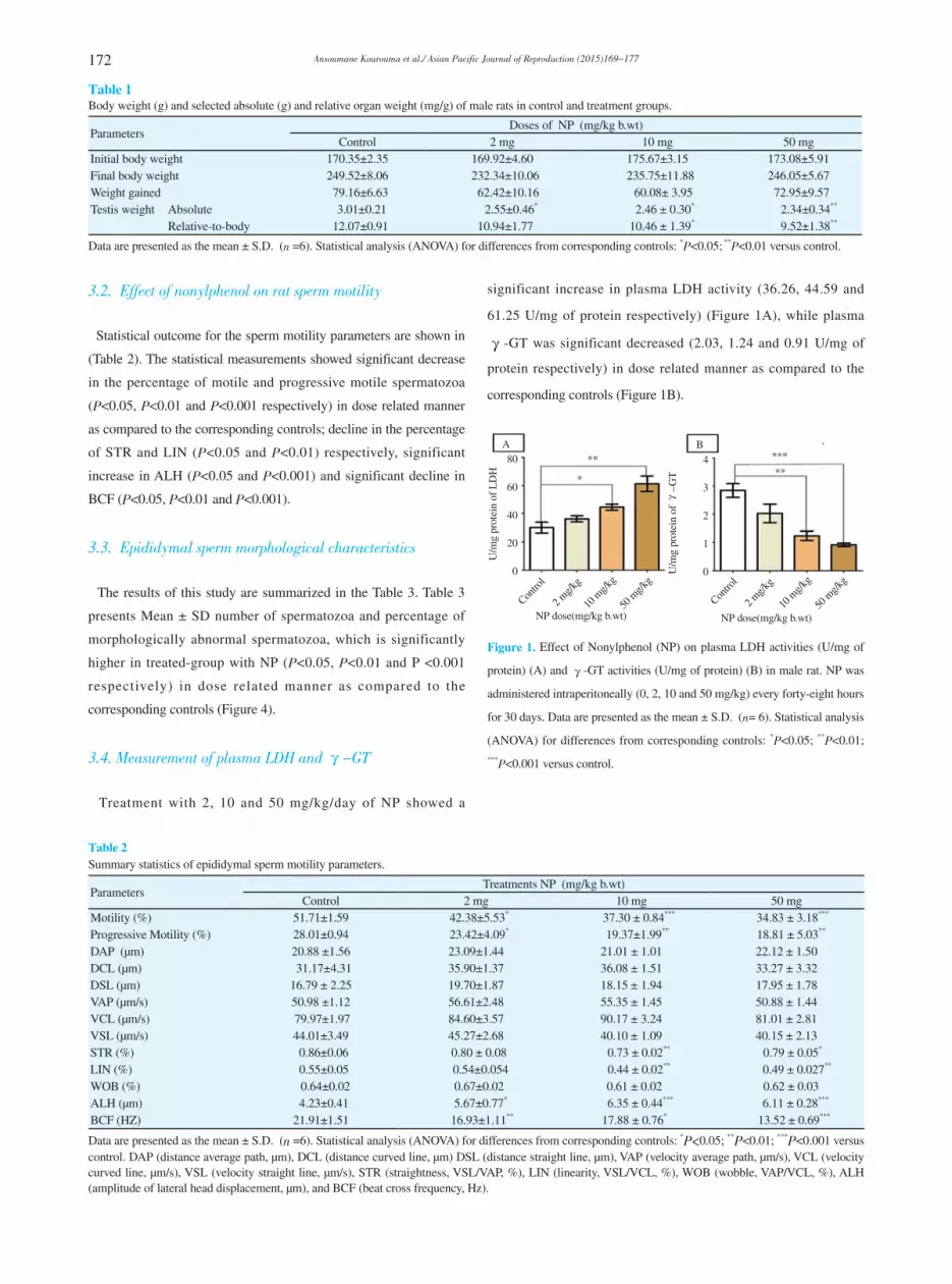
3.1. Body and testis weight
No significant differences in body weight happed among the
treated-group and control group during the experiment period, but
final body weight of all animal were increased significantly, while
a significant decrease in absolute and relative weights of testes has
been observed (P<0.05 and P<0.01) (Table 1).
172 Ansoumane Kourouma et al./ Asian Pacific Journal of Reproduction (2015)169-177
3.2. Effect of nonylphenol on rat sperm motility
Statistical outcome for the sperm motility parameters are shown in
(Table 2). The statistical measurements showed significant decrease
in the percentage of motile and progressive motile spermatozoa
(P<0.05, P<0.01 and P<0.001 respectively) in dose related manner
as compared to the corresponding controls; decline in the percentage
of STR and LIN (P<0.05 and P<0.01) respectively, significant
increase in ALH (P<0.05 and P<0.001) and significant decline in
BCF (P<0.05, P<0.01 and P<0.001).
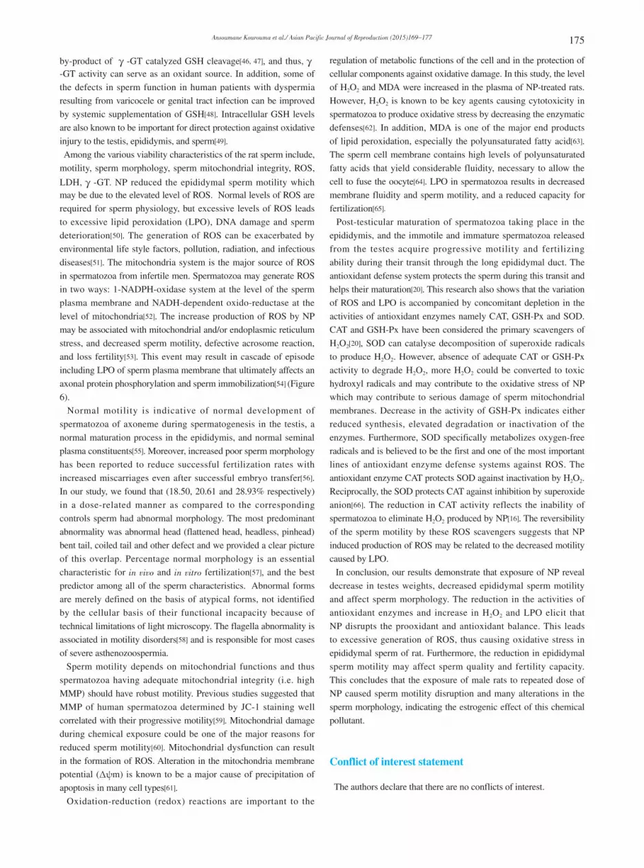
3.3. Epididymal sperm morphological characteristics
The results of this study are summarized in the Table 3. Table 3
presents Mean ± SD number of spermatozoa and percentage of
morphologically abnormal spermatozoa, which is significantly
higher in treated-group with NP (P<0.05, P<0.01 and P <0.001
respectively) in dose related manner as compared to the
corresponding controls (Figure 4).
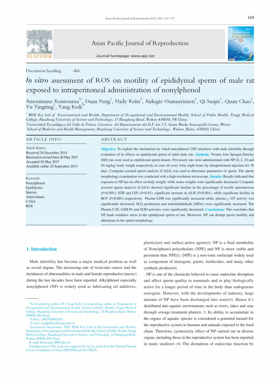
3.4. Measurement of plasma LDH and 毭-GT
Treatment with 2, 10 and 50 mg/kg/day of NP showed a
significant increase in plasma LDH activity (36.26, 44.59 and
61.25 U/mg of protein respectively) (Figure 1A), while plasma
毭-GT was significant decreased (2.03, 1.24 and 0.91 U/mg of
protein respectively) in dose related manner as compared to the
corresponding controls (Figure 1B).
A B80
60
40
20
0
U/m
g pr
otei
n of
LD
H
**
*
4
3
2
1
0U/m
g pr
otei
n of
毭-G
T
*****
Contro
l
Contro
l
2 mg/k
g
2 mg/k
g
10 m
g/kg
10 m
g/kg
50 m
g/kg
50 m
g/kg
NP dose(mg/kg b.wt) NP dose(mg/kg b.wt)
Figure 1. Effect of Nonylphenol (NP) on plasma LDH activities (U/mg of
protein) (A) and 毭-GT activities (U/mg of protein) (B) in male rat. NP was
administered intraperitoneally (0, 2, 10 and 50 mg/kg) every forty-eight hours
for 30 days. Data are presented as the mean ± S.D. (n= 6). Statistical analysis
(ANOVA) for differences from corresponding controls: *P<0.05; **P<0.01;
***P<0.001 versus control.
Table 2 Summary statistics of epididymal sperm motility parameters.
ParametersTreatments NP (mg/kg b.wt)
Control 2 mg 10 mg 50 mgMotility (%) 51.71±1.59 42.38±5.53* 37.30 ± 0.84*** 34.83 ± 3.18***
Progressive Motility (%) 28.01±0.94 23.42±4.09* 19.37±1.99** 18.81 ± 5.03**
DAP (µm) 20.88 ±1.56 23.09±1.44 21.01 ± 1.01 22.12 ± 1.50DCL (µm) 31.17±4.31 35.90±1.37 36.08 ± 1.51 33.27 ± 3.32DSL (µm) 16.79 ± 2.25 19.70±1.87 18.15 ± 1.94 17.95 ± 1.78VAP (µm/s) 50.98 ±1.12 56.61±2.48 55.35 ± 1.45 50.88 ± 1.44VCL (µm/s) 79.97±1.97 84.60±3.57 90.17 ± 3.24 81.01 ± 2.81VSL (µm/s) 44.01±3.49 45.27±2.68 40.10 ± 1.09 40.15 ± 2.13STR (%) 0.86±0.06 0.80 ± 0.08 0.73 ± 0.02** 0.79 ± 0.05*
LIN (%) 0.55±0.05 0.54±0.054 0.44 ± 0.02** 0.49 ± 0.027**
WOB (%) 0.64±0.02 0.67±0.02 0.61 ± 0.02 0.62 ± 0.03ALH (µm) 4.23±0.41 5.67±0.77* 6.35 ± 0.44*** 6.11 ± 0.28***
BCF (HZ) 21.91±1.51 16.93±1.11** 17.88 ± 0.76* 13.52 ± 0.69***
Data are presented as the mean ± S.D. (n =6). Statistical analysis (ANOVA) for differences from corresponding controls: *P<0.05; **P<0.01; ***P<0.001 versus control. DAP (distance average path, µm), DCL (distance curved line, µm) DSL (distance straight line, µm), VAP (velocity average path, µm/s), VCL (velocity curved line, µm/s), VSL (velocity straight line, µm/s), STR (straightness, VSL/VAP, %), LIN (linearity, VSL/VCL, %), WOB (wobble, VAP/VCL, %), ALH (amplitude of lateral head displacement, µm), and BCF (beat cross frequency, Hz).
Table 1 Body weight (g) and selected absolute (g) and relative organ weight (mg/g) of male rats in control and treatment groups.
ParametersDoses of NP (mg/kg b.wt)
Control 2 mg 10 mg 50 mgInitial body weight 170.35±2.35 169.92±4.60 175.67±3.15 173.08±5.91Final body weight 249.52±8.06 232.34±10.06 235.75±11.88 246.05±5.67Weight gained 79.16±6.63 62.42±10.16 60.08± 3.95 72.95±9.57Testis weight Absolute 3.01±0.21 2.55±0.46* 2.46 ± 0.30* 2.34±0.34**
Relative-to-body 12.07±0.91 10.94±1.77 10.46 ± 1.39* 9.52±1.38**
Data are presented as the mean ± S.D. (n =6). Statistical analysis (ANOVA) for differences from corresponding controls: *P<0.05; **P<0.01 versus control.
173Ansoumane Kourouma et al./ Asian Pacific Journal of Reproduction (2015)169-177
3.5. Oxidative status
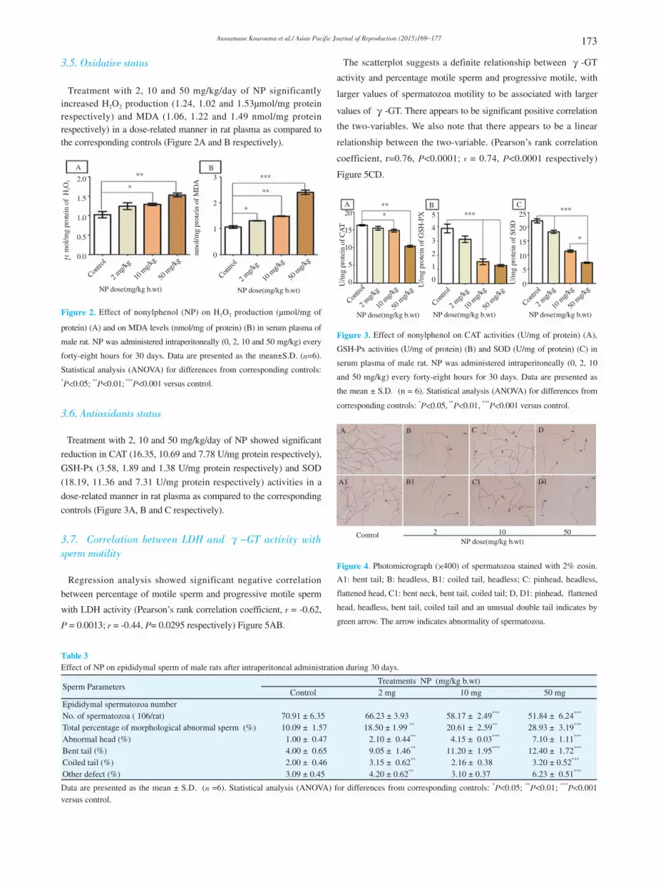
Treatment with 2, 10 and 50 mg/kg/day of NP significantly increased H2O2 production (1.24, 1.02 and 1.53µmol/mg protein respectively) and MDA (1.06, 1.22 and 1.49 nmol/mg protein respectively) in a dose-related manner in rat plasma as compared to the corresponding controls (Figure 2A and B respectively).
A B
2.0
1.5
1.0
0.5
0.0毺m
ol/m
g pr
otei
n of
H2O
2
***
3
2
1
0nmol
/mg
prot
ein
of M
DA
***
**
Contro
l
Contro
l
2 mg/k
g
2 mg/k
g
10 m
g/kg
10 m
g/kg
50 m
g/kg
50 m
g/kg
NP dose(mg/kg b.wt) NP dose(mg/kg b.wt)
*
Figure 2. Effect of nonylphenol (NP) on H2O2 production (µmol/mg of
protein) (A) and on MDA levels (nmol/mg of protein) (B) in serum plasma of
male rat. NP was administered intraperitoneally (0, 2, 10 and 50 mg/kg) every
forty-eight hours for 30 days. Data are presented as the mean±S.D. (n=6).
Statistical analysis (ANOVA) for differences from corresponding controls: *P<0.05; **P<0.01; ***P<0.001 versus control.
3.6. Antioxidants status
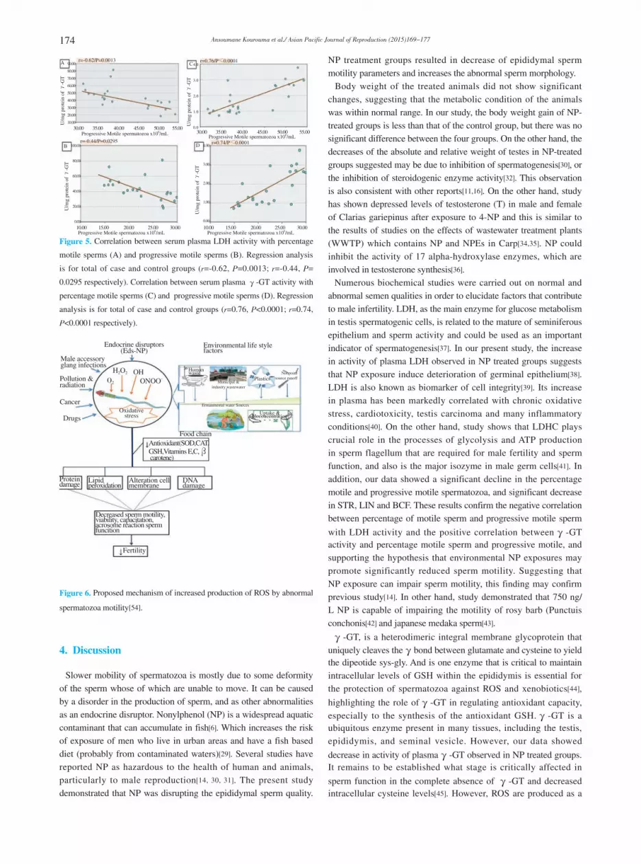
Treatment with 2, 10 and 50 mg/kg/day of NP showed significant
reduction in CAT (16.35, 10.69 and 7.78 U/mg protein respectively),
GSH-Px (3.58, 1.89 and 1.38 U/mg protein respectively) and SOD
(18.19, 11.36 and 7.31 U/mg protein respectively) activities in a
dose-related manner in rat plasma as compared to the corresponding
controls (Figure 3A, B and C respectively).
3.7. Correlation between LDH and 毭-GT activity with sperm motility
Regression analysis showed significant negative correlation
between percentage of motile sperm and progressive motile sperm
with LDH activity (Pearson’s rank correlation coefficient, r = -0.62,
P = 0.0013; r = -0.44, P= 0.0295 respectively) Figure 5AB.
The scatterplot suggests a definite relationship between 毭-GT
activity and percentage motile sperm and progressive motile, with
larger values of spermatozoa motility to be associated with larger
values of 毭-GT. There appears to be significant positive correlation
the two-variables. We also note that there appears to be a linear
relationship between the two-variable. (Pearson’s rank correlation
coefficient, r=0.76, P<0.0001; r = 0.74, P<0.0001 respectively)
Figure 5CD.
A B5
4
3
2
1
0
**** 25
20
15
10
5
0U/m
g pr
otei
n of
SO
D
*****
Contro
l
Contro
l
2 mg/k
g
2 mg/k
g
10 m
g/kg
10 m
g/kg
50 m
g/kg
50 m
g/kg
NP dose(mg/kg b.wt) NP dose(mg/kg b.wt)
*
50 m
g/kg
10 m
g/kg
2 mg/k
g
Contro
l
NP dose(mg/kg b.wt)
C
U/m
g pr
otei
n of
GSH
-PX20
15
10
5
0U/m
g pr
otei
n of
CA
TFigure 3. Effect of nonylphenol on CAT activities (U/mg of protein) (A),
GSH-Px activities (U/mg of protein) (B) and SOD (U/mg of protein) (C) in
serum plasma of male rat. NP was administered intraperitoneally (0, 2, 10
and 50 mg/kg) every forty-eight hours for 30 days. Data are presented as
the mean ± S.D. (n = 6). Statistical analysis (ANOVA) for differences from
corresponding controls: *P<0.05, **P<0.01, ***P<0.001 versus control.
A
A1
B
B1
C
C1
D
D1
Control 2 10 50NP dose(mg/kg b.wt)
Figure 4. Photomicrograph (伊400) of spermatozoa stained with 2% eosin.
A1: bent tail; B: headless, B1: coiled tail, headless; C: pinhead, headless,
flattened head, C1: bent neck, bent tail, coiled tail; D, D1: pinhead, flattened
head, headless, bent tail, coiled tail and an unusual double tail indicates by
green arrow. The arrow indicates abnormality of spermatozoa.
Table 3 Effect of NP on epididymal sperm of male rats after intraperitoneal administration during 30 days.
Sperm ParametersTreatments NP (mg/kg b.wt)
Control 2 mg 10 mg 50 mgEpididymal spermatozoa numberNo. of spermatozoa ( 106/rat) 70.91 ± 6.35 66.23 ± 3.93 58.17 ± 2.49*** 51.84 ± 6.24***
Total percentage of morphological abnormal sperm (%) 10.09 ± 1.57 18.50 ± 1.99 ** 20.61 ± 2.59** 28.93 ± 3.19***
Abnormal head (%) 1.00 ± 0.47 2.10 ± 0.44** 4.15 ± 0.03*** 7.10 ± 1.11***
Bent tail (%) 4.00 ± 0.65 9.05 ± 1.46** 11.20 ± 1.95*** 12.40 ± 1.72***
Coiled tail (%) 2.00 ± 0.46 3.15 ± 0.62** 2.16 ± 0.38 3.20 ± 0.52***
Other defect (%) 3.09 ± 0.45 4.20 ± 0.62** 3.10 ± 0.37 6.23 ± 0.51***
Data are presented as the mean ± S.D. (n =6). Statistical analysis (ANOVA) for differences from corresponding controls: *P<0.05; **P<0.01; ***P<0.001 versus control.
174 Ansoumane Kourouma et al./ Asian Pacific Journal of Reproduction (2015)169-177
A
B
C
D
r=-0.62/P=0.0013 r=0.76/P敿0.0001
r=-0.44/P=0.0295 r=0.74/P敿0.0001
30.00 35.00 40.00 45.00 50.00 55.00
90.00
80.00
70.00
60.00
50.00
40.00
30.00
20.00
10.00
4.0
3.0
2.0
1.0
0.0
4.00
3.00
2.00
1.00
0.00
100.00
80.00
60.00
40.00
20.00
0.00
10.00 15.00 20.00 25.00 30.00
30.00 35.00 40.00 45.00 50.00 55.00
10.00 15.00 20.00 25.00 30.00Progressive Motile spermatozoa x106/mL Progressive Motile spermatozoa x106/mL
Progressive Motile spermatozoa x106/mLProgressive Motile spermatozoa x106/mL
U/m
g pr
otei
n of
毭-G
T
U/m
g pr
otei
n of
毭-G
T
U/m
g pr
otei
n of
毭-G
T
U/m
g pr
otei
n of
毭-G
T
Figure 5. Correlation between serum plasma LDH activity with percentage
motile sperms (A) and progressive motile sperms (B). Regression analysis
is for total of case and control groups (r=-0.62, P=0.0013; r=-0.44, P=
0.0295 respectively). Correlation between serum plasma 毭-GT activity with
percentage motile sperms (C) and progressive motile sperms (D). Regression
analysis is for total of case and control groups (r=0.76, P<0.0001; r=0.74,
P<0.0001 respectively).
Endocrine disruptors(Eds-NP)
Male accessoryglang infections
Pollution &radiation
Cancer
Drugs
O2
H2O2 OHONOO-
Antioxidant(SOD,CAT,GSH,Vitamins E,C,毬 carotene)
DNAdamage
Alteration cellmembrane
Lipidperoxidation
Proteindamage
Decreased sperm motility,viability, capacitation,acrosome reaction spermfuncition
Fertility
Environmental life stylefactors
Food chain
Humanwaste
Municipal &industry wastewater
Ermanmental water Sources
Uptake &bloconcentration
Plastics Nonpoint source runoff
Oxidativestress
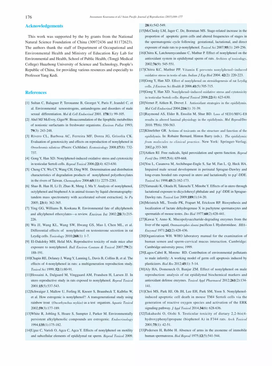
Figure 6. Proposed mechanism of increased production of ROS by abnormal
spermatozoa motility[54].
4. Discussion
Slower mobility of spermatozoa is mostly due to some deformity of the sperm whose of which are unable to move. It can be caused by a disorder in the production of sperm, and as other abnormalities as an endocrine disruptor. Nonylphenol (NP) is a widespread aquatic contaminant that can accumulate in fish[6]. Which increases the risk of exposure of men who live in urban areas and have a fish based diet (probably from contaminated waters)[29]. Several studies have reported NP as hazardous to the health of human and animals, particularly to male reproduction[14, 30, 31]. The present study demonstrated that NP was disrupting the epididymal sperm quality.
NP treatment groups resulted in decrease of epididymal sperm motility parameters and increases the abnormal sperm morphology. Body weight of the treated animals did not show significant changes, suggesting that the metabolic condition of the animals was within normal range. In our study, the body weight gain of NP-treated groups is less than that of the control group, but there was no significant difference between the four groups. On the other hand, the decreases of the absolute and relative weight of testes in NP-treated groups suggested may be due to inhibition of spermatogenesis[30], or the inhibition of steroidogenic enzyme activity[32]. This observation is also consistent with other reports[11,16]. On the other hand, study has shown depressed levels of testosterone (T) in male and female of Clarias gariepinus after exposure to 4-NP and this is similar to the results of studies on the effects of wastewater treatment plants (WWTP) which contains NP and NPEs in Carp[34,35]. NP could inhibit the activity of 17 alpha-hydroxylase enzymes, which are involved in testosterone synthesis[36]. Numerous biochemical studies were carried out on normal and abnormal semen qualities in order to elucidate factors that contribute to male infertility. LDH, as the main enzyme for glucose metabolism in testis spermatogenic cells, is related to the mature of seminiferous epithelium and sperm activity and could be used as an important indicator of spermatogenesis[37]. In our present study, the increase in activity of plasma LDH observed in NP treated groups suggests that NP exposure induce deterioration of germinal epithelium[38]. LDH is also known as biomarker of cell integrity[39]. Its increase in plasma has been markedly correlated with chronic oxidative stress, cardiotoxicity, testis carcinoma and many inflammatory conditions[40]. On the other hand, study shows that LDHC plays crucial role in the processes of glycolysis and ATP production in sperm flagellum that are required for male fertility and sperm function, and also is the major isozyme in male germ cells[41]. In addition, our data showed a significant decline in the percentage motile and progressive motile spermatozoa, and significant decrease in STR, LIN and BCF. These results confirm the negative correlation between percentage of motile sperm and progressive motile sperm
with LDH activity and the positive correlation between毭-GT activity and percentage motile sperm and progressive motile, and supporting the hypothesis that environmental NP exposures may promote significantly reduced sperm motility. Suggesting that NP exposure can impair sperm motility, this finding may confirm previous study[14]. In other hand, study demonstrated that 750 ng/L NP is capable of impairing the motility of rosy barb (Punctuis conchonis[42] and japanese medaka sperm[43].
毭-GT, is a heterodimeric integral membrane glycoprotein that uniquely cleaves the毭bond between glutamate and cysteine to yield the dipeotide sys-gly. And is one enzyme that is critical to maintain intracellular levels of GSH within the epididymis is essential for the protection of spermatozoa against ROS and xenobiotics[44],
highlighting the role of毭-GT in regulating antioxidant capacity, especially to the synthesis of the antioxidant GSH.毭-GT is a ubiquitous enzyme present in many tissues, including the testis, epididymis, and seminal vesicle. However, our data showed
decrease in activity of plasma毭-GT observed in NP treated groups. It remains to be established what stage is critically affected in
sperm function in the complete absence of 毭-GT and decreased intracellular cysteine levels[45]. However, ROS are produced as a
175Ansoumane Kourouma et al./ Asian Pacific Journal of Reproduction (2015)169-177
by-product of 毭-GT catalyzed GSH cleavage[46, 47], and thus,毭-GT activity can serve as an oxidant source. In addition, some of
the defects in sperm function in human patients with dyspermia
resulting from varicocele or genital tract infection can be improved
by systemic supplementation of GSH[48]. Intracellular GSH levels
are also known to be important for direct protection against oxidative
injury to the testis, epididymis, and sperm[49].
Among the various viability characteristics of the rat sperm include,
motility, sperm morphology, sperm mitochondrial integrity, ROS,
LDH,毭-GT. NP reduced the epididymal sperm motility which may be due to the elevated level of ROS. Normal levels of ROS are
required for sperm physiology, but excessive levels of ROS leads
to excessive lipid peroxidation (LPO), DNA damage and sperm
deterioration[50]. The generation of ROS can be exacerbated by
environmental life style factors, pollution, radiation, and infectious
diseases[51]. The mitochondria system is the major source of ROS
in spermatozoa from infertile men. Spermatozoa may generate ROS
in two ways: 1-NADPH-oxidase system at the level of the sperm
plasma membrane and NADH-dependent oxido-reductase at the
level of mitochondria[52]. The increase production of ROS by NP
may be associated with mitochondrial and/or endoplasmic reticulum
stress, and decreased sperm motility, defective acrosome reaction,
and loss fertility[53]. This event may result in cascade of episode
including LPO of sperm plasma membrane that ultimately affects an
axonal protein phosphorylation and sperm immobilization[54] (Figure
6).
Normal motility is indicative of normal development of
spermatozoa of axoneme during spermatogenesis in the testis, a
normal maturation process in the epididymis, and normal seminal
plasma constituents[55]. Moreover, increased poor sperm morphology
has been reported to reduce successful fertilization rates with
increased miscarriages even after successful embryo transfer[56].
In our study, we found that (18.50, 20.61 and 28.93% respectively)
in a dose-related manner as compared to the corresponding
controls sperm had abnormal morphology. The most predominant
abnormality was abnormal head (flattened head, headless, pinhead)
bent tail, coiled tail and other defect and we provided a clear picture
of this overlap. Percentage normal morphology is an essential
characteristic for in vivo and in vitro fertilization[57], and the best
predictor among all of the sperm characteristics. Abnormal forms
are merely defined on the basis of atypical forms, not identified
by the cellular basis of their functional incapacity because of
technical limitations of light microscopy. The flagella abnormality is
associated in motility disorders[58] and is responsible for most cases
of severe asthenozoospermia.
Sperm motility depends on mitochondrial functions and thus
spermatozoa having adequate mitochondrial integrity (i.e. high
MMP) should have robust motility. Previous studies suggested that
MMP of human spermatozoa determined by JC-1 staining well
correlated with their progressive motility[59]. Mitochondrial damage
during chemical exposure could be one of the major reasons for
reduced sperm motility[60]. Mitochondrial dysfunction can result
in the formation of ROS. Alteration in the mitochondria membrane
potential (Δψm) is known to be a major cause of precipitation of
apoptosis in many cell types[61].
Oxidation-reduction (redox) reactions are important to the
regulation of metabolic functions of the cell and in the protection of
cellular components against oxidative damage. In this study, the level
of H2O2 and MDA were increased in the plasma of NP-treated rats.
However, H2O2 is known to be key agents causing cytotoxicity in
spermatozoa to produce oxidative stress by decreasing the enzymatic
defenses[62]. In addition, MDA is one of the major end products
of lipid peroxidation, especially the polyunsaturated fatty acid[63].
The sperm cell membrane contains high levels of polyunsaturated
fatty acids that yield considerable fluidity, necessary to allow the
cell to fuse the oocyte[64]. LPO in spermatozoa results in decreased
membrane fluidity and sperm motility, and a reduced capacity for
fertilization[65].
Post-testicular maturation of spermatozoa taking place in the
epididymis, and the immotile and immature spermatozoa released
from the testes acquire progressive motility and fertilizing
ability during their transit through the long epididymal duct. The
antioxidant defense system protects the sperm during this transit and
helps their maturation[20]. This research also shows that the variation
of ROS and LPO is accompanied by concomitant depletion in the
activities of antioxidant enzymes namely CAT, GSH-Px and SOD.
CAT and GSH-Px have been considered the primary scavengers of
H2O2[20], SOD can catalyse decomposition of superoxide radicals
to produce H2O2. However, absence of adequate CAT or GSH-Px
activity to degrade H2O2, more H2O2 could be converted to toxic
hydroxyl radicals and may contribute to the oxidative stress of NP
which may contribute to serious damage of sperm mitochondrial
membranes. Decrease in the activity of GSH-Px indicates either
reduced synthesis, elevated degradation or inactivation of the
enzymes. Furthermore, SOD specifically metabolizes oxygen-free
radicals and is believed to be the first and one of the most important
lines of antioxidant enzyme defense systems against ROS. The
antioxidant enzyme CAT protects SOD against inactivation by H2O2.
Reciprocally, the SOD protects CAT against inhibition by superoxide
anion[66]. The reduction in CAT activity reflects the inability of
spermatozoa to eliminate H2O2 produced by NP[16]. The reversibility
of the sperm motility by these ROS scavengers suggests that NP
induced production of ROS may be related to the decreased motility
caused by LPO.
In conclusion, our results demonstrate that exposure of NP reveal
decrease in testes weights, decreased epididymal sperm motility
and affect sperm morphology. The reduction in the activities of
antioxidant enzymes and increase in H2O2 and LPO elicit that
NP disrupts the prooxidant and antioxidant balance. This leads
to excessive generation of ROS, thus causing oxidative stress in
epididymal sperm of rat. Furthermore, the reduction in epididymal
sperm motility may affect sperm quality and fertility capacity.
This concludes that the exposure of male rats to repeated dose of
NP caused sperm motility disruption and many alterations in the
sperm morphology, indicating the estrogenic effect of this chemical
pollutant.
Conflict of interest statement
The authors declare that there are no conflicts of interest.
176 Ansoumane Kourouma et al./ Asian Pacific Journal of Reproduction (2015)169-177
Acknowledgements
This work was supported by the by grants from the National Natural Science Foundation of China (30972436 and 81172623). The authors thank the staff of Department of Occupational and Environmental Health and Ministry of Education Key Lab for Environmental and Health, School of Public Health, (Tongji Medical College) Huazhong University of Science and Technology, People’s Republic of China, for providing various resources and especially to Professor Yang Kedi.
References
[1] Sultan C, Balaguer P, Terouanne B, Georget V, Paris F, Jeandel C, et
al. Environmental xenoestrogens, antiandrogens and disorders of male
sexual differentiation. Mol & Cell Endocrinol 2001. 178(1): 99-105.
[2] Ahel MJ McEvoy, GigerW. Bioaccumulation of the lipophilic metabolites
of nonionic surfactants in freshwater organisms. Environ Pollut 1993;
79(3): 243-248.
[3] Rivero CL, Barbosa AC, Ferreira MF, Dorea JG, Grisolia CK.
Evaluation of genotoxicity and effects on reproduction of nonylphenol in
Oreochromis niloticus (Pisces: Cichlidae). Ecotoxicology 2008;17(8): 732-
737.
[4] Gong Y, Han XD. Nonylphenol-induced oxidative stress and cytotoxicity
in testicular Sertoli cells. Reprod Toxicol 2006;22(4): 623-630.
[5] Cheng CY, Wu CY, Wang CH, Ding WH. Determination and distribution
characteristics of degradation products of nonylphenol polyethoxylates
in the rivers of Taiwan. Chemosphere 2006;65(11): 2275-2281.
[6] Shao B, Han H, Li D, Zhao R, Meng J, Ma Y. Analysis of nonylphenol,
octylphenol and bisphenol A in animal tissues by liquid chromatography-
tandem mass spectrometry with accelerated solvent extraction]. Se Pu 2005; 23(4): 362-365.
[7] Ying GG, Williams B, Kookana R. Environmental fate of alkylphenols
and alkylphenol ethoxylates—a review. Environ Int 2002;28(3):215-
226.
[8] Wu JJ, Wang KL, Wang SW, Hwang GS, Mao I, Chen ML, et al.
Differential effects of nonylphenol on testosterone secretion in rat
Leydig cells. Toxicology 2010;268(1): 1-7.
[9] El-Dakdoky MH, Helal MA. Reproductive toxicity of male mice after
exposure to nonylphenol. Bull Environ Contam & Toxicol 2007;79(2):
188-191.
[10] Chapin RE, Delaney J, Wang Y, Lanning L, Davis B, Collins B, et al. The
effects of 4-nonylphenol in rats: a multigeneration reproduction study.
Toxicol Sci 1999; 52(1):80-91.
[11] Hossaini A, Dalgaard M, Vinggaard AM, Frandsen H, Larsen JJ. In
utero reproductive study in rats exposed to nonylphenol. Reprod Toxicol 2001;15(5):537-543.
[12] Schwaiger J, Mallow U, Ferling H, Knoerr S, Braunbeck T, Kalbfus W,
et al. How estrogenic is nonylphenol?: A transgenerational study using
rainbow trout (Oncorhynchus mykiss) as a test organism. Aquatic Toxicol 2002;59(3):177-189.
[13] White R, Jobling S, Hoare S, Sumpter J, Parker M. Environmentally
persistent alkylphenolic compounds are estrogenic. Endocrinology 1994;135(1):175-182.
[14] Uguz C, Varisli O, Agca C, Agca Y. Effects of nonylphenol on motility
and subcellular elements of epididymal rat sperm. Reprod Toxicol 2009.
28(4):542-549.
[15] McClusky LM, Jager C. De, Bornman MS. Stage-related increase in the
proportion of apoptotic germ cells and altered frequencies of stages in
the spermatogenic cycle following gestational, lactational, and direct
exposure of male rats to p-nonylphenol. Toxicol Sci 2007;95(1): 249-256.
[16] Chitra K, Latchoumycandane C, Mathur P. Effect of nonylphenol on the
antioxidant system in epididymal sperm of rats. Archives of toxicology,
2002;76(9): 545-551.
[17] Chitra KC, Mathur PP. Vitamin E prevents nonylphenol-induced oxidative stress in testis of rats. Indian J Exp Biol 2004. 42(2): 220-223.
[18] Gong Y, Han XD. Effect of nonylphenol on steroidogenesis of rat Leydig cells. J Environ Sci Health B 2006;41(5):705-715.
[19] Gong Y, Han XD. Nonylphenol-induced oxidative stress and cytotoxicity in testicular Sertoli cells. Reprod Toxicol 2006;22(4): 623-630.
[20] Vernet P, Aitken R, Drevet J. Antioxidant strategies in the epididymis. Mol Cell Endocrinol 2004;216(1): 31-39.
[21] Raymond AS, Elder B, Ensslin M, Shur BD. Loss of SED1/MFG‐E8 results in altered luminal physiology in the epididymis. Mol Reprod/Dev 2010. 77(6): 550-563.
[22] Klinefelter GR. Actions of toxicants on the structure and function of the epididymis. In: Robaire Bernard, Hinton Barry (eds.). The epididymis: from molecules to clinical practice. New York: Springer-Verlag;
2002,p.353-369.
[23] Aitken RJ. Free radicals, lipid peroxidation and sperm function. Reprod Fertil Dev 1995;7(4): 659-668.
[24] You L, Casanova M, Archibeque-Engle S, Sar M, Fan L. Q, Heck HA.
Impaired male sexual development in perinatal Sprague-Dawley and
long-evans hooded rats exposed in utero and lactationally to p,p’-DDE.
Toxicol Sci 1998;45(2):162-173.
[25] Yamasaki K, Okuda H, Takeuchi T, Minobe Y. Effects of in utero through
lactational exposure to dicyclohexyl phthalate and p,p’-DDE in Sprague-
Dawley rats. Toxicol Lett 2009;189(1):14-20.
[26] Meistrich ML, Trostle PK, Frapart M, Erickson RP. Biosynthesis and
localization of lactate dehydrogenase X in pachytene spermatocytes and
spermatids of mouse testes. Dev Biol 1977;60(2):428-441.
[27] Kawai Y, Anno K. Mucopolysaccharide-degrading enzymes from the
liver of the squid, Ommastrephes sloani pacificus I. Hyaluronidase. BBA-Enzymol 1971;242(2):428-436.
[28] Organization WH. WHO laboratory manual for the examination of
human semen and sperm-cervical mucus interaction. Cambridge:
Cambridge university press; 1999.
[29] Lagos-Cabré R, Moreno RD. Contribution of environmental pollutants
to male infertily: A working model of germ cell apoptosis induced by
plasticizers. Biol Res 2012;45(1): 5-14.
[30] Aly HA, Domenech O, Banjar ZM. Effect of nonylphenol on male
reproduction: analysis of rat epididymal biochemical markers and
antioxidant defense enzymes. Toxicol Appl Pharmacol 2012;261(2):134-
141.
[31] Choi MS, Park HJ, Oh JH, Lee EH, Park SM, Yoon S. Nonylphenol-
induced apoptotic cell death in mouse TM4 Sertoli cells via the
generation of reactive oxygen species and activation of the ERK
signaling pathway. J Appl Toxicol 2014;34(6): 628-636.
[32] Takahashi O, Oishi S. Testicular toxicity of dietary 2,2-bis(4-
hydroxyphenyl)propane (bisphenol A) in F344 rats. Arch Toxicol 2001;75(1): 42-51.
[33] Pedersen H, Rebbe H. Absence of arms in the axoneme of immobile
human spermatozoa. Biol Reprod 1975;12(5):541-544.
177Ansoumane Kourouma et al./ Asian Pacific Journal of Reproduction (2015)169-177
[34] Folmar LC, Hemmer MJ, Denslow ND, Kroll K, Chen J, Cheek A, et al.,
A comparison of the estrogenic potencies of estradiol, ethynylestradiol,
diethylstilbestrol, nonylphenol and methoxychlor in vivo and in vitro.
Aquat Toxicol 2002;60(1): 101-110.
[35] Lavado R, Thibaut R, Raldúa D, Martın R, Porte C. First evidence of
endocrine disruption in feral carp from the Ebro River. Toxicol Appl Pharmacol 2004;196(2):247-257.
[36] Laurenzana EM, Weis CC, Bryant CW, Newbold R, Delclos KB. Effect
of dietary administration of genistein, nonylphenol or ethinyl estradiol
on hepatic testosterone metabolism, cytochrome P-450 enzymes, and
estrogen receptor alpha expression. Food Chem Toxicol 2002; 40(1):53-
63.
[37] Qiong L, Jun L, Jun Y, Yinzhu Z, Xiaoyan C, Mingliang Y. The effect
of Laminaria japonica polysaccharides on the recovery of the male
rat reproductive system and mating function damaged by multiple mini-
doses of ionizing radiations. Environ Toxicol Pharmacol 2011;31(2):286-
294.
[38] Pant N, Murthy R, Srivastava S. Male reproductive toxicity of sodium
arsenite in mice. Hum & Exp Toxicol 2004;23(8):399-403.
[39] Aly HA, Khafagy RM. 2,3,7,8-tetrachlorodibenzo-p-dioxin (TCDD)-
induced cytotoxicity accompanied by oxidative stress in rat Sertoli cells:
Possible role of mitochondrial fractions of Sertoli cells. Toxicol Appl Pharmacol 2011;252(3): 273-280.
[40] Jaiswal A, Parihar VK, Sudheer Kumar M, Manjula S, Krishnanand B,
Shanbhag R, et al. 5-Aminosalicylic acid reverses endosulfan-induced
testicular toxicity in male rats. Mutat Res /Gen Toxicol & Environ Mutagen 2005;585(1): 50-59.
[41] Odet F, Duan C, Willis WD, Goulding EH, Kung A, Eddy EM, et al.
Expression of the gene for mouse lactate dehydrogenase C (Ldhc) is
required for male fertility. Biol Reprod 2008;79(1): 26-34.
[42] Qin X, Shi-Cui Z, Jia-Hui H, Yu-Yan X. Sperm of rosy barb (Puntius
conchonius) as an in vitro assay system of nonylphenol cytotoxicity. J Environ Sci (China) 2006;18(3): 417-419.
[43] Hara Y, Strüssmann CA, Hashimoto S. Strüssmann, Hashimoto S.
Assessment of short-term exposure to nonylphenol in Japanese medaka
using sperm velocity and frequency of motile sperm. Arch Environ Contam & Toxicol 2007;53(3): 406-410.
[44] Markey CM, Rudolph DB, Labus JC, Hinton BT. Oxidative stress
differentially regulates the expression of gamma-glutamyl transpeptidase
mRNAs in the initial segment of the rat epididymis. J Androl 1998;19(1):
92-99.
[45] Kumar TR, Wiseman AL, Kala G, Kala SV, Matzuk MM, Lieberman
MW. Reproductive defects in gamma-glutamyl transpeptidase-deficient
mice. Endocrinology 2000;141(11):4270-4277.
[46] Paolicchi A, Dominici S, Pieri L, Maellaro E, Pompella A. Glutathione
catabolism as a signaling mechanism. Biochem Pharmacol 2002;64(5):
1027-1035.
[47] Filomeni G, Rotilio G, Ciriolo MR. Cell signalling and the glutathione
redox system. Biochem Pharmacol 2002;64(5):1057-1064.
[48] Lenzi A, Culasso F, Gandini L, Lombardo F, Dondero F. Andrology:
Placebo-controlled, double-blind, cross-over trial of glutathione therapy
in male infertility. Hum Reprod 1993;8(10):1657-1662.
[49] Aitken RJ, Roman SD. Antioxidant systems and oxidative stress in the
testes. Adv Exp Med Biol 2008; 636:154-171.
[50] Aitken RJ, Curry BJ. Redox regulation of human sperm function:
from the physiological control of sperm capacitation to the etiology of
infertility and DNA damage in the germ line. Antioxid & Redox Sign 2011;14(3):367-381.
[51] González-Flecha B. Oxidant mechanisms in response to ambient air
particles. Mol Aspects Med 2004;25(1):169-182.
[52] Aitken RJ, Fisher HM, Fulton N, Gomez E, Knox W, Lewis B, et al.
Reactive oxygen species generation by human spermatozoa is induced
by exogenous NADPH and inhibited by the flavoprotein inhibitors
diphenylene iodonium and quinacrine. Mol Reprod Dev 1997;47(4):468-
482.
[53] Aitken RJ, Harkiss D, Buckingham DW. Analysis of lipid peroxidation
mechanisms in human spermatozoa. Mol Reprod Dev 1993;35(3):302-
315.
[54] de Lamirande E, Gagnon C. Impact of reactive oxygen species on
spermatozoa: a balancing act between beneficial and detrimental effects.
Hum Reprod 1995;10(suppl 1):15-21.
[55] McConnell J. Abnormalities in sperm motility: techniques of evaluation
and treatment. In:Lipschultz LI, Howards SS. (eds.) Infertility in the male.
3rd ed. St. Louis, MO: Mosby-Year Book Inc.; 1997,p. 254-259.
[56] Kobayashi D, Tokino T, Watanabe N. Contribution of caspase-3 differs
by p53 status in apoptosis induced by X-irradiation. Jpn J Cancer Res 2001;92(4):475-481.
[57] Coetzee, K., Kruge T.F, and Lombard C.J, Predictive value of normal
sperm morphology: a structured literature review. Hum Reprod Update 1998;4(1):73-82.
[58] Afzelius BA, Eliasson R. Flagellar mutants in man: on the heterogeneity
of the immotile-cilia syndrome. J Ultrastructure Res 1979;69(1):43-52.
[59] Marchetti C, Jouy N, Leroy-Martin B, Defossez A, Formstecher P,
Marchetti P. Comparison of four fluorochromes for the detection of the
inner mitochondrial membrane potential in human spermatozoa and
their correlation with sperm motility. Hum Reprod 2004;19(10): 2267-
2276.
[60] Uguz C, Varisli O, Agca C, Evans T, Agca Y. In vitro effects of
nonylphenol on motility, mitochondrial, acrosomal and chromatin
integrity of ram and boar spermatozoa. Andrologia 2014;
[61] Das M, Chatterjee S, Basak P, Das P, Pereira JA, Dutta RK, et al., Sca-1
/c-Kit receptor expression and apoptosis pattern in ENU induced MDS
mice. J Stem Cells 2009;4(4): 229-241.
[62] Rao MV, Gangadharan B. Antioxidative potential of melatonin against
mercury induced intoxication in spermatozoa in vitro. Toxicol In Vitro
2008;22(4):935-942.
[63] Subermaniam K, Saad QHM, Das S, Othman F. Virgin coconut oil
(VCO) decreases the level of malondialdehyde (MDA) in the cardiac
tissue of experimental Sprague-Dawley rats fed with heated palm oil. J Med & Bio Eng 2014;3(2):102-106.
[64] Sikka SC. Andrology lab corner: Role of oxidative stress and antioxidants
in andrology and assisted reproductive technology. J Androl 2004;25(1):
5-18.
[65] Selvakumar E, Prahalathan C, Sudharsan PT, Varalakshmi P.
Chemoprotective effect of lipoic acid against cyclophosphamide-induced
changes in the rat sperm. Toxicology 2006;217(1):71-78.
[66] Selvakumar E, Prahalathan C, Mythili Y, Varalakshmi P. Protective effect
of dl-α-lipoic acid in cyclophosphamide induced oxidative injury in rat
testis. Reprod Toxicol 2004;19(2): 163-167.