histopathological alterations in some body organs of adult clarias gariepinus (burchell, 1822)...
TRANSCRIPT
8
Histopathological Alterations in some Body Organs of Adult Clarias gariepinus
(Burchell, 1822) Exposed to 4-Nonylphenol
Alaa El-Din H. Sayed1*, Imam A. Mekkawy1,2 and Usama M. Mahmoud1 1Zoology Department, Faculty of Science, Assiut University, Assiut,
2Biology Department, Faculty of Science, Taif University, Taif, 1Egypt
2Saudi Arabia
1. Introduction
Endocrine-disrupting chemicals (EDCs) include synthetic and naturally occurring chemicals that affect the balance of normal functions in animals (Razia et al. 2006). It has been found that exposure to natural and synthetic estrogenic chemicals may adversely affect wildlife and human health (Colborn et al. 1993). In vitro exposures (Soto et al. 1992; Soto et al. 1994; Toomey et al. 1999) have confirmed the effects of EDCs on tissue structure and cellular processes. Nonylphenol ethoxylates (NPEs) are EDCs which are used globally in the production of plastics, pesticides, and cleaning products and are present in sewage effluents around the world (Talmage, 1994). It has been reported that NP is the most important degradation product of NPEs because of its enhanced resistance towards biodegradation, toxicity, ability to bioaccumulate in aquatic organisms, and estrogenicity (Ahel et al. 1994). NP is found in surface waters, aquatic sediments, and ground water (Bennie, 1999; Talmage, 1994) and it is estrogenic in various aquatic animals (Nimrod and Benson, 1996; Talmage, 1994; Servos, 1999).
The application of environmental toxicological studies on non-mammalian vertebrates is rapidly expanding; and for aquatic system, fish have become valuable indicator for the evaluation of the effects of noxious compounds (Khidr and Mekkawy, 2008). Histology and histopathology can be used as biomonitoring tools for health in toxicity studies (Meyers and Hendricks, 1985). Histoplathological alterations are biomarkers of effect exposure to environmental stressors, revealing alterations in physiological and biochemical function (Hinton et al. 1992). Histopathology, the study of lesions or abnormalities on cellular and tissue levels is useful tool for assessing the degree of pollution, particularly for sublethal and chronic effects (Bernet et al. 1999). More than one tissue may be studied for assessment of the biological effects of a toxicant on localized portions of certain organs and also for assessment of subsequent derangements (degradations) in tissues or cells in other locations and this allows for diagnoses of the observed changes (Adeyemo, 2008). NP has been shown
*Corresponding Author
Zoology
164
to cause histopathological changes in the germ and Sertoli cells of the male eelpot (Christiansen et al. 1998). The skin of fish is continuously exposed to and in direct contact with the environment pollutants such as NP. Histological changes in skin of rainbow trout with mucosomes in goblet cells were recorded after exposure to 10 µg/l of 4-nonylphenol. Several studies demonstrated the high susceptibility of skin to environmental pollutant impacts (Burkhardt-Holm et al. 1997; Iger et al. 1995; Shephard, 1994). Burkhardt-Holm et al. (1997) hypothesized that in trout, xenobiotic estrogens might affect the skin, like natural estrogens, via the steroid receptor.
In trout species, nonylphenol was found to accumulate in the liver, gill, skin, gut, fat, and kidney tissue (Ahel et al. 1993; Coldham et al. 1998; Lewis and Lech, 1996). So that, 4-nonylphenol may affects those organs in corresponding with its impacts on reproductive ones. Most of NP studies revealed sever effects on the liver and gonads of fish tissues (Christiansen et al. 1998; Jobling et al. 1996; Lech et al. 1996) and the corresponding metabolism. The liver is important in many aspects of nutrition, including lipid and carbohydrates storage and alterations in liver structure may be useful as biomarker that indicate prior exposure to environmental stressors (Hinton and laurén, 1990). Stressors-associated alterations of hepatocytes may be found in the nucleus or cytoplasm or both (Marchand et al. 2008). Malik and Hodgson, (2002) reported that the liver plays a major role in complex enzymatic processes of thyroid hormones conversion. So, liver dysfunction and disease affects thyroid hormone metabolism. Although gills are not only the prime organs for gaseous exchange, they perform several other physiological functions including osmoregulation and excretion. Parashar and Banerjee, (2002) reported that changes in environmental parameters often damage this delicate vital organ which has direct contact with aquatic environment. Many studies demonstrated that increased concentrations of different pollutants including several heavy metals seriously damage the gills of teleostean fish (Dutta et al. 1996; Wendelaar Bonga, 1997)).
African catfish (Clarias gariepinus), an omnivore freshwater fish, is a popular delicacy relished throughout tropical Africa (Nguyen and Janssen, 2002) due to fast growth rate, high stocking-density capacities, high consumer acceptability and high resistance to poor water quality and oxygen depletion (Adewolu et al. 2008; Akinwole and Faturoti, 2007; Karami et al. 2010). Because it is a prominent culture species (Adeyemo, 2008), the African catfish has been used in many fundamental experimental researches (Mahmoud et al., 2009).
The present work is an extension of previous studies of the present authors (Mekkawy et al., 2011; Mahmoud et al., 2011; Sayed et al., 2011) to determine to what extent the histopathological variations in some organs of the adult catfish, Clarias gareipinus (Burchell, 1822) are simultaneously correlated with biochemical and physiological NP-induced changes especially in respect with endocrine disruption.
2. Materials and methods
2.1 Specimen collection
Specimens of adult catfish C. gariepinus were collected from the River Nile at Assiut and then were transported to Fish Biology Laboratory of Zoology Department, Faculty of Science, Assiut University. The fish (500–1200 g) were fed on a commercial pellet diet (3% of body weight per day) and kept together in 100 l rectangular tanks containing tap water
Histopathological Alterations in some Body Organs of Adult Clarias gariepinus (Burchell, 1822) Exposed to 4-Nonylphenol
165
(conductivity 2000 ls/cm; pH 7.5; oxygen 88–95% saturation; temperature 27-28 ºC; photoperiod 12:12 light: dark). After 2 week acclimatization, fishes were used for the experimental setup.
2.2 4-nonylphenol
4-Nonylphenol was obtained from Sigma- Aldrich (Schnelldrof, Germany)
2.3 Experimental setup
The adapted adult fish classified into four groups (10 fish per each): control, 4-nonylphenol-treated group (for15 day/ for 0.05mg/l day), 4-nonylphenol-treated group (for 15 day/for 0.08mg/l day), and 4-nonylphenol-treated group (for 15 day/for 0.1 h/ day). In the present study, the range of NP exposures was 0.05-0.1 mg/l and the exposure concentrations are environmentally relevant. The conditions of the experiment were as that of acclimatization with changing all the tap water and concentrations of 4-nonylphenol every day.
2.4 Hematoxylin-Eosin (HE) and Masson's Trichrome (TRI) histopathological preparations
For microscopic preparations, after 15 days, 3 surviving fish of each group were removed and dissected. Small pieces of the liver, kidneys, gills, and skin were taken and immediately fixed in 10% neutral buffered formalin. Fixed tissues were processed routinely for paraffin embedding technique. Embedded tissues were sectioned at 5-7μ in thickness and then stained with Harris' hematoxylin and eosin stain (H & E) and Masson's Trichrome (TRI) stain according to Bancroft and Steven, (1982). Sections were visualized and studied using OLYMPUS microscope model BX50F4 (Olympus optical Co., LTP. Japan).
2.5 Transmission electron microscope (TEM)
Small pieces of liver of newly scarified fish were fixed in 2 % glutaraldehyde, washed in cacodylate buffer and post-fixed in 1% osmium tetroxide. Dehydration was carried out in ascending grads of alcohol and then embedded in epon-araldite mixture. Semithin sections of liver were cut at 1µm and stained with toluidine blue for examination under a light microscope. Ultrathin sections were stained with uranyl acetate followed by lead nitrate (Johannessen, 1978). Electron micrographs were obtained using a Jeol JEM 1200 EX Transmission Electron Microscope at Electron microscope center of Assiut University.
2.6 Ethical statement
All experiments were carried out in accordance with the Egyptian laws and University guidelines for the care of experimental animals. All procedures of the current experiment have been approved by the Committee of the Faculty of Science, Assiut University, Egypt.
3. Results
Throughout the duration of the experiment, the number of fish that died was 1.2, 3.75, 5.5, and 8 % for control, 0.05, 0.08 and 0.1 mg/l 4-nonylphenol respectively. Lesions were observed in the gills, skin, kidneys, and liver of sampled fish for all 4-nonylphenol at all
Zoology
166
exposure concentrations and durations. The occurrence and degree of alterations were positively related with the concentrations of 4-nonylphenol while samples taken from the control group remained normal for all the organs throughout the duration of the experiment.
3.1 Histopathological changes in the gills
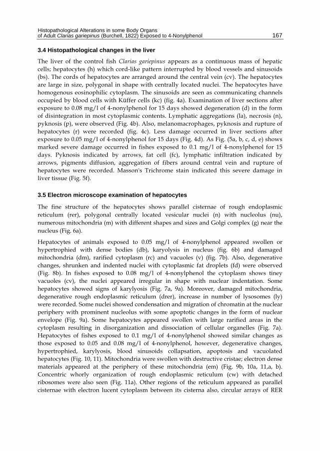
Histologically, the gills of the adult catfish Clarias gariepinus are composed of primary lamellae (pl), secondary lamellae (sl), epithelial cell (epc), mucous cell (mc), and chloride cell (chc) (Fig. 1a). The initial lesions in the gills were manifested in groups exposed to 0.05, 0.08, and 0.1 mg/l of 4-nonylphenol for 15 days (Fig. 1b, c, d, e). The anomalies include epithelial lifting, edema, deformed secondary lamellae in fishes exposed to 0.05 mg/l 4-nonylphenol (Fig. 1b) while in fishes exposed to 0.08 mg/l 4-nonylphenol, desquamation and necrosis were recorded (Fig. 1c). As Fig. (1d, e) shows gills with degeneration of cartilaginous bar malformed secondary lamellae, increase in chloride cell size and number, epithelial hyperplasia, diffusion of secondary lamellae and increase number of mucous cells in fishes exposed to 0.1 mg/l 4-nonylphenol for 15 days.
3.2 Histopathological changes in the skin
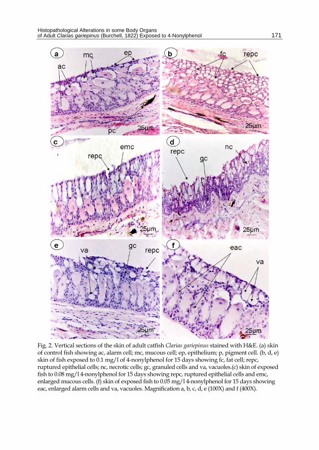
Normal structure of skin of adult catfish, Clarias gariepinus was shown in fig. (2a), where it consists of alarm cell (ac), mucous cell (mc) and epithelium (ep) with pigment cell (p). The fishes exposed to 0.05 mg/l of 4-nonylphenol showed enlarged alarm cell (eac), with vacuoles (va) in their skin structure (Fig. 2f). As shown in fig. (2c) other changes such as ruptured epithelial cells (repc) and enlarged mucous cells (emc) in the skin of fishes exposed to 0.08 mg/l of 4-nonylphenol for 15 days. Severe damage was recorded in fishes exposed to 0.1 mg/l of 4-nonylphenol as in Fig. (2b, d, e) in which ruptured epithelial cells, necrotic cell (nc), granuled cells (gc), vacuoles (va) and fat cells (fc) were recorded.
3.3 Histopathological changes in the kidney
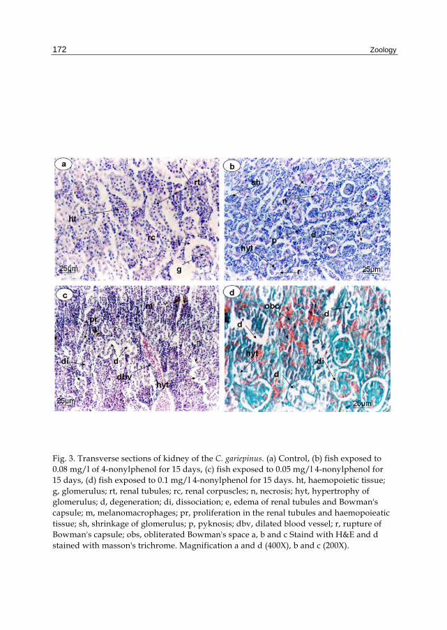
The functional units of the kidney of the control fish, Clarias gareipinus are nephrons which are composed of renal corpuscles (rc) and renal tubules (rt); these structures are surrounded by haemopoietic tissue (ht). The shape of the renal corpuscle is roughly spherical consisting of a double membraned capsule (Bowman's capsule) enclosing a tuft of blood capillaries (glomerulus) (g). Bowman's space; a space between the glomerulus and the capsule (Fig. 3a). Examination of kidney sections of fish exposed to 0.05 mg/l of 4-nonylphenol for 15 days revealed edema in the epithelium lining of some renal tubules (e) and some showed degeneration (d) and rupture of Bowman's capsule (r). Hypertrophy of the glomerulus (hyt) was observed with shrinkage (sh). Moreover, necrosis (n) and pyknosis (p) were observed in some renal tubules (fig. 3b). After 15 days of exposure to the 0.08 mg/l of 4-nonylphenol, similar histological changes were observed, however, proliferation in renal tubules and haemopoieatic tissue (pr) with dissociation in some tubules (di) were recorded. Dilated blood vessels (dbv) and mealnomacrophages (m) were also observed (fig. 3c). The sections of kidney in fishes exposed to 0.1 mg/l of 4-nonylphenol showed severe damage or complete degeneration with obliterated Bowman's space (obs). Masson's Trichrome stain indicated the degeneration of connective tissue and degeneration of renal tubule and glomerulus (fig. 3d).
Histopathological Alterations in some Body Organs of Adult Clarias gariepinus (Burchell, 1822) Exposed to 4-Nonylphenol
167
3.4 Histopathological changes in the liver
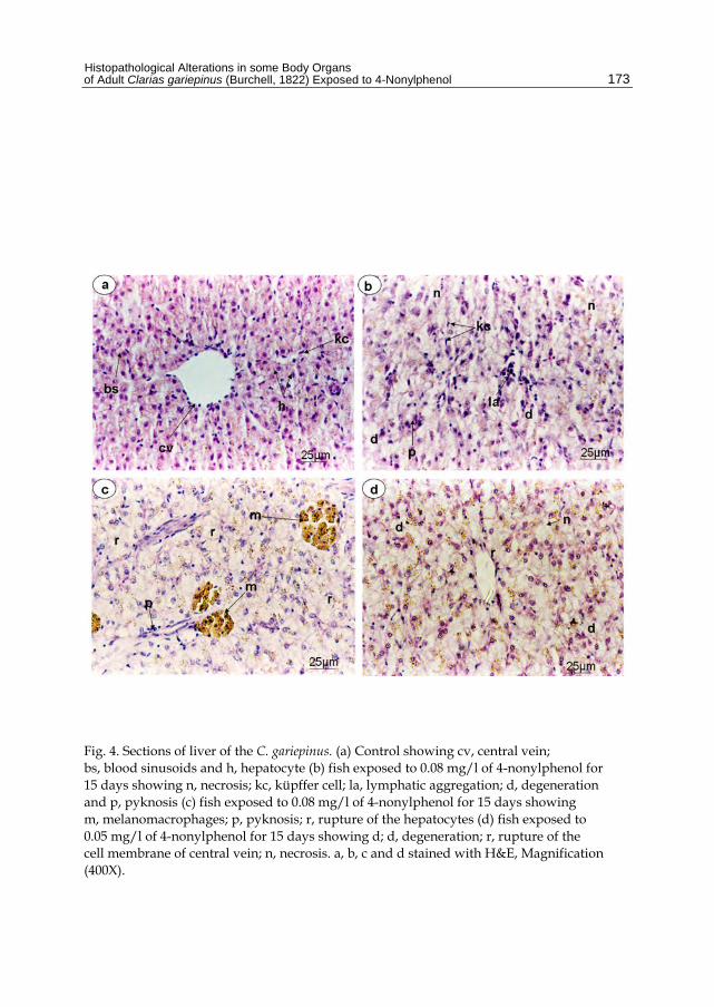
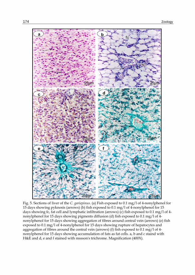
The liver of the control fish Clarias gariepinus appears as a continuous mass of hepatic cells; hepatocytes (h) which cord-like pattern interrupted by blood vessels and sinusoids (bs). The cords of hepatocytes are arranged around the central vein (cv). The hepatocytes are large in size, polygonal in shape with centrally located nuclei. The hepatocytes have homogenous eosinophilic cytoplasm. The sinusoids are seen as communicating channels occupied by blood cells with Küffer cells (kc) (fig. 4a). Examination of liver sections after exposure to 0.08 mg/l of 4-nonylphenol for 15 days showed degeneration (d) in the form of disintegration in most cytoplasmic contents. Lymphatic aggregations (la), necrosis (n), pyknosis (p), were observed (Fig. 4b). Also, melanomacrophages, pyknosis and rupture of hepatocytes (r) were recorded (fig. 4c). Less damage occurred in liver sections after exposure to 0.05 mg/l of 4-nonylphenol for 15 days (Fig. 4d). As Fig. (5a, b, c, d, e) shows marked severe damage occurred in fishes exposed to 0.1 mg/l of 4-nonylphenol for 15 days. Pyknosis indicated by arrows, fat cell (fc), lymphatic infiltration indicated by arrows, pigments diffusion, aggregation of fibers around central vein and rupture of hepatocytes were recorded. Masson's Trichrome stain indicated this severe damage in liver tissue (Fig. 5f).
3.5 Electron microscope examination of hepatocytes
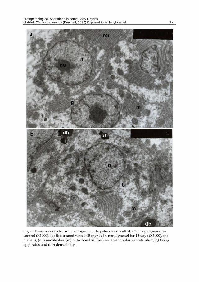
The fine structure of the hepatocytes shows parallel cisternae of rough endoplasmic reticulum (rer), polygonal centrally located vesicular nuclei (n) with nucleolus (nu), numerous mitochondria (m) with different shapes and sizes and Golgi complex (g) near the nucleus (Fig. 6a).
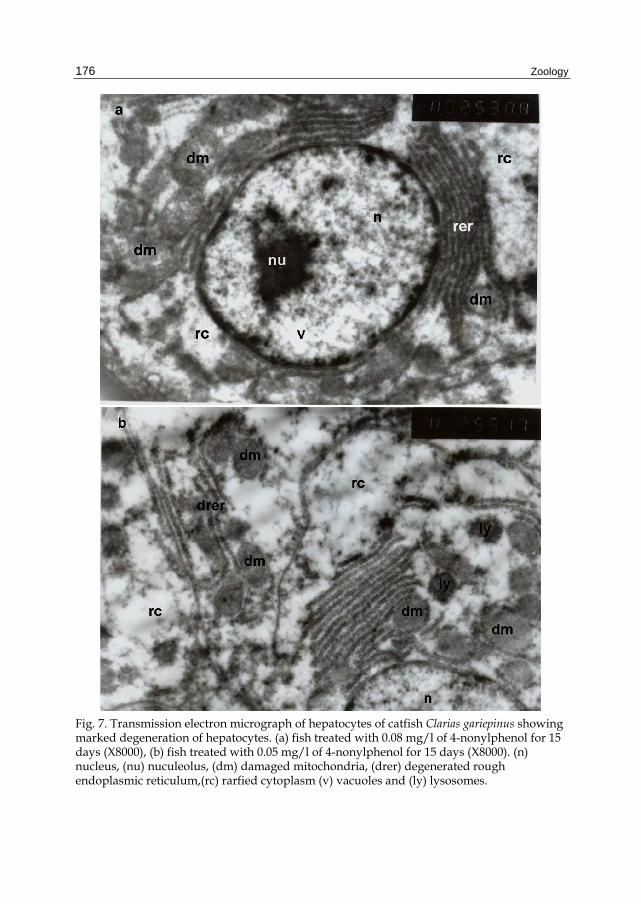
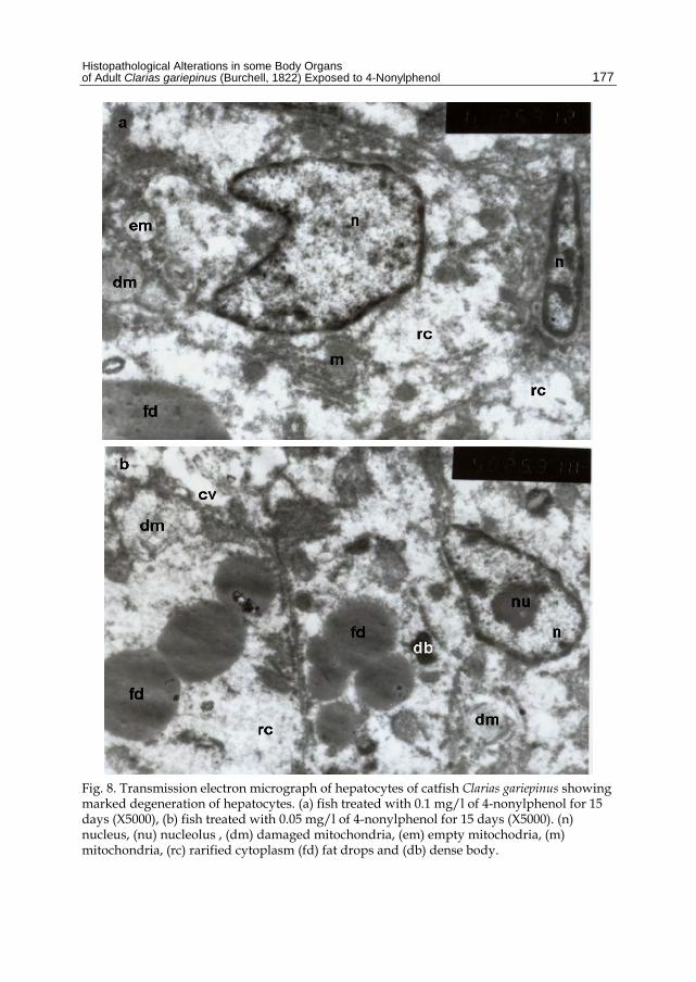
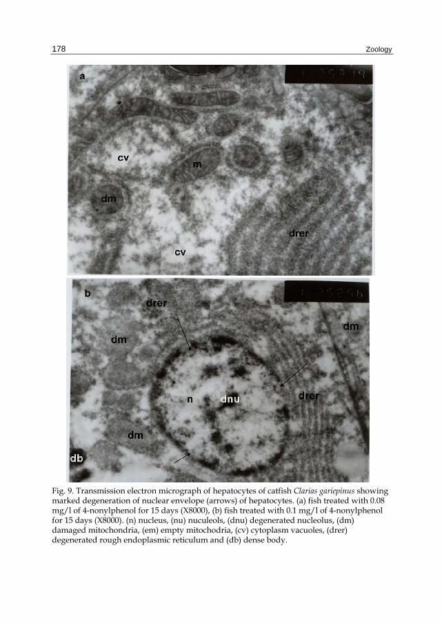
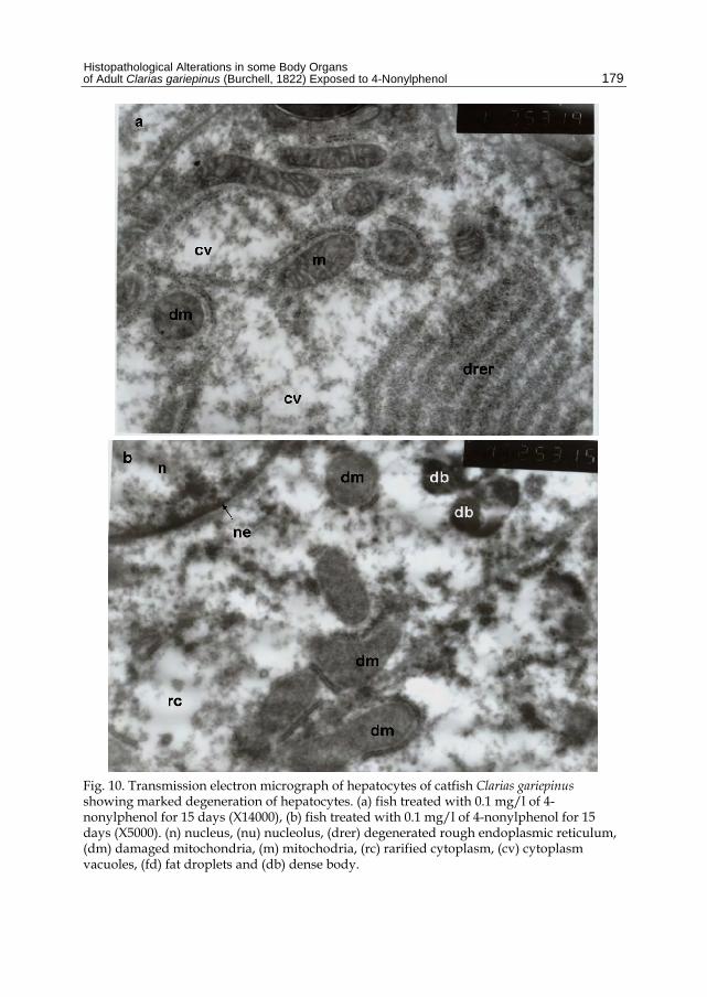
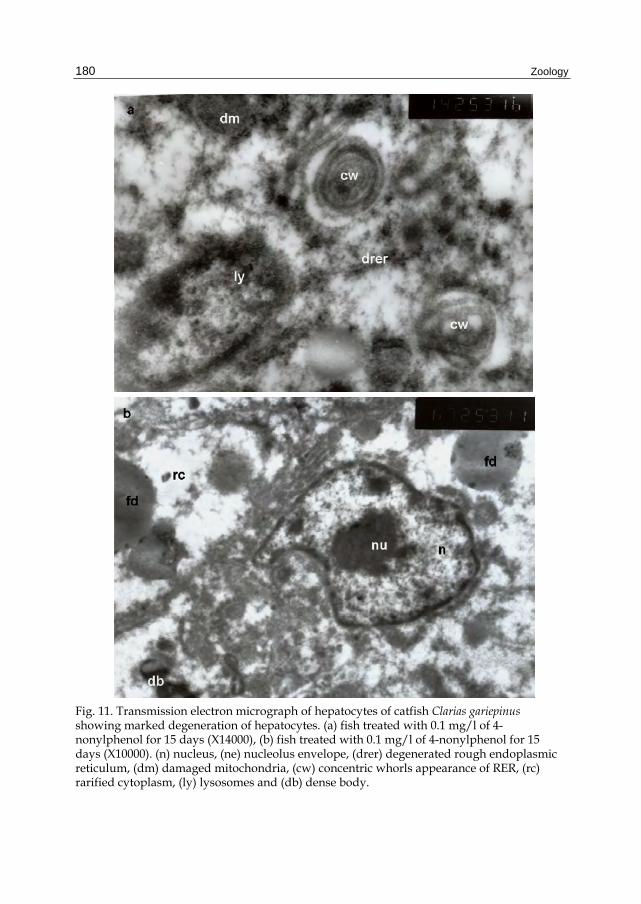
Hepatocytes of animals exposed to 0.05 mg/l of 4-nonylphenol appeared swollen or hypertrophied with dense bodies (db), karyolysis in nucleus (fig. 6b) and damaged mitochondria (dm), rarified cytoplasm (rc) and vacuoles (v) (fig. 7b). Also, degenerative changes, shrunken and indented nuclei with cytoplasmic fat droplets (fd) were observed (Fig. 8b). In fishes exposed to 0.08 mg/l of 4-nonylphenol the cytoplasm shows tiney vacuoles (cv), the nuclei appeared irregular in shape with nuclear indentation. Some hepatocytes showed signs of karylyosis (Fig. 7a, 9a). Moreover, damaged mitochondria, degenerative rough endoplasmic reticulum (drer), increase in number of lysosomes (ly) were recorded. Some nuclei showed condensation and migration of chromatin at the nuclear periphery with prominent nucleolus with some apoptotic changes in the form of nuclear envelope (Fig. 9a). Some hepatocytes appeared swollen with large rarified areas in the cytoplasm resulting in disorganization and dissociation of cellular organelles (Fig. 7a). Hepatocytes of fishes exposed to 0.1 mg/l of 4-nonylphenol showed similar changes as those exposed to 0.05 and 0.08 mg/l of 4-nonylphenol, however, degenerative changes, hypertrophied, karylyosis, blood sinusoids collapsation, apoptosis and vacuolated hepatocytes (Fig. 10, 11). Mitochondria were swollen with destructive cristae; electron dense materials appeared at the periphery of these mitochondria (em) (Fig. 9b, 10a, 11,a, b). Concentric whorly organization of rough endoplasmic reticulum (cw) with detached ribosomes were also seen (Fig. 11a). Other regions of the reticulum appeared as parallel cisternae with electron lucent cytoplasm between its cisterna also, circular arrays of RER
Zoology
168
were appeared (Fig. 10a, 11a). An increase in the number of lysosomes and fat drops were seen (Fig. 10b, 11b).
4. Discussion
It has been reported that NP like E2 is estrogenic and affects the histology of developing immune and endocrine organs and those in direct contact with aquatic environment (Yokota et al., 2001; Kang et al., 2003; Seki et al., 2003; Razia et al., 2006). Skin and gills are highly sensitive to pollutants due to their direct contact to aquatic environment. It has been reported that the skin is sensitive to steroid hormone activity (Pottinger and Pickering, 1985). The present results showed severe damage in the skin epithelial cells and necrosis reflecting such sensitivity to NP. NP exposure of rainbow trout resulted in a specific granulation pattern of epidermal mucous cells visible as irregularly shaped and large mucosomes (Burkhardt-Holm et al., 2000). The unique granulation pattern in skin of rainbow trout represents a suitable bioindicator for nonylphenol exposure (Burkhardt-Holm et al., 2000). These latter authors stated that the structural alterations in the skin of the estradiol-injected trout is pointed to a physiological response such as, detached pavement cells, vacuolation of the cytoplasm and severely deformed cell nuclei at a dose of 10µgl-1 which is lower than those in the present study. The damage occurred in the rainbow trout skin is similar to the quantitative changes of the mucous composition induced by hormones or environmental acidifications (Balm et al., 1995). Schwaiger et al., (1999) reported vitellogenin induction in the liver after exposure to 10 µgl-1 nonylphenol.
The present results exhibited severe damage in liver tissue of C. gariepinus including necrosis and decrease in the cell number along with vacuolation. Similar results were recorded by Uguz et al. (2003) who reported a significant increase in the Küpffer cells after one week of 4-nonylphenol exposure. Hughes et al. (2000) have shown NP-induced cell death. Galembeck et al. (1998), Hughes et al. (2000), and Uguz et al. (2003) reported that the disappearance of the cell membranes could be due to the lytic activity of alkylphenols.
In the present study, the liver cell borders disappeared and nuclei became larger after two weeks of exposure to 0.1mgl-1 of 4-nonylphenol, this is similar to the findings of Uguz et al. (2003) who reported that this may be due to the increase in the DNA/ RNA ratio which was been observed in carcinogenic cells induced by NP (Chiriboga et al., 2000a, b). The increase in the connective tissue with regenerating hepatocytes instead of normal liver tissues recorded in the present work was similar to those of Uguz et al. (2003). Such changes can be interpreted as an indication of carcinogenic development in the liver (Chiriboga et al., 2000a, b; Calmak, 2001). Generally, the lesions detected in cells, tissues or organs are represent an integration of cumulative effects of physiological and biochemical stressors and therefore, can be linked to the exposure and subsequent metabolism of chemical contaminants (Adeyemo, 2008).
The gills are the primary initial target of toxicity, and the cytological changes in gill morphology in fish usually occur as a result of contaminant exposure. Gills have an extensive surface area and minimal diffusion distance between dissolved O2 and blood capillary for efficient gaseous exchange. The fusion occurred in gills of fishes exposed to
Histopathological Alterations in some Body Organs of Adult Clarias gariepinus (Burchell, 1822) Exposed to 4-Nonylphenol
169
4-nonylphenbol in this investigation may cause a drastic reduction in the respiratory surface area. However, very little is known about the toxic impact of 4-nonylphenol on the functional morphology of the gills. The present results indicated such toxic impacts. Increase in the number of mucous cells in gills of fishes exposed to 0.1 mgl-1 of 4-nonylphenol was recorded. It has been reported that the immediate morpho-pathological response of the gills to ambient xenobiotics is often manifested by a significant increase in the density of its mucous cells (Dutta, 1997, Hemalatha and Banerjee, 1997). The large quantity of mucous secretion acts as a defense mechanism against several toxic substances (Handy and Eddy, 1991; Mazon et al., 1999). Similar to the findings of Dutta et al. (1996), the present results included many alterations such as increase in mucous and chloride cell number and size, necrosis, rupture of epithelium, desquamation, deformed secondary lamellae and oedema.
According to Peuranen et al. (1994) any discontinuity of epithelial lining of the gill lead to a negative ion balance and to changes in the haematocrite and mean cellular haemoglobin values of the blood. The number of chloride cells increased in the present study and this is similar to the results of Parashar and Banerjee, (2002). They stated that the number of chloride cells in the epithelial linings of both primary lamellae and secondary lamellae of Heteropneustes fossilis increased significantly following exposure to lead nitrate solution. Dutta et al. (1996) summarized the increased number of chloride cells in the gills of fishes following exposure to a variety of toxicants.
Increased ion permeability and sodium efflux of gill epithelial cells due to ethoxylate nonylphenol were reported in rainbow trout (Pärt et al., 1985). Similar results in the present work were recorded in the histology of gills under NP-stress and confirmed by the increased NP-induced anion gap.
The kidney of fishes receives the largest proportion of the post-branchial blood and therefore renal lesions might be expected to be good indicators of environmental pollution (Cengiz, 2006). Many studies used histological characteristics of kidney as an indicators of pollution especially nonylphenol (Srivastava et al., 1990; Banerjee and Bhattacharya, 1994; Ortiz et al., 2003; Cengiz, 2006). In the present work, histological changes in the kidney after exposure to 4- nonylphenol were necrosis, hypertrophy of glomerulus, degeneration and dissociation of renal tubules and Bowman's capsule, proliferation in the renal tubule and haemopoieatic tissue, shrinkage of glomerulus, pyknosis, dilated blood vessel, rupture of Bowman's capsule, and obliterated Bowman's space. Similar results were reported in fishes after exposure to other pollutants (Cengiz, 2006; Khidr and Mekkawy, 2008; Abdel-Tawab and Al-Salahy, 2009).
From the results of the current study, it could be suggested that the exposure of adult catfish, Clarias gariepinus to sublethel doses of 4-nonylphenol caused moderate and severe damage to some organs such as gills, skin, kidney, and liver. These adverse effects of NP in gills, skin, kidney and liver were simultaneously correlated with sever biochemical, physiological changes in addition to endocrine disruption (Mekkawy et al., 2011; Mahmoud et al., 2011; Sayed et al., 2011) So, it is concluded that NP works as estrogenic and non-estrogenic factor leadings to general and specific metabolism disruption in different pathway.
Zoology
170
Fig. 1. (a) Gill structure of control adult fish Clarias gariepinus. (pl), primary lamellae; (sl) secondary lamellae; (epc) epithelial cell; (mc) mucous cell; (chc) chloride cell. (b) Gill tissue exposed to 0.05 mg/l 4-nonylphenol for 15 days showing a, epithelial lifting and oedema and b, deformed secondary lamellae. (c) Gill tissue exposed to 0.08 mg/l of 4-nonylphenol for 15 days showing a, desquamation and necrosis. (d) ) Gill tissue exposed to 0.1 mg/l of 4-nonylphenol for 15 days showing a, degeneration of cartilaginous bar and b, malformed secondary lamellae. (e) Gill tissue exposed to 0.1 mg/l of 4-nonylphenol for 15 days showing a, increase in chloride cell size and number; b, epithelial hyperplasia and diffusion of secondary lamellae; c, degeneration and vacuolation of cartilaginous bar; d, desquamation and necrosis and e, increase number of mucous cells. Stain H& E. Magnification a, b, c, e (400X) and d (100X).
Histopathological Alterations in some Body Organs of Adult Clarias gariepinus (Burchell, 1822) Exposed to 4-Nonylphenol
171
Fig. 2. Vertical sections of the skin of adult catfish Clarias gariepinus stained with H&E. (a) skin of control fish showing ac, alarm cell; mc, mucous cell; ep, epithelium; p, pigment cell. (b, d, e) skin of fish exposed to 0.1 mg/l of 4-nonylphenol for 15 days showing fc, fat cell; repc, ruptured epithelial cells; nc, necrotic cells; gc, granuled cells and va, vacuoles.(c) skin of exposed fish to 0.08 mg/l 4-nonylphenol for 15 days showing repc, ruptured epithelial cells and emc, enlarged mucous cells. (f) skin of exposed fish to 0.05 mg/l 4-nonylphenol for 15 days showing eac, enlarged alarm cells and va, vacuoles. Magnification a, b, c, d, e (100X) and f (400X).
Zoology
172
Fig. 3. Transverse sections of kidney of the C. gariepinus. (a) Control, (b) fish exposed to 0.08 mg/l of 4-nonylphenol for 15 days, (c) fish exposed to 0.05 mg/l 4-nonylphenol for 15 days, (d) fish exposed to 0.1 mg/l 4-nonylphenol for 15 days. ht, haemopoietic tissue; g, glomerulus; rt, renal tubules; rc, renal corpuscles; n, necrosis; hyt, hypertrophy of glomerulus; d, degeneration; di, dissociation; e, edema of renal tubules and Bowman's capsule; m, melanomacrophages; pr, proliferation in the renal tubules and haemopoieatic tissue; sh, shrinkage of glomerulus; p, pyknosis; dbv, dilated blood vessel; r, rupture of Bowman's capsule; obs, obliterated Bowman's space a, b and c Staind with H&E and d stained with masson's trichrome. Magnification a and d (400X), b and c (200X).
Histopathological Alterations in some Body Organs of Adult Clarias gariepinus (Burchell, 1822) Exposed to 4-Nonylphenol
173
Fig. 4. Sections of liver of the C. gariepinus. (a) Control showing cv, central vein; bs, blood sinusoids and h, hepatocyte (b) fish exposed to 0.08 mg/l of 4-nonylphenol for 15 days showing n, necrosis; kc, küpffer cell; la, lymphatic aggregation; d, degeneration and p, pyknosis (c) fish exposed to 0.08 mg/l of 4-nonylphenol for 15 days showing m, melanomacrophages; p, pyknosis; r, rupture of the hepatocytes (d) fish exposed to 0.05 mg/l of 4-nonylphenol for 15 days showing d; d, degeneration; r, rupture of the cell membrane of central vein; n, necrosis. a, b, c and d stained with H&E, Magnification (400X).
Zoology
174
Fig. 5. Sections of liver of the C. gariepinus. (a) Fish exposed to 0.1 mg/l of 4-nonylphenol for 15 days showing pyknosis (arrows) (b) fish exposed to 0.1 mg/l of 4-nonylphenol for 15 days showing fc, fat cell and lymphatic infiltration (arrows) (c) fish exposed to 0.1 mg/l of 4-nonylphenol for 15 days showing pigments diffusion (d) fish exposed to 0.1 mg/l of 4-nonylphenol for 15 days showing aggregation of fibres around central vein (arrows) (e) fish exposed to 0.1 mg/l of 4-nonylphenol for 15 days showing rupture of hepatocytes and aggregation of fibres around the central vein (arrows) (f) fish exposed to 0.1 mg/l of 4-nonylphenol for 15 days showing accumulation of fats as fat cells. a, b and c staind with H&E and d, e and f stained with mssson's trichrome. Magnification (400X).
Histopathological Alterations in some Body Organs of Adult Clarias gariepinus (Burchell, 1822) Exposed to 4-Nonylphenol
175
Fig. 6. Transmission electron micrograph of hepatocytes of catfish Clarias gariepinus. (a) control (X5000), (b) fish treated with 0.05 mg/l of 4-nonylphenol for 15 days (X5000). (n) nucleus, (nu) nuculeolus, (m) mitochondria, (rer) rough endoplasmic reticulum,(g) Golgi apparatus and (db) dense body.
Zoology
176
Fig. 7. Transmission electron micrograph of hepatocytes of catfish Clarias gariepinus showing marked degeneration of hepatocytes. (a) fish treated with 0.08 mg/l of 4-nonylphenol for 15 days (X8000), (b) fish treated with 0.05 mg/l of 4-nonylphenol for 15 days (X8000). (n) nucleus, (nu) nuculeolus, (dm) damaged mitochondria, (drer) degenerated rough endoplasmic reticulum,(rc) rarfied cytoplasm (v) vacuoles and (ly) lysosomes.
Histopathological Alterations in some Body Organs of Adult Clarias gariepinus (Burchell, 1822) Exposed to 4-Nonylphenol
177
Fig. 8. Transmission electron micrograph of hepatocytes of catfish Clarias gariepinus showing marked degeneration of hepatocytes. (a) fish treated with 0.1 mg/l of 4-nonylphenol for 15 days (X5000), (b) fish treated with 0.05 mg/l of 4-nonylphenol for 15 days (X5000). (n) nucleus, (nu) nucleolus , (dm) damaged mitochondria, (em) empty mitochodria, (m) mitochondria, (rc) rarified cytoplasm (fd) fat drops and (db) dense body.
Zoology
178
Fig. 9. Transmission electron micrograph of hepatocytes of catfish Clarias gariepinus showing marked degeneration of nuclear envelope (arrows) of hepatocytes. (a) fish treated with 0.08 mg/l of 4-nonylphenol for 15 days (X8000), (b) fish treated with 0.1 mg/l of 4-nonylphenol for 15 days (X8000). (n) nucleus, (nu) nuculeols, (dnu) degenerated nucleolus, (dm) damaged mitochondria, (em) empty mitochodria, (cv) cytoplasm vacuoles, (drer) degenerated rough endoplasmic reticulum and (db) dense body.
Histopathological Alterations in some Body Organs of Adult Clarias gariepinus (Burchell, 1822) Exposed to 4-Nonylphenol
179
Fig. 10. Transmission electron micrograph of hepatocytes of catfish Clarias gariepinus showing marked degeneration of hepatocytes. (a) fish treated with 0.1 mg/l of 4-nonylphenol for 15 days (X14000), (b) fish treated with 0.1 mg/l of 4-nonylphenol for 15 days (X5000). (n) nucleus, (nu) nucleolus, (drer) degenerated rough endoplasmic reticulum, (dm) damaged mitochondria, (m) mitochodria, (rc) rarified cytoplasm, (cv) cytoplasm vacuoles, (fd) fat droplets and (db) dense body.
Zoology
180
Fig. 11. Transmission electron micrograph of hepatocytes of catfish Clarias gariepinus showing marked degeneration of hepatocytes. (a) fish treated with 0.1 mg/l of 4-nonylphenol for 15 days (X14000), (b) fish treated with 0.1 mg/l of 4-nonylphenol for 15 days (X10000). (n) nucleus, (ne) nucleolus envelope, (drer) degenerated rough endoplasmic reticulum, (dm) damaged mitochondria, (cw) concentric whorls appearance of RER, (rc) rarified cytoplasm, (ly) lysosomes and (db) dense body.
Histopathological Alterations in some Body Organs of Adult Clarias gariepinus (Burchell, 1822) Exposed to 4-Nonylphenol
181
5. References
Abdel-Tawab, H.S. & Al-Salahy, M.B. (2010). Biocemical and ultrastructural studies of the effect of garlic juice on the liver of the fish Clarias gariepinus. Journal of Egyptian Germany Society of Zoology Vol. 60C: 39-62.
Adewolu, M.A., Adeniji, C.A. & Adejobi, A.B. (2008). Feed utilization, growth and survival of Clarias gariepinus (Burchell 1822) fingerlings cultured under different photoperiods. Aquaculture Vol. 283: 64–67.
Adeyemo, O.K. (2008). Histological Alterations Observed in the Gills and Ovaries of Clarias gariepinus exposed to environmentally relevant lead concentrations. Journal of Environmental Health Vol. 70 (9): 48-51.
Ahel, M., Giger, W. & Koch, M. (1994). Behaviour of alkylphenol polyethoxylate surfactants in the aquatic environment - I. Occurrence and transformation in sewage treatment. Water Resources Vol. 28: 1131–1142.
Ahel, M., McEvoy, J. & Giger, W. (1993). Bioaccumulation of the lipophilic metabolites of nonionic surfactants in freshwater organisms. Environmental Pollution. Vol. 79: 243-248.
Akinwole, A.O. & Faturoti, E.O. (2007). Biological performance of African Catfish (Clarias gariepinus) cultured in recirculating system in Ibadan. Aquaculture Enginering. Vol. 36: 18–23.
Balm, P.H.M., Iger, Y., Prunet, P., Pottinger, T.G. & Wendelaar Bonga, S.E. (1995). Skin ultrastructure in relation to prolactin and MSH function in rainbow trout (Oncorhynchus mykiss) exposed to environmental acidification. Cell Tissue Research. Vol. 279: 351-358.
Bancroft, J. & Stevens A, (1982). Theory and Practice of Histological Techniques, 2nd Ed. Churchill-Livingston, NY, pp 131-135.
Banerjee, S. & Bhattacharya, S. (1994). Histopathology of kidney of Channa punctatus exposed to chronic nonlethal level of elsan, mercury and ammonia. Ecotoxicology and Environmental Safety. Vol. 29 (3): 65–275.
Bennie, D.T. (1999). Review of the environmental occurrence of alkylphenols and alkylphenol ethoxylates. Water Quality Research Journal of Canada. Vol. 34: 79–122.
Bernet, D., Schmidt, H., Meier, W., Brkhardt-Holm, P. & Wahli, T. (1999). Histopathology in fish: Proposal for a protocol to assess aquatic pollution. Journal of Fish Diseases. Vol. 22: 25–34.
Burkhardt-Holm, P., Escher, M. & Meier, W. (1997). Waste water management plant effluents cause cellular alterations in the skin of brown trout Salmo trutta. Journal of Fish Biology. Vol. 50: 744-758.
Burkhardt-Holm, P., Wahli, T. & Meier, W. (2000). Nonylphenol affects the granulation pattern of epidermal mucous cells in rainbow trout, Oncorhynchus mykiss. Ecotoxicology and Environmental Safety. Vol. 46: 34–40.
Calmak, G. (2001). Spectroscopic analysis of the livers exposed to nonylphenol in rainbowtrout (Onchorynchus mykiss). MS thesis, Biology Department, Middle East Technical University, Turkey.
Cengiz, E. I. (2006). Gill and kidney histopathology in the freshwater fish Cyprinus carpio after acute exposure to deltamethrin. Environmental Toxicology Pharmacology. Vol. 22: 200–204.
Chiriboga, L., Yee, H. & Diem, M. (2000a). Infrared spectroscopy of human cells and tissue. Part VI: a comparative study of histology and infrared microspectroscopy of normal, cirrhotic, and cancerous liver tissue. Applied Spectroscopy. Vol. 54 (1): 1–8.
Zoology
182
Chiriboga, L., Yee, H. & Diem, M. (2000b). Infrared spectroscopy of human cells and tissue. Part VII: FT-IR microspectroscopy of DNase- and RNase-treated normal, cirrhotic, and neoplastic liver tissue. Applied. Spectroscopy. Vol. 54 (4): 480–485.
Christiansen, T., Korsgaard, B. & Jespersen, A. (1998). Effects of nonylphenol and 17β-oestradiol on vitellogenin synthesis, testicular structure and cytology in male eelpout Zoarces viviparus. Journal of Experimental of Biology. Vol. 201: 179-192.
Colborn, T., Vom Saal, F.S. & Soto, A.M. (1993). Developmental effects of endocrine disrupting chemicals in wildlife and humans. Environmental Health Perspectives. Vol. 101: 378–384.
Coldham, N.G., Sivapathasundaram, S., Dave, M., Ashfleld, L.A., Pottinger, T.G., Goodall, C. & Sauer, M.J. (1998). Biotransformation, tissue distribution, and persistence of 4-nonylphenol residues in juvenile rainbow trout (Oncorhynchus mykiss). Drug Metabolism Dispososition. Vol. 26: 347-353.
Dutta, H.M. (1997). A composite approach for evaluation of the effect of pesticides on fish, In: Munshi, J. S. D., Dutta, H. M. (Eds.), Fish Morphology, Horizon of new research Science Publisher Inc., USA, pp. 249-277.
Dutta, H.M., Munshi, J.S.D., Roy, P.K., Singh, N.K., Adhikari, S. & Killius, J. (1996). Ultrastructural changes in the respiratory lamellae of the catfish, Heteropneustes fossilis after sublethal exposure to melathion. Environmental Pollution. Vol. 92: 329-341.
Galembeck, E., Alonso, A. & Meirelles, N.C. (1998). Effects of polyoxyethylene chain length on erythrocyte hemolysis induced by poly[oxyethylene(n)nonylphenol] non-ionic surfactants. Chemico-Biollogical Interactions. Vol. 113 (2): 91–103.
Handy, R.D. & Eddy, F.B. (1991). The absence of mucous on the secondary lamellae of unstressed rainbow trout, Oncorhynchus mykiss. Journal of Fish Biology. Vol. 38: 153-155.
Hemalatha, S. & Banerjee, T.K. (1997). Histopathological analysis of sublethal toxicity of zinc chloride to the respiratory organs of the air-breathing catfish Heteropneustes fossilis (Bloch). Biological Research. Vol. 30: 11-21.
Hinton, D.E., Baumann, P.C., Gardner, G.R., Hawkins, W.E., Hendricks, J.D., Murchelano, R.A. & Okihiro, M.S. (1992). Histopathological biomarkers, In: Huggett, R.J., Kimerle, R.A., Mehrle, P.M., Jr Bergman, H.L., (Eds.), Biomarkers: Biochemical, Physiological, and Histological Markers of Anthropogenic Stress, Lewis, USA, pp 155–196.
Hinton, D.E. & Lauren, D.J. (1990). Integrative histopathological approaches to detecting effects of environmental stressors on fishes. Am. Fish Soc. Sym. Vol. 8: 51–66.
Hughes, P.J., McLellan, H., Lowes, D.A., Khan, S.Z., Bilmen, J.G., Tovey, S.C., Godfrey, R.E., Michell, R.H., Kirk, C.J. & Michelangeli, F. (2000). Estrogenic alkylphenols induce cell death by inhibiting testis endoplasmic reticulum Ca2+ pumps. Biochemical and Biophysical Research Communications. Vol. 277: 68–574.
Iger, Y., Balm, P.H., Jenner, H.A. & Wendelaar Bonga, S.E. (1995). Cortisol induces stress-related changes in the skin of rainbow trout (Oncorhynchus mykiss). General and Comparative Endocrinology. Vol. 97: 188-198.
Jobling, S., Sheahan, D., Osborne, J.A., Matthiessen, P. & Sumpter, J. P. (1996). Inhibition of testicular growth in rainbow trout (Oncorhynchus mykiss) exposed to estrogenic alkylphenolic chemicals. Environmental Toxicology and Chemistry. Vol. 15: 194-202.
Johannessen, J. (1978). Instruction and techniques in Electron Micrscopy in human medicine. Mchgraw-Hill Int. Book Co.
Histopathological Alterations in some Body Organs of Adult Clarias gariepinus (Burchell, 1822) Exposed to 4-Nonylphenol
183
Karami, A., Christianus, A., Ishak, Z., Courtenay S. C., Syed, M. A., Noor Azlina, M. & Noorshinah H. (2010). Effect of triploidization on juvenile African catfish (Clarias gariepinus). Aquaculture International. Vol. 18: 851–858.
Khidr, M. B. & Mekkawy, I.A.A. (2008). Effect of separate and combined lead and selenium on the liver of the cichlid fish Oreochromis niloticus: ultrastructural study. Egyptian Journal of Zoology. Vol. 50: 89-119.
Lech, J.J., Lewis, S.K. & Ren, L. (1996). In vivo estrogenic activity of nonylphenol in rainbow trout. Fundam. Applied Toxicology. Vol. 30: 229-232.
Lewis, S.K. & Lech, J.J. (1996). Uptake, disposition and persistence of nonylphenol from water in rainbow trout (Oncorhynchus mykiss). Xenobiotica. Vol. 26: 813-819.
Malik, R. & Hodgson, H. (2002). The relationship between the thyroid gland and the liver. Quarterly Journal of Medicine. Vol. 95: 559-569.
Marchand, M.J., van Dyk, J.C., Pieterse, G.M., Barnhoorn, I.E.J. & Bornman, M.S., (2008). Histopathological Alterations in the Liver of the Sharptooth Catfish Clarias gariepinus from Polluted Aquatic Systems in South Africa. Environmental Toxicology. DOI 10.1002/tox.
Mazon, A.F., Cerqueira, C.C.C., Monteiro, E.A.S. & Fernandes, M.N. (1999). Acute copper exposure in freshwater fish, Morphological and physiological effects. In: Val, A.L., Almeida-Val, V.M.F., (Eds.), Biology of Tropical Fishes, INPA, Manaus, pp. 263-275.
Meyers, T.R. & Hendricks, J.D. (1985). Histopathology. In: Loux, D.B., Dorfman, M., (Eds.), Fundamentals of Aquatic Toxicology: Methods and Applications, Hemisphere USA, pp. 283–330.
Nguyen, L.T.H. & Janssen, C.R. (2002). Embryo-larval toxicity tests with the African catfish (Clarias gariepinus): comparative sensitivity of endpoints. Archives of Environmental Contamination and Toxicology. Vol. 42: 256–262.
Nimrod, A.C. & Benson, W.H. (1996). Environmental estrogenic effects of alkylphenol ethoxylates. Critical Reviews in Toxicology. Vol. 26: 335–364.
Ortiz, J.B., De Canales, M.L.G. & Sarasquete, C. (2003). Histopathological changes induced by lindane (gamma-HCH) in various organs of fishes. Scientia Marina. Vol. 67 (1): 53–61.
Parashar, R.S. & Banerjee, T.K. (2002). Toxic impact of lethal concentration of lead nitrate on the gills of air-breathing catfish Heteropneustes fossilis (Bloch). Veterinarski Arhiv. Vol. 72 (3): 167-183.
Pärt, P., Svanberg, O. & Bergstrom, E. (1985). The influence of surfactants on gill physiology and cadmium uptake in perfused rainbow trout gills. Ecotoxicology and Environmental Safety. Vol. 9: 135-144.
Peuranen, S., Vuorinen, P. J., Vuorinen, M. & Hollender, A. (1994). The effect of iron, humic acids and low pH on the gills and physiology of brown trout (Salmo trutta). Annales Zoologici Fennicii. Vol. 31: 389-396.
Pottinger, T.G. & Pickering, A.D. (1985). Changes in skin structure associated with elevated androgen levels in maturing male brown trout, Salmo trutta L. Journal of Fish Biology. Vol. 26: 745-753.
Razia, S., Maegawa, Y., Tamotsu, S. & Oishi, T. (2006). Histological changes in immune and endocrine organs of quail embryos: Exposure to estrogen and nonylphenol. Ecotoxicology and Environmental Safety. Vol. 65: 364–371.
Schwaiger, J., Spieser, O.H., Nardy, E., Kalbfus, W., Braunbeck, Th. & Negele, R. D. (1999). Toxic effects versus endocrine disruption Does the xenoestrogen nonylphenol influence physiological functions in fish? SETAC: 9th Annual Meeting, 25-29 May 1999, Leipzig, Germany.
Zoology
184
Servos, M.R. (1999). Review of the aquatic toxicity, estrogenic responses and bioaccumulation of alkylphenols and alkylphenol polyethoxylates. Water Qualality Research Journal of Canada. Vol. 31: 123–177.
Shephard, K.L. (1994). Functions for fish mucus. Reviews in Fish Biology and Fisheries. Vol. 4: 401-429.
Soto, A.M., Chung, K.L. & Sonnenschein, C. (1994). The pesticides endosulfan, toxaphene and dieldrin have estrogenic effects on human estrogen sensitive cells. Environmental Health Perspectives. Vol. 102: 380–385.
Soto, A.M., Lin, T.M., Justicia, H., Silvia, R.M. & Sonnenenschein, C. (1992). An ‘in culture’ bioassay to assess the estrogenicity of xenobiotics (E-screen), In: Colburn, T.C. (Eds.), Chemically Induced Alterations in Sexual and Functional Development; The Wildlife-human Connection. Princeton Scientific Publishing, Princeton, NJ, USA, pp. 295–309.
Srivastava, S.K., Tiwari, P.R. & Srivastav, A.K. (1990). Effects of chlorpyrifos on the kidney of freshwater catfish. Heteropneustes fossilis. Bulletin of Environmental Contamination and Toxicology. Vol. 45: 748–751.
Talmage, S.S. (1994). Environmental and Human Safety of Major Surfactants: Alcohol Ethoxylates and Alkylphenol Ethoxylates. Lewis Publishers, Boca Raton, FL.
Toomey, B.H., Monteverdi, G.H. & Di Giulia, R.T. (1999). Octylphenol induces vitellogenin production and cell death in fish hepatocytes. Environmental Toxicology and Chemistry. Vol. 18: 734–739.
Uguz, C., Iscan, M., Ergu, A., Belgin I.V. & Togan, I. (2003). The bioaccumulation of nonyphenol and its adverse effect on the liver of rainbowtrout (Onchorynchus mykiss). Environmental Research. Vol. 92: 262–270.
Wendelaar Bonga, S.E. (1997). The stress response in Fish. Physiological Reviews. Vol. 77: 591-625.
Sayed, A.H., Mekkawy, I.A. & Mahmoud, U.M. (2011). Histopathological alterations in some organs of adults of Clarias gariepinus (Burchell, 1822) exposed to 4-nonylphenol. The 19th Conference of Egyptian-German Scocity of Zoology, Bin Sueif University, Egypt.
Mekkawy, I.A., Mahmoud, U.M. & Sayed, A.H. (2011). Effects of 4-nonylphenol on blood cells of the African catfish Clarias gariepinus (Burchell, 1822) Tissue Cell, doi:10.1016/j.tice.2011.03.006
Mahmoud, U.M., Sayed, A.H. & Mekkawy, I.A. (2011). Biochemical changes of African catfish Clarias gariepinus exposed to 4-nonylphenol. African Journal of Biochemistery,11-053 (Accepted)
Yokota, H., Seki, M., Maeda, M., Oshima, Y., Tadokoro, H., Honjo, T. & Kobayashi, K. (2001). Life-cycle toxicity of 4-nonylphenol to medaka (Oryzias latipes), Environmental Toxicology and Chemistry. Vol. 20: 2552-2560.
Kang, I.J., Yokota, H., Oshima, Y., Tsuruda, Y., Hano, T., Maeda, M., Imada, N., Tadokoro, H. & Honjo, T. (2003). Effect of 4-nonylphenol on the reproduction of Japanese medaka, Oryzias latipes, Environmental Toxicology and Chemistery. Vol. 22: 2438-2445.
Seki, M., Yokota, H., Maeda, M., Tadokoro, H. & Kobayashi, K. (2003). Effect of 4-nonylphenol and 4- tert octylphenol on sex differentiation and vitellogenin induction in medaka (Oryzias Latipes), Environmental Toxicology and Chemistery. Vol. 22:1507-1516.