intrinsic respiratory gating in small-animal ct
TRANSCRIPT
Eur Radiol (2008) 18: 1375–1384DOI 10.1007/s00330-008-0903-3 EXPERIMENTAL
Soenke H. BartlingJulien DinkelWolfram StillerMichael GrasruckIjad MadischHans-Ulrich KauczorWolfhard SemmlerRajiv GuptaFabian Kiessling
Received: 21 September 2007Revised: 1 December 2007Accepted: 19 January 2008Published online: 23 April 2008# European Society of Radiology 2008
Intrinsic respiratory gating in small-animal CT
Abstract Gating in small-animal CTimaging can compensate artefactscaused by physiological motion duringscanning. However, all published gat-ing approaches for small animals relyon additional hardware to derive thegating signals. In contrast, in this studya novel method of intrinsic respiratorygating of rodents was developed andtested for mice (n=5), rats (n=5) andrabbits (n=2) in a flat-panel cone-beamCT system. In a consensus read imagequality was compared with that of non-gated and retrospective extrinsically
gated scans performed using a pneu-matic cushion. In comparison to non-gated images, image quality improvedsignificantly using intrinsic and extrin-sic gating. Delineation of diaphragmand lung structure improved in allanimals. Image quality of intrinsicallygatedCTwas judged to be equivalent toextrinsically gated ones. Additionally4D datasets were calculated using bothgating methods. Values for expiratory,inspiratory and tidal lung volumesdetermined with the two gating meth-ods were comparable and correlatedwell with values known from theliterature. We could show that intrinsicrespiratory gating in rodents makesadditional gating hardware and pre-paratory efforts superfluous. Thismethod improves image quality andallows derivation of functional data.Therefore it bears the potential to findwide applications in small-animal CTimaging.
Keywords CT .Small-animal imaging .Flat-panel detector . Intrinsic gating
Introduction
With the increasing utilization of small-animal CT inpreclinical and basic research, effective methods tocompensate for physiological motion during scanninghave been sought. Prospective [1–5] as well as retro-spective [6, 7] methods for respiratory and cardiac gatinghave been described, and their benefit for structural lungimaging has been shown [2, 6, 7]. Furthermore, gating
enables reconstruction of a 4D time series that can be usedto calculate functional parameters such as respiratory tidalvolume and cardiac ejection fraction [6–8].
Regardless of whether retrospective or prospectivegating is used, all gating methods that are currently inuse depend on an extrinsic sensor to derive a gatingreference signal. Such a sensor could be a respiratorycushion that is placed below the chest of the small animal[1, 6, 7] or an optical system to deduce the breathing
Soenke H. Bartling and Julien Dinkelcontributed equally.
S. H. Bartling . F. KiesslingJunior Group Molecular Imaging,German Cancer Research Center(DKFZ),Heidelberg, Germany
S. H. Bartling . W. Stiller .W. Semmler . F. KiesslingDepartment of Medical Physics inRadiology, German Cancer ResearchCenter (DKFZ),Heidelberg, Germany
J. Dinkel . H.-U. KauczorDepartment of Radiology, GermanCancer Research Center (DKFZ),Heidelberg, Germany
M. GrasruckSiemens Medical Solutions,Forchheim, Germany
I. Madisch . R. GuptaDepartment of Radiology,Massachusetts General Hospital,Boston, MA, USA
S. H. Bartling (*)Junior Group Molecular Imaging,Department Medical Physics inRadiology, German Cancer ResearchCenter (DKFZ),Im Neuenheimer Feld 280,69120 Heidelberg, Germanye-mail: [email protected].: +49-622-1422686Fax: +49-622-1422572
movements [9, 10]. For cardiac gating, EKG electrodes areroutinely used.
Intrinsic methods extract the gating information from theacquired projection data directly, obviating the need foradditional hardware or preparatory efforts. Intrinsic meth-ods have been described for human cardiac [11, 12] as wellas respiratory gated CT imaging [13, 14]. However,intrinsic gating methods for small animals have not yetbeen described.
Human and small-animal CT instrument characteristicsand data acquisition parameters vary substantially. To thebest of our knowledge, this paper describes the firstintrinsic respiratory gating method for small-animal imag-ing that takes into account the characteristics of small-animal cone-beam CT instruments such as a relatively largez-coverage, relatively slow data acquisition, continuousvolume acquisition and non-spiral scanning. In thisresearch we successfully demonstrated that, using intrinsicrespiratory gating in small animals, it is possible to achievethe same image quality as that from the establishedextrinsic gating using external gating hardware. It is alsopossible to reconstruct 4D data sets, thus enabling deriva-tion of functional parameters of respiration.
Materials and methods
Flat-panel-based volume CT instrumentsand data acquisition
A prototype CT instrument (Siemens Medical Solutions,Forchheim, Germany) was used for gating experiments.The technical CT setup is identical to that described in [7,15] where extrinsic gating was originally tested [7]. Itsmain features are a flat-panel detector and a modified X-raytube, both mounted on a multi-slice CT gantry. Taking thegeometry of the CT system setup into consideration, theinstrument’s total field of view is 25×25×18 cm3. For ratand mouse CT imaging, the active detector area was limitedto 192 lines in z-direction and 1,024 rows in x-y-directionto increase the frame rate. The detector was read out in a2×2 binning mode, meaning that four neighbouring pixelswere averaged. This resulted in a decreased field of view of25×25×4.5 cm3, which was still big enough to cover theentire thorax and diaphragm of a rat. The resulting framerate was 100 frames per second (fps), which translates intoan exposure time of 10 ms per projection.
Rabbits were examinedwithout reduction of the active CTdetector area, because the lung did not fit in the reduced fieldof view that was employed for rats and mice. The detectorread out frame rate was therefore reduced to 30 fps. All otherparameters were the same for rabbit CT imaging.
The spatial resolution of the CT system, as computed byexamining a tungsten wire phantom, is 24 lp/cm at 10%modulation transfer function. This isotropic spatial resolu-
tion translates into a minimal detectable feature size of200 μm.
Each projection image acquired during gantry rotation istime stamped and is labelled with the angle of acquisition.Gantry rotation times can be varied from 2 s to 19 s in stepsof 1 s, the maximum total CT data acquisition time being80 s. The tomographic image reconstruction is based on amodified Feldkamp algorithm [16].
For each animal examined, a total data acquisition timeof 80 s was used with a gantry rotation time of 5 s. Thisresulted in a projection data set over 16 full rotations. Atube voltage of 80 kV and a tube current of 50 mA withcontinuous radiation were selected. Both extrinsic as wellas intrinsic motion-gated reconstruction was performed foreach animal. The reconstruction field of view was 4.5 cmtransaxially with a reconstruction matrix of 512×512 pixelsand an axial slice spacing 0.2 mm resulting in a voxel sizeof 0.08×0.08×0.2 mm3. A sharp reconstruction kernel(H80s) was used for image reconstruction.
All gating algorithms contained a procedure to obtain agating reference signal. Essentially, both gating methodscompared herein differ only in the way the reference signalis derived.
Derivation of a gatingreference signal for extrinsic gating
The aim of this research is to show that the proposedintrinsic gating method leads to the same results as anestablished extrinsic gating method. Therefore the extrinsicrespiratory gating was performed as described previously[6, 7], including the extrinsic method of gating referencesignal derivation: A commercial small-animal monitoringunit (1025L and Signal Breakout Module, SA Instruments,Stony Brook, NY) was used to track the respirationmovements using a pneumatic cushion. The pneumaticcushion was placed beneath the thorax of the small animalin prone position. Commercially available software wasused to derive a respiratory gating reference signal.
Derivation of a gating reference signalfor intrinsic gating
In comparison to extrinsic gating, the gating referencesignal in intrinsic gating was derived from the image dataalone. The key innovation described in this paper is themethod used for deriving a gating reference signal from theraw projection data.
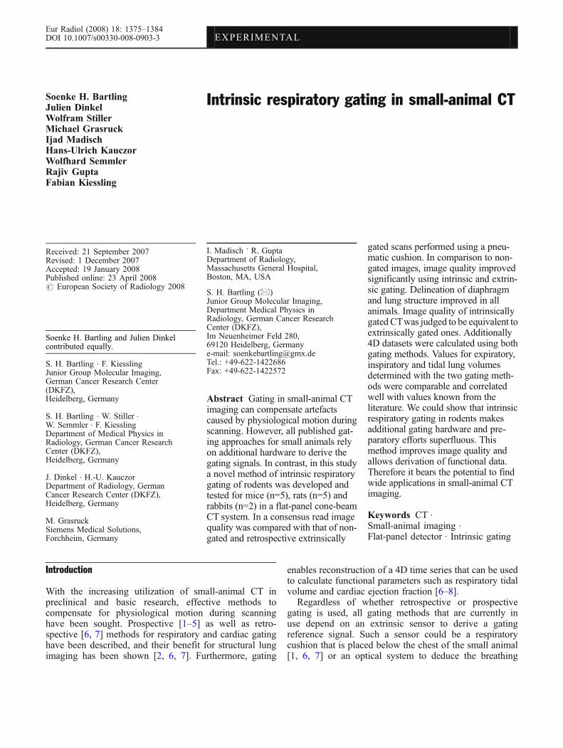
A region of interest (ROI) that covers the diaphragm andadjacent structures on all projections was defined for eachindividual CT raw data set (Fig. 1). Its position was thesame in all projections and was defined in absolute detectorcoordinates. The ROI extension in x-y direction was broad
1376
enough so that the ROI covered the lateral extension ofthe animal fully on all projections. Within this ROI, thecenter of mass (COM) in z-direction (P) was calculatedfrom the raw projection data. All raw projection valueswere gain calibrated, and the value of defective detectorelements was estimated using interpolation [15]. Forcalculating the COM, each line sum of projection values(mz) was multiplied with a weighting factor (z) thatdepends on z-position of the particular line. Weightedprojection values from all lines were summed anddivided by the total sum of projection values from theROI (M) as shown in:
P ¼ 1
M
X
z
mzZ; with M ¼X
z
mz (1)
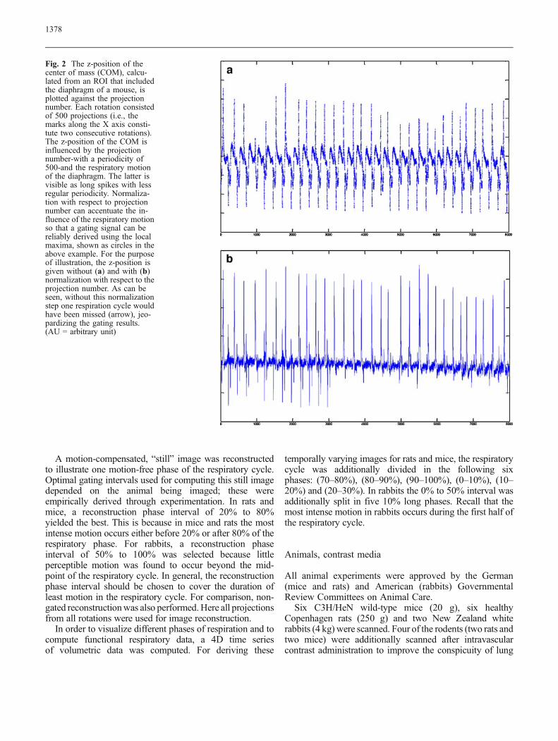
An example of a curve that shows the z-position of theCOM as a function of angular projection position is shownin (Fig. 2a). The z-position of the COM depends mainly ontwo factors: (a) the angular position of the imaging chainand (b) the position of the diaphragm in the selected ROIreflecting the phase of respiration. The variations due to theangular position of the gantry have a fixed periodicity of500 projections reflecting the number of projections in onegantry rotation around the animal. The phase of respirationhas a more irregular period and reflects breathing excursionof the diaphragm along z-axis. In order to derive a gatingreference signal, the influence of angular position on theresulting curve should be minimized while maximizing theeffect of breathing excursions. This is accomplished bybaseline correction described below. The COM of eachprojection position is normalized to the mean of the z-position values at this projection position for all acquiredrotations to decrease the influence of (a). The resultingcurve is shown in Fig. 2b. As can be seen, by explicitlyreducing the angular position dependent oscillations, thedependence on the effect position of the diaphragm can beincreased. Local maxima of this curve were used as gatingreference points as marked in Fig. 2b.
Motion-gated rebinning and CT reconstruction
As described above, gating reference points were derived forevery respiratory cycle for extrinsic gating by analysing thecompression of the respiratory cushion and for intrinsicgating by analysing the parameter P derived from rawprojections. All other steps, such as retrospective binning ofprojections from several rotations according to their phase,and volumetric reconstruction using these projection sets,were identical in both gating methods. In essence, this leavesthe derivation of the gating signal the only differencebetween the new intrinsic and established extrinsic gatingmethod. This assures that potential differences in retro-spective binning, interpolation and reconstruction algorithmscannot cause a difference in the two methods compared.
This is also the reason why all other steps of the gatingalgorithm beside the derivation of the gating reference signalare nearly identical to the already published procedure [7]and are therefore only briefly summarized below.
The starting point of each respiratory cycle (0% point) wasdefined to correspond to the gating reference point of everymotion cycle. In order to reconstruct a given phase of therespiratory cycle, the projections that were acquired within acertain time frame around that respiratory phasewere selectedfor image reconstruction. Time frames were defined by startand end points, given as the percentage of the cycle length.
The selected projections from the re-binning step,representing projections pertaining to a given phase ofthe respiratory cycle, were then interpolated to yield a new360° projection data set consisting of 600 evenlydistributed projections. If two or more selected projectionswere found to be at identical positions-recall that theangular position of each projection is recorded duringdifferent rotations-they were averaged to improve thesignal-to-noise ratio of the interpolated projection. If noprojections were found for a selected angular position,interpolation from the closest neighbouring projectionswas performed. Interpolation was weighted with respect toangular distance.
Fig. 1 An example of a manually selected region of interest (ROI)placed over the diaphragm and adjacent structures. Note that theROI box has been made deliberately larger in x-y direction andextends beyond the margins of the animal. This is to ensure that the
whole body is within the ROI in all projections. The selection ofROI is the only manual interaction necessary for the proposedintrinsic gating algorithm
1377
A motion-compensated, “still” image was reconstructedto illustrate one motion-free phase of the respiratory cycle.Optimal gating intervals used for computing this still imagedepended on the animal being imaged; these wereempirically derived through experimentation. In rats andmice, a reconstruction phase interval of 20% to 80%yielded the best. This is because in mice and rats the mostintense motion occurs either before 20% or after 80% of therespiratory phase. For rabbits, a reconstruction phaseinterval of 50% to 100% was selected because littleperceptible motion was found to occur beyond the mid-point of the respiratory cycle. In general, the reconstructionphase interval should be chosen to cover the duration ofleast motion in the respiratory cycle. For comparison, non-gated reconstructionwas also performed.Here all projectionsfrom all rotations were used for image reconstruction.
In order to visualize different phases of respiration and tocompute functional respiratory data, a 4D time seriesof volumetric data was computed. For deriving these
temporally varying images for rats and mice, the respiratorycycle was additionally divided in the following sixphases: (70–80%), (80–90%), (90–100%), (0–10%), (10–20%) and (20–30%). In rabbits the 0% to 50% interval wasadditionally split in five 10% long phases. Recall that themost intense motion in rabbits occurs during the first half ofthe respiratory cycle.
Animals, contrast media
All animal experiments were approved by the German(mice and rats) and American (rabbits) GovernmentalReview Committees on Animal Care.
Six C3H/HeN wild-type mice (20 g), six healthyCopenhagen rats (250 g) and two New Zealand whiterabbits (4 kg) were scanned. Four of the rodents (two rats andtwo mice) were additionally scanned after intravascularcontrast administration to improve the conspicuity of lung
Fig. 2 The z-position of thecenter of mass (COM), calcu-lated from an ROI that includedthe diaphragm of a mouse, isplotted against the projectionnumber. Each rotation consistedof 500 projections (i.e., themarks along the X axis consti-tute two consecutive rotations).The z-position of the COM isinfluenced by the projectionnumber-with a periodicity of500-and the respiratory motionof the diaphragm. The latter isvisible as long spikes with lessregular periodicity. Normaliza-tion with respect to projectionnumber can accentuate the in-fluence of the respiratory motionso that a gating signal can bereliably derived using the localmaxima, shown as circles in theabove example. For the purposeof illustration, the z-position isgiven without (a) and with (b)normalization with respect to theprojection number. As can beseen, without this normalizationstep one respiration cycle wouldhave been missed (arrow), jeo-pardizing the gating results.(AU = arbitrary unit)
1378
vessels. For this purpose, 2.5-ml Fenestra-VC (ARTAdvanced Research Technologies, Saint-Laurent, CA)-ablood pool contrast agent with 50mg iodine/ml-was injectedinto the tail vein of the rats, 5 min prior to the data acquisition[1, 17]. The mice were given 0.5 ml of the same contrastagent. The rodents were anaesthetized by continuousinhalation of 3% Sevoflurane (Sevorane, Abbot, Maiden-head, UK) in oxygen during preparation, the injection ofcontrast media and data acquisition. Rabbits were anaesthe-tized by intraperitoneal injection of 1 mg/kg acepromazine,40 mg/kg ketamine and 6 mg/kg xylazine. The pneumaticcushion was attached to the animals to record the respiratorymovements. The animals were free-breathing.
Post-processing
The reconstructed imaged datasets were supplementedby a DICOM3-header to enable importation into standardpost-processing software. InSpace (Siemens MedicalSolutions, Forchheim, Germany) was used for analysisand generation of appropriate reformations from both 3D and4D datasets. The Medical Imaging Interaction Toolkit(German Cancer Research Center, Heidelberg, Germany)[18] was used to semi-automatically segment the lungvolumes.
Evaluation and data analysis
Image quality of non-gated, intrinsically and extrinsicallygated datasets was compared with respect to the followingscore table:
A. Delineation of the diaphragm:B. Rib delineation:
0 points no clear delineation, severe motion artefacts1 point some blurring, contours predominantly assessable2 points clear delineation, no motion artefacts
C. Tracheobronchial tract:
0 points trachea and main bronchi not assessable1 point trachea and main bronchi assessable, but nosegmental bronchi can be delineated
2 points trachea, main bronchi and segmental bronchiclearly visualized
D. Delineation of central vessels within the mediastinum:
0 points no clear delineation, severe motion artefacts1 point some blurring, contours predominantly assessable2 points clear delineation (n, no motion artefacts
Three readers (SHB, FK, JD) decided scores inconsensus. Sum scores were calculated for each criterionand each animal. In addition, mean total sum scores(± standard deviation) of the non-gated, extrinsically andintrinsically gated datasets in mice, rats and rabbits werecalculated. Differences in the achieved total sum scoreswere statistically compared among all three groups using anon-parametric Friedmann test. Differences between twogroups were analyzed using the non-parametric Wilcoxonsigned rank test. A p-value of <0.05 was considered toindicate significant differences.
For the determination of respiratory tidal volume thetime points of maximum expiration and inspiration wereselected. Because it was not known whether the time pointsof maximum and minimum lung volumes lie within thesame reconstruction phase for each animal, and for eachgating method, the phases of maximum inspiration andexpiration were selected by visual inspection. The lungvolumes at these time points were segmented. Respiratorytidal volume was calculated as the difference between bothlung volumes.
Results
Comparison of image quality
When comparing the mean sum scores of non-gated andrespiratory-gated scans significant differences betweengroups were found by the Friedmann test (p=0.003).Using extrinsic gating image quality only improvedsignificantly in mice (p=0.05). In rats improvement ofimage quality after extrinsic gating hardly failed signifi-cance as compared to non-gated scans, although the overallsum score was higher. Intrinsic gating resulted in asignificantly image quality improvement for mice(p=0.03) and rats (p=0.03). However, statistical testingrevealed no significant differences in image qualitybetween extrinsically and intrinsically gated images. Alsoin the two rabbits examined higher sum scores for imagequality were found for each gating method.
By visual inspection the diaphragm was blurred and haddouble contour from motion-induced shadowing or ghost-ing in non-gated datasets. In contrast, it was sharplydelineated in the gated datasets (Figs. 3, 4 and 5). Lungstructures such as bronchi and vessels were also sharperand better delineated in the gated datasets (Figs. 3, 4 and 5).Motion artefacts, most pronounced around bony structuresof the rib cage, were considerably diminished in the gateddatasets (Fig. 4). The difference between gated and non-gated datasets was most pronounced in rabbit scans,probably because of greater excursion of the thoracic cage.While there was obviously a perceptible differencebetween the non-gated and gated scans, there was nosignificant difference in image quality between the twodifferent types of gating schemes tested when comparing
1379
the mean sum scores in mice, rats and rabbits. The scoringdetails are listed for each animal and criterion in Table 1.
Functional lung imaging
The rodents exhibited a gasping type of respiration withminimal changes in respiratory motion and ventilationfrequency once a steady-state of narcosis was reached.Respiration rate varied from 20 to 35min−1 inmice and 20 to39 min−1 in rats. In rabbits, the expiratory phase was almostas long as the inspiratory phase. Their respiratory rate rangedfrom 25–40 min−1. In the 4D time series, the respiratoryexcursions of both the diaphragm and the rib cage could bevisualized. Points of maximum inspiration and expirationcould be determined in all cases and for both gatingmethods.
Mean expiratory and inspiratory lung volumesrevealed by semi-automated segmentation are shownin Table 2. No outlier was found. The lung volumesobtained from the intrinsic and extrinsic datasets werewithin standard deviation of each other (Table 2). Aliterature search for the approximate values of lungvolumes and the respiratory tidal volumes was con-ducted. This revealed that the values quoted in theliterature correlated well with values computed usinggated images (Table 2). Minor differences in the valuesmay derive from different animal sizes, narcosis states,the physiological conditions under which the animalswere tested and measurement error.
Discussion
Intrinsic gating schemes in multi-detector CT where thegating signal is derived from the slice-by-slice projection
data have been implemented and shown to work for humansubjects [11–14]. MDCT machines, however, offer lessfavorable conditions for intrinsic gating than small-animalimagers that are based on flat-panel detectors. As aconsequence algorithms developed for MDCT machinesare specifically adapted for human respiratory physiologythat differs significantly from that of small animals.However, these intrinsic gating algorithms tended to becomplex and difficult to implement. Perhaps not surpris-ingly, none of them are being used in routine clinicalpractice so far.
The main result of this research was to show that in smallanimals an intrinsic gating signal derived solely from theprojection data using a relatively simple and easy toimplement algorithm can be used to improve image qualityto the same extent as extrinsic gating derived from externalhardware. We also show the feasibility of high-fidelity 4Ddatasets computed using intrinsic gating. The functionalparameters such as respiratory tidal volume, derived fromthe intrinsically gated 4D datasets, have the same level offidelity as those from extrinsic gating. These values alsocompare favourably with the values quoted in the literature,which were derived using other means.
The ratings and statistics performed in this study resultedin image quality improvements between gated and non-gated CT acquisitions-regardless which gating method wasused. No significant difference in image quality was foundbetween the two gating methods. The obvious similarity ofrespiratory functional parameters derived from both gatingmethods further support our conclusion that intrinsic aswell as extrinsic gating not only both improve imagequality, but also do that to a very similar extent.
In the following paragraphs, we briefly outline the maindifferences between the intrinsic gating schemes describedin the literature and that proposed here.
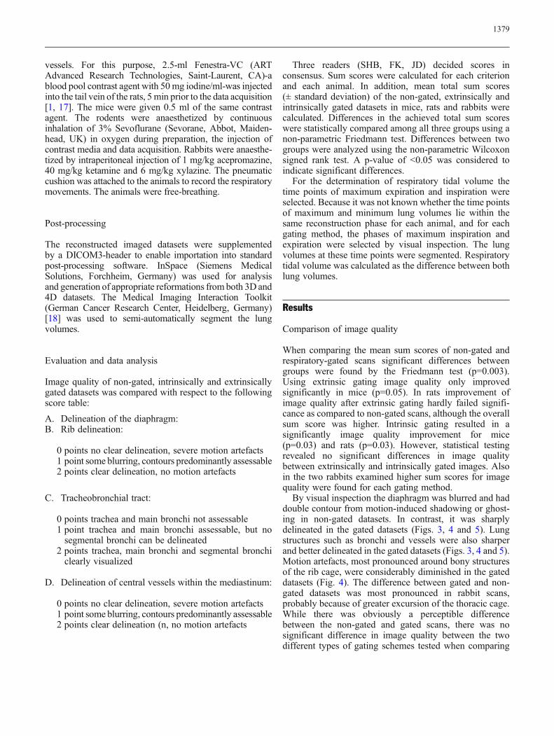
Fig. 3 Non-gated (a, d), extrinsically gated (b, e) and intrinsicallygated (c, f) coronal images of a mouse thorax enhanced with ablood-pool contrast media (same windowing). Magnified views of aregion of interest, as marked in the coronal reformations in the upperrow, are given in the lower row. Both extrinsic and intrinsic gating
improved the sharpness and definition of vessels (small arrows) andbronchi to a similar extent. The diaphragm, which was blurred andhad a ghost contour in the non-gated images (long arrows in d), wassharper and without the ghost contour in the gated datasets (longarrows in e, f)
1380
In multi-slice spiral CT only parts of the volume areexamined during one revolution around the patient. Sincethe detector width in MDCT machines is mostly 2 to 4 cm,the projection data are severely limited in Z extent.However the COM along x-y axis can be used to derivea cardiac gating signal [11]. This is in contrast to themethod presented here where the COM is quite sensitive tomovements of diaphragm and other structures thatpredominantly move along the subject’s z-axis.
Most methods for human CTwere developed for cardiacgating because a normal thorax scan usually does notrequire respiratory gating. It is possible to adapt theproposed method based on COM for cardiac gating.Cardiac motion-which entails Z shortening, helical screwmotion, oblique oscillatory undulations and in-plane (i.e.,XY) contraction-is much more complex than diaphrag-matic movement. Therefore, for this application morecomplex post-processing steps will be required, and thealgorithmic complexity may rival that in human CT usingintrinsic gating [11].
The high scan speed of modern human CT machinesensures that chest scanning can be concluded in one breathhold. Respiratory gating has been most intensivelyinvestigated in the domain of CT applications in radiationoncology. One of the proposed schemes in this field is verysimilar to the intrinsic gating approach proposed in thispaper [12]. Sonke et al. used projection data from a large
cone-beam flat-panel CT system integrated with a linearaccelerator. They used the projections to derive a 1D signalalong the z-axis that correlated well with the movement ofthe diaphragm in the z-direction. In contrast to ourapproach, this method assumes that respiratory phasescorrelate with the extent of the 1D signal.
Iterative reconstruction algorithms for derivation ofintrinsic gating signal have also been described [19].These techniques, however, tend to be computationallyintensive and are not (yet) used routinely.
The proposed method can probably be further improved.However, the similarity of images from intrinsic andextrinsic gating suggests that the quality of intrinsic gatingsignal is already near optimal. Therefore, any algorithmicimprovements would most likely result in small, second-order improvements in the final image quality. There is alsoa limit to which additional improvements add to the imagequality. This is because the positional reproducibility ofthoracic structures from one respiratory cycle to the next isof the order of 100 μm [20] in rodents. A spatial resolutionexceeding 100 μm is therefore not possible by collectingprojections over multiple respiratory cycles. The influenceof rotation time, total scan length and dose to the imagequality can be assessed and optimised in the future.
Since the proposed intrinsic gating method does not relyon any special features of the CT machine used, it can beimplemented in any small-animal cone-beam CT systems.
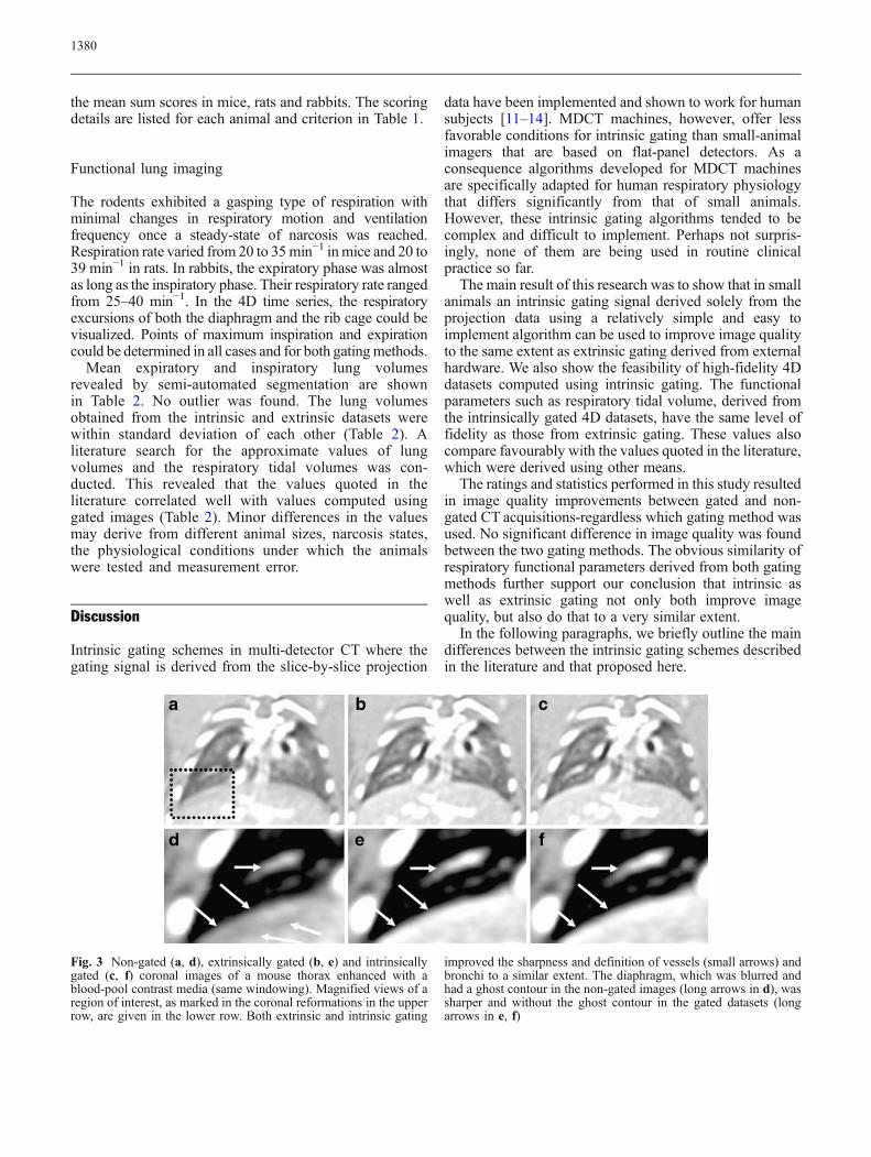
Fig. 4 Non-gated (a), extrinsically gated (b) and intrinsically gated(c) coronal images of a rat thorax after administration of blood-poolcontrast media. Extrinsic as well as intrinsic gating showedcomparable image quality. Using both methods, motion artefacts
around the ribs (black arrow) and double contour along thediaphragm from motion-induced shadowing (long white arrow)were diminished, and the visualization of the basal bronchi (shortwhite arrows) was improved as compared with the non-gated images
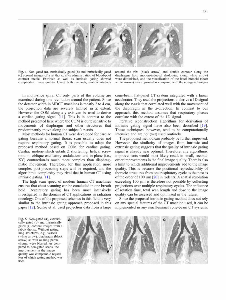
Fig. 5 Non-gated (a), extrinsi-cally gated (b) and intrinsicallygated (c) coronal images from arabbit thorax. Without gating,lung structures, e.g., vessels(white arrow), diaphragm (blackarrow) as well as lung paren-chyma, were blurred. As com-pared to non-gated scans, theimprovement in the imagequality was comparable regard-less of which gating method wasused
1381
Table 1 Results of the multireader analysis of image quality without and with extrinsic and intrinsic gating, respectively
Criterion Non-gated Extrinsic gating Intrinsic gating
Mice (n=6) with score Sum score Mice (n=6) with score Sum score Mice (n=6) with score Sum score
0 1 2 0 1 2 0 1 2
Diaphragm assessability 0 5 1 7 0 0 6 12 0 0 6 12
Rib delineation 2 4 0 4 0 3 3 9 0 2 4 10
Tracheobronchial tract 1 5 0 5 0 2 4 10 0 2 4 10
Delineation of central vessels 0 5 1 7 0 4 2 8 0 4 2 8
Mean sum score/ animal ± SD 3.8±1.3 (max. 8) 6.5±1.3 6.7±1.2
Diaphragm assessability 4 2 0 2 0 1 5 11 0 1 5 11
Rib delineation 0 4 2 8 0 1 5 11 0 2 4 10
Tracheobronchial tract 0 1 5 11 0 1 5 11 0 1 5 11
Delineation of central vessels 4 2 0 2 0 0 6 12 0 1 5 11
Mean sum score/ animal ± SD 3.8±1.16 (max. 8) 7.5±0.83 7.2±1.17
Diaphragm assessability 2 0 0 0 0 0 2 4 0 0 2 4
Rib delineation 1 1 0 1 0 0 2 4 0 0 2 4
Tracheobronchial tract 0 2 0 2 0 0 2 4 0 0 2 4
Delineation of central vessels 0 2 0 2 0 0 2 4 0 0 2 4
Mean sum score/ animal ± SD 2.5±0.7 (max. 8) 8.0±0.0 8.0±0.0
The number of scans that were assigned to score 0, 1 or 2 is indicated for each criterion. In addition the mean total sum scores of six mice,six rats and two rabbits considering all criteria are given. Extrinsic as well as intrinsic respiratory gating causes a general improvement ofimage quality in all animals (p<0.05 in mice and rats; p=0.05 in rabbits). The differences between extrinsic and intrinsic gating were notsignificant (SD: standard deviation)
Table 2 Lung volumes of mice (n=6), rats (n=6) and rabbits (n=2) after semi-automated segmentation from extrinsically and intrinsicallygated datasets (SD: standard deviation)
Mice (mean ± SD) [μl] Rat (mean ± SD) [ml] Rabbit (mean ± SD) [ml]
Extrinsic gating
Expiratory volume 583.7±54.1 4.33±0.10 53.2±2.0
Inspiratory volume 798.5±52.8 5.84±0.14 67.0±3.8
Tidal volume 214.8±21.9 1.56±0.21 15.1±1.8
Intrinsic gating
Expiratory volume 567.3±71.7 4.28±0.05 52.6±1.0
Inspiratory volume 791.8±93.8 5.62±0.13 69.5±4.6
Tidal volume 224.5±59.1 1.34±0.16 16.9±5.7
Difference: intrinsic vs. extrinsic gating
Expiratory volume 16.3±40.4 0.04±0.10 0.6±3.1
Inspiratory volume 6.7±65.2 0.22±0.23 −2.5±0.8Tidal volume −9.7±40.9 0.17±0.29 −3.1±3.8
Reference values from literature
Expiratory volume 400 (*) 430(7) 3.9 [21] 4.6 (7) 76.4 [22]
Inspiratory volume 640 (7) 6.17 (7)
Tidal volume 260 (*) 210 (7) 0.6–2.0 [21] 1.52 (7) 10.8 [23]
The differences between intrinsic and extrinsic gating are also tabulated. All differences are within 1 SD of the values being compared.Volumes correlate well with reference values from the literature. (*Mouse Phenome Project, The Jackson Laboratory, Bar Harbor, ME)
1382
A prerequisite would be access to some basic scanparameters such raw projection data. Even existing systemscan be easily adapted with this gating algorithm makinghardware changes superfluous.
Currently, selection of the ROI is the only manual step inthis algorithm. An automated method for the ROI selection,whichwouldmake the process fully user independent, wouldfacilitate the workflow.
In summary, this paper demonstrates a method forintrinsic gating from projection data that is suitable for flat-panel or cone-beam small-animal CT instruments. Theproposed algorithm is easy to implement and makesexternal gating hardware completely superfluous in small-animal CT imaging. Since the external gating hardwaretypically has to interface with the CT hardware in order tophase-stamp each projection, a secondary benefit of theproposed scheme is that it makes gating fully independent
from the CT hardware. This means that gating can beperformed as a post-processing step, without any changesto the existing CT hardware. Our experience to date showsthat this method is quite robust and reliable as far aschanges in the examination environment are concerned. Italso enables acquisition of 4D functional respiratory data.Finally, it may even be possible to derive a gating signalfrom several animals that are examined by CT simulta-neously, allowing a higher throughput of animals. Weexpect that all these salient features will lead to widespreadadoption of this gating method in small-animal CT.
Acknowledgment The work was supported by the trans-regionalgrant “Vascular Differentiation and Remodelling” of the GermanResearch Foundation (DFG). We thank Karin Leotta for her excellenttechnical assistance during data acquisition and post processing.
References
1. Buliev IG, Badea CT, Kolitsi Z,Pallikarakis N (2003) Estimation of theheart respiratory motion with applica-tions for cone beam computedtomography imaging: a simulationstudy. IEEE Trans Inf Technol Biomed7:404–411
2. Cody DD, Nelson CL, Bradley WM,Wislez M, Juroske D, Price RE, ZhouX, Bekele BN, Kurie JM (2004) Mu-rine lung tumor measurement usingrespiratory-gated micro-computedtomography. Invest Radiol 40:263–269
3. Walters EB, Panda K, Bankson JA,Brown E, Cody DD (2004) Improvedmethod of in vivo respiratory-gatedmicro-CT imaging. Phys Med Biol49:4163–4172
4. Ford NL, Nikolov HN, Norley CJ,Thornton MM, Foster PJ, Drangova M,Holdsworth DW (2005) Prospective res-piratory-gatedmicro-CTof free breathingrodents. Med Phys 32:2888–2898
5. Hu J, Haworth ST, Molthen RC,Dawson CA (2004) Dynamic smallanimal lung imaging via a postacquisi-tion respiratory gating technique usingmicro-cone beam computed tomogra-phy. Acad Radiol 11:961–970
6. Drangova M, Ford NL, Detombe SA,Wheatley AR, Holdsworth DW (2007)Fast retrospectively gated quantitativefour-dimensional (4D) cardiac microcomputed tomography imaging of free-breathing mice. Invest Radiol 42:85–94
7. Bartling S, Stiller W, Grasruck M,Schmidt B, Peschke P, Semmler W,Kiessling F (2007) Retrospective mo-tion-gating in small animal CT of miceand rats. Invest Radiol 42:704–714
8. Kiessling F, Greschus S, Lichy MP,Bock M, Fink C, Vosseler S, Moll J,Mueller MM, Fusenig NE, Traupe H,Semmler W (2004) Volumetric com-puted tomography (VCT): a new tech-nology for noninvasive, high-resolutionmonitoring of tumor angiogenesis. NatMed 10:1133–1138
9. Zaporozhan J, Ley S, UnterhinninghofenR, Saito Y, Fabel-Schulte M, Keller S,Szabo G, Kauczor HU (2006) Free-breathing. -dimensional computedtomography of the lung using prospectiverespiratory gating: charge-coupled devicecamera and laser sensor device in ananimal experiment. Invest Radiol41:468–475
10. Ritchie CJ, Hsieh J, Gard MF, GodwinJD, Kim Y, Crawford CR (1994)Predictive respiratory gating: a newmethod to reduce motion artifacts onCT scans. Radiology 190:847–852
11. Kachelrieß M, Sennst DA, MaxlmoserW, Kalender WA (2002) Kymogramdetection and kymogram-correlatedimage reconstruction from subsecondspiral computed tomography scans ofthe heart. Med Phys 29:1489–1503
12. Manzke R, Kohler T, Nielsen T,Hawkes D, Grass M (2004) Automaticphase determination for retrospectivelygated cardiac CT. Med Phys 31:3345–3362
13. Sonke JJ, Zijp L, Remeijer P, van HerkM (2005) Respiratory correlated conebeam CT. Med Phys 32:1176–1186
14. Zeng R, Fessler JA, Balter JM (2005)Respiratory motion estimation fromslowly rotating x-ray projections: the-ory and simulation. Med Phys 32:984–991
15. Gupta R, Grasruck M, Suess C,Bartling SH, Schmidt B, Stierstorfer K,Popescu S, Brady T, Flohr T (2006)Ultra-high resolution flat-panel volumeCT: fundamental principles, designarchitecture, and system characteriza-tion. Eur Radiol 30:337–343
16. Feldkamp LA, Davis LC, Kress JW(1984) Practical cone-beam algorithm.J Opt Soc Am 1:612–619
17. Bartling S, Stiller W, Semmler W,Kiessling F (2007) Small animal Com-puted Tomography Imaging. CMIR 3:45–59
18. Wolf I, Vetter M, Wegner I, Bottger T,Nolden M, Schobinger M, HastenteufelM, Kunert T, Meinzer HP (2004) Themedical imaging interaction toolkit(MITK): a toolkit facilitating the crea-tion of interactive software by extend-ing VTK and ITK. Proceedings of SPIE5367:16–27
19. De Man B, Edic P, Basu S (2005) Aniterative algorithm for time-resolvedreconstruction of a CT scan of a beatingheart. Proceedings of Fully 3D, The 8thinternational meeting on fully three-dimensional image reconstruction inradiology and nuclear medicine:pp 356–359
1383

20. Mai W, Badea CT, Wheeler CT,Hedlund LW, Johnson GA (2005)Effects of breathing and cardiac motionon spatial resolution in the microscopicimaging of rodents. Magn Reson Med53:858–665
21. Sharp P, La Regina M, LaRegina M(1998) The Laboratory Rat. CRC Press,Boca Raton, FL, USA
22. Krause MF, Hoehn T (1999) Decreasein lung volume depends on end-expiratory pressure in a rabbit model ofairway lavage. Respiration 66:259–264
23. Zhu GF, Zhang W, Zong H, Liang Y(2006) Effects of continuous trachealgas insufflation during pressure limitedventilation on pulmonary surfactant inrabbits with acute lung injury. ChinMed J (Engl) 119:1415–1420
1384