intrinsic disorder in proteins with pathogenic repeat
TRANSCRIPT
molecules
Review
Intrinsic Disorder in Proteins with PathogenicRepeat Expansions
April L. Darling 1,2,* ID and Vladimir N. Uversky 1,3,* ID
1 Department of Molecular Medicine, College of Medicine, Byrd Alzheimer’s Institute,University of South Florida, Tampa, FL 33612, USA
2 James A. Haley Veteran’s Hospital, Tampa, FL 33612, USA3 Institute for Biological Instrumentation of the Russian Academy of Sciences, Pushchino,
Moscow Region 142290, Russia* Correspondence: [email protected] (A.L.D.); [email protected] (V.N.U.);
Tel.: +1-813-396-9249 (A.L.D.); +1-813-974-5816 (V.N.U.)
Received: 8 November 2017; Accepted: 21 November 2017; Published: 24 November 2017
Abstract: Intrinsically disordered proteins and proteins with intrinsically disordered regions havebeen shown to be highly prevalent in disease. Furthermore, disease-causing expansions of the regionscontaining tandem amino acid repeats often push repetitive proteins towards formation of irreversibleaggregates. In fact, in disease-relevant proteins, the increased repeat length often positively correlateswith the increased aggregation efficiency and the increased disease severity and penetrance, beingnegatively correlated with the age of disease onset. The major categories of repeat extensions involvedin disease include poly-glutamine and poly-alanine homorepeats, which are often times located in theintrinsically disordered regions, as well as repeats in non-coding regions of genes typically encodingproteins with ordered structures. Repeats in such non-coding regions of genes can be expressed atthe mRNA level. Although they can affect the expression levels of encoded proteins, they are nottranslated as parts of an affected protein and have no effect on its structure. However, in some cases,the repetitive mRNAs can be translated in a non-canonical manner, generating highly repetitivepeptides of different length and amino acid composition. The repeat extension-caused aggregation ofa repetitive protein may represent a pivotal step for its transformation into a proteotoxic entity thatcan lead to pathology. The goals of this article are to systematically analyze molecular mechanisms ofthe proteinopathies caused by the poly-glutamine and poly-alanine homorepeat expansion, as well asby the polypeptides generated as a result of the microsatellite expansions in non-coding gene regionsand to examine the related proteins. We also present results of the analysis of the prevalence andfunctional roles of intrinsic disorder in proteins associated with pathological repeat expansions.
Keywords: protein repeat expansion; intrinsically disordered protein; intrinsically disordered proteinregion; homorepeats; protein aggregation; proteinopathies
1. Introduction
It is clear now that the protein universe includes not only globular, transmembrane, and fibrousproteins but also intrinsically disordered proteins (IDPs) and hybrid proteins with ordered domainsand intrinsically disordered protein regions (IDPRs) [1,2]. In fact, there is a growing amount ofevidence supporting the idea that many protein regions, and even entire proteins, lack stable tertiaryand/or secondary structure in solution, and instead exist as dynamic conformational ensembles ofinterconverting structures [3–7]. Although these IDPs and IDPRs fail to form specific 3D structures,they are biologically active [2,8–13].
Molecules 2017, 22, 2027; doi:10.3390/molecules22122027 www.mdpi.com/journal/molecules
Molecules 2017, 22, 2027 2 of 48
Being typically involved in regulation, signaling and control pathways, such as those involvedin the cell cycle, IDPs/IDPRs are characterized by specific functionality [14,15] that complement thefunctional repertoire of ordered proteins, which have evolved mainly to carry out efficient catalyticand transport functions. Some illustrative biological activities of IDPs/IDPs include regulation ofcell division, transcription and translation, signal transduction, protein phosphorylation and otherposttranslational modifications, storage of small molecules, chaperone action, and regulation of theself-assembly of large multi-protein complexes such as the ribosome [2,9,11,14–25]. As a matterof fact, the lack of rigid globular structure under physiological conditions provides IDPs/IDPRswith a remarkable set of unique functional advantages [26–31]. For example, large conformationalplasticity allows IDPs/IDPRs to be promiscuous binders and interact efficiently with multiple differenttargets [2,12,32,33]. The functional importance of being disordered has been intensively analyzed andsystemized in several dedicated reviews [2,10,18,24,34]. Many IDPs/IDPRs are known to undergo adisorder-to-order transition upon functioning [10,24,34,35]. When IDPRs bind to signaling partners,the free energy required to bring about the disorder to order transition takes away from the interfacial,contact free energy, with the net result that a highly specific interaction can be combined with a lownet free energy of association [10,36]. High specificity coupled with low affinity seems to be a usefulpair of properties for a signaling interaction so that the signaling interaction is reversible. In addition,a disordered protein can readily bind to multiple partners by changing shape to associate with differenttargets [32,33]. In addition to decoupled high binding specificity and low affinity, disorder has severalclear advantages for functions in signaling, regulation, and control [2,8,10,11,13,17–20,25,37–39].
Many IDPs/IDPRs are involved in the pathogenesis of numerous human diseases (disorders),giving rise to the “disorder in disorders” or D2 concept [40]. According to this concept, involvementof IDPs/IDPRs in the development of various diseases is defined by the unique structural andfunctional properties of these representatives of protein universe. Such diseases originate not onlyfrom the misfolding of causative IDPs/IDPRs but also from their misidentification, misregulation,and missignaling. Among human maladies associated with misbehavior of IDPs/IDPRs are variousneurodegenerative diseases [41–43].
The absence of ordered structure in IDPs/IDPRs proteins has been associated with some specificfeatures of their amino acid sequences, such as the presence of numerous uncompensated chargedgroups (often negative); i.e., a high net charge at neutral pH, which is a result of the extreme pI valuesin such proteins and a low content of hydrophobic amino acid residues [9]. In fact, IDPs/IDPRs wereshown to be significantly depleted in order-promoting amino acid residues, such as Trp, Tyr, Phe,Ile, Leu, Val, Cys, and Asn, and enriched in disorder-promoting amino acids, such as Ala, Arg, Gly,Gln, Ser, Glu, Lys, and Pro [10,44–46]. Furthermore, IDPs/IDPRs are typically characterized by lowsequence complexity and might contain numerous sequence repeats [37,47–49].
Amino acid tandem repeats are abundant in eukaryotes, where they are found within manyphysiologically important proteins playing important roles in protein functionality [50,51]. In general,the length of units in protein repeats can be different, with a special type being repeats with therepetitive unit equals to one amino acid residue (homorepeats). Repeats, especially those withpoly-glutamine and poly-alanine sequences, are overrepresented in DNA-binding proteins andtranscription factors [50], which, compared to the average protein, are engaged in more protein-proteininteractions [50,52]. It is likely that proteins utilize homorepeats for non-specific protein-proteininteractions or in modulation of specific interactions [53]. However, although homorepeats are engagedin important physiological interactions, they are prone for expansion via strand slippage of encodingDNA, which can lead to increased aggregation propensity of resulting proteins and subsequent disease.It was also pointed out that among various types of the protein repeats, the homorepeats have thehighest tendency to aggregate [54].

Molecules 2017, 22, 2027 3 of 48
Curiously, several human diseases are associated with pathological expansions of repeatedsequences in specific proteins [55,56]. Most known homorepeat expansions that correlate with diseaseare found in exons, where translation occurs in a canonical fashion leading to long stretches of peptidehomorepeats [55,57]. In most homorepeat disorders, length of the causing homorepeat expansion iscorrelated with disease severity and penetrance. Genetically, homorepeat disorders are characterizedby the lack of conventional Mendelian transmission and exhibit a phenomenon called anticipation,where the following generation is likely to inherit a longer repeat than the previous one, and thisresults in increased disease severity with earlier onset [58–60]. This is because at the genetic level, theexpanded repeats are characterized by meiotic or intergenerational instability and change in size whentransmitted from parents to offspring [61]. On the other hand, mitotic instability or somatic mosaicismof such homorepeats defines their size variation within the tissues of an affected individual [61].
In rarer cases, the repeat expansions occur in non-coding regions that normally are not translated.However, some expansions have been shown to lead to unique peptide species through non-canonicalmechanisms of translation [62]. In fact, these regions, like those found in exons, commonly undergoframe-shifts that lead to the production of several repeat peptide species [62]. This leads to the increasedcomplication that the interaction between different translation products must be considered becausebiochemical interactions between different species could alter aggregation propensity, depending onthe species, and could cause the peptides to undergo structural changes.
The other correlative feature of peptide repeat length is structure, with the longer repeats generallycorresponding to increased disorder [63]. These long disordered stretches of repeat peptides have apropensity to aggregate, which contrasts with shorter peptides that remain diffuse throughout thecell. The appearance of large aggregates leads to a cascade of cellular events that causes toxicity andcell death. Cell death from aggregated repeat peptides is emerging as a common feature in manyneurological and developmental diseases but the mechanism that causes it is not yet known.
Proteins affected by repeat expansion mutations are listed in Table 1. The goal of this work isto systematically examine proteins associated with proteinopathies caused by the poly-glutamineand poly-alanine homorepeat expansions, as well as by the polypeptides generated as a result of themicrosatellite expansions in non-coding gene regions, with the major focus on the roles of intrinsicdisorder in proteins associated with pathological repeat expansions. Here, we first discuss molecularmechanisms of various pathologies associated with repeat expansion and represent related proteins.Next, we represent the results of a systematic analysis of the intrinsic disorder status and the presence ofdisorder-based functional features (such as sites of posttranslational modifications and disorder-basedprotein binding sites, known as molecular recognition features, MoRFs) in proteins with pathologicalrepeat expansions.
Molecules 2017, 22, 2027 4 of 48
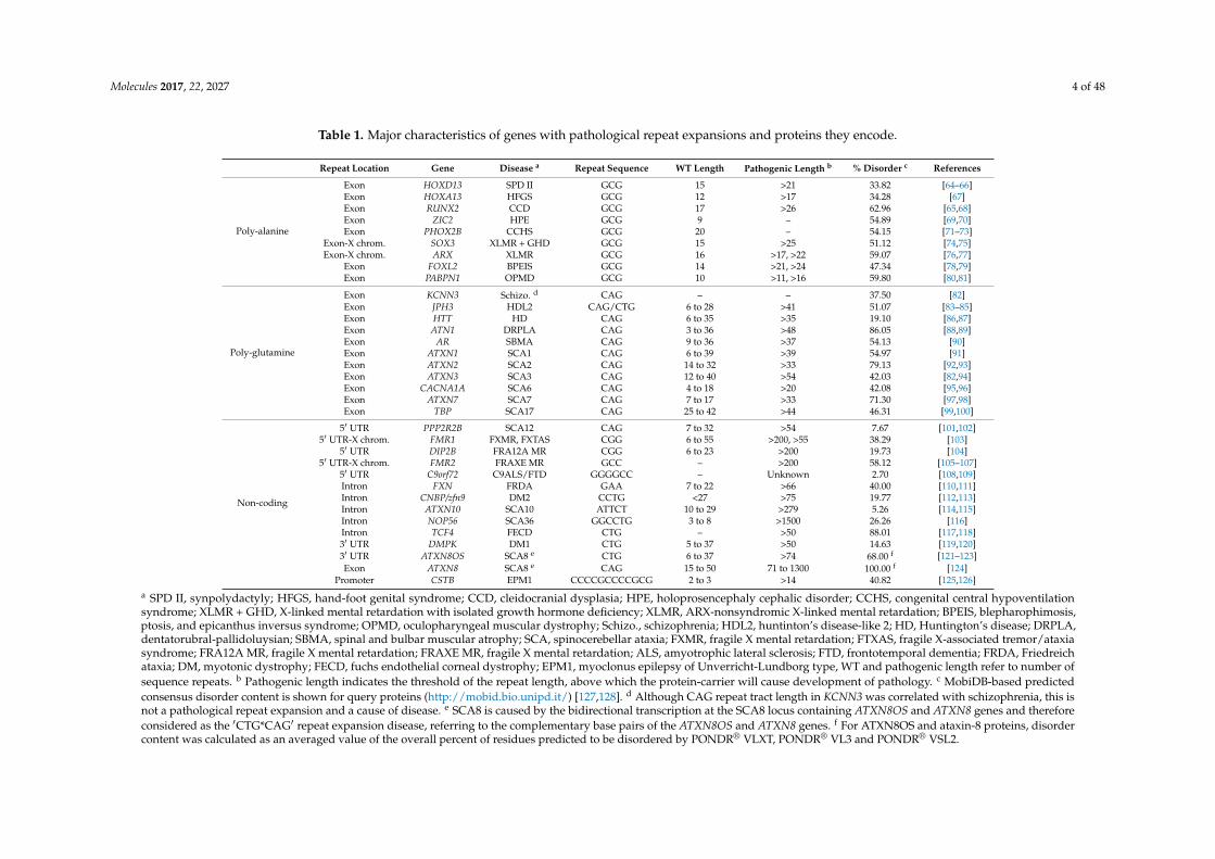
Table 1. Major characteristics of genes with pathological repeat expansions and proteins they encode.
Repeat Location Gene Disease a Repeat Sequence WT Length Pathogenic Length b % Disorder c References
Poly-alanine
Exon HOXD13 SPD II GCG 15 >21 33.82 [64–66]Exon HOXA13 HFGS GCG 12 >17 34.28 [67]Exon RUNX2 CCD GCG 17 >26 62.96 [65,68]Exon ZIC2 HPE GCG 9 – 54.89 [69,70]Exon PHOX2B CCHS GCG 20 – 54.15 [71–73]
Exon-X chrom. SOX3 XLMR + GHD GCG 15 >25 51.12 [74,75]Exon-X chrom. ARX XLMR GCG 16 >17, >22 59.07 [76,77]
Exon FOXL2 BPEIS GCG 14 >21, >24 47.34 [78,79]Exon PABPN1 OPMD GCG 10 >11, >16 59.80 [80,81]
Poly-glutamine
Exon KCNN3 Schizo. d CAG – – 37.50 [82]Exon JPH3 HDL2 CAG/CTG 6 to 28 >41 51.07 [83–85]Exon HTT HD CAG 6 to 35 >35 19.10 [86,87]Exon ATN1 DRPLA CAG 3 to 36 >48 86.05 [88,89]Exon AR SBMA CAG 9 to 36 >37 54.13 [90]Exon ATXN1 SCA1 CAG 6 to 39 >39 54.97 [91]Exon ATXN2 SCA2 CAG 14 to 32 >33 79.13 [92,93]Exon ATXN3 SCA3 CAG 12 to 40 >54 42.03 [82,94]Exon CACNA1A SCA6 CAG 4 to 18 >20 42.08 [95,96]Exon ATXN7 SCA7 CAG 7 to 17 >33 71.30 [97,98]Exon TBP SCA17 CAG 25 to 42 >44 46.31 [99,100]
Non-coding
5′ UTR PPP2R2B SCA12 CAG 7 to 32 >54 7.67 [101,102]5′ UTR-X chrom. FMR1 FXMR, FXTAS CGG 6 to 55 >200, >55 38.29 [103]
5′ UTR DIP2B FRA12A MR CGG 6 to 23 >200 19.73 [104]5′ UTR-X chrom. FMR2 FRAXE MR GCC – >200 58.12 [105–107]
5′ UTR C9orf72 C9ALS/FTD GGGGCC – Unknown 2.70 [108,109]Intron FXN FRDA GAA 7 to 22 >66 40.00 [110,111]Intron CNBP/zfn9 DM2 CCTG <27 >75 19.77 [112,113]Intron ATXN10 SCA10 ATTCT 10 to 29 >279 5.26 [114,115]Intron NOP56 SCA36 GGCCTG 3 to 8 >1500 26.26 [116]Intron TCF4 FECD CTG – >50 88.01 [117,118]3′ UTR DMPK DM1 CTG 5 to 37 >50 14.63 [119,120]3′ UTR ATXN8OS SCA8 e CTG 6 to 37 >74 68.00 f [121–123]Exon ATXN8 SCA8 e CAG 15 to 50 71 to 1300 100.00 f [124]
Promoter CSTB EPM1 CCCCGCCCCGCG 2 to 3 >14 40.82 [125,126]
a SPD II, synpolydactyly; HFGS, hand-foot genital syndrome; CCD, cleidocranial dysplasia; HPE, holoprosencephaly cephalic disorder; CCHS, congenital central hypoventilationsyndrome; XLMR + GHD, X-linked mental retardation with isolated growth hormone deficiency; XLMR, ARX-nonsyndromic X-linked mental retardation; BPEIS, blepharophimosis,ptosis, and epicanthus inversus syndrome; OPMD, oculopharyngeal muscular dystrophy; Schizo., schizophrenia; HDL2, huntinton’s disease-like 2; HD, Huntington’s disease; DRPLA,dentatorubral-pallidoluysian; SBMA, spinal and bulbar muscular atrophy; SCA, spinocerebellar ataxia; FXMR, fragile X mental retardation; FTXAS, fragile X-associated tremor/ataxiasyndrome; FRA12A MR, fragile X mental retardation; FRAXE MR, fragile X mental retardation; ALS, amyotrophic lateral sclerosis; FTD, frontotemporal dementia; FRDA, Friedreichataxia; DM, myotonic dystrophy; FECD, fuchs endothelial corneal dystrophy; EPM1, myoclonus epilepsy of Unverricht-Lundborg type, WT and pathogenic length refer to number ofsequence repeats. b Pathogenic length indicates the threshold of the repeat length, above which the protein-carrier will cause development of pathology. c MobiDB-based predictedconsensus disorder content is shown for query proteins (http://mobid.bio.unipd.it/) [127,128]. d Although CAG repeat tract length in KCNN3 was correlated with schizophrenia, this isnot a pathological repeat expansion and a cause of disease. e SCA8 is caused by the bidirectional transcription at the SCA8 locus containing ATXN8OS and ATXN8 genes and thereforeconsidered as the ′CTG*CAG′ repeat expansion disease, referring to the complementary base pairs of the ATXN8OS and ATXN8 genes. f For ATXN8OS and ataxin-8 proteins, disordercontent was calculated as an averaged value of the overall percent of residues predicted to be disordered by PONDR® VLXT, PONDR® VL3 and PONDR® VSL2.

Molecules 2017, 22, 2027 5 of 48
2. Polyalanine Repeat Expansions
Polyalanine (poly-Ala) tract expansions are linked to 9 inherited human diseases, such asblepharophimosis-ptosis-epicanthus inversus syndrome (BPEIS), cleidocranial dysplasia (CCD),congenital central hypo-ventilation syndrome (CCHS); hand–foot–genital syndrome (HFGS),holoprosencephaly (HPE), oculopharyngeal muscular dystrophy (OPMD), synpolydactyly syndrome(SPD), X-linked mental retardation and abnormal genitalia (XLAG), and X-linked mental retardationand growth hormone deficit (XLMR + GHD) [129]. At the genetic level, the expansion of homopolymericalanine is caused by the expansion of translated GCN trinucleotide repeats (where N refers to any ofthe four bases, among which GCG is significantly over-represented in the poly-Ala coding sequencesand [130] in the disease-associated genes. It was emphasized that genes coding for poly-Ala stretcheslonger than four alanines are rather common in human genome that contains 494 such proteinspossessing 604 poly-Ala domains [130]. Importantly, as shown in Figure 1, transcription factors areinvolved in 8 of the 9 poly-Ala expansion diseases and account for 36% of human proteins withpoly-Ala tracts [131]. Poly-Ala repeats hang on the cryptic edge of toxicity depending on their length,which is directly correlated with their structure. Unlike other repeat expansions seen in disease, thereis a low degree of polymorphism for poly-Ala tract repeats, most likely due to an altered mechanismof expansion as opposed to expansion of other repeats [76]. Poly-Ala tracts are thought to extend byunequal allelic homologous recombination during meiosis, while other disease relevant expansionsextend due to DNA polymerase slippage during translation [70]. These observations, in addition to thefact that proteins containing poly-Ala tracts are highly conserved in mammals, makes it conceivablethat diminutive extensions to alanine tracts are sufficient to cause cellular dysfunction with subsequentdisease pathology [129].
Molecules 2017, 22, 2027 6 of 46
2. Polyalanine Repeat Expansions
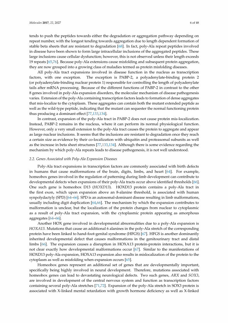
Polyalanine (poly-Ala) tract expansions are linked to 9 inherited human diseases, such as blepharophimosis-ptosis-epicanthus inversus syndrome (BPEIS), cleidocranial dysplasia (CCD), congenital central hypo-ventilation syndrome (CCHS); hand–foot–genital syndrome (HFGS), holoprosencephaly (HPE), oculopharyngeal muscular dystrophy (OPMD), synpolydactyly syndrome (SPD), X-linked mental retardation and abnormal genitalia (XLAG), and X-linked mental retardation and growth hormone deficit (XLMR + GHD) [129]. At the genetic level, the expansion of homopolymeric alanine is caused by the expansion of translated GCN trinucleotide repeats (where N refers to any of the four bases, among which GCG is significantly over-represented in the poly-Ala coding sequences and [130] in the disease-associated genes. It was emphasized that genes coding for poly-Ala stretches longer than four alanines are rather common in human genome that contains 494 such proteins possessing 604 poly-Ala domains [130]. Importantly, as shown in Figure 1, transcription factors are involved in 8 of the 9 poly-Ala expansion diseases and account for 36% of human proteins with poly-Ala tracts [131]. Poly-Ala repeats hang on the cryptic edge of toxicity depending on their length, which is directly correlated with their structure. Unlike other repeat expansions seen in disease, there is a low degree of polymorphism for poly-Ala tract repeats, most likely due to an altered mechanism of expansion as opposed to expansion of other repeats [76]. Poly-Ala tracts are thought to extend by unequal allelic homologous recombination during meiosis, while other disease relevant expansions extend due to DNA polymerase slippage during translation [70]. These observations, in addition to the fact that proteins containing poly-Ala tracts are highly conserved in mammals, makes it conceivable that diminutive extensions to alanine tracts are sufficient to cause cellular dysfunction with subsequent disease pathology [129].
Figure 1. Major known functions of proteins with pathogenic repeat expansions. Proteins with pathogenic expansions have varied functions depending on the type of expansion present. Poly-alanine (poly-Ala) expansions have the least variability in functions, with 8 of the 9 engaging in some sort of transcription regulation. Poly-glutamine (polyQ) expansions that cause pathology have more varied functions, but the majority participate in transcription regulation as well. Pathogenic repeats in non-coding regions occur in genes encoding proteins with the most varied functions. They include everything from catalytic proteins to receptors. Since the repeat extension occurs in the non-coding region of the gene, it is conceivable that there are not more synonymous functions among pathogenic repeat proteins in non-coding regions.
2.1. Molecular Mechanisms of Poly-Ala Expansion Diseases
Extension of poly-Ala tracts lead to structural changes in the repeat peptide that may indicate mechanisms of disease pathogenesis. Shorter length peptides are predominantly disordered with short α-helical sections, which, upon expansion, become more prevalent [132]. Increased length tends to push the peptides towards either the degradation or aggregation pathway depending on repeat
Figure 1. Major known functions of proteins with pathogenic repeat expansions. Proteins withpathogenic expansions have varied functions depending on the type of expansion present. Poly-alanine(poly-Ala) expansions have the least variability in functions, with 8 of the 9 engaging in some sortof transcription regulation. Poly-glutamine (polyQ) expansions that cause pathology have morevaried functions, but the majority participate in transcription regulation as well. Pathogenic repeats innon-coding regions occur in genes encoding proteins with the most varied functions. They includeeverything from catalytic proteins to receptors. Since the repeat extension occurs in the non-codingregion of the gene, it is conceivable that there are not more synonymous functions among pathogenicrepeat proteins in non-coding regions.
2.1. Molecular Mechanisms of Poly-Ala Expansion Diseases
Extension of poly-Ala tracts lead to structural changes in the repeat peptide that may indicatemechanisms of disease pathogenesis. Shorter length peptides are predominantly disordered withshort α-helical sections, which, upon expansion, become more prevalent [132]. Increased length
Molecules 2017, 22, 2027 6 of 48
tends to push the peptides towards either the degradation or aggregation pathway depending onrepeat number, with the longest tending towards aggregation due to length dependent formation ofstable beta sheets that are resistant to degradation [68]. In fact, poly-Ala repeat peptides involvedin disease have been shown to form large intracellular inclusions of the aggregated peptides. Theselarge inclusions cause cellular dysfunction; however, this is not observed unless their length exceeds19 repeats [65,76]. Because poly-Ala extensions cause misfolding and subsequent protein aggregation,they are now grouped into a growing class of maladies termed as protein misfolding diseases.
All poly-Ala tract expansions involved in disease function in the nucleus as transcriptionfactors, with one exception. The exception is PABP-2, a polyadenylate-binding protein 2(or polyadenylate-binding nuclear protein 1) responsible for controlling the length of polyadenylatetails after mRNA processing. Because of the different functions of PABP-2 in contrast to the other8 genes involved in poly-Ala expansion disorders, the molecular mechanism of disease pathogenesisvaries. Extension of the poly-Ala containing transcription factors leads to formation of dense aggregatesthat mis-localize to the cytoplasm. These aggregates can contain both the mutant extended peptide aswell as the wild-type peptide, indicating that the mutant can sequester the normal functioning proteinthus producing a dominant effect [77,133,134].
In contrast, expansion of the poly-Ala tract in PABP-2 does not cause protein mis-localization.Instead, PABP-2 remains in the nucleus, where it can perform its normal physiological function.However, only a very small extension to the poly-Ala tract causes the protein to aggregate and appearas large nuclear inclusions. It seems that the inclusions are resistant to degradation once they reacha certain size as evidence by their co-localization with ubiquitin and proteasomal subunits as wellas the increase in beta sheet structures [77,133,134]. Although there is some evidence regarding themechanism by which poly-Ala repeats leads to disease pathogenesis, it is not well understood.
2.2. Genes Associated with Poly-Ala Expansion Diseases
Poly-Ala tract expansions in transcription factors are commonly associated with birth defectsin humans that cause malformations of the brain, digits, limbs, and heart [64]. For example,homeobox genes involved in the regulation of patterning during limb development can contribute todevelopmental defects when expansions of their poly-Ala tracts occur above identified thresholds [66].One such gene is homeobox D13 (HOXD13). HOXD13 protein contains a poly-Ala tract inthe first exon, which upon expansion above an 8-alanine threshold, is associated with humansynpolydactyly (SPD) [64–66]. SPD is an autosomal-dominant disease resulting in limb malformations,usually including digit duplication [64,66]. The mechanism by which the expansion contributes tomalformation is unclear, but the localization of the protein changes from nuclear to cytoplasmicas a result of poly-Ala tract expansion, with the cytoplasmic protein appearing as amorphousaggregates [64–66].
Another HOX gene involved in developmental abnormalities due to a poly-Ala expansion isHOXA13. Mutations that cause an additional 6 alanines in the poly-Ala stretch of the correspondingprotein have been linked to hand-foot-genital syndrome (HFGS) [67]. HFGS is another dominantlyinherited developmental defect that causes malformations in the genitourinary tract and distallimbs [66]. The expansion causes a disruption in HOXA13 protein-protein interactions, but it isnot clear exactly how developmental malformations occur [67]. Similar to the manifestations ofHOXD13 poly-Ala expansion, HOXA13 expansion also results in mislocalization of the protein to thecytoplasm as well as misfolding when expansion occurs [65].
Homeobox genes represent an additional set of genes that are developmentally important,specifically being highly involved in neural development. Therefore, mutations associated withhomeobox genes can lead to devastating neurological defects. Two such genes, ARX and SOX3,are involved in development of the central nervous system and function as transcription factorscontaining several poly-Ala stretches [71,72]. Expansion of the poly-Ala stretch in SOX3 protein isassociated with X-linked mental retardation with growth hormone deficiency as well as X-linked
Molecules 2017, 22, 2027 7 of 48
hypopituitarism [72,74]. The latter is due to two specific poly-Ala expansions, addition of 7 or11 alanines, with the longer being associated with a more severe disease phenotype [74].
ARX is exceedingly dangerous due to the presence of multiple poly-Ala tracts, potentiating therisk of disease due to expansion. Because ARX has multiple stretches with expansion potential, itis associated with several developmental diseases of varying severity. The most common maladyarising from the aberrant poly-Ala expansion is ARX-linked mental retardation, a non-syndromicform of X-linked mental retardation; however, syndromic X-linked mental retardation is also acommon outcome [77,133,134]. Other disorders that have been linked to ARX poly-alanine tractexpansions include hydrocephaly with abnormal genitalia, myoclonic epilepsy with spasticity andmental retardation, Partington syndrome, and X-linked infantile spasms [77,133,134].
Two additional transcription factors associated with developmental defects due to poly-Alaexpansions include RUNX2 and ZIC2. RUNX2 is a bone-specific transcription factor with a poly-alaninetract [77,133,134]. When expansion occurs above a threshold of 10 repeats cleidocranial dysplasia,a bone developmental defect, occurs [68,131]. ZIC2 is a zinc finger transcription repressor that hasbeen extensively linked to holoprosencephaly, a developmental defect where the midline of the braindoes not properly form and therefore there is no separation of the brain into hemispheres [73,131].Similar to other poly-Ala expansion disorders, expansion in these two transcription factors causesprotein mislocalization, misfolding, and aggregation [68].
Development and regulation of the autonomic nervous system occur by various transcriptionfactors in addition to homeobox genes. PHOX2B is an example that is involved in autonomic nervoussystem development [72]. PHOX2B contains a 20-residue C-terminal poly-alanine tract that, uponexpansion, can result in congenital central hypoventilation syndrome (CCHS) [72,73]. CCHS is adisorder that effects breathing due to a disruption in autonomic nervous system regulation, whichis especially problematic when sleeping [72]. The poly-Ala expansion in PHOX2B results in nuclearlocalization of the repeat peptide and defects in nuclear import [72]. Unlike in the previously mentionedpoly-Ala expansion related disorders, expansion in PHOX2B causes nuclear localization of the proteinas opposed to cytoplasmic, offering evidence that not all poly-Ala tract expansions in transcriptionfactors result in similar cellular defects.
Transcription factors in the Forkhead family are known to play important roles in themaintenance of differentiated cells lines, embryogenesis, and tumorigenesis [132]. Therefore, they arecommonly involved in human developmental diseases with most of them resulting in aberrant ocularmanifestations [78]. One member of this family, FOXL2, is a transcription factor involved in both eyeand ovary development [77]. It contains a poly-alanine stretch and upon expansion is correlated withblepharophimosis syndrome (BPES), a developmental disorder that causes defects in the ovary andeyelid [78,135]. The WT protein functions in the nucleus and appears diffuse throughout, but poly-Alaexpansions cause FOXL2 to mislocalize to the cytoplasm in aggregated form and diminish its role as atranscription regulator [78].
The only gene associated with disease due to poly-Ala repeat expansions that is not a transcriptionfactor is PABPN1 encoding PABP-2 protein. This gene contains the shortest poly-alanine expansionassociated with disease, with the addition of only 2 alanines sufficient to cause a disease phenotype [65].Like in most other cases, the repeat length closely correlates with disease penetrance and severity,and extended stretches lead to muscle weakness with accompanying nuclear inclusions that are slowto evolve [77]. Expansions in the poly-alanine tract of PABP-2 have been linked to oculopharyngealmuscular dystrophy (OPMD), which is an adult-onset disorder marked by the presentation ofprogressive dysphagia, eyelid ptosis, and proximal limb weakness [81]. In addition, the skeletalmuscle of those affected contains intranuclear filament inclusions that contain PABP-2 and biopsiesshow that inclusions are accompanied by myopathic and neurogenic changes [81]. OPMD is the onlypoly-Ala expansion disorder where onset occurs in adulthood and its presentation, pathology, andonset is more similar to polyglutamine expansion diseases than to other poly-Ala tract expansions [77].
Molecules 2017, 22, 2027 8 of 48
PABP-2 demonstrates the ability of poly-Ala expansions to cause dysfunction even when they are notpart of a transcription regulatory protein.
Therefore, while most proteins-carriers of pathogenic poly-Ala expansions behave similarly andengage in analogous functions, there are exceptions. Those exceptions demonstrate the ability forpoly-Ala tract extensions to cause pathology in a multitude of ways, depending on the functionalityand structure of the translated protein product. The very short extensions needed for poly-Ala tractexpansion disease phenotypes to penetrate is directly correlative to structural changes in the resultanttranslational product. This highlights the fact that the structural transition of the protein containingthe extended alanine repeat region may be the defining step that leads to cellular dysfunction andsubsequent disease presentation.
3. Poly-Glutamine Repeat Expansions
Currently, there are at least twelve known hereditary diseases in which the expansion of a CAGrepeat in the gene leads to neurodegeneration [136,137]. Table 1 shows that these poly-glutaminerepeat diseases includes Huntington’s disease, Huntington’s disease-like 2, Kennedy disease (alsoknown as spinal and bulbar muscular atrophy, SBMA), dentatorubral-pallidoluysian atrophy (DRPLA),spinocerebellar ataxia type 1 (SCA1), spinocerebellar ataxia type 2 (SCA2), SCA3 (also known asMachado-Joseph disease, MJD), SCA6, SCA7, SCA17, and schizophrenia. Note that although theCAG repeat tract length is somehow correlated with schizophrenia, this is not a pathological repeatexpansion and a cause of disease. Similarly, expanded poly-glutamine (polyQ) tracts may occur in thecase of the JPH3 gene too, but the related HDL2 is not a typical polyQ disease. The majority of thesediseases are accompanied by the progressive death of neurons, with insoluble, granular, and fibrousdeposits being found in the cell nuclei of the affected neurons. The neurotoxicity in these diseases isdue to the expansion of the (CAG)N-encoded polyQ repeat, which leads to the formation of amyloidfibrils and neuronal death. As a matter of fact, polyQ repeat expansions represent the most well studiedgroup of trinucleotide repeat expansions involved in disease, with the discovery of the link betweenrepeat expansion regions and disease being made when a polyQ expansion in the gene that encodes theandrogen receptor was linked to SBMA [90,138]. The CAG trinucleotide repeat is highly unstable andtherefore the repeat tract length has a high level of polymorphism across affected individuals as well asacross different tissue types. Similar to poly-Ala expansions, most polyQ expansions occur in proteinsthat share common functions. Most disease-related proteins with polyQ expansions are involvedin the regulation of neurogenesis or transcription in a DNA dependent manner [66]. In addition,most proteins with polyQ expansions are engaged in physiologically and functionally importantpromiscuous binding and interact with multiple partners [139]. PolyQ containing proteins have thepotential to cause cellular dysfunction in a variety of ways. However, because of the multitude offunctional interactions that most of such homorepeat-containing proteins participate in, their ability toaggregate in particular has several pathological implications.
3.1. Molecular Mechanisms of PolyQ Repeat Expansion Diseases
PolyQ expansion diseases are considered protein misfolding diseases that arise by a toxic gainof function mechanism that is not well understood (see Figure 1). The categorization as proteinmisfolding disease comes from the fact that polyQ expansions are associated with highly stableβ-rich amyloid-like protein inclusions [140]. Patients affected by the polyQ-expansion-related diseasespresent with polyQ-containing intracellular inclusions, which serve as a hallmark of this categoryof diseases [139]. These inclusions have been identified as both nuclear and cytoplasmic, and inaddition to proteins with polyQ repeat expansions, contain ubiquitin, chaperone proteins, proteasomeunits, and various proteinaceous complexes with which the functional proteins are known to beassociated [140,141]. Furthermore, these polyQ-expansion-containing proteins become resistant todegradation once they form these large inclusions.
Molecules 2017, 22, 2027 9 of 48
Proteins with polyQ expanded repeats can cause pathology in several ways. First, expansionof the homorepeat region increases the probability that the polyQ containing protein will interactwith itself, thereby forming pathological aggregates and deposits [142]. Additionally, when proteinswith polyQ expansions aggregate they sequester other polyQ containing proteins, both of biologicaland pathological repeat lengths, rendering them unable to perform their biological function [143,144].Aggregation can also cause the sequestration of non-repeat containing proteins such as molecularchaperones, which can be trapped by aggregates when unable to facilitate proper folding [145]. Sincethe homorepeat region is often involved in protein-protein interactions, expansion accompanied byan increased propensity to aggregate can alter the binding of biological partners and thus lead topathology [144,145].
However, while it is simplistic to consider that these aggregates are pathological, there are afew perplexing examples of polyQ expansion diseases that cause neuronal toxicity in the absence ofany visible intracellular inclusions [146]. In fact, some studies have shown that large amyloid-likeinclusions of polyQ, instead of being cytotoxic, play a protective role in the cell by sequesteringmisfolded toxic proteins [147]. Therefore, these large aggregates may not be the toxic species, andinstead the small soluble β-sheet rich oligomers may be the species responsible for pathology [146].
Mechanistically, the linkage of the CAG repeat expansions to cytotoxicity involves the tendencyof longer polyQ sequences, regardless of protein context, to form insoluble aggregates [148–156].Some biophysical properties of a series of simple polyglutamine peptides have been analyzed togain information on potential mechanisms of cytotoxicity [154]. In this study, the close similarityof the far-UV CD spectra of polyQ peptides with repeat lengths of 5, 15, 28 and 44 residues to eachother and to that of a polypeptide with a high degree of random coil structure suggested that thelength-dependence of disease is not related to a conformational change in the monomeric states ofproteins with expanded polyQ sequences [154]. However, spontaneous formation of amyloid-likefibrils was dramatically accelerated for polyQ peptides with repeat lengths exceeding 37 residues [154].
3.2. Genes Associated with PolyQ Expansion Diseases
The human genome contains many genes with CAG repeat stretches that are translated intopolyQ repeats involved in neurogenesis, transcription factor regulation, and modulate the bindingof transcription factor co-activators [157]. All polyQ containing proteins have not been linked topathology, but several have successfully been identified in repeat expansion diseases. All polyQexpansion-related diseases are inherited in an autosomal dominant manner except for SBMA, whichis X-linked. In addition, expansion of the polyQ tract does not lead to a disease phenotype unless acertain threshold repeat number is reached. Unlike in poly-Ala expansions, the pathogenic repeatlength is significantly longer and has a stronger inverse relationship with disease severity, age of onset,and penetrance [61].
The most well studied polyQ expanded gene is HTT, which upon expansion of the homopeptideregion, is responsible for the pathogenesis associated with Huntington’s disease. Huntington’s diseaseis a dominantly inherited motor neuron disease that generally affects middle aged adults but canalso present as early-onset and in a juvenile-form if repeat lengths exceeds 70. The decline in motorfunctioning generally begins with chorea and progresses over an average period of 10–15 years [148].Huntington’s disease ultimately results in death, most commonly from bulbar dysfunction and itsrelated complications [148].
The gene product from HTT is the Huntingtin protein, a highly interactive protein. Huntingtincontains several hydrophobic alpha-helices responsible for the mediation of several protein-proteininteractions [86]. Using a yeast two-hybrid system, it was shown that Huntingtin directly interactswith 186 other proteins in its interaction network [87]. Because of the hydrophobic nature of manystructural features in Huntingtin and its physiologically important promiscuous binding propensity,long extensions in its N-terminus tend to be poorly tolerated by the cell.

Molecules 2017, 22, 2027 10 of 48
Huntington’s Disease Like-2’s (HDL2) only known genetic link comes from a CAG expansion inthe Junctophilin-3 gene, which is detected in 100% of cases [83,84]. The expansion is inherited in anautosomal dominant manner and can be traced back to Africa [158]. When the expansion exceeds thethreshold of 50 repeats, it results in disease [159]. HDL2’s clinical manifestations are very similar tothose seen in Huntington’s disease, but motor and cognitive symptoms are more variable betweenpatients [158,159]. The disease generally presents itself with chorea, and its progression results infatality after an average of 15–20 years, although this is inversely correlated with repeat length [84,87].
Junctophilin-3 is normally expressed in the brain and functions to enable the establishment ofa junctional complex established between the cytoplasmic membrane and endoplasmic reticulummembrane [86]. The exact mechanism by which the repeat expansion leads to cell death is not known;however there is a decrease is the expression of Junctophilin-3 when the mutation occurs, suggestingthat haploinsufficiency of the gene may be key to driving disease pathogenesis in HDL2. The expressionis down-regulated when the expansion occurs due to the sequestration of the wild type version of thegene into aggregates of the mutant protein [83].
Atrophin 1 (ATN1) is a gene coding transcription repressor, that upon expansion of a CAG repeat,leads to dentatorubral and pallidoluysian atrophy (DRPLA). DRPLA is a neurodegenerative diseasecharacterized by epilepsy, cerebral ataxia, dementia, chorea, and myoclonus [160]. Healthy individualshave a repeat length between 7–23 in ATN1, but when the expansion exceeds 48, disease ensues [160].There is an inverse correlation between repeat size and age of onset and disease progression [160].
The gene that encodes the androgen receptor has been convincingly linked to spinal andbulbar muscular atrophy (SBMA). SBMA is a slowly progressing motorneuron disease possessingX-linked inheritance; therefore, only males are affected. Disease presentation includes muscleweakness and atrophy, gynecomastia, testicular atrophy, reduced fertility, and mild androgeninsensitivity [90,161,162]. The X-linked inheritance of SBMA stems from the fact that disease isgenetically linked to a CAG expansion in the AR gene (encoding the androgen receptor) located on theproximal arm of the X-chromosome. The polyQ expansion is located on the amino-terminus of theandrogen receptor and upon elongation causes the translated protein to assume an altered structuregoing from an unfolded state to a stable beta sheet structure [90,161,162]. The change in the structureof the protein is believed to be in favor of the rate limiting structure of aggregation, a soluble oligomercapable of seeding additional aggregation reactions [162]. In fact, histological staining has revealedlarge insoluble fibers present in the nucleus of SBMA patients [163]. In the human androgen receptor,there are three polyglutamine repeats ranging in size from 5 to 22 residues, stretches of seven prolinesand five alanines, and a polyglycine repeat of 24 residues. Polymorphisms of the largest polyglutamineand the polyglycine repeats of this protein were found in a number of human diseases, such as prostatecancer, benign prostatic hyperplasia, male infertility, and rheumatoid arthritis [164].
The seven remaining genes that undergo disease causing expansion of their polyQ regions are allinvolved in spinocereberal ataxia (SCA), but different genes lead to different forms of SCA. SCA is adominantly inherited disorder with the primary feature being ataxia, which involves problems withbalance, speech, and eye movements. There have been 40 characterized SCAs which differ in age ofonset and disease presentation, and so far, 28 have been genetically linked [165].
SCA1, 2, 3, and 17 are all linked to expansions in the coding regions of related ATXN genes. SCA1,3, and 17 are all associated with ATXN genes that specifically encode for a nuclear version of theataxin protein, while SCA2 is associated with a cytoplasmic ataxin protein [140]. The cytoplasmicprotein is referred to as ataxin 2 and causes a version of ataxia that resembles Parkinson’s diseasebut has associated eye degeneration as well [93]. SCA1 is specifically caused by a CAG expansion inATXN1 [166]. It causes peripheral neuropathy and hypometric cascades [97,167]. CAG expansions inATXN3 lead to SCA3, which is characterized by peripheral neuropathy and ophthalmic change [97].Expansions in the CAG repeat region in ATXN7 cause SCA7. SCA7 is mainly a degenerative eyedisorder characterized by retinal degeneration with associated visual loss [167]. All three genesinvolved in the above listed in the nuclear ataxin associated SCAs are involved in functions where
Molecules 2017, 22, 2027 11 of 48
DNA binding is necessary, and upon expansion their localization changes from nuclear to cytoplasmic,meaning the protein is no longer able to perform its wild type functions.
Another nuclear protein involved in spinocereberal ataxia that undergoes mis-localization to thecytoplasm upon expansion is the TATA-box-binding protein, TBP [97]. This protein is encoded by theTBP gene, in which expansion of either CAG or CAA repeat regions (both codes for the polyQ tracts inthe corresponding protein) leads to SCA17 [141]. SCA17 is an ataxia with symptoms ranging frominvoluntary movements and dementia to psychosis [97]. It can present in children in their first twoyears and lead to developmental delays and an early death [163].
Not only are DNA-binding proteins involved in ataxia upon extension of CAG repeats, but certainphosphatases and channel proteins are also engaged. For example, CAG repeat expansions in the geneCACNA1A that encodes for the P/Q voltage-dependent calcium channel can cause SCA6 [168]. Theseexpansions are generally very small and lead to a very slowly progressing disease that is presented aspure ataxia and occurs for the lifetime of the patient [157,168]. Similarly, CAG long repeat expansionsin the KCNN3 gene that affect the N-terminal region of the small conductance calcium-activatedpotassium channel KCNN3 (also known as hKCa3 or SK3) might be related to the pathology ofschizophrenia and bipolar disorder [169–172]. Curiously, it was also reported that the polymorphismof schizophrenia symptom can be associated with both the CAG repeat numbers and the difference inallele sizes [82,170].
One of the general trends found in many polyQ extension-related diseases is a noticeablecorrelation between the number of CAG repeats and the probability of disease onset. For example, inthe 3142-residue-long huntingtin, polyQ repeat encoded by the CAG repeat expansion of the exon 1varies between 16 and 37 residues in healthy individuals, whereas patients with Huntington’s diseasehave repeats of >38 glutamine residues [173]. Similarly, the age of onset and the severity of theprogression of SCA1 are both directly linked to the length of the polyQ tract in ataxin-1 [174–176], withthe length of the polyQ tract exceeding a threshold of 39–44 glutamine residues being associatedwith the formation of granular or fibrillar intranuclear aggregates of ataxin-1 and eventual celldeath [177,178]. In SBMA, which is associated with the polyQ expansion of the androgen receptor,healthy individuals have a polyQ segment of 15 to 31 residues, whereas the SBMA afflicted individualshave 40–62 glutamine residues [179]. Finally, the age of onset of the DRPLA is inversely correlated withthe length of the polyQ track repeat size in atrophin-1, which varies from 7–23 in normal individualsand is expanded to 49–75 in DRPLA patients [88].
PolyQ-related diseases have received the most attention among any repeat expansion disease dueto a higher prevalence in the population. These diseases, while considered protein misfolding diseases,have many possible mechanisms that lead to cell pathology. However, there is a common theme ofloss of function of the wild type protein in which the extension occurs. Because of the complex natureof the possibly toxicity mechanisms created by polyQ extensions, much more work needs to be doneto understand their role in disease to develop effective therapies for those suffering with disease.
4. Non-Coding Region Repeat Expansion
Microsatellites are tandem arrays of short (usually <10 bp) units commonly found in eukaryoticgenomes [180]. Microsatellite expansions in non-coding regions of genes are the most peculiar type ofgenetic alteration, since despite the lack of start codons, in some cases, non-canonical translationstill ensues generating polypeptides with highly repeated sequences. This aberrant translationcomplicates the cellular mechanisms of pathology by throwing further insults onto an already injuredcell. Generally, these polypeptides are involved in a gain of function toxicity, and their translationis often correlated with promoter methylation of the gene they are located on, rendering it inactive.Therefore, expansions in the noncoding region of genes are especially dangerous and lead to severaldevelopmental and neurological diseases.
Molecules 2017, 22, 2027 12 of 48
4.1. Molecular Mechanisms of Diseases Associated with Non-Coding Region Repeat Expansions
The mechanisms of pathology for non-coding repeat expansions are not as straight forward asexpansions in coding regions since non-canonical translational processes often occur, and there isstill little understood about these processes themselves. In addition, not all non-coding expansionshave identified repeat peptide products associated with DNA expansions, so while it is plausible thataberrant peptides may arise in all cases, this has not been confirmed. However, for a few expansions,such as those in the 5′ UTR of C9orf72, there is confirmation of peptide translation due to expansionsin non-coding regions [108,109,181]. These aberrant peptide products may contribute to cell deaththrough a gain of function toxicity.
In addition, expansions in non-coding regions of DNA lead to the transcription of long mRNAtranscripts known to form stable structures that are toxic to the cell. The mRNA itself can causedamage, but the main toxicity is due to its sequestration of RNA binding proteins [55]. Once the RNAtranscript has successfully sequestered the RNA binding protein, it forms nuclear foci in cells that arevisible using fluorescent histology. In addition, expansions in non-coding regions often lead to loss ofexpression of the translated protein, which causes a loss of function toxicity [55]. It is not clear whetherone of these mechanisms is suffice to cause some expansion diseases or if multiple mechanisms ensuethat add insult to injury and lead to cell death [55].
4.2. Genes Associated with Non-Coding Region Repeat Expansions
Many of the genes involved in disease that undergo expansions in non-coding regions containwhat are known as fragile sites. These fragile sites are specific loci on chromosomes which, followingpartial inhibition of DNA synthesis, during metaphase, appear as visible gaps [55]. In addition, thesites are associated with the activation of the DNA damage response at stalled replication forks andare considered to create a high level of genome instability [55]. Fragile sites can be classified into twomain categories depending on the frequency with which they occur in the population. They can beclassified as either common fragile sites or rare fragile sites. Rare fragile sites occur in less than 5% ofthe population and have increased incidents of breakage that are generally associated with expansionof nucleotide repeats [55]. Rare fragile sites are then further classified by the conditions by whichthey are induced when in cell culture, with folate-sensitive fragile sites representing the largest group.These folate-sensitive fragile sites have a high prevalence of being located on the X-chromosome andupon expansions of nucleotide repeats, are involved in several diseases.
Fragile X Mental Retardation (FXMR), the most prevalent form of mental retardation in males,has been genetically linked to a fragile site on the X-chromosome known as FRAXA [182]. FXMRcauses intellectual disabilities stemming from defects in cognitive development and learning [183]. It isgenerally more severe in males due to the X-linked inheritance pattern and can cause the characteristicappearance of a long face and prominent forehead and ears in those severely affected [183]. Individualsaffected with this disorder also tend to engage in behavioral abnormalities such as hyperactivity,especially at adolescent age, and commonly mimic symptoms that appear in autism [182].
The FRAXA site implicated in FXMR is located on FMR1 [183–185]. FMR1 encodes an RNAbinding protein that has both a nuclear export and import signal, implicating that it may have somerole in the nuclear transport of mRNA [184]. Upon an expansion of CGG in FRAXA, the promoterregion of FMR1 is methylated leading to the loss of gene expression. The loss of expression of FMR1 isthe driving factor for disease because the expansion in the absence of downregulation does not lead toa disease phenotype [186].
Although methylation of the promoter region in FMR1 is required for pathology, the phenotypeproduced from the CGG expansion is dependent on length and comes in several forms. The first formis present in healthy individuals and contains 6–40 repeats, followed by the intermediate form whichhas 41–60 repeats, both of which do not lead to any disease phenotypes [182,183]. When the expansionis extended to 61–200 repeats, it is called a pre-mutation and is involved in less severe diseasesthan FXMR [183]. The permutation is involved in Fragile X-associated tremor/ataxia syndrome,
Molecules 2017, 22, 2027 13 of 48
which is a late onset disease characterized by motor degeneration and FMR1-related primary ovarianinsufficiency [183,187–189].
FRAXE is another rare folate-sensitive fragile site located on the FMR2 gene of the X-chromosomeand is also associated with mental retardation [106,107,110]. The FMR2 transcript is expressedin placenta and adult brains and is found in high levels in the fetal brain [106]. The gene istranslated into a 1311-amino acid protein that is nuclear localized and possess putative transcriptiontransactivation potential [106]. Like in FMR1 expansions, expansion in FRAXE in FMR2 is not sufficientto causes disease and must also accompany methylation of the promoter region of the gene anddown-regulation [186]. The degree of the amplification of the GCC region is also classified in FMR2as either normal, permutation, or full mutation with only the full mutation leading to disease [107].The mental retardation associated with this expansion is similar to FXMR except it is generally muchmilder [106].
The last rare folate-sensitive fragile site that undergoes disease causing CGG nucleotideexpansions and subsequent methylation is referred to as FRA12A [104]. It is located on the 5’ UTRregion of DIP2b, which encodes a protein involved in DNA methylation [104]. Upon expansion ofCGG, methylation of the promoter region of DIP2b occurs leading to loss of expression that results in adisease phenotype [104]. The expansion induced silencing of DIP2b leads to mental retardation that ismilder than that seen in FXMR.
The fragile nature of the expansion sites in non-coding regions is becoming more and moreevident. Recently, it was found that another expansion involved in disease located at a locus on theFXN gene for frataxin displays chromosomal fragility [190]. Expansions of GAA occur in the firstintron of FXN at an Alu repeat region and lead to a reduction of GAA in the transcribed product dueto the impediment of elongation during transcription [110]. This phenomenon can be exacerbatedupon an increase in the repeat tract length [110]. Long expansions in frataxin lead to the most commonform of ataxia, Frederich’s Ataxia (FRDA). FRDA is an autosomal recessive disease characterized bydegeneration in the central and peripheral nervous system as well as the heart [111]. It is one of themore severe forms of ataxia and generally reduces mobility and causes early death most commonlythrough cardiac complications [111].
One of the most recently discovered repeat expansions involved in disease is a G4C2
hexanucleotide repeat expansion (HRE) found on the 5’ untranslated region of C9orf72 [108,109,181].This expansion is the major genetic cause of Amyotrophic Lateral Sclerosis (ALS) and FrontotemporalDementia (FTD). ALS is a motor neuron disease characterized by degeneration of upper and lowermotor neurons that leads to paralysis and eventually death. FTD is a neurodegenerative disorderwhere neuron death causes atrophy of the frontal lobe in the brain and leads to a loss in executivefunctioning and changes in behavior and personality. Since the discovery of the genetic link, rapidprogress has been made to identify the molecular mechanism involved in disease.
The C9orf72 (C9) HRE leads to the partial loss in functioning of the C9 protein, a multifunctionalhomologue of DENN proteins [191]. DENN proteins function as guanine nucleotide exchange factorsfor small GTPases [192]. Due to the similarity in structure of DENN proteins to C9, it is predictedthat C9 engages in similar functioning, specifically acting as a guanine nucleotide exchange factor forRAB [191,192]. C9 has also been found to play a functional role in endosomal trafficking and autophagyin neurons [191,192]. HRE expansions in C9 lead to decreased expression of the translational productwhich, in zebrafish, leads to age dependent motor deficits [193]. In addition, the expansions aretranscribed into long stretches of mRNA which form nuclear foci in cells that cause toxicity throughthe sequestration of RNA binding proteins [57].
The last known mechanism by which C9 expansions may lead to pathology is through a gain offunction toxicity mechanism. The gain of function comes from the non-canonical repeat associatednon-ATG (RAN) translation products of the C9 expansion [194,195]. These products contain tandempeptide repeats and are termed dipeptide repeat (DPR) proteins. The C9 expansion results in sixDPRs, one from each of the three reading frames of the sense mRNA (poly-GA/GP/GR) and one from
Molecules 2017, 22, 2027 14 of 48
each of the antisense (poly-PR/PG/PA). Note that although C9 expansions are translated into the sixreading frames and although all these reading frames are utilized in protein biosynthesis, only fivedifferent DPRs are synthesized. This is because at the protein level, it is impossible to discriminatepoly-GP and poly-PG generated from the sense and antisense mRNAs. The sense and antisensepeptides are sometimes translated in the same cell and have been shown to cause toxicity thoughvarious mechanisms such as blocking nuclear transport and impairing the assembly of membrane-lessorganelles [196–200]. These aberrant peptides are newly defined products of intronic repeat expansionsand offer evidence into the possibility of the translation of non-coding DNA upon expansion of repeatregions. Therefore, further studies should be performed to try to identify similar peptide products innon-coding microsatellite expansions that are implicated in disease.
Like in C9, many of the disease-causing expansions in non-coding regions lead to neuro andmuscular degenerations. For example, myotonic dystrophy (DM), the most common form of musculardystrophy that occurs in adults, is linked to DNA repeat expansions [112]. The symptoms of diseaseinclude myotonia, muscular dystrophy, cataracts, diabetes, and cardiac conduction defects [201]. DMcomes in several forms, and the forms DM1 and DM2 are linked to regions of repeat extensions.DM1 and DM2 are distinguishable by the fact that DM2 is generally a later onset disease and is notpresent from birth like DM1 [201]. DM1 is associated with CTG expansions on the 3’ end of thegene DMPK, while DM2 is genetically linked to CCTG expansions on the first intron of the geneZNF9 [112,201]. Both diseases are inherited in a dominant manner and present with multisystemicclinical features [201].
Other form of spinocerebellar ataxias (SCA) are associated with repeats in non-coding regions asopposed to the majority which are linked to polyQ expansions. These include SCA8, 10 and 36, whichare dominantly inherited and characterized by seizures, cerebellar ataxia, and anticipation [115,116,202].SCA36 can be further classified by distinct tongue atrophy and motor neuron degeneration [202],while SCA8 commonly has oculomotor incoordination as a main symptom [123]. SCA8 was the firstform of SCA that was linked to a non-CAG repeat expansion [123]. It is genetically linked to a CTGexpansion of the ATXN8OS gene [122,123] and a complementary CAG repeat expansion in the ATXN8gene [124]. In fact, SCA8 is caused by the bidirectional transcription at the SCA8 locus containingATXN8OS (ataxin 8 opposite strand) and ATXN8 genes and is therefore considered as the ′CTG*CAG′
repeat expansion disease, referring to the complementary expanded base pairs of the ATXN8OS (CTG)and ATXN8 genes (CAG) [124]. Although ataxin-8 protein encoded by the ATXN8 gene representsa polyglutamine protein, SCA8 is not considered as typical polyQ disease. SCA10 is linked to anATTCT expansion in the ninth intron of ataxin 10 [115]. The expansion on SCA10 does not have to becontinuous to cause disease. In fact, it was shown that several repeat interruptions of varying lengthsand sequences can be present in an individual expressing the disease phenotype [115]. SCA36 is linkedto a GGCCTG hexanucleotide expansion on nop56 [202]. The disease causing repeat expansion seen onnop56 mRNA exceeds 1500 repeats and is longer than that seen in any other neuromuscular disorderassociated with repeat expansions and shows the most dramatic shift from the wild type number of3–8 repeats to the disease-causing expansion [202].
Progressive myoclonus epilepsy (EPM1) is a mitochondrial myopathy, meaning that the cellpathology is produced from the mitochondria and without sufficient energy production, high energyconsuming tissue is compromised. EPM1 is inherited as an autosomal recessive disorder and ischaracterized by severe, stimulus-sensitive myoclonus and tonic-clonic seizures and myoclonus that isstimulus-sensitive [203]. The disease has been genetically linked to an extension of a repeat region inthe promoter region of the cystatin B gene [125]. Cystatin B, without an extension mutation, generallyonly contains two copies of the dodecamer repeat CCCCGCCCCGCG, but extension beyond 14 repeatsleads to EPM1 [125]. However, unlike in most repeat expansion diseases, the length of extension isnot correlated with age of onset or severity of disease most likely due to the fact that it occurs in thepromoter region [204]. This also suggests that once the repeat is beyond a critical threshold, geneexpression is reduced and disease ensues at the same rate regardless of the expansion size [125].
Molecules 2017, 22, 2027 15 of 48
Another repeat expansion disease linked to the non-coding region is involved in oculardegeneration, specifically Fuchs’ endothelial corneal dystrophy (FECD). FECD is an inheriteddegenerative disease that affects the corneal endothelium which helps to maintain corneal clarity [117].It generally is an asymptomatic disease in the early stages but later stages present with corneal edema,associated eye pain, and vision loss [117]. FECD has been genetically linked to an intronic CTGexpansion in a transcription factor, namely, TCF4 [117]. Cells containing the repeat expansion containRNA foci and have reduced expression of TCF4, both of which may contribute to cellular pathogenesis.
CAG repeat expansions in the gene PPP2R2B, which encodes for protein phosphatase 2, has beengenetically linked to spinocerebellar ataxia type 12 (SCA12) [205]. Although CAG repeat expansionmutations are located in exon 7 of PPP2R2B, there is no evidence that this CAG expansion results inpolyQ production [206]. In fact, it was emphasized that the CAG expansion in PPP2R2B has a promoterfunction [206], and it was also mentioned that this expansion occurs in a 5′-untranslated region ofthe of PPP2R2B gene [102]. CAG repeats numbers 7–28 in normal individuals and 55–78 in SCA12patients [206]. SCA12 is an ataxia that most closely resembles Parkinson’s disease with symptoms suchas loss of movement, tremors, and dementia [90] and which is relatively rare worldwide [207].
Repeat expansion mutations found in non-coding regions are the most variable due to the diversityin the functionality of genes that involved in disease. However, like in the other expansion mutationcategories, non-coding expansions still share some common traits with a few exceptions. Most of thenon-coding region repeats have an inverse correlation between repeat size and disease severity andpenetrance, with the exception of the cystatin B extension. Also, like the polyQ and poly-Ala repeats,most associated diseases are neurodegenerative or neuromuscular in nature. The exception to thisis the TCF4 extension, which is associated with opthamalic problems; however these are generallyclosely associated with neurodegenerative disease presentation.
5. Intrinsic Disorder in Proteins Associated with Pathological Repeat Expansions
Another important feature linking various diseases associated with the pathogenic repeatexpansions is the presence of noticeable disorder in the corresponding carrier proteins, even beforethe introduction of repeat expansion mutations. This observation is illustrated by Figure 2 thatrepresents the PONDR® VSL2 predictor, which is one of the more accurate stand-alone tools forevaluating intrinsic disorder status in a target protein, being statistically better for proteins containingboth ordered and disordered regions [208,209]. Additional information on the intrinsic disorderstatus and on the presence of disorder-based functional features (such as sites of posttranslationalmodifications and disorder-based protein binding sites, known as molecular recognition features,MoRFs) for the majority of proteins considered in this review is presented in Supplementary Materials(see Figure S1) as outputs of the D2P2 database (http://d2p2.pro/) [210] that provides disorderevaluations by several computational tools (such as IUPred [211], PONDR® VLXT [212], PrDOS [213],PONDR® VSL2B [208,209], PV2 [210], and ESpritz [214]). These D2P2 outputs of multiple disorderpredictors are complemented with some important disorder-related functional information (such aslocation of various curated PTMs and ANCHOR-predicted disorder-based protein-protein interactionsites [215,216], known as molecular recognition features, MoRFs, see [35,217–219]). Therefore, foreach protein, D2P2 represents location of IDPRs predicted by various computational tools (shown bydifferently colored bars). Next, positions of known and predicted functional domains are indicated.This is followed by a blue-green-white bar in the middle of the plot that shows the predicted disorderagreement between nine predictors, with blue and green parts corresponding to disordered regionsby consensus. Yellow bars show locations of the predicted disorder-based binding sites (molecularrecognition features, MoRFs), whereas differently colored circles at the bottom of the plot show locationof various PTMs. Note that D2P2 information is not available for KCNN3 and CACNA1A, bothassociated with the polyQ expansion diseases. We also utilized the outputs of the MobiDB database(http://mobid.bio.unipd.it/) [127,128], for further characterization of the disorder status of queryproteins. This tool was used because MobiDB generates consensus disorder scores by aggregating
Molecules 2017, 22, 2027 16 of 48
the output from ten predictors, such as two versions of IUPred [211], two versions of ESpritz [214],two versions of DisEMBL [220], JRONN [221], PONDR® VSL2B [208,209,222], and GlobPlot [223].MobiDB also has manually curated annotations related to protein function and structure derived fromUniProt [224] and DisProt [225], as well as from Pfam [226] and PDB [227]. Sections below provide abrief overview of the disorder status of all 33 proteins listed in Table 1.
5.1. Disorder in Proteins Associated with Poly-Ala Expansion Diseases
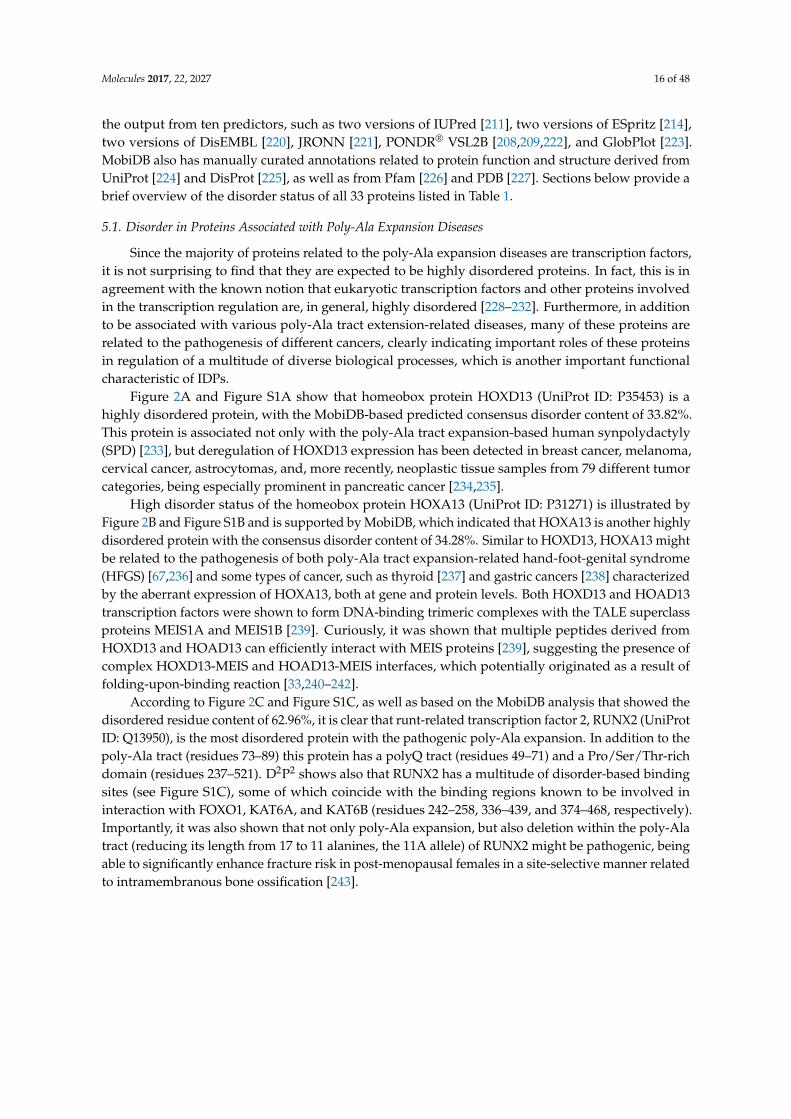
Since the majority of proteins related to the poly-Ala expansion diseases are transcription factors,it is not surprising to find that they are expected to be highly disordered proteins. In fact, this is inagreement with the known notion that eukaryotic transcription factors and other proteins involvedin the transcription regulation are, in general, highly disordered [228–232]. Furthermore, in additionto be associated with various poly-Ala tract extension-related diseases, many of these proteins arerelated to the pathogenesis of different cancers, clearly indicating important roles of these proteinsin regulation of a multitude of diverse biological processes, which is another important functionalcharacteristic of IDPs.
Figure 2A and Figure S1A show that homeobox protein HOXD13 (UniProt ID: P35453) is ahighly disordered protein, with the MobiDB-based predicted consensus disorder content of 33.82%.This protein is associated not only with the poly-Ala tract expansion-based human synpolydactyly(SPD) [233], but deregulation of HOXD13 expression has been detected in breast cancer, melanoma,cervical cancer, astrocytomas, and, more recently, neoplastic tissue samples from 79 different tumorcategories, being especially prominent in pancreatic cancer [234,235].
High disorder status of the homeobox protein HOXA13 (UniProt ID: P31271) is illustrated byFigure 2B and Figure S1B and is supported by MobiDB, which indicated that HOXA13 is another highlydisordered protein with the consensus disorder content of 34.28%. Similar to HOXD13, HOXA13 mightbe related to the pathogenesis of both poly-Ala tract expansion-related hand-foot-genital syndrome(HFGS) [67,236] and some types of cancer, such as thyroid [237] and gastric cancers [238] characterizedby the aberrant expression of HOXA13, both at gene and protein levels. Both HOXD13 and HOAD13transcription factors were shown to form DNA-binding trimeric complexes with the TALE superclassproteins MEIS1A and MEIS1B [239]. Curiously, it was shown that multiple peptides derived fromHOXD13 and HOAD13 can efficiently interact with MEIS proteins [239], suggesting the presence ofcomplex HOXD13-MEIS and HOAD13-MEIS interfaces, which potentially originated as a result offolding-upon-binding reaction [33,240–242].
According to Figure 2C and Figure S1C, as well as based on the MobiDB analysis that showed thedisordered residue content of 62.96%, it is clear that runt-related transcription factor 2, RUNX2 (UniProtID: Q13950), is the most disordered protein with the pathogenic poly-Ala expansion. In addition to thepoly-Ala tract (residues 73–89) this protein has a polyQ tract (residues 49–71) and a Pro/Ser/Thr-richdomain (residues 237–521). D2P2 shows also that RUNX2 has a multitude of disorder-based bindingsites (see Figure S1C), some of which coincide with the binding regions known to be involved ininteraction with FOXO1, KAT6A, and KAT6B (residues 242–258, 336–439, and 374–468, respectively).Importantly, it was also shown that not only poly-Ala expansion, but also deletion within the poly-Alatract (reducing its length from 17 to 11 alanines, the 11A allele) of RUNX2 might be pathogenic, beingable to significantly enhance fracture risk in post-menopausal females in a site-selective manner relatedto intramembranous bone ossification [243].
Molecules 2017, 22, 2027 17 of 48Molecules 2017, 22, 2027 17 of 46
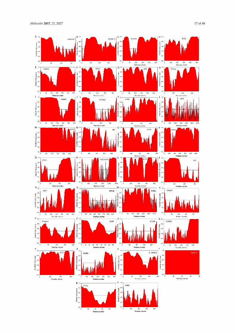
Figure 2. Evaluation of intrinsic disorder propensities of 33 proteins associated with the proteins caused by the nucleotide expansions. Intrinsic disorder predisposition was evaluated by PONDR®
Molecules 2017, 22, 2027 18 of 48
Figure 2. Evaluation of intrinsic disorder propensities of 33 proteins associated with the proteins causedby the nucleotide expansions. Intrinsic disorder predisposition was evaluated by PONDR® VSL2predictor, which is one of the more accurate stand-alone tools for prediction of the intrinsic disorderstatus of a target protein. This tool is known to be statistically better for proteins containing both orderedand disordered regions [208,209]. (A) Homeobox protein HOXD13 (UniProt ID: P35453); (B) Homeoboxprotein HOXA13 (UniProt ID: P31271); (C) runt-related transcription factor 2, RUNX2 (UniProt ID:Q13950); (D) Zinc finger protein ZIC2 (UniProt ID: O95409); (E) Paired mesoderm homeobox protein2B (UniProt ID: Q99453); (F) Transcription factor SOX3 (UniProt ID: P41225); (G) Homeobox proteinARX (UniProt ID: Q96QS3); (H) Human FOXL2 (UniProt ID: P58012); (I) PABP2/PABPN1 (UniProtID: Q86U42); (J) Small conductance calcium-activated potassium channel protein 3 (SK3, UniProt ID:Q9UGI6); (K) Human junctophilin-3 (JP-3, UniProt ID: Q8WXH2); (L) Human huntingtin (UniProtID: P42858); (M) Atrophin-1 (UniProt ID: P54259); (N) Human androgen receptor (AR, UniProt ID:P10275); (O) Ataxin-1 (UniProt ID: P54253); (P) Ataxin-2 (UniProt ID: Q99700); (Q) Human ataxin-3(UniProt ID: P54252); (R) Voltage-dependent P/Q-type calcium channel subunit α1A (CACNA1A,UniProt ID: O00555); (S) Ataxin-7 (UniProt ID: O15265); (T) TATA-box-binding protein (TBP, UniProtID: P20226); (U) Synaptic functional regulator FMR1 (UniProt ID: Q06787); (V) Disco-interactingprotein 2 homolog B (UniProt ID: Q9P265); (W) AF4/FMR2 family member 2 (UniProt ID: P51816);(X) C9orf72 (UniProt ID: Q96LT7); (Y) Frataxin (UniProt ID: Q16595); (Z) Cellular nucleic acid-bindingprotein (CNBP, UniProt ID: P62633); (a) Ataxin-10 (UniProt ID: Q9UBB4); (b) Nucleolar protein 56(UniProt ID: O00567); (c) Transcription factor 4 (UniProt ID: P15884); (d) Myotonin-protein kinase(UniProt ID: Q09013); (e) Ataxin-8 (UniProt ID: Q156A1); (f) ATXN8OS protein (UniProt ID: P0DMR3);(g) Cystatin-B (UniProt ID: P04080); (h) Serine/threonine-protein phosphatase 2A 55 kDa regulatorysubunit B β isoform (PPP2R2B, UniProt ID: Q00005). In this analysis, scores above 0.5 correspond tointrinsic disorder.
Zinc finger protein ZIC2 (UniProt ID: O95409) has a MobiDB-defined disorder content of54.89%. Figure 2D and Figure S1D illustrate that this protein has long IDPRs located in its N- andC-terminal tails. The protein is predicted to have a multitude of functional domains, possess severaldisorder-based binding regions, and have several PTM sites (see Figure S1D). Zic2 has multipleregions with compositional biases, such as a poly-Gly region (residues 490–508), two poly-His regions(residues 20–23 and 231–239), and four poly-Ala regions (residues 25–33, 89–97, 226–230, and 456–470).It has multiple zinc-finger domains (C2H2-type 1, 2, 3, 4, and 5, residues 256–291, 300–327, 332–357,363–387, and 393–415, respectively) needed for transcription activation. Furthermore, a 100–255 regionof ZIC2 known to be necessary for interaction with MDFIC and transcriptional activation or repressionis predicted to have multiple disorder-based protein-protein interaction sites (see Figure S1D). Finally,similar to HOXD13 and HOAD13, deregulated expression of ZIC2 was shown to be associated withhepatocellular carcinoma, with this protein being required for the self-renewal maintenance of livercancer stem cells [244].
Paired mesoderm homeobox protein 2B (PHOX2B homeodomain protein, UniProt ID: Q99453) isexpected to have 54.14% disordered residues (as evaluated by the MobiDB-based consensus disordercontent). In agreement with these MobiDB predictions, Figure 2E and Figure S1E show high levels ofintrinsic disorder in N- and C-terminal regions of this protein. There are two poly-Ala tracts in humanPHOX2B (residues 159–167 and 241–260) complemented by a poly-Gly region (residues 212–217).In addition to involvement in the poly-Ala expansion-related congenital central hypoventilationsyndrome [245–247], mutations in PHOX2B are associated with neuroblastoma-2 [248,249]. Figure S1Eshows that PHOX2B has several C-terminally-located disorder-based protein-protein interaction sitesand also possess several PTM sites.
With its MobiDB consensus disorder score of 51.12%, transcription factor SOX3 (Sex-determiningregion Y-box3, UniProt ID: P41225) definitely belongs to the category of highly disordered proteins.This is further illustrated by Figure 2F and Figure S1F both showing high levels of intrinsic disorderalmost evenly distributed throughout the entire protein sequence. Human SOX3 has a poly-Gly
Molecules 2017, 22, 2027 19 of 48
and poly-Pro tracts (residues 129–133 and 290–294, respectively) and four poly-Ala regions (residues234–248, 324–330, 340–347, and 353–364). The protein is predicted to have 9 MoRF regions and multiplephosphorylation sites (see Figure S1F). Besides being associated with X-linked mental retardation withgrowth hormone deficiency (via its poly-Ala tract expansion mutations) [250], as well as with X-linkedhypopituitarism (via its over- and under-dosage) [251] and SOX3 copy number variation-related 46,XX sex reversal 3 (SRXX3) [252], SOX3 overexpression was shown to play a crucial role in tumorprogression [253–257], placing this transcription factors into the oncogene category.
Homeobox protein ARX (Aristaless-related homeobox, UniProt ID: Q96QS3) is predicted byMobiDB consensus to have 59.07% disordered residues. It is not surprising since ARX has two longAla-rich regions (residues 100–155 and 425–544), as well as a long Pro-rich region (residues 395–459)and a long Glu-rich region (residues 224–253). In fact, Figure 2G and Figure S1G indicate that intrinsicdisorder is spread over the entire protein sequence and Figure S1G shows that this disorder is offunctional importance, since ARX is predicted to have 8 MoRFs (two of very significant length, 44 and157 residues), as well as several phosphorylation and methylation sites. Again, besides being relatedto the poly-Ala expansion-driven X-linked mental retardation [258], mutations ARX are related toagenesis of the corpus callosum in females and X-linked lissencephaly with abnormal genitalia inmales [259], early infantile epileptic encephalopathy-1 [260,261], Partington syndrome [261], andX-linked lissencephaly-2 [259,262]. It was also pointed out that duplication mutation of ARX can causebenign bilateral cystic-like cavities in the cerebral and cerebellar hemispheres [263].
Figure 2H and Figure S1H show that human forkhead box protein L2 (FOXL2, UniProt ID:P58012) is predicted to have very high levels of intrinsic disorder, possessing the MobiDB consensusdisorder score of 47.34%. As a matter of fact, it is unlikely that any significant part of this proteincan spontaneously gain ordered structure. Instead, several regions of FOXL2 can fold at interactionswith specific binding partners and this protein is shown to have multiple sites of phosphorylation,acetylation and SUMOylation (see Figure S1H). As the majority proteins discussed in this section,human FOXL2 contains a poly-Gly, a poly-Pro, and two poly-Ala stretches (residues 35–43, 284–292,221–234, and 301–304, respectively). Besides multiple point mutations several different poly-Ala tractexpansions associated with blepharophimosis, ptosis, and epicanthus inversus syndrome [264] severalmutations in this proteins are directly linked to the premature ovarian failure 3 [265,266]. Furthermore,C134W mutation in FOXL2 was shown to be one of the causative agents of the adult granulosa celltumor, one of the malignant ovarian sex cord-stromal tumors [267].
A last member of the diverse family of proteins with pathogenic poly-Ala expansions ispolyadenylate-binding protein 2/polyadenine-binding protein nuclear-1 (PABP2/PABPN1, UniProtID: Q86U42) that is predicted to be highly disordered by MobiDB (consensus disorder score of 59.80%),PONDR® VSL2 (Figure 2I) and D2p2 (Figure S1I). In fact, all computational tools agree that the first160 and the last 60 residues of PABP2/PABPN1 are expected to be disordered, whereas central regionconsisting of residues 170–250 should be ordered. In agreement with these predictions, X-ray structurewas solved for the RNA binding domain also known as RNA recognition motif (RRM) of this proteinspanning residues 167–254 (see, e.g., PDB ID: 3B4D; [268]). Figure S1I suggests that N- and C-terminallylocated intrinsic disorder is differently used in functionality of PABP2/PABPN1, with long N-terminalIDPR clearly serving protein-protein interaction roles (it has all 6 MoRFs found in this protein andpossesses multiple phosphorylation and ubiquitination sites), whereas disordered C-tail mostlyfunctions in PABP2/PABPN1 regulation, possessing a whole host of methylation sites. In agreementwith this hypothesis, known protein interaction sites, such as regions needed for interaction withSKIP (residues 2–145), stimulation of poly(A) polymerase alpha (PAPOLA, residues 119–147) andcoiled-coil-based interaction (residues 115–151), are all located within the N-terminal IDPR. Beingthe only non-transcription factor with the pathogenic poly-Ala tract, PABP2/PABPN1 does not forman exception from the multipathogenicity rule, being associated with oculopharyngeal musculardystrophy via the expansion mutations in its poly-Ala tract [269] and also being involved in metastaticduodenal cancer [270] and non-small cell lung cancer [271].
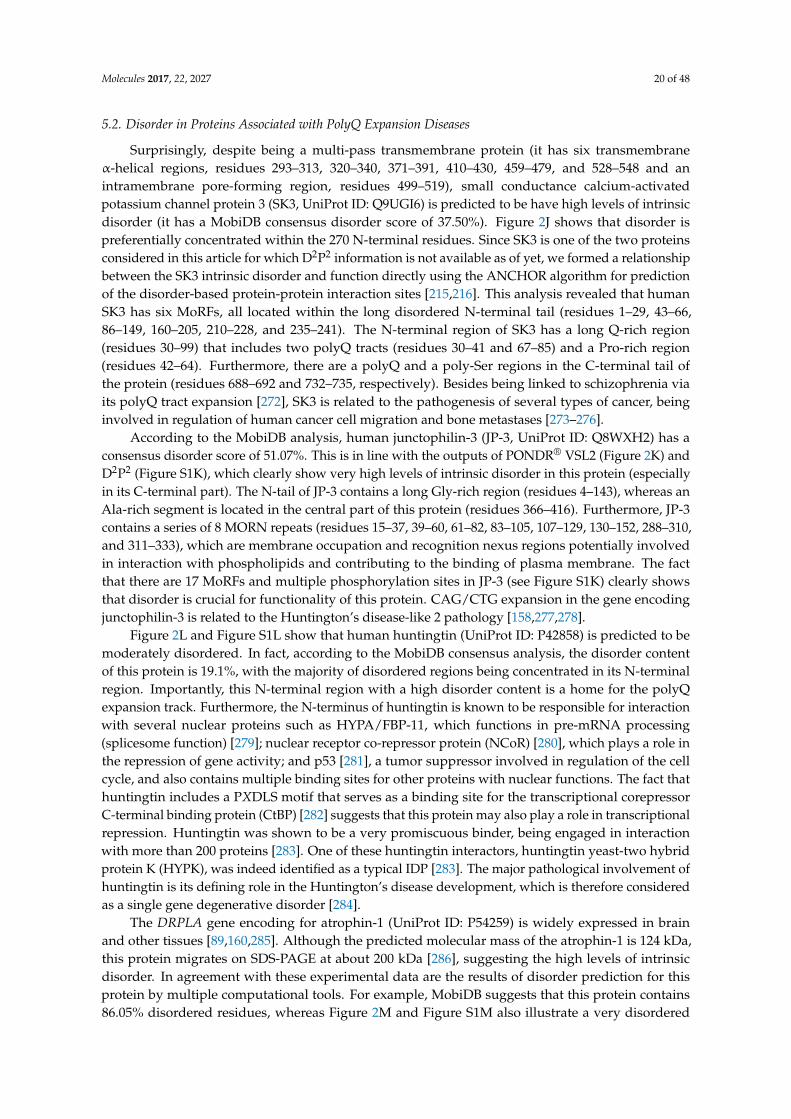
Molecules 2017, 22, 2027 20 of 48
5.2. Disorder in Proteins Associated with PolyQ Expansion Diseases
Surprisingly, despite being a multi-pass transmembrane protein (it has six transmembraneα-helical regions, residues 293–313, 320–340, 371–391, 410–430, 459–479, and 528–548 and anintramembrane pore-forming region, residues 499–519), small conductance calcium-activatedpotassium channel protein 3 (SK3, UniProt ID: Q9UGI6) is predicted to be have high levels of intrinsicdisorder (it has a MobiDB consensus disorder score of 37.50%). Figure 2J shows that disorder ispreferentially concentrated within the 270 N-terminal residues. Since SK3 is one of the two proteinsconsidered in this article for which D2P2 information is not available as of yet, we formed a relationshipbetween the SK3 intrinsic disorder and function directly using the ANCHOR algorithm for predictionof the disorder-based protein-protein interaction sites [215,216]. This analysis revealed that humanSK3 has six MoRFs, all located within the long disordered N-terminal tail (residues 1–29, 43–66,86–149, 160–205, 210–228, and 235–241). The N-terminal region of SK3 has a long Q-rich region(residues 30–99) that includes two polyQ tracts (residues 30–41 and 67–85) and a Pro-rich region(residues 42–64). Furthermore, there are a polyQ and a poly-Ser regions in the C-terminal tail ofthe protein (residues 688–692 and 732–735, respectively). Besides being linked to schizophrenia viaits polyQ tract expansion [272], SK3 is related to the pathogenesis of several types of cancer, beinginvolved in regulation of human cancer cell migration and bone metastases [273–276].
According to the MobiDB analysis, human junctophilin-3 (JP-3, UniProt ID: Q8WXH2) has aconsensus disorder score of 51.07%. This is in line with the outputs of PONDR® VSL2 (Figure 2K) andD2P2 (Figure S1K), which clearly show very high levels of intrinsic disorder in this protein (especiallyin its C-terminal part). The N-tail of JP-3 contains a long Gly-rich region (residues 4–143), whereas anAla-rich segment is located in the central part of this protein (residues 366–416). Furthermore, JP-3contains a series of 8 MORN repeats (residues 15–37, 39–60, 61–82, 83–105, 107–129, 130–152, 288–310,and 311–333), which are membrane occupation and recognition nexus regions potentially involvedin interaction with phospholipids and contributing to the binding of plasma membrane. The factthat there are 17 MoRFs and multiple phosphorylation sites in JP-3 (see Figure S1K) clearly showsthat disorder is crucial for functionality of this protein. CAG/CTG expansion in the gene encodingjunctophilin-3 is related to the Huntington’s disease-like 2 pathology [158,277,278].
Figure 2L and Figure S1L show that human huntingtin (UniProt ID: P42858) is predicted to bemoderately disordered. In fact, according to the MobiDB consensus analysis, the disorder contentof this protein is 19.1%, with the majority of disordered regions being concentrated in its N-terminalregion. Importantly, this N-terminal region with a high disorder content is a home for the polyQexpansion track. Furthermore, the N-terminus of huntingtin is known to be responsible for interactionwith several nuclear proteins such as HYPA/FBP-11, which functions in pre-mRNA processing(splicesome function) [279]; nuclear receptor co-repressor protein (NCoR) [280], which plays a role inthe repression of gene activity; and p53 [281], a tumor suppressor involved in regulation of the cellcycle, and also contains multiple binding sites for other proteins with nuclear functions. The fact thathuntingtin includes a PXDLS motif that serves as a binding site for the transcriptional corepressorC-terminal binding protein (CtBP) [282] suggests that this protein may also play a role in transcriptionalrepression. Huntingtin was shown to be a very promiscuous binder, being engaged in interactionwith more than 200 proteins [283]. One of these huntingtin interactors, huntingtin yeast-two hybridprotein K (HYPK), was indeed identified as a typical IDP [283]. The major pathological involvement ofhuntingtin is its defining role in the Huntington’s disease development, which is therefore consideredas a single gene degenerative disorder [284].
The DRPLA gene encoding for atrophin-1 (UniProt ID: P54259) is widely expressed in brainand other tissues [89,160,285]. Although the predicted molecular mass of the atrophin-1 is 124 kDa,this protein migrates on SDS-PAGE at about 200 kDa [286], suggesting the high levels of intrinsicdisorder. In agreement with these experimental data are the results of disorder prediction for thisprotein by multiple computational tools. For example, MobiDB suggests that this protein contains86.05% disordered residues, whereas Figure 2M and Figure S1M also illustrate a very disordered
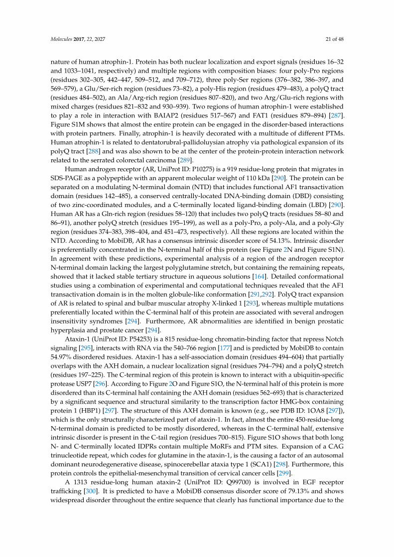
Molecules 2017, 22, 2027 21 of 48
nature of human atrophin-1. Protein has both nuclear localization and export signals (residues 16–32and 1033–1041, respectively) and multiple regions with composition biases: four poly-Pro regions(residues 302–305, 442–447, 509–512, and 709–712), three poly-Ser regions (376–382, 386–397, and569–579), a Glu/Ser-rich region (residues 73–82), a poly-His region (residues 479–483), a polyQ tract(residues 484–502), an Ala/Arg-rich region (residues 807–820), and two Arg/Glu-rich regions withmixed charges (residues 821–832 and 930–939). Two regions of human atrophin-1 were establishedto play a role in interaction with BAIAP2 (residues 517–567) and FAT1 (residues 879–894) [287].Figure S1M shows that almost the entire protein can be engaged in the disorder-based interactionswith protein partners. Finally, atrophin-1 is heavily decorated with a multitude of different PTMs.Human atrophin-1 is related to dentatorubral-pallidoluysian atrophy via pathological expansion of itspolyQ tract [288] and was also shown to be at the center of the protein-protein interaction networkrelated to the serrated colorectal carcinoma [289].
Human androgen receptor (AR, UniProt ID: P10275) is a 919 residue-long protein that migrates inSDS-PAGE as a polypeptide with an apparent molecular weight of 110 kDa [290]. The protein can beseparated on a modulating N-terminal domain (NTD) that includes functional AF1 transactivationdomain (residues 142–485), a conserved centrally-located DNA-binding domain (DBD) consistingof two zinc-coordinated modules, and a C-terminally located ligand-binding domain (LBD) [290].Human AR has a Gln-rich region (residues 58–120) that includes two polyQ tracts (residues 58–80 and86–91), another polyQ stretch (residues 195–199), as well as a poly-Pro, a poly-Ala, and a poly-Glyregion (residues 374–383, 398–404, and 451–473, respectively). All these regions are located within theNTD. According to MobiDB, AR has a consensus intrinsic disorder score of 54.13%. Intrinsic disorderis preferentially concentrated in the N-terminal half of this protein (see Figure 2N and Figure S1N).In agreement with these predictions, experimental analysis of a region of the androgen receptorN-terminal domain lacking the largest polyglutamine stretch, but containing the remaining repeats,showed that it lacked stable tertiary structure in aqueous solutions [164]. Detailed conformationalstudies using a combination of experimental and computational techniques revealed that the AF1transactivation domain is in the molten globule-like conformation [291,292]. PolyQ tract expansionof AR is related to spinal and bulbar muscular atrophy X-linked 1 [293], whereas multiple mutationspreferentially located within the C-terminal half of this protein are associated with several androgeninsensitivity syndromes [294]. Furthermore, AR abnormalities are identified in benign prostatichyperplasia and prostate cancer [294].
Ataxin-1 (UniProt ID: P54253) is a 815 residue-long chromatin-binding factor that repress Notchsignaling [295], interacts with RNA via the 540–766 region [177] and is predicted by MobiDB to contain54.97% disordered residues. Ataxin-1 has a self-association domain (residues 494–604) that partiallyoverlaps with the AXH domain, a nuclear localization signal (residues 794–794) and a polyQ stretch(residues 197–225). The C-terminal region of this protein is known to interact with a ubiquitin-specificprotease USP7 [296]. According to Figure 2O and Figure S1O, the N-terminal half of this protein is moredisordered than its C-terminal half containing the AXH domain (residues 562–693) that is characterizedby a significant sequence and structural similarity to the transcription factor HMG-box containingprotein 1 (HBP1) [297]. The structure of this AXH domain is known (e.g., see PDB ID: 1OA8 [297]),which is the only structurally characterized part of ataxin-1. In fact, almost the entire 450-residue-longN-terminal domain is predicted to be mostly disordered, whereas in the C-terminal half, extensiveintrinsic disorder is present in the C-tail region (residues 700–815). Figure S1O shows that both longN- and C-terminally located IDPRs contain multiple MoRFs and PTM sites. Expansion of a CAGtrinucleotide repeat, which codes for glutamine in the ataxin-1, is the causing a factor of an autosomaldominant neurodegenerative disease, spinocerebellar ataxia type 1 (SCA1) [298]. Furthermore, thisprotein controls the epithelial-mesenchymal transition of cervical cancer cells [299].
A 1313 residue-long human ataxin-2 (UniProt ID: Q99700) is involved in EGF receptortrafficking [300]. It is predicted to have a MobiDB consensus disorder score of 79.13% and showswidespread disorder throughout the entire sequence that clearly has functional importance due to the
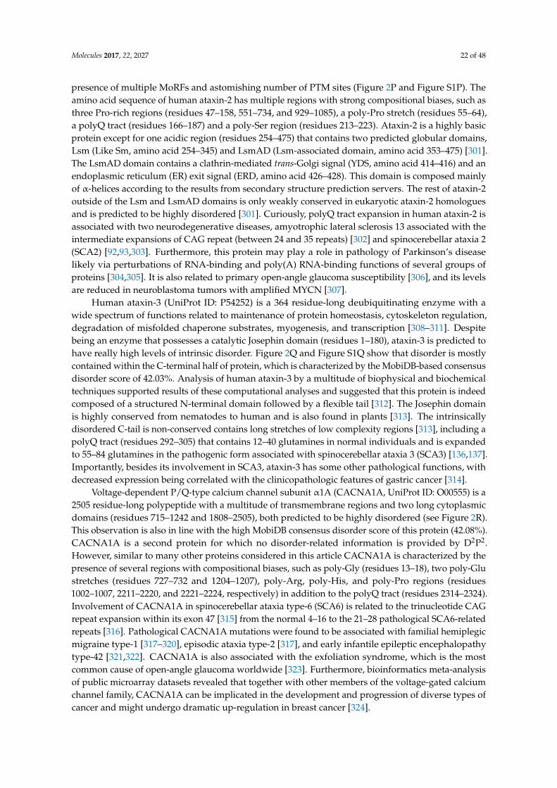
Molecules 2017, 22, 2027 22 of 48
presence of multiple MoRFs and astomishing number of PTM sites (Figure 2P and Figure S1P). Theamino acid sequence of human ataxin-2 has multiple regions with strong compositional biases, such asthree Pro-rich regions (residues 47–158, 551–734, and 929–1085), a poly-Pro stretch (residues 55–64),a polyQ tract (residues 166–187) and a poly-Ser region (residues 213–223). Ataxin-2 is a highly basicprotein except for one acidic region (residues 254–475) that contains two predicted globular domains,Lsm (Like Sm, amino acid 254–345) and LsmAD (Lsm-associated domain, amino acid 353–475) [301].The LsmAD domain contains a clathrin-mediated trans-Golgi signal (YDS, amino acid 414–416) and anendoplasmic reticulum (ER) exit signal (ERD, amino acid 426–428). This domain is composed mainlyof α-helices according to the results from secondary structure prediction servers. The rest of ataxin-2outside of the Lsm and LsmAD domains is only weakly conserved in eukaryotic ataxin-2 homologuesand is predicted to be highly disordered [301]. Curiously, polyQ tract expansion in human ataxin-2 isassociated with two neurodegenerative diseases, amyotrophic lateral sclerosis 13 associated with theintermediate expansions of CAG repeat (between 24 and 35 repeats) [302] and spinocerebellar ataxia 2(SCA2) [92,93,303]. Furthermore, this protein may play a role in pathology of Parkinson’s diseaselikely via perturbations of RNA-binding and poly(A) RNA-binding functions of several groups ofproteins [304,305]. It is also related to primary open-angle glaucoma susceptibility [306], and its levelsare reduced in neuroblastoma tumors with amplified MYCN [307].
Human ataxin-3 (UniProt ID: P54252) is a 364 residue-long deubiquitinating enzyme with awide spectrum of functions related to maintenance of protein homeostasis, cytoskeleton regulation,degradation of misfolded chaperone substrates, myogenesis, and transcription [308–311]. Despitebeing an enzyme that possesses a catalytic Josephin domain (residues 1–180), ataxin-3 is predicted tohave really high levels of intrinsic disorder. Figure 2Q and Figure S1Q show that disorder is mostlycontained within the C-terminal half of protein, which is characterized by the MobiDB-based consensusdisorder score of 42.03%. Analysis of human ataxin-3 by a multitude of biophysical and biochemicaltechniques supported results of these computational analyses and suggested that this protein is indeedcomposed of a structured N-terminal domain followed by a flexible tail [312]. The Josephin domainis highly conserved from nematodes to human and is also found in plants [313]. The intrinsicallydisordered C-tail is non-conserved contains long stretches of low complexity regions [313], including apolyQ tract (residues 292–305) that contains 12–40 glutamines in normal individuals and is expandedto 55–84 glutamines in the pathogenic form associated with spinocerebellar ataxia 3 (SCA3) [136,137].Importantly, besides its involvement in SCA3, ataxin-3 has some other pathological functions, withdecreased expression being correlated with the clinicopathologic features of gastric cancer [314].
Voltage-dependent P/Q-type calcium channel subunit α1A (CACNA1A, UniProt ID: O00555) is a2505 residue-long polypeptide with a multitude of transmembrane regions and two long cytoplasmicdomains (residues 715–1242 and 1808–2505), both predicted to be highly disordered (see Figure 2R).This observation is also in line with the high MobiDB consensus disorder score of this protein (42.08%).CACNA1A is a second protein for which no disorder-related information is provided by D2P2.However, similar to many other proteins considered in this article CACNA1A is characterized by thepresence of several regions with compositional biases, such as poly-Gly (residues 13–18), two poly-Glustretches (residues 727–732 and 1204–1207), poly-Arg, poly-His, and poly-Pro regions (residues1002–1007, 2211–2220, and 2221–2224, respectively) in addition to the polyQ tract (residues 2314–2324).Involvement of CACNA1A in spinocerebellar ataxia type-6 (SCA6) is related to the trinucleotide CAGrepeat expansion within its exon 47 [315] from the normal 4–16 to the 21–28 pathological SCA6-relatedrepeats [316]. Pathological CACNA1A mutations were found to be associated with familial hemiplegicmigraine type-1 [317–320], episodic ataxia type-2 [317], and early infantile epileptic encephalopathytype-42 [321,322]. CACNA1A is also associated with the exfoliation syndrome, which is the mostcommon cause of open-angle glaucoma worldwide [323]. Furthermore, bioinformatics meta-analysisof public microarray datasets revealed that together with other members of the voltage-gated calciumchannel family, CACNA1A can be implicated in the development and progression of diverse types ofcancer and might undergo dramatic up-regulation in breast cancer [324].
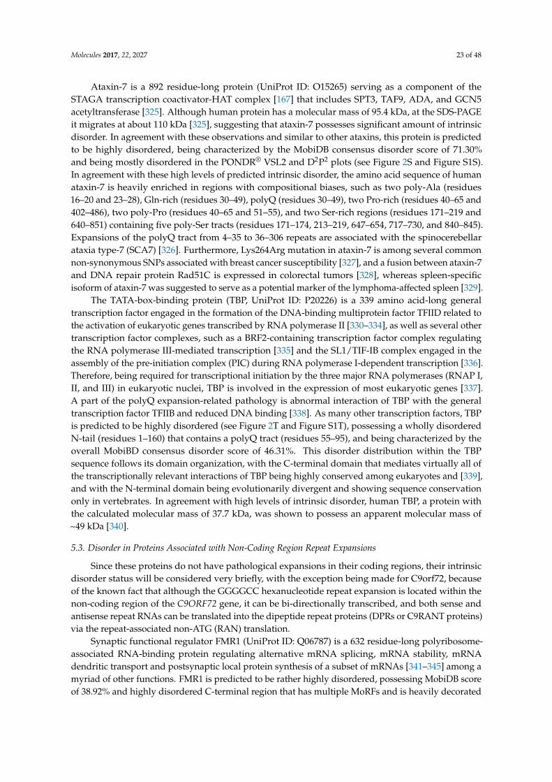
Molecules 2017, 22, 2027 23 of 48
Ataxin-7 is a 892 residue-long protein (UniProt ID: O15265) serving as a component of theSTAGA transcription coactivator-HAT complex [167] that includes SPT3, TAF9, ADA, and GCN5acetyltransferase [325]. Although human protein has a molecular mass of 95.4 kDa, at the SDS-PAGEit migrates at about 110 kDa [325], suggesting that ataxin-7 possesses significant amount of intrinsicdisorder. In agreement with these observations and similar to other ataxins, this protein is predictedto be highly disordered, being characterized by the MobiDB consensus disorder score of 71.30%and being mostly disordered in the PONDR® VSL2 and D2P2 plots (see Figure 2S and Figure S1S).In agreement with these high levels of predicted intrinsic disorder, the amino acid sequence of humanataxin-7 is heavily enriched in regions with compositional biases, such as two poly-Ala (residues16–20 and 23–28), Gln-rich (residues 30–49), polyQ (residues 30–49), two Pro-rich (residues 40–65 and402–486), two poly-Pro (residues 40–65 and 51–55), and two Ser-rich regions (residues 171–219 and640–851) containing five poly-Ser tracts (residues 171–174, 213–219, 647–654, 717–730, and 840–845).Expansions of the polyQ tract from 4–35 to 36–306 repeats are associated with the spinocerebellarataxia type-7 (SCA7) [326]. Furthermore, Lys264Arg mutation in ataxin-7 is among several commonnon-synonymous SNPs associated with breast cancer susceptibility [327], and a fusion between ataxin-7and DNA repair protein Rad51C is expressed in colorectal tumors [328], whereas spleen-specificisoform of ataxin-7 was suggested to serve as a potential marker of the lymphoma-affected spleen [329].
The TATA-box-binding protein (TBP, UniProt ID: P20226) is a 339 amino acid-long generaltranscription factor engaged in the formation of the DNA-binding multiprotein factor TFIID related tothe activation of eukaryotic genes transcribed by RNA polymerase II [330–334], as well as several othertranscription factor complexes, such as a BRF2-containing transcription factor complex regulatingthe RNA polymerase III-mediated transcription [335] and the SL1/TIF-IB complex engaged in theassembly of the pre-initiation complex (PIC) during RNA polymerase I-dependent transcription [336].Therefore, being required for transcriptional initiation by the three major RNA polymerases (RNAP I,II, and III) in eukaryotic nuclei, TBP is involved in the expression of most eukaryotic genes [337].A part of the polyQ expansion-related pathology is abnormal interaction of TBP with the generaltranscription factor TFIIB and reduced DNA binding [338]. As many other transcription factors, TBPis predicted to be highly disordered (see Figure 2T and Figure S1T), possessing a wholly disorderedN-tail (residues 1–160) that contains a polyQ tract (residues 55–95), and being characterized by theoverall MobiBD consensus disorder score of 46.31%. This disorder distribution within the TBPsequence follows its domain organization, with the C-terminal domain that mediates virtually all ofthe transcriptionally relevant interactions of TBP being highly conserved among eukaryotes and [339],and with the N-terminal domain being evolutionarily divergent and showing sequence conservationonly in vertebrates. In agreement with high levels of intrinsic disorder, human TBP, a protein withthe calculated molecular mass of 37.7 kDa, was shown to possess an apparent molecular mass of~49 kDa [340].
5.3. Disorder in Proteins Associated with Non-Coding Region Repeat Expansions
Since these proteins do not have pathological expansions in their coding regions, their intrinsicdisorder status will be considered very briefly, with the exception being made for C9orf72, becauseof the known fact that although the GGGGCC hexanucleotide repeat expansion is located within thenon-coding region of the C9ORF72 gene, it can be bi-directionally transcribed, and both sense andantisense repeat RNAs can be translated into the dipeptide repeat proteins (DPRs or C9RANT proteins)via the repeat-associated non-ATG (RAN) translation.
Synaptic functional regulator FMR1 (UniProt ID: Q06787) is a 632 residue-long polyribosome-associated RNA-binding protein regulating alternative mRNA splicing, mRNA stability, mRNAdendritic transport and postsynaptic local protein synthesis of a subset of mRNAs [341–345] among amyriad of other functions. FMR1 is predicted to be rather highly disordered, possessing MobiDB scoreof 38.92% and highly disordered C-terminal region that has multiple MoRFs and is heavily decorated
Molecules 2017, 22, 2027 24 of 48
with different PTMs (see Figure 2U and Figure S1U). However, it does not possesses regions withcompositional bias.
Disco-interacting protein 2 homolog B (UniProt ID: Q9P265) is a moderately disordered1576 residue-long protein involved in DNA methylation [346], with the MobiDB score of 19.73% andhighly disordered N-terminal tail with several disorder-based protein binding regions and multiplesites of different PTMs (see Figure 2V and Figure S1V). This protein has a Ser-rich (residues 145–234),a poly-Ala (residues 1118–1121), and two poly-Val regions (residues 1503–1506 and 1534–1540).
AF4/FMR2 family member 2 (UniProt ID: P51816) is a 1311 residue-long RNA binding proteininvolved in alternative splicing regulation [347]. FMR2 is predicted by MobiDB to have 58.12%disordered residues occupying the N-terminal and central parts of this protein that have multipledisorder-based binding regions and several PTM sites (see Figure 2W and Figure S1W). There is nocompositional bias in this protein.
Although the 481 residue-long protein C9orf72 (UniProt ID: Q96LT7) is the most ordered proteinanalyzed in this study (it has a MobiDB score of 2.70% and is expected to have short disorderedtails, three to four short disordered loops, and a long disordered/flexible region located between theresidues 130 and 210 as shown in Figure 2X and Figure S1X), it clearly deserves more attention inrelation to the topic of this article. Besides the fact that the GGGGCC (G4C2) hexanucleotide repeatexpansions in the non-coding intronic region between the non-coding exons 1a and 1b of the C9ORF72gene represent the major cause of ALS and FTD [348,349] and that these expansions can vary from 10to thousands of repeats [108,109,181,350,351], the expanded GGGGCC hexanucleotide repeats can bebi-directionally transcribed, and both sense and antisense repeat RNAs are engaged in the formationof RNA foci [352–355]. Furthermore, resulting hexanucleotide repeat RNA can be translated in a seriesof the dipeptide repeat proteins (DPRs or C9RANT proteins) by the RAN translation and these DPRsare commonly found as major constituents of proteinaceous inclusions throughout the CNS of theALS and FTD patients [356]. This RAN translation of sense GGGGCC repeat RNAs generates threedifferent proteins, poly(GA), poly(GR), and poly(GP) [194,195], and poly(PA), poly(PR), and poly(GP)proteins are synthesized as a result of translation of the antisense RNAs [353,355,357]. It is knownthat repeat-containing proteins are often intrinsically disordered, with the more perfect repeats beingmore disordered [47]. In agreement with these earlier observations, all DPRs synthesized as a resultof the RAN translation of the sense and antisense GGGGCC hexanucleotide repeat RNAs, poly(GA),poly(GR), poly(GP), poly(PA), and poly(PR), were predicted to be highly disordered [358]. It was alsoemphasized that based on their positions within the CH-CDF phase space, DPRs can be classified eitheras native molten globules (Poly(GA) and Poly(PA)) or native coils or pre-molten globules (Poly(GP),Poly(GR), and Poly(PR)) [358].
Frataxin is a 210 residue-long iron-binding protein (UniProt ID: Q16595) that takes part in theheme biosynthesis [359] and biosynthesis and repair of iron-sulfur clusters [360] and acts as aniron chaperone [361]. Despite being involved in catalytic detoxification of redox-active iron [362],frataxin is predicted to have a MobiDB score of 40.00%, with the majority of disordered residuesbeing concentrated within the N- and C-tails containing several MoRFs and PTMs (see Figure 2Y andFigure S1Y). There are no regions with compositional bias in this protein.
The cellular nucleic acid-binding protein is a 177 residue-long single-stranded DNA-bindingprotein (CNBP, UniProt ID: P62633) with moderate level of intrinsic disorder as evidenced by theMobiDB score of 19.77% and presence of several IDPRs enriched in MoRFs and PTM sites (see Figure 2Zand Figure S1Z). CNBP has an Arg/Gly-rich region (residues 22–42) seven zinc finger motifs of theCCHC type.
Ataxin-10 is a 475 residue-long protein (UniProt ID: Q9UBB4) needed for the survival of cerebellarneurons. With the MobiDB score of 5.26%, mostly short IDPRs (see Figure 2a and Figure S1a), and lackof compositional biases, this protein is the most ordered ataxin considered in this article.
Nucleolar protein 56 is a 594 residue-long protein (UniProt ID: O00567) serving as a corecomponent of the box C/D small nucleolar ribonucleoprotein (snoRNP) particles [363] and related
Molecules 2017, 22, 2027 25 of 48
to the early to middle stages of the biogenesis of 60S ribosomal subunit [364]. It is characterized bythe moderate-to-high overall levels of intrinsic disorder, has a MobiDB score of 26.26%, and possessesa highly disordered C-terminal domain that contains multiple MoRFs, a multitude of divers PTMs(see Figure 2b and Figure S1b), and a long Lys-rich stretch (residues 438–589).
Transcription factor 4 is a 667 residue-long transcription factor (UniProt ID: P15884) known forits binding to the immunoglobulin enhancer Mu-E5/KE5-motif and involvement in the initiation ofneuronal differentiation. This is one of the most disordered proteins analyzed in this study, beingcharacterized by a MobiDB score of 88.01%, containing long IDPRs and a whole host of different PTMs,using almost an entire sequence for the disorder-based protein-protein and protein-DNA interactions(see Figure 2c and Figure S1c), and containing a short poly-Ser region (residues 228–231).
Myotonin-protein kinase is a 629 residue-long non-receptor serine/threonine protein kinase(UniProt ID: Q09013) needed for the upkeep of skeletal muscle structure and function. It has amoderate intrinsic disorder content of 14.63%, with the majority of disordered residues, disorder-basedbinding regions, and PTM sites being present in the C-terminal part of this protein (see Figure 2d andFigure S1d). There are no compositional bias regions in this kinase.
Ataxin-8 and ATXN8OS are 80 and 200 residue-long proteins, respectively. Biological functionsof the ataxin-8 (UniProt ID: Q156A1) and ATXN8OS proteins (UniProt ID: P0DMR3) are unknown.ATXN8OS is a putative protein with the disorder content of 68% (see Figure 2e). It is predicted tocontain a couple of MoRFs located in the N-terminal region. Since ataxin-8 is simply a polyglutaminepolypeptide (in fact, according to UniProt, it contains only 80 glutamine residues); it is not surprisingthat this protein is predicted to be completely disordered (see Figure 2f).
Cystatin-B is a short, 98 residue-long, protein (UniProt ID: P04080) that serves as an inhibitor ofintracellular thiol proteinase. Cystatin-B is predicted by MobiDB to have 40.82% disordered residuesand is expected to have flexible tails and a less flexible central region (see Figure 2g and Figure S1g).
Serine/threonine-protein phosphatase 2A 55 kDa regulatory subunit B β isoform (PPP2R2B,UniProt ID: Q00005) is a 443 residue-long protein modulating substrate selectivity and catalytic activityof the PP2A phosphatase. It is characterized by a MobiDB score of 7.67% and has several short IDPRs,one short MoRF, and several PTM sites (see Figure 2h and Figure S1h). There are also seven WDrepeats that are 40-residue-long conserved domains containing a centrally located Trp-Asp motif.
6. Summary
IDPs and IDPRs are characteristically comprised of low complexity domains, often containingrepetitive amino acid sequences [47]. Expansion of these repetitive domains frequently results inpathology seen in several neurological and neuromuscular diseases. Many of the genes linked to repeatexpansion diseases are translated into proteins predicted to contain a large percentage of disorderedregions. Even expansions in regions that are not normally translated, such as those in C9orf72, result inaberrant dipeptide repeat products predicted to be disordered. The resultant disorder in these peptidesoften decreases in a length-dependent manner, causing many repetitive proteins to aggregate andsequester other proteins into the aggregated structures [365].
Trinucleotide repeats are the most common type seen in coding regions of genes, with polyQbeing the most commonly occurring followed by polyAla [65]. Both pathological extensions result indiseases classified as protein misfolding disorders and share many pathological characteristics suchas intracellular protein inclusions and the appearance of RNA foci. Since both Ala and Gln repeatexpansions correlate with protein aggregation and formation of the β-structure-enriched amyloid-likefibrils [366,367], it is likely that the fibrillation process triggered by expanded repeats of these twocompletely different residues has some common molecular mechanisms. In agreement with thishypothesis, computational analysis of non-aggregated polyAla and polyQ peptides composed of7, 10, 14 or 20 amino acids revealed that both types of these homo-oligopeptides are characterizedby the presence of similar secondary structural elements (type I and type III β-turns, antiparallelβ-strand, α-helix, and 310-helix) and that characteristic H-bonding patterns containing i–i + 3 and
Molecules 2017, 22, 2027 26 of 48
i–i + 4 H-bonds are formed [368]. Furthermore, both polyAla and polyQ repeats were shown to formcoiled-coil structures, the stability of which increased with the length of the expansion, and which werealso able to form higher-order multimers and aggregates in vitro [369]. Also, it was pointed out that inthe majority of expanded CAG and GCG repeat proteins, the polyQ or polyAla sequence is typicallylocated within the protein, thereby possessing specific N- and C-terminal flanking sequences that mayplay a crucial role in regulation of the polyQ and polyAla aggregation propensity [367]. Finally, manyof the proteins involved in both types of trinucleotide repeat expansions are associated with signaling,regulation, and RNA metabolism. Many of the diseases associated with both expansion types aredevelopmental, neuromuscular, and neurodegenerative in nature [365].
The similarities shared by the two common trinucleotide repeats involved in disease are moreabundant than the characteristics that set them apart. For example, trinucleotide repeats exceedingspecific thresholds show replication-related instability that increases in a repeat length-dependentmanner, with most instabilities causing repeat expansion [370]. Due to the effect of increased probabilityof repeat expansion on replication, and due to the fact that the increase in the rate of mutations thatadd additional codons and thereby increase the expansion length become more likely with each newgeneration, in successive generations, the age of the onset of trinucleotide expansion diseases typicallybecomes younger, whereas the severity of these maladies increases [370]. Importantly, one shouldkeep in mind that expanded repeats can undergo further expansion-biased somatic instability, leadingto further increases in the expansion length [371–375]. Furthermore, pathogenic processes associatedwith the expansions of both the polyQ and polyAla repeats are usually age-dependent (i.e., they donot happen before a particular age). This is likely due to the presence of the aforementioned somaticinstability of the trinucleotide expansions [372] and because of other age-related processes, such asimpairment of proteostasis [376–381].
However, not everything is similar for the repeat-containing proteins. In fact, one stark differencebetween polyQ and polyAla repeats is the threshold size of the repeat needed to cause disease. PolyQrepeats require much longer extensions for disease presentation, while polyAla repeats often can causesdisease with negligible extensions. This size difference may have to do with the fact that extensions inpolyAla repeats induce structural changes more readily than those in polyQ repeats because of thebiophysical properties of the repeated amino acids in the peptide sequence [65].
Repeats in non-coding regions are more diverse than those seen in coding regions. The functionsof the genes that are involved in disease vary widely as compared to those found in coding regionsthat are involved in disease. Often times in non-coding expansion diseases, there is a knock down inexpression of the gene involved. However, in most cases this is not sufficient to cause disease andtherefore there must be other mechanisms at play. For example, we now know that in the case ofC9orf72, not only is the expression of the protein reduced, but aberrant mRNA and repeat peptidespecies are also produced [108,109,181].
The peptide species produced through non-canonical translation of the repeat region in C9orf72are predicted to be disordered at short repeat lengths, but upon extension have been shown toform toxic stable aggregates in cells that cause the sequestration of proteins with low complexitydomains [196]. Therefore, it is important to consider that these species may account for, at least in part,the pathology that is associated with the expansion of repeats in non-coding regions. In addition, it isimportant to note that the expansion in the 5’ UTR of C9orf72 results in five different dipeptide repeatspecies. Three of the peptides are produced from the three available reading frames of the repeatregion and their antisense partners make up the remaining three species present in disease. This couldtheoretically be the case for all expansions that occur in non-coding regions and should be consideredfor other similar cases. All of the repeat regions may also have the propensity to undergo translationinto toxic repeat peptide species with unique biochemical properties. The predicted peptide speciesfor each non-coding expansion linked to disease can be found in Table 2. Although, except for theDPRs generated as a result of RAN translation of the mRNA produced as a result of hexanucleotideexpansion in the 5’ UTR of the C9orf72 gene, the presence of such polypeptides was not demonstrated
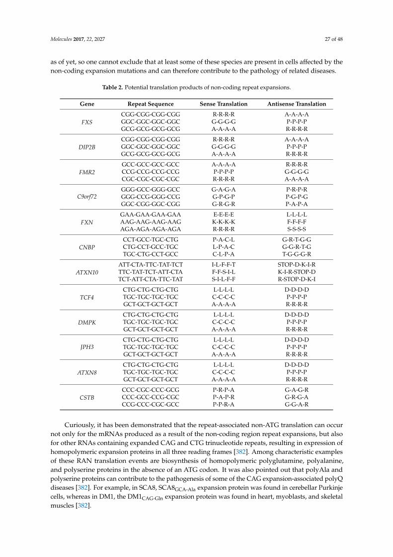
Molecules 2017, 22, 2027 27 of 48
as of yet, so one cannot exclude that at least some of these species are present in cells affected by thenon-coding expansion mutations and can therefore contribute to the pathology of related diseases.
Table 2. Potential translation products of non-coding repeat expansions.
Gene Repeat Sequence Sense Translation Antisense Translation
FXSCGG-CGG-CGG-CGG R-R-R-R A-A-A-AGGC-GGC-GGC-GGC G-G-G-G P-P-P-PGCG-GCG-GCG-GCG A-A-A-A R-R-R-R
DIP2BCGG-CGG-CGG-CGG R-R-R-R A-A-A-AGGC-GGC-GGC-GGC G-G-G-G P-P-P-PGCG-GCG-GCG-GCG A-A-A-A R-R-R-R
FMR2GCC-GCC-GCC-GCC A-A-A-A R-R-R-RCCG-CCG-CCG-CCG P-P-P-P G-G-G-GCGC-CGC-CGC-CGC R-R-R-R A-A-A-A
C9orf72GGG-GCC-GGG-GCC G-A-G-A P-R-P-RGGG-CCG-GGG-CCG G-P-G-P P-G-P-GGGC-CGG-GGC-CGG G-R-G-R P-A-P-A
FXNGAA-GAA-GAA-GAA E-E-E-E L-L-L-LAAG-AAG-AAG-AAG K-K-K-K F-F-F-FAGA-AGA-AGA-AGA R-R-R-R S-S-S-S
CNBPCCT-GCC-TGC-CTG P-A-C-L G-R-T-G-GCTG-CCT-GCC-TGC L-P-A-C G-G-R-T-GTGC-CTG-CCT-GCC C-L-P-A T-G-G-G-R
ATXN10ATT-CTA-TTC-TAT-TCT I-L-F-F-T STOP-D-K-I-RTTC-TAT-TCT-ATT-CTA F-F-S-I-L K-I-R-STOP-DTCT-ATT-CTA-TTC-TAT S-I-L-F-F R-STOP-D-K-I
TCF4CTG-CTG-CTG-CTG L-L-L-L D-D-D-DTGC-TGC-TGC-TGC C-C-C-C P-P-P-PGCT-GCT-GCT-GCT A-A-A-A R-R-R-R
DMPKCTG-CTG-CTG-CTG L-L-L-L D-D-D-DTGC-TGC-TGC-TGC C-C-C-C P-P-P-PGCT-GCT-GCT-GCT A-A-A-A R-R-R-R
JPH3CTG-CTG-CTG-CTG L-L-L-L D-D-D-DTGC-TGC-TGC-TGC C-C-C-C P-P-P-PGCT-GCT-GCT-GCT A-A-A-A R-R-R-R
ATXN8CTG-CTG-CTG-CTG L-L-L-L D-D-D-DTGC-TGC-TGC-TGC C-C-C-C P-P-P-PGCT-GCT-GCT-GCT A-A-A-A R-R-R-R
CSTBCCC-CGC-CCC-GCG P-R-P-A G-A-G-RCCC-GCC-CCG-CGC P-A-P-R G-R-G-ACCG-CCC-CGC-GCC P-P-R-A G-G-A-R
Curiously, it has been demonstrated that the repeat-associated non-ATG translation can occurnot only for the mRNAs produced as a result of the non-coding region repeat expansions, but alsofor other RNAs containing expanded CAG and CTG trinucleotide repeats, resulting in expression ofhomopolymeric expansion proteins in all three reading frames [382]. Among characteristic examplesof these RAN translation events are biosynthesis of homopolymeric polyglutamine, polyalanine,and polyserine proteins in the absence of an ATG codon. It was also pointed out that polyAla andpolyserine proteins can contribute to the pathogenesis of some of the CAG expansion-associated polyQdiseases [382]. For example, in SCA8, SCA8GCA-Ala expansion protein was found in cerebellar Purkinjecells, whereas in DM1, the DM1CAG-Gln expansion protein was found in heart, myoblasts, and skeletalmuscles [382].
Molecules 2017, 22, 2027 28 of 48
Diseases found in microsatellite expansion regions are numerous and, in most cases, devastating.The broad commonalities between the functions of the genes involved, the structural change ofthe translated products as a result of the expansion, and the resulting disease phenotypes areoverwhelmingly evident. This points to the question of whether there is a shared mechanism thatoccurs in most disease as a result of the expansions. Elucidation of the common mechanisms involvedin pathology of each expansion disorder may shed light on this question and open new avenues for abroader therapeutic approach to treating repeat expansion disorders.
Supplementary Materials: Supplementary materials are available online. Figure S1: Intrinsic disorder propensityand some important disorder-related functional information generated for human proteins encoded by genes withnucleotide expansions by the D2P2 database (http://d2p2.pro/). Here, the outputs of several disorder predictorsare shown by differently colored bars, whereas the blue-green-and-white bar in the middle of the plot shows thepredicted disorder agreement between nine predictors, with blue and green parts corresponding to disorderedregions by consensus. Yellow bar shows the location of the predicted disorder-based binding sites (molecularrecognition features, MoRFs), whereas colored circles at the bottom of the plot show location of various PTMs.
Acknowledgments: This work was supported in part by a grant “Studies of strutural and functional propertiesof metal binding and intrinsically disordered proteins” from the Program of the Russian Academy of Sciences“Molecular and Cellular Biology” (registration number 114111870001).
Author Contributions: V.N.U. conceived the idea; A.L.D. and V.N.U. collected and analyzed literature data;V.N.U. conducted bioinformatics analysis; A.L.D. and V.N.U. wrote the paper.
Conflicts of Interest: The authors declare no conflict of interest. The founding sponsors had no role in the designof the study; in the collection, analyses, or interpretation of data; in the writing of the manuscript, and in thedecision to publish the results.
References
1. Turoverov, K.K.; Kuznetsova, I.M.; Uversky, V.N. The protein kingdom extended: Ordered and intrinsicallydisordered proteins, their folding, supramolecular complex formation, and aggregation. Prog. Biophys.Mol. Biol. 2010, 102, 73–84. [CrossRef] [PubMed]
2. Wright, P.E.; Dyson, H.J. Intrinsically unstructured proteins: Re-assessing the protein structure-functionparadigm. J. Mol. Biol. 1999, 293, 321–331. [CrossRef] [PubMed]
3. Romero, P.; Obradovic, Z.; Kissinger, C.R.; Villafranca, J.E.; Garner, E.; Guilliot, S.; Dunker, A.K. Thousandsof proteins likely to have long disordered regions. Pac. Symp. Biocomput. 1998, 3, 437–448.
4. Dunker, A.K.; Obradovic, Z.; Romero, P.; Garner, E.C.; Brown, C.J. Intrinsic protein disorder in completegenomes. Genome Inform. Ser. Workshop Genome Inform. 2000, 11, 161–171. [PubMed]
5. Ward, J.J.; Sodhi, J.S.; McGuffin, L.J.; Buxton, B.F.; Jones, D.T. Prediction and functional analysis of nativedisorder in proteins from the three kingdoms of life. J. Mol. Biol. 2004, 337, 635–645. [CrossRef] [PubMed]
6. Xue, B.; Dunker, A.K.; Uversky, V.N. Orderly order in protein intrinsic disorder distribution: Disorder in 3500proteomes from viruses and the three domains of life. J. Biomol. Struct. Dyn. 2012, 30, 137–149. [CrossRef][PubMed]
7. Peng, Z.; Yan, J.; Fan, X.; Mizianty, M.J.; Xue, B.; Wang, K.; Hu, G.; Uversky, V.N.; Kurgan, L. Exceptionallyabundant exceptions: Comprehensive characterization of intrinsic disorder in all domains of life. Cell. Mol.Life Sci. 2015, 72, 137–151. [CrossRef] [PubMed]
8. Dunker, A.K.; Garner, E.; Guilliot, S.; Romero, P.; Albrecht, K.; Hart, J.; Obradovic, Z.; Kissinger, C.;Villafranca, J.E. Protein disorder and the evolution of molecular recognition: Theory, predictions andobservations. Pac. Symp. Biocomput. 1998, 3, 473–484.
9. Uversky, V.N.; Gillespie, J.R.; Fink, A.L. Why are “natively unfolded” proteins unstructured underphysiologic conditions? Proteins 2000, 41, 415–427. [CrossRef]
10. Dunker, A.K.; Lawson, J.D.; Brown, C.J.; Williams, R.M.; Romero, P.; Oh, J.S.; Oldfield, C.J.; Campen, A.M.;Ratliff, C.M.; Hipps, K.W.; et al. Intrinsically disordered protein. J. Mol. Graph. Model. 2001, 19, 26–59.[CrossRef]
11. Tompa, P. Intrinsically unstructured proteins. Trends Biochem. Sci. 2002, 27, 527–533. [CrossRef]12. Uversky, V.N.; Dunker, A.K. Understanding protein non-folding. Biochim. Biophys. Acta 2010, 1804, 1231–1264.
[CrossRef] [PubMed]
Molecules 2017, 22, 2027 29 of 48
13. Uversky, V.N. Unusual biophysics of intrinsically disordered proteins. Biochim. Biophys. Acta 2013, 1834,932–951. [CrossRef] [PubMed]
14. Iakoucheva, L.M.; Brown, C.J.; Lawson, J.D.; Obradovic, Z.; Dunker, A.K. Intrinsic disorder in cell-signalingand cancer-associated proteins. J. Mol. Biol. 2002, 323, 573–584. [CrossRef]
15. Dunker, A.K.; Cortese, M.S.; Romero, P.; Iakoucheva, L.M.; Uversky, V.N. Flexible nets: The roles of intrinsicdisorder in protein interaction networks. FEBS J. 2005, 272, 5129–5148. [CrossRef] [PubMed]
16. Dunker, A.K.; Obradovic, Z. The protein trinity—Linking function and disorder. Nat. Biotechnol. 2001, 19,805–806. [CrossRef] [PubMed]
17. Dunker, A.K.; Brown, C.J.; Obradovic, Z. Identification and functions of usefully disordered proteins.Adv. Protein Chem. 2002, 62, 25–49. [PubMed]
18. Dunker, A.K.; Brown, C.J.; Lawson, J.D.; Iakoucheva, L.M.; Obradovic, Z. Intrinsic disorder and proteinfunction. Biochemistry 2002, 41, 6573–6582. [CrossRef] [PubMed]
19. Uversky, V.N. Natively unfolded proteins: A point where biology waits for physics. Protein Sci. 2002, 11,739–756. [CrossRef] [PubMed]
20. Uversky, V.N. What does it mean to be natively unfolded? Eur. J. Biochem. 2002, 269, 2–12. [CrossRef][PubMed]
21. Uversky, V.N.; Oldfield, C.J.; Dunker, A.K. Showing your id: Intrinsic disorder as an id for recognition,regulation and cell signaling. J. Mol. Recognit. 2005, 18, 343–384. [CrossRef] [PubMed]
22. Dunker, A.K.; Silman, I.; Uversky, V.N.; Sussman, J.L. Function and structure of inherently disorderedproteins. Curr. Opin. Struct. Biol. 2008, 18, 756–764. [CrossRef] [PubMed]
23. Uversky, V.N. The mysterious unfoldome: Structureless, underappreciated, yet vital part of any givenproteome. J. Biomed. Biotechnol. 2010, 2010, 568068. [CrossRef] [PubMed]
24. Dyson, H.J.; Wright, P.E. Intrinsically unstructured proteins and their functions. Nat. Rev. Mol. Cell Biol. 2005,6, 197–208. [CrossRef] [PubMed]
25. Tompa, P. The interplay between structure and function in intrinsically unstructured proteins. FEBS Lett.2005, 579, 3346–3354. [CrossRef] [PubMed]
26. Dunker, A.K.; Bondos, S.E.; Huang, F.; Oldfield, C.J. Intrinsically disordered proteins and multicellularorganisms. Semin. Cell Dev. Biol. 2015, 37, 44–55. [CrossRef] [PubMed]
27. Van der Lee, R.; Buljan, M.; Lang, B.; Weatheritt, R.J.; Daughdrill, G.W.; Dunker, A.K.; Fuxreiter, M.; Gough, J.;Gsponer, J.; Jones, D.T.; et al. Classification of intrinsically disordered regions and proteins. Chem. Rev. 2014,114, 6589–6631. [CrossRef] [PubMed]
28. Habchi, J.; Tompa, P.; Longhi, S.; Uversky, V.N. Introducing protein intrinsic disorder. Chem. Rev. 2014, 114,6561–6588. [CrossRef] [PubMed]
29. Fuxreiter, M.; Toth-Petroczy, A.; Kraut, D.A.; Matouschek, A.; Lim, R.Y.; Xue, B.; Kurgan, L.; Uversky, V.N.Disordered proteinaceous machines. Chem. Rev. 2014, 114, 6806–6843. [CrossRef] [PubMed]
30. Oldfield, C.J.; Dunker, A.K. Intrinsically disordered proteins and intrinsically disordered protein regions.Annu. Rev. Biochem. 2014, 83, 553–584. [CrossRef] [PubMed]
31. Jakob, U.; Kriwacki, R.; Uversky, V.N. Conditionally and transiently disordered proteins: Awakening crypticdisorder to regulate protein function. Chem. Rev. 2014, 114, 6779–6805. [CrossRef] [PubMed]
32. Hsu, W.L.; Oldfield, C.; Meng, J.; Huang, F.; Xue, B.; Uversky, V.N.; Romero, P.; Dunker, A.K. Intrinsic proteindisorder and protein-protein interactions. Pac. Symp. Biocomput. 2012, 116–127. [CrossRef]
33. Oldfield, C.J.; Meng, J.; Yang, J.Y.; Yang, M.Q.; Uversky, V.N.; Dunker, A.K. Flexible nets: Disorder andinduced fit in the associations of p53 and 14–3-3 with their partners. BMC Genom. 2008, 9 (Suppl. 1), S1.[CrossRef] [PubMed]
34. Dyson, H.J.; Wright, P.E. Coupling of folding and binding for unstructured proteins. Curr. Opin. Struct. Biol.2002, 12, 54–60. [CrossRef]
35. Oldfield, C.J.; Cheng, Y.; Cortese, M.S.; Romero, P.; Uversky, V.N.; Dunker, A.K. Coupled folding and bindingwith alpha-helix-forming molecular recognition elements. Biochemistry 2005, 44, 12454–12470. [CrossRef][PubMed]
36. Schulz, G.E. Nucleotide binding proteins. In Molecular Mechanism of Biological Recognition; Balaban, M., Ed.;Elsevier/North-Holland Biomedical Press: New York, NY, USA, 1979; pp. 79–94.
Molecules 2017, 22, 2027 30 of 48
37. Ng, K.P.; Potikyan, G.; Savene, R.O.; Denny, C.T.; Uversky, V.N.; Lee, K.A. Multiple aromatic sidechains within a disordered structure are critical for transcription and transforming activity of EWS familyoncoproteins. Proc. Natl. Acad. Sci. USA 2007, 104, 479–484. [CrossRef] [PubMed]
38. Cortese, M.S.; Uversky, V.N.; Dunker, A.K. Intrinsic disorder in scaffold proteins: Getting more from less.Prog. Biophys. Mol. Biol. 2008, 98, 85–106. [CrossRef] [PubMed]
39. Uversky, V.N. Intrinsic disorder-based protein interactions and their modulators. Curr. Pharm. Des. 2013, 19,4191–4213. [CrossRef] [PubMed]
40. Uversky, V.N.; Oldfield, C.J.; Dunker, A.K. Intrinsically disordered proteins in human diseases: Introducingthe D2 concept. Annu. Rev. Biophys. 2008, 37, 215–246. [CrossRef] [PubMed]
41. Uversky, V.N. Intrinsically disordered proteins and their (disordered) proteomes in neurodegenerativedisorders. Front. Aging Neurosci. 2015, 7, 18. [CrossRef] [PubMed]
42. Uversky, V.N. The triple power of D(3): Protein intrinsic disorder in degenerative diseases. Front. Biosci.(Landmark Ed.) 2014, 19, 181–258. [CrossRef] [PubMed]
43. Uversky, V.N. Intrinsic disorder in proteins associated with neurodegenerative diseases. Front. Biosci.(Landmark Ed.) 2009, 14, 5188–5238. [CrossRef] [PubMed]
44. Williams, R.M.; Obradovi, Z.; Mathura, V.; Braun, W.; Garner, E.C.; Young, J.; Takayama, S.; Brown, C.J.;Dunker, A.K. The protein non-folding problem: Amino acid determinants of intrinsic order and disorder.Pac. Symp. Biocomput. 2001, 89–100. [CrossRef]
45. Radivojac, P.; Iakoucheva, L.M.; Oldfield, C.J.; Obradovic, Z.; Uversky, V.N.; Dunker, A.K. Intrinsic disorderand functional proteomics. Biophys. J. 2007, 92, 1439–1456. [CrossRef] [PubMed]
46. Vacic, V.; Uversky, V.N.; Dunker, A.K.; Lonardi, S. Composition profiler: A tool for discovery andvisualization of amino acid composition differences. BMC Bioinform. 2007, 8, 211. [CrossRef] [PubMed]
47. Jorda, J.; Xue, B.; Uversky, V.N.; Kajava, A.V. Protein tandem repeats—The more perfect, the less structured.FEBS J. 2010, 277, 2673–2682. [CrossRef] [PubMed]
48. Simon, M.; Hancock, J.M. Tandem and cryptic amino acid repeats accumulate in disordered regions ofproteins. Genome Biol. 2009, 10, R59. [CrossRef] [PubMed]
49. Tompa, P. Intrinsically unstructured proteins evolve by repeat expansion. BioEssays 2003, 25, 847–855.[CrossRef] [PubMed]
50. Alba, M.M.; Guigo, R. Comparative analysis of amino acid repeats in rodents and humans. Genome Res. 2004,14, 549–554. [CrossRef] [PubMed]
51. Kalita, M.K.; Ramasamy, G.; Duraisamy, S.; Chauhan, V.S.; Gupta, D. Protrepeatsdb: A database of aminoacid repeats in genomes. BMC Bioinform. 2006, 7, 336. [CrossRef] [PubMed]
52. Li, X.; Wang, W.; Wang, J.; Malovannaya, A.; Xi, Y.; Li, W.; Guerra, R.; Hawke, D.H.; Qin, J.; Chen, J. Proteomicanalyses reveal distinct chromatin-associated and soluble transcription factor complexes. Mol. Syst. Biol.2015, 11, 775. [CrossRef] [PubMed]
53. Mier, P.; Alanis-Lobato, G.; Andrade-Navarro, M.A. Context characterization of amino acid homorepeatsusing evolution, position, and order. Proteins 2017, 85, 709–719. [CrossRef] [PubMed]
54. Kajava, A.V. Tandem repeats in proteins: From sequence to structure. J. Struct. Biol. 2012, 179, 279–288.[CrossRef] [PubMed]
55. Gatchel, J.R.; Zoghbi, H.Y. Diseases of unstable repeat expansion: Mechanisms and common principles.Nat. Rev. Genet. 2005, 6, 743–755. [CrossRef] [PubMed]
56. La Spada, A.R.; Taylor, J.P. Repeat expansion disease: Progress and puzzles in disease pathogenesis.Nat. Rev. Genet. 2010, 11, 247–258. [CrossRef] [PubMed]
57. Usdin, K. The biological effects of simple tandem repeats: Lessons from the repeat expansion diseases.Genome Res. 2008, 18, 1011–1019. [CrossRef] [PubMed]
58. Koshy, B.T.; Zoghbi, H.Y. The CAG/polyglutamine tract diseases: Gene products and molecular pathogenesis.Brain Pathol. 1997, 7, 927–942. [CrossRef] [PubMed]
59. Ashley, C.T., Jr.; Warren, S.T. Trinucleotide repeat expansion and human disease. Annu. Rev. Genet. 1995, 29,703–728. [CrossRef] [PubMed]
60. Carpenter, N.J. Genetic anticipation. Expanding tandem repeats. Neurol. Clin. 1994, 12, 683–697. [PubMed]
Molecules 2017, 22, 2027 31 of 48
61. La Spada, A.R. Trinucleotide repeat instability: Genetic features and molecular mechanisms. Brain Pathol.1997, 7, 943–963. [CrossRef] [PubMed]
62. Pearson, C.E. Repeat associated non-atg translation initiation: One DNA, two transcripts, seven readingframes, potentially nine toxic entities! PLoS Genet. 2011, 7, e1002018. [CrossRef] [PubMed]
63. Dosztanyi, Z.; Chen, J.; Dunker, A.K.; Simon, I.; Tompa, P. Disorder and sequence repeats in hub proteinsand their implications for network evolution. J. Proteome Res. 2006, 5, 2985–2995. [CrossRef] [PubMed]
64. Xin, Q.; Li, L.; Li, J.; Qiu, R.; Guo, C.; Gong, Y.; Liu, Q. Eight-alanine duplication in homeobox d13 in achinese family with synpolydactyly. Gene 2012, 499, 48–51. [CrossRef] [PubMed]
65. Albrecht, A.N.; Kornak, U.; Boddrich, A.; Suring, K.; Robinson, P.N.; Stiege, A.C.; Lurz, R.; Stricker, S.;Wanker, E.E.; Mundlos, S. A molecular pathogenesis for transcription factor associated poly-alanine tractexpansions. Hum. Mol. Genet. 2004, 13, 2351–2359. [CrossRef] [PubMed]
66. Muragaki, Y.; Mundlos, S.; Upton, J.; Olsen, B.R. Altered growth and branching patterns in synpolydactylycaused by mutations in hoxd13. Science 1996, 272, 548–551. [CrossRef] [PubMed]
67. Utsch, B.; Becker, K.; Brock, D.; Lentze, M.J.; Bidlingmaier, F.; Ludwig, M. A novel stable polyalanine[poly(A)] expansion in the HOXA13 gene associated with hand-foot-genital syndrome: Proper function ofpoly(A)-harbouring transcription factors depends on a critical repeat length? Hum. Genet. 2002, 110, 488–494.[CrossRef] [PubMed]
68. Shibata, A.; Machida, J.; Yamaguchi, S.; Kimura, M.; Tatematsu, T.; Miyachi, H.; Matsushita, M.; Kitoh, H.;Ishiguro, N.; Nakayama, A.; et al. Characterisation of novel runx2 mutation with alanine tract expansionfrom japanese cleidocranial dysplasia patient. Mutagenesis 2016, 31, 61–67. [CrossRef] [PubMed]
69. Paulussen, A.D.; Schrander-Stumpel, C.T.; Tserpelis, D.C.; Spee, M.K.; Stegmann, A.P.; Mancini, G.M.;Brooks, A.S.; Collee, M.; Maat-Kievit, A.; Simon, M.E.; et al. The unfolding clinical spectrum ofholoprosencephaly due to mutations in shh, zic2, six3 and tgif genes. Eur. J. Hum. Genet. 2010, 18,999–1005. [CrossRef] [PubMed]
70. Cohen, M.M., Jr. Holoprosencephaly: Clinical, anatomic, and molecular dimensions. Birth Defects Res. Part AClin. Mol. Teratol. 2006, 76, 658–673. [CrossRef] [PubMed]
71. Klaskova, E.; Drabek, J.; Hobzova, M.; Smolka, V.; Seda, M.; Hyjanek, J.; Slavkovsky, R.; Stranska, J.;Prochazka, M. Significant phenotype variability of congenital central hypoventilation syndrome in a familywith polyalanine expansion mutation of the PHOX2B gene. Biomed. Pap. Med. Fac. Univ. Palacky OlomoucCzech. Repub. 2016, 160, 495–498. [CrossRef] [PubMed]
72. Di Lascio, S.; Belperio, D.; Benfante, R.; Fornasari, D. Alanine expansions associated with congenital centralhypoventilation syndrome impair PHOX2B homeodomain-mediated dimerization and nuclear import.J. Biol. Chem. 2016, 291, 13375–13393. [CrossRef] [PubMed]
73. Bachetti, T.; Di Duca, M.; Della Monica, M.; Grappone, L.; Scarano, G.; Ceccherini, I. Recurrence of CCHSassociated PHOX2B poly-alanine expansion mutation due to maternal mosaicism. Pediatr. Pulmonol. 2014,49, E45–E47. [CrossRef] [PubMed]
74. Wong, J.; Farlie, P.; Holbert, S.; Lockhart, P.; Thomas, P.Q. Polyalanine expansion mutations in the X-linkedhypopituitarism gene SOX3 result in aggresome formation and impaired transactivation. Front. Biosci. 2007,12, 2085–2095. [CrossRef] [PubMed]
75. Albrecht, A.; Mundlos, S. The other trinucleotide repeat: Polyalanine expansion disorders. Curr. Opin.Genet. Dev. 2005, 15, 285–293. [CrossRef] [PubMed]
76. Cossee, M.; Faivre, L.; Philippe, C.; Hichri, H.; de Saint-Martin, A.; Laugel, V.; Bahi-Buisson, N.; Lemaitre, J.F.;Leheup, B.; Delobel, B.; et al. Arx polyalanine expansions are highly implicated in familial cases of mentalretardation with infantile epilepsy and/or hand dystonia. Am. J. Med. Genet. Part A 2011, 155, 98–105.[CrossRef] [PubMed]
77. Lisik, M.; Sieron, A.L. [Arx—One gene—Many phenotypes]. Neurologia i Neurochirurgia Polska 2008, 42,338–344. [PubMed]
78. Fan, J.; Zhou, Y.; Huang, X.; Zhang, L.; Yao, Y.; Song, X.; Chen, J.; Hu, J.; Ge, S.; Song, H.; et al. Thecombination of polyalanine expansion mutation and a novel missense substitution in transcription factorFOXL2 leads to different ovarian phenotypes in blepharophimosis-ptosis-epicanthus inversus syndrome(BPES) patients. Hum. Reprod. (Oxf. Engl.) 2012, 27, 3347–3357. [CrossRef] [PubMed]
79. Beysen, D.; De Paepe, A.; De Baere, E. Foxl2 mutations and genomic rearrangements in BPES. Hum. Mutat.2009, 30, 158–169. [CrossRef] [PubMed]
Molecules 2017, 22, 2027 32 of 48
80. Muller, T.; Schroder, R.; Zierz, S. Gcg repeats and phenotype in oculopharyngeal muscular dystrophy.Muscle Nerve 2001, 24, 120–122. [CrossRef]
81. Brais, B. Oculopharyngeal muscular dystrophy: A late-onset polyalanine disease. Cytogenet. Genome Res.2003, 100, 252–260. [CrossRef] [PubMed]
82. Ivkovic, M.; Rankovic, V.; Tarasjev, A.; Orolicki, S.; Damjanovic, A.; Paunovic, V.R.; Romac, S. Schizophreniaand polymorphic CAG repeats array of calcium-activated potassium channel (KCNN3) gene in serbianpopulation. Int. J. Neurosci. 2006, 116, 157–164. [CrossRef] [PubMed]
83. Holmes, S.E.; O’Hearn, E.; Rosenblatt, A.; Callahan, C.; Hwang, H.S.; Ingersoll-Ashworth, R.G.; Fleisher, A.;Stevanin, G.; Brice, A.; Potter, N.T.; et al. A repeat expansion in the gene encoding junctophilin-3 is associatedwith huntington disease-like 2. Nat. Genet. 2001, 29, 377–378. [CrossRef] [PubMed]
84. Margolis, R.L.; Holmes, S.E.; Rosenblatt, A.; Gourley, L.; O’Hearn, E.; Ross, C.A.; Seltzer, W.K.; Walker, R.H.;Ashizawa, T.; Rasmussen, A.; et al. Huntington’s disease-like 2 (HDL2) in North America and Japan.Ann. Neurol. 2004, 56, 670–674. [CrossRef] [PubMed]
85. Todd, P.K.; Paulson, H.L. RNA-mediated neurodegeneration in repeat expansion disorders. Ann. Neurol.2010, 67, 291–300. [CrossRef] [PubMed]
86. Andrade, M.A.; Bork, P. Heat repeats in the huntington’s disease protein. Nat. Genet. 1995, 11, 115–116.[CrossRef] [PubMed]
87. Goehler, H.; Lalowski, M.; Stelzl, U.; Waelter, S.; Stroedicke, M.; Worm, U.; Droege, A.; Lindenberg, K.S.;Knoblich, M.; Haenig, C.; et al. A protein interaction network links GIT1, an enhancer of huntingtinaggregation, to huntington’s disease. Mol. Cell 2004, 15, 853–865. [CrossRef] [PubMed]
88. Nagafuchi, S.; Yanagisawa, H.; Ohsaki, E.; Shirayama, T.; Tadokoro, K.; Inoue, T.; Yamada, M. Structure andexpression of the gene responsible for the triplet repeat disorder, dentatorubral and pallidoluysian atrophy(DRPLA). Nat. Genet. 1994, 8, 177–182. [CrossRef] [PubMed]
89. Koide, R.; Ikeuchi, T.; Onodera, O.; Tanaka, H.; Igarashi, S.; Endo, K.; Takahashi, H.; Kondo, R.; Ishikawa, A.;Hayashi, T.; et al. Unstable expansion of CAG repeat in hereditary dentatorubral-pallidoluysian atrophy(DRPLA). Nat. Genet. 1994, 6, 9–13. [CrossRef] [PubMed]
90. La Spada, A.R.; Wilson, E.M.; Lubahn, D.B.; Harding, A.E.; Fischbeck, K.H. Androgen receptor genemutations in X-linked spinal and bulbar muscular atrophy. Nature 1991, 352, 77–79. [CrossRef] [PubMed]
91. Chong, S.S.; McCall, A.E.; Cota, J.; Subramony, S.H.; Orr, H.T.; Hughes, M.R.; Zoghbi, H.Y. Gametic andsomatic tissue-specific heterogeneity of the expanded SCA1 CAG repeat in spinocerebellar ataxia type 1.Nat. Genet. 1995, 10, 344–350. [CrossRef] [PubMed]
92. Imbert, G.; Saudou, F.; Yvert, G.; Devys, D.; Trottier, Y.; Garnier, J.M.; Weber, C.; Mandel, J.L.; Cancel, G.;Abbas, N.; et al. Cloning of the gene for spinocerebellar ataxia 2 reveals a locus with high sensitivity toexpanded CAG/glutamine repeats. Nat. Genet. 1996, 14, 285–291. [CrossRef] [PubMed]
93. Sanpei, K.; Takano, H.; Igarashi, S.; Sato, T.; Oyake, M.; Sasaki, H.; Wakisaka, A.; Tashiro, K.; Ishida, Y.;Ikeuchi, T.; et al. Identification of the spinocerebellar ataxia type 2 gene using a direct identification of repeatexpansion and cloning technique, direct. Nat. Genet. 1996, 14, 277–284. [CrossRef] [PubMed]
94. Durr, A.; Stevanin, G.; Cancel, G.; Duyckaerts, C.; Abbas, N.; Didierjean, O.; Chneiweiss, H.; Benomar, A.;Lyon-Caen, O.; Julien, J.; et al. Spinocerebellar ataxia 3 and machado-joseph disease: Clinical, molecular, andneuropathological features. Ann. Neurol. 1996, 39, 490–499. [CrossRef] [PubMed]
95. Riess, O.; Schols, L.; Bottger, H.; Nolte, D.; Vieira-Saecker, A.M.; Schimming, C.; Kreuz, F.;Macek, M., Jr.; Krebsova, A.; Macek, M.S.; et al. Sca6 is caused by moderate CAG expansion in thealpha1a-voltage-dependent calcium channel gene. Hum. Mol. Genet. 1997, 6, 1289–1293. [CrossRef][PubMed]
96. Jodice, C.; Mantuano, E.; Veneziano, L.; Trettel, F.; Sabbadini, G.; Calandriello, L.; Francia, A.; Spadaro, M.;Pierelli, F.; Salvi, F.; et al. Episodic ataxia type 2 (EA2) and spinocerebellar ataxia type 6 (SCA6) due toCAG repeat expansion in the CACNA1A gene on chromosome 19p. Hum. Mol. Genet. 1997, 6, 1973–1978.[CrossRef] [PubMed]
97. David, G.; Abbas, N.; Stevanin, G.; Durr, A.; Yvert, G.; Cancel, G.; Weber, C.; Imbert, G.; Saudou, F.;Antoniou, E.; et al. Cloning of the SCA7 gene reveals a highly unstable CAG repeat expansion. Nat. Genet.1997, 17, 65–70. [CrossRef] [PubMed]
Molecules 2017, 22, 2027 33 of 48
98. David, G.; Durr, A.; Stevanin, G.; Cancel, G.; Abbas, N.; Benomar, A.; Belal, S.; Lebre, A.S.; Abada-Bendib, M.;Grid, D.; et al. Molecular and clinical correlations in autosomal dominant cerebellar ataxia with progressivemacular dystrophy (SCA7). Hum. Mol. Genet. 1998, 7, 165–170. [CrossRef] [PubMed]
99. Nakamura, K.; Jeong, S.Y.; Uchihara, T.; Anno, M.; Nagashima, K.; Nagashima, T.; Ikeda, S.; Tsuji, S.;Kanazawa, I. SCA17, a novel autosomal dominant cerebellar ataxia caused by an expanded polyglutaminein tata-binding protein. Hum. Mol. Genet. 2001, 10, 1441–1448. [CrossRef] [PubMed]
100. Maltecca, F.; Filla, A.; Castaldo, I.; Coppola, G.; Fragassi, N.A.; Carella, M.; Bruni, A.; Cocozza, S.; Casari, G.;Servadio, A.; et al. Intergenerational instability and marked anticipation in SCA-17. Neurology 2003, 61,1441–1443. [CrossRef] [PubMed]
101. Fujigasaki, H.; Verma, I.C.; Camuzat, A.; Margolis, R.L.; Zander, C.; Lebre, A.S.; Jamot, L.; Saxena, R.;Anand, I.; Holmes, S.E.; et al. SCA12 is a rare locus for autosomal dominant cerebellar ataxia: A study of anindian family. Ann. Neurol. 2001, 49, 117–121. [CrossRef]
102. Holmes, S.E.; O’Hearn, E.E.; McInnis, M.G.; Gorelick-Feldman, D.A.; Kleiderlein, J.J.; Callahan, C.;Kwak, N.G.; Ingersoll-Ashworth, R.G.; Sherr, M.; Sumner, A.J.; et al. Expansion of a novel CAG trinucleotiderepeat in the 5′ region of PPP2R2B is associated with SCA12. Nat. Genet. 1999, 23, 391–392. [PubMed]
103. Gray, S.J.; Gerhardt, J.; Doerfler, W.; Small, L.E.; Fanning, E. An origin of DNA replication in the promoterregion of the human fragile X mental retardation (FMR1) gene. Mol. Cell. Biol. 2007, 27, 426–437. [CrossRef][PubMed]
104. Winnepenninckx, B.; Debacker, K.; Ramsay, J.; Smeets, D.; Smits, A.; FitzPatrick, D.R.; Kooy, R.F. Cgg-repeatexpansion in the DIP2B gene is associated with the fragile site FRA12A on chromosome 12Q13.1. Am. J.Hum. Genet. 2007, 80, 221–231. [CrossRef] [PubMed]
105. Gecz, J.; Bielby, S.; Sutherland, G.R.; Mulley, J.C. Gene structure and subcellular localization of FMR2,a member of a new family of putative transcription activators. Genomics 1997, 44, 201–213. [CrossRef][PubMed]
106. Gecz, J.; Gedeon, A.K.; Sutherland, G.R.; Mulley, J.C. Identification of the gene FMR2, associated with fraxemental retardation. Nat. Genet. 1996, 13, 105–108. [CrossRef] [PubMed]
107. Gu, Y.; Shen, Y.; Gibbs, R.A.; Nelson, D.L. Identification of FMR2, a novel gene associated with the fraxeCCG repeat and CPG island. Nat. Genet. 1996, 13, 109–113. [CrossRef] [PubMed]
108. DeJesus-Hernandez, M.; Mackenzie, I.R.; Boeve, B.F.; Boxer, A.L.; Baker, M.; Rutherford, N.J.;Nicholson, A.M.; Finch, N.A.; Flynn, H.; Adamson, J.; et al. Expanded GGGGCC hexanucleotide repeatin noncoding region of C9ORF72 causes chromosome 9p-linked FTD and ALS. Neuron 2011, 72, 245–256.[CrossRef] [PubMed]
109. Renton, A.E.; Majounie, E.; Waite, A.; Simon-Sanchez, J.; Rollinson, S.; Gibbs, J.R.; Schymick, J.C.;Laaksovirta, H.; van Swieten, J.C.; Myllykangas, L.; et al. A hexanucleotide repeat expansion in C9ORF72 isthe cause of chromosome 9p21-linked ALS-FTD. Neuron 2011, 72, 257–268. [CrossRef] [PubMed]
110. Grabczyk, E.; Usdin, K. The GAA*TTC triplet repeat expanded in friedreich’s ataxia impedes transcriptionelongation by T7 RNA polymerase in a length and supercoil dependent manner. Nucleic Acids Res. 2000, 28,2815–2822. [CrossRef] [PubMed]
111. Campuzano, V.; Montermini, L.; Molto, M.D.; Pianese, L.; Cossee, M.; Cavalcanti, F.; Monros, E.; Rodius, F.;Duclos, F.; Monticelli, A.; et al. Friedreich’s ataxia: Autosomal recessive disease caused by an intronic GAAtriplet repeat expansion. Science 1996, 271, 1423–1427. [CrossRef] [PubMed]
112. Liquori, C.L.; Ricker, K.; Moseley, M.L.; Jacobsen, J.F.; Kress, W.; Naylor, S.L.; Day, J.W.; Ranum, L.P. Myotonicdystrophy type 2 caused by a CCTG expansion in intron 1 of ZNF9. Science 2001, 293, 864–867. [CrossRef][PubMed]
113. Day, J.W.; Ricker, K.; Jacobsen, J.F.; Rasmussen, L.J.; Dick, K.A.; Kress, W.; Schneider, C.; Koch, M.C.;Beilman, G.J.; Harrison, A.R.; et al. Myotonic dystrophy type 2: Molecular, diagnostic and clinical spectrum.Neurology 2003, 60, 657–664. [CrossRef] [PubMed]
114. Matsuura, T.; Fang, P.; Pearson, C.E.; Jayakar, P.; Ashizawa, T.; Roa, B.B.; Nelson, D.L. Interruptions in theexpanded ATTCT repeat of spinocerebellar ataxia type 10: Repeat purity as a disease modifier? Am. J.Hum. Genet. 2006, 78, 125–129. [CrossRef] [PubMed]
Molecules 2017, 22, 2027 34 of 48
115. Matsuura, T.; Yamagata, T.; Burgess, D.L.; Rasmussen, A.; Grewal, R.P.; Watase, K.; Khajavi, M.; McCall, A.E.;Davis, C.F.; Zu, L.; et al. Large expansion of the ATTCT pentanucleotide repeat in spinocerebellar ataxiatype 10. Nat. Genet. 2000, 26, 191–194. [CrossRef] [PubMed]
116. Obayashi, M.; Stevanin, G.; Synofzik, M.; Monin, M.L.; Duyckaerts, C.; Sato, N.; Streichenberger, N.;Vighetto, A.; Desestret, V.; Tesson, C.; et al. Spinocerebellar ataxia type 36 exists in diverse populations andcan be caused by a short hexanucleotide GGCCTG repeat expansion. J. Neurol. Neurosurg. Psychiatry 2015,86, 986–995. [CrossRef] [PubMed]
117. Mootha, V.V.; Hussain, I.; Cunnusamy, K.; Graham, E.; Gong, X.; Neelam, S.; Xing, C.; Kittler, R.;Petroll, W.M. TCF4 triplet repeat expansion and nuclear RNA foci in fuchs’ endothelial corneal dystrophy.Investig. Ophthalmol. Vis. Sci. 2015, 56, 2003–2011. [CrossRef] [PubMed]
118. Nakano, M.; Okumura, N.; Nakagawa, H.; Koizumi, N.; Ikeda, Y.; Ueno, M.; Yoshii, K.; Adachi, H.; Aleff, R.A.;Butz, M.L.; et al. Trinucleotide repeat expansion in the tcf4 gene in fuchs’ endothelial corneal dystrophy injapanese. Investig. Ophthalmol. Vis. Sci. 2015, 56, 4865–4869. [CrossRef] [PubMed]
119. Fu, Y.H.; Pizzuti, A.; Fenwick, R.G., Jr.; King, J.; Rajnarayan, S.; Dunne, P.W.; Dubel, J.; Nasser, G.A.;Ashizawa, T.; de Jong, P.; et al. An unstable triplet repeat in a gene related to myotonic muscular dystrophy.Science 1992, 255, 1256–1258. [CrossRef] [PubMed]
120. Mahadevan, M.; Tsilfidis, C.; Sabourin, L.; Shutler, G.; Amemiya, C.; Jansen, G.; Neville, C.; Narang, M.;Barcelo, J.; O’Hoy, K.; et al. Myotonic dystrophy mutation: An unstable CTG repeat in the 3’ untranslatedregion of the gene. Science 1992, 255, 1253–1255. [CrossRef] [PubMed]
121. Tsilfidis, C.; MacKenzie, A.E.; Mettler, G.; Barcelo, J.; Korneluk, R.G. Correlation between CTG trinucleotiderepeat length and frequency of severe congenital myotonic dystrophy. Nat. Genet. 1992, 1, 192–195.[CrossRef] [PubMed]
122. Worth, P.F.; Houlden, H.; Giunti, P.; Davis, M.B.; Wood, N.W. Large, expanded repeats in SCA8 are notconfined to patients with cerebellar ataxia. Nat. Genet. 2000, 24, 214–215. [CrossRef] [PubMed]
123. Ikeda, Y.; Dalton, J.C.; Moseley, M.L.; Gardner, K.L.; Bird, T.D.; Ashizawa, T.; Seltzer, W.K.; Pandolfo, M.;Milunsky, A.; Potter, N.T.; et al. Spinocerebellar ataxia type 8: Molecular genetic comparisons and haplotypeanalysis of 37 families with ataxia. Am. J. Hum. Genet. 2004, 75, 3–16. [CrossRef] [PubMed]
124. Ikeda, Y.; Daughters, R.S.; Ranum, L.P. Bidirectional expression of the SCA8 expansion mutation: Onemutation, two genes. Cerebellum 2008, 7, 150–158. [CrossRef] [PubMed]
125. Lalioti, M.D.; Mirotsou, M.; Buresi, C.; Peitsch, M.C.; Rossier, C.; Ouazzani, R.; Baldy-Moulinier, M.;Bottani, A.; Malafosse, A.; Antonarakis, S.E. Identification of mutations in cystatin b, the gene responsiblefor the unverricht-lundborg type of progressive myoclonus epilepsy (epm1). Am. J. Hum. Genet. 1997, 60,342–351. [PubMed]
126. Lalioti, M.D.; Scott, H.S.; Genton, P.; Grid, D.; Ouazzani, R.; M’Rabet, A.; Ibrahim, S.; Gouider, R.; Dravet, C.;Chkili, T.; et al. A pcr amplification method reveals instability of the dodecamer repeat in progressivemyoclonus epilepsy (epm1) and no correlation between the size of the repeat and age at onset. Am. J.Hum. Genet. 1998, 62, 842–847. [CrossRef] [PubMed]
127. Di Domenico, T.; Walsh, I.; Martin, A.J.; Tosatto, S.C. Mobidb: A comprehensive database of intrinsic proteindisorder annotations. Bioinformatics 2012, 28, 2080–2081. [CrossRef] [PubMed]
128. Potenza, E.; Domenico, T.D.; Walsh, I.; Tosatto, S.C. Mobidb 2.0: An improved database of intrinsicallydisordered and mobile proteins. Nucleic Acids Res. 2014, 43, D315–D320. [CrossRef] [PubMed]
129. Amiel, J.; Trochet, D.; Clement-Ziza, M.; Munnich, A.; Lyonnet, S. Polyalanine expansions in human.Hum. Mol. Genet. 2004, 13, R235–R243. [CrossRef] [PubMed]
130. Lavoie, H.; Debeane, F.; Trinh, Q.D.; Turcotte, J.F.; Corbeil-Girard, L.P.; Dicaire, M.J.; Saint-Denis, A.; Page, M.;Rouleau, G.A.; Brais, B. Polymorphism, shared functions and convergent evolution of genes with sequencescoding for polyalanine domains. Hum. Mol. Genet. 2003, 12, 2967–2979. [CrossRef] [PubMed]
131. Yamasaki, M.; Kanemura, Y. Molecular biology of pediatric hydrocephalus and hydrocephalus-relateddiseases. Neurol. Med. Chir. 2015, 55, 640–646. [CrossRef] [PubMed]
132. Bernacki, J.P.; Murphy, R.M. Length-dependent aggregation of uninterrupted polyalanine peptides.Biochemistry 2011, 50, 9200–9211. [CrossRef] [PubMed]
133. Ruggieri, M.; Pavone, P.; Scapagnini, G.; Romeo, L.; Lombardo, I.; Li Volti, G.; Corsello, G.; Pavone, L. Thearistaless (arx) gene: One gene for many “interneuronopathies”. Front. Biosci. (Elite Ed.) 2010, 2, 701–710.[CrossRef] [PubMed]
Molecules 2017, 22, 2027 35 of 48
134. Sherr, E.H. The arx story (epilepsy, mental retardation, autism, and cerebral malformations): One gene leadsto many phenotypes. Curr. Opin. Pediatr. 2003, 15, 567–571. [CrossRef] [PubMed]
135. Beysen, D.; Moumne, L.; Veitia, R.; Peters, H.; Leroy, B.P.; De Paepe, A.; De Baere, E. Missense mutationsin the forkhead domain of FOXL2 lead to subcellular mislocalization, protein aggregation and impairedtransactivation. Hum. Mol. Genet. 2008, 17, 2030–2038. [CrossRef] [PubMed]
136. Cummings, C.J.; Zoghbi, H.Y. Trinucleotide repeats: Mechanisms and pathophysiology. Annu. Rev. Genom.Hum. Genet. 2000, 1, 281–328. [CrossRef] [PubMed]
137. Cummings, C.J.; Zoghbi, H.Y. Fourteen and counting: Unraveling trinucleotide repeat diseases.Hum. Mol. Genet. 2000, 9, 909–916. [CrossRef] [PubMed]
138. Fischbeck, K.H.; Souders, D.; La Spada, A. A candidate gene for X-linked spinal muscular atrophy.Adv. Neurol. 1991, 56, 209–213. [PubMed]
139. Ferrigno, P.; Silver, P.A. Polyglutamine expansions: Proteolysis, chaperones, and the dangers of promiscuity.Neuron 2000, 26, 9–12. [CrossRef]
140. La Spada, A.R.; Weydt, P.; Pineda, V.V. Frontiers in neuroscience huntington’s disease pathogenesis:Mechanisms and pathways. In Neurobiology of Huntington’s Disease: Applications to Drug Discovery; Lo, D.C.,Hughes, R.E., Eds.; CRC Press/Taylor & Francis LLC.: Boca Raton, FL, USA, 2011.
141. Brouwer, J.R.; Willemsen, R.; Oostra, B.A. Microsatellite repeat instability and neurological disease. BioEssays2009, 31, 71–83. [CrossRef] [PubMed]
142. Li, S.H.; Li, X.J. Aggregation of n-terminal huntingtin is dependent on the length of its glutamine repeats.Hum. Mol. Genet. 1998, 7, 777–782. [CrossRef] [PubMed]
143. Kazantsev, A.; Preisinger, E.; Dranovsky, A.; Goldgaber, D.; Housman, D. Insoluble detergent-resistantaggregates form between pathological and nonpathological lengths of polyglutamine in mammalian cells.Proc. Natl. Acad. Sci. USA 1999, 96, 11404–11409. [CrossRef] [PubMed]
144. Perez, M.K.; Paulson, H.L.; Pendse, S.J.; Saionz, S.J.; Bonini, N.M.; Pittman, R.N. Recruitment and the role ofnuclear localization in polyglutamine-mediated aggregation. J. Cell Biol. 1998, 143, 1457–1470. [CrossRef][PubMed]
145. Yang, H.; Hu, H.Y. Sequestration of cellular interacting partners by protein aggregates: Implication in aloss-of-function pathology. FEBS J. 2016, 283, 3705–3717. [CrossRef] [PubMed]
146. Poirier, M.A.; Jiang, H.; Ross, C.A. A structure-based analysis of huntingtin mutant polyglutamineaggregation and toxicity: Evidence for a compact beta-sheet structure. Hum. Mol. Genet. 2005, 14, 765–774.[CrossRef] [PubMed]
147. Wolfe, K.J.; Cyr, D.M. Amyloid in neurodegenerative diseases: Friend or foe? Semin. Cell Dev. Biol. 2011, 22,476–481. [CrossRef] [PubMed]
148. Zoghbi, H.Y.; Orr, H.T. Polyglutamine diseases: Protein cleavage and aggregation. Curr. Opin. Neurobiol.1999, 9, 566–570. [CrossRef]
149. Ross, C.A.; Wood, J.D.; Schilling, G.; Peters, M.F.; Nucifora, F.C., Jr.; Cooper, J.K.; Sharp, A.H.; Margolis, R.L.;Borchelt, D.R. Polyglutamine pathogenesis. Philos. Trans. R. Soc. Lond. B Biol. Sci. 1999, 354, 1005–1011.[CrossRef] [PubMed]
150. Preisinger, E.; Jordan, B.M.; Kazantsev, A.; Housman, D. Evidence for a recruitment and sequestrationmechanism in huntington’s disease. Philos. Trans. R. Soc. Lond. B Biol. Sci. 1999, 354, 1029–1034. [CrossRef][PubMed]
151. Wanker, E.E. Protein aggregation and pathogenesis of huntington’s disease: Mechanisms and correlations.Biol. Chem. 2000, 381, 937–942. [CrossRef] [PubMed]
152. McCampbell, A.; Taylor, J.P.; Taye, A.A.; Robitschek, J.; Li, M.; Walcott, J.; Merry, D.; Chai, Y.; Paulson, H.;Sobue, G.; et al. CREB-binding protein sequestration by expanded polyglutamine. Hum. Mol. Genet. 2000, 9,2197–2202. [CrossRef] [PubMed]
153. McCampbell, A.; Fischbeck, K.H. Polyglutamine and CBP: Fatal attraction? Nat. Med. 2001, 7, 528–530.[CrossRef] [PubMed]
154. Chen, S.; Berthelier, V.; Yang, W.; Wetzel, R. Polyglutamine aggregation behavior in vitro supports arecruitment mechanism of cytotoxicity. J. Mol. Biol. 2001, 311, 173–182. [CrossRef] [PubMed]
Molecules 2017, 22, 2027 36 of 48
155. Chen, S.; Berthelier, V.; Hamilton, J.B.; O’Nuallain, B.; Wetzel, R. Amyloid-like features of polyglutamineaggregates and their assembly kinetics. Biochemistry 2002, 41, 7391–7399. [CrossRef] [PubMed]
156. Perutz, M.F.; Pope, B.J.; Owen, D.; Wanker, E.E.; Scherzinger, E. Aggregation of proteins with expandedglutamine and alanine repeats of the glutamine-rich and asparagine-rich domains of sup35 and of theamyloid beta-peptide of amyloid plaques. Proc. Natl. Acad. Sci. USA 2002, 99, 5596–5600. [CrossRef][PubMed]
157. Butland, S.L.; Devon, R.S.; Huang, Y.; Mead, C.L.; Meynert, A.M.; Neal, S.J.; Lee, S.S.; Wilkinson, A.;Yang, G.S.; Yuen, M.M.; et al. CAG-encoded polyglutamine length polymorphism in the human genome.BMC Genom. 2007, 8, 126. [CrossRef] [PubMed]
158. Krause, A.; Mitchell, C.; Essop, F.; Tager, S.; Temlett, J.; Stevanin, G.; Ross, C.; Rudnicki, D.; Margolis, R.Junctophilin 3 (JPH3) expansion mutations causing huntington disease like 2 (HDL2) are common in southafrican patients with african ancestry and a huntington disease phenotype. Am. J. Med. Genet. Part BNeuropsychiatr. Genet. 2015, 168, 573–585. [CrossRef] [PubMed]
159. Seixas, A.I.; Holmes, S.E.; Takeshima, H.; Pavlovich, A.; Sachs, N.; Pruitt, J.L.; Silveira, I.; Ross, C.A.;Margolis, R.L.; Rudnicki, D.D. Loss of junctophilin-3 contributes to huntington disease-like 2 pathogenesis.Ann. Neurol. 2012, 71, 245–257. [CrossRef] [PubMed]
160. Margolis, R.L.; Li, S.H.; Young, W.S.; Wagster, M.V.; Stine, O.C.; Kidwai, A.S.; Ashworth, R.G.; Ross, C.A.Drpla gene (atrophin-1) sequence and mRNA expression in human brain. Brain Res. Mol. Brain Res. 1996, 36,219–226. [CrossRef]
161. Giorgetti, E.; Lieberman, A.P. Polyglutamine androgen receptor-mediated neuromuscular disease. Cell. Mol.Life Sci. 2016, 73, 3991–3999. [CrossRef] [PubMed]
162. La Spada, A. Spinal and bulbar muscular atrophy. In Genereviews(r); Pagon, R.A., Adam, M.P., Ardinger, H.H.,Wallace, S.E., Amemiya, A., Bean, L.J.H., Bird, T.D., Ledbetter, N., Mefford, H.C., Smith, R.J.H., et al. Eds.;Gene Reviews is a registered trademark of the University of Washington; University of Washington: Seattle,WA, USA, 1993.
163. Sun, Y.M.; Lu, C.; Wu, Z.Y. Spinocerebellar ataxia: Relationship between phenotype and genotype—A review.Clin. Genet. 2016, 90, 305–314. [CrossRef] [PubMed]
164. McEwan, I.J. Structural and functional alterations in the androgen receptor in spinal bulbar muscular atrophy.Biochem. Soc. Trans. 2001, 29, 222–227. [CrossRef] [PubMed]
165. Duenas, A.M.; Goold, R.; Giunti, P. Molecular pathogenesis of spinocerebellar ataxias. Brain 2006, 129,1357–1370. [CrossRef] [PubMed]
166. Sobczak, K.; Krzyzosiak, W.J. Patterns of CAG repeat interruptions in SCA1 and SCA2 genes in relation torepeat instability. Hum. Mutat. 2004, 24, 236–247. [CrossRef] [PubMed]
167. Nakamura, Y.; Tagawa, K.; Oka, T.; Sasabe, T.; Ito, H.; Shiwaku, H.; La Spada, A.R.; Okazawa, H. Ataxin-7associates with microtubules and stabilizes the cytoskeletal network. Hum. Mol. Genet. 2012, 21, 1099–1110.[CrossRef] [PubMed]
168. Seidel, K.; Brunt, E.R.; de Vos, R.A.; Dijk, F.; van der Want, H.J.; Rub, U.; den Dunnen, W.F. The p62 antibodyreveals various cytoplasmic protein aggregates in spinocerebellar ataxia type 6. Clin. Neuropathol. 2009, 28,344–349. [CrossRef] [PubMed]
169. Chandy, K.G.; Fantino, E.; Wittekindt, O.; Kalman, K.; Tong, L.L.; Ho, T.H.; Gutman, G.A.; Crocq, M.A.;Ganguli, R.; Nimgaonkar, V.; et al. Isolation of a novel potassium channel gene HSKCA3 containing apolymorphic CAG repeat: A candidate for schizophrenia and bipolar disorder? Mol. Psychiatry 1998, 3,32–37. [CrossRef] [PubMed]
170. Ritsner, M.; Modai, I.; Ziv, H.; Amir, S.; Halperin, T.; Weizman, A.; Navon, R. An association of CAGrepeats at the KCNN3 locus with symptom dimensions of schizophrenia. Biol. Psychiatry 2002, 51, 788–794.[CrossRef]
171. Dror, V.; Shamir, E.; Ghanshani, S.; Kimhi, R.; Swartz, M.; Barak, Y.; Weizman, R.; Avivi, L.; Litmanovitch, T.;Fantino, E.; et al. HKCA3/KCNN3 potassium channel gene: Association of longer CAG repeats withschizophrenia in israeli ashkenazi jews, expression in human tissues and localization to chromosome 1q21.Mol. Psychiatry 1999, 4, 254–260. [CrossRef] [PubMed]
Molecules 2017, 22, 2027 37 of 48
172. Gargus, J.J.; Fantino, E.; Gutman, G.A. A piece in the puzzle: An ion channel candidate gene for schizophrenia.Mol. Med. Today 1998, 4, 518–524. [CrossRef]
173. Perutz, M.F. Glutamine repeats and inherited neurodegenerative diseases: Molecular aspects. Curr. Opin.Struct. Biol. 1996, 6, 848–858. [CrossRef]
174. Ross, C.A. Polyglutamine pathogenesis: Emergence of unifying mechanisms for huntington’s disease andrelated disorders. Neuron 2002, 35, 819–822. [CrossRef]
175. Bates, G. Huntingtin aggregation and toxicity in huntington’s disease. Lancet 2003, 361, 1642–1644. [CrossRef]176. Soto, C. Unfolding the role of protein misfolding in neurodegenerative diseases. Nat. Rev. Neurosci. 2003, 4,
49–60. [CrossRef] [PubMed]177. Yue, S.; Serra, H.G.; Zoghbi, H.Y.; Orr, H.T. The spinocerebellar ataxia type 1 protein, ataxin-1, has
RNA-binding activity that is inversely affected by the length of its polyglutamine tract. Hum. Mol. Genet.2001, 10, 25–30. [CrossRef] [PubMed]
178. Klement, I.A.; Skinner, P.J.; Kaytor, M.D.; Yi, H.; Hersch, S.M.; Clark, H.B.; Zoghbi, H.Y.; Orr, H.T. Ataxin-1nuclear localization and aggregation: Role in polyglutamine-induced disease in SCA1 transgenic mice. Cell1998, 95, 41–53. [CrossRef]
179. Gusella, J.; MacDonald, M. No post-genetics era in human disease research. Nat. Rev. Genet. 2002, 3, 72–79.[CrossRef] [PubMed]
180. Charlesworth, B.; Sniegowski, P.; Stephan, W. The evolutionary dynamics of repetitive DNA in eukaryotes.Nature 1994, 371, 215–220. [CrossRef] [PubMed]
181. Gitler, A.D.; Tsuiji, H. There has been an awakening: Emerging mechanisms of C9orf72 mutations inFTD/ALS. Brain Res. 2016, 1647, 19–29. [CrossRef] [PubMed]
182. Yu, S.; Pritchard, M.; Kremer, E.; Lynch, M.; Nancarrow, J.; Baker, E.; Holman, K.; Mulley, J.C.; Warren, S.T.;Schlessinger, D.; et al. Fragile x genotype characterized by an unstable region of DNA. Science 1991, 252,1179–1181. [CrossRef] [PubMed]
183. Saul, R.A.; Tarleton, J.C. FMR1-related disorders. In Genereviews(r); Pagon, R.A., Adam, M.P., Ardinger, H.H.,Wallace, S.E., Amemiya, A., Bean, L.J.H., Bird, T.D., Ledbetter, N., Mefford, H.C., Smith, R.J.H., et al. Eds.;Gene Reviews is a registered trademark of the University of Washington; University of Washington: Seattle,WA, USA, 1993.
184. Eberhart, D.E.; Malter, H.E.; Feng, Y.; Warren, S.T. The fragile x mental retardation protein is aribonucleoprotein containing both nuclear localization and nuclear export signals. Hum. Mol. Genet.1996, 5, 1083–1091. [CrossRef] [PubMed]
185. Verkerk, A.J.; Pieretti, M.; Sutcliffe, J.S.; Fu, Y.H.; Kuhl, D.P.; Pizzuti, A.; Reiner, O.; Richards, S.; Victoria, M.F.;Zhang, F.P.; et al. Identification of a gene (FMR-1) containing a cgg repeat coincident with a breakpointcluster region exhibiting length variation in fragile x syndrome. Cell 1991, 65, 905–914. [CrossRef]
186. Knight, S.J.; Hirst, M.C.; Roche, A.; Christodoulou, Z.; Huson, S.M.; Winter, R.; Fitchett, M.; McKinley, M.J.;Lindenbaum, R.H.; Nakahori, Y.; et al. Molecular studies of the fragile x syndrome. Am. J. Med. Genet. 1992,43, 217–223. [CrossRef] [PubMed]
187. Hagerman, P.J.; Hagerman, R.J. Fragile X-associated tremor/ataxia syndrome (FXTAS). Ment. Retard. Dev.Disabil. Res. Rev. 2004, 10, 25–30. [CrossRef] [PubMed]
188. Murray, A.; Webb, J.; Grimley, S.; Conway, G.; Jacobs, P. Studies of fraxa and fraxe in women with prematureovarian failure. J. Med. Genet. 1998, 35, 637–640. [CrossRef] [PubMed]
189. Sullivan, A.K.; Marcus, M.; Epstein, M.P.; Allen, E.G.; Anido, A.E.; Paquin, J.J.; Yadav-Shah, M.; Sherman, S.L.Association of FMR1 repeat size with ovarian dysfunction. Hum. Reprod. (Oxf. Engl.) 2005, 20, 402–412.[CrossRef] [PubMed]
190. Kumari, D.; Hayward, B.; Nakamura, A.J.; Bonner, W.M.; Usdin, K. Evidence for chromosome fragility at thefrataxin locus in friedreich ataxia. Mutat. Res. 2015, 781, 14–21. [CrossRef] [PubMed]
191. Levine, T.P.; Daniels, R.D.; Gatta, A.T.; Wong, L.H.; Hayes, M.J. The product of C9orf72, a gene stronglyimplicated in neurodegeneration, is structurally related to DENN Rab-GEFs. Bioinformatics 2013, 29, 499–503.[CrossRef] [PubMed]
192. Farg, M.A.; Sundaramoorthy, V.; Sultana, J.M.; Yang, S.; Atkinson, R.A.; Levina, V.; Halloran, M.A.;Gleeson, P.A.; Blair, I.P.; Soo, K.Y.; et al. C9orf72, implicated in amytrophic lateral sclerosis and frontotemporaldementia, regulates endosomal trafficking. Hum. Mol. Genet. 2014, 23, 3579–3595. [CrossRef] [PubMed]
Molecules 2017, 22, 2027 38 of 48
193. Ciura, S.; Lattante, S.; Le Ber, I.; Latouche, M.; Tostivint, H.; Brice, A.; Kabashi, E. Loss of function of C9orf72causes motor deficits in a zebrafish model of amyotrophic lateral sclerosis. Ann. Neurol. 2013, 74, 180–187.[CrossRef] [PubMed]
194. Ash, P.E.; Bieniek, K.F.; Gendron, T.F.; Caulfield, T.; Lin, W.L.; Dejesus-Hernandez, M.; van Blitterswijk, M.M.;Jansen-West, K.; Paul, J.W., 3rd; Rademakers, R.; et al. Unconventional translation of C9ORF72 GGGGCCexpansion generates insoluble polypeptides specific to C9FTD/ALS. Neuron 2013, 77, 639–646. [CrossRef][PubMed]
195. Mori, K.; Weng, S.M.; Arzberger, T.; May, S.; Rentzsch, K.; Kremmer, E.; Schmid, B.; Kretzschmar, H.A.;Cruts, M.; Van Broeckhoven, C.; et al. The C9ORF72 GGGGCC repeat is translated into aggregatingdipeptide-repeat proteins in FTLD/ALS. Science 2013, 339, 1335–1338. [CrossRef] [PubMed]
196. Lee, K.H.; Zhang, P.; Kim, H.J.; Mitrea, D.M.; Sarkar, M.; Freibaum, B.D.; Cika, J.; Coughlin, M.; Messing, J.;Molliex, A.; et al. C9ORF72 dipeptide repeats impair the assembly, dynamics, and function of membrane-lessorganelles. Cell 2016, 167, 774–788. [CrossRef] [PubMed]
197. Shi, K.Y.; Mori, E.; Nizami, Z.F.; Lin, Y.; Kato, M.; Xiang, S.; Wu, L.C.; Ding, M.; Yu, Y.; Gall, J.G.; et al. Toxicprn poly-dipeptides encoded by the C9orf72 repeat expansion block nuclear import and export. Proc. Natl.Acad. Sci. USA 2017, 114, E1111–e1117. [CrossRef] [PubMed]
198. Zhang, K.; Donnelly, C.J.; Haeusler, A.R.; Grima, J.C.; Machamer, J.B.; Steinwald, P.; Daley, E.L.; Miller, S.J.;Cunningham, K.M.; Vidensky, S.; et al. The C9orf72 repeat expansion disrupts nucleocytoplasmic transport.Nature 2015, 525, 56–61. [CrossRef] [PubMed]
199. Kwon, I.; Xiang, S.; Kato, M.; Wu, L.; Theodoropoulos, P.; Wang, T.; Kim, J.; Yun, J.; Xie, Y.; McKnight, S.L.Poly-dipeptides encoded by the C9orf72 repeats bind nucleoli, impede RNA biogenesis, and kill cells. Science2014, 345, 1139–1145. [CrossRef] [PubMed]
200. Lin, Y.; Mori, E.; Kato, M.; Xiang, S.; Wu, L.; Kwon, I.; McKnight, S.L. Toxic PR poly-dipeptides encoded bythe C9orf72 repeat expansion target LC domain polymers. Cell 2016, 167, 789–802. [CrossRef] [PubMed]
201. Ranum, L.P.; Day, J.W. Myotonic dystrophy: Clinical and molecular parallels between myotonic dystrophytype 1 and type 2. Curr. Neurol. Neurosci. Rep. 2002, 2, 465–470. [CrossRef] [PubMed]
202. Kobayashi, H.; Abe, K.; Matsuura, T.; Ikeda, Y.; Hitomi, T.; Akechi, Y.; Habu, T.; Liu, W.; Okuda, H.;Koizumi, A. Expansion of intronic GGCCTG hexanucleotide repeat in nop56 causes SCA36, a type ofspinocerebellar ataxia accompanied by motor neuron involvement. Am. J. Hum. Genet. 2011, 89, 121–130.[CrossRef] [PubMed]
203. Lalioti, M.D.; Scott, H.S.; Antonarakis, S.E. Altered spacing of promoter elements due to the dodecamerrepeat expansion contributes to reduced expression of the cystatin B gene in EPM1. Hum. Mol. Genet. 1999,8, 1791–1798. [CrossRef] [PubMed]
204. Virtaneva, K.; D’Amato, E.; Miao, J.; Koskiniemi, M.; Norio, R.; Avanzini, G.; Franceschetti, S.; Michelucci, R.;Tassinari, C.A.; Omer, S.; et al. Unstable minisatellite expansion causing recessively inherited myoclonusepilepsy, EPM1. Nat. Genet. 1997, 15, 393–396. [CrossRef] [PubMed]
205. Williams, A.J.; Paulson, H.L. Polyglutamine neurodegeneration: Protein misfolding revisited. Trends Neurosci.2008, 31, 521–528. [CrossRef] [PubMed]
206. O’Hearn, E.; Holmes, S.E.; Margolis, R.L. Spinocerebellar ataxia type 12. Handb. Clin. Neurol. 2012, 103,535–547. [PubMed]
207. Dong, Y.; Wu, J.J.; Wu, Z.Y. Identification of 46 CAG repeats within PPP2R2B as probably the shortestpathogenic allele for SCA12. Parkinsonism Relat. Disord. 2015, 21, 398–401. [CrossRef] [PubMed]
208. Obradovic, Z.; Peng, K.; Vucetic, S.; Radivojac, P.; Dunker, A.K. Exploiting heterogeneous sequence propertiesimproves prediction of protein disorder. Proteins 2005, 61 (Suppl. 7), 176–182. [CrossRef] [PubMed]
209. Peng, K.; Radivojac, P.; Vucetic, S.; Dunker, A.K.; Obradovic, Z. Length-dependent prediction of proteinintrinsic disorder. BMC Bioinform. 2006, 7, 208. [CrossRef] [PubMed]
210. Oates, M.E.; Romero, P.; Ishida, T.; Ghalwash, M.; Mizianty, M.J.; Xue, B.; Dosztanyi, Z.; Uversky, V.N.;Obradovic, Z.; Kurgan, L.; et al. D(2)p(2): Database of disordered protein predictions. Nucleic Acids Res.2013, 41, D508–D516. [CrossRef] [PubMed]
211. Dosztanyi, Z.; Csizmok, V.; Tompa, P.; Simon, I. Iupred: Web server for the prediction of intrinsicallyunstructured regions of proteins based on estimated energy content. Bioinformatics 2005, 21, 3433–3434.[CrossRef] [PubMed]
Molecules 2017, 22, 2027 39 of 48
212. Romero, P.; Obradovic, Z.; Li, X.; Garner, E.C.; Brown, C.J.; Dunker, A.K. Sequence complexity of disorderedprotein. Proteins 2001, 42, 38–48. [CrossRef]
213. Ishida, T.; Kinoshita, K. Prdos: Prediction of disordered protein regions from amino acid sequence.Nucleic Acids Res. 2007, 35, W460–W464. [CrossRef] [PubMed]
214. Walsh, I.; Martin, A.J.; Di Domenico, T.; Tosatto, S.C. Espritz: Accurate and fast prediction of protein disorder.Bioinformatics 2012, 28, 503–509. [CrossRef] [PubMed]
215. Meszaros, B.; Simon, I.; Dosztanyi, Z. Prediction of protein binding regions in disordered proteins.PLoS Comput. Biol. 2009, 5, e1000376. [CrossRef] [PubMed]
216. Dosztanyi, Z.; Meszaros, B.; Simon, I. Anchor: Web server for predicting protein binding regions in disorderedproteins. Bioinformatics 2009, 25, 2745–2746. [CrossRef] [PubMed]
217. Mohan, A.; Oldfield, C.J.; Radivojac, P.; Vacic, V.; Cortese, M.S.; Dunker, A.K.; Uversky, V.N. Analysis ofmolecular recognition features (MoRFs). J. Mol. Biol. 2006, 362, 1043–1059. [CrossRef] [PubMed]
218. Vacic, V.; Oldfield, C.J.; Mohan, A.; Radivojac, P.; Cortese, M.S.; Uversky, V.N.; Dunker, A.K. Characterizationof molecular recognition features, MoRFs, and their binding partners. J. Proteome Res. 2007, 6, 2351–2366.[CrossRef] [PubMed]
219. Cheng, Y.; Oldfield, C.J.; Meng, J.; Romero, P.; Uversky, V.N.; Dunker, A.K. Mining alpha-helix-formingmolecular recognition features with cross species sequence alignments. Biochemistry 2007, 46, 13468–13477.[CrossRef] [PubMed]
220. Linding, R.; Jensen, L.J.; Diella, F.; Bork, P.; Gibson, T.J.; Russell, R.B. Protein disorder prediction: Implicationsfor structural proteomics. Structure 2003, 11, 1453–1459. [CrossRef] [PubMed]
221. Yang, Z.R.; Thomson, R.; McNeil, P.; Esnouf, R.M. Ronn: The bio-basis function neural network techniqueapplied to the detection of natively disordered regions in proteins. Bioinformatics 2005, 21, 3369–3376.[CrossRef] [PubMed]
222. Peng, Z.L.; Kurgan, L. Comprehensive comparative assessment of in-silico predictors of disordered regions.Curr. Protein Pept. Sci. 2012, 13, 6–18. [CrossRef] [PubMed]
223. Linding, R.; Russell, R.B.; Neduva, V.; Gibson, T.J. Globplot: Exploring protein sequences for globularity anddisorder. Nucleic Acids Res. 2003, 31, 3701–3708. [CrossRef] [PubMed]
224. Apweiler, R.; Bairoch, A.; Wu, C.H.; Barker, W.C.; Boeckmann, B.; Ferro, S.; Gasteiger, E.; Huang, H.;Lopez, R.; Magrane, M.; et al. Uniprot: The universal protein knowledgebase. Nucleic Acids Res. 2004, 32,D115–D119. [CrossRef] [PubMed]
225. Sickmeier, M.; Hamilton, J.A.; LeGall, T.; Vacic, V.; Cortese, M.S.; Tantos, A.; Szabo, B.; Tompa, P.; Chen, J.;Uversky, V.N.; et al. Disprot: The database of disordered proteins. Nucleic Acids Res. 2007, 35, D786–D793.[CrossRef] [PubMed]
226. Finn, R.D.; Bateman, A.; Clements, J.; Coggill, P.; Eberhardt, R.Y.; Eddy, S.R.; Heger, A.; Hetherington, K.;Holm, L.; Mistry, J.; et al. Pfam: The protein families database. Nucleic Acids Res. 2014, 42, D222–D230.[CrossRef] [PubMed]
227. Berman, H.M.; Westbrook, J.; Feng, Z.; Gilliland, G.; Bhat, T.N.; Weissig, H.; Shindyalov, I.N.; Bourne, P.E.The protein data bank. Nucleic Acids Res. 2000, 28, 235–242. [CrossRef] [PubMed]
228. Liu, J.G.; Perumal, N.B.; Oldfield, C.J.; Su, E.W.; Uversky, V.N.; Dunker, A.K. Intrinsic disorder in transcriptionfactors. Biochemistry 2006, 45, 6873–6888. [CrossRef] [PubMed]
229. Toth-Petroczy, A.; Oldfield, C.J.; Simon, I.; Takagi, Y.; Dunker, A.K.; Uversky, V.N.; Fuxreiter, M. Malleablemachines in transcription regulation: The mediator complex. PLoS Comput. Biol. 2008, 4, e1000243. [CrossRef][PubMed]
230. Dunker, A.K.; Uversky, V.N. Drugs for ‘protein clouds’: Targeting intrinsically disordered transcriptionfactors. Curr. Opin. Pharmacol. 2010, 10, 782–788. [CrossRef] [PubMed]
231. Westerheide, S.D.; Raynes, R.; Powell, C.; Xue, B.; Uversky, V.N. HSF transcription factor family, heat shockresponse, and protein intrinsic disorder. Curr. Protein Pept. Sci. 2012, 13, 86–103. [CrossRef] [PubMed]
232. Minezaki, Y.; Homma, K.; Kinjo, A.R.; Nishikawa, K. Human transcription factors contain a high fraction ofintrinsically disordered regions essential for transcriptional regulation. J. Mol. Biol. 2006, 359, 1137–1149.[CrossRef] [PubMed]
233. Malik, S.; Grzeschik, K.H. Synpolydactyly: Clinical and molecular advances. Clin. Genet. 2008, 73, 113–120.[CrossRef] [PubMed]
Molecules 2017, 22, 2027 40 of 48
234. Cantile, M.; Franco, R.; Tschan, A.; Baumhoer, D.; Zlobec, I.; Schiavo, G.; Forte, I.; Bihl, M.; Liguori, G.;Botti, G.; et al. HOX D13 expression across 79 tumor tissue types. Int. J. Cancer 2009, 125, 1532–1541.[CrossRef] [PubMed]
235. Cantile, M.; Franco, R.; Forte, I.; Cerrone, M.; Anniciello, A.; Liguori, G.; Manna, A.; Corrado, A.; Aquino, G.;Terracciano, L.; et al. HOX D13 expression across 79 tumor tissue types and its prognostic role in pancreaticcancer. Virchows Arch. 2009, 455, 391.
236. Lappin, T.R.; Grier, D.G.; Thompson, A.; Halliday, H.L. HOX genes: Seductive science, mysteriousmechanisms. Ulster Med. J. 2006, 75, 23–31. [PubMed]
237. Cantile, M.; Scognamiglio, G.; La Sala, L.; La Mantia, E.; Scaramuzza, V.; Valentino, E.; Tatangelo, F.; Losito, S.;Pezzullo, L.; Chiofalo, M.G.; et al. Aberrant expression of posterior HOX genes in well differentiatedhistotypes of thyroid cancers. Int. J. Mol. Sci. 2013, 14, 21727–21740. [CrossRef] [PubMed]
238. Chang, S.; Liu, J.S.; Guo, S.C.; He, S.C.; Qiu, G.L.; Lu, J.; Wang, J.; Fan, L.; Zhao, W.; Che, X.M. Hottipand HOXA13 are oncogenes associated with gastric cancer progression. Oncol. Rep. 2016, 35, 3577–3585.[CrossRef] [PubMed]
239. Williams, T.M.; Williams, M.E.; Innis, J.W. Range of HOX/TALE superclass associations and proteins domainrequirements for HOXA13 : MEIS interaction. Dev. Biol. 2005, 277, 457–471. [CrossRef] [PubMed]
240. Gunasekaran, K.; Tsai, C.J.; Nussinov, R. Analysis of ordered and disordered protein complexes revealsstructural features discriminating between stable and unstable monomers. J. Mol. Biol. 2004, 341, 1327–1341.[CrossRef] [PubMed]
241. Fong, J.H.; Panchenko, A.R. Intrinsic disorder and protein multibinding in domain, terminal, and linkerregions. Mol. Biosyst. 2010, 6, 1821–1828. [CrossRef] [PubMed]
242. Wu, Z.H.; Hu, G.; Yang, J.Y.; Peng, Z.L.; Uversky, V.N.; Kurgan, L. In various protein complexes, disorderedprotomers have large per-residue surface areas and area of protein-, DNA- and RNA-binding interfaces.FEBS Lett. 2015, 589, 2561–2569. [CrossRef] [PubMed]
243. Morrison, N.A.; Stephens, A.S.; Osato, M.; Pasco, J.A.; Fozzard, N.; Stein, G.S.; Polly, P.; Griffiths, L.R.;Nicholson, G.C. Polyalanine repeat polymorphism in runx2 is associated with site-specific fracture inpost-menopausal females. PLoS ONE 2013, 8, e72740. [CrossRef] [PubMed]
244. Zhu, P.P.; Wang, Y.Y.; He, L.; Huang, G.L.; Du, Y.; Zhang, G.; Yan, X.L.; Xia, P.Y.; Ye, B.Q.; Wang, S.; et al.ZIC2-dependent OCT4 activation drives self-renewal of human liver cancer stem cells. J. Clin. Investig. 2015,125, 3795–3808. [CrossRef] [PubMed]
245. Sasaki, A.; Kanai, M.; Kijima, K.; Akaba, K.; Hashimoto, M.; Hasegawa, H.; Otaki, S.; Koizumi, T.; Kusuda, S.;Ogawa, Y.; et al. Molecular analysis of congenital central hypoventilation syndrome. Hum. Genet. 2003, 114,22–26. [CrossRef] [PubMed]
246. Amiel, J.; Laudier, B.; Attie-Bitach, T.; Trang, H.; de Pontual, L.; Gener, B.; Trochet, D.; Etchevers, H.;Ray, P.; Simonneau, M.; et al. Polyalanine expansion and frameshift mutations of the paired-like homeoboxgene PHOX2B in congenital central hypoventilation syndrome. Nat. Genet. 2003, 33, 459–461. [CrossRef][PubMed]
247. Trochet, D.; O’Brien, L.M.; Gozal, D.; Trang, H.; Nordenskjold, A.; Laudier, B.; Svensson, P.J.; Uhrig, S.;Cole, T.; Munnich, A.; et al. PHOX2B genotype allows for prediction of tumor risk in congenital centralhypoventilation syndrome. Am. J. Hum. Genet. 2005, 76, 421–426. [CrossRef] [PubMed]
248. Bourdeaut, F.; Trochet, D.; Janoueix-Lerosey, I.; Ribeiro, A.; Deville, A.; Coz, C.; Michiels, J.F.; Lyonnet, S.;Amiel, J.; Delattre, O. Germline mutations of the paired-like homeobox 2b (PHOX2B) gene in neuroblastoma.Cancer Lett. 2005, 228, 51–58. [CrossRef] [PubMed]
249. Trochet, D.; Bourdeaut, F.; Janoueix-Lerosey, I.; Deville, A.; de Pontual, L.; Schleiermacher, G.; Coze, C.;Philip, N.; Frebourg, T.; Munnich, A.; et al. Germline mutations of the paired-like homeobox 2b (PHOX2B)gene in neuroblastoma. Am. J. Hum. Genet. 2004, 74, 761–764. [CrossRef] [PubMed]
250. Laumonnier, F.; Ronce, N.; Hamel, B.C.; Thomas, P.; Lespinasse, J.; Raynaud, M.; Paringaux, C.;Van Bokhoven, H.; Kalscheuer, V.; Fryns, J.P.; et al. Transcription factor SOX3 is involved in x-linkedmental retardation with growth hormone deficiency. Am. J. Hum. Genet. 2002, 71, 1450–1455. [CrossRef][PubMed]
251. Woods, K.S.; Cundall, M.; Turton, J.; Rizotti, K.; Mehta, A.; Palmer, R.; Wong, J.; Chong, W.K.; Al-Zyoud, M.;El-Ali, M.; et al. Over- and underdosage of SOX3 is associated with infundibular hypoplasia andhypopituitarism. Am. J. Hum. Genet. 2005, 76, 833–849. [CrossRef] [PubMed]
Molecules 2017, 22, 2027 41 of 48
252. Sutton, E.; Hughes, J.; White, S.; Sekido, R.; Tan, J.; Arboleda, V.; Rogers, N.; Knower, K.; Rowley, L.; Eyre, H.;et al. Identification of SOX3 as an XX male sex reversal gene in mice and humans. J. Clin. Investig. 2011, 121,328–341. [CrossRef] [PubMed]
253. Li, K.; Wang, R.W.; Jiang, Y.G.; Zou, Y.B.; Guo, W. Overexpression of SOX3 is associated with diminishedprognosis in esophageal squamous cell carcinoma. Ann. Surg. Oncol. 2013, 20 (Suppl. 3), S459–S466.[CrossRef] [PubMed]
254. Vural, B.; Chen, L.C.; Saip, P.; Chen, Y.T.; Ustuner, Z.; Gonen, M.; Simpson, A.J.; Old, L.J.; Ozbek, U.;Gure, A.O. Frequency of SOX group b (SOX1, 2, 3) and zic2 antibodies in turkish patients with small cell lungcarcinoma and their correlation with clinical parameters. Cancer 2005, 103, 2575–2583. [CrossRef] [PubMed]
255. Kim, R.; Trubetskoy, A.; Suzuki, T.; Jenkins, N.A.; Copeland, N.G.; Lenz, J. Genome-based identification ofcancer genes by proviral tagging in mouse retrovirus-induced T-cell lymphomas. J. Virol. 2003, 77, 2056–2062.[CrossRef] [PubMed]
256. Xia, Y.; Papalopulu, N.; Vogt, P.K.; Li, J. The oncogenic potential of the high mobility group box proteinSOX3. Cancer Res. 2000, 60, 6303–6306. [PubMed]
257. Gure, A.O.; Stockert, E.; Scanlan, M.J.; Keresztes, R.S.; Jager, D.; Altorki, N.K.; Old, L.J.; Chen, Y.T. Serologicalidentification of embryonic neural proteins as highly immunogenic tumor antigens in small cell lung cancer.Proc. Natl. Acad. Sci. USA 2000, 97, 4198–4203. [CrossRef] [PubMed]
258. Bienvenu, T.; Poirier, K.; Friocourt, G.; Bahi, N.; Beaumont, D.; Fauchereau, F.; Ben Jeema, L.; Zemni, R.;Vinet, M.C.; Francis, F.; et al. ARX, a novel Prd-class-homeobox gene highly expressed in the telencephalon,is mutated in x-linked mental retardation. Hum. Mol. Genet. 2002, 11, 981–991. [CrossRef] [PubMed]
259. Kato, M.; Das, S.; Petras, K.; Kitamura, K.; Morohashi, K.; Abuelo, D.N.; Barr, M.; Bonneau, D.;Brady, A.F.; Carpenter, N.J.; et al. Mutations of ARX are associated with striking pleiotropy and consistentgenotype-phenotype correlation. Hum. Mutat. 2004, 23, 147–159. [CrossRef] [PubMed]
260. Turner, G.; Partington, M.; Kerr, B.; Mangelsdorf, M.; Gecz, J. Variable expression of mental retardation,autism, seizures, and dystonic hand movements in two families with an identical ARX gene mutation. Am. J.Med. Genet. 2002, 112, 405–411. [CrossRef] [PubMed]
261. Stromme, P.; Mangelsdorf, M.E.; Shaw, M.A.; Lower, K.M.; Lewis, S.M.; Bruyere, H.; Lutcherath, V.;Gedeon, A.K.; Wallace, R.H.; Scheffer, I.E.; et al. Mutations in the human ortholog of aristaless causex-linked mental retardation and epilepsy. Nat. Genet. 2002, 30, 441–445. [CrossRef] [PubMed]
262. Kitamura, K.; Yanazawa, M.; Sugiyama, N.; Miura, H.; Iizuka-Kogo, A.; Kusaka, M.; Omichi, K.; Suzuki, R.;Kato-Fukui, Y.; Kamiirisa, K.; et al. Mutation of ARX causes abnormal development of forebrain and testesin mice and x-linked lissencephaly with abnormal genitalia in humans. Nat. Genet. 2002, 32, 359–369.[CrossRef] [PubMed]
263. Stromme, P.; Bakke, S.J.; Dahl, A.; Gecz, J. Brain cysts associated with mutation in the aristaless relatedhomeobox gene, ARX. J. Neurol. Neurosurg. Psychiatry 2003, 74, 536–538. [CrossRef] [PubMed]
264. Verdin, H.; De Baere, E. Blepharophimosis, ptosis, and epicanthus inversus. In Genereviews(r); Pagon, R.A.,Adam, M.P., Ardinger, H.H., Wallace, S.E., Amemiya, A., Bean, L.J.H., Bird, T.D., Ledbetter, N., Mefford, H.C.,Smith, R.J.H., et al. Eds.; University of Washington: Seattle, WA, USA, 1993.
265. Harris, S.E.; Chand, A.L.; Winship, I.M.; Gersak, K.; Aittomaki, K.; Shelling, A.N. Identification of novelmutations in FOXL2 associated with premature ovarian failure. Mol. Hum. Reprod. 2002, 8, 729–733.[CrossRef] [PubMed]
266. Laissue, P.; Lakhal, B.; Benayoun, B.A.; Dipietromaria, A.; Braham, R.; Elghezal, H.; Philibert, P.; Saad, A.;Sultan, C.; Fellous, M.; et al. Functional evidence implicating FOXL2 in non-syndromic premature ovarianfailure and in the regulation of the transcription factor osr2. J. Med. Genet. 2009, 46, 455–457. [CrossRef][PubMed]
267. Fuller, P.J.; Leung, D.; Chu, S. Genetics and genomics of ovarian sex cord-stromal tumors. Clin. Genet. 2017,91, 285–291. [CrossRef] [PubMed]
268. Ge, H.; Zhou, D.; Tong, S.; Gao, Y.; Teng, M.; Niu, L. Crystal structure and possible dimerization of the singleRRM of human PABPN1. Proteins 2008, 71, 1539–1545. [CrossRef] [PubMed]
269. Van der Sluijs, B.M.; van Engelen, B.G.; Hoefsloot, L.H. Oculopharyngeal muscular dystrophy (OPMD) dueto a small duplication in the PABPN1 gene. Hum. Mutat. 2003, 21, 553. [CrossRef] [PubMed]
Molecules 2017, 22, 2027 42 of 48
270. Ohshima, K.; Kanto, K.; Hatakeyama, K.; Ide, T.; Wakabayashi-Nakao, K.; Watanabe, Y.;Sakura, N.; Terashima, M.; Yamaguchi, K.; Mochizuki, T. Exosome-mediated extracellular release ofpolyadenylate-binding protein 1 in human metastatic duodenal cancer cells. Proteomics 2014, 14, 2297–2306.[CrossRef] [PubMed]
271. Ichinose, J.; Watanabe, K.; Sano, A.; Nagase, T.; Nakajima, J.; Fukayama, M.; Yatomi, Y.; Ohishi, N.; Takai, D.Alternative polyadenylation is associated with lower expression of PABPN1 and poor prognosis in non-smallcell lung cancer. Cancer Sci. 2014, 105, 1135–1141. [CrossRef] [PubMed]
272. Grube, S.; Gerchen, M.F.; Adamcio, B.; Pardo, L.A.; Martin, S.; Malzahn, D.; Papiol, S.; Begemann, M.;Ribbe, K.; Friedrichs, H.; et al. A CAG repeat polymorphism of KCNN3 predicts SK3 channel function andcognitive performance in schizophrenia. EMBO Mol. Med. 2011, 3, 309–319. [CrossRef] [PubMed]
273. Gueguinou, M.; Harnois, T.; Crottes, D.; Uguen, A.; Deliot, N.; Gambade, A.; Chantome, A.; Haelters, J.P.;Jaffres, P.A.; Jourdan, M.L.; et al. SK3/TRPC1/Orai1 complex regulates SOCE-dependent colon cancer cellmigration: A novel opportunity to modulate anti-EGFR mab action by the alkyl-lipid ohmline. Oncotarget2016, 7, 36168–36184. [CrossRef] [PubMed]
274. Steinestel, K.; Eder, S.; Ehinger, K.; Schneider, J.; Genze, F.; Winkler, E.; Wardelmann, E.; Schrader, A.J.;Steinestel, J. The small conductance calcium-activated potassium channel 3 (SK3) is a molecular target forEdelfosine to reduce the invasive potential of urothelial carcinoma cells. Tumour Biol. 2016, 37, 6275–6283.[CrossRef] [PubMed]
275. Clarysse, L.; Gueguinou, M.; Potier-Cartereau, M.; Vandecasteele, G.; Bougnoux, P.; Chevalier, S.;Chantome, A.; Vandier, C. cAMP-PKA inhibition of SK3 channel reduced both Ca2+ entry and cancercell migration by regulation of SK3-Orai1 complex. Pflugers Arch. 2014, 466, 1921–1932. [CrossRef] [PubMed]
276. Chantome, A.; Potier-Cartereau, M.; Clarysse, L.; Fromont, G.; Marionneau-Lambot, S.; Gueguinou, M.;Pages, J.C.; Collin, C.; Oullier, T.; Girault, A.; et al. Pivotal role of the lipid raft SK3-Orai1 complex in humancancer cell migration and bone metastases. Cancer Res. 2013, 73, 4852–4861. [CrossRef] [PubMed]
277. Stevanin, G.; Camuzat, A.; Holmes, S.E.; Julien, C.; Sahloul, R.; Dode, C.; Hahn-Barma, V.; Ross, C.A.;Margolis, R.L.; Durr, A.; et al. CAG/CTG repeat expansions at the huntington’s disease-like 2 locus are rarein huntington’s disease patients. Neurology 2002, 58, 965–967. [CrossRef] [PubMed]
278. Walker, R.H.; Rasmussen, A.; Rudnicki, D.; Holmes, S.E.; Alonso, E.; Matsuura, T.; Ashizawa, T.;Davidoff-Feldman, B.; Margolis, R.L. Huntington’s disease—Like 2 can present as chorea-acanthocytosis.Neurology 2003, 61, 1002–1004. [CrossRef] [PubMed]
279. Faber, P.W.; Barnes, G.T.; Srinidhi, J.; Chen, J.; Gusella, J.F.; MacDonald, M.E. Huntingtin interacts with afamily of WW domain proteins. Hum. Mol. Genet. 1998, 7, 1463–1474. [CrossRef] [PubMed]
280. Boutell, J.M.; Thomas, P.; Neal, J.W.; Weston, V.J.; Duce, J.; Harper, P.S.; Jones, A.L. Aberrant interactions oftranscriptional repressor proteins with the huntington’s disease gene product, huntingtin. Hum. Mol. Genet.1999, 8, 1647–1655. [CrossRef] [PubMed]
281. Steffan, J.S.; Kazantsev, A.; Spasic-Boskovic, O.; Greenwald, M.; Zhu, Y.Z.; Gohler, H.; Wanker, E.E.;Bates, G.P.; Housman, D.E.; Thompson, L.M. The huntington’s disease protein interacts with p53 andCREB-binding protein and represses transcription. Proc. Natl. Acad. Sci. USA 2000, 97, 6763–6768. [CrossRef][PubMed]
282. Schaeper, U.; Boyd, J.M.; Verma, S.; Uhlmann, E.; Subramanian, T.; Chinnadurai, G. Molecular cloningand characterization of a cellular phosphoprotein that interacts with a conserved C-terminal domain ofadenovirus E1A involved in negative modulation of oncogenic transformation. Proc. Natl. Acad. Sci. USA1995, 92, 10467–10471. [CrossRef] [PubMed]
283. Raychaudhuri, S.; Majumder, P.; Sarkar, S.; Giri, K.; Mukhopadhyay, D.; Bhattacharyya, N.P. Huntingtininteracting protein HYPK is intrinsically unstructured. Proteins 2008, 71, 1686–1698. [CrossRef] [PubMed]
284. Nopoulos, P.C. Huntington disease: A single-gene degenerative disorder of the striatum. Dialogues Clin.Neurosci. 2016, 18, 91–98. [PubMed]
285. Onodera, O.; Oyake, M.; Takano, H.; Ikeuchi, T.; Igarashi, S.; Tsuji, S. Molecular cloning of a full-lengthcDNA for dentatorubral-pallidoluysian atrophy and regional expressions of the expanded alleles in the CNS.Am. J. Hum. Genet. 1995, 57, 1050–1060. [PubMed]
Molecules 2017, 22, 2027 43 of 48
286. Yazawa, I.; Nukina, N.; Hashida, H.; Goto, J.; Yamada, M.; Kanazawa, I. Abnormal gene product identifiedin hereditary dentatorubral-pallidoluysian atrophy (DRPLA) brain. Nat. Genet. 1995, 10, 99–103. [CrossRef][PubMed]
287. Hou, R.; Sibinga, N.E. Atrophin proteins interact with the fat1 cadherin and regulate migration andorientation in vascular smooth muscle cells. J. Biol. Chem. 2009, 284, 6955–6965. [CrossRef] [PubMed]
288. Suzuki, Y.; Yazawa, I. Pathological accumulation of Atrophin-1 in dentatorubralpallidoluysian atrophy. Int. J.Clin. Exp. Pathol. 2011, 4, 378–384. [PubMed]
289. Chen, H.; Fang, Y.; Zhu, H.; Li, S.; Wang, T.; Gu, P.; Fang, X.; Wu, Y.; Liang, J.; Zeng, Y.; et al. Protein-proteininteraction analysis of distinct molecular pathways in two subtypes of colorectal carcinoma. Mol. Med. Rep.2014, 10, 2868–2874. [CrossRef] [PubMed]
290. Claessens, F.; Denayer, S.; Van Tilborgh, N.; Kerkhofs, S.; Helsen, C.; Haelens, A. Diverse roles of androgenreceptor (AR) domains in AR-mediated signaling. Nucl. Recept. Signal. 2008, 6, e008. [CrossRef] [PubMed]
291. Lavery, D.N.; McEwan, I.J. Structural characterization of the native NH2-terminal transactivation domainof the human androgen receptor: A collapsed disordered conformation underlies structural plasticity andprotein-induced folding. Biochemistry 2008, 47, 3360–3369. [CrossRef] [PubMed]
292. McEwan, I.J.; Lavery, D.; Fischer, K.; Watt, K. Natural disordered sequences in the amino terminal domain ofnuclear receptors: Lessons from the androgen and glucocorticoid receptors. Nucl. Recept. Signal. 2007, 5,e001. [CrossRef] [PubMed]
293. Echaniz-Laguna, A.; Rousso, E.; Anheim, M.; Cossee, M.; Tranchant, C. A family with early-onset andrapidly progressive x-linked spinal and bulbar muscular atrophy. Neurology 2005, 64, 1458–1460. [CrossRef][PubMed]
294. Shukla, G.C.; Plaga, A.R.; Shankar, E.; Gupta, S. Androgen receptor-related diseases: What do we know?Andrology 2016, 4, 366–381. [CrossRef] [PubMed]
295. Tong, X.; Gui, H.; Jin, F.; Heck, B.W.; Lin, P.; Ma, J.; Fondell, J.D.; Tsai, C.C. Ataxin-1 and brother of ataxin-1are components of the notch signalling pathway. EMBO Rep. 2011, 12, 428–435. [CrossRef] [PubMed]
296. Hong, S.; Kim, S.J.; Ka, S.; Choi, I.; Kang, S. USP7, a ubiquitin-specific protease, interacts with ataxin-1, theSCA1 gene product. Mol. Cell. Neurosci. 2002, 20, 298–306. [CrossRef] [PubMed]
297. Chen, Y.W.; Allen, M.D.; Veprintsev, D.B.; Lowe, J.; Bycroft, M. The structure of the AXH domain ofspinocerebellar ataxin-1. J. Biol. Chem. 2004, 279, 3758–3765. [CrossRef] [PubMed]
298. Servadio, A.; Koshy, B.; Armstrong, D.; Antalffy, B.; Orr, H.T.; Zoghbi, H.Y. Expression analysis of the ataxin-1protein in tissues from normal and spinocerebellar ataxia type 1 individuals. Nat. Genet. 1995, 10, 94–98.[CrossRef] [PubMed]
299. Kang, A.R.; An, H.T.; Ko, J.; Kang, S. Ataxin-1 regulates epithelial-mesenchymal transition of cervical cancercells. Oncotarget 2017, 8, 18248–18259. [CrossRef] [PubMed]
300. Nonis, D.; Schmidt, M.H.; van de Loo, S.; Eich, F.; Dikic, I.; Nowock, J.; Auburger, G. Ataxin-2 associates withthe endocytosis complex and affects EGF receptor trafficking. Cell. Signal. 2008, 20, 1725–1739. [CrossRef][PubMed]
301. Albrecht, M.; Golatta, M.; Wullner, U.; Lengauer, T. Structural and functional analysis of ataxin-2 and ataxin-3.Eur. J. Biochem. 2004, 271, 3155–3170. [CrossRef] [PubMed]
302. Elden, A.C.; Kim, H.J.; Hart, M.P.; Chen-Plotkin, A.S.; Johnson, B.S.; Fang, X.; Armakola, M.; Geser, F.;Greene, R.; Lu, M.M.; et al. Ataxin-2 intermediate-length polyglutamine expansions are associated withincreased risk for ALS. Nature 2010, 466, 1069–1075. [CrossRef] [PubMed]
303. Pulst, S.M.; Nechiporuk, A.; Nechiporuk, T.; Gispert, S.; Chen, X.N.; Lopes-Cendes, I.; Pearlman, S.;Starkman, S.; Orozco-Diaz, G.; Lunkes, A.; et al. Moderate expansion of a normally biallelic trinucleotiderepeat in spinocerebellar ataxia type 2. Nat. Genet. 1996, 14, 269–276. [CrossRef] [PubMed]
304. Nkiliza, A.; Chartier-Harlin, M.C. ATXN2 a culprit with multiple facets. Oncotarget 2017, 8, 34028. [CrossRef][PubMed]
305. Nkiliza, A.; Mutez, E.; Simonin, C.; Lepretre, F.; Duflot, A.; Figeac, M.; Villenet, C.; Semaille, P.; Comptdaer, T.;Genet, A.; et al. RNA-binding disturbances as a continuum from spinocerebellar ataxia type 2 to parkinsondisease. Neurobiol. Dis. 2016, 96, 312–322. [CrossRef] [PubMed]
306. Bailey, J.N.; Loomis, S.J.; Kang, J.H.; Allingham, R.R.; Gharahkhani, P.; Khor, C.C.; Burdon, K.P.; Aschard, H.;Chasman, D.I.; Igo, R.P., Jr.; et al. Genome-wide association analysis identifies TXNRD2, ATXN2 and FOXC1as susceptibility loci for primary open-angle glaucoma. Nat. Genet. 2016, 48, 189–194. [CrossRef] [PubMed]
Molecules 2017, 22, 2027 44 of 48
307. Wiedemeyer, R.; Westermann, F.; Wittke, I.; Nowock, J.; Schwab, M. Ataxin-2 promotes apoptosis of humanneuroblastoma cells. Oncogene 2003, 22, 401–411. [CrossRef] [PubMed]
308. Mao, Y.; Senic-Matuglia, F.; Di Fiore, P.P.; Polo, S.; Hodsdon, M.E.; De Camilli, P. Deubiquitinating functionof ataxin-3: Insights from the solution structure of the Josephin Domain. Proc. Natl. Acad. Sci. USA 2005, 102,12700–12705. [CrossRef] [PubMed]
309. Seki, T.; Gong, L.; Williams, A.J.; Sakai, N.; Todi, S.V.; Paulson, H.L. JOSD1, a membrane-targeteddeubiquitinating enzyme, is activated by ubiquitination and regulates membrane dynamics, cell motility,and endocytosis. J. Biol. Chem. 2013, 288, 17145–17155. [CrossRef] [PubMed]
310. Tzvetkov, N.; Breuer, P. Josephin Domain-containing proteins from a variety of species are activede-ubiquitination enzymes. Biol. Chem. 2007, 388, 973–978. [CrossRef] [PubMed]
311. Li, F.; Macfarlan, T.; Pittman, R.N.; Chakravarti, D. Ataxin-3 is a histone-binding protein with twoindependent transcriptional corepressor activities. J. Biol. Chem. 2002, 277, 45004–45012. [CrossRef][PubMed]
312. Masino, L.; Musi, V.; Menon, R.P.; Fusi, P.; Kelly, G.; Frenkiel, T.A.; Trottier, Y.; Pastore, A. Domain architectureof the polyglutamine protein ataxin-3: A globular domain followed by a flexible tail. FEBS Lett. 2003, 549,21–25. [CrossRef]
313. Albrecht, M.; Hoffmann, D.; Evert, B.O.; Schmitt, I.; Wullner, U.; Lengauer, T. Structural modeling of ataxin-3reveals distant homology to adaptins. Proteins 2003, 50, 355–370. [CrossRef] [PubMed]
314. Zeng, L.X.; Tang, Y.; Ma, Y. Ataxin-3 expression correlates with the clinicopathologic features of gastriccancer. Int. J. Clin. Exp. Med. 2014, 7, 973–981. [PubMed]
315. Zhuchenko, O.; Bailey, J.; Bonnen, P.; Ashizawa, T.; Stockton, D.W.; Amos, C.; Dobyns, W.B.; Subramony, S.H.;Zoghbi, H.Y.; Lee, C.C. Autosomal dominant cerebellar ataxia (SCA6) associated with small polyglutamineexpansions in the alpha 1a-voltage-dependent calcium channel. Nat. Genet. 1997, 15, 62–69. [CrossRef][PubMed]
316. Terwindt, G.; Kors, E.; Haan, J.; Vermeulen, F.; Van den Maagdenberg, A.; Frants, R.; Ferrari, M. Mutationanalysis of the CACNA1A calcium channel subunit gene in 27 patients with sporadic hemiplegic migraine.Arch. Neurol. 2002, 59, 1016–1018. [CrossRef] [PubMed]
317. Ophoff, R.A.; Terwindt, G.M.; Vergouwe, M.N.; van Eijk, R.; Oefner, P.J.; Hoffman, S.M.; Lamerdin, J.E.;Mohrenweiser, H.W.; Bulman, D.E.; Ferrari, M.; et al. Familial hemiplegic migraine and episodic ataxiatype-2 are caused by mutations in the Ca2+ channel gene cacnl1a4. Cell 1996, 87, 543–552. [CrossRef]
318. Carrera, P.; Piatti, M.; Stenirri, S.; Grimaldi, L.M.; Marchioni, E.; Curcio, M.; Righetti, P.G.; Ferrari, M.;Gelfi, C. Genetic heterogeneity in italian families with familial hemiplegic migraine. Neurology 1999, 53,26–33. [CrossRef] [PubMed]
319. Ducros, A.; Denier, C.; Joutel, A.; Cecillon, M.; Lescoat, C.; Vahedi, K.; Darcel, F.; Vicaut, E.; Bousser, M.G.;Tournier-Lasserve, E. The clinical spectrum of familial hemiplegic migraine associated with mutations in aneuronal calcium channel. N. Engl. J. Med. 2001, 345, 17–24. [CrossRef] [PubMed]
320. Stam, A.H.; Vanmolkot, K.R.; Kremer, H.P.; Gartner, J.; Brown, J.; Leshinsky-Silver, E.; Gilad, R.; Kors, E.E.;Frankhuizen, W.S.; Ginjaar, H.B.; et al. CACNA1A R1347Q: A frequent recurrent mutation in hemiplegicmigraine. Clin. Genet. 2008, 74, 481–485. [CrossRef] [PubMed]
321. Epi4K Consortium De novo mutations in SLC1A2 and CACNA1A are important causes of epilepticencephalopathies. Am. J. Hum. Genet. 2016, 99, 287–298.
322. Reinson, K.; Oiglane-Shlik, E.; Talvik, I.; Vaher, U.; Ounapuu, A.; Ennok, M.; Teek, R.; Pajusalu, S.;Murumets, U.; Tomberg, T.; et al. Biallelic CACNA1A mutations cause early onset epileptic encephalopathywith progressive cerebral, cerebellar, and optic nerve atrophy. Am. J. Med. Genet. A 2016, 170, 2173–2176.[CrossRef] [PubMed]
323. Aung, T.; Ozaki, M.; Mizoguchi, T.; Allingham, R.R.; Li, Z.; Haripriya, A.; Nakano, S.; Uebe, S.; Harder, J.M.;Chan, A.S.; et al. A common variant mapping to CACNA1A is associated with susceptibility to exfoliationsyndrome. Nat. Genet. 2015, 47, 387–392. [CrossRef] [PubMed]
324. Wang, C.Y.; Lai, M.D.; Phan, N.N.; Sun, Z.; Lin, Y.C. Meta-analysis of public microarray datasets revealsvoltage-gated calcium gene signatures in clinical cancer patients. PLoS ONE 2015, 10, e0125766. [CrossRef][PubMed]

Molecules 2017, 22, 2027 45 of 48
325. Palhan, V.B.; Chen, S.; Peng, G.H.; Tjernberg, A.; Gamper, A.M.; Fan, Y.; Chait, B.T.; La Spada, A.R.;Roeder, R.G. Polyglutamine-expanded ataxin-7 inhibits staga histone acetyltransferase activity to produceretinal degeneration. Proc. Natl. Acad. Sci. USA 2005, 102, 8472–8477. [CrossRef] [PubMed]
326. Stevanin, G.; Durr, A.; Brice, A. Clinical and molecular advances in autosomal dominant cerebellar ataxias:From genotype to phenotype and physiopathology. Eur. J. Hum. Genet. 2000, 8, 4–18. [CrossRef] [PubMed]
327. Milne, R.L.; Burwinkel, B.; Michailidou, K.; Arias-Perez, J.I.; Zamora, M.P.; Menendez-Rodriguez, P.;Hardisson, D.; Mendiola, M.; Gonzalez-Neira, A.; Pita, G.; et al. Common non-synonymous SNPs associatedwith breast cancer susceptibility: Findings from the breast cancer association consortium. Hum. Mol. Genet.2014, 23, 6096–6111. [CrossRef] [PubMed]
328. Kalvala, A.; Gao, L.; Aguila, B.; Dotts, K.; Rahman, M.; Nana-Sinkam, S.P.; Zhou, X.; Wang, Q.E.; Amann, J.;Otterson, G.A.; et al. Rad51C-ATXN7 fusion gene expression in colorectal tumors. Mol. Cancer 2016, 15, 47.[CrossRef] [PubMed]
329. Bharti, B.; Mishra, R. Spleen-specific isoforms of Pax5 and ataxin-7 as potential proteomic markers oflymphoma-affected spleen. Mol. Cell. Biochem. 2015, 402, 181–191. [CrossRef] [PubMed]
330. He, Y.; Yan, C.; Fang, J.; Inouye, C.; Tjian, R.; Ivanov, I.; Nogales, E. Near-atomic resolution visualization ofhuman transcription promoter opening. Nature 2016, 533, 359–365. [CrossRef] [PubMed]
331. LeRoy, G.; Orphanides, G.; Lane, W.S.; Reinberg, D. Requirement of RSF and fact for transcription ofchromatin templates in vitro. Science 1998, 282, 1900–1904. [CrossRef] [PubMed]
332. Hoffman, A.; Sinn, E.; Yamamoto, T.; Wang, J.; Roy, A.; Horikoshi, M.; Roeder, R.G. Highly conserved coredomain and unique N terminus with presumptive regulatory motifs in a human tata factor (TFIID). Nature1990, 346, 387–390. [CrossRef] [PubMed]
333. Peterson, M.G.; Tanese, N.; Pugh, B.F.; Tjian, R. Functional domains and upstream activation properties ofcloned human TATA binding protein. Science 1990, 248, 1625–1630. [CrossRef] [PubMed]
334. Kao, C.C.; Lieberman, P.M.; Schmidt, M.C.; Zhou, Q.; Pei, R.; Berk, A.J. Cloning of a transcriptionally activehuman TATA binding factor. Science 1990, 248, 1646–1650. [CrossRef] [PubMed]
335. Gouge, J.; Satia, K.; Guthertz, N.; Widya, M.; Thompson, A.J.; Cousin, P.; Dergai, O.; Hernandez, N.;Vannini, A. Redox signaling by the RNA polymerase III TFIIB-related factor Brf2. Cell 2015, 163, 1375–1387.[CrossRef] [PubMed]
336. Friedrich, J.K.; Panov, K.I.; Cabart, P.; Russell, J.; Zomerdijk, J.C. TBP-TAF complex SL1 directs Rnapolymerase I pre-initiation complex formation and stabilizes upstream binding factor at the rDNA promoter.J. Biol. Chem. 2005, 280, 29551–29558. [CrossRef] [PubMed]
337. Hochheimer, A.; Tjian, R. Diversified transcription initiation complexes expand promoter selectivity andtissue-specific gene expression. Genes Dev. 2003, 17, 1309–1320. [CrossRef] [PubMed]
338. Friedman, M.J.; Shah, A.G.; Fang, Z.H.; Ward, E.G.; Warren, S.T.; Li, S.; Li, X.J. Polyglutamine domainmodulates the TBP-TFIIB interaction: Implications for its normal function and neurodegeneration.Nat. Neurosci. 2007, 10, 1519–1528. [CrossRef] [PubMed]
339. Burley, S.K. The TATA box binding protein. Curr. Opin. Struct. Biol. 1996, 6, 69–75. [CrossRef]340. Lescure, A.; Lutz, Y.; Eberhard, D.; Jacq, X.; Krol, A.; Grummt, I.; Davidson, I.; Chambon, P.; Tora, L. The
N-terminal domain of the human TATA-binding protein plays a role in transcription from TATA-containingRNA polymerase II and III promoters. EMBO J. 1994, 13, 1166–1175. [PubMed]
341. Kenny, P.J.; Zhou, H.; Kim, M.; Skariah, G.; Khetani, R.S.; Drnevich, J.; Arcila, M.L.; Kosik, K.S.; Ceman, S.MOV10 and fmrp regulate AGO2 association with microRNA recognition elements. Cell Rep. 2014, 9,1729–1741. [CrossRef] [PubMed]
342. Ascano, M., Jr.; Mukherjee, N.; Bandaru, P.; Miller, J.B.; Nusbaum, J.D.; Corcoran, D.L.; Langlois, C.;Munschauer, M.; Dewell, S.; Hafner, M.; et al. FMRP targets distinct mRNA sequence elements to regulateprotein expression. Nature 2012, 492, 382–386. [CrossRef] [PubMed]
343. Bechara, E.G.; Didiot, M.C.; Melko, M.; Davidovic, L.; Bensaid, M.; Martin, P.; Castets, M.; Pognonec, P.;Khandjian, E.W.; Moine, H.; et al. A novel function for fragile X mental retardation protein in translationalactivation. PLoS Biol. 2009, 7, e16. [CrossRef] [PubMed]
344. Didiot, M.C.; Tian, Z.; Schaeffer, C.; Subramanian, M.; Mandel, J.L.; Moine, H. The G-quartet containingFMRP binding site in FMR1 mRNA is a potent exonic splicing enhancer. Nucleic Acids Res. 2008, 36,4902–4912. [CrossRef] [PubMed]
Molecules 2017, 22, 2027 46 of 48
345. Antar, L.N.; Li, C.; Zhang, H.; Carroll, R.C.; Bassell, G.J. Local functions for FMRP in axon growth conemotility and activity-dependent regulation of filopodia and spine synapses. Mol. Cell. Neurosci. 2006, 32,37–48. [CrossRef] [PubMed]
346. Hayashi, T.; Lombaert, I.M.; Hauser, B.R.; Patel, V.N.; Hoffman, M.P. Exosomal microRNA transport fromsalivary mesenchyme regulates epithelial progenitor expansion during organogenesis. Dev. Cell 2017, 40,95–103. [CrossRef] [PubMed]
347. Bensaid, M.; Melko, M.; Bechara, E.G.; Davidovic, L.; Berretta, A.; Catania, M.V.; Gecz, J.; Lalli, E.; Bardoni, B.FRAXE-associated mental retardation protein (FMR2) is an RNA-binding protein with high affinity forG-quartet RNA forming structure. Nucleic Acids Res. 2009, 37, 1269–1279. [CrossRef] [PubMed]
348. Majounie, E.; Renton, A.E.; Mok, K.; Dopper, E.G.; Waite, A.; Rollinson, S.; Chio, A.; Restagno, G.;Nicolaou, N.; Simon-Sanchez, J.; et al. Frequency of the C9orf72 hexanucleotide repeat expansion in patientswith amyotrophic lateral sclerosis and frontotemporal dementia: A cross-sectional study. Lancet Neurol. 2012,11, 323–330. [CrossRef]
349. Chio, A.; Borghero, G.; Restagno, G.; Mora, G.; Drepper, C.; Traynor, B.J.; Sendtner, M.; Brunetti, M.; Ossola, I.;Calvo, A.; et al. Clinical characteristics of patients with familial amyotrophic lateral sclerosis carrying thepathogenic GGGGCC hexanucleotide repeat expansion of C9orf72. Brain 2012, 135, 784–793. [CrossRef][PubMed]
350. Daoud, H.; Suhail, H.; Sabbagh, M.; Belzil, V.; Szuto, A.; Dionne-Laporte, A.; Khoris, J.; Camu, W.; Salachas, F.;Meininger, V.; et al. C9orf72 hexanucleotide repeat expansions as the causative mutation for chromosome9p21-linked amyotrophic lateral sclerosis and frontotemporal dementia. Arch. Neurol. 2012, 69, 1159–1163.[CrossRef] [PubMed]
351. Garcia-Redondo, A.; Dols-Icardo, O.; Rojas-Garcia, R.; Esteban-Perez, J.; Cordero-Vazquez, P.;Munoz-Blanco, J.L.; Catalina, I.; Gonzalez-Munoz, M.; Varona, L.; Sarasola, E.; et al. Analysis of theC9orf72 gene in patients with amyotrophic lateral sclerosis in spain and different populations worldwide.Hum. Mutat. 2013, 34, 79–82. [CrossRef] [PubMed]
352. Cooper-Knock, J.; Bury, J.J.; Heath, P.R.; Wyles, M.; Higginbottom, A.; Gelsthorpe, C.; Highley, J.R.;Hautbergue, G.; Rattray, M.; Kirby, J.; et al. C9orf72 GGGGCC expanded repeats produce splicingdysregulation which correlates with disease severity in amyotrophic lateral sclerosis. PLoS ONE 2015,10, e0127376. [CrossRef] [PubMed]
353. Zu, T.; Liu, Y.; Banez-Coronel, M.; Reid, T.; Pletnikova, O.; Lewis, J.; Miller, T.M.; Harms, M.B.; Falchook, A.E.;Subramony, S.H.; et al. RAN proteins and RNA FOCI from antisense transcripts in C9orf72 als andfrontotemporal dementia. Proc. Natl. Acad. Sci. USA 2013, 110, E4968–E4977. [CrossRef] [PubMed]
354. Mizielinska, S.; Lashley, T.; Norona, F.E.; Clayton, E.L.; Ridler, C.E.; Fratta, P.; Isaacs, A.M. C9orf72frontotemporal lobar degeneration is characterised by frequent neuronal sense and antisense RNA FOCI.Acta Neuropathol. 2013, 126, 845–857. [CrossRef] [PubMed]
355. Gendron, T.F.; Bieniek, K.F.; Zhang, Y.J.; Jansen-West, K.; Ash, P.E.; Caulfield, T.; Daughrity, L.; Dunmore, J.H.;Castanedes-Casey, M.; Chew, J.; et al. Antisense transcripts of the expanded C9orf72 hexanucleotiderepeat form nuclear RNA FOCI and undergo repeat-associated non-ATG translation in C9FTD/ALS.Acta Neuropathol. 2013, 126, 829–844. [CrossRef] [PubMed]
356. Al-Sarraj, S.; King, A.; Troakes, C.; Smith, B.; Maekawa, S.; Bodi, I.; Rogelj, B.; Al-Chalabi, A.; Hortobagyi, T.;Shaw, C.E. P62 positive, TDP-43 negative, neuronal cytoplasmic and intranuclear inclusions in the cerebellumand hippocampus define the pathology of C9orf72-linked ftld and MND/ALS. Acta Neuropathol. 2011, 122,691–702. [CrossRef] [PubMed]
357. Mori, K.; Arzberger, T.; Grasser, F.A.; Gijselinck, I.; May, S.; Rentzsch, K.; Weng, S.M.; Schludi, M.H.;van der Zee, J.; Cruts, M.; et al. Bidirectional transcripts of the expanded C9orf72 hexanucleotide repeatare translated into aggregating dipeptide repeat proteins. Acta Neuropathol. 2013, 126, 881–893. [CrossRef][PubMed]
358. Santamaria, N.; Alhothali, M.; Alfonso, M.H.; Breydo, L.; Uversky, V.N. Intrinsic disorder in proteins involvedin amyotrophic lateral sclerosis. Cell. Mol. Life Sci. 2017, 74, 1297–1318. [CrossRef] [PubMed]
359. Schoenfeld, R.A.; Napoli, E.; Wong, A.; Zhan, S.; Reutenauer, L.; Morin, D.; Buckpitt, A.R.; Taroni, F.;Lonnerdal, B.; Ristow, M.; et al. Frataxin deficiency alters heme pathway transcripts and decreasesmitochondrial heme metabolites in mammalian cells. Hum. Mol. Genet. 2005, 14, 3787–3799. [CrossRef][PubMed]
Molecules 2017, 22, 2027 47 of 48
360. Yoon, T.; Cowan, J.A. Iron-sulfur cluster biosynthesis. Characterization of frataxin as an iron donor forassembly of [2Fe-2S] clusters in ISU-type proteins. J. Am. Chem. Soc. 2003, 125, 6078–6084. [CrossRef][PubMed]
361. Bulteau, A.L.; O’Neill, H.A.; Kennedy, M.C.; Ikeda-Saito, M.; Isaya, G.; Szweda, L.I. Frataxin acts as aniron chaperone protein to modulate mitochondrial aconitase activity. Science 2004, 305, 242–245. [CrossRef][PubMed]
362. O’Neill, H.A.; Gakh, O.; Park, S.; Cui, J.; Mooney, S.M.; Sampson, M.; Ferreira, G.C.; Isaya, G. Assembly ofhuman frataxin is a mechanism for detoxifying redox-active iron. Biochemistry 2005, 44, 537–545. [CrossRef][PubMed]
363. Watkins, N.J.; Lemm, I.; Ingelfinger, D.; Schneider, C.; Hossbach, M.; Urlaub, H.; Luhrmann, R. Assemblyand maturation of the U3 snoRNP in the nucleoplasm in a large dynamic multiprotein complex. Mol. Cell2004, 16, 789–798. [CrossRef] [PubMed]
364. Hayano, T.; Yanagida, M.; Yamauchi, Y.; Shinkawa, T.; Isobe, T.; Takahashi, N. Proteomic analysis of humanNop56p-associated pre-ribosomal ribonucleoprotein complexes. Possible link between Nop56p and thenucleolar protein treacle responsible for treacher collins syndrome. J. Biol. Chem. 2003, 278, 34309–34319.[CrossRef] [PubMed]
365. Orr, H.T.; Zoghbi, H.Y. Trinucleotide repeat disorders. Annu. Rev. Neurosci. 2007, 30, 575–621. [CrossRef][PubMed]
366. Scheuermann, T.; Schulz, B.; Blume, A.; Wahle, E.; Rudolph, R.; Schwarz, E. Trinucleotide expansions leadingto an extended poly-L-alanine segment in the poly(A) binding protein PABPN1 cause fibril formation. ProteinSci. 2003, 12, 2685–2692. [CrossRef] [PubMed]
367. Wetzel, R. Physical chemistry of polyglutamine: Intriguing tales of a monotonous sequence. J. Mol. Biol.2012, 421, 466–490. [CrossRef] [PubMed]
368. Leitgeb, B.; Kerenyi, A.; Bogar, F.; Paragi, G.; Penke, B.; Rakhely, G. Studying the structural properties ofpolyalanine and polyglutamine peptides. J. Mol. Model. 2007, 13, 1141–1150. [CrossRef] [PubMed]
369. Pelassa, I.; Cora, D.; Cesano, F.; Monje, F.J.; Montarolo, P.G.; Fiumara, F. Association of polyalanineand polyglutamine coiled coils mediates expansion disease-related protein aggregation and dysfunction.Hum. Mol. Genet. 2014, 23, 3402–3420. [CrossRef] [PubMed]
370. Walker, F.O. Huntington’s disease. Lancet 2007, 369, 218–228. [CrossRef]371. Lee, J.M.; Pinto, R.M.; Gillis, T.; St Claire, J.C.; Wheeler, V.C. Quantification of age-dependent somatic CAG
repeat instability in HDH CAG knock-in mice reveals different expansion dynamics in striatum and liver.PLoS ONE 2011, 6, e23647. [CrossRef] [PubMed]
372. Swami, M.; Hendricks, A.E.; Gillis, T.; Massood, T.; Mysore, J.; Myers, R.H.; Wheeler, V.C. Somatic expansionof the huntington’s disease CAG repeat in the brain is associated with an earlier age of disease onset.Hum. Mol. Genet. 2009, 18, 3039–3047. [CrossRef] [PubMed]
373. Dragileva, E.; Hendricks, A.; Teed, A.; Gillis, T.; Lopez, E.T.; Friedberg, E.C.; Kucherlapati, R.; Edelmann, W.;Lunetta, K.L.; MacDonald, M.E.; et al. Intergenerational and striatal CAG repeat instability in huntington’sdisease knock-in mice involve different DNA repair genes. Neurobiol. Dis. 2009, 33, 37–47. [CrossRef][PubMed]
374. Gonitel, R.; Moffitt, H.; Sathasivam, K.; Woodman, B.; Detloff, P.J.; Faull, R.L.; Bates, G.P. DNA instability inpostmitotic neurons. Proc. Natl. Acad. Sci. USA 2008, 105, 3467–3472. [CrossRef] [PubMed]
375. Wheeler, V.C.; Lebel, L.A.; Vrbanac, V.; Teed, A.; te Riele, H.; MacDonald, M.E. Mismatch repair gene MSH2modifies the timing of early disease in Hdh (Q111) striatum. Hum. Mol. Genet. 2003, 12, 273–281. [CrossRef][PubMed]
376. Mizushima, N.; Levine, B.; Cuervo, A.M.; Klionsky, D.J. Autophagy fights disease through cellularself-digestion. Nature 2008, 451, 1069–1075. [CrossRef] [PubMed]
377. Powers, E.T.; Morimoto, R.I.; Dillin, A.; Kelly, J.W.; Balch, W.E. Biological and chemical approaches todiseases of proteostasis deficiency. Annu. Rev. Biochem. 2009, 78, 959–991. [CrossRef] [PubMed]
378. Hartl, F.U.; Bracher, A.; Hayer-Hartl, M. Molecular chaperones in protein folding and proteostasis. Nature2011, 475, 324–332. [CrossRef] [PubMed]
379. Koga, H.; Kaushik, S.; Cuervo, A.M. Protein homeostasis and aging: The importance of exquisite qualitycontrol. Ageing Res. Rev. 2011, 10, 205–215. [CrossRef] [PubMed]

Molecules 2017, 22, 2027 48 of 48
380. Lopez-Otin, C.; Blasco, M.A.; Partridge, L.; Serrano, M.; Kroemer, G. The hallmarks of aging. Cell 2013, 153,1194–1217. [CrossRef] [PubMed]
381. Tanaka, K.; Matsuda, N. Proteostasis and neurodegeneration: The roles of proteasomal degradation andautophagy. Biochim. Biophys. Acta 2014, 1843, 197–204. [CrossRef] [PubMed]
382. Zu, T.; Gibbens, B.; Doty, N.S.; Gomes-Pereira, M.; Huguet, A.; Stone, M.D.; Margolis, J.; Peterson, M.;Markowski, T.W.; Ingram, M.A.; et al. Non-ATG-initiated translation directed by microsatellite expansions.Proc. Natl. Acad. Sci. USA 2011, 108, 260–265. [CrossRef] [PubMed]
© 2017 by the authors. Licensee MDPI, Basel, Switzerland. This article is an open accessarticle distributed under the terms and conditions of the Creative Commons Attribution(CC BY) license (http://creativecommons.org/licenses/by/4.0/).