innate immunity. a spaetzle-like role for nerve growth factor β in vertebrate immunity to...
TRANSCRIPT
A Spaetzle-like role for Nerve Growth Factor β in vertebrate immunity to Staphylococcus aureus
Lucy Hepburn#1,2, Tomasz K. Prajsnar#3,4,5, Catherine Klapholz1,2, Pablo Moreno1, Catherine A. Loynes5,6, Nikolay V. Ogryzko5, Karen Brown1,2,7, Mark Schiebler1,2, Krisztina Hegyi1,2,*, Robin Antrobus1, Katherine L. Hammond5,6, John Connolly3,4, Bernardo Ochoa8, Clare Bryant9, Michael Otto10, Bas Surewaard11, Suranjith L. Seneviratne12, Dorothy M. Grogono2,7, Julien Cachat13, Tor Ny14, Arthur Kaser2, M. Estée Török2, Sharon J. Peacock2,15, Matthew Holden15, Tom Blundell8, Lihui Wang16, Petros Ligoxygakis16, Liliana Minichiello17, C. Geoff Woods1,18, Simon J. Foster3,4, Stephen A. Renshaw3,5,6, and R. Andres Floto1,2,7
1Cambridge Institute for Medical Research, University of Cambridge, UK 2Department of Medicine, University of Cambridge, UK 3Krebs Institute, University of Sheffield, Western Bank, Sheffield, S10 2TN, UK 4Department of Molecular Biology and Biotechnology, University of Sheffield, Western Bank, Sheffield, S10 2TN, UK 5Bateson Centre, University of Sheffield, Western Bank, Sheffield, S10 2TN, UK 6Department of Infection and Immunity, University of Sheffield, Western Bank, Sheffield, S10 2TN, UK 7Cambridge Centre for Lung Infection, Papworth Hospital, Cambridge, UK 8Department of Biochemistry, University of Cambridge, UK 9Department of Veterinary Medicine, University of Cambridge, UK 10Laboratory of Human Bacterial Pathogenesis NIAID, NIH, Bethesda USA 11Dept of Medical Microbiology, University Medical Centre, Utrecht, Netherlands 12Department of Clinical Immunology, Royal Free Hospital London, UK 13Dept. of Pathology and Immunology, Geneva University, Switzerland 14Dept. of Medical Biochemistry and Biophysics, Umea University, Sweden 15Wellcome Trust Sanger Institute, Hinxton, UK 16Biochemistry Department, Oxford University. UK 17Pharmacology Department, Oxford University. UK 18Department of Medical Genetics, University of Cambridge, UK
# These authors contributed equally to this work.
Abstract
Many key components of innate immunity to infection are shared between Drosophila and
humans. However, the fly Toll ligand Spaetzle is not thought to have a vertebrate equivalent. We
have found that the structurally related cystine-knot protein, Nerve Growth Factor β (NGFβ), plays
an unexpected Spaetzle-like role in immunity to Staphylococcus aureus infection in chordates.
Deleterious mutations of either human NGFβ or its high-affinity receptor TRKA were associated
with severe S. aureus infections. NGFβ was released by macrophages in response to S. aureus
Correspondence: Dr R. Andres Floto. Cambridge Institute for Medical Research, University of Cambridge, Hills Road Cambridge, CB2 0XY, United Kingdom (+44 1223 768801), [email protected] Or Dr Stephen A. Renshaw, Bateson Centre, University of Sheffield, Firth Court, Western Bank, Sheffield S10 2TN,United Kingdom (+44 114 222 2334) [email protected].*Deceased
Europe PMC Funders GroupAuthor ManuscriptScience. Author manuscript; available in PMC 2014 December 04.
Published in final edited form as:Science. 2014 October 31; 346(6209): 641–646. doi:10.1126/science.1258705.
Europe PM
C Funders A
uthor Manuscripts
Europe PM
C Funders A
uthor Manuscripts
exoproteins through activation of NLRP3 and NLRP4, and enhanced phagocytosis and
superoxide-dependent killing, stimulated pro-inflammatory cytokine production, and promoted
calcium-dependent neutrophil recruitment. TrkA knockdown in zebrafish increased susceptibility
to S. aureus infection, confirming an evolutionarily conserved role for NGFβ-TRKA signaling in
pathogen-specific host immunity.
Staphylococcus aureus causes a range of serious infections including skin ulceration,
osteomyelitis, pneumonia and septicaemia (1, 2). Several evolutionarily conserved
components of anti-staphylococcal immunity have been identified using Drosophila as a
model organism (3,4). One of the key mediators of immunity to Gram-positive bacteria in
Drosophila is the soluble protein Spaetzle which, when activated by Spaetzle processing
enzyme (SPE) upon infection, triggers effector immunity in an autocrine and paracrine
manner through Toll receptor activation (3, 5-8). To detect potential vertebrate equivalents
of Spaetzle, we searched the human proteome using a relatively tolerant PROSITE pattern
(C-X(35,45)-C-X(10,15)-C-X(25,32)-C-X-C; modified from ref. 9) to identify 166 soluble
proteins potentially containing a >10 membered cystine knot domain (See Supplementary
Material). We identified the neurotrophin Nerve Growth Factor β (NGFβ) as a possible
vertebrate orthologue of Spaetzle (Figure 1A, B). NGFβ regulates the survival,
differentiation and function of central and peripheral neurons (10, 11), predominantly
through activation of its high affinity receptor, tropomyosin-related kinase receptor A
(TRKA). Like Spaetzle, NGFβ is generated by enzymatic cleavage of a precursor pro-
protein to form a biologically active cystine knot dimer (Ref. 11; Figure 1C). Because NGFβ
is implicated in the modulation of inflammation in non-neuronal cells (12 – 14), we asked
whether NGFβ could play a Spaetzle-like role in co-ordinating vertebrate immunity to S.
aureus.
Deleterious biallelic mutations in the genes encoding NGFβ (NGF; Ref. 15, 16) or TRKA
(NTRK1; Ref. 17) lead to a profound congenital sensory and autonomic neuropathy (termed
Hereditary Sensory and Autonomic Neuropathy (HSAN) 4 and 5). We found that these
individuals also had frequent severe S. aureus infections of skin, teeth, joints and bone
(Figure S1) suggesting a pathogen-specific immune defect. To further explore the role of
NGFβ in staphylococcal immunity, we measured its release from primary human
macrophages obtained from healthy individuals. Infection of cells with live, but not killed, S.
aureus stimulated de novo synthesis and secretion of both pro-NGF and mature NGFβ
(Figure 1D-F). We found considerable variation in NGFβ stimulation by clinical isolates of
S. aureus. Clones triggering lower levels of NGFβ were associated with increased all-cause
patient mortality (Figure S1); again suggesting a protective role for NGFβ during S. aureus
infection. The exact mechanisms generating mature NGFβ remain unclear but it is likely that
endogenous and exogenous host proteases (such as furins (18), matrix metalloproteinase
(MMP) 7 and plasmin (19)) as well as bacterial proteases (Figure S1) combine to cleave
Pro-NGF during S. aureus infection, suggesting similarities with the regulation of Spaetzle
processing (20).
We next examined whether other bacterial species were also able to stimulate NGFβ release
from macrophages. Although a low level response was seen with some other bacteria (such
Hepburn et al. Page 2
Science. Author manuscript; available in PMC 2014 December 04.
Europe PM
C Funders A
uthor Manuscripts
Europe PM
C Funders A
uthor Manuscripts
as Enterococcus faecalis), only S. aureus effectively triggered NGFβ release (Figure S1).
Indeed, the closely-related skin commensal Staphylococcus epidermidis was unable to
stimulate significant NGFβ production, suggesting that macrophages can discriminate
between pathogenic and non-pathogenic staphylococcal species. Furthermore macrophages
only secreted NGFβ and not other neurotrophins (BDNF, NT3 and NT4) in response to
infection (Figure S1). Thus, NGFβ may act as a specific and sensitive signal for S. aureus
infection in man, potentially explaining the clinical phenotype of patients with HSAN 4 and
5 and suggesting a non-redundant and pathogen-specific role for NGFβ in innate immunity.
We then explored the cellular pathways triggering NGFβ generation. Rather than involving
conventional surface pattern recognition receptors, S. aureus elicits NGFβ production
through activation of NOD-like receptors (NLRs; Figure S2), a well-recognised
consequence of infection with this bacteria (21), and suggests an additional potential role for
NGFβ during tissue damage.
To define the bacterial components responsible for NGFβ release from macrophages, we
screened the Nebraska library of S. aureus transposon mutants (22) for their ability to
stimulate NGFβ release from THP-1 cells. This identified a number of genes involved in
bacterial cell wall synthesis, macromolecular transport, metabolism and cellular regulation
(Figure S3; Additional Data Table S1) including the saeR/saeS 2 component gene system
and autolysin, which regulate exoprotein and peptidoglycan release, respectively (23,24). As
expected, a number of purified S. aureus-derived exoproducts (protein A, peptidoglycan and
α-haemolysin) were able to stimulate NGFβ release in a proteinase K-dependent manner
(Figure S3). Because most single exoprotein deletion mutants were still capable of
stimulating NGFβ release suggesting redundancy (Figure S4), we turned to comparative
mass spectroscopy of conditioned media from wild type and saeS- mutant S. aureus to
define further bacterial components mediating NGFβ release (Figure S4) and identified
alpha phenol soluble modulins (α-PSMs), a recently described family of secreted peptides
capable of membrane rupture (25), as putative factors (Figure S4). Thus, multiple S. aureus
exoproteins can stimulate NGFβ release from macrophages. We asked whether this
regulatory mechanism might be evolutionarily conserved to control Spaetzle production in
Drosophila. Intriguingly, although the regulation of Spaetzle activity has focused on its
SPE-mediated activation (26), pro-Spaetzle levels in Drosophila phagocytes (S2 cells) were
stimulated by wild type but not saeR- S. aureus, by conditioned media and by peptidoglycan
(Figure S4), mirroring our results with NGFβ.
We then evaluated the effects of NGFβ on macrophage function. Primary human
macrophages, which have constitutively high surface expression of TRKA but not the low
affinity NGF receptor p75, responded to NGFβ with sustained calcium signaling (Figure
2A), which could be reconstituted in HeLa cells expressing wild type TRKA but not the
HSAN5-associated mutation G517E (Figure 2B). TRKA signaling in macrophages also
triggered rapid activation of calcium-dependent PKC isoforms (Figure 2C) as well as other
recognised components of TRKA signaling observed in neuronal cells (Additional Data
Table S2). Since TRKA is thought to continue signaling following internalisation, thereby
permitting signal transmission along axons (27), we examined whether phagosomal TRKA
activation might occur and found persistent tyrosine phosphorylation of TRKA within S.
Hepburn et al. Page 3
Science. Author manuscript; available in PMC 2014 December 04.
Europe PM
C Funders A
uthor Manuscripts
Europe PM
C Funders A
uthor Manuscripts
aureus-containing phagosomes (Figure 2D). Functionally, TRKA activation led to enhanced
phagocytosis (Figure 2E), pro-inflammatory cytokine release from uninfected cells (Figure
2F) and increased S. aureus-induced phagosomal superoxide generation (Figure 2G). TRKA
activation also enhanced intracellular killing of S. aureus in human and mouse macrophages
(Figure 2H) and in TRKA-transfected, but not control, THP-1 cells (Figure 2I). This
increased killing was dependent on intact receptor signaling (since it was not observed in
cells from HSAN4 patients), and was principally mediated through enhanced superoxide
generation (Figure S5) and autophagy (Figure S6). TRKA-dependent effector responses also
depended on intact TLR signaling, because intracellular killing in S. aureus-infected cells
and cytokine production in uninfected cells were abrogated in Myd88−/− and Trif−/−
macrophages (Figure S7), suggesting an evolutionarily conserved interaction between
cystine knot proteins and Toll family receptors.
We next determined the role of NGFβ-TRKA in human neutrophils, which are critical
components of the host response to S. aureus infection (28). Neutrophils constitutively
expressed TRKA (Figure 3A) and released NGFβ in response to live S. aureus and
peptidoglycan (Figure 3B). As seen in macrophages, NGFβ stimulated neutrophils to
generate superoxide (Figure 3C), secrete pro-inflammatory cytokines (Figure 3D) and
enhanced intracellular killing of S. aureus (Figure 3E). NGFβ also stimulated chemokinesis
and chemotaxis in a TRKA- and calcium-dependent manner (Figure 3F,G; Movie S1; Figure
S8) suggesting that NGFβ may be an important chemotactic signal for neutrophil
recruitment to sites of S. aureus infection.
To establish whether NGFβ-TRKA signaling represents a critical, evolutionarily conserved
component of vertebrate immunity to S. aureus infection, we examined its role during in
vivo infection of zebrafish. Effective morpholino knockdown of trkA was confirmed by
immunohistochemistry, where we observed the expected loss of trkA protein in the forebrain
and nose of zebrafish larvae (Figure 4A). Knockdown of trkA had a major effect on the host
response to S. aureus: trkA morphants were more susceptible to S. aureus infection than
controls; a phenotype that could be rescued by concomitant injection of morpholino-resistant
trkA RNA (Figure 4B) and was only partially rescued in a transgenic line expressing trkA
specifically in macrophages (Figure S9), suggesting the critical importance of trkA signaling
in other cells (such as neutrophils). Bacterial counts in trkA-deficient fish rose faster and
remained significantly higher than in controls (Figure 4C). We then explored the
relationship between the ability of bacteria to stimulate NGFβ release from macrophages and
the in vivo impact of silencing trkA expression during infection (Figure 4D). We observed a
greater effect of trkA knockdown in fish infected with wild type (SH1000) S. aureus
compared to animals infected with bacteria less able to trigger NGFβ release from
macrophages: the saeR- S. aureus mutant (causing a mild infection) and Enterococcus
(causing a severe infection). Furthermore trkA knockdown compromised neutrophil
migration to sites of S. aureus infection (Figure 4E,F) as well as sterile inflammation (Figure
4G,H) supporting a role for NGFβ as an ‘alarmin’ for both S. aureus infection and for non-
specific tissue damage.
In summary, our results indicate a critical role for NGFβ-TRKA signaling in controlling
vertebrate innate immunity during S. aureus infection. It is also conceivable that other
Hepburn et al. Page 4
Science. Author manuscript; available in PMC 2014 December 04.
Europe PM
C Funders A
uthor Manuscripts
Europe PM
C Funders A
uthor Manuscripts
vertebrate cystine-knot proteins might play similar roles to NGFβ for other bacterial
pathogens. The recent finding that Spaetzle also functions as a neurotrophin in Drosophila
(29) suggests an evolutionarily conserved dual function for cystine knot proteins in both
nerve development and anti-staphylococcal immunity and may explain stimulation of
aberrant nerve growth by soft tissue infection by S. aureus (30). Our findings reveal
pleotropic effects of the NGFβ-TRKA pathway that may particularly influence innate
immunity to S. aureus infection suggesting that, potentially, person-to-person variability in
phagocyte secretion of, or response to, NGFβ may influence vulnerability to S. aureus
infection and may provide opportunities for therapeutic intervention, particularly in multi-
drug resistant disease.
Supplementary Material
Refer to Web version on PubMed Central for supplementary material.
Acknowledgments
We would like to thank S. Clegg and E. Henderson for help with patient samples, R. Mifsud and D. Cusens for initial phylogenetic and functional analysis, A. Segal for provision of Nod2−/− mouse bone marrow and the aquarium staff of the Bateson Centre, University of Sheffield for zebrafish husbandry. This work was supported by The Wellcome Trust (Senior Clinical Research Fellowship to RAF (084953), project grant to SJF/SAR (089981), The Medical Research Council, UK (Research centre grant (G0700091), Senior Clinical Fellowship to SAR (G0701932)), Papworth Hospital and NIHR Cambridge Biomedical Research Centre, the Intramural Research Program of the National Institute of Allergy and Infectious Diseases (NIAID), U.S. National Institutes of Health (NIH).
References and Notes
1. Lowy FD. Staphylococcus aureus infections. N. Engl. J. Med. 1998; 339:520–532. [PubMed: 9709046]
2. Thwaites GE, et al. Clinical management of Staphylococcus aureus bacteraemia. Lancet Infect. Dis. 2011; 11:208. [PubMed: 21371655]
3. Lemaitre B, Hoffmann J. The host defense of Drosophila melanogaster. Annu. Rev. Immunol. 2007; 25:697–743. [PubMed: 17201680]
4. Stuart LM, Ezekowitz RA. Phagocytosis and comparative innate immunity: learning on the fly. Nat. Rev. Immunol. 2008; 8:131–141. [PubMed: 18219310]
5. Hoffmann JA. The immune response of Drosophila. Nature. 2003; 426:33–38. [PubMed: 14603309]
6. Valanne S, Wang J-H, Rämet M. The Drosophila Toll signaling pathway. J. Immunol. 2011; 186:649–656. [PubMed: 21209287]
7. Belvin MP, Anderson KV. A conserved signaling pathway: the Drosophila toll-dorsal pathway. Annu. Rev. Cell. Dev. Biol. 1994; 12:393–416. [PubMed: 8970732]
8. Weber N, et al. Binding of the Drosophila cytokine Spätzle to Toll is direct and establishes signalling. Nat. Immunol. 2003; 4:794–800. [PubMed: 12872120]
9. Vitt UA, Hsu SY, Hsueh AJ. Evolution and classification of cystine knot-containing hormones and related extracellular signaling molecules. Mol. Endocrinol. 2001; 15:681–694. [PubMed: 11328851]
10. Cohen S, Levi-Montalcini R. A nerve growth-stimulating factor isolated from snake venom. Proc. Natl. Acad. Sci. U.S.A. 1956; 42:571. [PubMed: 16589907]
11. Sofroniew MV, Howe CL, Mobley WC. Nerve growth factor signaling, neuroprotection, and neural repair. Annu. Rev. Neurosci. 2001; 24:1217. [PubMed: 11520933]
12. Aloe F, Levi-Montalcini R. Mast cells increase in tissues of neonatal rats injected with the nerve growth factor. Brain Res. 1977; 133:358–366. [PubMed: 902100]
Hepburn et al. Page 5
Science. Author manuscript; available in PMC 2014 December 04.
Europe PM
C Funders A
uthor Manuscripts
Europe PM
C Funders A
uthor Manuscripts
13. Otten U, Ehrhard P, Peck R. Nerve growth factor induces growth and differentiation of human B lymphocytes. Proc. Natl. Acad. Sci. U.S.A. 1989; 86:10059. [PubMed: 2557615]
14. Bischoff SC, Dahinden SA. Effect of nerve growth factor on the release of inflammatory mediators by mature human basophils. Blood. 1992; 79:2662. [PubMed: 1586715]
15. Einarsdottir E, et al. A mutation in the nerve growth factor beta gene (NGFB) causes loss of pain perception. Hum. Mol. Genet. 2004; 13:799. [PubMed: 14976160]
16. Carvalho OP, et al. A novel NGF mutation clarifies the molecular mechanism and extends the phenotypic spectrum of the HSAN5 neuropathy. J. Med. Genet. 2011; 48:131. [PubMed: 20978020]
17. Rotthier A, Baets J, Timmerman V, Janssens K. Mechanisms of disease in hereditary sensory and autonomic neuropathies. Nat. Rev. Neurol. 2012; 8:73. [PubMed: 22270030]
18. Seidah NG, Benjannet S, Pareek S, Chrétien M, Murphy RA. Cellular processing of the neurotrophin precursors of NT3 and BDNF by the mammalian proprotein convertases. FEBS Letts. 1996; 379:247. [PubMed: 8603699]
19. Bruno MA, Cuello AC. Activity-dependent release of precursor nerve growth factor, conversion to mature nerve growth factor, and it’s degradation by a protease cascade. PNAS. 2006; 103:6735–6740. [PubMed: 16618925]
20. El Chamy L, Leclerc V, Calderari I, Reichhart JM. Sensing of danger signals and pathogen-associated molecular patterns defines binary signaling pathways ‘upstream’ of Toll. Nat. Immunol. 9:1165–70. [PubMed: 18724373]
21. Muñoz-Planillo R, Franchi L, Miller LS, Núñez G. A critical role for hemolysins and bacterial lipoproteins in Staphylococcus aureus-induced activation of the Nlrp3 inflammasome. J. Immunol. 2009; 183:3942–3948. [PubMed: 19717510]
22. Fey PD, et al. A genetic resource for rapid and comprehensive phenotype screening of nonessential Staphylococcus aureus genes. Mbio. 2013; 12:e00537–12. [PubMed: 23404398]
23. Benson MA, Lilo S, Nygaard T, Voyich JM, Torres VJ. Rot and SaeRS cooperate to activate expression of the staphylococcal superantigen-like exoproteins. J Bacteriol. 2012; 194:4355–4365. [PubMed: 22685286]
24. Vollmer W, Joris B, Charlier P, Foster S. Bacterial peptidoglycan (murein) hydrolases. FEMS Microbiol. Rev. 2008; 32:259–286. [PubMed: 18266855]
25. Peschel A, Otto M. Phenol-soluble modulins and staphylococcal infection. Nat. Rev. Microbiol. 2013; 11:667–673. [PubMed: 24018382]
26. Jang IH, et al. A Spätzle-processing enzyme required for toll signaling activation in Drosophila innate immunity. Dev Cell. 2006; 10:45–55. [PubMed: 16399077]
27. Howe CL, Valletta JS, Rusnak AS, Mobley WC. NGF signaling from clathrin-coated vesicles: evidence that signaling endosomes serve as a platform for the Ras-MAPK pathway. Neuron. 2001; 32:801–814. [PubMed: 11738027]
28. Rigby KM, DeLeo FR. Neutrophils in innate host defense against Staphylococcus aureus infections. Semin. Immunopathol. 2012; 34:237–259. [PubMed: 22080185]
29. Zhu B, et al. Drosophila Neurotrophins Reveal a Common Mechanism for Nervous System Formation. PLoS Biol. 2008; 6:2476.
30. Chiu IM, et al. Bacteria activate sensory neurons that modulate pain and inflammation. Nature. 2013; 501:52–57. [PubMed: 23965627]
Hepburn et al. Page 6
Science. Author manuscript; available in PMC 2014 December 04.
Europe PM
C Funders A
uthor Manuscripts
Europe PM
C Funders A
uthor Manuscripts

Figure 1. NGFβ is implicated in anti-staphylococcal immunity and is released from macrophages following S. aureus infectionA. Bioinformatic identification of potential human orthologues of Spaetzle. The human
proteome was search using a PROSITE pattern to find soluble proteins potentially
containing a >10 membered cystine knot domain, which were then subjected to
multifactorial analysis, incorporating structural prediction of disulphide bond formation with
other structural and sequence parameters (See Supplementary Methods for details) to
identify Nerve growth factor (NGF; red) as the closest human orthologue to Spaetzle (Spz;
blue). The other human neurotrophins (brain-derived growth factor (BDNF), neurotrophic
factor 3 (NTF3) and 4 (NTF4); black) are also highlighted. B. Phylogenetic alignment of
vertebrate neurotrophic factors (NGF, BDNF, NTF 3 & 4), Drosophila neurotrophin (NT) 1
& 2 and the Drosophila immune regulator Spaetzle, with bootstrap values. C. Dimeric
protein structures (from PDB) of Spaetzle and human NGFβ. D. Intracellular staining of
NGFβ (green) in primary human macrophages uninfected (top) or infected with S. aureus
(SH1000) (red; bottom) and then treated with monensin for 14h to prevent secretion. Scale:
5 μm. E. Time course of NGFβ release from primary human macrophages following
infection with S. aureus. NGFβ secretion requires live bacteria since heat-killed (HK) or
paraformaldehyde (PFA)-killed S. aureus do not trigger NGFβ release. F. Release of Pro-
NGF and NGFβ from differentiated THP-1 cells upon infection with S. aureus for 12 h. *
denotes p ≤ 0.05; ** denotes p ≤ 0.005. All experiments were carried out in at least triplicate
and are representative of at least 3 independent repeats.
Hepburn et al. Page 7
Science. Author manuscript; available in PMC 2014 December 04.
Europe PM
C Funders A
uthor Manuscripts
Europe PM
C Funders A
uthor Manuscripts
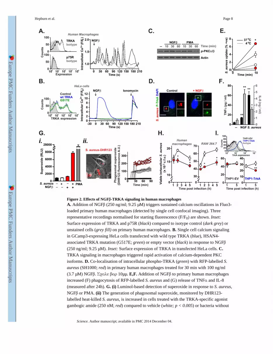
Figure 2. Effects of NGFβ-TRKA signaling in human macrophagesA. Addition of NGFβ (250 ng/ml; 9.25 μM) triggers sustained calcium oscillations in Fluo3-
loaded primary human macrophages (detected by single cell confocal imaging). Three
representative recordings normalised for starting fluorescence (F/F0) are shown. Inset:
Surface expression of TRKA and p75R (black) compared to isotype control (dark grey) or
unstained cells (grey fill) on primary human macrophages. B. Single cell calcium signaling
in GCamp3-expressing HeLa cells transfected with wild type TRKA (blue), HSAN4-
associated TRKA mutation (G517E; green) or empty vector (black) in response to NGFβ
(250 ng/ml; 9.25 μM). Inset: Surface expression of TRKA in transfected HeLa cells. C. TRKA signaling in macrophages triggered rapid activation of calcium-dependent PKC
isoforms. D. Co-localisation of intracellular phospho-TRKA (green) with RFP-labelled S.
aureus (SH1000; red) in primary human macrophages treated for 30 min with 100 ng/ml
(3.7 μM) NGFβ. Σχαλε βαρ 10μμ. E,F. Addition of NGFβ to primary human macrophages
increased (F) phagocytosis of RFP-labelled S. aureus and (G) release of TNFα and IL-8
(measured after 24h). G. (i) Luminol-based detection of superoxide in response to S. aureus,
NGFβ or PMA. (ii) The generation of phagosomal superoxide, monitored by DHR123-
labelled heat-killed S. aureus, is increased in cells treated with the TRKA-specific agonist
gambogic amide (250 nM; red) compared to vehicle (white; p < 0.005) or bacteria without
Hepburn et al. Page 8
Science. Author manuscript; available in PMC 2014 December 04.
Europe PM
C Funders A
uthor Manuscripts
Europe PM
C Funders A
uthor Manuscripts
cells (black). Four representative fluorescence traces from individual cells are shown for
each group. H,I. TRKA activation (by gambogic amide; 250 nM) enhanced intracellular
killing of S. aureus in (H) primary human macrophages (left) and the mouse macrophage
cell line RAW 264.7 (right) and in (I) TRKA-transfected (blue), but not control (black)
THP-1 cells. Inset: surface TRKA expression in THP-1 cells transfected with TRKA (blue)
or empty vector (black) compared to isotype control (grey) and unstained cells (grey fill).
All experiments were carried out in at least triplicate and are representative of at least 3
independent repeats.
Hepburn et al. Page 9
Science. Author manuscript; available in PMC 2014 December 04.
Europe PM
C Funders A
uthor Manuscripts
Europe PM
C Funders A
uthor Manuscripts
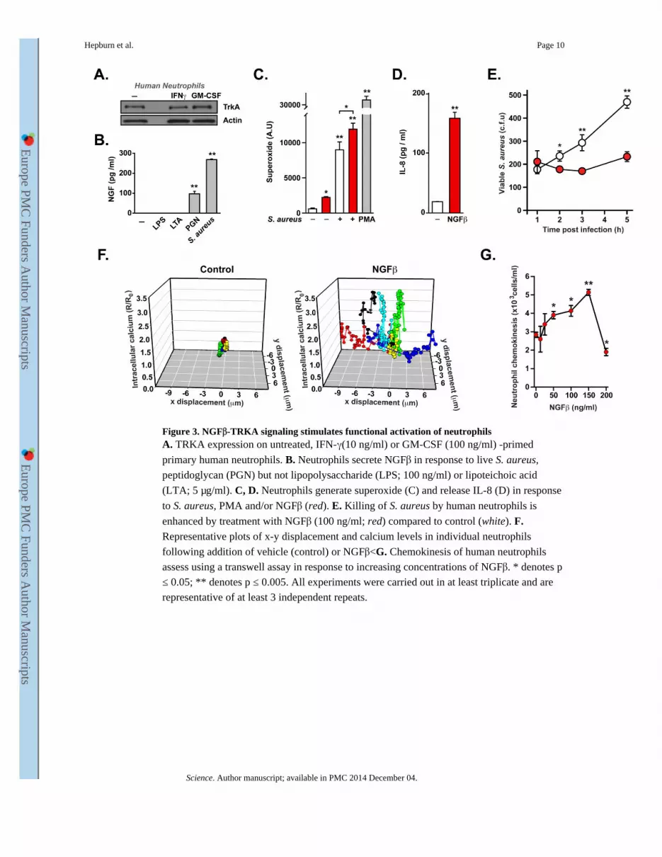
Figure 3. NGFβ-TRKA signaling stimulates functional activation of neutrophilsA. TRKA expression on untreated, IFN-γ(10 ng/ml) or GM-CSF (100 ng/ml) -primed
primary human neutrophils. B. Neutrophils secrete NGFβ in response to live S. aureus,
peptidoglycan (PGN) but not lipopolysaccharide (LPS; 100 ng/ml) or lipoteichoic acid
(LTA; 5 μg/ml). C, D. Neutrophils generate superoxide (C) and release IL-8 (D) in response
to S. aureus, PMA and/or NGFβ (red). E. Killing of S. aureus by human neutrophils is
enhanced by treatment with NGFβ (100 ng/ml; red) compared to control (white). F. Representative plots of x-y displacement and calcium levels in individual neutrophils
following addition of vehicle (control) or NGFβ<G. Chemokinesis of human neutrophils
assess using a transwell assay in response to increasing concentrations of NGFβ. * denotes p
≤ 0.05; ** denotes p ≤ 0.005. All experiments were carried out in at least triplicate and are
representative of at least 3 independent repeats.
Hepburn et al. Page 10
Science. Author manuscript; available in PMC 2014 December 04.
Europe PM
C Funders A
uthor Manuscripts
Europe PM
C Funders A
uthor Manuscripts
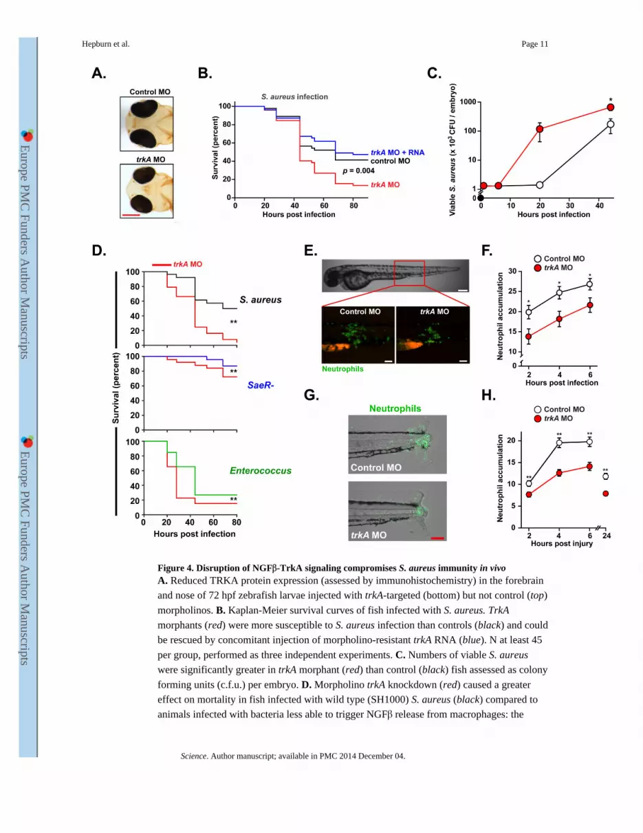
Figure 4. Disruption of NGFβ-TrkA signaling compromises S. aureus immunity in vivoA. Reduced TRKA protein expression (assessed by immunohistochemistry) in the forebrain
and nose of 72 hpf zebrafish larvae injected with trkA-targeted (bottom) but not control (top)
morpholinos. B. Kaplan-Meier survival curves of fish infected with S. aureus. TrkA
morphants (red) were more susceptible to S. aureus infection than controls (black) and could
be rescued by concomitant injection of morpholino-resistant trkA RNA (blue). N at least 45
per group, performed as three independent experiments. C. Numbers of viable S. aureus
were significantly greater in trkA morphant (red) than control (black) fish assessed as colony
forming units (c.f.u.) per embryo. D. Morpholino trkA knockdown (red) caused a greater
effect on mortality in fish infected with wild type (SH1000) S. aureus (black) compared to
animals infected with bacteria less able to trigger NGFβ release from macrophages: the
Hepburn et al. Page 11
Science. Author manuscript; available in PMC 2014 December 04.
Europe PM
C Funders A
uthor Manuscripts
Europe PM
C Funders A
uthor Manuscripts
saeR- S. aureus mutant (causing a mild infection; blue) and Enterococcus faecalis (causing a
severe infection; green).E-H. Reduced migration of GFP-tagged neutrophils to sites of S.
aureus infection (E,F) or sterile inflammation (G,H) in trkA morphants (red) compared to
controls (white). Representative images at 4h post infection (E; scale bar: brightfield 200
μm, fluorescence 100 μm) or tail injury (G; scale bar 100 μm). N at least 32 per group,
performed as 3 independent experiments. * denotes p ≤ 0.05; ** p ≤ 0.005 and *** p ≤
0.0005. Unless otherwise stated, data shown is representative of at least 3 independent
experiments.
Hepburn et al. Page 12
Science. Author manuscript; available in PMC 2014 December 04.
Europe PM
C Funders A
uthor Manuscripts
Europe PM
C Funders A
uthor Manuscripts