inhibition of superoxide production in human polymorphonuclear leukocytes by oral treponemal factors
TRANSCRIPT
Vol. 56, No. 3INFECTION AND IMMUNITY, Mar. 1988, p. 589-5940019-9567/88/030589-06$02.00/0Copyright X 1988, American Society for Microbiology
Inhibition of Superoxide Production in Human PolymorphonuclearLeukocytes by Oral Treponemal Factors
MICHAEL N. SELA,1* AARON WEINBERG,' RUTH BORINSKY,1 STANLEY C. HOLT,2AND THEODOR DISHON'
Department of Oral Biology, Hadassah Faculty of Dental Medicine, Hebrew University, Jerusalem, Israel 91010,1 and theDepartment of Periodontics, The University of Texas Health Science Center at San Antonio, San Antonio, Texas 782842
Received 5 June 1987/Accepted 15 November 1987
The inhibition of superoxide (02-) production by human peripheral blood polymorphonuclear leukocytes(PMNs) in the presence of oral treponemes, their cellular components, and their culture supernatants was
investigated. Superoxide production was inhibited 56% by a 25-,ug/ml phenol extract of a human clinicalisolate. Inhibition by culture supernatants of both the clinical isolate and a reference strain was related to thebacterial phase of growth and viability, though inhibition also persisted in the decline phase. Inhibition ofsuperoxide production was not evident when either opsonized or nonopsonized whole spirochetes were reactedwith PMNs. The suppressive activity depended, therefore, on the treponemes either being disrupted or growingand releasing the inhibitory factor into the culture medium. These results suggest that oral treponemes possessfactors which interfere with the activity of PMNs and thereby alter the inflammatory process in the diseasedperiodontal pocket.
Several studies have suggested that oral spirochetes havea contributory role in the pathogenesis of periodontal dis-eases. These studies include observations that spirochetesare found primarily on the apical surfaces of subgingivalplaque, in direct contact with the pocket epithelium andmigrating leukocytes (20), and that they invade the gingivalconnective tissue in acute necrotizing ulcerative gingivitis(19). The number of oral spirochetes increases dramaticallyin periodontally diseased, as opposed to nondiseased, sites(3, 21, 22), but it is markedly reduced after periodontaltreatment (24). It was also suggested in a study on themicroflora of chronic periodontitis that specific treponemesare among "the most suspect species" of the flora in thisdisease (27).With the advent of new culture techniques that facilitate
the isolation and growth of these fastidious microorganisms(2, 7, 18, 23, 36), a number of in vitro studies have beenperformed that show the effects of oral treponemes on avariety of host cells (4, 22, 34, 38). Moreover, proteolyticand fibrinolytic properties have been identified with oraltreponemes (15, 28, 41). Since polymorphonuclear leuko-cytes (PMNs) are the predominant host defense cells thatinteract with the pocket microflora (35, 39, 43), their specificfunctions after challenge with the potential pathogens (i.e.,phagocytosis, enzyme release, chemotaxis, etc.) are beinginvestigated. It is now becoming evident that the productionof free radicals by phagocytic cells plays an important role inthe killing of ingested microorganisms (1, 16). When PMNsare stimulated by bacteria, they undergo a respiratory burstcharacterized by increased oxygen consumption (1, 16).More than 90% of the oxygen consumed goes into thegeneration of superoxide free radicals (02-) (J. C. Fantoneand P. A. Ward, Current concepts, The Upjohn Co., p. 12,1985). Superoxide is the primary source of hydrogen perox-ide (H202) through spontaneous dismutation and through anenzyme-catalyzed dismutation with superoxide dismutase(10). Whereas 02 alone is not a potent bactericidal agent,
* Corresponding author.
H202 and its subsequent metabolites are a primary means bywhich phagocytes kill bacteria (1, 6, 16). Therefore, anymechanism inhibiting 02 production will directly affect theability of phagocytic cells to destroy bacteria.The purpose of the present study was to investigate the
interaction of PMNs with oral treponemes, their cellularcomponents, and their extracellular products in the processof oxygen metabolite production.
(This research was conducted by A. Weinberg in partialfulfillment of the requirements for the Ph.D. degree fromHebrew University, Jerusalem, Israel, 1987.)
MATERIALS AND METHODSBacterial samples. Samples of human subgingival fluid
were obtained from periodontal pockets of at least 6 mm.After the pocket was isolated and the gingiva was dried,sterile paper points were inserted for approximately 15 s,removed, and placed into sterile capped vials that containedreduced transport fluid (0.3 ml per vial) (37). Within 5 min oftaking the samples, they were processed in a Coy anaerobicchamber (Coy Laboratory Products, Ann Arbor, Mich.) (seebelow).
Isolation of spirochetes. A 10-,u portion of the bacterialsample was placed on a 0.2-,um-pore-size Millipore filter disk(Millipore Corp., Bedford, Mass.) positioned on the surfaceof prereduced GM-1 agar plates containing 2 RI of rifampinper ml (18) (see below). The plates were incubated for 7 daysin an anaerobic chamber in an atmosphere of 5% H2, 10%C02, and 85% N2. The filters were then removed with sterileforceps, and the plates were allowed to incubate for anadditional 5 days; a sample of the hazy, cloudy spreadinggrowth was then removed from the periphery of the hazewith a sterile loop and placed in 5 ml of GM-1 broth. Thistube was vortexed for 45 s, and 100 [lI of the resultantsolution was spread with an L-shaped sterile glass rod overthe surface of a fresh GM-1 plate. This plate was incubatedfor 7 days, after which isolated colonies of treponemesappeared. Colonies of similar and dissimilar morphologywere isolated by conventional subculturing techniques. The
589
on August 26, 2015 by guest
http://iai.asm.org/
Dow
nloaded from

590 SELA ET AL.
colonies were subcultured, and isolated cells were cloned atleast four times. Purity was confirmed by dark-field andtransmission electron microscopy. The latter used a conven-
tional negative-stain technique with 1% phosphotungsticacid (14).
Media. The isolation of treponemes was performed on
both GM-1 (2) and NOS (18) media. The results reported inthe present study relate to a clinical isolate grown in GM-1medium. ATCC reference strain 35405 was grown in NOSmedium. Growth curve profiles were established using a
Klett-Summerson photoelectric colorimeter (Klett Manufac-turing Co., Long Island City, N.Y.). The cultures were
harvested after 15 days of growth and processed to yield thedifferent oral treponemal components.
Preparation of human leukocytes. Experiments were per-
formed with preparations containing approximately 70%neutrophils obtained from freshly drawn normal adult wholeblood in adenine, citrate, and dextrose, kindly provided bythe Hadassah Hospital Blood Bank, Jerusalem, Israel. Theleukocytes were separated by mixing 10 ml of blood with 1.5ml of 6% dextran (Sigma Chemical Co., St. Louis, Mo.;molecular weight, 225,000) in saline. Erythrocytes were
lysed by adding 3 ml of distilled water for 30 s and thenwashed with 0.8% saline. The washed leukocytes were
suspended in Hanks balanced salt solution buffered with 3mM HEPES (N-2-hydroxyethylpiperazine-N'-2-ethanesul-fonic acid; pH 7.33) or in Hanks balanced salt solution withHEPES to which 10 mM sodium azide (NaN3) was added(40). The leukocyte suspensions were kept in polypropylenetubes in crushed ice. The viability of each leukocyte harvestwas measured by the trypan blue exclusion technique and bythe amount of lactate dehydrogenase released from leuko-cytes treated with and without the treponemal components.
Determination of superoxide (02-) Superoxide was mea-
sured as the superoxide dismutase-inhibitable cytochrome c
reduction by the method of Babior (1). Reaction mixturescontained 1.5 x 10⁶ leukocytes in 0.8 ml of Hanks balancedsalt solution with 10 mM NaN3 and the different spirochetalcomponents at various concentrations (see below) were
incubated for 10 min at 37°C. Thereafter, 50 RI of group Astreptococci (type 3, strain C203S) opsonized with histone(type II-A, 10,000 molecular weight; Sigma), cytochalasin B(2.5 ,ug/ml; Makor Chemicals, Jerusalem, Israel) (11), and 80,uM cytochrome c (type III; Sigma) were added and incu-bated for an additional 10 min at 37°C. Superoxide dismutase(30 ,ug/ml; Sigma) was added to the controls. Reactionmixtures also included preincubation of the various culturesupernatants with the challenge bacteria before or simulta-neously with the PMNs. The final volume for experimentaland control vials was 1 ml. The reaction mixtures were
centrifuged for 5 min at 1,000 x g, and the supematant fluidswere read at 575, 550, and 525 nm in a Kontron/Uvikon 810Pdouble-beam spectrophotometer. The concentration of su-
peroxide production was calculated from the formula E550 =2.1 x 10⁻⁴ M-1 by the method of Babior (1) and expressedas nanomoles per 106 cells per 10 min. The inhibition of 02-production, observed after incubation of the different spiro-chetal components with the leukocytes, was expressed as
the percentage of 02 produced when leukocytes were
challenged with histone-opsonized streptococci alone.Determination of hydrogen peroxide (H202). H202 was
determined by the method of Thurman et al. (40). Thereaction mixtures were the same as those described forsuperoxide determination, but without cytochrome c andsuperoxide dismutase. Briefly, after incubation of 1.5 x 10⁶leukocytes in 0.8 ml of Hanks balanced salt solution with 10
mM NaN3 for 10 min at 37°C with the different spirochetalcomponents, 50 ,ul of histone-opsonized streptococci and 2.5,ug of cytochalasin B per ml were added to a final volume of1 ml and incubated for an additional 10 min at 37°C.Thereafter, 200 [lI of 30% trichloroacetic acid was added toeach test tube. The supernatants were transferred to cleantest tubes, and 200 RId of FeNH4 (SO4)2. 12H20 (19 mg/ml ofwater) and 100 [lI of 25% KSCN were then added. Thecontents of the tubes were thoroughly mixed, incubated for5 min at room temperature, and centrifuged at 1,000 x g for5 min. The brown-red color which developed was read in thespectrophotometer at 480 nm. Results were expressed asnanomoles of H202 per 1.5 x 10⁶ leukocytes per 10 min,according to a standard curve of H202. The inhibition ofH202 production obtained by incubating the different spiro-chetal components with the leukocytes was expressed as apercentage of H202 produced when leukocytes were chal-lenged with streptococci opsonized by histone alone.
Extraction of oral treponemal components. Growth me-dium supernatant (GMS) was separated from the bacterialpellet after various growth periods by centrifugation at10,000 x g for 30 min. Growth medium kept in the Coyanaerobic chamber for the same number of days underwentsimilar centrifugation. GMS and growth medium wereheated at 56 and 98°C (boiling) for 30 min and treated withtrypsin in an initial attempt to characterize the °2 inhibi-tory factor found in the treponemal supernatants.The bacterial pellet was washed and suspended in 0.5 ml
of normal saline at an optical density of 200 Klett units perml, read at 540 nm. The suspension was then opsonized at37°C for 30 min with fresh normal human serum at a finalconcentration of 10% (vol/vol).
Sonic extracts of the two oral treponemes and of Actino-bacillus actinomycetemcomitans Y4 and Capnocytophagasputigena 4 were obtained by disrupting lyophilized bacteriasuspended in 4 ml of saline with a Sonicator cell disruptor(Heat Systems Ultrasonics, Inc., Plainview, N.Y.). In eachcase, samples were examined by dark-field microscopy toensure homogeneous sonication. The sonicates were thencentrifuged at 10,000 x g for 30 min, and the supernatantswere dialyzed against double-distilled water for 3 days withwater changes twice daily. Lyophilization was performed,followed by protein content analysis with the Bio-Rad pro-tein kit (Bio-Rad, Chemical Div., Richmond, Calif.).
Phenol extracts of the two oral treponemes and lipopoly-saccharides (LPSs) of the two nontreponemal microorgan-isms were obtained by the method of Nowotny (29). Briefly,lyophilized treponemes were mixed with an equal volume ofdistilled water and 90% phenol in a 70°C water bath. Aftercentrifugation at 3,000 x g for 30 min, the upper phase wasseparated and the extraction procedure was repeated twice.The water phases were pooled and dialyzed against distilledwater in the cold room for 3 days. After lyophilization, thecrude extract was dissolved in cold methanol that contained20% MgCI2 in ethanol. The preparation was left standing ina cold room for 1 to 3 days for a precipitate to develop. Aftercentrifugation at 3,000 x g for 30 min, the sediment wasdissolved in distilled water that contained cold methanolwithout MgCl2. After standing again in the cold room for 1 to3 days, the sediment was centrifuged, and dissolution wascarried out for a third time as described above. The finalsediment was dissolved in distilled water and lyophilized.This procedure was repeated to remove the methanol fromthe solution. For further purification, the extracts werecentrifuged at 100,000 to 110,000 x g for 2 h. Protein contentanalysis was done as described above.
INFECT. IMMUN.
on August 26, 2015 by guest
http://iai.asm.org/
Dow
nloaded from
PMN 02- INHIBITION BY TREPONEMAL FACTORS 591
Statistical analysis. All the data were analyzed using theStudent t test (25). A value of P c 0.05 was accepted assignificant.
RESULTS
The results presented in the present study are from one ofour clinical isolates, whose morphology is similar to that ofTreponema denticola, with an axial fibril configuration of2-4-2 and a protoplasmic cylinder width of 0.25 ,um. The T.denticola reference strain described here has a similar mor-phology.
Cell viability in response to spirochete extracts and super-natants. PMNs incubated with either sonic extracts, phenolextracts, or supernatants of the spirochetes tested showedvery little evidence of cell death. In all cases, no more than6% of the PMNs counted showed uptake of trypan blue, andlactate dehydrogenase release was no more than 10% in anygiven assay (control was 8.9%).
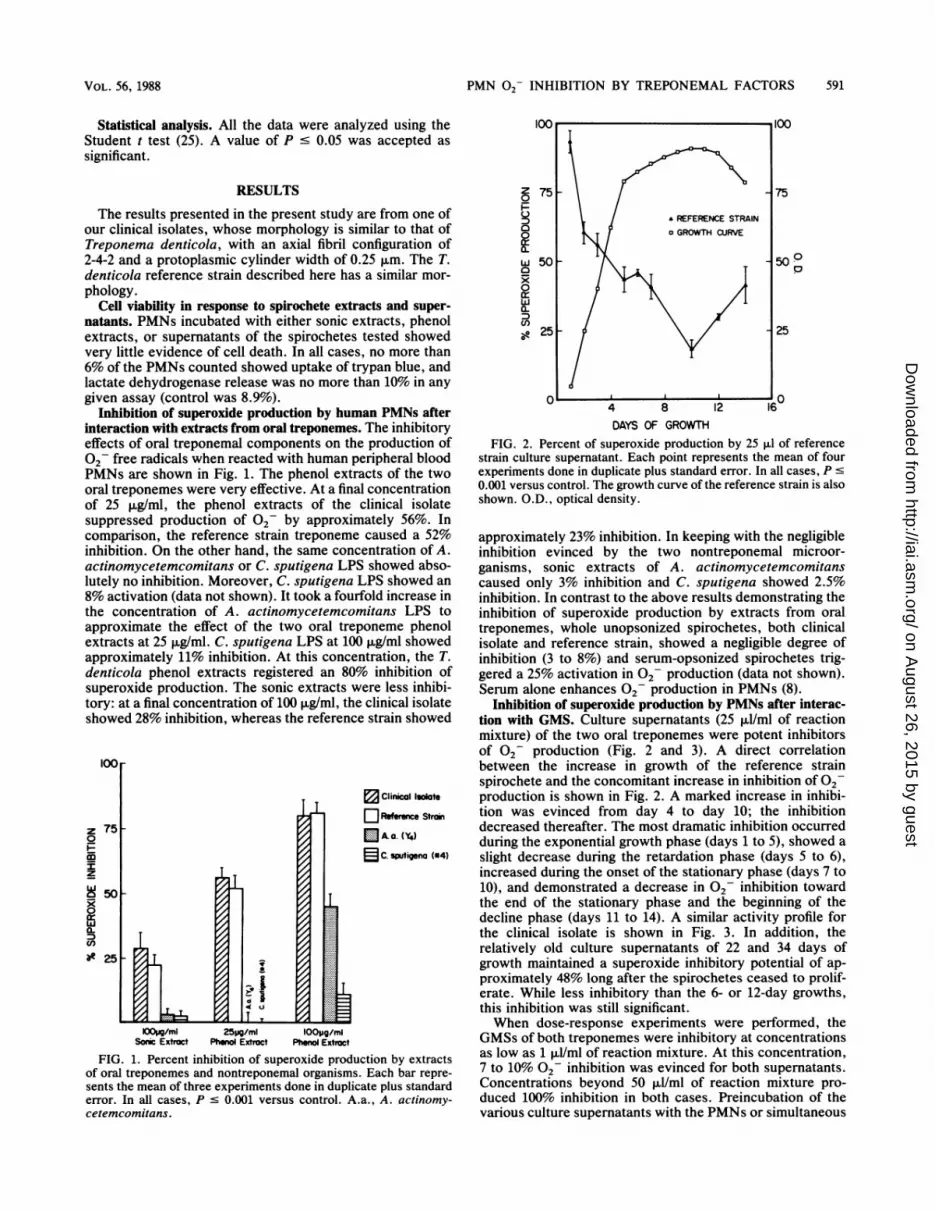
Inhibition of superoxide production by human PMNs afterinteraction with extracts from oral treponemes. The inhibitoryeffects of oral treponemal components on the production of02 free radicals when reacted with human peripheral bloodPMNs are shown in Fig. 1. The phenol extracts of the twooral treponemes were very effective. At a final concentrationof 25 ,ug/ml, the phenol extracts of the clinical isolatesuppressed production Of 02- by approximately 56%. Incomparison, the reference strain treponeme caused a 52%inhibition. On the other hand, the same concentration of A.actinomycetemcomitans or C. sputigena LPS showed abso-lutely no inhibition. Moreover, C. sputigena LPS showed an8% activation (data not shown). It took a fourfold increase inthe concentration of A. actinomycetemcomitans LPS toapproximate the effect of the two oral treponeme phenolextracts at 25 ,ug/ml. C. sputigena LPS at 100 ,ug/ml showedapproximately 11% inhibition. At this concentration, the T.denticola phenol extracts registered an 80% inhibition ofsuperoxide production. The sonic extracts were less inhibi-tory: at a final concentration of 100 ,ug/ml, the clinical isolateshowed 28% inhibition, whereas the reference strain showed
IOOr
z rDOT0I
z
50
0.
CLcn
?e 25
i-
IF 1
I10 Clinical Isolate
Referene Stroin
EJA.a. (Y4)
2 C. sputigena (#4)
lOolAg/ml 25Mg/mi lOOpg/mlSonic Extract Phenol Extroct Phenol Extroct
FIG. 1. Percent inhibition of superoxide production by extractsof oral treponemes and nontreponemal organisms. Each bar repre-sents the mean of three experiments done in duplicate plus standarderror. In all cases, P - 0.001 versus control. A.a., A. actinomy-cetemcomitans.
z0
wacx0wa-(I)
75
50 _
25 -
4 8 12DAYS OF GROWTH
75
50Pp
25
16
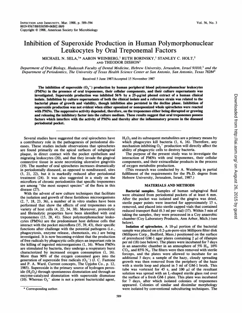
FIG. 2. Percent of superoxide production by 25 ,ul of referencestrain culture supernatant. Each point represents the mean of fourexperiments done in duplicate plus standard error. In all cases, P s0.001 versus control. The growth curve of the reference strain is alsoshown. O.D., optical density.
approximately 23% inhibition. In keeping with the negligibleinhibition evinced by the two nontreponemal microor-ganisms, sonic extracts of A. actinomycetemcomitanscaused only 3% inhibition and C. sputigena showed 2.5%inhibition. In contrast to the above results demonstrating theinhibition of superoxide production by extracts from oraltreponemes, whole unopsonized spirochetes, both clinicalisolate and reference strain, showed a negligible degree ofinhibition (3 to 8%) and serum-opsonized spirochetes trig-gered a 25% activation in 02 production (data not shown).Serum alone enhances 02 production in PMNs (8).
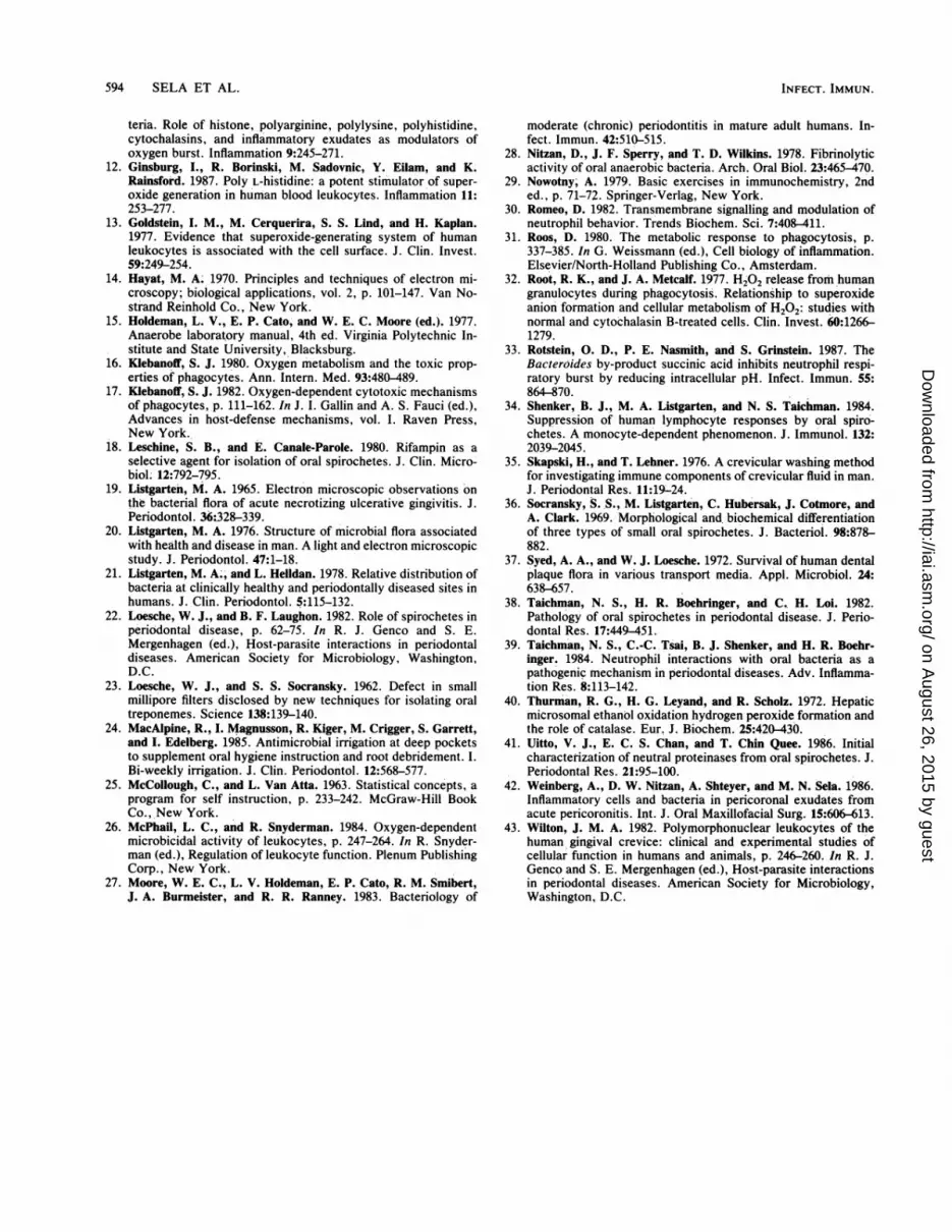
Inhibition of superoxide production by PMNs after interac-tion with GMS. Culture supernatants (25 ,ul/ml of reactionmixture) of the two oral treponemes were potent inhibitorsof 02 production (Fig. 2 and 3). A direct correlationbetween the increase in growth of the reference strainspirochete and the concomitant increase in inhibition of 02production is shown in Fig. 2. A marked increase in inhibi-tion was evinced from day 4 to day 10; the inhibitiondecreased thereafter. The most dramatic inhibition occurredduring the exponential growth phase (days 1 to 5), showed aslight decrease during the retardation phase (days 5 to 6),increased during the onset of the stationary phase (days 7 to10), and demonstrated a decrease in 02 inhibition towardthe end of the stationary phase and the beginning of thedecline phase (days 11 to 14). A similar activity profile forthe clinical isolate is shown in Fig. 3. In addition, therelatively old culture supernatants of 22 and 34 days ofgrowth maintained a superoxide inhibitory potential of ap-proximately 48% long after the spirochetes ceased to prolif-erate. While less inhibitory than the 6- or 12-day growths,this inhibition was still significant.When dose-response experiments were performed, the
GMSs of both treponemes were inhibitory at concentrationsas low as 1 ul/ml of reaction mixture. At this concentration,7 to 10% 02 inhibition was evinced for both supernatants.Concentrations beyond 50 ,ul/ml of reaction mixture pro-duced 100% inhibition in both cases. Preincubation of thevarious culture supernatants with the PMNs or simultaneous
VOL. 56, 1988
-7C. L
;F
;j, .;d I.0u
T 11
on August 26, 2015 by guest
http://iai.asm.org/
Dow
nloaded from

592 SELA ET AL.
z
0
()
w
0
w
0Ne
751-
501-
251-
5 10 15 20 2
DAYS OF GROWTH
90g
80F
70
P 60M
z50
cm 40
e30
20
10
5 30 35
FIG. 3. Percent of superoxide production by 25 >1. of clinicalisolate culture supernatant. Each point represents the mean of fourexperiments done in duplicate plus standard error. In all cases, P <
0.001 versus control.
in isolate
Refrence Stroin
A.a. (Y4)
C. sputigeno (*4)
IF
I
K)Opg/ml 25pg/ml ioopg/mlSonic Extoct Phenol Extroct Phenol Extroct
FIG. 4. Percent inhibition of H202 production by extracts of oraltreponemes and nontreponemal organisms. Each bar represents themean of three experiments done in duplicate plus standard error. Inall cases, P - 0.001 versus control. A.a., A. actinomycetemcomi-tans.
incubation with the reaction mixture components gave sim-ilar results. However, when opsonized streptococci werepreincubated with the various culture supernatants, washed,and reacted with PMNs, no inhibition of 02 production wasobserved.H202 inhibition by oral treponemal extracts and GMS.
Profiles of H202 production by peripheral blood PMNstreated with the spirochetal extracts and GMS indicated aninhibitory trend. Table 1 shows that on days 9, 14, and 16,the clinical isolate GMS was most inhibitory, approximating50%. The reference strain was less inhibitory, with a meaninhibition of 36% for the same days of growth. However,when all data from days 3 through 28 were examinedtogether, the mean inhibitions of H202 by both oral trepo-neme GMSs, 32% for the reference strain and 36% for theclinical isolate, was quite similar. Moreover, relatively oldGMS (28 days) maintained H202 inhibitory potentials for theclinical isolate and the reference strain of 38% and 28%,respectively. The only effective inhibition of H202 produc-tion by the spirochetal extracts was evinced by their phenolextracts at a final concentration of 100 jig/ml (Fig. 4). At thisconcentration, the clinical isolate produced a 41.5% inhibi-tion, whereas the reference strain showed a 35% inhibition.However, in relation to the two nontreponemal microorgan-isms, the two oral treponemes continued to demonstrateconsistently more inhibition of free-radical production (Fig.1 and 4).Heating and trypsinization of the culture supernatants.
Initial results, after the actively inhibitory culture superna-
tants of both T. denticola strains were heated at 56°C for 30min and boiled for 30 min, showed that the factor probablyresponsible for the inhibition of superoxide production isthermostable. On the other hand, trypsinization of theseculture supernatants reduced inhibitory activity by 60%.
DISCUSSION
The results of this study suggest the presence of an oraltreponemal factor (OTF) which inhibits 02 production inhuman peripheral blood PMNs without any detrimentaleffect on PMN viability. Inhibition of superoxide produc-tion, while observed to some degree with the treponemalsonic extracts, was demonstrated mainly with the phenolextract and the culture supernatants. On the other hand, wefound no inhibitory activity when PMNs were incubatedwith whole opsonized or nonopsonized spirochetes. Thesuppressive activity, therefore, depended on the spirocheteseither being disrupted or growing and releasing the inhibitoryfactor into the medium. The increased suppressive effect on02 production was initially directly related to oral trepo-nemal growth and persisted without the need for treponemalviability (Fig. 2 and 3), even in the decline phase (22 and 35days; Fig. 3). While lower than suppression during the earlystages of growth, this suppression in 02 production pla-teaued to a significant 48%. These findings suggest (i) thatthe OTF found in the culture supernatant is released by thespirochetes during the exponential growth phase, (ii) thatOTF activity is not lost from the medium even during the
TABLE 1. Percent inhibition of H202 by reference strain and clinical isolate treponeme GMSsa
% Inhibition' at day: MeanStrain cumulative
3 5 7 9 12 14 16 19 28 % inhibition
Reference 29.8 + 3.0 31.0 + 2.0 20.0 + 7.0 32.3 + 4.0 31.0 ± 3.5 40.0 ± 2.0 36.0 ± 4.5 42.0 ± 4.5 28.0 ± 4.0 32.2 ± 3.8strain
Clinical 26.5 ± 4.2 17.0 ± 7.0 21.3 ± 3.0 49.4 ± 5.0 34.0 ± 3.0 57.0 ± 5.2 48.4 ± 6.2 36.0 ± 3.5 37.9 ± 5.4 36.0 ± 4.7isolatea Mean ± standard error of four experiments done in duplicate. P s 0.001 versus control by the Student t test (25).
o CLINICAL ISOLATE
INFECT. IMMUN.
loor
on August 26, 2015 by guest
http://iai.asm.org/
Dow
nloaded from

PMN 02- INHIBITION BY TREPONEMAL FACTORS 593
decline phase, and (iii) that the activity found during thedecline phase could be due to components of the spiro-chetes, e.g., LPS-like macromolecules, which are liberatedonly during and after the disruption of the bacteria. Thisphenomenon is being further investigated.
Since in initial experiments each culture supernatant waspreincubated with PMNs before challenge with opsonizedstreptococci, it was postulated that the site for OTF activitywas the PMN itself. To exclude the possibility that theinhibition could have been due to attachment of OTF to thehistone-opsonized streptococci, thereby affecting receptorrecognition on the PMN surface membrane, the variousculture supernatants were preincubated with the challengingbacteria (followed by washing) before reacting with thePMNs. The results showed that there was no interference in02- production by the supernatants, indicating that theinhibitory effect was not a result of OTF affinity for theopsonized streptococci, but rather of affinity for the PMNitself. Furthermore, pretreatment of PMNs with GMSs wasnot a prerequisite for the inhibition of 02- production.Simultaneous incubation of PMNs, GMS, and opsonizedstreptococci caused an inhibition of 02 production of thesame magnitude as when PMNs were pretreated with culturesupernatant. This shows that OTF is capable of competitiveinhibition with potential bacterial stimulants, a situationwhich could be of pathogenic importance in the periodontalpocket.The current consensus is that 02 iS formed by a one-
electron reduction of molecular oxygen and is then dismu-tated to H202, with a theoretical quantitative ratio of two02 to one H202 (1, 13, 17, 26, 30-32). Our results indicatethat in some cases there was inhibition approximating thisratio (Fig. 1 and 4; sonic and phenol extracts of 100 ,ug/ml forthe oral treponemes). However, in other cases this ratio wasnot upheld (Fig. 1 and 4 phenol extracts of 25 ,ug/ml for theoral treponemes and phenol extracts for A. actinomycetem-comitans and C. sputigena). Moreover, this ratio was notalways maintained in the inhibition of H202 production bythe two oral treponemal culture supernatants when com-pared with superoxide inhibition (Fig. 2 and 3, Table 1).These discrepancies are not fully understood. However, wedid notice that variability in donor PMN responses to thesame stimuli was most pronounced in those cases whereinhibition of 02 was less than 20%. Moreover, not allstimuli of the respiratory burst induce the generation ofequivalent amounts of superoxide and hydrogen peroxide (9,13, 31, 32). Furthermore, it was recently demonstrated thatmuch larger amounts of H202 than 2- were produced byhistone-opsonized streptococci (12), suggesting that lessinhibition of H202 in relation to 02- would ensue if aninhibitory agent were employed. It has been further sug-gested that, in histone-opsonized streptococci, peroxidemight not only originate by dismutation from 02, but mightalso be generated by a two-electron reduction of molecularoxygen by pathways analogous to those of glucose oxidase(12). These observations could explain in part the in vitrodiscrepancy in those cases where the ratio of inhibition didnot approximate 2:1.
It has recently been suggested that succinic acid, a small-molecular-weight fatty acid and a major by-product of Bac-teroides metabolism, is responsible for inhibiting superoxideproduction in PMNs (33). Our preliminary results of thecharacterization of the OTF seem to indicate a thermostableproteinlike substance responsible for the inhibition of super-oxide production. Moreover, succinic acid was effective ininhibiting the neutrophil respiratory burst only at pH 5.5
(33), while the OTF is effective at neutral pH. Furthermore,succinic acid is not a major metabolic by-product of T.denticola (5). Further work on the isolation and characteri-zation of the OTF is under way.
Disease-related properties of oral treponemes have beendetermined in vitro in recent years. T. denticola possessesproteolytic (15) and fibrinolytic properties (28), has a tryp-sinlike activity (22, 41), suppresses lymphocyte blastogen-esis by mitogens (34), inhibits fibroblast proliferation (4),and, together with T. vincentii, is cytotoxic to epithelial cells(P. Baehni and G. Cimasoni, J. Dent. Res. 65:767, abstr. no.372, 1986). Recently, it was suggested that oral spirochetes"may limit fusion of lysosomes to phagosomes" by demon-strating a significant decrease in zymosan-induced degranu-lation when PMNs were pretreated with soluble extracts ofspirochetes (3). Moreover, recent data on acute pericoronitisdemonstrated that, although spirochetes are the predomi-nant microorganisms in the exudates, they are not observedbeing phagocytosed by PMNs (42). In addition, whereasphagocytosis of other bacteria by PMNs was observed in allthe exudates, lysis of the host PMNs was seen withouteradication of the ingested bacteria (42). These in vivoobservations, together with the in vitro results of Boehringeret al. (3), suggest that oral treponemes may interfere withongoing neutralization of microorganisms by human PMNs.The results presented here further strengthen this conten-tion. The significant inhibition of 02 production by the OTFand other oral treponemal components may alter the abilityof PMNs to dispose of other noxious microorganisms,thereby enhancing the periodontal disease process. There-fore, it is important to determine the OTF mechanism ofaction and to learn whether it affects other cellular activities.
ACKNOWLEDGMENT
We thank Ronit Naor for her technical assistance throughout thisstudy.
LITERATURE CITED1. Babior, B. M. 1984. Oxidants from phagocytes: agents of
defense and destruction. Blood 64:959-966.2. Blakemore, R. P., and E. Canale-Parola. 1976. Arginine catab-
olism by Treponema denticola. J. Bacteriol. 128:616-622.3. Boehringer, H. R., P. H. Berthold, and N. S. Taichman. 1986.
Studies on the interaction of human neutrophils with plaquespirochetes. J. Periodontal Res. 21:195-209.
4. Boehringer, H. R., N. S. Taichman, and B. J. Shenker. 1984.Suppression of fibroblast proliferation by oral spirochetes. In-fect. Immun. 45:155-159.
5. Canale-Parola, E. 1977. Physiology and evolution of spiro-chetes. Bacteriol. Rev. 41:181-204.
6. Chance, B., H. Sies, and A. Boveris. 1979. Hydroperoxidemetabolism in mammalian organs. Physiol. Rev. 59:527-605.
7. Cheng, S. I., and E. C. S. Chan. 1984. The routine isolation,growth and maintenance of the intermediate-size anaerobic oralspirochetes from periodontal pockets. J. Periodontal Res. 18:362-368.
8. Curnutte, J. T., and B. M. Babior. 1974. Biological defensemechanisms, the effect of bacteria and serum on superoxideproduction by granulocytes. J. Clin. Invest. 53:1662-1672.
9. Curnutte, J. T., and A. I. Tauber. 1983. Failure to detectsuperoxide in human neutrophils stimulated with latex particles.Pediatr. Res. 17:281-284.
10. Fee, J. A., and J. S. Valentine. 1977. Chemical and physicalproperties of superoxide, p. 19-66. In A. M. Michelson, J. M.McCoral, and I. Fridovich (ed.), Superoxide and superoxidedismutases. Academic Press, Inc., New York.
11. Ginsburg, I., R. Borinski, D. Malamud, F. Struckmayer, and V.Klimetzek. 1985. Chemiluminescence and superoxide genera-tion by leukocytes stimulated by polyelectrolyte-opsonized bac-
VOL. 56, 1988
on August 26, 2015 by guest
http://iai.asm.org/
Dow
nloaded from
594 SELA ET AL.
teria. Role of histone, polyarginine, polylysine, polyhistidine,cytochalasins, and inflammatory exudates as modulators ofoxygen burst. Inflammation 9:245-271.
12. Ginsburg, I., R. Borinski, M. Sadovnic, Y. Eilam, and K.Rainsford. 1987. Poly L-histidine: a potent stimulator of super-oxide generation in human blood leukocytes. Inflammation 11:253-277.
13. Goldstein, I. M., M. Cerquerira, S. S. Lind, and H. Kaplan.1977. Evidence that superoxide-generating system of humanleukocytes is associated with the cell surface. J. Clin. Invest.59:249-254.
14. Hayat, M. A. 1970. Principles and techniques of electron mi-croscopy; biological applications, vol. 2, p. 101-147. Van No-strand Reinhold Co., New York.
15. Holdeman, L. V., E. P. Cato, and W. E. C. Moore (ed.). 1977.Anaerobe laboratory manual, 4th ed. Virginia Polytechnic In-stitute and State University, Blacksburg.
16. Klebanoff, S. J. 1980. Oxygen metabolism and the toxic prop-erties of phagocytes. Ann. Intern. Med. 93:480-489.
17. Klebanoff, S. J. 1982. Oxygen-dependent cytotoxic mechanismsof phagocytes, p. 111-162. In J. I. Gallin and A. S. Fauci (ed.),Advances in host-defense mechanisms, vol. I. Raven Press,New York.
18. Leschine, S. B., and E. Canale-Parole. 1980. Rifampin as aselective agent for isolation of oral spirochetes. J. Clin. Micro-biol. 12:792-795.
19. Listgarten, M. A. 1965. Electron microscopic observations onthe bacterial flora of acute necrotizing ulcerative gingivitis. J.Periodontol. 36:328-339.
20. Listgarten, M. A. 1976. Structure of microbial flora associatedwith health and disease in man. A light and electron microscopicstudy. J. Periodontol. 47:1-18.
21. Listgarten, M. A., and L. Helidan. 1978. Relative distribution ofbacteria at clinically healthy and periodontally diseased sites inhumans. J. Clin. Periodontol. 5:115-132.
22. Loesche, W. J., and B. F. Laughon. 1982. Role of spirochetes inperiodontal disease, p. 62-75. In R. J. Genco and S. E.Mergenhagen (ed.), Host-parasite interactions in periodontaldiseases. American Society for Microbiology, Washington,D.C.
23. Loesche, W. J., and S. S. Socransky. 1962. Defect in smallmillipore filters disclosed by new techniques for isolating oraltreponemes. Science 138:139-140.
24. MacAlpine, R., I. Magnusson, R. Kiger, M. Crigger, S. Garrett,and I. Edelberg. 1985. Antimicrobial irrigation at deep pocketsto supplement oral hygiene instruction and root debridement. I.Bi-weekly irrigation. J. Clin. Periodontol. 12:568-577.
25. McCollough, C., and L. Van Atta. 1963. Statistical concepts, aprogram for self instruction, p. 233-242. McGraw-Hill BookCo., New York.
26. McPhail, L. C., and It. Snyderman. 1984. Oxygen-dependentmicrobicidal activity of leukocytes, p. 247-264. In R. Snyder-man (ed.), Regulation of leukocyte function. Plenum PublishingCorp., New York.
27. Moore, W. E. C., L. V. Holdeman, E. P. Cato, R. M. Smibert,J. A. Burmeister, and R. R. Ranney. 1983. Bacteriology of
moderate (chronic) periodontitis in mature adult humans. In-fect. Immun. 42:510-515.
28. Nitzan, D., J. F. Sperry, and T. D. Wilkins. 1978. Fibrinolyticactivity of oral anaerobic bacteria. Arch. Oral Biol. 23:465-470.
29. Nowotny, A. 1979. Basic exercises in immunochemistry, 2nded., p. 71-72. Springer-Verlag, New York.
30. Romeo, D. 1982. Transmembrane signalling and modulation ofneutrophil behavior. Trends Biochem. Sci. 7:408-411.
31. Roos, D. 1980. The metabolic response to phagocytosis, p.337-385. In G. Weissmann (ed.), Cell biology of inflammation.Elsevier/North-Holland Publishing Co., Amsterdam.
32. Root, R. K., and J. A. Metcalf. 1977. H202 release from humangranulocytes during phagocytosis. Relationship to superoxideanion formation and cellular metabolism of H202: studies withnormal and cytochalasin B-treated cells. Clin. Invest. 60:1266-1279.
33. Rotstein, 0. D., P. E. Nasmith, and S. Grinstein. 1987. TheBacteroides by-product succinic acid inhibits neutrophil respi-ratory burst by reducing intracellular pH. Infect. Immun. 55:864-870.
34. Shenker, B. J., M. A. Listgarten, and N. S. Taichman. 1984.Suppression of human lymphocyte responses by oral spiro-chetes. A monocyte-dependent phenomenon. J. Immunol. 132:2039-2045.
35. Skapski, H., and T. Lehner. 1976. A crevicular washing methodfor investigating immune components of crevicular fluid in man.J. Periodontal Res. 11:19;-24.
36. Socransky, S. S., M. Listgarten, C. Hubersak, J. Cotmore, andA. Clark. 1969. Morphological and, biochemical differentiationof three types of small oral spirochetes. J. Bacteriol. 98:878-882.
37. Syed, A. A., and W. J. Loesche. 1972. Survival of human dentalplaque flora in various transport media. Appl. Microbiol. 24:638-657.
38. Taichman, N. S., H. R. Boehringer, and C., H. Loi. 1982.Pathology of oral spirochetes in periodontal disease. J. Perio-dontal Res. 17:449-451.
39. Taichman, N. S., C.-C. Tsai, B. J. Shenker, and H. R. Boehr-inger. 1984. Neutrophil interactions with oral bacteria as apathogenic mechanism in periodontal diseases. Adv. Inflamma-tion Res. 8:113-142.
40. Thurman, R. G., H. G. Leyand, and R. Scholz. 1972. Hepaticmicrosomal ethanol oxidation hydrogen peroxide formation andthe role of catalase. Eur. J. Biochem. 25:420-430.
41. Uitto, V. J., E. C. S. Chan, and T. Chin Quee. 1986. Initialcharacterization of neutral proteinases from oral spirochetes. J.Periodontal Res. 21:95-100.
42. Weinberg, A., D. W. Nitzan, A. Shteyer, and M. N. Sela. 1986.Inflammatory cells and bacteria in pericoronal exudates fromacute pericoronitis. Int. J. Oral Maxillofacial Surg. 15:606-613.
43. Wilton, J. M. A. 1982. Polymorphonuclear leukocytes of thehuman gingival crevice: clinical and experimental studies ofcellular function in humans and animals, p. 246-260. In R. J.Genco and S. E. Mergenhagen (ed.), Host-parasite interactionsin periodontal diseases. American Society for Microbiology,Washington, D.C.
INFECT. IMMUN.
on August 26, 2015 by guest
http://iai.asm.org/
Dow
nloaded from