digestive vacuoles of plasmodium falciparum are selectively phagocytosed by and impair killing...
TRANSCRIPT
PHAGOCYTES, GRANULOCYTES, AND MYELOPOIESIS
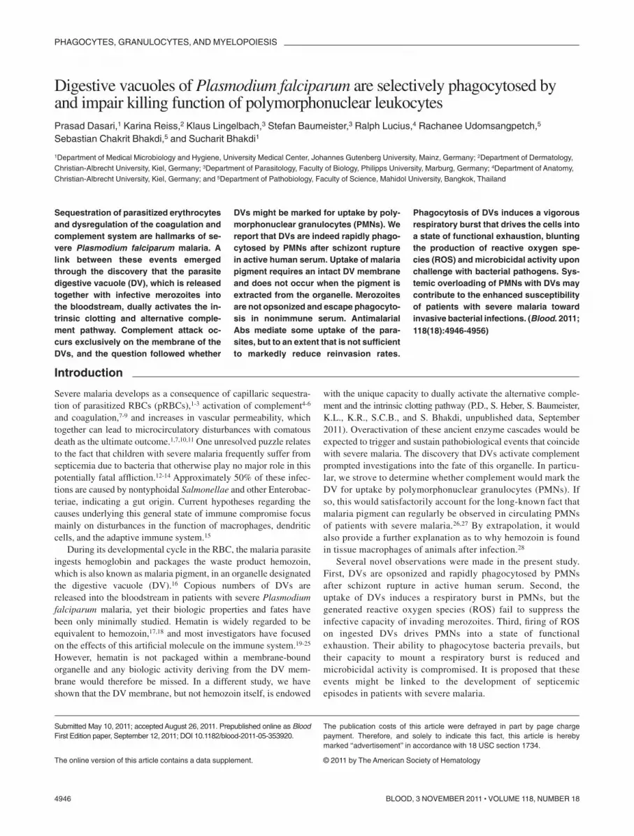
Digestive vacuoles of Plasmodium falciparum are selectively phagocytosed byand impair killing function of polymorphonuclear leukocytesPrasad Dasari,1 Karina Reiss,2 Klaus Lingelbach,3 Stefan Baumeister,3 Ralph Lucius,4 Rachanee Udomsangpetch,5
Sebastian Chakrit Bhakdi,5 and Sucharit Bhakdi1
1Department of Medical Microbiology and Hygiene, University Medical Center, Johannes Gutenberg University, Mainz, Germany; 2Department of Dermatology,Christian-Albrecht University, Kiel, Germany; 3Department of Parasitology, Faculty of Biology, Philipps University, Marburg, Germany; 4Department of Anatomy,Christian-Albrecht University, Kiel, Germany; and 5Department of Pathobiology, Faculty of Science, Mahidol University, Bangkok, Thailand
Sequestration of parasitized erythrocytesand dysregulation of the coagulation andcomplement system are hallmarks of se-vere Plasmodium falciparum malaria. Alink between these events emergedthrough the discovery that the parasitedigestive vacuole (DV), which is releasedtogether with infective merozoites intothe bloodstream, dually activates the in-trinsic clotting and alternative comple-ment pathway. Complement attack oc-curs exclusively on the membrane of theDVs, and the question followed whether
DVs might be marked for uptake by poly-morphonuclear granulocytes (PMNs). Wereport that DVs are indeed rapidly phago-cytosed by PMNs after schizont rupturein active human serum. Uptake of malariapigment requires an intact DV membraneand does not occur when the pigment isextracted from the organelle. Merozoitesare not opsonized and escape phagocyto-sis in nonimmune serum. AntimalarialAbs mediate some uptake of the para-sites, but to an extent that is not sufficientto markedly reduce reinvasion rates.
Phagocytosis of DVs induces a vigorousrespiratory burst that drives the cells intoa state of functional exhaustion, bluntingthe production of reactive oxygen spe-cies (ROS) and microbicidal activity uponchallenge with bacterial pathogens. Sys-temic overloading of PMNs with DVs maycontribute to the enhanced susceptibilityof patients with severe malaria towardinvasive bacterial infections. (Blood. 2011;118(18):4946-4956)
Introduction
Severe malaria develops as a consequence of capillaric sequestra-tion of parasitized RBCs (pRBCs),1-3 activation of complement4-6
and coagulation,7-9 and increases in vascular permeability, whichtogether can lead to microcirculatory disturbances with comatousdeath as the ultimate outcome.1,7,10,11 One unresolved puzzle relatesto the fact that children with severe malaria frequently suffer fromsepticemia due to bacteria that otherwise play no major role in thispotentially fatal affliction.12-14 Approximately 50% of these infec-tions are caused by nontyphoidal Salmonellae and other Enterobac-teriae, indicating a gut origin. Current hypotheses regarding thecauses underlying this general state of immune compromise focusmainly on disturbances in the function of macrophages, dendriticcells, and the adaptive immune system.15
During its developmental cycle in the RBC, the malaria parasiteingests hemoglobin and packages the waste product hemozoin,which is also known as malaria pigment, in an organelle designatedthe digestive vacuole (DV).16 Copious numbers of DVs arereleased into the bloodstream in patients with severe Plasmodiumfalciparum malaria, yet their biologic properties and fates havebeen only minimally studied. Hematin is widely regarded to beequivalent to hemozoin,17,18 and most investigators have focusedon the effects of this artificial molecule on the immune system.19-25
However, hematin is not packaged within a membrane-boundorganelle and any biologic activity deriving from the DV mem-brane would therefore be missed. In a different study, we haveshown that the DV membrane, but not hemozoin itself, is endowed
with the unique capacity to dually activate the alternative comple-ment and the intrinsic clotting pathway (P.D., S. Heber, S. Baumeister,K.L., K.R., S.C.B., and S. Bhakdi, unpublished data, September2011). Overactivation of these ancient enzyme cascades would beexpected to trigger and sustain pathobiological events that coincidewith severe malaria. The discovery that DVs activate complementprompted investigations into the fate of this organelle. In particu-lar, we strove to determine whether complement would mark theDV for uptake by polymorphonuclear granulocytes (PMNs). Ifso, this would satisfactorily account for the long-known fact thatmalaria pigment can regularly be observed in circulating PMNsof patients with severe malaria.26,27 By extrapolation, it wouldalso provide a further explanation as to why hemozoin is foundin tissue macrophages of animals after infection.28
Several novel observations were made in the present study.First, DVs are opsonized and rapidly phagocytosed by PMNsafter schizont rupture in active human serum. Second, theuptake of DVs induces a respiratory burst in PMNs, but thegenerated reactive oxygen species (ROS) fail to suppress theinfective capacity of invading merozoites. Third, firing of ROSon ingested DVs drives PMNs into a state of functionalexhaustion. Their ability to phagocytose bacteria prevails, buttheir capacity to mount a respiratory burst is reduced andmicrobicidal activity is compromised. It is proposed that theseevents might be linked to the development of septicemicepisodes in patients with severe malaria.
Submitted May 10, 2011; accepted August 26, 2011. Prepublished online as BloodFirst Edition paper, September 12, 2011; DOI 10.1182/blood-2011-05-353920.
The online version of this article contains a data supplement.
The publication costs of this article were defrayed in part by page chargepayment. Therefore, and solely to indicate this fact, this article is herebymarked ‘‘advertisement’’ in accordance with 18 USC section 1734.
© 2011 by The American Society of Hematology
4946 BLOOD, 3 NOVEMBER 2011 � VOLUME 118, NUMBER 18

Methods
Cell preparation
P falciparum culture, isolation of digestive vacuoles and hemozoin, andstaining procedures for DNA and C3 were undertaken as describedpreviously (P.D., S.H., S. Baumeister, K.L., K.R., S.C.B., and S. Bhakdi,unpublished data, September 2011) and as detailed in supplementalMethods (available on the Blood Web site; see the Supplemental Materialslink at the top of the online article). Human PMNs were isolated fromheparinized blood of healthy volunteers using conventional procedures29
and were kept in PBS on ice until use. Surface labeling of PMNs wasperformed with PE-conjugated polyclonal rabbit anti–human CD16 IgG(BD Biosciences) following the manufacturer’s recommendation. Afterlabeling, 50 �L of Ab solution was added to 0.5 mL of cells (107 PMNs/mL)for 20 minutes at room temperature, and the cells were subsequentlywashed twice with PBS. By microscopic assessment, it was ascertained thatsurface labeling had no effect on phagocytic function. Erythrocytes, bankedhuman blood, and human sera, all group O, Rh�, were kindly provided byRoland Conradi and Walter Hitzler (Blood Transfusion Center of theUniversity of Mainz, Mainz, Germany). C8-deficient human serum wasprepared as described previously.30 Polyclonal rabbit anti–human C3 IgGwas obtained from Dakocytomation.
PMNs phagocytosis of DVs
PMNs suspended at 5 � 106 cells/mL in 10% active or heat-inactivatedserum were challenged with 3-5 DVs/cell and phagocytosis was analyzedby microscopy of Giemsa-stained smears. To obtain quantitative data, DVswere fluorescently labeled by 20 minutes of incubation with 7.5 �g/mLCellMask Deep Red plasma membrane stain (Invitrogen), washed twice,and added to the PMNs. After the given times at 37°C, 50 �L of the reactionmixture was removed, diluted in 500 �L of veronal buffered saline (VBS),and analyzed in a FACSScan flow cytometer (BD Biosciences).
Detection of ROS generation
Single-cell observations were conducted using PMNs loaded with 0.1�M2�,7�-dichlorodihydrofluorescein diacetate (Sigma-Aldrich) for 30 minutesat 37°C, and washed 4 times with PBS. Next, 0.2 mL of cell suspensions(3 � 106/mL in VBS with 10% active serum) were transferred to Eppendorftubes and DVs/PMNs at a ratio of 2:5 were added. Incubation wasconducted at 37°C. Smears were prepared every 2 minutes and viewed in anAxioskop 2 microscope (Carl Zeiss) at a 1000� magnification. Imageswere obtained using AxioVision software.
Luminol-based quantification of ROS
Experiments were performed in microtiter plates using a thermostatedluminometer (MicroLumat LB96P; Berthold Technologies). Each wellcontained 0.2 mL of PMNs (3 � 106/mL in VBS) with 10% active orinactive serum. Luminol (Sigma-Aldrich) was added to a final concentra-tion of 125�M to each well. DVs were applied at a ratio of 3:5 DVs/PMNsand chemiluminescence was measured every 4 minutes. In bacterialchallenge experiments, luminol was re-added after 60 minutes, a ratio of10:15 Staphylococcus aureus/PMNs was applied, and measurements werecontinued for another 60 minutes.
Assessment of uptake of DVs and merozoites by PMNs afterrupture of pRBCs
Synchronized enriched cultures containing 60%-70% late-stage pRBCswere diluted to 0.2% hematocrit in RPMI 1640 medium and maintainedat 37°C. Giemsa-stained smears were prepared every 30 minutes. Whenthe first schizont rupture was observed, cultures were continued foranother 30 minutes, during which time � 75% of the parasitized cellslysed. The culture was then centrifuged in a Beckman Coulter Allegra6KR centrifuge at 200g for 5 minutes to sediment unlysed cells. Thesupernatant was centrifuged at 3000g for 10 minutes in a Sorvall RC2Bcentrifuge to pellet the merozoites and DVs. The pellet was resuspended
in RPMI and centrifuged once again at low speed (400g) for 1 minute toremove any residual unlysed cells. The supernatant was then centrifugedat 17 000g for 2 minutes, washed 3 times, and stained with Hoechst33342. Isolated DVs and merozoites were added to Eppendorf tubes,each containing 3 � 106/mL of PMNs (prelabeled with PE-conjugatedpolyclonal rabbit anti–human CD16 IgG) in 0.2 mL of VBS with 10%active serum (3-5 DVs and 50-100 merozoites/PMNs). After 30 minutesat 37°C, thin smears were prepared and air dried, and Z-stacked imageswere taken in a fluorescence microscope (Axiovert 200M; Carl Zeiss)using AxioVision Version 4.7 software.
Infection experiments
Synchronized late-stage pRBCs were enriched to 55%-70% and suspendedat 0.4% hematocrit in RPMI with 20% active human serum; 0.5 mL wasapplied per well in a 24-well cell culture plate (Greiner Bio-One). To this,0.1 mL of 10% noninfected RBCs � 106 PMNs were added, and the totalvolume brought to 1 mL with RPMI to give a final ratio of PMNs:pRBCs:RBCs of � 1:10:100. After 24 hours, 0.2 mL of the culture was stained with100�M hydroethidine for 30 minutes at 37°C, and infection rates werequantified by flow cytometry.31 Slides were prepared and stained withGiemsa for microscopy.
Ab protection experiments
Assessment of Ab protection was undertaken using a 10-fold higher numberof PMNs and the 3H-hypoxanthine incorporation assay.32 For this assay,5 � 106 schizonts and 5 � 107 noninfected RBCs were suspended in0.2 mL total volume in 96-well flat-bottom microtiter suspense cultureplates (Greiner Bio-One) and supplemented as follows: (1) 20% activeserum, (2) 20% active serum � 107 PMNs, (3) 20% active serum � 0.2 mgof IgG, and (4) 20% active serum � 0.2 mg of IgG and 107 PMNs.Experiments were performed in duplicate. Plates were incubated for 10-12hours at 37°C, after which time the medium was exchanged and 1�Ci3H-hypoxanthine (Perkin Elmer) was added per well. After another12 hours, genomic DNA was isolated using the DNeasy Blood and TissueKit (QIAGEN) and hypoxanthine incorporation was measured in a liquidscintillation counter (LS 6000 TA; Beckman Coulter).
Abs
Sera were obtained from Thai patients with acute P falciparum malaria,which was confirmed by microscopic examination of Giemsa-stained bloodfilms on admission. The sera were tested for Abs to malarial antigens withthe indirect fluorescent Ab test using methanol-fixed films of P falciparum–infected RBCs. Five patients with high titers (� 1:625) had parasitemiaranging from 4.5%-21%; 4 patients had low indirect fluorescent Ab titers(� 1:25) and parasitemia ranging from 0.5%-1.1%. Pools of the high- andlow-titered sera were prepared. Experiments were performed with both theindividual sera and the 2 serum pools. IgG was purified using Spintrapcolumns (GE Healthcare Life Sciences) and concentrated using Vivaspin6 Centricon tubes (Sartorius) to their original volume. The IgG sampleswere dialyzed against RPMI 1640, the protein concentrations weremeasured using Bradford reagent and the Bio-Rad protein assay, and storedat 20°C until use.
Phagocytosis and bacterial killing assays
Experiments with S aureus were performed with cell-rich plasma obtainedafter dextran sedimentation of heparinized blood samples from healthyvolunteers.33 DVs were added to 0.9 mL of cell-rich plasma to give a ratioof 3:5 DVs/PMNs and incubated at 37°C for 1 hour. Thereafter, 0.1 mL ofS aureus in PBS was added to give a ratio of 10:20 bacteria/PMNs. At giventime points, 0.1-mL samples were withdrawn and admixed with 0.1 mL of a1% 3-[(3-cholamidopropyl) dimethylammonio]-1-propanesulfonate(CHAPS) solution. After 5 minutes at 37°C, 0.8 mL of water was added andincubation continued for 3 minutes at 37°C. This treatment ensuredcomplete liberation of bacteria from the cells under retention of theirviability.33 Finally, 20-�L samples were diluted 1:200 in saline and 20 �Lof this solution was plated in duplicate for colony counting.
Experiments conducted with a clinical isolate of Salmonella typhimu-rium (obtained from the Diagnostics Laboratory of the Department of
P falciparum VACUOLES IMPAIR KILLING FUNCTION OF PMNs 4947BLOOD, 3 NOVEMBER 2011 � VOLUME 118, NUMBER 18
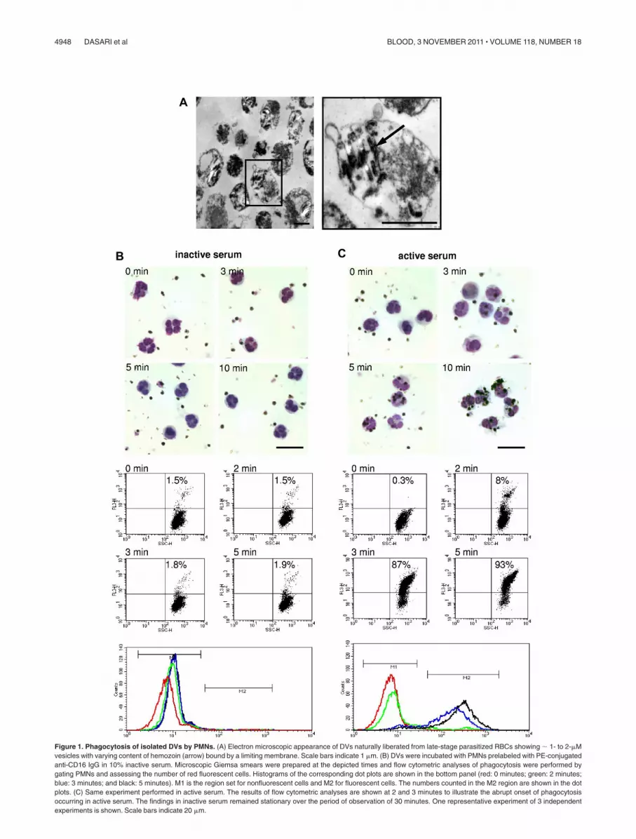
Figure 1. Phagocytosis of isolated DVs by PMNs. (A) Electron microscopic appearance of DVs naturally liberated from late-stage parasitized RBCs showing � 1- to 2-�Mvesicles with varying content of hemozoin (arrow) bound by a limiting membrane. Scale bars indicate 1 �m. (B) DVs were incubated with PMNs prelabeled with PE-conjugatedanti-CD16 IgG in 10% inactive serum. Microscopic Giemsa smears were prepared at the depicted times and flow cytometric analyses of phagocytosis were performed bygating PMNs and assessing the number of red fluorescent cells. Histograms of the corresponding dot plots are shown in the bottom panel (red: 0 minutes; green: 2 minutes;blue: 3 minutes; and black: 5 minutes). M1 is the region set for nonfluorescent cells and M2 for fluorescent cells. The numbers counted in the M2 region are shown in the dotplots. (C) Same experiment performed in active serum. The results of flow cytometric analyses are shown at 2 and 3 minutes to illustrate the abrupt onset of phagocytosisoccurring in active serum. The findings in inactive serum remained stationary over the period of observation of 30 minutes. One representative experiment of 3 independentexperiments is shown. Scale bars indicate 20 �m.
4948 DASARI et al BLOOD, 3 NOVEMBER 2011 � VOLUME 118, NUMBER 18

Medical Microbiology and Hygiene, University of Mainz, Mainz, Ger-many) were performed using isolated PMNs and C8-deficient serum thathad been prepared as described previously.30 PMNs were suspended at2 � 106 cells/mL in 50% C8-deficient serum/VBS and experiments wereconducted as described above.
Transmission electron microscopy
DVs were centrifuged, fixed with 2.5% glutaraldehyde in PBS at 4°C overnight,washed in PBS, post-fixed in 2% OsO4, dehydrated in ethanol, and embedded inAraldite (Sigma-Aldrich). Ultrathin sections were mounted on Formvar-coatedgrids and double stained with a saturated solution of uranyl acetate in 70%methanol and lead citrate. The grids were examined with a Zeiss EM 900transmission electron microscope equipped with a digital camera system.
Statistical analysis
The assumptions for normality and equal variance were verified with SigmaStat3.1 software (Erkrath; SYSSTAT). The Holm-Sidak test was used for compari-sons against a control group. Results represent means � SEM of at least3 independent experiments or the means of at least triplicates of 1 experi-ment � SD as indicated. P � .05 was considered statistically significant.
Results
Rapid complement–dependent phagocytosis of intact DVsby PMNs
DVs were isolated from supernatants of hemolyzed pRBCs and,in accordance with descriptions in the literature,34,35 were foundto comprise a population of dispersed, 1- to 2-�M sized vesicleswith various contents of hemozoin crystals and amorphousmaterial (Figure 1A). No merozoites or RBCs membrane debriscould be discerned in the electron micrographs.
DVs and PMNs were admixed at a ratio of 5:1 in 10% activeor 10% inactive human serum and phagocytosis was followedmicroscopically and by flow cytometry. For the latter analyses,DVs were fluorescently labeled, the PMNs were gated based onside and forward scatter and the uptake of fluorescently labeledDVs was assessed. No phagocytosis of the DVs by PMNsoccurred in inactive serum within the 30-minute period ofobservation, as was apparent from the Giemsa-stained smearsand from the corresponding absence of a red fluorescent shift ofthe cells (Figure 1B). In contrast, phagocytosis in active serumcould be readily observed microscopically, commencing within3 minutes and essentially reaching completion at 5-8 minutes.Correspondingly, PMNs abruptly started to assume red fluores-cence within 2-3 minutes and, in agreement with the micro-scopic observations, � 90% of the cells were laden with DVsafter just 5 minutes (Figure 1C).
PMNs were loaded with dichlorofluorescein, which allowedvisualization of ROS production in single cells. In agreement withthe classic response during phagocytosis, a burst of ROS couldalways be observed surrounding a DV during and immediatelyafter its uptake (Figure 2A). The single-cell observations wereborne out by luminol-based quantification of ROS generation,which revealed a bell-shaped chemiluminescence response cover-ing a time span of � 40 minutes that was not seen whenheat-inactivated serum was used (Figure 2B).
Merozoites escape phagocytosis by PMNs in nonimmuneserum
Late-stage synchronized pRBCs were cultured at 0.2% hematocritin medium containing 10% active, nonimmune human serum untilschizont rupture occurred, and merozoites and DVs were harvested
Figure 2. Phagocytosis of DVs induces ROS productionin PMNs. (A) PMNs were laden with dichlorol-fluorescein tovisualize ROS generation at the single-cell level. Phagocyto-sis of DVs was accompanied by triggering of the respiratoryburst, and ROS generation was seen to surround intracellularDVs. ROS generation was not observed when DVs were justattached to the cells (arrows). Scale bar indicates 10 �m.(B) Luminol-based chemiluminescence assay for ROS gen-eration in PMNs during phagocytosis of DVs revealed abell-shaped response curve covering a time span of 30-40minutes. PMNs were challenged with 3-5 DVs/cell and thechemiluminescence response was recorded in the presenceof 10% active or heat-inactivated human serum. One repre-sentative experiment of 3 independent experiments is shown.
P falciparum VACUOLES IMPAIR KILLING FUNCTION OF PMNs 4949BLOOD, 3 NOVEMBER 2011 � VOLUME 118, NUMBER 18

and stained for DNA and bound C3 (Figure 3A). The polyclonalAbs used recognized native and activated C3 (C3b). Hemozoincontained within the DVs was visualized in the phase-contrastimage. Merozoites were not seen here, but showed up in the bluefluorescence image generated by the DNA stain. Immunofluores-cence staining revealed the presence of C3 exclusively on the DVs,as was strikingly apparent in merged images, in which the red C3fluorescent stain is seen colocalizing with DVs, but was invariablyabsent on merozoites. Weaker stainings were also obtained using anAb against C3d, which reacts with the remnant molecule after C3bis cleaved and removed (data not shown). This experiment wasperformed 3 times with serum from different donors. Mergedimages were randomly taken and 100 hemozoin particles wereevaluated for the presence of C3. In all 3 experiments, C3colocalized with � 85% of the DVs. DVs liberated just beforeharvest would have had insufficient time to become sufficientlycoated with C3b. In striking contrast to these findings, not a singlemerozoite was ever observed to carry C3 deposits.
It followed that selective phagocytosis might occur as a naturalconsequence of intravascular schizont rupture. Therefore, in the nextexperiment, merozoites and DVs were isolated as above and DNAstaining was undertaken to stain the parasites. PE-surface-labeled PMNs(1 PMN:3-5 DVs: 50-100 merozoites) were then added together with10% NHS, and stacked fluorescence microscopic images were preparedafter 30 minutes. These revealed virtually exclusive uptake of DVs, and
merozoites were rarely seen to colocalize with the DVs in the centralplane of the cells (Figure 3Biii and 3Biv).
It has been reported that PMNs may phagocytose late-stage parasit-ized RBCs.36 Such cells were stained with 2�,7�-bis-(2-carboxyethyl)-5-(and-6)-carboxyfluorescein acetoxymethyl ester (BCECF-AM), whichenabled intracellular parasites to be visualized (P.D., S. Heber, S.Baumeister, K.L., K.R., S.C.B., and S. Bhakdi, unpublished data,September 2011), and incubated with PE-labeled PMNs in the presenceof active serum for 30 minutes. However, erythrophagocytosis couldnever be observed (Figure 4A).
Isolated hemozoin lacking the encasing DV-membrane is notphagocytosed
Malaria pigment is microscopically detectable in circulating PMNsof patients with malaria, and the relative number of pigment-containing PMNs provide a prognostic criterion for disease out-come.26,27 The question of how the pigment enters the cell hasnever been addressed. A tacit assumption is that DVs are labilestructures from which hemozoin is released and then phagocytosed.However, we have found that the DV membrane is remarkablystable, requiring extreme measures to effect its disruption in vitro(P.D., S. Heber, S. Baumeister, K.L., K.R., S.C.B., and S. Bhakdi,unpublished data, September 2011). Once freed of the encasingmembrane, the pigment disperses into small, crystalline unit
Figure 3. Selective opsonization and phagocytosisof DVs after pRBC rupture. (A) Immediately after lysisof late-stage pRBCs, merozoites and DVs were har-vested and stained with polyclonal Abs directed againstnative and activated C3 (C3b) and DNA. C3 immunore-activity was exclusively restricted to DV membranes.Scale bar indicates 20 �m. (B) DNA staining was used toidentify merozoites and PE-surface-labeled PMNs wereadded to the mixture with DVs. Stacked images (i-vi: topto bottom) were prepared that revealed extracellularlocalization of merozoites. Left rows: fluorescence; rightrows: merged fluorescence and phase-contrast images.The micrographs shown are representative of 3 indepen-dent experiments. Scale bar indicates 20 �m.
4950 DASARI et al BLOOD, 3 NOVEMBER 2011 � VOLUME 118, NUMBER 18

structures devoid of complement-activating properties and morpho-logically distinct from the compact deposits visible within intactDVs (P.D., S. Heber, S. Baumeister, K.L., K.R., S.C.B., and S.Bhakdi, unpublished data, September 2011). This agrees withpublished data showing that the intravesicular malaria pigmentconsists of multiple aggregates of hemozoin crystals.17 It is theseaggregates and not the dispersed crystals that present as malaria
pigment in circulating leukocytes.26,27 In the next experiment, DVswere disrupted by sonication or by detergent lysis, and hemozoinwas purified by centrifugation through Percoll. The isolatedhemozoin was incubated in 10% NHS with PMNs for 1 hour. Nomicroscopic evidence for attraction of PMNs to and uptake ofisolated hemozoin could be obtained, and cells presenting with thecharacteristic malaria pigment were not observed (Figure 4B). Inagreement with these observations, chemiluminescence assaysrevealed that sonication of DVs immediately destroyed theirROS-inducing property (Figure 4C). The same was found whenisolated hemozoin was applied to the cells (not shown).
DV-induced ROS-production in PMNs does not suppressmerozoite reinvasion
Merozoites have been reported to be susceptible to the cytotoxic actionof ROS.37 This conclusion was based on experiments with murinemalaria merozoites and may not be extrapolatable to P falciparum.Nevertheless, it was of interest to determine whether ROS generationmight indirectly serve a protective function because of bystander killingof the parasites. In the next experiment, a 10-fold excess of noninfectedRBCs was added to enriched, late-stage pRBCs and cultures weremaintained for 24 hours in the presence or absence of PMNs, whichwere added in supraphysiological numbers (� 1:100 RBCs) so that noeffects would be missed. Figure 5Ai depicts the control without PMNs,showing the presence of freshly infected cells with ring-stage parasites.When PMNs were present in the culture, these were seen to havephagocytosed the DVs (Figure 5Aii). However, ring forms could still bediscerned and quantification of infected cells by flow cytometricanalyses of DNA-stained samples generated superimposable curves(Figure 5Aiii left peaks, uninfected nonfluorescing cells; right peaks,infected cells). Therefore, ROS generation accompanying phagocyticuptake of DVs by PMNs led to no reduction in the infective capacity ofmerozoites. This experiment was performed 3 times with PMNs fromdifferent donors with the same result.
At this juncture, it was of interest to determine whether thefailure of PMNs to prevent parasite reinvasion might be rectified inthe presence of specific Abs. Experiments were undertaken withIgG from 5 patients with high Ab titers, from a pool of high-titeredsera from 10 patients, and from 5 patients with low Ab titers.
Phagocytosis experiments were performed as in the experimentshown in Figure 3, and reinvasion was quantified by measuringDNA incorporation of 3H-hypoxanthine. No effects of low-titeredAbs could be discerned in any experiments. However, in thepresence of high-titered Abs, merozoites were observed to bephagocytosed along with comparable numbers of DVs (Figure 5B).Inspection of 100 PMNs revealed that, although the merozoitesoutnumbered DVs by an order of magnitude in the incubationmixture, a phagocyte was never seen harboring merozoites alone.Therefore, it appeared that preferential uptake of DVs persistedeven in the presence of the Abs. This might have reduced eachcell’s capacity to phagocytose merozoites, allowing the majority toescape phagocytosis. Indeed, only small Ab-mediated reductions ofparasite reinvasion could be detected in the 3H-hypoxanthineincorporation assays, even though the PMNs were present in10-fold higher numbers in these experiments (Figure 5C).
The capacity to produce ROS is blunted in DV-laden PMNs
ROS generation is subject to multiple pathways of feedbackregulation,38,39 so the possibility that ingestion of DVs might leadto impaired respiratory burst on subsequent bacterial challengeemerged. PMNs in 10% serum were incubated for 60 minutes in thepresence or absence of 2-4 DVs per cell and subsequently
Figure 4. PMNs do not phagocytose late-stage pRBCs or hemozoin in nonim-mune serum. (A) pRBCs were stained with BCECF-AM, a nonfluorescent acetoxym-ethylester that is enzymatically hydrolyzed to fluorescent BCECF, to visualizeparasites and intracellular DVs, and incubated with PE-labeled PMNs in activehuman serum. No erythrophagocytosis could be discerned after 30 minutes. Scalebar indicates 20 �m. (B) DVs were disrupted by sonication and hemozoin wasisolated from a Percoll gradient and incubated with PMNs in active serum. Noevidence for phagocytic uptake and no appearance of cells with characteristicmalaria pigment could be discerned in Giemsa-stained smears. Scale bar indicates10 �m. (C) Luminol-based chemiluminescence assay were performed in PMNs andactive serum upon incubation with intact DVs or with sonicated DVs (hemozoin).Representative results are shown from 1 of 3 similar experiments.
P falciparum VACUOLES IMPAIR KILLING FUNCTION OF PMNs 4951BLOOD, 3 NOVEMBER 2011 � VOLUME 118, NUMBER 18

challenged with S aureus. Figure 6A shows the chemiluminescencerecordings observed. In control PMNs, S aureus challenge pro-voked a sustained generation of ROS. DVs induced the initialbell-shaped chemiluminescence response, but thereafter, the sec-ond burst of ROS provoked by S aureus was blunted. Identicalresults were found with PMNs from 4 different donors.
DV uptake reduces microbicidal activity of PMNs
These findings prompted phagocytosis and killing assays.Cell-rich plasma obtained after dextran sedimentation of eryth-rocytes from heparinized blood was spiked with 2-5 DV/PMNand incubated for 60 minutes at 37°C. The PMNs were thenchallenged with 10-20 S aureus per cell. Microscopy revealedthat the capacity of DV-loaded PMNs to subsequently ingest thebacteria remained unimpaired, and most of the bacteria werephagocytosed as in the controls after 15-20 minutes (Figure 6B).However, colony counting undertaken after detergent solubiliza-tion of the PMNs led to the striking finding that the capacity ofDV-laden cells to kill ingested bacteria was significantly compro-mised (Figure 6C). ROS reduction did not directly impact oxygen-independent microbicidal mechanisms, so the killing function wasreduced but not entirely abrogated.
Five to 10% of African children with severe malaria suffer fromsepsis episodes, half of which are caused by enteric nontyphoidSalmonellae.12-14 Phagocytic killing assays were therefore alsoperformed with a clinical isolate of S typhimurium. Enteric Salmo-nellae display widely varying degrees of serum complementresistance. This unpredictable potential confounder was eliminatedby conducting assays with isolated PMNs in the presence of 50%C8-deficient human serum. Killing could then be attributed solelyto the microbicidal function of the neutrophils. As found withS aureus, a significant reduction of bactericidal capacity wasobserved for S typhimurium in PMNs that had been laden with DVs(20-minute kill in controls: 48% � 5% and in DV-laden cells:20% � 2%; n 4, *P � .001).
Discussion
Lysis of each parasitized erythrocyte in P falciparum malarialiberates one DV along with infectious merozoites into thebloodstream.16,40 High parasitemia is therefore inseparably associ-ated with high loads of DVs at the sites of RBC rupture. It is all themore remarkable that, whereas seminal work on the biogenesis andbiochemical events occurring within the DV is ongoing,34,35,41-43
only one group of investigators has been conducting studies withnaturally released DVs, and these are devoted to their effects onmonocyte functions.44-48 The first study was conducted withunpurified malarial pigment obtained by hypotonic lysis of infectederythrocytes. In that study, evidence was presented that the pigmentwas phagocytosed by human macrophages, and that this provokeda respiratory burst that was blunted upon subsequent provocation.Bactericidal assays were not performed, but it was surmised thatthis immune suppression might bear general clinical relevancewhen malarial pigment reached the macrophages in the spleen andother organs.44 The fact that most DVs will probably be ingestednot by tissue macrophages but rather by the surrounding PMNs hasnever been considered before the present work, an oversight thatlikely derives from several reasons. There is a tendency to assumethat the malaria pigment itself is endowed with biologic properties,and that synthetic hematin, which is considered equivalent tonatural malaria pigment,17,18 is readily available. The popularity of
hematin as a research tool is understandable, but its use could nothave led to present discoveries because free pigment lacks all ofproperties described herein. In this context, it is not commonknowledge that the DV is released as an intact, enveloped organelleand it has not been recognized that the DV membrane is remarkablystable. Therefore, a widespread assumption is that malaria pigmentitself rapidly contacts the host environment. Consequently, thepossibility has been missed that the DV membrane may fulfillbiologic functions that are entirely distinct from those of isolatedhematin or hemozoin.
The starting point of the present investigation was our recentdiscovery that the DV membrane is endowed with the capacity todually activate the alternative complement and intrinsic clottingpathways (P.D., S. Heber, S. Baumeister, K.L., K.R., S.C.B., andS. Bhakdi, unpublished data, September 2011). Binding of comple-ment marks a particle for phagocytosis, and experiments describedherein naturally followed. Isolated DVs were indeed found to berapidly phagocytosed in a complement-dependent fashion, whichraised the possibility that engulfment of DVs represents the majorpathway leading to the presence of malaria pigment in PMNs ofpatients. The latter is a widespread finding, attesting to thegenerality of the phenomenon. Previous concepts have envisagedphagocytosis of hemozoin crystals or schizonts to be responsible,but no evidence for either could be obtained in this study. SonicatedDVs or isolated hemozoin did not induce a respiratory burst andwere not detectably taken up by the PMNs. We also could notobserve any phagocytosis of late-stage parasitized RBCs in nonim-mune serum. The possibility that specific Abs might alter the lattersituation, as suggested in an early study,36 is not excluded.However, PMNs containing ingested parasitized RBCs are seldomseen in clinical samples, so we propose at this stage that malariapigment in the PMNs of patients derives mainly from phagocytosisof intact DVs after their extrusion into the blood. Electronmicrographs of both PMNs and macrophages bearing malarialpigment have been published.49 The findings are highly suggestivefor 2 reasons. First, although the cells had not phagocytosedparasitized erythrocytes, aggregates of hemozoin crystals could beseen in the cell cytoplasm. Such aggregates do not persist when thepigment is artificially liberated from the organelle in vitro. Second,the aggregates can be seen to be surrounded by membranestructures in the electron micrographs. These findings are fully inagreement with our hypothesis that phagocytosis of DVs underliesthe appearance of malaria pigment in the cells.
Whether DVs might be preferentially phagocytosed was inves-tigated in the presence and absence of high-titered Abs againstP falciparum. Synchronized late-stage pRBCs were used, enablingthe time point of schizont rupture to be closely monitored so thatthe notoriously short-lived merozoites could be retrieved togetherwith the DVs. Culture was performed in active instead of heat-inactivated serum so that complement activation would immedi-ately occur upon erythrocyte rupture. Staining of merozoite DNAwas undertaken before the addition of surface-labeled PMNs,rendering rapid fluorescent microscopic analyses feasible. Theseexperiments revealed the striking fact that DVs but not merozoiteswere selectively opsonized and phagocytosed by PMNs in thepresence of active, nonimmune serum. In the presence of high-titered Abs, phagocytosis of merozoites was also observed. How-ever, preferential uptake of DVs still appeared to persist. Therefore,although merozoites outnumbered DVs by an order of magnitude,as also occurs in vivo, PMNs were never observed to containmerozoites only. If preferential uptake of DVs really takes place,this might impinge on each cell’s capacity to ingest merozoites.
4952 DASARI et al BLOOD, 3 NOVEMBER 2011 � VOLUME 118, NUMBER 18

Figure 5. Lack of protective effect of PMNs on merozoite reinvasion. (A) Late-stage pRBCs were allowed to rupture and new infection was allowed to proceed in activehuman serum in the absence (i) or presence (ii) of PMNs. Twenty-four hours later, ring forms could be seen microscopically in both cases. Infected erythrocytes were detectedby fluorescent staining of parasite DNA with hydroethidium-bromide (iii). Flow cytometric analyses revealed identical infection rates in the presence and absence of PMNs(right peaks: fluorescing infected cells; left peaks: nonfluorescing, noninfected cells). (B) Detection of merozoite phagocytosis mediated by Abs against P falciparum. Theexperiment shown in Figure 3B was repeated in the presence of 1 mg/mL of IgG isolated from a pool of 5 sera containing high-titered Abs against P falciparum. Stacked images(i-vi, top to bottom) now revealed that merozoites (blue) had been phagocytosed alongside with the DVs. Left rows: fluorescence; right rows: merged fluorescence andphase-contrast images. Arrows: phagocytosed merozoites colocalizing with DVs in the central plane (iii-iv) of a cell. Similar results were obtained with IgG from 5 individual serawith high-titered Abs against P falciparum. Scale bar indicates 20 �m. (C) Paucity of protective effects of PMNs and specific Abs upon parasite reinvasion. The3H-hypoxanthine DNA-incorporation assay was used and values obtained in active serum alone were defined as 100%. Results obtained with 5 high-titered Abs are shown.3H-hypoxanthine incorporation was assessed in active serum (AS) in the presence of either PMNs or IgG (Ab) or in the presence of PMNs and IgG. A small but significantreduction in 3H-hypoxanthine incorporation was observed in the presence of Abs plus PMNs compared with the control (n 5; *P � .001).
P falciparum VACUOLES IMPAIR KILLING FUNCTION OF PMNs 4953BLOOD, 3 NOVEMBER 2011 � VOLUME 118, NUMBER 18

ROS generation, the central element in the microbicidal machin-ery of mammalian phagocytes, is subject to feedback regulation viamultiple pathways.38,39 Induction of the respiratory burst byphagocytosed DVs might therefore be followed by a state ofhyporesponsiveness. Indeed, the capacity to mount a respiratoryburst on subsequent bacterial challenge was severely compromisedafter DV uptake. PMNs preloaded with DVs were still fully capableof phagocytosing bacteria. However, their microbicidal capacitywas reduced. This was shown using the classic target S aureus anda clinical isolate of S typhimurium. The latter was used becausenoninvasive Salmonella are the leading cause of septicemia inAfrican children with severe malaria, and these experiments nowoffer a possible explanation for this finding.
Having served its physiologic purpose, the liberated DVappears to function as a decoy, and is used by the parasite to divertand derange central elements of the innate immune system. Theintrinsic clotting and alternative pathway simultaneously becomeactivated on its surface. Both pathways generate potent mediatormolecules that may trigger and sustain microcirculatory distur-bances and vascular leakage, which contribute to the clinicalsyndromes of malaria. DVs from other Plasmodium species areprobably endowed with similar properties. Complement activationhas been shown in monkeys infected with P coatneyi50 and in miceinfected with P berghei.51 High levels of parasitemia exceeding10% are found almost exclusively in P falciparum infections, andcapillaric sequestration of pRBCs will further heighten the localload of DVs. Other than the living parasite, the organelle is thenpreferentially taken up by blood phagocytes. This may initiallyprovide benefit to the host by restricting activation of complementand coagulation. Differences in PMN-dependent clearing capacitymay explain why high parasitemia is sometimes tolerated and viceversa. However, the price for this indirectly beneficial PMNfunction may ultimately be high. As parasitemia increases, so willthe detraction of the cells away from their true targets. We foundthat high-titer-specific Abs invoked some phagocytosis of merozo-ites. This finding would appear to confirm earlier studies in whichAbs from patients reportedly promoted phagocytosis of merozoitesby neutrophils.52,53 However, merozoite preparations described inthose studies were retrieved as a dark band from Percoll gradientsand thus may have contained appreciable numbers of DVs. We arenot aware of any previous study in which phagocytosis ofmerozoites and DVs has been cleanly differentiated, such as isillustrated herein in Figures 3 and 5.
Possibly because efficient ingestion of DVs still prevailed,PMNs contained relatively small numbers of merozoites and themajority of parasites remained outside of the cells. In agreementwith this observation, substantive reductions in the rates ofreinvasion could also not be discerned. These findings are prelimi-nary because they were performed with only a small number ofantisera and one laboratory strain of P falciparum. HomologousAbs might more efficiently redirect PMN phagocytosis towardmerozoites to afford some protection. However, our findings agreewith earlier studies in which little32 or no54,55 protective effects ofspecific Abs plus PMNs could be observed in vitro. Monocytesapparently synergized more efficiently with the Abs,55 but thesignificance of this finding remains unclear because the cells wereused at unphysiologically high numbers. Most recently, the pres-ence of Abs provoking strong ROS responses in PMNs uponincubation with a mixture of merozoites and DVs was reported tobe correlated with better in vivo protection compared with Abs thatinduced little ROS production.56 Whether ROS generation iscorrelated with uptake of merozoites requires further investigation.These open questions notwithstanding, it appears quite clear that in
Figure 6. Phagocytosis of DVs leads to impairment of PMNs-function. (A) ROSgeneration was detected using the luminol-based chemiluminescence assay. ControlPMNs incubated for 60 minutes in medium and subsequently challenged with S aureusmounted a vigorous response. Incubation of cells with DVs induced an initial bell-shapedresponse, but ROS generation upon subsequent bacterial challenge was conspicuouslyblunted. (B) Microscopic examination undertaken 20 minutes after S aureus challengerevealed effective phagocytic uptake of the bacteria in both cases (left: control PMNs; right:PMNs preloaded with DVs). Scale bar indicates 10 �m. (C) Assessment of bacterial killingrevealed marked impairment of bactericidal activity in DV-laden cells. One representativeexperiment of 4 independent experiments is shown. Results are expressed as means oftriplicates � SD; *P � .001 compared with control.
4954 DASARI et al BLOOD, 3 NOVEMBER 2011 � VOLUME 118, NUMBER 18

vivo protection mediated by anti-merozoite Abs can generally notbe very efficient, because patients in endemic regions often havecirculating Abs that can neither inhibit infection nor totallysuppress disease progression.
DV uptake and cellular activation could cause PMNs to remainmainly sequestered in the microcirculation. If that is the case, then
the actual number of DV-laden phagocytes would probably beconsiderably higher than suggested by the mere numbers ofcirculating, hemozoin-containing cells. Sequestered, activated PMNspossibly augment pathologic processes in the microcirculation. Atthe same time, their systemic overloading may gradually set thestage for septicemic complications to develop whenever bacterialpathogens chance to gain entry into the circulation (Figure 7). Theleading roles played by Salmonellae and Enterobacteriaceae inAfrican children may derive simply from the high endemicprevalence of these agents. Should the principle tenets of thisinvestigation turn out to be correct, the DV would emerge as amajor, multifaceted determinant of parasitic pathogenicity. Clini-cally oriented studies are called for to test this hypothesis.
Acknowledgments
The authors thank Walter Hitzler and Roland Conradi for continuedsupply of erythrocytes, banked human blood, and human sera, andMarkus Radsak for the gift of anti-CD16 Abs.
This work was supported by the Deutsche Forschungsgemein-schaft, Sonderforschungsbereich 490 (to S. Bhakdi), Sonderforsc-hungsbereich 593 (to K.L. and S. Baumeister), Sonderforschungs-bereich 877 (to K.R.), the Cluster of Excellence “Inflammation atInterfaces” (to K.R.), and the Thai Infectious Disease Network (toP.D., R.U., S.C.B., and S. Bhakdi).
Authorship
Contribution: S. Bhakdi and S.C.B. conceived the project; S.Bhakdi, K.R., and P.D. designed the research; P.D. and S. Bhakdiperformed the experiments; R.L. performed the electron micros-copy; S. Bhakdi, P.D., S. Baumeister, K.L., R.U., and K.R.analyzed the data; and S. Bhakdi wrote the manuscript.
Conflict-of-interest disclosure: The authors declare no compet-ing financial interests.
Correspondence: Sucharit Bhakdi, Department of MedicalMicrobiology and Hygiene, University Medical Center, JohannesGutenberg University, Hochhaus Augustusplatz, 55202 Mainz,Germany; e-mail: [email protected].
References
1. Berendt AR, Tumer GD, Newbold CI. Cerebralmalaria: the sequestration hypothesis. ParasitolToday. 1994;10(10):412-414.
2. MacPherson GG, Warrell MJ, White NJ, Looaree-suwan S, Warrell DA. Human cerebral malaria. Aquantitative ultrastructural analysis of parasitizederythrocyte sequestration. Am J Pathol. 1985;119(3):385-401.
3. Bridges DJ, Bunn J, van Mourik JA, et al. Rapidactivation of endothelial cells enables Plasmo-dium falciparum adhesion to platelet-decoratedvon Willebrand factor strings. Blood. 2010;115(7):1472-1474.
4. Srichaikul T, Puwasatien P, Karnjanajetanee J,Bokisch VA, Pawasatien P. Complement changesand disseminated intravascular coagulation inPlasmodium falciparum malaria. Lancet. 1975;1(7910):770-772.
5. Neva FA, Howard WA, Glew RH, et al. Relationshipof serum complement levels to events of the malarialparoxysm. J Clin Invest. 1974;54(2):451-460.
6. Helegbe GK, Goka BQ, Kurtzhals JA, et al.Complement activation in Ghanaian children with
severe Plasmodium falciparum malaria. Malar J.2007;6:165.
7. Moxon CA, Heyderman RS, Wassmer SC. Dys-regulation of coagulation in cerebral malaria. MolBiochem Parasitol. 2009;166(2):99-108.
8. Francischetti IM. Does activation of the blood co-agulation cascade have a role in malaria patho-genesis? Trends Parasitol. 2008;24(6):258-263.
9. Ghosh K, Shetty S. Blood coagulation in falci-parum malaria–a review. Parasitol Res. 2008;102(4):571-576.
10. van der Heyde HC, Nolan J, Combes V, Gramaglia I,Grau GE. A unified hypothesis for the genesis of ce-rebral malaria: sequestration, inflammation and he-mostasis leading to microcirculatory dysfunction.Trends Parasitol. 2006;22(11):503-508.
11. Francischetti IM, Seydel KB, Monteiro RQ. Bloodcoagulation, inflammation, and malaria. Microcir-culation. 2008;15(2):81-107.
12. Mabey DC, Brown A, Greenwood BM. Plasmo-dium falciparum malaria and Salmonella infec-tions in Gambian children. J Infect Dis. 1987;155(6):1319-1321.
13. Bronzan RN, Taylor TE, Mwenechanya J, et al.
Bacteremia in Malawian children with severe ma-laria: prevalence, etiology, HIV coinfection, andoutcome. J Infect Dis. 2007;195(6):895-904.
14. Berkley J, Mwarumba S, Bramham K, Lowe B,Marsh K. Bacteraemia complicating severe ma-laria in children. Trans R Soc Trop Med Hyg.1999;93(3):283-286.
15. Wykes MN, Good MF. What really happens todendritic cells during malaria? Nat Rev Microbiol.2008;6(11):864-870.
16. Bannister LH, Hopkins JM, Fowler RE, Krishna S,Mitchell GH. A brief illustrated guide to the ultra-structure of Plasmodium falciparum asexualblood stages. Parasitol Today. 2000;16(10):427-433.
17. Pagola S, Stephens PW, Bohle DS, Kosar AD,Madsen SK. The structure of malaria pigmentbeta-haematin. Nature. 2000;404(6775):307-310.
18. Bohle DS, Kosar AD, Stephens PW. Phase ho-mogeneity and crystal morphology of the malariapigment beta-hematin. Acta Crystallogr D BiolCrystallogr. 2002;58(10 pt 1):1752-1756.
19. Pawluczkowycz AW, Lindorfer MA, Waitumbi JN,
Figure 7. Schematic presentation of the concept of immune decoy by the DV.Rupture of a parasitized cell liberates one DV along with 32 merozoites. Innonimmune serum, complement is activated only on the DV, so exclusive uptake ofthe vesicle follows, leaving merozoites free to invade new cells. ROS generation inresponse to DV uptake is unable to harm the merozoites, but instead drives the cellsinto a state of functional exhaustion so that efficient killing of subsequently engulfedbacteria is no longer ensured.
P falciparum VACUOLES IMPAIR KILLING FUNCTION OF PMNs 4955BLOOD, 3 NOVEMBER 2011 � VOLUME 118, NUMBER 18

Taylor RP. Hematin promotes complement alter-native pathway-mediated deposition of C3 activa-tion fragments on human erythrocytes: potentialimplications for the pathogenesis of anemia inmalaria. J Immunol. 2007;179(8):5543-5552.
20. Jaramillo M, Plante I, Ouellet N, Vandal K,Tessier PA, Olivier M. Hemozoin-inducible proin-flammatory events in vivo: potential role in ma-laria infection. J Immunol. 2004;172(5):3101-3110.
21. Pichyangkul S, Saengkrai P, Webster HK. Plas-modium falciparum pigment induces monocytesto release high levels of tumor necrosis factor-alpha and interleukin-1 beta. Am J Trop Med Hyg.1994;51(4):430-435.
22. Jaramillo M, Godbout M, Olivier M. Hemozoininduces macrophage chemokine expressionthrough oxidative stress-dependent and -inde-pendent mechanisms. J Immunol. 2005;174(1):475-484.
23. Parroche P, Lauw FN, Goutagny N, et al. Malariahemozoin is immunologically inert but radicallyenhances innate responses by presenting ma-laria DNA to Toll-like receptor 9. Proc Natl AcadSci U S A. 2007;104(6):1919-1924.
24. Griffith JW, Sun T, McIntosh MT, Bucala R. PureHemozoin is inflammatory in vivo and activatesthe NALP3 inflammasome via release of uricacid. J Immunol. 2009;183(8):5208-5220.
25. Carney CK, Schrimpe AC, Halfpenny K, et al. Thebasis of the immunomodulatory activity of malariapigment (hemozoin). J Biol Inorg Chem. 2006;11(7):917-929.
26. Nguyen PH, Day N, Pram TD, Ferguson DJ,White NJ. Intraleucocytic malaria pigment andprognosis in severe malaria. Trans R Soc TropMed Hyg. 1995;89(2):200-204.
27. Lyke KE, Diallo DA, Dicko A, et al. Association ofintraleukocytic Plasmodium falciparum malariapigment with disease severity, clinical manifesta-tions, and prognosis in severe malaria. Am J TropMed Hyg. 2003;69(3):253-259.
28. Levesque MA, Sullivan AD, Meshnick SR.Splenic and hepatic hemozoin in mice after ma-laria parasite clearance. J Parasitol. 1999;85(3):570-573.
29. Walev I, Tappe D, Gulbins E, Bhakdi S. Streptoly-sin O-permeabilized granulocytes shed L-selectinconcomitantly with ceramide generation via neu-tral sphingomyelinase. J Leukoc Biol. 2000;68(6):865-872.
30. Bhakdi S, Tranum-Jensen J. C5b-9 assembly:average binding of one C9 molecule to C5b-8without poly-C9 formation generates a stabletransmembrane pore. J Immunol. 1986;136(8):2999-3005.
31. van der Heyde HC, Elloso MM, vande Waa J,Schell K, Weidanz WP. Use of hydroethidine andflow cytometry to assess the effects of leukocyteson the malarial parasite Plasmodium falciparum.Clin Diagn Lab Immunol. 1995;2(4):417-425.
32. Kumaratilake LM, Ferrante A, Rzepczyk CM. Tu-mor necrosis factor enhances neutrophil-medi-ated killing of Plasmodium falciparum. Infect Im-mun. 1990;58(3):788-793.
33. Martin E, Bhakdi S. Flow cytometric assay forquantifying opsonophagocytosis and killing ofStaphylococcus aureus by peripheral blood leu-kocytes. J Clin Microbiol. 1992;30(9):2246-2255.
34. Goldberg DE, Slater AF, Cerami A, Henderson GB.Hemoglobin degradation in the malaria parasitePlasmodium falciparum: an ordered process in aunique organelle. Proc Natl Acad Sci U S A.1990;87(8):2931-2935.
35. Dluzewski AR, Ling IT, Hopkins JM, et al. Forma-tion of the food vacuole in Plasmodium falci-parum: a potential role for the 19 kDa fragment ofmerozoite surface protein 1 (MSP1(19)). PLoSOne. 2008;3(8):e3085.
36. Celada A, Cruchaud A, Perrin LH. Phagocytosisof Plasmodium falciparum-parasitized erythro-cytes by human polymorphonuclear leukocytes.J Parasitol. 1983;69(1):49-53.
37. Dockrell HM, Playfair JH. Killing of blood-stagemurine malaria parasites by hydrogen peroxide.Infect Immun. 1983;39(1):456-459.
38. Jandl RC, Andre-Schwartz J, Borges-DuBois L,Kipnes RS, McMurrich BJ, Babior BM. Termina-tion of the respiratory burst in human neutrophils.J Clin Invest. 1978;61(5):1176-1185.
39. Lee C, Miura K, Liu X, Zweier JL. Biphasic regula-tion of leukocyte superoxide generation by nitricoxide and peroxynitrite. J Biol Chem. 2000;275(50):38965-38972.
40. Abkarian M, Massiera G, Berry L, Roques M,Braun-Breton C. A novel mechanism for egress ofmalarial parasites from red blood cells. Blood.2011;117(15):4118-4124.
41. Elliott DA, McIntosh MT, Hosgood HD, 3rd, et al.Four distinct pathways of hemoglobin uptake inthe malaria parasite Plasmodium falciparum.Proc Natl Acad Sci U S A. 2008;105(7):2463-2468.
42. Fidock DA, Nomura T, Talley AK, et al. Mutationsin the P. falciparum digestive vacuole transmem-brane protein PfCRT and evidence for their role inchloroquine resistance. Mol Cell. 2000;6(4):861-871.
43. Abu Bakar N, Klonis N, Hanssen E, Chan C,Tilley L. Digestive-vacuole genesis and endocyticprocesses in the early intraerythrocytic stages ofPlasmodium falciparum. J Cell Sci. 2010;123(Pt3):441-450.
44. Schwarzer E, Turrini F, Ulliers D, Giribaldi G,Ginsburg H, Arese P. Impairment of macrophagefunctions after ingestion of Plasmodium falci-parum-infected erythrocytes or isolated malarialpigment. J Exp Med. 1992;176(4):1033-1041.
45. Skorokhod OA, Alessio M, Mordmuller B, Arese P,Schwarzer E. Hemozoin (malarial pigment) inhib-its differentiation and maturation of human mono-
cyte-derived dendritic cells: a peroxisome prolif-erator-activated receptor-gamma-mediatedeffect. J Immunol. 2004;173(6):4066-4074.
46. Prato M, Giribaldi G, Polimeni M, Gallo V, Arese P.Phagocytosis of hemozoin enhances matrix met-alloproteinase-9 activity and TNF-alpha produc-tion in human monocytes: role of matrix metallo-proteinases in the pathogenesis of falciparummalaria. J Immunol. 2005;175(10):6436-6442.
47. Giribaldi G, Prato M, Ulliers D, et al. Involvementof inflammatory chemokines in survival of humanmonocytes fed with malarial pigment. Infect Im-mun. 2010;78(11):4912-4921.
48. Prato M, Gallo V, Giribaldi G, Aldieri E, Arese P.Role of the NF-kappaB transcription pathway inthe haemozoin- and 15-HETE-mediated activa-tion of matrix metalloproteinase-9 in human ad-herent monocytes. Cell Microbiol. 2010;12(12):1780-1791.
49. Wickramasinghe SN, Phillips RE, Looareesuwan S,Warrell DA, Hughes M. The bone marrow in hu-man cerebral malaria: parasite sequestrationwithin sinusoids. Br J Haematol. 1987;66(3):295-306.
50. Glew RH, Atkinson JP, Frank MM, Collins WE,Neva FA. Serum complement and immunity inexperimental simian malaria. I. Cyclical altera-tions in C4 related to schizont rupture. J InfectDis. 1975;131(1):17-25.
51. Krettli AU, Nussenzweig V, Nussenzweig RS.Complement alterations in rodent malaria. Am JTrop Med Hyg. 1976;25(1):34-41.
52. Kumaratilake LM, Ferrante A. Opsonization andphagocytosis of Plasmodium falciparum merozo-ites measured by flow cytometry. Clin Diagn LabImmunol. 2000;7(1):9-13.
53. Kumaratilake LM, Ferrante A, Jaeger T,Rzepczyk CM. Effects of cytokines, complement,and antibody on the neutrophil respiratory burstand phagocytic response to Plasmodium falci-parum merozoites. Infect Immun. 1992;60(9):3731-3738.
54. Lunel F, Druilhe P. Effector cells involved in non-specific and antibody-dependent mechanismsdirected against Plasmodium falciparum bloodstages in vitro. Infect Immun. 1989;57(7):2043-2049.
55. Bouharoun-Tayoun H, Attanath P, Sabchareon A,Chongsuphajaisiddhi T, Druilhe P. Antibodies thatprotect humans against Plasmodium falciparumblood stages do not on their own inhibit parasitegrowth and invasion in vitro, but act in coopera-tion with monocytes. J Exp Med. 1990;172(6):1633-1641.
56. Joos C, Marrama L, Polson HE, et al. Clinical pro-tection from falciparum malaria correlates withneutrophil respiratory bursts induced by merozo-ites opsonized with human serum antibodies.PLoS One. 2010;5(3):e9871.
4956 DASARI et al BLOOD, 3 NOVEMBER 2011 � VOLUME 118, NUMBER 18