impact of template overhang-binding region of hiv-1 rt on the binding and orientation of the duplex...
TRANSCRIPT

This article was published in the above mentioned Springer issue.The material, including all portions thereof, is protected by copyright;all rights are held exclusively by Springer Science + Business Media.
The material is for personal use only;commercial use is not permitted.
Unauthorized reproduction, transfer and/or usemay be a violation of criminal as well as civil law.
ISSN 0300-8177, Volume 338, Combined 1-2
Impact of template overhang-binding region of HIV-1 RTon the binding and orientation of the duplex regionof the template-primer
Alok K. Upadhyay • Tanaji T. Talele •
Virendra N. Pandey
Received: 20 August 2009 / Accepted: 29 October 2009 / Published online: 17 November 2009
� Springer Science+Business Media, LLC. 2009
Abstract Fingers domain of HIV-1 RT is one of the
constituents of the dNTP-binding pocket that is involved in
binding of both dNTP and the template-primer. In the
ternary complex of HIV-1 RT, two residues Trp-24 and
Phe-61 located on the b1 and b3, respectively, are seen
interacting with N ? 1 to N ? 3 nucleotides in the tem-
plate overhang. We generated nonconservative and con-
servative mutant derivatives of these residues and
examined their impact on the template-primer binding and
polymerase function of the enzyme. We noted that W24A,
F61A, and F61Y and the double mutant (W24A/F61A)
were significantly affected in their ability to bind template-
primer and also to catalyze the polymerase reaction while
W24F remained unaffected. Using a specially designed
template-primer with photoactivatable bromo-dU base in
the duplex region at the penultimate position to the primer
terminus, we demonstrated that F61A, W24A, F61Y as
well as the double mutant were also affected in their cross-
linking ability with the duplex region of the template-pri-
mer. We also isolated the E–TP covalent complexes of
these mutants and examined their ability to catalyze single
dNTP incorporation onto the immobilized primer terminus.
The E–TP covalent complexes from W24F mutant dis-
played wild-type activity while those from W24A, F61A,
F61Y, and the double mutant (W24A/F61A) were
significantly impaired in their ability to catalyze dNTP
incorporation onto the immobilized primer terminus. This
unusual observation indicated that amino acid residues
involved in the positioning of the template overhang may
also influence the binding and orientation of the duplex
region of the template-primer. Molecular modeling studies
based on our biochemical results suggested that confor-
mation of both W24 and F61 are interdependent on their
interactions with each other, which together are required
for proper positioning of the ?1 template nucleotide in the
binary and ternary complexes.
Keywords HIV-1 reverse transcriptase �Template overhang � HIV-1 RT-DNA binary complex �HIV-1 RT-DNA-dNTP ternary complex �Duplex DNA binding track � HIV-1 RT dimer
Abbreviations
HIV-1 Human immunodeficiency virus
type 1
RT Reverse transcriptase
BSA Bovine serum albumin
DTT Dithiothreitol
dNTP Deoxyribonucleoside
triphosphate
ddNTP Dideoxy-nucleoside
triphosphate
TP Template-primer
U5-PBS RNA template HIV-1 genomic RNA template
corresponding to the primer
binding sequence region
U5-PBS DNA template HIV-1 genomic DNA template
corresponding to the primer
binding sequence region
A. K. Upadhyay � V. N. Pandey (&)
Department of Biochemistry and Molecular Biology,
UMDNJ-New Jersey Medical School, 185 South Orange
Avenue, Newark, NJ 07103, USA
e-mail: [email protected]
T. T. Talele
Department of Pharmaceutical Sciences, College of Pharmacy
and Allied Health Professions, St. John’s University,
8000 Utopia Parkway, Jamaica, NY 11439, USA
123
Mol Cell Biochem (2010) 338:19–33
DOI 10.1007/s11010-009-0316-x Author's personal copy

E–DNA Enzyme bound with DNA
template-primer in the binary
complex
E–DNA–dNTP Enzyme bound with DNA
template-primer and dNTP in
the ternary complex
Introduction
Retroviruses containing a single-stranded (?) RNA genome
cause a variety of diseases, including leukemia, sarcoma,
anemia, and arthritis, as well as immunodeficiency states.
Among these, the human immunodeficiency virus (HIV),
which causes acquired immunodeficiency syndrome
(AIDS) in humans, has been the target of a massive crusade.
A unique feature of their replication is reverse transcription
of their single-stranded RNA genome and its integration as
duplex DNA into the host chromosome [1, 2]. Reverse
transcription is carried out at an early stage after infection
by the virion-encapsidated reverse transcriptase enzyme
(HIV-1 RT). The enzyme is multifunctional, exhibiting
both RNA- and DNA-dependent DNA polymerase activi-
ties, as well as an RNase-H activity that is both exo- and
endonucleolytic [2–5]. These features, combined with the
apparent lack of reverse transcription in the normal
metabolism of eukaryotic cells, have made reverse trans-
criptase an attractive target in the search for chemothera-
peutic agents to combat the spread of retroviral infections.
Human immunodeficiency virus-1 reverse transcriptase
(HIV-1 RT) is a 117-kDa heterodimeric protein (p66/p51);
the smaller p51 subunit is probably generated from p66 of
p66/66 homodimeric enzyme by proteolytic cleavage in
one of the subunits and removal of the COOH-terminal
15-kDa RNase-H domain [2]. In vitro, the enzyme is
functional in both homodimeric (p66/66) and hetero-
dimeric (p66/51) forms with identical kinetic constants [6];
in vivo, however, it exists only as a heterodimeric enzyme
[2]. The catalytic activity of the enzyme resides in the
larger subunit, which is folded into an open structure
containing the polymerase-active site cleft, while the cat-
alytically inactive smaller subunit is closed and compact
[7]. The polymerase cleft is further folded into three dis-
tinct subdomains that resemble the palm, finger, and thumb
of a right hand [8]. Each subdomain has been implicated in
facilitating different functions of HIV-1 RT. The catalysis
of DNA synthesis is performed by highly conserved
aspartate residues in the palm subdomain [8, 9]. The fingers
have been implicated in the binding of incoming dNTP
substrate [10]. The thumb subdomain has been postulated
to participate in translocation of the enzyme along the
template-primer (TP) [11–14]. In the crystal structures of
TP bound HIV-1 RT binary complex [15–17], and TP-
dNTP bound HIV-1 RT ternary complex [18], the position
of the duplex region of the TP appears to be well estab-
lished, while the position and orientation of the single-
stranded template overhang is not well defined.
In the crystal structures of binary and ternary complexes
of different polymerases, the single-stranded region of the
template bends various degrees ‘‘away’’ from the active
site. In the DNA pol I family of enzymes, the first nucleo-
tide in the single-stranded region of the TP is flipped out of
the active site by *90�–180� in both binary and ternary
complexes [19, 20]. In DNA polymerase b, the downstream
duplex region of the TP has an *90� bend at the single-
stranded template overhang of the gapped DNA TP [21]. In
HIV-1 RT, in contrast, the only nucleotide in the overhang
in the binary complex of HIV-1 RT is protruded partially
into the dNTP-binding pocket [16, 17] while, in the ternary
complex, the first unpaired nucleotide is displaced away
from the active site [18]. Also, the bend at the junction of
the duplex region and template overhang is not as sharp as
that in the DNA pol I and DNA pol b family of enzymes.
In the crystal structure of the ternary complex of HIV-1
RT, a number of amino acid residues are seen interacting
with the template overhang [18]. The side chains of two
aromatic amino acids, W24 and F61, are seen interacting
directly with the template overhang. F61 has been impli-
cated in conferring fidelity and sensitivity to nucleoside
analog RT inhibitors [22] and also in strand displacement
activity of the enzyme [23]. While F61 interacts with the
N ? 1 and N ? 2 template nucleotides, the bulky side
chain of W24 is positioned to interact with the N ? 2 and
N ? 3 template nucleotide overhang. However, the inter-
actions of these residues are conspicuously absent in the
crystal structure of enzyme–TP (E–TP) binary complex
HIV-1 RT. Both the residues are far away from the tem-
plate, which contains only a single N ? 1 template
nucleotide overhang. Based on their strategic positions,
both W24 and F61 are expected to be important in binding
the TP in the binary complex, as well as for productive
positioning of the N ? 1 template nucleotide overhang for
the incoming dNTP substrate. Recently, both these residues
have been shown to be involved in accurate binding of TP
to the enzyme [24]. In this study, we generated conserva-
tive and nonconservative mutant derivatives of residues
W24 and F61 and demonstrated that the functions of both
the residues are interdependent on each other. Using a
specially designed photoactivatable self-annealing tem-
plate-primer (SATP), we demonstrated that both W24 and
F61 residues, in spite of being located outside of the duplex
DNA binding tract, significantly influence the productive
binding to the duplex region of TP and positioning of the
N ? 1 template nucleotide in the binary and ternary
complexes.
20 Mol Cell Biochem (2010) 338:19–33
123
Author's personal copy

Materials and methods
Materials
Restriction endonucleases were procured from Promega or
Boehringer Mannheim; DNA sequencing reagents were
from Stratagene (Texas). The HPLC-purified dNTPs were
obtained from Boehringer Mannheim; [32P]-labeled dNTPs
and ATP were obtained from Perkin Elmer Life Science.
All the synthetic oligonucleotides were synthesized at the
Molecular Resource Facility of the University of Medicine
and Dentistry, New Jersey. All other reagents, of the
highest purity grade, were purchased from Fisher, Milli-
pore Corp, and Bio-Rad.
In vitro mutagenesis and protein purification
We used a recombinant clone of HIV-1 RT (pKK223-
RT66) as the template for site-directed mutagenesis. Two
primers corresponding to the sense and antisense strands
containing the desired mutation were used to amplify the
plasmid by high-fidelity PCR using a QuickChangeTM Site-
Directed Mutagenesis Kit (Stratagene). We confirmed the
mutation by sequencing. The wild-type and mutant
enzymes were expressed in E. coli JM109; the homodi-
meric p66 form of enzyme was purified using ion exchange
chromatography as previously described [25, 26].
Thermolysin digestion of wild-type HIV-1 RT
and its mutant derivatives
Five micrograms of the enzyme was treated with 0.03 lg
of thermolysin in a total volume of 10 ll containing
50 mM Tris–HCl (pH 7.5), 2 mM MgCl2, 2 mM CaCl2,
10% glycerol, and 0.2 mM DTT. Reactions were done at
45�C for 30 min and quenched by addition of 5 ll of
protein gel sample buffer (50 mM Tris–HCl, pH 8.5,
100 mM 2-mercaptoethanol, 0.1% SDS, 20% glycerol,
0.01% bromophenol blue) containing 100 mM EDTA.
Samples were resolved on 12% denaturing SDS-poly-
acrylamide gel and visualized by staining with Coomassie
Brilliant Blue.
Preparation of HIV-1 U5-PBS RNA template
We used an HIV-RNA expression clone (pHIV-PBS) for
preparation of the U5-PBS HIV-1 genomic RNA template
as described before [27–29]. We linearized the plasmid
pHIV-PBS with AccI and in vitro transcribed it using T7
RNA polymerase. The enzyme, buffer, and rNTP solutions
were from Ambion. The transcription reaction was done
according to the manufacturer’s protocol.
Polymerase activity assay using primer extension gel
assay
We assayed the DNA- and RNA-dependent DNA poly-
merase activity of wild-type HIV-1 RT and its mutant
derivatives on U5-PBS HIV-1 RNA and U5-PBS DNA
templates primed with 50[32P]-labeled 21-mer DNA PBS
primer. The U5-PBS HIV-1 RNA template was transcribed
from the plasmid pHIV-PBS, which contains a 947-bp
fragment of the HIV-1 genome (?473 to ?1420) corre-
sponding to the PBS region [27]. We did the reaction in a
5 ll volume containing 50 mM Tris–HCl (pH 7.8),
100 lg/ml bovine serum albumin, 2 mM MgCl2, 10 mM
dithiothreitol, 50 mM KCl, 0.5 pmol [32P]-labeled TP
(10 K Cerenkov cpm), 20 lM dNTP, and 7.5 nM enzyme.
We initiated the reaction by adding the enzyme and, after
incubation for 60 s at room temperature, stopped the
reaction by adding an equal volume of gel loading dye
containing 0.025% bromophenol blue, 0.025% xylene
cyanol, 10 mM EDTA, and 40% formamide. We resolved
the reaction products on 8% denaturing polyacrylamide gel
containing 8 M urea and visualized them by phosphorim-
aging analysis.
Photoaffinity cross-linking of enzyme
with the template-primer
We used 50[32P]-labeled 37-mer or 51-mer self-annealing
TPs (SATP) containing photoactivatable bromo-dU base
pairing with the N - 1 template base at the penultimate
position from the 30 primer terminus (Chart 1). We labeled
the TPs at the 50 position using [c-32P] ATP and T4 poly-
nucleotide kinase according to the standard protocol [30].
The labeled TP was separated from free [c-32P] ATP on an
8% polyacrylamide–urea gel. The radioactive band corre-
sponding to the labeled TP was excised, eluted in 0.5 M
ammonium acetate, and purified on an NAP-10 column
(Pharmacia). The cross-linking reaction mixture contained
50 mM Tris–HCl (pH 7.8), 2 mM MgCl2, 1 mM DTT,
50 nM of labeled TP (40 K Cerenkov CPM/pmol), and
512 nM of enzyme in a final volume of 50 ll. The mixture
was exposed to 312 nm UV for 3 min in a Spectrolinker;
the TP-cross-linked enzyme species were resolved by
electrophoresis on SDS-polyacrylamide gel and detected
by phosphorimaging.
Catalytic activity of the E–TP covalent complex
The ability of the cross-linked E–TP covalent complex to
catalyze incorporation of the incoming dNTP onto the
immobilized primer terminus was assessed as described
previously [31, 32]. We incubated 15 pmol of the enzyme
on ice with 25 pmol of unlabeled self-annealing TP
Mol Cell Biochem (2010) 338:19–33 21
123
Author's personal copy

containing the photoactivatable bromo-dU base, then irra-
diated the E–TP complex at 312 nM UV for 3 min in a
Spectrolinker.
The cross-linked E–TP covalent complexes were puri-
fied as follows: the irradiated samples were loaded on a
DEAE-Sephadex column (0.5 ml) pre-equilibrated with
50 mM Tris–HCl (pH 8.0), 1 mM DTT, 200 mM NaCl,
and 5% glycerol. After extensive washing of the column
with the same buffer to remove the uncross-linked free
enzyme, the E–TP covalent complexes were recovered
from the column by elution with 1.0 M NaCl in the same
buffer. The eluate was desalted and concentrated using
Centriprep-30. The final preparation was free of the
uncross-linked enzyme, as judged by the lack of activity on
the externally added synthetic TP. Incorporation of a single
incoming dNTP was determined by the addition of 10 lCi
of [a32P]-TTP at 0.5 lM concentrations. The reaction, run
for 25 min at room temperature, was terminated by the
addition of 1% SDS and 20 mM EDTA. An aliquot of the
reaction mixture was subjected to SDS-polyacrylamide gel
electrophoresis, then autoradiography.
Gel retardation analysis of E–TP binary complex
and determination of Kd (DNA) value
For gel retardation analysis of E–TP binary complex, an
aliquot of enzyme was incubated with 1 nM of 50[32P]-
labeled TP in an incubation buffer containing 50 mM Tris–
HCl (pH 7.8), 5 mM MgCl2, and 0.01% BSA in a total
volume of 10 ll. After 10 min incubation on ice, we added
to the reaction mixture an equal volume of 29 gel loading
dye containing 0.25% bromophenol blue and 20% glycerol.
We resolved the E–TP binary complexes by electrophore-
sis on a 6% native polyacrylamide gel at 4�C followed by
phosphorimaging.
Using gel retardation analysis, we determined the dis-
sociation constants (Kd) of E–TP binary complexes of the
wild-type HIV-1 RT and of its mutant derivatives, as pre-
viously described [28, 33]. We used a 50[32P]-labeled
21-mer PBS primer annealed with either a 22-mer DNA
template with N ? 1 template overhang or a 26-mer tem-
plate with N ? 5 template overhang. In a total volume of
10 ll, we incubated a fixed concentration of [32P]-labeled
1. U5-PBS HIV-1 RNA containing the primer binding site: 3’-CAG GGA CAA GCC CGC GGU GAC GAU CUC UAA AAG GUG UGA CUG AUU UUC CCA GAC UCC CUA GAG AUC AAU GGU CUC AGU GUG UUG UCU GCC CGU GUG UGA UGA ACU UCC UGA GUU CCG UUC GAA AUA ACU CCG AAU UCG UCA CCC AAG GGA UCA UCG GUC UCU CGA GGG UCC GAG UCU AGA-5’
2. 21-mer DNA PBS primer: 5’-GTCCCTGTTCGGGCGCCACTG-3’
U5-PBS DNA templates corresponding to U5-PBS HIV-1 RNA sequences: 22-mer-3’-CAG GGA CAA GCC CGC GGT GAC G-5’ 23-mer-3’-CAG GGA CAA GCC CGC GGT GAC GA-5’24-mer-3’-CAG GGA CAA GCC CGC GGT GAC GAT-5’ 25-mer-3’-CAG GGA CAA GCC CGC GGT GAC GATC-5’ 26-mer-3’-CAG GGA CAA GCC CGC GGT GAC GATCT-5’
49-mer-3’-CAG GGA CAA GCC CGC GGT GAC GAT CTC TAA AAG GTG TGA CTG ATT TTC C-5’
3. ddC-terminated 21-mer DNA primer and corresponding templates: 5’-GTCCCTGTTCGGGCGCCACTddC-3’ (Primer)
22-mer-3’-CAG GGA CAA GCC CGC GGT GAG G-5’ 26-mer-3’-CAG GGA CAA GCC CGC GGT GAG GATCT-5’
4. Photoactivatable self-annealing template primers
Chart 1 Sequence of template
primers used
22 Mol Cell Biochem (2010) 338:19–33
123
Author's personal copy

TP (0.3 nM) with increasing concentrations of enzyme in
the incubation buffer. After 10 min of incubation on ice,
we resolved the E–TP binary complexes by native poly-
acrylamide electrophoresis as described. Using Image-
Quant software, we determined the amount of TP bound to
the enzyme in the binary complex and plotted it against
enzyme concentration using Graph Pad software. We cal-
culated the percent of TP bound to the enzyme in the form
of E–TP complex, then fitted it in a nonlinear regression
equation (b = (Vmax * [E])/(Kd ? [E]) as a function of
enzyme concentration. The Kd (DNA) was defined as the
enzyme concentration at which 50% of DNA was bound.
Stable ternary complex formation
We evaluated the ability of HIV-1 RT and its mutant
derivatives to form stable ternary complexes using the
method previously described [34, 35]. Briefly, in a total
volume of 10 ll, we incubated duplicate samples of each
enzyme with 50-[32P]-labeled ddC-terminated 21-mer pri-
mer annealed with a complementary 22-mer or 26-mer
template (0.3 nM). The incubation buffer and conditions
were similar to those used in the binary complex formation.
We first predetermined the amount of each enzyme that
give rise to a complete shift of the labeled TP in the E–TP
binary complex formation. We further incubated the reac-
tion mixture for 10 min in the presence of 200 lM dGTP, a
nucleotide complementary to the first templating base. We
assessed the stability of the ternary complexes by the
degree of labeled TP that remained bound in E–TP–dNTP
complex forms upon the addition of a 1,000-fold excess of
DNA trap. We resolved these complexes on 6% poly-
acrylamide native gel, then did phosphorimaging using
ImageQuant Software.
Glycerol gradient ultracentrifugation
To analyze monomeric and dimeric conformation of the
mutant enzymes, we used glycerol gradient ultracentrifu-
gation analysis [26, 36]. In brief, 50 lg of the enzyme protein
in 50 mM Tris–HCl (pH 8.0), and 400 mM NaCl was
applied on the top of a 5-ml 10–30% linear glycerol gradient
prepared in the same buffer. Gradients were centrifuged at
48,000 rpm in an SW 50.I rotor for 22 h at 4�C and frac-
tionated from the bottom. Sixty-two fractions (80 ll each)
were collected. Protein peaks in the fractions were deter-
mined by OD280 using a Nanodrop spectrophotometer.
CD spectra analysis
We did CD spectra analysis of the wild-type HIV-1 RT and
its mutant derivatives (0.75 lM) in the wavelength range of
195–260 nm at 0.5-nm intervals in an Aviv spectropolarimeter,
model 400 (Lakewood, NJ), using cylindrical fused quartz
cells with a path length of 0.1 cm.
Molecular modeling
The coordinates of 3D structures of HIV-1 RT in E–TP
binary complex and E–TP–dNTP ternary complex were
taken, respectively, from the RCSB PDB 2HMI and 1RTD
files [16, 18]. Due to the lack of template overhang in the
binary structures, we extracted the modeled template
overhang from the binary structure published by Peletskaya
et al. [37], incorporated it into the binary complex crystal
structure of Ding et al. [16], and subjected it to energy
minimization (Macromodel v9.5) to relieve steric hin-
drance between the modeled template overhang and the
protein. To analyze the interaction of mutant derivatives of
W24 and F61, the crystal structure of RT was altered to
include the respective amino acid substitutions. Mutations
were introduced using Maestro v8.0 (Schrodinger, LLC,
New York). Structure display was done using the Maestro
graphical interface. The wild-type and mutant enzyme
complexes were energy-minimized according to a protein
refinement protocol implemented in Macromodel v9.5
(Schrodinger LLC, New York), using the OPLS-AA force-
field parameters [38].
Results
Construction and purification of the mutant enzymes
Five mutants were generated, including a conservative and
a nonconservative mutant at both positions 24 (W24A,
W24F) and 61 (F61A and F61Y), as well as a double
mutant carrying Ala substitutions at both positions (W24A/
F61A). The mutants were expressed in E. coli and purified
to homogeneity with greater than 95% purity. The levels of
their expression, solubility, and electrophoretic profile
were identical to the wild-type enzyme (data not shown).
We also did thermolysin digestion [39] of the mutant
enzymes at 45�C and found no change in their digestion
pattern as compared to the WT enzyme (data not shown)
suggesting that substitution at these positions did not cause
any perturbation in the enzyme structure.
Effect of side chain substitutions at positions 24 and 61
on the polymerase activity of the enzyme
Using both U5-PBS RNA and 49-mer U5-PBS DNA
templates annealed with 21-mer PBS primer, we examined
the effect of conservative and nonconservative substitu-
tions at these positions on the polymerase activity of the
enzyme. Reactions were done for 30 and 60 s, after which
Mol Cell Biochem (2010) 338:19–33 23
123
Author's personal copy

the reaction products were analyzed on an 8% polyacryl-
amide–urea gel. Nonconservative substitutions at positions
24 and 61 resulted in significant loss of the primer exten-
sion ability of the enzyme (Fig. 1). The W24A mutant was
nearly inactive on the RNA template, while displaying
approximately 50% of wild-type activity on the DNA
template. In contrast, the conservative W24F mutant
exhibited wild-type activity on both the RNA and DNA
templates. Similar patterns of primer extension activity
were obtained with the nonconservative and conservative
mutants of F61. As expected, the primer extension ability
of the double mutant (W24A/F61A) was negligible on both
RNA and DNA templates.
Effect of Ala substitution at positions 61 and 24
on dimer stability of enzyme
Since Gly substitution at position 61 and 24 destabilizes
dimeric conformation of RT [40], we determined whether
Ala substitution at these positions also affects dimeric
conformation leading to reduced TP binding and poly-
merase activity of the enzyme. We used glycerol gradient
ultracentrifugation analysis of the wild-type and mutant
enzymes to determine their dimeric-monomeric conforma-
tions [26, 36]. A dimerization-defective mutant of HIV-1 RT
(W401L) was used as a positive control [41]. Gradients
were fractionated from the bottom and each fraction was
measured for OD280 absorbance. The results indicated that
the sedimentation profiles (35th fraction) of the wild-type
enzyme and all the mutant derivatives of W24 and F61
were identical, having sedimentation peak in the 35th
fraction (Fig. 2). In contrast, the sedimentation peaks of the
dimerization-defective p66W401L mutant of HIV-1 RT and
the p51 subunits were, respectively, in the 39th and 45th
fractions. These results suggest that, unlike Gly substitu-
tion, Ala substitutions at positions 24 and 61 have no
significant impact on the dimer stability of the enzyme. We
also confirmed that Trp ? Ala and Phe ? Ala substitu-
tions at these positions do not alter the secondary structure
of the enzyme since their CD spectra were similar to that of
the wild-type enzyme (Fig. 3).
Effect of template length on the polymerase activity
of the wild-type and its mutant derivatives
In the ternary complex of HIV-1 RT, W24 interacts with
the phosphate backbone of the N ? 2 template nucleotide
and F61 interacts with the base moiety of N ? 2 and with
the sugar ring of the N ? 1 template nucleotides. We,
therefore, postulate that these interactions are crucial for
appropriate positioning of the N ? 1 template for binding
and incorporation of the incoming dNTP substrate. Any
nonconservative substitutions at positions 24 and 61 may
significantly impair the enzyme’s ability to position the
N ? 1 template, thus affecting the polymerase function of
the enzyme. To ascertain this, we examined the polymerase
activity of these mutants on TPs with varying template
overhangs. We used 50-[32P] labeled 21-mer PBS primer
annealed with U5-PBS DNA templates carrying N ? 1 to
N ? 5 nucleotide overhangs (Chart 1). The gel extension
profile of the polymerase products obtained with these
mutants is shown in Fig. 4.
The primer extension ability of the conservative W24F
mutant was similar to that of the wild-type enzyme on all
the template overhangs. The nonconservative W24A
mutant was significantly affected in its primer extension
ability on the templates with a N ? 1 nucleotide overhang,
but had approximately 50% of wild-type activity on tem-
plates with N ? 2 to N ? 5 template nucleotide overhangs.
In contrast, the primer extension ability of F61A and
double mutant W24A/F61A was severely affected on all
template overhangs. The conservative F61Y mutant was
also impaired with a N ? 1 template overhang and was
unable to incorporate the last nucleotide on the other two
templates (N ? 2 and N ? 3) when it encountered a situ-
ation similar to a N ? 1 template. However, the F61Y
mutant synthesized full-length products on templates with
N ? 4 and N ? 5 nucleotide overhangs, probably due to
additional interactions with the longer template overhangs.
The impaired ability of F61A and F61Y to extend the
primer with a N ? 1 template suggests that the Phe side
chain at the 61 position may be required for proper
Fig. 1 Polymerase activity of the wild-type enzyme and its mutant
derivatives on RNA and DNA templates. The polymerase activity of
the wild-type and mutant enzymes was determined on U5-PBS RNA
and U5-PBS DNA templates primed with 50[32P]-labeled 21-mer PBS
primer; a primer extension gel assay was done. Lanes 1 and 2
represent the products formed during the reaction, which was carried
out for 30 and 60 s, respectively. The DM indicates double mutant
(W24A/F61A) while P indicate the position of the labeled primer
24 Mol Cell Biochem (2010) 338:19–33
123
Author's personal copy

positioning of a N ? 1 template. However, the impaired
ability of W24A on a template with a N ? 1 template was
unexpected, since this mutant had a wild-type Phe residue at
position 61. It is possible that the side chain conformations
of both F61 and W24 are dependent on their interaction
with each other, which may be lost by Ala substitution at
either of the positions.
Photoaffinity cross-linking of mutant enzymes
with the template-primer
The impaired or reduced ability of mutant derivatives of
F61 and W24 to extend the primer on short template
overhangs may be due to either reduced DNA binding or
improper positioning of the template overhang. We exam-
ined the DNA binding function of these mutant enzymes by
covalent cross-linking of the 50[32P]-labeled self-annealing
37-mer TP containing a photoactivatable BrdU base at the
penultimate position from the primer terminus pairing with
N - 1 template in the duplex region. Since 30-OH of the
primer terminus is positioned in the catalytic cleft, the BrdU
base pairing with a N - 1 template is expected to cross-link
selectively with the interacting residue in the catalytic cleft
upon irradiation of the E–TP complex at 312 nm. At this
Fig. 3 CD spectrum of the wild-type HIV-RT and its mutant
derivatives of W24 and F61. CD spectrum analysis was carried out
in the wavelength range of 195–260 nm at 0.5-nm intervals in an
Aviv spectropolarimeter, model 400 (Lakewood, NJ) using cylindri-
cal fused quartz cells with a path length of 0.1 cm
Fig. 2 Glycerol gradient ultracentrifugation analysis of the WT HIV-1
RT and its mutant derivatives of W24 and F61. The wild-type p66/66
HIV-1 RT and its mutant derivatives were individually resolved on
10–30% linear glycerol gradient ultracentrifugation at 48,000 rpm in
an SW 50.I rotor for 22 h at 4�C. Gradients were fractionated from
the bottom and measured for OD280 absorbance using a Nanodrop
spectrophotometer. We also included p51 monomer and a dimeriza-
tion-defective W401L mutant as a positive control
b
Mol Cell Biochem (2010) 338:19–33 25
123
Author's personal copy

wavelength, only the BrdU base can be activated to cross-
link with the enzyme. We have earlier shown that this TP
selectively crosslinks to the catalytic subunit upon irradia-
tion at 312 nm UV [31]. The cross-linked covalent complex
was resolved on SDS-PAGE. All the mutant enzymes
except the double mutant were able to form a covalent
complex with the 50-[32P]-labeled self-annealing 37-mer
TP, although the extent of the E–TP complexes formed by
nonconservative W24A and F61A mutants was significantly
lower than that produced by the wild-type enzyme
(Fig. 5A). Among these, the double mutant (W24A/F61A)
was severely affected in its ability to form E–TP binary
complex. Since 37-mer SATP had only a 12 bp duplex
region, we also used a 51-mer self-annealing TP with a
19 bp duplex region to rule out the possibility that the
variations in cross-linking may have been due to a shorter
duplex region. Although the extent of cross-linking with
51-mer SATP was marginally improved, the overall pattern
remained identical to 37-mer SATP (Fig. 5B). The two
bands that are seen in the gel are common with these tem-
plat- primers, possibly existing as a mixture of monomer
and dimer due to the presence of complementary sequences.
Ability of mutant enzymes to incorporate a single
incoming dNTP onto the immobilized primer terminus
We examined whether the N ? 1 template nucleotide in
these E–TP covalent complexes is appropriately positioned
to facilitate incorporation of the first incoming dNTP
substrate onto the primer terminus of the immobilized TP.
We incubated the wild-type and mutant enzymes with
unlabeled 37-mer or 51-mer SATP containing a photoac-
tivatable BrdU base pairing with the N - 1 template base
in the duplex region (Chart 1). The E–TP complexes were
then UV irradiated at 312 nm and the E–TP covalent
complex formed was purified. We supplemented the puri-
fied E–TP covalent complexes with [32P]-labeled TTP and
examined their ability to incorporate the first incoming
dNTP substrate onto the immobilized primer terminus.
The results indicated differences in the ability of the
E–TP covalent complexes of the wild-type and mutant
derivatives to incorporate the first incoming dNTP sub-
strate (Fig. 6). While E–TP covalent complexes from
W24F displayed wild-type activity, the complexes from
F61Y, W24A, and F61A mutants were significantly
Fig. 4 DNA polymerase activity of the wild-type enzyme and its
mutant derivatives on template-primers with varying template over-
hangs. Primer extension gel assay was done using the 50-[32P]-labeled
21-mer PBS primer annealed with DNA templates carrying N ? 1,
N ? 2, N ? 3, N ? 4, or N ? 5 template nucleotide 50 overhangs.
The reaction was done for 60 s in a total volume of 5 ll and the
products resolved on denaturing polyacrylamide-urea gel. The
number with (N?) sign below each lane indicates the number of
nucleotides in the 50 template overhang
Fig. 5 Photoaffinity cross-linking of 50-[32P]-labeled A 37-mer and
B 51-mer self-annealing template-primer to the wild-type enzyme and
its mutant derivatives. The 50[32P]-labeled SATP contained photoac-
tivatable bromo-dU base that base pair with the N - 1 template base
at the penultimate nucleotide from the 30 primer terminus (Chart 1).
The labeled SATP (50 nM, 100K Cerenkov cpm) was incubated with
512 nM of each enzyme in a solution containing 50 mM Tris–HCl
(pH 7.8), 2 mM MgCl2, and 1 mM DTT in a final volume of 50 ll.
The mixture was exposed to 312 nm UV for 3 min in a Spectrolinker.
The TP-cross-linked enzyme species were resolved by electrophoresis
on SDS-polyacrylamide gel and detected by phosphorimaging. The
two bands seen in the gel are common with self-annealing TPs since,
due to complementary sequences, they exist as both monomer and
primer-dimer. The control lane represents 50-[32P] labeled SATP
irradiated without the enzyme which has ran out of the gel upon SDS-
PAGE
26 Mol Cell Biochem (2010) 338:19–33
123
Author's personal copy

impaired in their ability to incorporate the first incoming
dNTP onto the immobilized primer terminus of the cross-
linked TP. As expected, only a trace of nucleotidyltrans-
ferase activity could be detected with E–TP covalent
complexes from the double mutant (W24A/F61A). This is
consistent with its low level of E–TP binary complex for-
mation (Fig. 6A, B). These results support the contention
that W24 and F61 residues are involved not only in binary
complex formation, but also in appropriate positioning of
the N ? 1 template nucleotide for dNTP-binding and
polymerase function of the enzyme.
Effect of template overhang on the DNA binding
affinity of mutant enzymes
To determine whether TP binding affinity of mutant
enzymes is influenced by template nucleotide overhangs,
we first examined the TP binding pattern of the enzymes,
using gel retardation with 22/21 and 26/21-mer TP
carrying, respectively, N ? 1 and N ? 5 template nucleo-
tide overhangs. We incubated each mutant and WT enzyme
at a 1 nM concentration with 1 nM of [32P]-labeled TP. As
shown in Fig. 7, the extent of E–TP binary complex for-
mation by the nonconservative W24A and F61A mutants
depended on the length of the template overhang. Both
mutants were unable to form the E–TP binary complex
when a template had only a single nucleotide overhang.
However, when a template overhang was increased to
N ? 5 nucleotides, the ability of both W24A and F61A
mutants to form the binary complex was slightly improved,
but remained at a significantly lower level than did for-
mation by the wild-type enzyme. As expected, the double
mutant (W24A/F61A) was unable to form detectable bin-
ary complex with both the TPs. In contrast, the extent of
E–TP binary complex formation with conservative W24F
and F61Y mutants was similar to the wild-type enzyme
with both shorter and longer template overhangs.
After qualitatively evaluating E–TP binary complex
formed by these mutants, we determined the equilibrium
dissociation constant (Kd) of E–TP binary complexes for
the wild-type enzyme and its mutant derivatives on the two
TPs carrying N ? 1 and N ? 5 nucleotide template over-
hangs. The gel retardation analyses of E–TP complexes of
the wild-type and its mutant derivatives are shown in
Fig. 8. The values for Kd determined (Table 1) demonstrate
a strong correlation between the polymerase activity of the
mutants and their Kd values. The mutant enzymes W24A
and F61A, which had reduced enzyme activity on all five
Fig. 6 Catalytic activity of the E–TP covalent complex. We incu-
bated 15 pmol of enzyme with 25 pmol of unlabeled cold A 37-mer
SATP or B 51-mer SATP and irradiated the mixture at 312 nm UV
for 3 min. The ability of E–TP covalent complex to catalyze the
incorporation of nucleotide onto the immobilized primer terminus
was examined by incubating the complex with 10 lCi of the first
incoming [a32P]-TTP at a 0.5 lM concentration. The reaction mixture
was incubated for 25 min at room temperature. The reaction was
terminated by the addition of 1% SDS and 20 mM EDTA. An aliquot
of the reaction mixture was subjected to SDS-polyacrylamide gel
electrophoresis, then autoradiography. The control lane represents
incubation of uncrosslinked wild-type enzyme–SATP complex with
[a32P]-TTP and the extended SATP ran out of the gel upon SDS-
PAGE
Fig. 7 Effect of 50 template nucleotide overhang of the template-
primer on binding with the enzyme. One nanomolar of the wild-type
HIV-1 RT or its mutant derivatives was incubated with 1 nM of
50-[32P]-labeled 22/21-mer TP with an N ? 1 template overhang or
26/21 TP with an N ? 5 template overhang. Following 10 min
incubation on ice in an incubation buffer of 50 mM Tris–HCl
(pH 7.8), 5 mM MgCl2, and 0.01% BSA, the E–TP binary complex
formed was analyzed by nondenaturing polyacrylamide gel
electrophoresis
Mol Cell Biochem (2010) 338:19–33 27
123
Author's personal copy

TPs, also exhibited 10–15-fold lower DNA binding affinity
than did the wild-type enzyme. The DNA binding affinity
of the double mutant was reduced by approximately 125-
fold. As expected, the conservative W24F mutant with
wild-type polymerase activity displayed no change in its
DNA binding affinity.
Stable ternary complex formation
In the crystal structure of the ternary complex of HIV-1 RT
(E–DNA–dNTP), a significant movement of the fingers
subdomain toward the polymerase cleft has been noted
upon binding of dNTP to the E–TP binary complex [18].
Specifically, the fingers subdomain moves 20 A toward the
palm subdomain (fingers closing) so that, following dNTP
binding, the TP in the E–TP binary complex is locked in a
stable ternary complex poised for catalysis. An in vitro
assay for ternary complex formation using dideoxy termi-
nated primer annealed with the template allows the next
correct dNTP to bind in the ternary complex without
actually being incorporated [34]. We used this assay to
examine the ability of these mutant enzymes to transform
E–TP binary complexes carrying (N ? 1) and (N ? 5)
template nucleotide overhangs into stable E–TP–dNTP
ternary complexes in the presence of first incoming dNTP
substrate. Since the binding of dNTP to the enzyme is an
ordered mechanism that occurs only after the formation of
E–TP binary complex, the extent of labeled TP remaining
bound to the enzyme in the presence of dNTP and a large
excess DNA trap represents the extent of the stable ternary
complex formed.
We determined the amount of DNA trap required for
complete dissociation of labeled E–TP binary complex and
the extent of stable ternary complex formed by the wild-
type enzyme (Fig. 9A, B). We then incubated the preformed
E–TP binary complexes of the WT and mutant enzymes in
the presence of a 1,000-fold molar excess of DNA trap
(Fig. 9C, lane 2) or in the presence the correct incoming
dNTP (dGTP; 200 lM), followed by addition of the DNA
trap (Fig. 9C, lane 4). The E–TP binary complexes of all the
mutants and WT enzymes were completely dissociated in
the presence of a large excess of DNA trap (Fig. 9C, lane 2).
In contrast, a significant amount of E–TP binary complexes
were converted to E–TP–dNTP ternary complexes, which
remained resistant to competition with the DNA trap
(Fig. 9C, lane 4). The nonconservative W24A and F61A
mutants and the conservative F61Y mutant were unable to
form stable ternary complexes when TP carried a single
N ? 1 template nucleotide overhang (22/21-TP, lane 4).
Only a trace amount of the ternary complex could be formed
when the template overhang was increased to N ? 5
Fig. 8 Determination of the equilibrium dissociation constant of
E–TP binary complex (Kd [TP]) with TP carrying ?1 and ?5 template
nucleotide overhangs. The 50-[32P]-labeled 21-mer primer was
annealed with complementary 22-mer and 26-mer templates carrying
50 template overhangs of ?1 and ?5 nucleotides, respectively. The
labeled TP was incubated with varying concentrations of the enzyme.
The E–TP binary complexes were resolved by electrophoresis on 6%
native nondenaturing polyacrylamide gel. The amount of labeled TP
bound to the enzyme in the binary complex was determined and
plotted against the enzyme concentration. The Kd was defined as the
concentration of enzyme at which 50% of DNA was bound
Table 1 Kd of the wild-type enzyme and mutant enzymes on TP with
different template overhangsa
Enzyme Kd (nM)
22/21-mer
(N ? 1 overhang)
26/21-mer
(N ? 5 overhang)
WT 0.24 ± 0.12 0.25 ± 0.03
W24A 3.50 ± 0.14 2.0 ± 0.21
W24F 0.30 ± 0.03 0.30 ± 0.01
F61A 4.0 ± 0.08 2.5 ± 0.08
F61Y 1.60 ± 0.15 0.56 ± 0.12
DM (W24A/F61A) 30 ± 7.5 30 ± 9.5
a The data represent the average ± standard deviation of three
experiments
28 Mol Cell Biochem (2010) 338:19–33
123
Author's personal copy
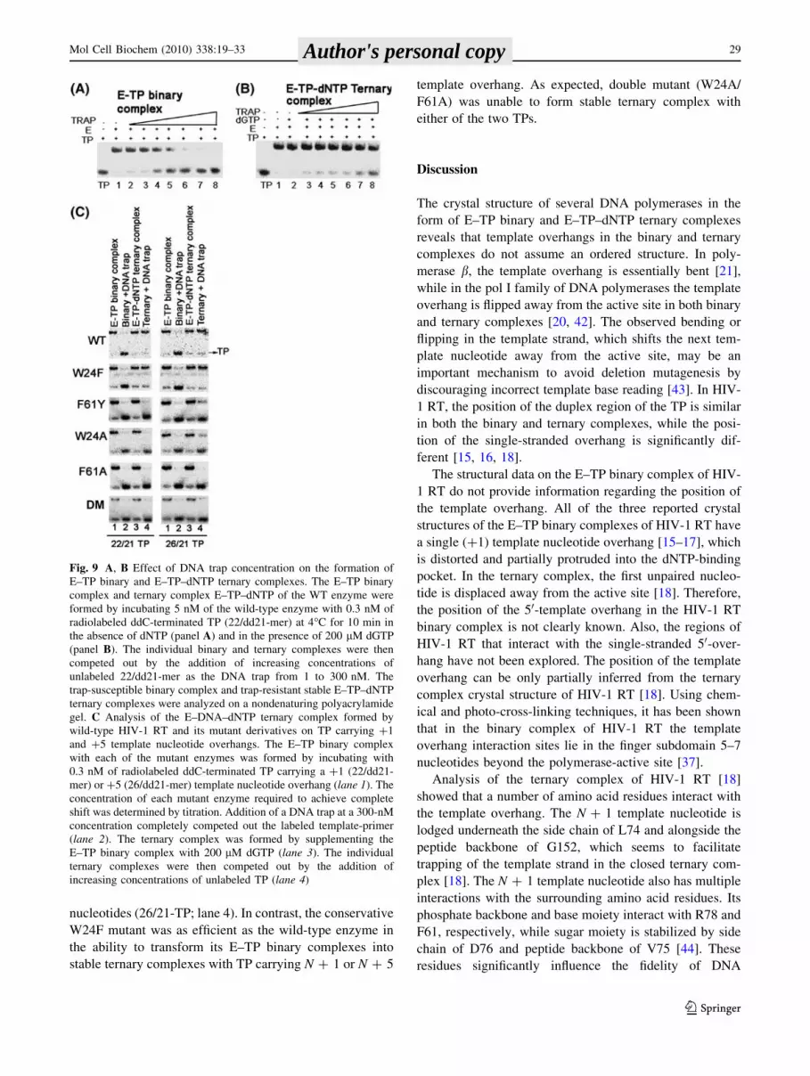
nucleotides (26/21-TP; lane 4). In contrast, the conservative
W24F mutant was as efficient as the wild-type enzyme in
the ability to transform its E–TP binary complexes into
stable ternary complexes with TP carrying N ? 1 or N ? 5
template overhang. As expected, double mutant (W24A/
F61A) was unable to form stable ternary complex with
either of the two TPs.
Discussion
The crystal structure of several DNA polymerases in the
form of E–TP binary and E–TP–dNTP ternary complexes
reveals that template overhangs in the binary and ternary
complexes do not assume an ordered structure. In poly-
merase b, the template overhang is essentially bent [21],
while in the pol I family of DNA polymerases the template
overhang is flipped away from the active site in both binary
and ternary complexes [20, 42]. The observed bending or
flipping in the template strand, which shifts the next tem-
plate nucleotide away from the active site, may be an
important mechanism to avoid deletion mutagenesis by
discouraging incorrect template base reading [43]. In HIV-
1 RT, the position of the duplex region of the TP is similar
in both the binary and ternary complexes, while the posi-
tion of the single-stranded overhang is significantly dif-
ferent [15, 16, 18].
The structural data on the E–TP binary complex of HIV-
1 RT do not provide information regarding the position of
the template overhang. All of the three reported crystal
structures of the E–TP binary complexes of HIV-1 RT have
a single (?1) template nucleotide overhang [15–17], which
is distorted and partially protruded into the dNTP-binding
pocket. In the ternary complex, the first unpaired nucleo-
tide is displaced away from the active site [18]. Therefore,
the position of the 50-template overhang in the HIV-1 RT
binary complex is not clearly known. Also, the regions of
HIV-1 RT that interact with the single-stranded 50-over-
hang have not been explored. The position of the template
overhang can be only partially inferred from the ternary
complex crystal structure of HIV-1 RT [18]. Using chem-
ical and photo-cross-linking techniques, it has been shown
that in the binary complex of HIV-1 RT the template
overhang interaction sites lie in the finger subdomain 5–7
nucleotides beyond the polymerase-active site [37].
Analysis of the ternary complex of HIV-1 RT [18]
showed that a number of amino acid residues interact with
the template overhang. The N ? 1 template nucleotide is
lodged underneath the side chain of L74 and alongside the
peptide backbone of G152, which seems to facilitate
trapping of the template strand in the closed ternary com-
plex [18]. The N ? 1 template nucleotide also has multiple
interactions with the surrounding amino acid residues. Its
phosphate backbone and base moiety interact with R78 and
F61, respectively, while sugar moiety is stabilized by side
chain of D76 and peptide backbone of V75 [44]. These
residues significantly influence the fidelity of DNA
Fig. 9 A, B Effect of DNA trap concentration on the formation of
E–TP binary and E–TP–dNTP ternary complexes. The E–TP binary
complex and ternary complex E–TP–dNTP of the WT enzyme were
formed by incubating 5 nM of the wild-type enzyme with 0.3 nM of
radiolabeled ddC-terminated TP (22/dd21-mer) at 4�C for 10 min in
the absence of dNTP (panel A) and in the presence of 200 lM dGTP
(panel B). The individual binary and ternary complexes were then
competed out by the addition of increasing concentrations of
unlabeled 22/dd21-mer as the DNA trap from 1 to 300 nM. The
trap-susceptible binary complex and trap-resistant stable E–TP–dNTP
ternary complexes were analyzed on a nondenaturing polyacrylamide
gel. C Analysis of the E–DNA–dNTP ternary complex formed by
wild-type HIV-1 RT and its mutant derivatives on TP carrying ?1
and ?5 template nucleotide overhangs. The E–TP binary complex
with each of the mutant enzymes was formed by incubating with
0.3 nM of radiolabeled ddC-terminated TP carrying a ?1 (22/dd21-
mer) or ?5 (26/dd21-mer) template nucleotide overhang (lane 1). The
concentration of each mutant enzyme required to achieve complete
shift was determined by titration. Addition of a DNA trap at a 300-nM
concentration completely competed out the labeled template-primer
(lane 2). The ternary complex was formed by supplementing the
E–TP binary complex with 200 lM dGTP (lane 3). The individual
ternary complexes were then competed out by the addition of
increasing concentrations of unlabeled TP (lane 4)
Mol Cell Biochem (2010) 338:19–33 29
123
Author's personal copy

synthesis of the enzyme [22, 23, 45, 46]. Substitutions of
Arg ? Ala and Asp ? Val at positions 78 and 76,
respectively, were also shown to affect the DNA binding
affinity of the enzyme; while R78A decreased the TP
binding affinity by 8-fold, D76V mutation enhanced it by
7-fold [45]. Another important residue involved in stabiliz-
ing the template overhang in the ternary complex is W24,
which interacts with the phosphate backbone between N ? 2
and N ? 3 nucleotides, as well as their sugar moieties, in
coordination with F61, which interacts with the sugar moi-
eties of N ? 1 and N ? 2 and also with the N ? 1 base.
We noted that Ala substitutions at either position 61 or
24 have adverse effects on the binary complex formation of
the enzyme. The double mutant (W24A/F61A) was most
severely affected in its ability to form the binary complex
(Fig. 5A). For this purpose, we used specifically designed
self-annealing TPs that had bromo-dU at the penultimate
position from the primer terminus pairing with the N - 1
template base. Upon irradiation of the E–TP complex at
312 nm UV, only bromo-dU was excited to crosslink with
the enzyme. This result was intriguing since both W24 and
F61 are in the vicinity of the single-stranded template
overhang, while the cross-linking site (BrdU base) is on the
primer strand in the duplex region. In the crystal structure
of binary complex, the side chain of Y183 is close to the
penultimate nucleotide base from the primer terminus,
which may crosslink with the BrdU base upon 312 nm UV
irradiation. This contention is supported by the fact that
Y183A mutant does not crosslink with the BrdU containing
SATPs when exposed to 312 nm UV (data not shown). The
reduced crosslinking observed with W24A, F61A, and the
double mutant enzymes suggests possible alteration in the
positioning of the duplex region of TP in their binary
complexes. It is possible that misalignment of the N ? 1
template overhang also influences positioning of the primer
terminus in the duplex region of the TP.
This contention was supported by the fact that ability of
E–TP covalent complexes of the mutant enzymes to
incorporate a single incoming [a-32P]-dNTP onto the
immobilized primer terminus was significantly reduced as
compared to the wild-type enzyme (Fig. 6) suggesting
improper positioning of the primer terminus and/or the
N ? 1 templating nucleotide. Involvement of both W24
and F61 in productive positioning of the N ? 1 template
overhang was further supported by the fact that their
mutant derivatives, except W24F, had severely limited
ability to catalyze dNTP incorporation on TP with a single
N ? 1 template nucleotide overhang (Fig. 4). The poly-
merase activity of all but one of these mutants was
improved upon increase in the template overhang. The
exception was the double mutant (W24A/F61A), which did
not use these TPs, thereby suggesting significant destabi-
lization of the template position.
We further noted that binary complex formation with
these mutant derivatives is influenced by the length of the
template overhang. A 14–16-fold increase in the Kd (DNA)
for both the W24A and F61A mutants occurred with a
N ? 1 template overhang, in contrast to the 8–10 fold
increase when the template overhang was increased to ?5
(Table 1). As expected, the decrease in the TP binding
affinity of the double mutant (W24A/F61A) was greater
than 125-fold with either an N ? 1 or N ? 5 template
overhang.
In a study in which both W24 and F61 were substituted
with Gly, the double mutant displayed lowest binding
affinity for the TP and about a reduction of 80% in the
steady-state rate of RT activity [24]. These authors have
suggested that Gly substitution at positions 24 and 61
destabilizes the dimeric conformation of the enzyme,
which may have affected the DNA binding function of
the enzyme [40]. Unlike Gly substitutions, we found that
Ala substitutions at these positions have no effect on
dimerization of the enzyme. The observed reduction in
the DNA binding affinity could be due to loss of inter-
action with the N ? 1 template nucleotide. A similar
decrease in DNA binding affinity was noted for a non-
conservative mutant derivative of R78 (R78A), which
interacts with the phosphate backbone of an N ? 1 tem-
plate nucleotide [45]. These results suggest that amino
acid residues interacting with N ? 1 template nucleotide
also affect binding of the duplex region of the TP.
Interaction of both W24 and F61 with the template
overhang is required for binary complex formation. Loss
of this interaction due to Ala substitution at either or both
positions greatly affected the formation of stable ternary
complexes, suggesting a possible role of these residues in
productive positioning of the N ? 1 template nucleotide
in the dNTP-binding pocket.
Vertical scanning mutagenesis at position 61 has shown
that among the conservative and nonconservative mutants,
F61A displayed the highest fidelity and large reductions in
sensitivity to ddNTPs [22]. A similar increase in fidelity
has been reported for W24G mutant [24]. The enhanced
fidelity of F61A and W24G mutants could be due to
improper positioning of the N ? 1 template nucleotide in
their binary complexes causing restraint on the flexible
conformation of the dNTP-binding pocket. Also, because
Ala substitution at either position affects the function of the
other, it is apparent that the side chains at positions 24 and
61 may be interdependent with regard to the stability of
their side chain conformation. In addition to W24 and F61,
other residues such as R78, D76, and V75 interacting with
N ? 1 template may be involved in facilitating the posi-
tioning of the templating base, since mutations at these
positions significantly affect the DNA binding affinity and
fidelity of DNA synthesis.
30 Mol Cell Biochem (2010) 338:19–33
123
Author's personal copy

To examine the interaction of these residues at the
structural level, we used the binary complex crystal
structure [16] with modeled extended template overhang
[37] and the ternary complex structure with an N ? 3
template overhang [18]. In the binary complex, the fingers
are in the open conformation, giving the extended template
the opportunity to interact, respectively, with W24 and F61
in the b1 and b3 sheets.
In the binary complex, the solvent-exposed W24 is away
from the template strand but within interacting distance
with F61, establishing an edge-to-face contact via its indole
moiety and the phenyl ring of F61 (Fig. 10A, WT). A
conformational search of the F61 side chain, followed by
energy minimization of the resulting E–TP complex,
showed that in one of the allowed conformations, it inter-
acts with the ?1 template nucleotide and exhibits inter-
action with the side chain of W24. These interactions seem
to be essential for proper conformation of the side chains,
which together facilitate positioning of the N ? 1 template
overhang. Ala substitution at position 24 abolishes these
interactions and destabilizes the side chain conformation of
F61, resulting in loss of its key interaction with the N ? 1
template nucleotide (Fig. 10A, W24A). This contention is
supported by the observed inability of both W24A and
F61A mutants to form binary complex and stable ternary
complex with TP carrying a single N ? 1 template nucle-
otide overhang (Fig. 10A, F61A).
In the ternary complex, both W24 and F61 are solvent-
exposed and close to the template overhang (Fig. 10B,
WT). The W24 interacts with N ? 2 and N ? 3 template
nucleotides, whereas F61 establishes interaction with
N ? 1 and N ? 2 template nucleotides. As in the binary
complex, the edge-to-face interactions between aromatic
side chains of W24 and F61 are maintained in the ternary
complex but lost upon Ala substitution at either of these
positions. Taken together, our biochemical data and
molecular modeling study imply that aromatic amino acid
residues at positions 24 and 61 have important functions in
the formation of binary complex and positioning of the
N ? 1 templating base to facilitate the transition of binary
complex into productive, stable ternary complex.
Acknowledgment This research was partly supported by grants
from the NIAID/NIH (AI074477 and AI42520 to VNP).
References
1. Sarafianos SG, Marchand B, Das K, Himmel DM, Parniak MA,
Hughes SH, Arnold E (2009) Structure and function of HIV-1
reverse transcriptase: molecular mechanisms of polymerization
and inhibition. J Mol Biol 385:693–713
2. Telesnitsky A, Goff SP (1997) Reverse transcriptase and gener-
ation of retroviral DNA. In: Coffin JH, Hughes SH, Varmus HE
(eds) Retroviruses. Cold Spring Harbor Laboratory Press, NY
Fig. 10 Interactions between W24 and F61 and the DNA template
overhang. Molecular models of binary and ternary complexes of wild-
type and mutant derivatives of RT. The figure was created on Maestro
8.0 using PDB coordinates of 2HMI and 1RTD. Color scheme:
template is shown in orange, the primer in cyan, amino acid residues
as green ball-and-stick, the incoming dTTP substrate in color by
element as ball-and-stick, and Mg?2 ions in light pink. The positions
of the templating base and the downstream template nucleotides are
labeled ?1, ?2, ?3, and ?4 in the binary complex; template
overhangs in the ternary complex are labeled ?1, ?2, and ?3
Mol Cell Biochem (2010) 338:19–33 31
123
Author's personal copy

3. Basavapathruni A, Anderson KS (2007) Reverse transcription of
the HIV-1 pandemic. FASEB J 21:3795–3808
4. Katz RA, Skalka AM (1994) The retroviral enzymes. Annu Rev
Biochem 63:133–173
5. Schatz O, Mous J, Le Grice SF (1990) HIV-1 RT-associated
ribonuclease H displays both endonuclease and 30–50 exonuclease
activity. EMBO J 9:1171–1176
6. Beard WA, Wilson SH (1993) Kinetic analysis of template-pri-
mer interactions with recombinant forms of HIV-1 reverse
transcriptase. Biochemistry 32:9745–9753
7. Wang J, Smerdon SJ, Jager J, Kohlstaedt LA, Rice PA, Friedman
JM, Steitz TA (1994) Structural basis of asymmetry in the human
immunodeficiency virus type 1 reverse transcriptase heterodimer.
Proc Natl Acad Sci USA 91:7242–7246
8. Kohlstaedt LA, Wang J, Friedman JM, Rice PA, Steitz TA (1992)
Crystal structure at 3.5 A resolution of HIV-1 reverse transcrip-
tase complexed with an inhibitor. Science 256:1783–1790
9. Kaushik N, Rege N, Yadav PN, Sarafianos SG, Modak MJ,
Pandey VN (1996) Biochemical analysis of catalytically crucial
aspartate mutants of human immunodeficiency virus type 1
reverse transcriptase. Biochemistry 35:11536–11546
10. Dash C, Fisher TS, Prasad VR, Le Grice SF (2006) Examining
interactions of HIV-1 reverse transcriptase with single-stranded
template nucleotides by nucleoside analog interference. J Biol
Chem 281:27873–27881
11. Powell MD, Beard WA, Bebenek K, Howard KJ, Le Grice SF,
Darden TA, Kunkel TA, Wilson SH, Levin JG (1999) Residues in
the alphaH and alphaI helices of the HIV-1 reverse transcriptase
thumb subdomain required for the specificity of RNase H-cata-
lyzed removal of the polypurine tract primer. J Biol Chem
274:19885–19893
12. Hermann T, Meier T, Gotte M, Heumann H (1994) The ‘helix
clamp’ in HIV-1 reverse transcriptase: a new nucleic acid binding
motif common in nucleic acid polymerases. Nucleic Acids Res
22:4625–4633
13. Hermann T, Heumann H (1996) Strained template under the
thumbs. How reverse transcriptase of human immunodeficiency
virus type 1 moves along its template. Eur J Biochem 242:
98–103
14. Hsiou Y, Ding J, Das K, Clark AD Jr, Hughes SH, Arnold E
(1996) Structure of unliganded HIV-1 reverse transcriptase at
2.7 A resolution: implications of conformational changes for
polymerization and inhibition mechanisms. Structure 4:853–860
15. Jacobo-Molina A, Ding J, Nanni RG, Clark AD Jr, Lu X, Tantillo
C, Williams RL, Kamer G, Ferris AL, Clark P et al (1993) Crystal
structure of human immunodeficiency virus type 1 reverse
transcriptase complexed with double-stranded DNA at 3.0 A
resolution shows bent DNA. Proc Natl Acad Sci USA 90:6320–
6324
16. Ding J, Das K, Hsiou Y, Sarafianos SG, Clark AD Jr, Jacobo-
Molina A, Tantillo C, Hughes SH, Arnold E (1998) Structure and
functional implications of the polymerase active site region in a
complex of HIV-1 RT with a double-stranded DNA template-
primer and an antibody Fab fragment at 2.8 A resolution. J Mol
Biol 284:1095–1111
17. Sarafianos SG, Das K, Tantillo C, Clark AD Jr, Ding J, Whitcomb
JM, Boyer PL, Hughes SH, Arnold E (2001) Crystal structure of
HIV-1 reverse transcriptase in complex with a polypurine tract
RNA:DNA. EMBO J 20:1449–1461
18. Huang H, Chopra R, Verdine GL, Harrison SC (1998) Structure
of a covalently trapped catalytic complex of HIV-1 reverse
transcriptase: implications for drug resistance. Science 282:1669–
1675
19. Kiefer JR, Mao C, Braman JC, Beese LS (1998) Visualizing
DNA replication in a catalytically active Bacillus DNA poly-
merase crystal. Nature 391:304–307
20. Li Y, Korolev S, Waksman G (1998) Crystal structures of open
and closed forms of binary and ternary complexes of the large
fragment of Thermus aquaticus DNA polymerase I: structural
basis for nucleotide incorporation. EMBO J 17:7514–7525
21. Sawaya MR, Prasad R, Wilson SH, Kraut J, Pelletier H (1997)
Crystal structures of human DNA polymerase beta complexed
with gapped and nicked DNA: evidence for an induced fit
mechanism. Biochemistry 36:11205–11215
22. Fisher TS, Prasad VR (2002) Substitutions of Phe61 located in
the vicinity of template 50-overhang influence polymerase fidelity
and nucleoside analog sensitivity of HIV-1 reverse transcriptase.
J Biol Chem 277:22345–22352
23. Fisher TS, Darden T, Prasad VR (2003) Substitutions at Phe61 in
the beta3-beta4 hairpin of HIV-1 reverse transcriptase reveal a
role for the Fingers subdomain in strand displacement DNA
synthesis. J Mol Biol 325:443–459
24. Agopian A, Depollier J, Lionne C, Divita G (2007) p66 Trp24
and Phe61 are essential for accurate association of HIV-1 reverse
transcriptase with primer/template. J Mol Biol 373:127–140
25. Hsieh JC, Zinnen S, Modrich P (1993) Kinetic mechanism of the
DNA-dependent DNA polymerase activity of human immuno-
deficiency virus reverse transcriptase. J Biol Chem 268:24607–
24613
26. Pandey PK, Kaushik N, Singh K, Sharma B, Upadhyay AK,
Kumar S, Harris D, Pandey VN (2002) Insertion of a small
peptide of six amino acids into the beta7-beta8 loop of the p51
subunit of HIV-1 reverse transcriptase perturbs the heterodimer
and affects its activities. BMC Biochem 3:18
27. Arts EJ, Li X, Gu Z, Kleiman L, Parniak MA, Wainberg MA
(1994) Comparison of deoxyoligonucleotide and tRNA(Lys-3) as
primers in an endogenous human immunodeficiency virus-1 in
vitro reverse transcription/template-switching reaction. J Biol
Chem 269:14672–14680
28. Sharma B, Kaushik N, Singh K, Kumar S, Pandey VN (2002)
Substitution of conserved hydrophobic residues in motifs B and C
of HIV-1 RT alters the geometry of its catalytic pocket. Bio-
chemistry 41:15685–15697
29. Lee R, Kaushik N, Modak MJ, Vinayak R, Pandey VN (1998)
Polyamide nucleic acid targeted to the primer binding site of the
HIV-1 RNA genome blocks in vitro HIV-1 reverse transcription.
Biochemistry 37:900–910
30. Ausubel F, Brent R, Kingston RE, Moore DD, Seidman JS, Smith
JA, Struhl K (1987) Current protocols in molecular biology.
Greene Publishing Associates and Wiley-Intersciences, New
York
31. Harris D, Lee R, Misra HS, Pandey PK, Pandey VN (1998) The
p51 subunit of human immunodeficiency virus type 1 reverse
transcriptase is essential in loading the p66 subunit on the tem-
plate primer. Biochemistry 37:5903–5908
32. Pandey VN, Kaushik N, Rege N, Sarafianos SG, Yadav PN,
Modak MJ (1996) Role of methionine 184 of human immuno-
deficiency virus type-1 reverse transcriptase in the polymerase
function and fidelity of DNA synthesis. Biochemistry 35:2168–
2179
33. Astatke M, Grindley ND, Joyce CM (1995) Deoxynucleoside
triphosphate and pyrophosphate binding sites in the catalytically
competent ternary complex for the polymerase reaction catalyzed
by DNA polymerase I (Klenow fragment). J Biol Chem
270:1945–1954
34. Tong W, Lu CD, Sharma SK, Matsuura S, So AG, Scott WA
(1997) Nucleotide-induced stable complex formation by HIV-1
reverse transcriptase. Biochemistry 36:5749–5757
35. Sharma B, Kaushik N, Upadhyay A, Tripathi S, Singh K, Pandey
VN (2003) A positively charged side chain at position 154 on the
beta8-alphaE loop of HIV-1 RT is required for stable ternary
complex formation. Nucleic Acids Res 31:5167–5174
32 Mol Cell Biochem (2010) 338:19–33
123
Author's personal copy

36. Pandey PK, Kaushik N, Talele TT, Yadav PN, Pandey VN (2001)
The beta7-beta8 loop of the p51 subunit in the heterodimeric
(p66/p51) human immunodeficiency virus type 1 reverse trans-
criptase is essential for the catalytic function of the p66 subunit.
Biochemistry 40:9505–9512
37. Peletskaya EN, Boyer PL, Kogon AA, Clark P, Kroth H, Sayer
JM, Jerina DM, Hughes SH (2001) Cross-linking of the fingers
subdomain of human immunodeficiency virus type 1 reverse
transcriptase to template-primer. J Virol 75:9435–9445
38. Jorgensen W, Maxwell D, Tirado-Rives J (1996) Development
and testing of the OPLS all atom force field on conformational
energetics and properties of organic liquids. J Am Chem Soc
118:11225–11236
39. Polesky AH, Steitz TA, Grindley ND, Joyce CM (1990) Identi-
fication of residues critical for the polymerase activity of the
Klenow fragment of DNA polymerase I from Escherichia coli.J Biol Chem 265:14579–14591
40. Depollier J, Hourdou ML, Aldrian-Herrada G, Rothwell P, Restle
T, Divita G (2005) Insight into the mechanism of a peptide
inhibitor of HIV reverse transcriptase dimerization. Biochemistry
44:1909–1918
41. Tachedjian G, Radzio J, Sluis-Cremer N (2005) Relationship
between enzyme activity and dimeric structure of recombinant
HIV-1 reverse transcriptase. Proteins 60:5–13
42. Doublie S, Tabor S, Long AM, Richardson CC, Ellenberger T
(1998) Crystal structure of a bacteriophage T7 DNA replication
complex at 2.2 A resolution. Nature 391:251–258
43. Ling H, Boudsocq F, Woodgate R, Yang W (2001) Crystal
structure of a Y-family DNA polymerase in action: a mechanism
for error-prone and lesion-bypass replication. Cell 107:91–102
44. Matamoros T, Kim B, Menendez-Arias L (2008) Mechanistic
insights into the role of Val75 of HIV-1 reverse transcriptase in
misinsertion and mispair extension fidelity of DNA synthesis.
J Mol Biol 375:1234–1248
45. Kim B, Ayran JC, Sagar SG, Adman ET, Fuller SM, Tran NH,
Horrigan J (1999) New human immunodeficiency virus, type 1
reverse transcriptase (HIV-1 RT) mutants with increased fidelity
of DNA synthesis. Accuracy, template binding, and processivity.
J Biol Chem 274:27666–27673
46. Kim B, Hathaway TR, Loeb LA (1998) Fidelity of mutant HIV-1
reverse transcriptases: interaction with the single-stranded tem-
plate influences the accuracy of DNA synthesis. Biochemistry
37:5831–5839
Mol Cell Biochem (2010) 338:19–33 33
123
Author's personal copy