iawa journal, vol. 29 (1), 2008: 79–106 - wood anatomy
TRANSCRIPT
IAWA Journal, Vol. 29 (1), 2008: 79–106
WOOD ANATOMY OF ANACARDIACEAE FROM INDIA WITH SPE-CIAL REFERENCE TO SYSTEMATIC POSITION OF RHUS
Sangeeta Gupta and Manisha AgarwalWood Anatomy Discipline, Forest Research Institute, Dehradun 248006, India
[E-mail: [email protected]]
SUMMARY
The present publication provides a comprehensive wood anatomical sur-vey of woods of Indian tree species of the family Anacardiaceae. Thirty-five species belonging to 19 genera are described as per the feature list of IAWA. Intrusive fibre cavities and perforated ray cells have been reported in Holigarna arnottiana and Pistacia terebinthus respectively. Two spe-cies, Choerospondias axillaris and Rhus hookeri, lacked helical thicken-ing despite being ring-porous. Most tribes of the Anacardiaceae appear to be heterogeneous wood anatomically, except Semecarpeae which are homogeneous. The tribes Mangiferae and Semecarpeae are quite similar and may be placed together. Interesting findings were made regarding Indian species of Rhus, which can be divided into two groups. It is sug-gested to recognise Rhus Group II as a separate section. The ecological trends suggest that anatomical differentiation exists between tropical and temperate species as well as deciduous and evergreen species.
Key words: Anacardiaceae, systematic wood anatomy, intrusive fibre cav-ities, perforated ray cells, India.
INTRODUCTION
The family Anacardiaceae consists of about 77 genera and 700 species of trees, shrubs, woody climbers and, very rarely, herbs. In India, the family is represented by 20 genera and 60 species of trees and 3 genera and 8 species of shrubs (Singh et al. 2000). With the exception of Solenocarpus (small trees) all of the genera occurring in India have been studied. Anacardiaceae is distributed in the tropical and subtropical regions of the world with a few extending into the temperate zones. The family is of considerable economic value. It produces edible fruits, gums and resins, tannins, dyes, drugs and also several timbers of commercial importance. The timber of Anacardium occidentale is used for boat build-ing and charcoal. Bouea is suitable for making furniture, boxes, heavy packing cases, agricultural implements and small articles such as knife handles etc. Rhus wallichii is employed for making saw frames and musical instruments. The wood of Lannea coromandelica is used for veneers and plywood and is also suitable for papermaking. Mangifera indica wood is used in making light and heavy packing cases. However, its high grade timber and figured stock (curly grained) are suitable for furniture and cabinetwork (Anonymous 1963).
Associate Editor: Susan Anagnost
Downloaded from Brill.com01/27/2022 03:45:08PMvia free access
IAWA Journal, Vol. 29 (1), 200880 81Gupta & Agarwal — Wood anatomy of Anacardiaceae from India
Table 1. List of wood samples studied (species name – DDw accession numbers – prov-enance).
1 Anacardium occidentale — DDw 4624, 6836 — Travancore, Myanmar. 2 Bouea oppositifolia — DDw 2213, 5382, 5517, 8419 — Andaman, Myanmar. 3 Buchanania axillaris — DDw 5490, 5671, 6071 — Andhra Pradesh, Madras. 4 B. lancifolia — DDw 6354, 6361, 7128 — Myanmar. 5 B. lanzan — DDw 1124, 1249, 3531, 4425, 5300, 5880, 6548, 7895, 7896, 7897, 7898, 7899,
8342, 8490 — Bombay, Orissa, Dehradun, Madhya Pradesh, Myanmar, Bihar, Tamil Nadu, Karnataka.
6 Choerospondias axillaris — DDw 4804 — Darjeeling. 7 Cotinus coggygria — DDw 85 — Shimla. 8 Dracontomelon dao — DDw 6405, 6487, 7825 — Andaman, Myanmar. 9 Drimycarpus racemosus — DDw 6724, 6778, 8242 — Calcutta, Arunachal, Bangladesh.10 Gluta travancorica — DDw 1065, 3155, 4540, 5816, 7440 — Travancore, Madras.11 Holigarna arnottiana — DDw 4676, 6321 — Travancore, Mysore.12 H. beddomei — DDw 4603, 4715 — Travancore.13 H. grahamii — DDw 7537, 8485 — Mysore, Karnataka.14 H. longifolia — DDw 6305, 6729 — Myanmar, Bangladesh.15 Lannea coromandelica — DDw 202, 226, 447, 516, 661, 1103, 1414, 2342, 2992, 3529. 5306,
6076, 8327, 8377, 8413, 8559 — Andaman, Assam, Orissa, Darjeeling, Bombay, Rajasthan, Uttranchal, Madhya Pradesh.
16 Mangifera sylvatica — DDw 594, 952, 5796, 6103, 6173, 8092 — Darjeeling, Assam, Anda-man.
17 M. indica — DDw 637, 4901, 6060, 7396, 7443, 7444, 7445, 7446, 7447, 7623, 7900, 7901, 7902, 7903, 7904, 8038 — Orissa, Bihar, Mysore, Andhra Pradesh, Uttar Pradesh, West Bengal.
18 Melanorrhoea usitata — DDw 551, 2518, 4491, 5566, 5765 — Myanmar.19 Nothopegia colebrookiana — DDw 3860, 6055 — Madras, Andhra Pradesh.20 Parishia insignis — DDw 5828, 6411, 7486 — Andaman, Myanmar.21 Pistacia integerrima — DDw 227, 898, 926, 2930, 6059, 6062 — Pakistan, Punjab, Shimla,
Uttranchal.22 P. terebinthus — DDw 4522 — Dehradun, Uttranchal.23 Rhus chinensis — DDw 89, 2340, 3079 — Shimla, Darjeeling.24 R. hookeri — DDw 3104 — Darjeeling.25 R. parviflora — DDw 4814 — Uttranchal.26 R. punjabensis — DDw 19, 3051, 4451, 4767 — Shimla, Uttranchal.27 R. sinuata — DDw 3248 — Rajasthan.28 R. succedanea — DDw 2907, 6835 — Myanmar, Shimla.29 R. wallichii — DDw 4826 — Dehradun.30 Semecarpus anacardium — DDw 578, 627, 1157, 2341, 2746, 5305 — Dehradun, West Ben-
gal, Bombay.31 S. auriculata — DDw 4616 — Kerala.32 S. kurzii — DDw 6551, 8302 — Myanmar, Andaman.33 S. travancorica — DDw 4602 — Kerala.34 Spondias pinnata — DDw 499, 560, 1296, 5299, 5953, 6288, 7625, 8464 — Karnataka, My-
sore, Myanmar, Assam, Dehradun.35 Swintonia floribunda — DDw 6068, 6069, 6278, 6344, 6396, 7416 — Myanmar, Bangladesh.
Downloaded from Brill.com01/27/2022 03:45:08PMvia free access
IAWA Journal, Vol. 29 (1), 200880 81Gupta & Agarwal — Wood anatomy of Anacardiaceae from India
Solereder (1899) and Metcalfe and Chalk (1950) provided early literature on the wood anatomy of this family. Many wood anatomical descriptions, mostly of few species and often from restricted regions, have been published since, by Dadswell and Record (1936); Heimsch (1940, 1942); Record and Hess (1943); Hess (1946); Dads-well and Ingle (1948); Desch (1954); Chattaway (1955, 1956); Brazier and Franklin (1961); Grundwag and Werker (1976); Metcalfe and Chalk (1983); Das (1984); Baas and Schweingruber (1987); Carlquist (1988); Ilic (1991); Dong and Baas (1993) and Terrazas (1994, in Wheeler et al. 2004-onwards). On the Indian front, Gamble (1922) described general features of the Indian woods in brief. Pearson and Brown (1932) described 12 species of this family. Anonymous (1963) described only gross structure and general properties of the wood. Rao and Juneja (1971), Purkayastha and Juneja (1976), Chauhan and Dayal (1990), Agarwal and Pandey (1991, 1992), Negi and Raturi (1993), Negi and Madhwal (1995) and Agarwal et al. (2002) covered macro- and/or microstructure of few species in the booklet series on zonal timbers of India. The present study provides a wood anatomical survey of woods of 35 Indian tree species (including two species not native to India but widely planted) of the family Anacardiaceae as per the feature list of IAWA. The differences found between the present study and the earlier studies will be given at the end of the description of each genus.
MATERIAL AND METHODS
This study is based on 138 wood samples of 35 species belonging to 19 genera that are housed in the Xylarium of the Wood Anatomy Discipline, Forest Research Institute, Dehradun, India. This includes two exotic species that are widely cultivated in India, viz. Anacardium occidentale and Pistacia terebinthus. The details of the wood specimens studied are given in Table 1, along with their accession numbers. All samples were sectioned and macerated according to standard techniques. For microstructure, the terminology given by the International Association of Wood Anato-mists (IAWA 1989) has been followed for writing the descriptions. The nomenclature, distribution, and habit of Indian species are as given by Singh et al. (2000). The two ratios ̒ Vulnerability ̓(vessel diameter divided by number of vessels per mm2) and ʻMesomorphy ̓(vulnerability times vessel element length) given by Carlquist (1988) have been calculated to evaluate relationships between vessel characteristics and ecol-ogy.
RESULTS
Wood anatomical survey of the family Anacardiaceae
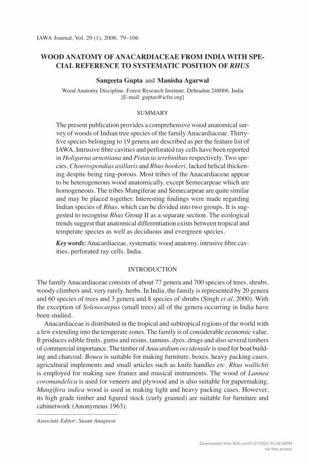
Growth boundaries In Anacardiaceae growth boundaries are usually indistinct or faint (Fig. 1). Distinct boundaries occur in species with marginal parenchyma bands and those having ring porosity (Fig. 2–4).
Downloaded from Brill.com01/27/2022 03:45:08PMvia free access
IAWA Journal, Vol. 29 (1), 200882 83Gupta & Agarwal — Wood anatomy of Anacardiaceae from India
1 2
5 6
43
Downloaded from Brill.com01/27/2022 03:45:08PMvia free access
IAWA Journal, Vol. 29 (1), 200882 83Gupta & Agarwal — Wood anatomy of Anacardiaceae from India
7 8 9
10 11 12
13 14 15
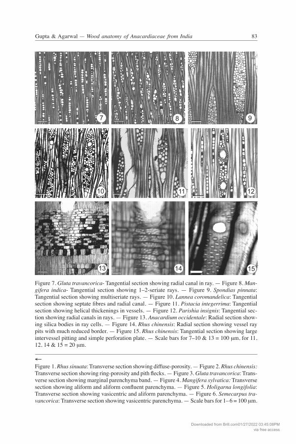
Figure 7. Gluta travancorica- Tangential section showing radial canal in ray. — Figure 8. Man-gifera indica- Tangential section showing 1–2-seriate rays. — Figure 9. Spondias pinnata: Tangential section showing multiseriate rays. — Figure 10. Lannea coromandelica: Tangential section showing septate fibres and radial canal. — Figure 11. Pistacia integerrima: Tangential section showing helical thickenings in vessels. — Figure 12. Parishia insignis: Tangential sec-tion showing radial canals in rays. — Figure 13. Anacardium occidentale: Radial section show-ing silica bodies in ray cells. — Figure 14. Rhus chinensis: Radial section showing vessel ray pits with much reduced border. — Figure 15. Rhus chinensis: Tangential section showing large intervessel pitting and simple perforation plate. — Scale bars for 7–10 & 13 = 100 μm, for 11, 12, 14 & 15 = 20 μm.
←Figure 1. Rhus sinuata: Transverse section showing diffuse-porosity. — Figure 2. Rhus chinensis: Transverse section showing ring-porosity and pith flecks. — Figure 3. Gluta travancorica: Trans-verse section showing marginal parenchyma band. — Figure 4. Mangifera sylvatica: Transverse section showing aliform and aliform confluent parenchyma. — Figure 5. Holigarna longifolia: Transverse section showing vasicentric and aliform parenchyma. — Figure 6. Semecarpus tra-vancorica: Transverse section showing vasicentric parenchyma. — Scale bars for 1–6 = 100 μm.
Downloaded from Brill.com01/27/2022 03:45:08PMvia free access
IAWA Journal, Vol. 29 (1), 200884 85Gupta & Agarwal — Wood anatomy of Anacardiaceae from India
16 17 18
19 20 21
22 23 24
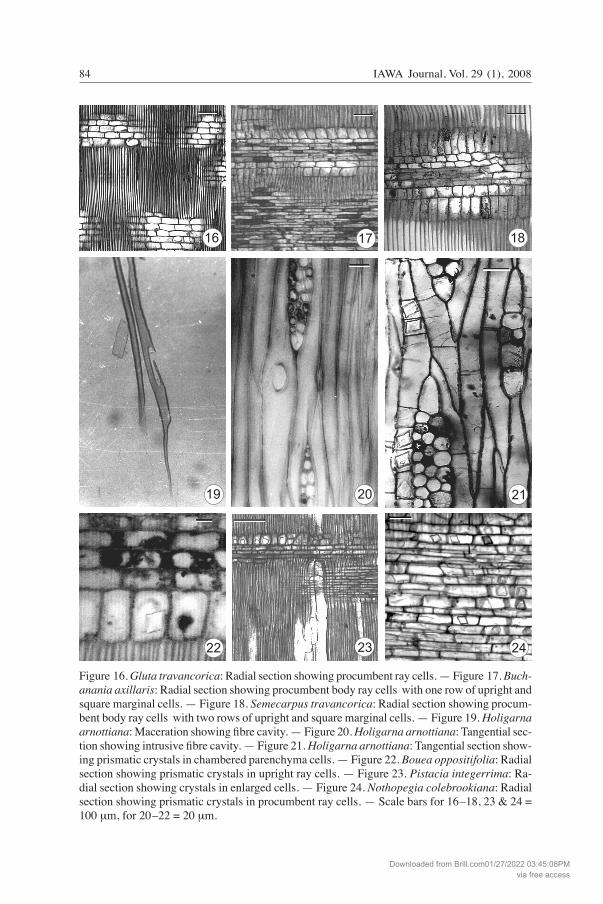
Figure 16. Gluta travancorica: Radial section showing procumbent ray cells. — Figure 17. Buch-anania axillaris: Radial section showing procumbent body ray cells with one row of upright and square marginal cells. — Figure 18. Semecarpus travancorica: Radial section showing procum-bent body ray cells with two rows of upright and square marginal cells. — Figure 19. Holigarna arnottiana: Maceration showing fibre cavity. — Figure 20. Holigarna arnottiana: Tangential sec-tion showing intrusive fibre cavity. — Figure 21. Holigarna arnottiana: Tangential section show-ing prismatic crystals in chambered parenchyma cells. — Figure 22. Bouea oppositifolia: Radial section showing prismatic crystals in upright ray cells. — Figure 23. Pistacia integerrima: Ra-dial section showing crystals in enlarged cells. — Figure 24. Nothopegia colebrookiana: Radial section showing prismatic crystals in procumbent ray cells. — Scale bars for 16–18, 23 & 24 = 100 μm, for 20–22 = 20 μm.
Downloaded from Brill.com01/27/2022 03:45:08PMvia free access
IAWA Journal, Vol. 29 (1), 200884 85Gupta & Agarwal — Wood anatomy of Anacardiaceae from India
Vessels Vessel grouping and distribution – The species of Choerospondias, Cotinus, Pistacia and Rhus growing in temperate climates are ring-porous (Fig. 2). Other species grow-ing in the tropics with weak seasonal climate changes are exclusively diffuse-porous (Fig. 1). Vessel frequency and size – Vessel frequency ranges from 2–73/mm2. Average tan-gential vessel diameter ranges from 75–214 μm and average vessel member length from 246–600 μm. Vessel perforation – Exclusively simple perforation plates (Fig. 15). Wall pitting – Intervessel pits alternate and non-vestured. The pit shape ranges from circular to polygonal. Average intervessel pit size in horizontal direction ranges from 3 to 16 μm.
1) Minute intervessel pits (< 4 μm) occur in two species, viz. Rhus parviflora and R. sinuata.
2) Medium-sized intervessel pits (7–10 μm) occur in 11 species (Fig. 15).3) Large intervessel pits (>10 μm) are present in 24 species.
Vessel-ray pits are usually with much reduced borders to apparently simple, and round to oval (Fig. 14). Along with this category in some samples large horizontal (gash-like) vessel-ray pits are also present. Buchanania lanzan and Bouea oppositifolia have both horizontal and vertical pitting. Tyloses are usually present. Vessel-wall thickenings – Helical thickenings, when present, cover the vessel wall completely (Fig. 11). They may be fine and weak. Helical thickenings are commonly present in the narrow vessel elements of ring-porous species of Cotinus, Pistacia and Rhus, while absent in Choerospondias axillaris and Rhus hookeri.
Fibres Fibres are septate or non-septate libriform fibres with simple to minutely bordered pits mainly in the radial walls. The fibre wall thickness falls under the IAWA categories very thin and thin- to thick-walled. Indian Anacardiaceae do not have thick-walled fibres. The fibre length ranges between 481–1819 μm. Seventy-five percent of the spe-cies of Indian Anacardiaceae have average fibre length between 800–1200 μm. Fibres are all septate in 10 (28%) species (Fig. 10) while in the rest of the species they are mixed with non-septate fibres. Intrusive cavities in the fibre walls, similar to the ones described by Dias-Leme and Angyalossy-Alfonso (1998) in four species of Euphor-biaceae, have been observed in Holigarna arnottiana (Fig. 19–20).
Axial parenchyma All types of parenchyma were observed in the Indian Anacardiaceae except for diffuse and diffuse-in-aggregates. Parenchyma distributions are of the following types: scanty paratracheal (Fig. 1), vasicentric, aliform, aliform to confluent (Fig. 4–6), paratracheal and apotracheal bands (Fig. 3 & 4).
Downloaded from Brill.com01/27/2022 03:45:08PMvia free access
IAWA Journal, Vol. 29 (1), 200886 87Gupta & Agarwal — Wood anatomy of Anacardiaceae from India
25 26
27 28
29 30
Downloaded from Brill.com01/27/2022 03:45:08PMvia free access
IAWA Journal, Vol. 29 (1), 200886 87Gupta & Agarwal — Wood anatomy of Anacardiaceae from India
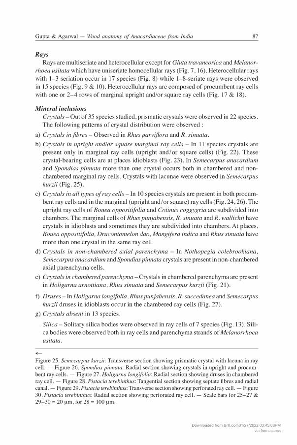
Rays Rays are multiseriate and heterocellular except for Gluta travancorica and Melanor-rhoea usitata which have uniseriate homocellular rays (Fig. 7, 16). Heterocellular rays with 1–3 seriation occur in 17 species (Fig. 8) while 1–8-seriate rays were observed in 15 species (Fig. 9 & 10). Heterocellular rays are composed of procumbent ray cells with one or 2–4 rows of marginal upright and/or square ray cells (Fig. 17 & 18).
Mineral inclusions Crystals – Out of 35 species studied, prismatic crystals were observed in 22 species. The following patterns of crystal distribution were observed :a) Crystals in fibres – Observed in Rhus parviflora and R. sinuata.b) Crystals in upright and/or square marginal ray cells – In 11 species crystals are
present only in marginal ray cells (upright and/or square cells) (Fig. 22). These crystal-bearing cells are at places idioblasts (Fig. 23). In Semecarpus anacardium and Spondias pinnata more than one crystal occurs both in chambered and non-chambered marginal ray cells. Crystals with lacunae were observed in Semecarpus kurzii (Fig. 25).
c) Crystals in all types of ray cells – In 10 species crystals are present in both procum-bent ray cells and in the marginal (upright and/or square) ray cells (Fig. 24, 26). The upright ray cells of Bouea oppositifolia and Cotinus coggygria are subdivided into chambers. The marginal cells of Rhus punjabensis, R. sinuata and R. wallichii have crystals in idioblasts and sometimes they are subdivided into chambers. At places, Bouea oppositifolia, Dracontomelon dao, Mangifera indica and Rhus sinuata have more than one crystal in the same ray cell.
d) Crystals in non-chambered axial parenchyma – In Nothopegia colebrookiana, Semecarpus anacardium and Spondias pinnata crystals are present in non-chambered axial parenchyma cells.
e) Crystals in chambered parenchyma – Crystals in chambered parenchyma are present in Holigarna arnottiana, Rhus sinuata and Semecarpus kurzii (Fig. 21).
f) Druses – In Holigarna longifolia, Rhus punjabensis, R. succedanea and Semecarpus kurzii druses in idioblasts occur in the chambered ray cells (Fig. 27).
g) Crystals absent in 13 species. Silica – Solitary silica bodies were observed in ray cells of 7 species (Fig. 13). Sili-
ca bodies were observed both in ray cells and parenchyma strands of Melanorrhoea usitata.
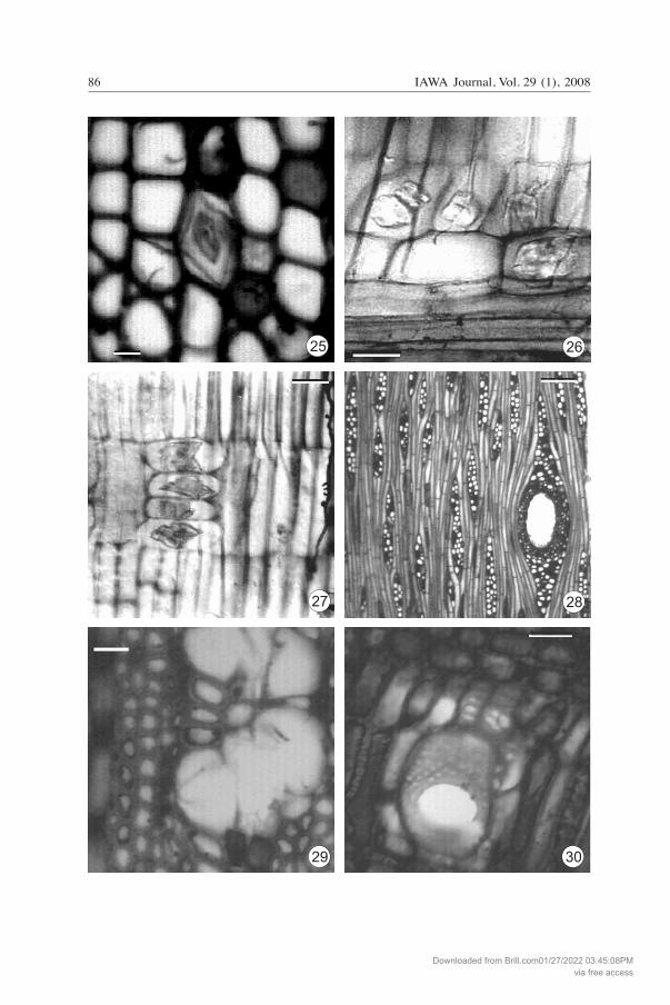
←Figure 25. Semecarpus kurzii: Transverse section showing prismatic crystal with lacuna in ray cell. — Figure 26. Spondias pinnata: Radial section showing crystals in upright and procum-bent ray cells. — Figure 27. Holigarna longifolia: Radial section showing druses in chambered ray cell. — Figure 28. Pistacia terebinthus: Tangential section showing septate fibres and radial canal. — Figure 29. Pistacia terebinthus: Transverse section showing perforated ray cell. — Figure 30. Pistacia terebinthus: Radial section showing perforated ray cell. — Scale bars for 25–27 & 29–30 = 20 μm, for 28 = 100 μm.
Downloaded from Brill.com01/27/2022 03:45:08PMvia free access
IAWA Journal, Vol. 29 (1), 200888 89Gupta & Agarwal — Wood anatomy of Anacardiaceae from India
Radial canals Radial canals were observed in 15 species. Radial canals are either single within rays (Fig. 7), or more than one canal is present within a ray (Fig. 10, 12), occurring in the widest portions of multiseriate rays.
GENERIC WOOD ANATOMICAL DESCRIPTION
1. Anacardium L. — A genus of shrubs and trees indigenous to tropical South America, Africa and Asia; 15 species are present in the world, 1 naturalized in India (Singh et al. 2000). Species studied: A. occidentale L. A diffuse-porous wood. Growth rings indistinct. Vessels 3–13/mm2, usually in pairs or in groups of 3–4 and solitary, somewhat rounded in outline, tangential diameter 135 (54–200) μm. Vessel members 387 (214–535) μm long. Intervessel pits non-vestured, alternate, bordered, polygonal, 10–12 μm in diameter with slit-like apertures, vessel-ray pits simple or with reduced borders, round to oval. Tyloses present. Fibres nonseptate, 600 (481–856) μm long, average diameter 14–17 μm, wall thickness 2.5 μm, thin- to thick-walled, pits minutely bordered, mainly confined to the radial walls. Fibre/Vessel element length (F/V) ratio 1.66–1.9. Parenchyma vasicentric to aliform, in 2–4-celled strands. Rays 4–7/mm, uni- to biseriate, heterocellular: body ray cells with procumbent, square and upright cells; ray height 400 (182–642) μm or 3–20 cells. Silica bodies present in rays. Crystals absent. Dong and Baas (1993) reported crystals in rays, but in our samples crystals were absent. Détienne and Jacquet (1983) also reported crystals absent in rays (in Wheeler et al. 2004-onwards).
2. Bouea Meissner — Distributed in tropical South-East Asia and Malaysia; 4 species are present in the world, 1 in India (Singh et al. 2000). Species studied: B. oppositifolia (Roxb.) Meissner A diffuse-porous wood. Growth rings indistinct to distinct. Vessels 2–8/mm2, mostly solitary, occasionally in radial multiples of 2–3 or more, round to oval in outline, tan-gential diameter 134 (75–193) μm, vessel members 337 (214–642) μm long. Intervessel pits non-vestured, alternate, bordered, and circular to oval, 6–10 μm in diameter with slit-like apertures. Vessel-ray pits with much-reduced borders to apparently simple, round to elongated pits horizontal to vertical. Tyloses common. Pith flecks occasion-ally present. Fibres non-septate, 896 (535–1177) μm long, average diameter 14 μm, wall thickness 2.8 μm, thin- to thick-walled with minutely bordered pits mainly con-fined to the radial wall, F/V ratio 2.6. Parenchyma apotracheal, in marginal bands and paratracheal, vasicentric forming a thin sheath, in 2–5-celled strands. Rays 2–4/mm, uni- to biseriate, heterocellular: body ray cells procumbent with 1–3 rows of marginal upright or square cells; ray height 481 (182–781) μm or 3–16 cells. Prismatic crystals present in ray cells. Radial canals absent.
3. Buchanania Sprengel — The genus consists of 25 species of trees distributed in Tropical Asia, Malaysia, Australia, Micronesia and Polynesia, 8 species are present in India (Singh et al. 2000), out of which 3 were studied.
Downloaded from Brill.com01/27/2022 03:45:08PMvia free access
IAWA Journal, Vol. 29 (1), 200888 89Gupta & Agarwal — Wood anatomy of Anacardiaceae from India
Species studied: B. axillaris (Desr.) Ramamurthy, B. lancifolia Roxb. and B. lanzan Sprengel. A diffuse-porous wood. Growth rings indistinct, however, tangential bands of thick-walled fibres often give the impression of growth periodicity. Vessels 6–18/mm2, solitary and in radial multiples of 2–5, rarely in clusters, round to oval, average tan-gential diameter 139–176 μm, average vessel member length 362–502 μm (Table 2). Intervessel pits alternate, oval to polygonal, 7–14 μm in diameter. Vessel-ray pits sim-ple to indistinctly bordered, round to elongate, at places vertical in B. lanzan. Tyloses present. Fibres non-septate, 932–1087 μm long, average diameter range 16–19.8 μm, wall thickness 2.5–4 μm, thin- to thick-walled with minutely bordered pits mainly confined to the radial walls, F/V ratio 2.0–3.0. Parenchyma vasicentric to aliform, in 2–4-celled strands in B. axillaris and B. lancifolia, in 3–8-celled strands in B. lanzan. Rays 3–5/mm, heterocellular: body ray cells procumbent with one row of marginal upright cells; 1–3-seriate in B. axillaris and B. lancifolia and 1–5-seriate in B. lanzan. Average ray heights 385–556/m (see Table 2). Radial canals present with 1–2 layers of partly lignified thin- to thick-walled epithelial cells. Silica bodies present in ray cells. Crystals absent. Dong and Baas (1993) studied three species from China and reported crystals in septate fibres of Buchanania arborescens only. Metcalfe and Chalk (1950) quoted Janssonius on the rare occurrence of solitary crystals in fibres in a single specimen of Buchanania florida. Septate fibres and crystals in fibres have not been observed in the Indian species studied. Pearson and Brown (1932) described 2 species (B. axillaris and B. lanzan) and used the term “several crystals in same cavity” probably for silica bodies.
4. Choerospondias Burtt & Hill — Distributed in South-East Asia; 2 species occur in India (Singh et al. 2000), one of which was studied. Species studied: C. axillaris (Roxb.) Burtt & Hill. A ring-porous wood. Growth rings distinct and marked by differences in vessel diameter and fibre wall thickness. Earlywood vessels solitary and in radial multiples of 2–4, tangential diameter 185 (171–203) μm, latewood vessels small, tangential diameter 100 (85–139) μm, vessel member length 438 (278–599) μm. Intervessel pits alternate, 8–11 μm in diameter, polygonal with slit-like apertures. Vessel-ray pits simple or with reduced borders. Fibres septate, average fibre length 1155 (963–1348) μm, average diameter18 μm, wall thickness 2 μm, thin- to thick-walled with minutely bordered pits, confined to radial walls, F/V ratio 2.6. Parenchyma scanty vasicentric, in
Table 2. Variation in wood anatomical features of Buchanania.
Wood anatomical features B. axillaris B. lancifolia B. lanzan
Vessel diameter (μm) 176 (85–267) 139 (64–214) 145 (64–214) Vessel member length (μm) 396 (321–588) 502 (267–749) 362 (214–535) Ray seriation 1–3 1–3 1–5 Ray height (μm) 556 (149–963) 385 (128–642) 442 (139–706) Ray height (no. of cells) 4–38 2–28 3–63 Fibre length (μm) 1087 (856–1391) 1086 (749–1391) 932 (695–1177)
Downloaded from Brill.com01/27/2022 03:45:08PMvia free access
IAWA Journal, Vol. 29 (1), 200890 91Gupta & Agarwal — Wood anatomy of Anacardiaceae from India
3–7-celled strands. Rays 2–5/mm, 1–5-seriate, heterocellular: body ray cells composed of procumbent cells with 1–2 rows of upright or square marginal cells; ray height 406 (214–599) μm or 9–30 cells. Radial canals present with 1–2 layers of partly lignified thin- to thick-walled epithelial cells. Terrazas (1994, in Wheeler et al. 2004-onwards) reported rays 1–3-seriate and pres-ence of crystals in rays and parenchyma. Dong and Baas (1993) reported crystals in ray cells in one sample and absent in another sample, but in our sample crystals were absent.
5. Cotinus Miller — A small genus of trees distributed in North America, Africa and Asia; 3 species reported in the world, 2 in India (Singh et al. 2000), one of which was studied. Species studied: C. coggygria Scop. A ring-porous wood. Growth rings distinct and marked by differences in vessel diameter and fibre wall thickness. Earlywood vessels solitary and in radial multiples of 2–3, tangential diameter 154 (107–214) μm, latewood vessels much more numerous, mostly in radial multiples and clusters arranged in a radial or oblique pattern, tangential diameter 65 (49–81) μm, vessel member length 272 (171–374) μm, Perforation plates simple. Intervessel pits 8–10 μm in diameter, alternate, round, with slit-like apertures. Vessel-ray pits indistinctly bordered, round and simple. Helical thickenings present in narrow vessel elements. Tyloses present. Fibres nonseptate, 694 (535–856) μm long, average diameter 13 μm, wall thickness 3 μm, thin- to thick-walled with minutely bor-dered pits, confined to radial walls, F/V ratio 2.5. Parenchyma scanty vasicentric, in 2–4-celled strands. Rays 4–6/mm, 1–4-seriate, heterocellular: body ray cells procum-bent with 1–4 rows of upright to square marginal cells; ray height 245 (118–375) μm or 5–17 cells. Prismatic crystals present in ray cells. Terrazas (1994, in Wheeler et al. 2004-onwards) reported vascular and vasicentric tracheids and prismatic crystals in axial parenchyma cells but in our sample these were not observed. They are reported as variable by Wheeler et al. (2004-onwards).
6. Dracontomelon Blume — Distributed in South-East Asia, Myanmar, Thailand, China, Malaysia and the Pacific Islands; c. 8 species, 1 in India (Singh et al. 2000). Species studied: D. dao (Blanco) Merr. & Rolfe. A diffuse-porous wood. Growth rings absent or faintly marked by dark coloured bands of dense fibres. Vessels 2–10 /mm2 , mostly solitary, remainder in short radial multiples of 2–4, rarely more, or in clusters, round, tangential diameter 176 (75–278) μm, vessel members 398 (214–642) μm long. Intervessel pits alternate, polygonal, 10–14 μm in diameter with slit-like apertures. Vessel-ray pits simple or with reduced borders, round to elongate. Fibres septate, septa fine, average fibre length 1206 (749–1551) μm, aver-age diameter 15–17 μm, wall thickness 3.6 μm, thin- to thick-walled, with indistinctly bordered pits mainly confined to the radial walls. F/V ratio 2.5. Parenchyma scanty, or vasicentric to aliform in very few places, in 5–6-celled strands. Rays 3–5 /mm, 1–4-seriate (mostly 3–4-seriate), heterocellular: body ray cells procumbent with 2–4 rows of marginal upright or square cells; ray height 695 (214–1177) μm or 3–40 cells. Pris-matic crystals present in ray cells.
Downloaded from Brill.com01/27/2022 03:45:08PMvia free access
IAWA Journal, Vol. 29 (1), 200890 91Gupta & Agarwal — Wood anatomy of Anacardiaceae from India
Terrazas (1994, in Wheeler et al. 2004-onwards) reported prismatic crystals in non-chambered axial parenchyma cells but in our sample these were not observed. This character is reported as variable by Wheeler et al. (2004-onwards).
7. Drimycarpus Hook. f. — A small genus of trees distributed in South-East Asia, Myanmar and Bangladesh; c. 2 species, 1 in India (Singh et al. 2000). Species studied: D. racemosus (Roxb.) Hook. f. A diffuse-porous wood. Growth rings very faint, demarcated by narrow layers of dark-coloured fibres. Vessels 6–31/mm2 , solitary or few in radial multiples of 2–5, rarely in small clusters, round to oval, tangential diameter 123 (75–170) μm, vessel members 511 (321–642) μm long. Intervessel pits alternate, polygonal, 8–10 μm in diameter, with slit-like apertures. Vessel-ray pits simple or with much reduced border, round to elongated. Tyloses present. Fibres nonseptate, 1307 (909–1605) μm long, average diameter 15 μm, wall thickness 4 μm, thin- to thick-walled, with simple or indistinctly bordered pits mainly confined to the radial walls. F/V ratio 2.5–3. Paren-chyma vasicentric to aliform, rarely in apotracheal bands, in 3–7-celled strands. Rays 2-5/mm, 1–3-seriate, heterocellular: body ray cells procumbent with 1–2 rows of mar-ginal, upright or square cells; ray height 684 (299–1070) μm or 4 to 28 cells. Prismatic crystals present in marginal and procumbent ray cells.
8. Gluta L. — The genus consists of 12 species distributed in South-East Asia, one in India (Singh et al. 2000). Species studied: G. travancorica Bedd. A diffuse-porous wood. Growth rings indistinct, but zonate bands of parenchyma and zones of thicker-walled fibrous tissue may give the impression of growth rings. Vessels 2–6/mm2, predominantly solitary, sometimes in radial multiples of 2–5, round to oval, tangential diameter 208 (128–288) μm. Vessel members 412 (321–642) μm long. Intervessel pits alternate, polygonal, 8–10 μm in diameter. Vessel-ray pits simple or with reduced borders, round to elongate. Tyloses common. Fibres non-septate, 1151 (963–1498) μm long, average diameter 13–14 μm, wall thickness 2.8 μm, thin- to thick-walled, with simple to minutely bordered pits mainly confined to radial walls. F/V ratio 1.8–2.8. Parenchyma apotracheal, in marginal or seemingly marginal bands, 2–5 cells wide, and scanty vasicentric, in 2–6-celled strands. Rays 3–5/mm, uniseriate, rarely multiseriate (only those containing radial canals), homocellular (procumbent cells) or weakly heterocellular: procumbent cells with few square and upright marginal ray cells; ray height 353 (85–620) μm or 3 to 22 cells. Radial canals present with 1–3 layers of partly lignified, thin-walled epithelial cells. Silica bodies present in ray cells. Pearson and Brown (1932) used the term aggregate crystals for silica bodies.
9. Holigarna Buch.-Ham. — A genus of large trees restricted to India and Myanmar; 7 species occur in India (Singh et al. 2000), four of which were studied. Species studied: H. arnottiana Hook. f., H. beddomei Hook. f., H. grahamii (Wight) Kurz and H. longifolia Roxb. Diffuse-porous woods. Growth rings usually absent or faintly demarcated by dark coloured fibrous bands. Vessels 2–10/mm2 in H. arnottiana and 2–8/mm2 in H. bed-
Downloaded from Brill.com01/27/2022 03:45:08PMvia free access
IAWA Journal, Vol. 29 (1), 200892 93Gupta & Agarwal — Wood anatomy of Anacardiaceae from India
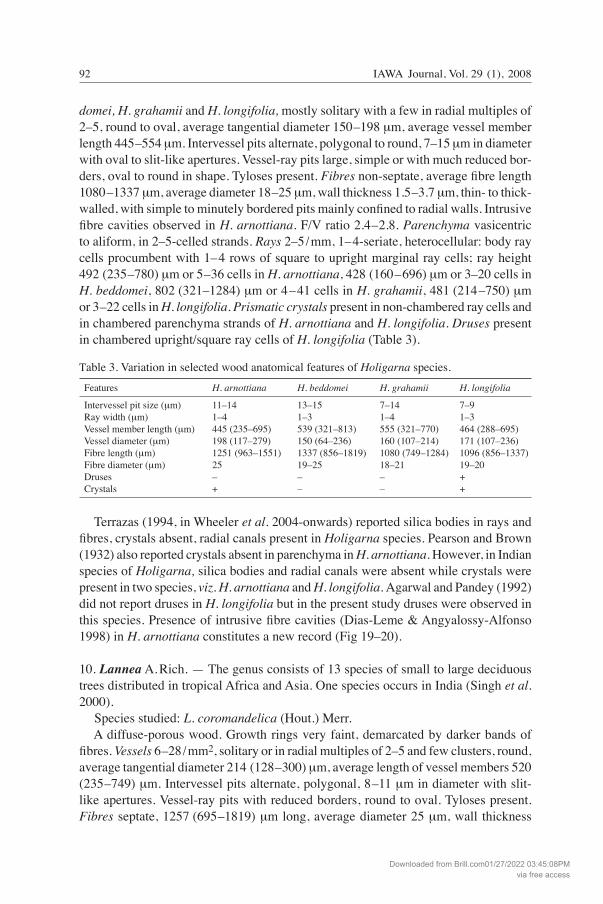
domei, H. grahamii and H. longifolia, mostly solitary with a few in radial multiples of 2–5, round to oval, average tangential diameter 150–198 μm, average vessel member length 445–554 μm. Intervessel pits alternate, polygonal to round, 7–15 μm in diameter with oval to slit-like apertures. Vessel-ray pits large, simple or with much reduced bor-ders, oval to round in shape. Tyloses present. Fibres non-septate, average fibre length 1080–1337 μm, average diameter 18–25 μm, wall thickness 1.5–3.7 μm, thin- to thick-walled, with simple to minutely bordered pits mainly confined to radial walls. Intrusive fibre cavities observed in H. arnottiana. F/V ratio 2.4–2.8. Parenchyma vasicentric to aliform, in 2–5-celled strands. Rays 2–5/mm, 1–4-seriate, heterocellular: body ray cells procumbent with 1–4 rows of square to upright marginal ray cells; ray height 492 (235–780) μm or 5–36 cells in H. arnottiana, 428 (160–696) μm or 3–20 cells in H. beddomei, 802 (321–1284) μm or 4–41 cells in H. grahamii, 481 (214–750) μm or 3–22 cells in H. longifolia. Prismatic crystals present in non-chambered ray cells and in chambered parenchyma strands of H. arnottiana and H. longifolia. Druses present in chambered upright/square ray cells of H. longifolia (Table 3).
Table 3. Variation in selected wood anatomical features of Holigarna species.
Features H. arnottiana H. beddomei H. grahamii H. longifolia
Intervessel pit size (μm) 11–14 13–15 7–14 7–9 Ray width (μm) 1–4 1–3 1–4 1–3 Vessel member length (μm) 445 (235–695) 539 (321–813) 555 (321–770) 464 (288–695) Vessel diameter (μm) 198 (117–279) 150 (64–236) 160 (107–214) 171 (107–236) Fibre length (μm) 1251 (963–1551) 1337 (856–1819) 1080 (749–1284) 1096 (856–1337) Fibre diameter (μm) 25 19–25 18–21 19–20 Druses – – – + Crystals + – – +
Terrazas (1994, in Wheeler et al. 2004-onwards) reported silica bodies in rays and fibres, crystals absent, radial canals present in Holigarna species. Pearson and Brown (1932) also reported crystals absent in parenchyma in H. arnottiana. However, in Indian species of Holigarna, silica bodies and radial canals were absent while crystals were present in two species, viz. H. arnottiana and H. longifolia. Agarwal and Pandey (1992) did not report druses in H. longifolia but in the present study druses were observed in this species. Presence of intrusive fibre cavities (Dias-Leme & Angyalossy-Alfonso 1998) in H. arnottiana constitutes a new record (Fig 19–20).
10. Lannea A. Rich. — The genus consists of 13 species of small to large deciduous trees distributed in tropical Africa and Asia. One species occurs in India (Singh et al. 2000). Species studied: L. coromandelica (Hout.) Merr. A diffuse-porous wood. Growth rings very faint, demarcated by darker bands of fibres. Vessels 6–28/mm2, solitary or in radial multiples of 2–5 and few clusters, round, average tangential diameter 214 (128–300) μm, average length of vessel members 520 (235–749) μm. Intervessel pits alternate, polygonal, 8–11 μm in diameter with slit-like apertures. Vessel-ray pits with reduced borders, round to oval. Tyloses present. Fibres septate, 1257 (695–1819) μm long, average diameter 25 μm, wall thickness
Downloaded from Brill.com01/27/2022 03:45:08PMvia free access
IAWA Journal, Vol. 29 (1), 200892 93Gupta & Agarwal — Wood anatomy of Anacardiaceae from India
4–5 μm, thin- to thick-walled, with simple pits mainly confined to the radial walls. F/V ratio 2.5. Parenchyma scanty vasicentric, in 2–4-celled strands. Rays 3–5/mm, 1–5-seriate, heterocellular: with body ray cells procumbent and 1–2 rows of upright or square marginal cells; ray height 497 (192–802) μm or 5 to 33 cells. Radial canals present with 2–3 layers of partly lignified thin-walled epithelial cells, in some rays two radial canals present. Silica bodies present in ray cells. Prismatic crystals present in procumbent and marginal ray cells and idioblasts. Pearson and Brown (1932) used the term aggregate crystals for silica bodies.
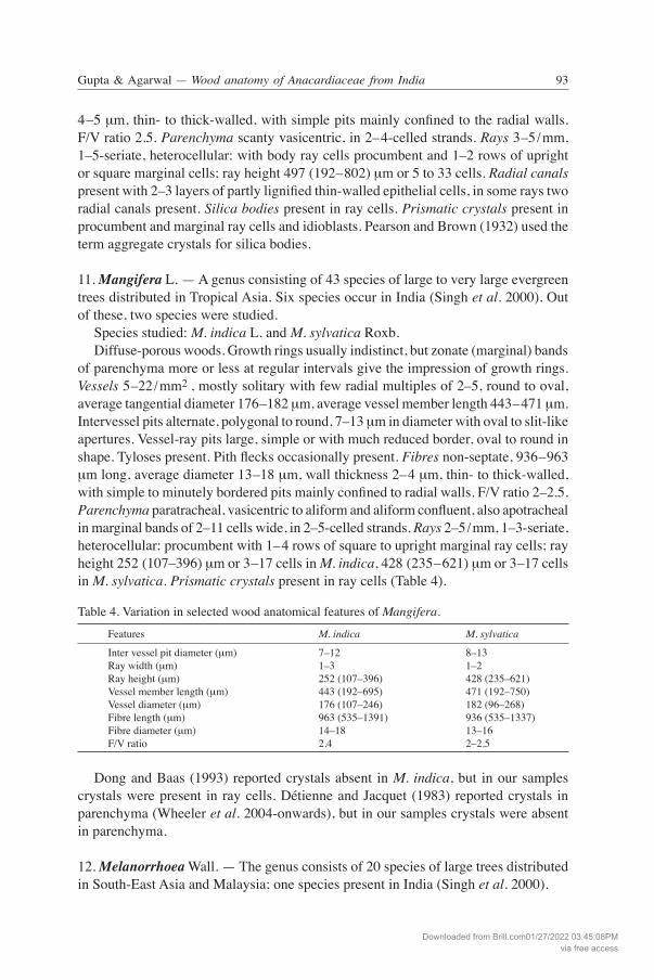
11. Mangifera L. — A genus consisting of 43 species of large to very large evergreen trees distributed in Tropical Asia. Six species occur in India (Singh et al. 2000). Out of these, two species were studied. Species studied: M. indica L. and M. sylvatica Roxb. Diffuse-porous woods. Growth rings usually indistinct, but zonate (marginal) bands of parenchyma more or less at regular intervals give the impression of growth rings. Vessels 5–22/mm2 , mostly solitary with few radial multiples of 2–5, round to oval, average tangential diameter 176–182 μm, average vessel member length 443–471 μm. Intervessel pits alternate, polygonal to round, 7–13 μm in diameter with oval to slit-like apertures. Vessel-ray pits large, simple or with much reduced border, oval to round in shape. Tyloses present. Pith flecks occasionally present. Fibres non-septate, 936–963 μm long, average diameter 13–18 μm, wall thickness 2–4 μm, thin- to thick-walled, with simple to minutely bordered pits mainly confined to radial walls. F/V ratio 2–2.5. Parenchyma paratracheal, vasicentric to aliform and aliform confluent, also apotracheal in marginal bands of 2–11 cells wide, in 2–5-celled strands. Rays 2–5/mm, 1–3-seriate, heterocellular: procumbent with 1–4 rows of square to upright marginal ray cells; ray height 252 (107–396) μm or 3–17 cells in M. indica, 428 (235–621) μm or 3–17 cells in M. sylvatica. Prismatic crystals present in ray cells (Table 4).
Table 4. Variation in selected wood anatomical features of Mangifera.
Features M. indica M. sylvatica
Inter vessel pit diameter (μm) 7–12 8–13Ray width (μm) 1–3 1–2Ray height (μm) 252 (107–396) 428 (235–621) Vessel member length (μm) 443 (192–695) 471 (192–750) Vessel diameter (μm) 176 (107–246) 182 (96–268) Fibre length (μm) 963 (535–1391) 936 (535–1337) Fibre diameter (μm) 14–18 13–16F/V ratio 2.4 2–2.5
Dong and Baas (1993) reported crystals absent in M. indica, but in our samples crystals were present in ray cells. Détienne and Jacquet (1983) reported crystals in parenchyma (Wheeler et al. 2004-onwards), but in our samples crystals were absent in parenchyma.
12. Melanorrhoea Wall. — The genus consists of 20 species of large trees distributed in South-East Asia and Malaysia; one species present in India (Singh et al. 2000).
Downloaded from Brill.com01/27/2022 03:45:08PMvia free access
IAWA Journal, Vol. 29 (1), 200894 95Gupta & Agarwal — Wood anatomy of Anacardiaceae from India
Species studied: M. usitata Wall. A diffuse-porous wood. Growth rings distinct due to terminal parenchyma. Vessels 3–9/mm2, mostly solitary, few radial multiples of 2–5, large, tangential diameter 176 (107–235) μm, vessel member length 445 (214–695) μm. Intervessel pits alternate, 9–15 μm in diameter with oval to slit-like apertures, vessel-ray pits large, round and simple with reduced borders. Tyloses common. Fibres nonseptate, 1096 (749–1444) μm in length, average diameter 14–15 μm, wall thickness 3.7–4.5 μm, thin- to thick-walled, with simple pits mainly confined to the radial walls. F/V ratio 2.4. Parenchyma apo-tracheal, in tangential bands of 3–9 cells wide, and vasicentric, in 4–8-celled strands. Rays 3–5/mm, uniseriate, rarely multiseriate (only those containing radial canals), homocellular (procumbent cells) or weakly heterocellular: composed of procumbent cells with a few marginal square and upright ray cells; ray height 321 (107–535) μm or 4 to 23 cells. Radial canals present with 1–3 layers of partly lignified thin-walled epithelial cells. Silica bodies present in ray cells and parenchyma strands. Pearson and Brown (1932) used the term aggregate crystals for silica bodies.
13. Nothopegia Blume — A small genus of 10 species distributed in India and Sri Lanka; 7 species occur in India (Singh et al. 2000), out of which one was studied. Species studied: N. colebrookiana (Wight) Blume. A diffuse-porous wood. Growth rings faintly marked, demarcated by thin tangential bands of fibres. Vessels 5–30/mm2 , mainly solitary, remainder in radial multiples of 2–5, round to oval, tangential diameter 117 (80–160) μm, vessel member length 486 (267–706) μm, perforations simple. Intervessel pits alternate, polygonal, 6–8 μm in diameter, vessel-ray pits with much reduced borders to apparently simple, horizontal. Fibres non-septate, 1096 (856–1337) μm in length, average diameter 13–15 μm, wall thickness 3 μm, thin- to thick-walled, with simple pits numerous in both radial and tan-gential walls. F/V ratio 2.2. Parenchyma aliform confluent, in wavy tangential lines of 2–5 cells wide enclosing the vessels, at places vasicentric to winged aliform, in strands of 2–4 cells. Rays 3–5 per mm, 1–3-seriate, heterocellular: procumbent body ray cells with 1–2 rows of upright or square marginal cells; 476 (192–760) μm or 4–26 cells high. Prismatic crystals present in all types of ray cells and also in axial parenchyma strands. Kryn (1952) reported crystals in rays (Wheeler et al. 2004-onwards), but our study revealed crystals in both rays and parenchyma.
14. Parishia Hook. f. — A small genus of tall, evergreen trees, distributed in South-East Asia; 5 species in the world, one in India (Singh et al. 2000). Species studied: P. insignis Hook. f. A diffuse-porous wood. Growth rings indistinct or faint, demarcated by a faint line of denser latewood fibres. Vessels 5–10/mm2, solitary and in radial multiples of 2–4, rarely in clusters, round to oval, tangential diameter 128 (86–172) μm. Vessel members 444 (160–749) μm long, perforation plates simple. Intervessel pits alternate, polygo-nal, 8–10 μm in diameter, vessel-ray pits simple or with much reduced borders, round to elongate. Tyloses present. Fibres non-septate, 1096 (749–1444) μm long, average
Downloaded from Brill.com01/27/2022 03:45:08PMvia free access
IAWA Journal, Vol. 29 (1), 200894 95Gupta & Agarwal — Wood anatomy of Anacardiaceae from India
diameter 27 μm, wall thickness 3.7–5.2 μm, thin- to thick-walled, with simple pits mainly confined to the radial walls. F/V ratio 2.5. Parenchyma scanty paratracheal and vasicentric, in 2–4–celled strands. Rays 2–4/mm, 1–3-seriate, heterocellular: procum-bent body ray cells with 1–2 rows of upright or square marginal cells; 470 (171–770) μm or 4–27 cells high. Radial canals present with 1–2 layers of partly lignified thin-walled epithelial cells. Silica bodies present in ray cells. Crystals absent.
15. Pistacia L. — A genus of trees or shrubs distributed in the southern USA and Central America, Tropical Africa, the Mediterranean region, Asia to Malaysia; 10 species in the world, 3 in India (Singh et al. 2000). Wood samples of two tree species were studied, of which Pistacia terebinthus L. is exotic, but naturalised in India. Species studied: P. chinensis Bunge and Pistacia terebinthus L. Pistacia chinensis Bunge —Wood ring-porous. Growth rings distinct, demarcated by large earlywood vessels. Earlywood vessels solitary and in radial multiples of 2–3, tangential diameter 145 (107–203) μm, latewood vessels much more numerous, mostly in multiples and clusters in a radial to oblique pattern, tangential diameter 54 (32–75) μm, length 304 (117–492) μm. Perforation plates simple. Intervessel pits 10–12 μm in diameter, alternate, round with slit-like apertures. Vessel-ray pits with much reduced borders, to apparently simple, round to elongate. Helical thickenings present in narrow vessel elements. Tyloses present. Fibres nonseptate, 898 (620–1177) μm long, average diameter 14 μm, wall thickness 4 μm, thin- to thick-walled, with simple pits mainly confined to the radial walls. F/V ratio 3. Parenchyma vasicentric, in 2–5-celled strands. Rays 2–4/mm, 1–5-seriate, heterocellular: procumbent body ray cells with one to two rows of marginal upright or square cells; 214–749 μm or 4–40 cells high. Radial canals present with 2–3 layers of epithelial cells. Prismatic crystals present both in ordinary cells and idioblasts of procumbent and square ray cells. Pistacia terebinthus L. — This species is common on the islands and along the shores of the Mediterranean and is planted in India as a garden tree (Anonymous 1963). Growth rings indistinct. Wood diffuse-porous. Vessels 50–80/mm2, solitary or fre-quently in radial multiples of 2–4 and in small clusters, tangential diameter 48 (32–64) μm, vessel members 358 (235–481) μm long, partially filled with tyloses. Perforation plates simple. Helical thickenings present. Intervessel pits alternate, bordered, polygo-nal, 5–7 μm in diameter with slit-like apertures, vessel-ray pits larger than intervessel pits, with much reduced borders. Fibres septate (Fig. 28), 700 (536–757) μm long, average diameter 16 μm, wall thickness 4 μm, thin- to thick-walled. F/V ratio 2. Paren-chyma scanty. Rays 2–4/mm, 1–4-seriate, heterocellular: procumbent body ray cells with one row of marginal upright or square cells; 86–567 μm or 5–54 cells high. Ra-dial canals present (Fig. 28). Crystals present in ray cells. Perforated ray cells present (Fig. 29 & 30). On the basis of the literature and the present study of 10 samples of Pistacia (7 Indian wood samples of P. chinensis, 3 foreign wood samples of P. atlantica (F 9221, 9222, 9223 from Israel) and herbarium material of P. khinjuk) it was noted that all the species of Pistacia are ring-porous and have non-septate fibres, except for P. terebinthus, which is diffuse-porous, has septate fibres and perforated ray cells (other
Downloaded from Brill.com01/27/2022 03:45:08PMvia free access
IAWA Journal, Vol. 29 (1), 200896 97Gupta & Agarwal — Wood anatomy of Anacardiaceae from India
features remaining the same). Also, the radial canals of P. terebinthus were found to be very large with 3–7 layers of epithelial cells, canal diameter being 30–95 μm against 2–3 layers and canal diameter being 40–50 μm in other species. Perforated ray cells in the family Anacardiaceae were reported for the first time in Pistacia terebinthus (Agarwal et al. 2002). Further studies should be carried out to assess the systematic position of this species.
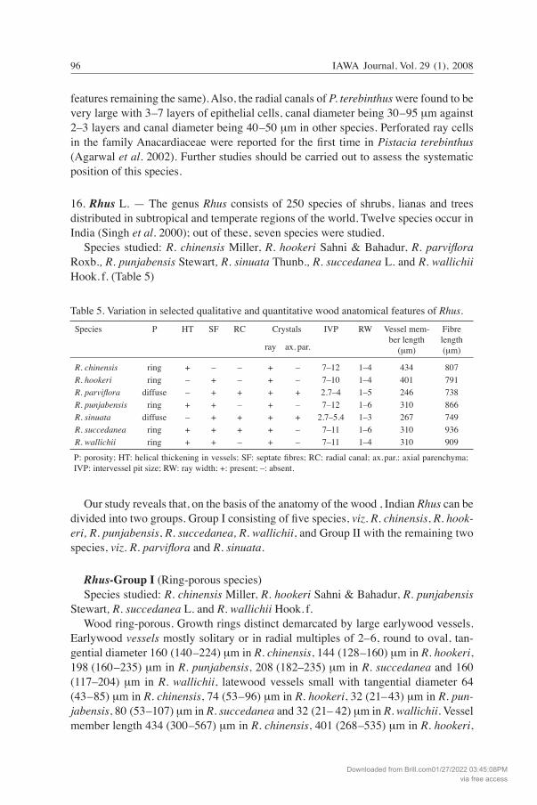
16. Rhus L. — The genus Rhus consists of 250 species of shrubs, lianas and trees distributed in subtropical and temperate regions of the world. Twelve species occur in India (Singh et al. 2000); out of these, seven species were studied. Species studied: R. chinensis Miller, R. hookeri Sahni & Bahadur, R. parviflora Roxb., R. punjabensis Stewart, R. sinuata Thunb., R. succedanea L. and R. wallichii Hook. f. (Table 5)
Table 5. Variation in selected qualitative and quantitative wood anatomical features of Rhus.
Species P HT SF RC Crystals IVP RW Vessel mem- Fibre ber length length ray ax. par. (μm) (μm)
R. chinensis ring + – – + – 7–12 1–4 434 807 R. hookeri ring – + – + – 7–10 1–4 401 791 R. parviflora diffuse – + + + + 2.7–4 1–5 246 738 R. punjabensis ring + + – + – 7–12 1–6 310 866 R. sinuata diffuse – + + + + 2.7–5.4 1–3 267 749 R. succedanea ring + + + + – 7–11 1–6 310 936 R. wallichii ring + + – + – 7–11 1–4 310 909
P: porosity; HT: helical thickening in vessels; SF: septate fibres; RC: radial canal; ax. par.: axial parenchyma; IVP: intervessel pit size; RW: ray width; +: present; –: absent.
Our study reveals that, on the basis of the anatomy of the wood , Indian Rhus can be divided into two groups. Group I consisting of five species, viz. R. chinensis, R. hook-eri, R. punjabensis, R. succedanea, R. wallichii, and Group II with the remaining two species, viz. R. parviflora and R. sinuata.
Rhus-Group I (Ring-porous species) Species studied: R. chinensis Miller, R. hookeri Sahni & Bahadur, R. punjabensis Stewart, R. succedanea L. and R. wallichii Hook. f. Wood ring-porous. Growth rings distinct demarcated by large earlywood vessels. Earlywood vessels mostly solitary or in radial multiples of 2–6, round to oval, tan-gential diameter 160 (140–224) μm in R. chinensis, 144 (128–160) μm in R. hookeri, 198 (160–235) μm in R. punjabensis, 208 (182–235) μm in R. succedanea and 160 (117–204) μm in R. wallichii, latewood vessels small with tangential diameter 64 (43–85) μm in R. chinensis, 74 (53–96) μm in R. hookeri, 32 (21–43) μm in R. pun-jabensis, 80 (53–107) μm in R. succedanea and 32 (21– 42) μm in R. wallichii. Vessel member length 434 (300–567) μm in R. chinensis, 401 (268–535) μm in R. hookeri,
Downloaded from Brill.com01/27/2022 03:45:08PMvia free access
IAWA Journal, Vol. 29 (1), 200896 97Gupta & Agarwal — Wood anatomy of Anacardiaceae from India
310 (193–428) μm in R. punjabensis, 310 (214–406) μm in R. succedanea and 310 (214–406) μm in R. wallichii. Perforation plates simple. Intervessel pits alternate, po-lygonal to round, 7–12 μm in diameter with oval to slit-like apertures. Vessel-ray pits large, simple or with much reduced borders, oval to round in shape. Helical thicken-ings present in narrow vessel elements of R. chinensis, R. punjabensis, R. succedanea and R. wallichii but absent in R. hookeri. Pith flecks present in R. chinensis. Fibres 791–936 μm long, septate, mixed with nonseptate fibres in R. hookeri, R. succedanea, R. wallichii and R. punjabensis (rarely septate) but in R. chinensis fibres are non-sep-tate, average fibre diameter 16–24 μm, thin- to thick-walled, wall thickness 3–4 μm, with minutely bordered pits mainly confined to the radial walls. F/V ratio 1.9–2.9. Parenchyma scanty paratracheal, in 2–7-celled strands. Rays 2–4/mm, 1–4-seriate in R. chinensis, R. hookeri and R. wallichii and 1–6-seriate in R. punjabensis and R. succedanea, heterocellular: procumbent with 1–2 rows of square to upright marginal ray cells; ray height 348 (160–535) μm or 7–26 cells in R. chinensis, R. hookeri and R. wallichii, 444 (140–749) μm or 7–37 cells in R. punjabensis and 401 (160–642) μm or 5–31 cells in R. succedanea. Radial canals present in R. succedanea with 1–2 layers of epithelial cells. Druses present in subdivided ray cells of R. punjabensis and R. succedanea. All the species have prismatic crystals in ray cells. Terrazas (1994, in Wheeler et al. 2004-onwards) reported radial canals absent in R. succedanea, but in our samples radial canals were observed in R. succedanea. Dong and Baas (1993) reported non-septate fibres in R. punjabensis, but in our samples both septate and non-septate fibres were observed (Table 5).
Rhus-Group II (Diffuse-porous species) Species studied: R. parviflora Roxb. and R. sinuata Thunb. Wood diffuse-porous. Growth rings distinct due to thick-walled latewood fibres. Vessels mostly solitary, or sometimes in radial multiples of 2–6, 40–73/mm2, tangential diameter 85 (52–117) μm in R. parviflora and 75 (50–110) μm in R. sinuata, vessel member length 246 (171–321) μm in R. parviflora and 267 (171–363) μm in R. sinu-ata. Perforation plates simple. Intervessel pits alternate, polygonal to round, diameter 2.7–5.4 μm with oval to slit-like apertures. Vessel-ray pits simple or with much reduced borders, oval to round in shape. Tyloses present. Fibres septate, mixed with nonsep-tate fibres, 738–749 μm long, average diameter 13–15 μm, thin- to thick-walled, wall thickness 3–4 μm, with minutely bordered pits mainly confined to the radial walls. F/V ratio 2.8–3. Parenchyma scanty paratracheal and in discontinuous unicellular marginal bands, in 2–7-celled strands. Rays heterocellular, 1–5-seriate, 358 (161–556) μm with 4–22 cells high in R. parviflora and 1–3-seriate, 391 (140–642) μm with 4–29 cells high in R. sinuata. Radial canals present with 1–2 layers of epithelial cells. Prismatic crystals present in ray cells, axial parenchyma cells and fibres in R. sinuat; present in ray cells and fibres in R. parviflora (Table 5).
17. Semecarpus L. f. — The genus consists of 60 species of trees, distributed in Tropi-cal South-East Asia, Australia and Fiji; 7 in India (Singh et al. 2000); out of these, 4 were studied
Downloaded from Brill.com01/27/2022 03:45:08PMvia free access
IAWA Journal, Vol. 29 (1), 200898 99Gupta & Agarwal — Wood anatomy of Anacardiaceae from India
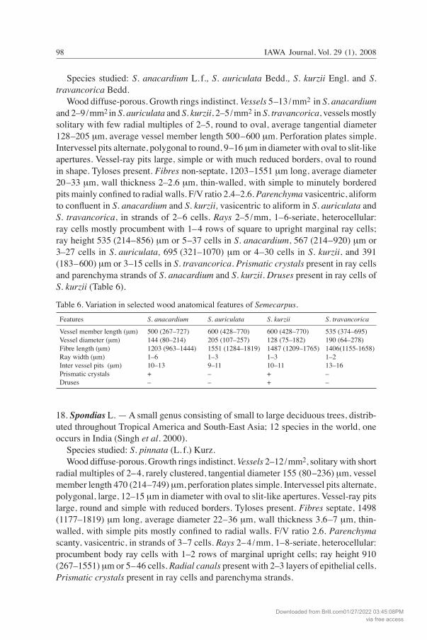
Species studied: S. anacardium L. f., S. auriculata Bedd., S. kurzii Engl. and S. travancorica Bedd. Wood diffuse-porous. Growth rings indistinct. Vessels 5–13/mm2 in S. anacardium and 2–9/mm2 in S. auriculata and S. kurzii, 2–5/mm2 in S. travancorica, vessels mostly solitary with few radial multiples of 2–5, round to oval, average tangential diameter 128–205 μm, average vessel member length 500–600 μm. Perforation plates simple. Intervessel pits alternate, polygonal to round, 9–16 μm in diameter with oval to slit-like apertures. Vessel-ray pits large, simple or with much reduced borders, oval to round in shape. Tyloses present. Fibres non-septate, 1203–1551 μm long, average diameter 20–33 μm, wall thickness 2–2.6 μm, thin-walled, with simple to minutely bordered pits mainly confined to radial walls. F/V ratio 2.4–2.6. Parenchyma vasicentric, aliform to confluent in S. anacardium and S. kurzii, vasicentric to aliform in S. auriculata and S. travancorica, in strands of 2–6 cells. Rays 2–5/mm, 1–6-seriate, heterocellular: ray cells mostly procumbent with 1–4 rows of square to upright marginal ray cells; ray height 535 (214–856) μm or 5–37 cells in S. anacardium, 567 (214–920) μm or 3–27 cells in S. auriculata, 695 (321–1070) μm or 4–30 cells in S. kurzii, and 391 (183–600) μm or 3–15 cells in S. travancorica. Prismatic crystals present in ray cells and parenchyma strands of S. anacardium and S. kurzii. Druses present in ray cells of S. kurzii (Table 6).
Table 6. Variation in selected wood anatomical features of Semecarpus.
Features S. anacardium S. auriculata S. kurzii S. travancorica
Vessel member length (μm) 500 (267–727) 600 (428–770) 600 (428–770) 535 (374–695) Vessel diameter (μm) 144 (80–214) 205 (107–257) 128 (75–182) 190 (64–278) Fibre length (μm) 1203 (963–1444) 1551 (1284–1819) 1487 (1209–1765) 1406(1155-1658) Ray width (μm) 1–6 1–3 1–3 1–2 Inter vessel pits (μm) 10–13 9–11 10–11 13–16 Prismatic crystals + – + – Druses – – + –
18. Spondias L. — A small genus consisting of small to large deciduous trees, distrib-uted throughout Tropical America and South-East Asia; 12 species in the world, one occurs in India (Singh et al. 2000). Species studied: S. pinnata (L. f.) Kurz. Wood diffuse-porous. Growth rings indistinct. Vessels 2–12/mm2, solitary with short radial multiples of 2–4, rarely clustered, tangential diameter 155 (80–236) μm, vessel member length 470 (214–749) μm, perforation plates simple. Intervessel pits alternate, polygonal, large, 12–15 μm in diameter with oval to slit-like apertures. Vessel-ray pits large, round and simple with reduced borders. Tyloses present. Fibres septate, 1498 (1177–1819) μm long, average diameter 22–36 μm, wall thickness 3.6–7 μm, thin-walled, with simple pits mostly confined to radial walls. F/V ratio 2.6. Parenchyma scanty, vasicentric, in strands of 3–7 cells. Rays 2–4/mm, 1–8-seriate, heterocellular: procumbent body ray cells with 1–2 rows of marginal upright cells; ray height 910 (267–1551) μm or 5–46 cells. Radial canals present with 2–3 layers of epithelial cells. Prismatic crystals present in ray cells and parenchyma strands.
Downloaded from Brill.com01/27/2022 03:45:08PMvia free access
IAWA Journal, Vol. 29 (1), 200898 99Gupta & Agarwal — Wood anatomy of Anacardiaceae from India
Kryn (1952) reported crystals absent in parenchyma, but in our samples crystals were observed in parenchyma (see also Wheeler et al. 2004-onwards). Pearson and Brown (1932) noted tracheids but we could not locate them in the same wood samples.
19. Swintonia Griffith — The genus consists of tall, evergreen trees which are distributed in South-East Asia; 16 species in the world, one in India (Singh et al. 2000). Species studied: S. floribunda Griffith. Wood diffuse-porous. Growth rings indistinct, occasionally fine concentric bands of parenchyma seem to delimit growth rings. Vessels 2–9/mm2, often solitary or in radial multiples of 2–4, rarely in clusters, round to oval, tangential diameter 193 (107–278) μm, vessel member length 470 (342–600) μm. Perforation plates simple. Intervessel pits alternate, polygonal, 11–15 μm in diameter with slit-like apertures. Vessel-ray pits with reduced borders or simple, round to elongate. Tyloses present. Fibres non-septate, 909 (535–1284) μm long, average diameter 15–18.5 μm, wall thickness 2.7–4 μm, thin- to thick-walled, with simple pits mainly confined to the radial walls. F/V ratio 1.5–1.9. Parenchyma vasicentric and in bands of 2–6 cells wide, strands composed of 2–4 cells. Rays 2–4/mm, 1–3-seriate, rarely uniseriate, heterocellular: procumbent body ray cells with one to two rows of marginal upright cells; ray height 433 (172–695) μm or 6–30 cells. Radial canals present with 2–3 layers of epithelial cells. Silica bodies present in ray cells. Terrazas (1994, in Wheeler et al. 2004-onwards) reported crystals in axial paren-chyma cells, but in our samples crystals were not observed. This feature is reported as variable by Wheeler et al. (2004-onwards).
DISCUSSION
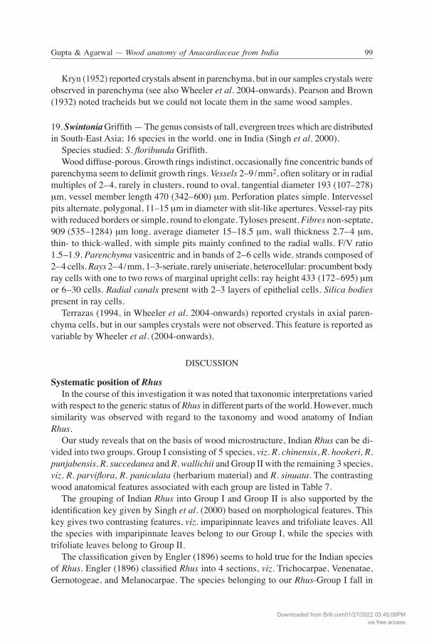
Systematic position of Rhus In the course of this investigation it was noted that taxonomic interpretations varied with respect to the generic status of Rhus in different parts of the world. However, much similarity was observed with regard to the taxonomy and wood anatomy of Indian Rhus. Our study reveals that on the basis of wood microstructure, Indian Rhus can be di-vided into two groups. Group I consisting of 5 species, viz. R. chinensis, R. hookeri, R. punjabensis, R. succedanea and R. wallichii and Group II with the remaining 3 species, viz. R. parviflora, R. paniculata (herbarium material) and R. sinuata. The contrasting wood anatomical features associated with each group are listed in Table 7. The grouping of Indian Rhus into Group I and Group II is also supported by the identification key given by Singh et al. (2000) based on morphological features. This key gives two contrasting features, viz. imparipinnate leaves and trifoliate leaves. All the species with imparipinnate leaves belong to our Group I, while the species with trifoliate leaves belong to Group II. The classification given by Engler (1896) seems to hold true for the Indian species of Rhus. Engler (1896) classified Rhus into 4 sections, viz. Trichocarpae, Venenatae, Gernotogeae, and Melanocarpae. The species belonging to our Rhus-Group I fall in
Downloaded from Brill.com01/27/2022 03:45:08PMvia free access
IAWA Journal, Vol. 29 (1), 2008100 101Gupta & Agarwal — Wood anatomy of Anacardiaceae from India
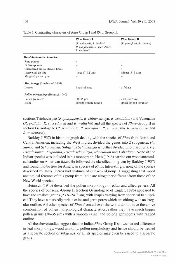
sections Trichocarpae (R. punjabensis, R. chinensis syn. R. semialata) and Venenatae (R. griffithii, R. succedanea and R. wallichii) and all the species of Rhus-Group II in section Gernotogeae (R. paniculata, R. parviflora, R. sinuata syn. R. mysorensis and R. tomentosa). Barkley (1937) in his monograph dealing with the species of Rhus from North and Central America, including the West Indies, divided the genus into 2 subgenera, viz. Sumac and Schomaltzia. Subgenus Schomaltzia is further divided into 5 sections, viz. Pseudosumac, Styphonia, Pseudoschmaltzia, Rhoeidium and Lobadium. None of the Indian species was included in his monograph. Hess (1946) carried out wood anatomi-cal studies on American Rhus. He followed the classification given by Barkley (1937) and found it to be true for American species of Rhus. Interestingly, none of the species described by Hess (1946) had features of our Rhus-Group II suggesting that wood anatomical features of this group from India are altogether different from those of the New World species. Heimsch (1940) described the pollen morphology of Rhus and allied genera. All the species of our Rhus-Group II (section Gernotogeae of Engler, 1896) appeared to have the smallest grains (23.8–24.7 μm) with shapes varying from spherical to ellipti-cal. They have a markedly striate exine and germ pores which are oblong with an irreg-ular outline. All other species of Rhus from all over the world do not have the above combination of pollen morphological characteristics; rather they have much bigger pollen grains (30–35 μm) with a smooth exine, and oblong germpores with ragged outline. All the above studies suggest that the Indian Rhus-Group II shows marked difference in leaf morphology, wood anatomy, pollen morphology and hence should be treated as a separate section or subgenus, or all its species may even be raised to a separate genus.
Table 7. Contrasting characters of Rhus Group I and Rhus Group II.
Rhus Group I Rhus Group II (R. chinensis, R. hookeri, (R. parviflora, R. sinuata) R. punjabensis, R. succedanea, R. wallichii) Wood Anatomical characters Ring-porous + – Diffuse-porous – + Chambered crystalliferous fibres – + Intervessel pit size large (7–12 μm) minute (3–5 μm) Marginal parenchyma – +
Morphology (Singh et al. 2000) Leaves imparipinnate trifoliate
Pollen morphology (Heimsch 1940) Pollen grain size 30–35 μm 23.8–24.7 μm Exine smooth oblong ragged striate oblong irregular
Downloaded from Brill.com01/27/2022 03:45:08PMvia free access
IAWA Journal, Vol. 29 (1), 2008100 101Gupta & Agarwal — Wood anatomy of Anacardiaceae from India
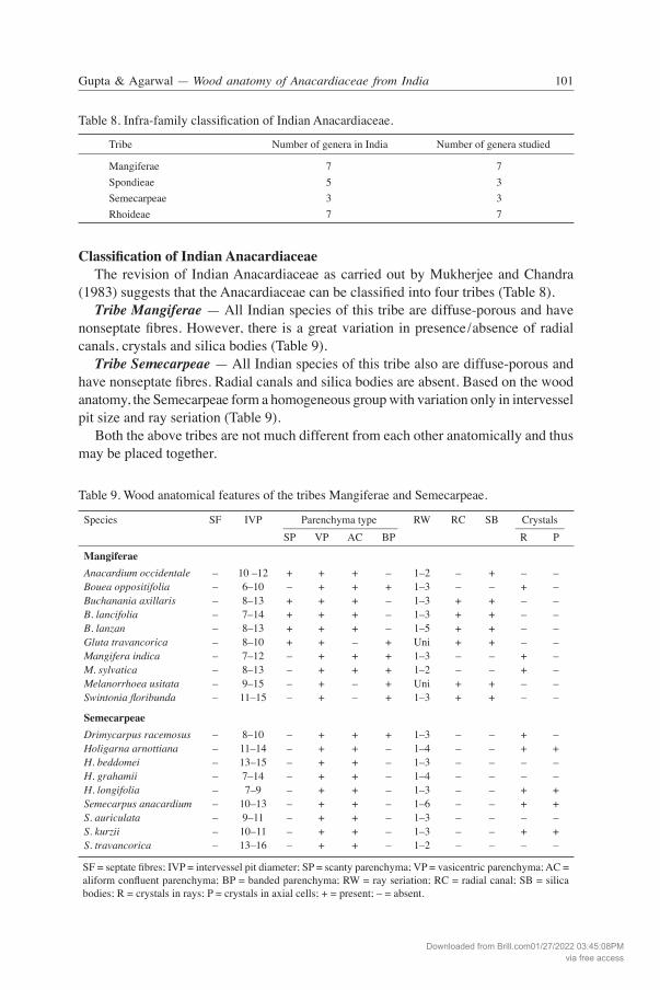
Classification of Indian Anacardiaceae The revision of Indian Anacardiaceae as carried out by Mukherjee and Chandra (1983) suggests that the Anacardiaceae can be classified into four tribes (Table 8). Tribe Mangiferae — All Indian species of this tribe are diffuse-porous and have nonseptate fibres. However, there is a great variation in presence/absence of radial canals, crystals and silica bodies (Table 9). Tribe Semecarpeae — All Indian species of this tribe also are diffuse-porous and have nonseptate fibres. Radial canals and silica bodies are absent. Based on the wood anatomy, the Semecarpeae form a homogeneous group with variation only in intervessel pit size and ray seriation (Table 9). Both the above tribes are not much different from each other anatomically and thus may be placed together.
Table 8. Infra-family classification of Indian Anacardiaceae.
Tribe Number of genera in India Number of genera studied
Mangiferae 7 7Spondieae 5 3Semecarpeae 3 3Rhoideae 7 7
Table 9. Wood anatomical features of the tribes Mangiferae and Semecarpeae.
Species SF IVP Parenchyma type RW RC SB Crystals ––––––––––––––––––––– ––––––––– SP VP AC BP R P
Mangiferae Anacardium occidentale – 10 –12 + + + – 1–2 – + – – Bouea oppositifolia – 6–10 – + + + 1–3 – – + – Buchanania axillaris – 8–13 + + + – 1–3 + + – – B. lancifolia – 7–14 + + + – 1–3 + + – – B. lanzan – 8–13 + + + – 1–5 + + – – Gluta travancorica – 8–10 + + – + Uni + + – – Mangifera indica – 7–12 – + + + 1–3 – – + – M. sylvatica – 8–13 – + + + 1–2 – – + – Melanorrhoea usitata – 9–15 – + – + Uni + + – – Swintonia floribunda – 11–15 – + – + 1–3 + + – –
Semecarpeae Drimycarpus racemosus – 8–10 – + + + 1–3 – – + – Holigarna arnottiana – 11–14 – + + – 1–4 – – + + H. beddomei – 13–15 – + + – 1–3 – – – – H. grahamii – 7–14 – + + – 1–4 – – – – H. longifolia – 7–9 – + + – 1–3 – – + + Semecarpus anacardium – 10–13 – + + – 1–6 – – + + S. auriculata – 9–11 – + + – 1–3 – – – – S. kurzii – 10–11 – + + – 1–3 – – + + S. travancorica – 13–16 – + + – 1–2 – – – –
SF = septate fibres; IVP = intervessel pit diameter; SP = scanty parenchyma; VP = vasicentric parenchyma; AC = aliform confluent parenchyma; BP = banded parenchyma; RW = ray seriation; RC = radial canal; SB = silica bodies; R = crystals in rays; P = crystals in axial cells; + = present; – = absent.
Downloaded from Brill.com01/27/2022 03:45:08PMvia free access
IAWA Journal, Vol. 29 (1), 2008102 103Gupta & Agarwal — Wood anatomy of Anacardiaceae from India
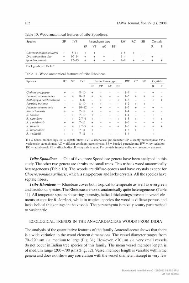
Tribe Spondieae — Out of five, three Spondieae genera have been analysed in this study. The other two genera are shrubs and small trees. This tribe is wood anatomically heterogeneous (Table 10). The woods are diffuse-porous and have crystals except for Choerospondias axillaris, which is ring-porous and lacks crystals. All the species have septate fibres. Tribe Rhoideae — Rhoideae cover both tropical to temperate as well as evergreen and deciduous species. The Rhoideae are wood anatomically quite heterogeneous (Table 11). All temperate species show ring-porosity, helical thickenings present in vessel ele-ments except for R. hookeri, while in tropical species the wood is diffuse-porous and lacks helical thickenings in the vessels. The parenchyma is mostly scanty paratracheal to vasicentric.
ECOLOGICAL TRENDS IN THE ANACARDIACEAE WOODS FROM INDIA
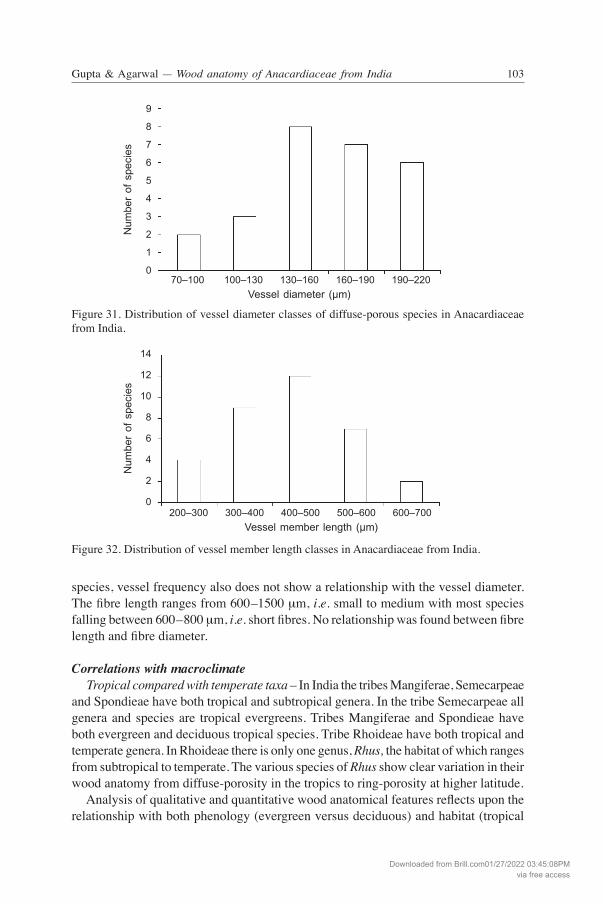
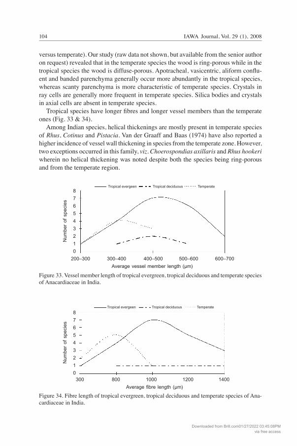
The analysis of the quantitative features of the family Anacardiaceae shows that there is a wide variation in the wood element dimensions. The vessel diameter ranges from 70–220 μm, i.e. medium to large (Fig. 31). However, <70 μm, i.e. very small vessels do not occur in Indian tree species of this family. The mean vessel member length is of medium range (200–700 μm) (Fig. 32). Vessel member length is variable within the genera and does not show any correlation with the vessel diameter. Except in very few
Table 10. Wood anatomical features of tribe Spondieae.
Species SF IVP Parenchyma type RW RC SB Crystals ––––––––––––––––––––– ––––––––– SP VP AC BP R P
Choerospondias axillaris + 8–11 + + – – 1–5 + – – – Dracontomelon dao + 10–14 + + + – 1–4 – – + – Spondias pinnata + 12–15 + + – – 1–8 + – + + For legends, see Table 9.
Table 11. Wood anatomical features of tribe Rhoideae.
Species HT SF IVP Parenchyma type RW RC SB Crystals ––––––––––––––––––––– ––––––––– SP VP AC BP R P
Cotinus coggygria + – 8–10 + – – – 1–4 – – + – Lannea coromandelica – + 8–11 + + – – 1–5 + + + – Nothopegia colebrookiana – – 6–8 – + + + 1–3 – – + + Parishia insignis – – 8–10 + + – – 1–2 + + – – Pistacia integerrimia + – 10–12 – + – – 1–5 + – + – Rhus chinensis + – 7–12 + – – – 1–4 – – + – R. hookeri – + 7–10 + – – – 1–4 – – + – R. parviflora – + 2.7–4 + – – + 1–5 + – + + R. punjabensis + + 7–12 + – – – 1–6 – – + – R. sinuata – + 2.7–5 + – – + 1–3 + – + + R. succedanea + + 7–11 + – – – 1–6 + – + – R. wallichii + + 7–11 + – – – 1–4 – – + –
HT = helical thickenings; SF = septate fibres; IVP = intervessel pit diameter; SP = scanty parenchyma; VP = vasicentric parenchyma; AC = aliform confluent parenchyma; BP = banded parenchyma; RW = ray seriation; RC = radial canal; SB = silica bodies; R = crystals in rays; P = crystals in axial cells; + = present; – = absent.
Downloaded from Brill.com01/27/2022 03:45:08PMvia free access
IAWA Journal, Vol. 29 (1), 2008102 103Gupta & Agarwal — Wood anatomy of Anacardiaceae from India
species, vessel frequency also does not show a relationship with the vessel diameter. The fibre length ranges from 600–1500 μm, i.e. small to medium with most species falling between 600–800 μm, i.e. short fibres. No relationship was found between fibre length and fibre diameter.
Correlations with macroclimate Tropical compared with temperate taxa – In India the tribes Mangiferae, Semecarpeae and Spondieae have both tropical and subtropical genera. In the tribe Semecarpeae all genera and species are tropical evergreens. Tribes Mangiferae and Spondieae have both evergreen and deciduous tropical species. Tribe Rhoideae have both tropical and temperate genera. In Rhoideae there is only one genus, Rhus, the habitat of which ranges from subtropical to temperate. The various species of Rhus show clear variation in their wood anatomy from diffuse-porosity in the tropics to ring-porosity at higher latitude. Analysis of qualitative and quantitative wood anatomical features reflects upon the relationship with both phenology (evergreen versus deciduous) and habitat (tropical
9
8
7
6
5
4
3
2
1
0 70–100 100–130 130–160 160–190 190–220
Vessel diameter (µm)
Num
ber
of s
peci
es
200–300 300–400 400–500 500–600 600–700
Vessel member length (µm)
14
12
10
8
6
4
2
0
Num
ber
of s
peci
es
Figure 31. Distribution of vessel diameter classes of diffuse-porous species in Anacardiaceae from India.
Figure 32. Distribution of vessel member length classes in Anacardiaceae from India.
Downloaded from Brill.com01/27/2022 03:45:08PMvia free access
IAWA Journal, Vol. 29 (1), 2008104 105Gupta & Agarwal — Wood anatomy of Anacardiaceae from India
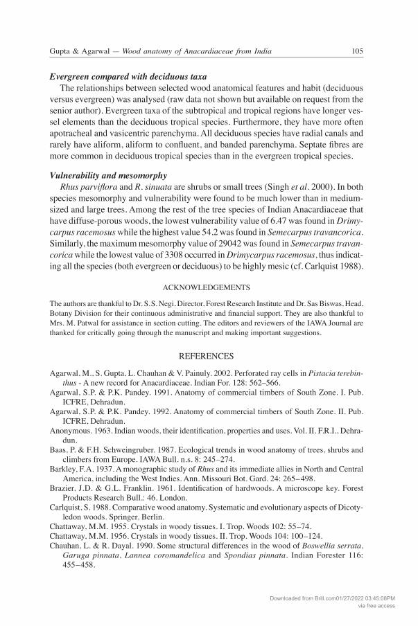
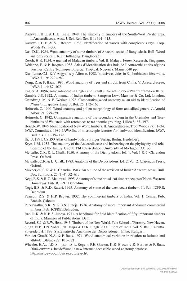
versus temperate). Our study (raw data not shown, but available from the senior author on request) revealed that in the temperate species the wood is ring-porous while in the tropical species the wood is diffuse-porous. Apotracheal, vasicentric, aliform conflu-ent and banded parenchyma generally occur more abundantly in the tropical species, whereas scanty parenchyma is more characteristic of temperate species. Crystals in ray cells are generally more frequent in temperate species. Silica bodies and crystals in axial cells are absent in temperate species. Tropical species have longer fibres and longer vessel members than the temperate ones (Fig. 33 & 34). Among Indian species, helical thickenings are mostly present in temperate species of Rhus, Cotinus and Pistacia. Van der Graaff and Baas (1974) have also reported a higher incidence of vessel wall thickening in species from the temperate zone. However, two exceptions occurred in this family, viz. Choerospondias axillaris and Rhus hookeri wherein no helical thickening was noted despite both the species being ring-porous and from the temperate region.
8
7
6
5
4
3
2
1
0
Num
ber
of s
peci
es
200–300 300–400 400–500 500–600 600–700
Average vessel member length (µm)
Tropical evergeen Tropical deciduous Temperate
8
7
6
5
4
3
2
1
0 300 800 1000 1200 1400
Average fibre length (µm)
Tropical evergeen Tropical deciduous Temperate
Num
ber
of s
peci
es
Figure 33. Vessel member length of tropical evergreen, tropical deciduous and temperate species of Anacardiaceae in India.
Figure 34. Fibre length of tropical evergreen, tropical deciduous and temperate species of Ana-cardiaceae in India.
Downloaded from Brill.com01/27/2022 03:45:08PMvia free access
IAWA Journal, Vol. 29 (1), 2008104 105Gupta & Agarwal — Wood anatomy of Anacardiaceae from India
Evergreen compared with deciduous taxa The relationships between selected wood anatomical features and habit (deciduous versus evergreen) was analysed (raw data not shown but available on request from the senior author). Evergreen taxa of the subtropical and tropical regions have longer ves-sel elements than the deciduous tropical species. Furthermore, they have more often apotracheal and vasicentric parenchyma. All deciduous species have radial canals and rarely have aliform, aliform to confluent, and banded parenchyma. Septate fibres are more common in deciduous tropical species than in the evergreen tropical species.
Vulnerability and mesomorphy Rhus parviflora and R. sinuata are shrubs or small trees (Singh et al. 2000). In both species mesomorphy and vulnerability were found to be much lower than in medium-sized and large trees. Among the rest of the tree species of Indian Anacardiaceae that have diffuse-porous woods, the lowest vulnerability value of 6.47 was found in Drimy-carpus racemosus while the highest value 54.2 was found in Semecarpus travancorica. Similarly, the maximum mesomorphy value of 29042 was found in Semecarpus travan-corica while the lowest value of 3308 occurred in Drimycarpus racemosus, thus indicat-ing all the species (both evergreen or deciduous) to be highly mesic (cf. Carlquist 1988).
ACKNOWLEDGEMENTS
The authors are thankful to Dr. S.S. Negi, Director, Forest Research Institute and Dr. Sas Biswas, Head, Botany Division for their continuous administrative and financial support. They are also thankful to Mrs. M. Patwal for assistance in section cutting. The editors and reviewers of the IAWA Journal are thanked for critically going through the manuscript and making important suggestions.
REFERENCES
Agarwal, M., S. Gupta, L. Chauhan & V. Painuly. 2002. Perforated ray cells in Pistacia terebin-thus - A new record for Anacardiaceae. Indian For. 128: 562–566.
Agarwal, S.P. & P.K. Pandey. 1991. Anatomy of commercial timbers of South Zone. I. Pub. ICFRE, Dehradun.
Agarwal, S.P. & P.K. Pandey. 1992. Anatomy of commercial timbers of South Zone. II. Pub. ICFRE, Dehradun.
Anonymous. 1963. Indian woods, their identification, properties and uses. Vol. II. F.R.I., Dehra-dun.
Baas, P. & F.H. Schweingruber. 1987. Ecological trends in wood anatomy of trees, shrubs and climbers from Europe. IAWA Bull. n.s. 8: 245–274.
Barkley, F.A. 1937. A monographic study of Rhus and its immediate allies in North and Central America, including the West Indies. Ann. Missouri Bot. Gard. 24: 265–498.
Brazier, J.D. & G.L. Franklin. 1961. Identification of hardwoods. A microscope key. Forest Products Research Bull.: 46. London.
Carlquist, S. 1988. Comparative wood anatomy. Systematic and evolutionary aspects of Dicoty-ledon woods. Springer, Berlin.
Chattaway, M.M. 1955. Crystals in woody tissues. I. Trop. Woods 102: 55–74.Chattaway, M.M. 1956. Crystals in woody tissues. II. Trop. Woods 104: 100–124.Chauhan, L. & R. Dayal. 1990. Some structural differences in the wood of Boswellia serrata,
Garuga pinnata, Lannea coromandelica and Spondias pinnata. Indian Forester 116: 455–458.
Downloaded from Brill.com01/27/2022 03:45:08PMvia free access
IAWA Journal, Vol. 29 (1), 2008106
Dadswell, H.E. & H.D. Ingle. 1948. The anatomy of timbers of the South-West Pacific area. I. Anacardiaceae. Aust. J. Sci. Res. Ser. B 1: 391–415.
Dadswell, H.E. & S.J. Record. 1936. Identification of woods with conspicuous rays. Trop. Woods 48: 1–30.
Das, D.K. 1984. Wood anatomy of some timbers of Anacardiaceae of Bangladesh. Bull. Wood anatomy series. F.R.I. Chittagong, Bangladesh.
Desch, H.E. 1954. A manual of Malayan timbers. Vol. II. Malaya. Forest Research, Singapore.Détienne, P. & P. Jacquet. 1983. Atlas dʼidentification des bois de lʼAmazonie et des régions
voisines. Centre Technique Forestier Tropical, Nogent s /Marne. 640 pp.Dias-Leme, C.L. & V. Angyalossy-Alfonso. 1998. Intrusive cavities in Euphorbiaceae fibre walls.
IAWA J. 19: 279 –283.Dong, Z. & P. Baas. 1993. Wood anatomy of trees and shrubs from China. V. Anacardiaceae.
IAWA J. 14: 87–102.Engler, A. 1896. Anacardiaceae in Engler and Prantlʼs Die natiirlichen Pflanzenfamilien III. 5.Gamble, J.S. 1922. A manual of Indian timbers. Sampson Low, Marston & Co. Ltd, London.Grundwag, M. & E. Werker. 1976. Comparative wood anatomy as an aid to identification of
Pistacia L. species. Israel J. Bot. 25: 152–167.Heimsch, C. 1940. Wood anatomy and pollen morphology of Rhus and allied genera. J. Arnold
Arbor. 21: 279–291.Heimsch, C. 1942. Comparative anatomy of the secondary xylem in the Gruinales and Tere-
binthales of Weitstein with references to taxonomic grouping. Lilloa 8: 83–197.Hess, R.W. 1946. Identification of New World timbers. II. Anacardiaceae. Trop. Woods 87: 11–34.IAWA Committee. 1989. IAWA list of microscopic features for hardwood identification. IAWA Bull. n.s. 10: 219–332.Ilic, J. 1991. CSIRO Atlas of hardwoods. Springer Verlag, Berlin, Heidelberg.Kryn, J.M. 1952. The anatomy of the Anacardiaceae and its bearing on the phylogeny and rela-
tionship of the family. Unpub. PhD Dissertation, University of Michigon. 331 pp.Metcalfe, C.R. & L. Chalk. 1950. Anatomy of the Dicotyledons. Ed. 1. Vol. 1 & 2. Clarendon
Press, Oxford.Metcalfe, C.R. & L. Chalk. 1983. Anatomy of the Dicotyledons. Ed. 2. Vol. 2. Clarendon Press,
Oxford.Mukherjee, S.K. & D. Chandra. 1983. An outline of the revision of Indian Anacardiaceae. Bull.
Bot. Sur. India. 25 (1-4): 52–61.Negi, B.S. & R.C. Madhwal. 1995. Anatomy of some broad leaf timber species of North-Western
Himalayas. Pub. ICFRE, Dehradun.Negi, B.S. & R.D. Raturi. 1993. Anatomy of some of the west coast timbers. II. Pub. ICFRE,
Dehradun.Pearson, R.S. & H.P. Brown. 1932. The commercial timbers of India. Vol. 1. Central Pub.
Branch, Calcutta.Purkayastha, S.K. & K.B.S. Juneja. 1976. Anatomy of more important Andaman commercial
timbers. Pub. ICFRE, Dehradun.Rao, R.K. & K.B.S. Juneja. 1971. A handbook for field identification of fifty important timbers
of India. Manager of Publications, Delhi.Record, S.J. & R.W. Hess. 1943. Timbers of the New World. Yale School of Forestry, New Haven.Singh, N.P., J.N. Vohra, P.K. Hajra & D.K. Singh. 2000. Flora of India. Vol. 5. BSI, Calcutta.Solereder, H. 1899. Systematische Anatomie der Dicotyledonen. Enke, Stuttgart.Van der Graaff, N.A. & P. Baas. 1974. Wood anatomical variation in relation to latitude and
altitude. Blumea 22: 101–121.Wheeler, E.A., T.D. Simpson, S.L. Rogers, P.E. Gasson, K.R. Brown, J.R. Bartlett & P. Baas.
2004-onwards. InsideWood: a new internet-accessible wood anatomy database: http://insidewood/lib.ncsu.edu/search/.
Downloaded from Brill.com01/27/2022 03:45:08PMvia free access