hyperbaric oxygen therapy reduces the severity of necrotizing enterocolitis in a neonatal rat model
TRANSCRIPT

www.elsevier.com/locate/jpedsurg
Journal of Pediatric Surgery (2009) 44, 534–540
Hyperbaric oxygen therapy reduces the severity ofnecrotizing enterocolitis in a neonatal rat model☆
Ahmet Guvena,⁎, Gokhan Gundogdua, Bulent Uysalb, Hakan Cermikc, Mustafa Kuld,Suzi Demirbaga, Haluk Ozturka, Sukru Oterb
aDepartment of Pediatric Surgery, Gulhane Military Medical Academy, 06017 Etlik, Ankara, TurkeybDepartment of Physiology, Gulhane Military Medical Academy, 06017 Etlik, Ankara, TurkeycService of Pathology, Army Hospital, Etimesgut, Ankara, TurkeydDepartment of Pediatrics, Gulhane Military Medical Academy, Haydarpasa Training Hospital, Istanbul, Turkey
Received 24 February 2008; revised 9 May 2008; accepted 15 June 2008
R
0d
Key words:Necrotizing enterocolitis;Hyperbaric oxygen therapy;Nitric oxide;Tumor necrosis factor α;Oxidant/antioxidant system
AbstractIntroduction: Hyperbaric oxygen (HBO) therapy is known to increase oxygen concentration in tissuesleading to induction of an adaptive increase in antioxidants, stimulation of angiogenesis, improvement ofwhite blood cell action, and regulation of inflammatory process. Therefore, we tested the potentialbeneficial effect of HBO in neonatal rat model of necrotizing enterocolitis (NEC).Materials and Methods: Thirty newborn Sprague-Dawley rats, provided by the Experimental ResearchCouncil, Gulhane Military Medical Academy, Ankara,Turkey, were randomly divided into 3 groups asfollows: NEC, NEC +HBO, and control. Necrotizing enterocolitis was induced by enteral formula feedingand exposure to hypoxia after cold stress at 4°C and oxygen. The NEC + HBO group received HBO at2.8 atmosphere absolute (ATA) for 90minutes daily for 3 days. The pups were killed on the fourth day, andtheir intestinal tissues were harvested for biochemical and histopathologic analysis. Blood samples werealso obtained from the pups.Results: The mortality rate was highest in the NEC group (3 pups in the NEC group vs 1 pup in the NEC +HBO group). Malondialdehyde and protein carbonyl content were significantly increased, whereassuperoxide dismutase and glutathione peroxidase were significantly decreased in the NEC group. All thesechanges were similar to control levels in the NEC group by HBO treatment. Nitrate plus nitrite (NOx)levels and serum tumor necrosis factor awere increased in the NEC group and histopathologic injury scoreand apoptosis index in the NEC group were significantly higher than in the NEC + HBO group.Conclusion: Hyperbaric oxygen significantly reduced the severity of NEC in our study.© 2009 Elsevier Inc. All rights reserved.
☆ This study was supported by Gulhane Military Medical Academyesearch Fund (project no. GATA 2007-048) (GATA, Ankara, Turkey).⁎ Corresponding author. Tel.: +90 312 3045483; fax: +90 312 3042150.E-mail address: [email protected] (A. Guven).
022-3468/$ – see front matter © 2009 Elsevier Inc. All rights reserved.oi:10.1016/j.jpedsurg.2008.06.008
Necrotizing enterocolitis (NEC) is the most commongastrointestinal emergency in the premature infant, and theincidence is rising because of advances in neonatology.Although NEC is most commonly observed in prematureinfants, 10% of affected patients are born at term. Itsincidence varies between 0.3 and 2.4 infants/1000 births and
535Effect of hyperbaric oxygen therapy on NEC
between 7% and 11% among infants of less than 1500 g. Theaverage mortality is 20% to 40%, and survivors after eithermedical or surgical therapy can present with failure to thrive,feeding abnormalities, diarrhea, or bowel obstruction [1-3].
Despite a hundred years having passed since therecognition of NEC, the etiology and pathogenesis of NECremain poorly understood. Several factors appear to playeither a primary or a secondary role as follows: immature gutfunctions, impaired intestinal barrier, disturbed gastrointest-inal motility, circulatory regulation, lack of some protectiveenzymes of cytokine-dependent injury, and undevelopedantioxidant capacity [1,3,4]. Intestinal ischemia, formulafeeding, and bacterial infections are also suggested to be riskfactors initiating cellular injury in premature intestine [5].Ongoing studies demonstrated that mucosal injury ismediated by oxygen-derived free radicals, inflammatorymediators including tumor necrosis factor (TNF), plateletactivating factor (PAF), and leukotrienes.
Of the many experimental models used to study NEC, theneonatal rat model has been shown to be one of the best. Themajor risk factors for human NEC (intestinal immaturity,enteral formula feeding, and hypoxia/ischemia) are essentialfor developing NEC in a neonatal rat model [6,7].
Therapeutic use of oxygen under pressure is known ashyperbaric oxygen (HBO) therapy. The Undersea andHyperbaric Medicine Society (Durham, NC, USA) approvedthe use of HBO therapy in disorders including necrotizinginfections, severe infection by anaerobic bacteria, andischemic injuries [8,9]. Although some of the mechanismsof action of HBO therapy are still to be discovered, it isknown that HBO therapy increases oxygen concentration inall body tissues, even with reduced or blocked blood flow;stimulates the growth of new blood vessels improving bloodflow to compromised organs; stimulates an adaptive increasein superoxide dismutase (SOD); and aids the treatment ofinfection by enhancing white blood cell action [8].
The clarification of the pathophysiology and introductionof new strategies for prevention and/or therapy for NEC areimportant to increase the rate of survival. Because thereseems to be a relationship between the pathophysiology ofNEC and the action mechanisms of HBO therapy, wedesigned an experimental study to evaluate the efficacy ofHBO therapy in an experimental model of NEC in pups.
1. Materials and methods
1.1. Animals and experimental design
The project was approved by the Experimental AnimalEthics Committee at Gulhane Military Medical Academy(Ankara, Turkey) and the National Institutes of Health Guide(Washington, D.C., USA) for the Care and Use of LaboratoryAnimals was followed.
The pregnant rats were kept in identical cages and were fedwith regular laboratory chow and water. Thirty newborn pups
from 4 time-mated Sprague-Dawley pregnant rats weredivided equally into 3 groups as follows: NEC (subjected toNEC), NEC+HBO (subjected toNEC and treated withHBO),and control (left with their mother to breast feed freely).
1.1.1. Necrotizing enterocolitis procedureIn the NEC and NEC + HBO groups, based on the
preventive effect of mother milk, newborn pups wereimmediately separated from their mothers and kept at 37°Cin a humidified incubator. These animals were fed 0.2 mL ofspecial rodent formula (15 g Similac 60/40 [Ross Pediatrics,Columbus, Ohio]) prepared with 75 mL of puppy-caninemilk replacement (Beaphar-Bogena, BV Sedel, Netherlands)3 times daily and subjected to 100% CO2 inhalation for 10minutes, +4°C cold exposure for 5 minutes, and 97% O2 for5 minutes twice daily to induce NEC. This procedure wasapplied to the pups for 3 days. The pups were weighed daily,and their weight loss or gain was recorded.
1.1.2. Hyperbaric oxygen applicationAfter the first day of the NEC procedure, animals in the
NEC + HBO group were subjected to hyperbaric oxygen at2.8 atmospheric pressure with 100% O2 inhalation for 90minutes daily for 3 days.
All rats were killed on the fourth day of experiment viadecapitation; the abdomen was opened; and the intestineswere inspected for evidence of NEC such as intestinaldiscoloration, fragility, weakness of tissue integrity, edema,intestinal hemorrhage, ileal distension, pneumotosis intesti-nalis, perforation, and necrosis, and 2 cm of the terminalileum was harvested for biochemical and histopathologicevaluations. Tissue specimens were flushed with cold salinesolution and half of the intestine was fixed in 10% bufferedformalin for histopathologic evaluation. The other half of fullthickness intestine wall was frozen in liquid nitrogen andthen stored at −70°C for biochemical examination. Bloodsamples were also collected for analysis of serum TNF-a.
Frozen tissues were homogenized in phosphate buffer (pH7.4) on an ice cube, by a homogenizer (Heidolph Diax 900;Heidolph Elektro GmbH, Kelheim, Germany).
1.2. Biochemical analysis
Serum TNF-a concentration was measured in duplicatewith a commercially available enzyme-linked immunosor-bent assay kit (BioSource Europe S.A., Nivelles, Belgium)according to the manufacturer's instructions.
The protein content of tissue homogenates was measuredas described by Lowry [10] with bovine serum albumin asthe standard.
The efficacy of HBO was assessed by the tissue level ofmalondialdehyde (MDA) as described by Ohkawa et al [11],protein carbonyl content (PCC) as described by Levine et al[12], SOD as described by Sun et al [13], and glutathioneperoxidase (GSH-Px) as described by Paglia and Valentine[14]. Nitrate plus nitrite (NOx) levels, end products of nitric

536 A. Guven et al.
oxide degradation, were measured using the methoddescribed by Miranda et al [15].
1.3. Histopathologic examination andapoptosis evaluation
Tissue histopathologic examination and apoptosis scoringwere evaluated by the same pathologist in blinded fashion.For each tissue specimen, corresponding blocks of both H&Eand transferase mediated dUTP nick end labeling (TUNEL)staining were scored.
For both light microscopy and apoptosis evaluation, thedistal ileum was kept in 10% neutral buffered formalinsolution. Specimens were paraffin embedded, and blockswere sliced into 5-μm portions and stained with H&E. Thehistopathologic evaluation was graded as follows: grade 0,normal; grade 1 (mild), separation of villous core, withoutother abnormalities; grade 2 (moderate), villous coreseparation, submucosal edema, and epithelial sloughing;and grade 3 (severe), denudation of epithelium with loss ofvillous, full thickness necrosis, or perforation [16].
For apoptosis evaluation, the sections were deparaffinizedand rehydrated through graded alcohols to water. Thesections were then incubated with 20 mg/mL of proteinaseK (Dako, Glostrup, Denmark) for 10 minutes at roomtemperature (RT). Endogenous peroxidase activity wasquenched by incubation with 3% H2O2 in phosphate buffersolution (PBS) (50 mmol/L sodium phosphate, pH 7.4,200 mmol/L NaCl) for 5 minutes at RT. After washing withdeionized water (dH2O), the equilibration buffer wasincubated for 10 seconds. Enzymatic incorporation ofnucleotides labeled with Working Strength TdT (ApopTagPeroxidase in Situ Apoptosis Detection Kit, S7101, Milli-pore, Billerica, MA, USA) were placed at 37°C in ahumidified chamber for 60 minutes. Then the sections weretreated with Working Strength Stop/Wash buffer for 10 min-utes at RT. The slides were washed twice in PBS. They wereincubated with antidigoxigenin-peroxidase at RT in ahumidified chamber for 30 minutes. They were washed inPBS and then they were incubated with 3,3 9-diaminoben-zidine tetrahydrochloride (DAB) (0.5 mg/mL), which wasused as chromogen substrate, for 6 minutes at RT in ahumidified chamber. Tissue sections were lightly counter-
Table 1 Biochemical evaluations for each group
Control (n = 10)
MDA (nmol/g protein) 0.32 ± 0.05PCC (nmol/g protein) 0.34 ± 0.04NOx (µmol/g tissue) 15.9 ± 2.1SOD (U/g protein) 2375.9 ± 118.2GSH-Px (U/g protein) 44.9 ± 3.1TNF-α (pg/mL) 49.8 ± 8.1
a Significantly different from control.b Significantly different from NEC group.
stained with 0.5% methyl green to reveal nuclei, and theslides were examined using an Axiolab microscope (CarlZeiss, Jena, Germany). Apoptosis scoring was graded asfollows: grade 0, normal; grade 1, apoptotic nuclei presents atvillous tips; grade 2, apoptotic nuclei covering all villous tipsbut crypts protected; and grade 3, transmural spread ofapoptotic nuclei [17].
1.4. Statistical analysis
Statistical analyses were carried out using SPSS (SPSS forWindows, Version 11.0; Chicago, IL). Data were expressedas means ± SEM. Differences among the groups wereanalyzed by the Kruskal-Wallis test. Dual comparisonsamong groups with significant values were evaluated withthe Mann-Whitney U test. Statistical significance wasdefined as P b .05.
2. Results
Twenty-six pups survived until the end of the study; 3pups in the NEC group and 1 pup in the NEC + HBOgroup died. The mortality rate in the NEC group was higherthan that in the NEC + HBO and control groups; however,this differences was not statistically significant. Theaverage weights of animals were similar for each groupat birth (5.6 ± 0.2 g). Weight loss in the NEC groupanimals was significantly higher than that in the NEC +HBO and control groups (P b .05). The control groupanimals were 8.2 ± 0.3 g, whereas in the NEC and NEC +HBO groups, they were 4.9 ± 0.2 g and 6.4 ± 0.3 g at timeof killing, respectively.
2.1. Oxidative and nitrosative stress markers
The MDA, PCC, and NOx levels in the intestine areshown in Table 1. A sharp increase in the tissue MDA andPCC levels was seen in the NEC group, suggesting increasedlipid peroxidation and protein oxidation (P b .001). TheMDA levels and PCC in the NEC + HBO group weresignificantly lower than those in the NEC group (P b .05).The NOx levels were significantly increased in the NEC
NEC (n = 7) NEC + HBO (n = 9)
0.79 ± 0.06 a 0.46 ± 0.08a,b
0.68 ± 0.13 a 0.42 ± 0.06 a
49.7 ± 3.4 a 22.6 ± 3.5a,b
887.5 ± 270.5 a 1625.1 ± 263.8a,b
24.7 ± 3.4 a 34.8 ± 3.9a,b
155.7 ± 13.4 a 96.6 ± 11.9a,b

Table 2 Comparison of histopathologic evaluation
Intestinalinjury scoring
Apoptosisindex scoring
Control (n = 10) 0 0NEC (n = 7) 2.6 ± 0.5 a 2.3 ± 0.4 a
NEC + HBO (n = 9) 1.2 ± 0.4a,b 0.9 ± 0.3a,b
a Significantly different from control.b Significantly different from NEC group.
537Effect of hyperbaric oxygen therapy on NEC
group, suggesting increased tissue nitric oxide (NO)production (P b .001). These increased levels weresignificantly attenuated in the treatment group; however,their levels remained significantly higher than those of thecontrol group (P b .05).
2.2. Antioxidant enzymes
The SOD and GSH-Px enzyme activities are shown inTable 1. Antioxidant enzyme activities were significantlylower in the NEC group than they were in the other groups(P b .001). Although those in the NEC + HBO group weresignificantly higher than those in the NEC group (P b .05),they were still lower than those in the control group (P b .05).
2.3. Serum TNF-a
Serum TNF-a levels are shown in Table 1. Necrotizingenterocolitis resulted in a significant increase in serum TNF-a level in the NEC group (P b .001). The HBO administrationdecreased serum TNF-a level (P b .05), but it was still higherthan that in the control group (P b .05).
2.4. Morphologic evaluation of intestines
After the abdomen was opened, the small intestine wasvisually evaluated and various degrees of signs of NEC suchas gross necrotic changes, pneumatosis intestinalis, fragility,
Fig. 1 Representative histopathologic evaluation of the terminal ileumIntestinal architecture of a rat from the control (breast feed) group (A(B) displayed severe injury findings consisting of denudation of epitheliuThe NEC + HBO group (C) displayed moderate histologic changes cons
weakness of tissue integrity, edema, and discoloration,especially in the ileocecal region, were seen in the NECgroup. Immediate postmortem examination of the 4 animalslost during the course of the experiment also displayednecrosis and perforation in the intestines. On the other hand,less severe degrees of signs of NEC such as edema andminimal weakness of tissue integrity were seen in the NEC +HBO group. No animals in the control group had evidence ofmacroscopic injury. These findings were consistent with themicroscopic evaluation.
The histologic grading and apoptotic index of intestinalinjury are shown in Table 2. Moderate or severe injuryfindings were seen in the pups subjected to NEC.Conversely, the intestine was evaluated as normal in thegroup treated with HBO. Representative histologic samplesfrom all of the groups are shown in Figs. 1 and 2.
3. Discussion
In this study, we evaluated the effects of HBO therapy inan experimental NEC model of newborn rats. The findings ofour study showed that HBO therapy had a significant effecton weight gain and survival rates in pups with induced NEC.In addition, HBO had an ameliorating effect on oxidativestress, nitrosative stress, and antioxidant enzymes activitiesin the intestine of pups subjected to NEC. We also found thatHBO had an ameliorating effect on the histopathologicchanges in the intestine in which NEC was induced.Moreover, serum TNF-a levels were decreased in pupstreated with HBO.
Focusing on cellular injury mechanisms has clarifiedsome steps of NEC pathogenesis. Presumably an initial stresstriggers the release of a proinflammatory mediator such asPAF [18,19]. Subsequent release of inflammatory cytokines(TNF-a, interleukin 6 [IL-6]) and migration of activatedpolymorphonuclear leukocytes result in excess generation ofreactive oxygen species (ROS) and reactive nitrogen species
from each experimental group (H&E, ×40 original magnification).) revealed normal histologic feature of villi (a). The NEC groupm with loss of villi (b), perforation (c), and full thickness necrosis.isting of shortened villi (d) and submucosal edema (e).
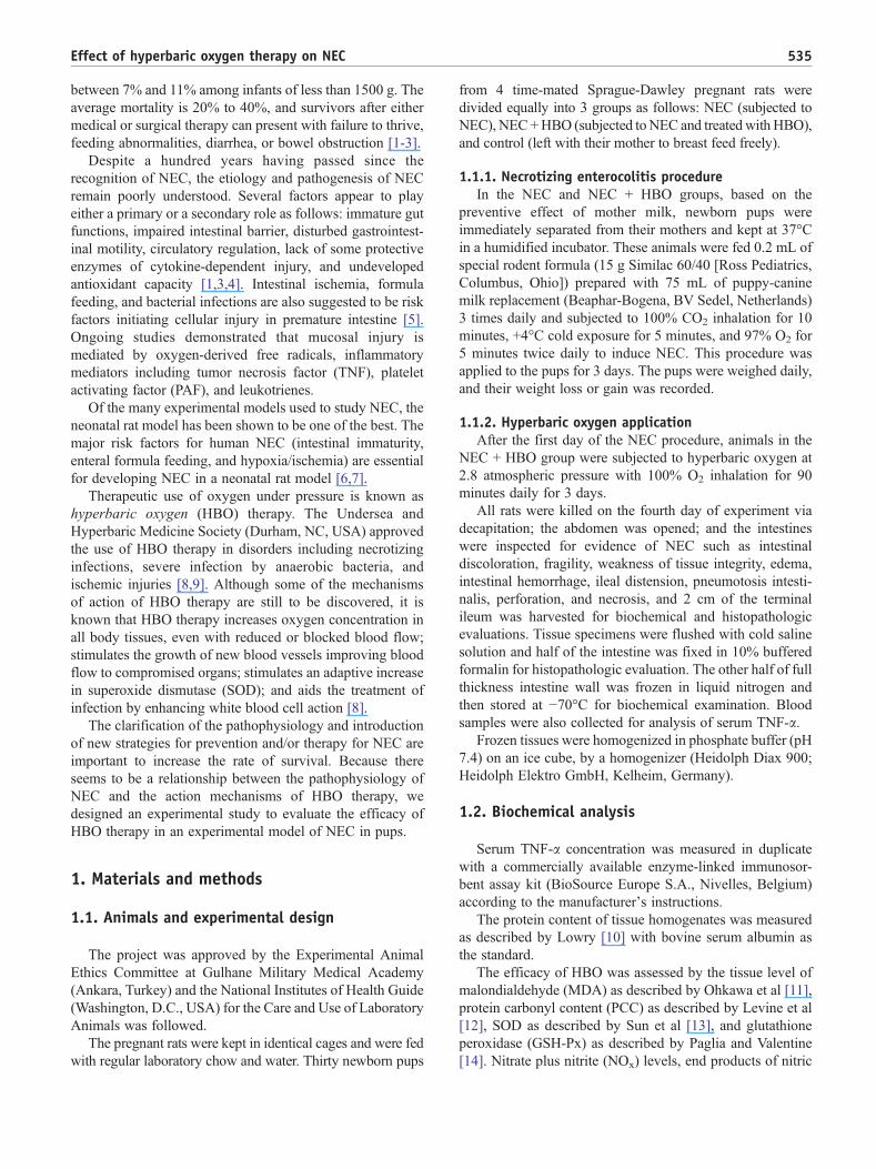
Fig. 2 Representative transferase mediated dUTP nick end labeling (TUNEL) staining section of the terminal ileum from each experimentalgroup (×40 original magnification). Intestinal architecture of a rat from the control (breastfeed) group (A) revealed normal histologic villi (a),NEC group (B) displayed apoptotic cells continuing to crypts, and NEC + HBO group (C) displayed apoptotic cells at the tip of villi (c).
538 A. Guven et al.
(RNS) [20]. Under normal conditions, the detrimental effectsof ROS/RNS are limited by antioxidant enzymes andscavengers such as SOD and GSH-Px. However, anyimbalance in this equilibrium caused by either excessiveROS/RNS production or limitation in antioxidant capacitymay result in cellular damage and subsequent necrosis viadifferent mechanisms including especially peroxidation ofcellular membrane lipids and oxidation of cellular proteins,which could be determined by MDA and PCC levels,respectively [20]. Moreover, deficient level of antioxidantenzyme capacity is another factor predisposing to tissueinjury in NEC and other diseases in newborns [4,21].
In our study, we found that NEC caused a sharp increase inintestinal tissue levels ofMDA and PCC, indicating increasedoxidative stress. We also observed a significant decrease inSOD and GSH-Px activities, indicating a reduction inantioxidant capacity. There is some evidence that freeradicals play a significant role in the development of NEC.In experimental models of NEC, administration of agents thateither inhibit ROS production or act as antioxidants has beenshown to reduce tissue damage compared with controls[1,3,22,23]. It was also reported that MDA increases andSOD decreases in intestinal tissue hypoxia/reoxygenation-induced intestinal injury in newborn rats [24,25], and ourresults are consistent with these previous studies.
We also found that HBO reduced lipid peroxidation andprotein oxidation and ameliorated antioxidant enzymesactivities in intestinal tissue of animals in which NEC wasinduced. The mechanism of action of HBO is attributed tothe immediate direct physical effects of oxygen and to thedelayed secondary physiologic and biochemical effects. It iswell known that HBO stimulates or upregulates somebiological processes such as angiogenesis, collagen synth-esis, leukocyte adhesion, erythropoietin, and release ofvascular endothelial growth factor (VGEF), which providetissue healing [8,9,26]. Hyperbaric oxygen is also known forproducing oxidative stress in an appropriate exposurepressure and duration-dependent manner [27,28], and it issuggested that this oxidative stress may be another of itsbeneficial mechanisms via stimulating antioxidant enzymes.
At this point, all data for oxidant/antioxidant systememphasize that oxidative stress is crucial in the pathogenesisof NEC, presumably because of immature functions ofpremature intestine, and HBO therapy has a beneficial effecton this process.
In our study, the histomorphologic evaluation of theintestine revealed preserved integrity and lower degrees ofedema and necrosis in the NEC + HBO group. Severalanimal studies have suggested that the presence of bacteriaand/or bacterial translocation is critical for the initiation ofintestinal inflammation and NEC, and probiotic organismscan reduce the incidence of NEC [29,30]. It is assumed thatoxygen enhances the white cells' ability to kill aerobic/anaerobic bacteria when administered at pressures aboveatmospheric level pressure. In addition, the main purpose ofHBO therapy is to save partially damaged hypoxic tissue andto reduce the extent of necrosis. The vasoconstrictive effectof HBO reduces edema while still increasing tissueoxygenation. This breaks the vicious circle of ischemia-edema-ischemia, improves tissue and cellular oxygenation,and enhances phagocytosis and bacterial killing by theleukocytes [26,31,32]. Therefore, the lower degree of edemaand perforation of intestines in the NEC + HBO group maybe also explained through enhancing tissue and cellularoxygenation after enhancement of macrophage activity andinhibition of bacterial growth.
In another part of this study, we found that TNF-a wasincreased in pups in which NEC was induced, and theseincreased levels were lowered by HBO therapy. Thepathophysiology of NEC involves a complex interaction ofinflammatory mediators such as TNF-a and PAF [3,5,18,19].Tumor necrosis factor a triggers the production of furtherinflammatory cytokines, which play important roles in theregulation of intestinal epithelial cell apoptosis [3,19,33,34].Many studies indicate that HBO therapy has antiinflamma-tory properties. Yamashita [35] reported that HBO therapyattenuates the induction of cytokines, such as TNF-a and IL-6, after massive hemorrhage. Similarly, Weisz et al [36]concluded that decreased IL-1, IL-6, and TNF-a secretion isbecause of HBO treatment in patients with perianal Crohn's

539Effect of hyperbaric oxygen therapy on NEC
disease. Although the interaction of TNF-a with HBO is notclearly understood, our finding is in agreement with studiesdemonstrating that HBO attenuates proinflammatory cyto-kine production in animal models of systemic inflammation.Given these effects, we can conclude that HBO may have abeneficial impact on inflammation in NEC.
In this study, we also found that intestinal NOx levels wereincreased in animals in which NEC was induced, and HBOdecreased elevated NOx levels. Nitric oxide or peroxynitriteare eventually converted to nitrite/nitrate (as accepted NOx),and the determination of NOx level gives an indirect butreliable idea regarding NO production in the tissue, althoughwe could not evaluate inducible nitric oxide synthase (iNOS)expression in the tissue. Nitric oxide has a key role in bothproviding intestinal integrity and causing tissue damage[34,37]. Analyses of intestinal segments from infants withacute NEC and various animal NEC models have shownupregulation of NO metabolism and/or iNOS expression[16,22,37], and it is known that iNOS synthesis is stronglyinduced by IL-1 and TNF-a. The findings of NOx and TNF-avalues in this work seem to support these reports. In addition,NO production downregulated during HBO exposure may beanother explanation for the reduction in the severity of NECin the NEC + HBO group.
In conclusion, based on the observations in our NECmodel, HBO therapy exerts a beneficial effect in themanagement of NEC. The rationale of using HBO in thetreatment of NEC is based on the data showing that HBOincreased tissue antioxidant enzymes activities, reducedtissue damage indices and NOx level, reduced serum TNF-alevel, and improved the integrity of the intestinal barrier.Whether HBO affords its protective effects by directlyintervening in the apoptotic pathway or by changing theoxidative stress remains to be determined.
Acknowledgments
We thank Russell Fraser for checking the English ofthis manuscript.
References
[1] Lee JS, Polin RA. Treatment and prevention of necrotizingenterocolitis. Semin Neonatol 2003;8:449-59.
[2] Ryder RW, Shelton JD, Guinan ME. Necrotizing enterocolitis: aprospective multicenter investigation. Am J Epidemiol 1980;112:113-23.
[3] Lin PW, Stoll BJ. Necrotising enterocolitis. Lancet 2006;368:1271-83.[4] Hsueh W, Caplan MS, Qu XW, et al. Neonatal necrotizing
enterocolitis: clinical considerations and pathogenetic concepts.Pediatr Dev Pathol 2003;6:6-23.
[5] Covert RF, Neu J, Elliott MJ, et al. Factors associated with age of onsetof necrotizing enterocolitis. Am J Perinatol 1989;6:455-60.
[6] Crissinger KD. Animal models of necrotizing enterocolitis. J PediatrGastroenterol Nutr 1995;20:17-22.
[7] Dvorak B, Halpern MD, Holubec H, et al. Maternal milk reducesseverity of necrotizing enterocolitis and increases intestinal IL-10 in aneonatal rat model. Pediatr Res 2003;53:426-33.
[8] Gill AL, Bell CN. Hyperbaric oxygen: its uses, mechanisms of actionand outcomes. QJM 2004;97:385-95.
[9] Oter S, Korkmaz A. Relevance of hyperbaric oxygen to ozone therapy.Arch Med Res 2006;37:917-8.
[10] Lowry OH, Rosebrough NJ, Farr AL, et al. Protein measurement withthe Folin phenol reagent. J Biol Chem 1951;193:265-75.
[11] Ohkawa H, Ohishi N, Yagi K. Assay for lipid peroxides in animaltissues by thiobarbituric acid reaction. Anal Biochem 1979;95:351-8.
[12] Levine RL, Garland D, Oliver CN, et al. Determination of carbonylcontent in oxidatively modified proteins. Methods Enzymol1990;186:464-78.
[13] Sun Y, Oberley LW, Li Y. A simple method for clinical assay ofsuperoxide dismutase. Clin Chem 1988;34:497-500.
[14] Paglia DE, Valentine WN. Studies on the quantitative and qualitativecharacterization of erythrocyte glutathione peroxidase. J Lab Clin Med1967;70:158-69.
[15] Miranda KM, Espey MG, Wink DA. A rapid, simple spectro-photometric method for simultaneous detection of nitrate and nitrite.Nitric Oxide 2001;5:62-71.
[16] Nadler EP, Dickinson E, Knisely A, et al. Expression of inducible nitricoxide synthase and interleukin-12 in experimental necrotizingenterocolitis. J Surg Res 2000;92:71-7.
[17] Jilling T, Lu J, Jackson M, et al. Intestinal epithelial apoptosis initiatesgross bowel necrosis in an experimental rat model of neonatalnecrotizing enterocolitis. Pediatr Res 2004;55:622-9.
[18] Caplan MS, Sun XM, Hseuh W, et al. Role of platelet activating factorand tumor necrosis factor-alpha in neonatal necrotizing enterocolitis.J Pediatr 1990;116:960-4.
[19] Edelson MB, Bagwell CE, Rozycki HJ. Circulating pro- andcounterinflammatory cytokine levels and severity in necrotizingenterocolitis. Pediatrics 1999;103:766-71.
[20] Li C, Jackson RM. Reactive species mechanisms of cellularhypoxia-reoxygenation injury. Am J Physiol Cell Physiol 2002;282:C227-41.
[21] Dani C, Martelli E, Bertini G, et al. Plasma bilirubin level andoxidative stress in preterm infants. Arch Dis Child Fetal Neonatal Ed2003;88:F119-23.
[22] Ergun O, Ergun G, Oktem G, et al. Enteral resveratrol supplementationattenuates intestinal epithelial inducible nitric oxide synthase activityand mucosal damage in experimental necrotizing enterocolitis.J Pediatr Surg 2007;42:1687-94.
[23] Travadi J, Patole S, Charles A, et al. Pentoxifylline reduces theincidence and severity of necrotizing enterocolitis in a neonatal ratmodel. Pediatr Res 2006;60:185-9.
[24] Ceylan H, Yuncu M, Gurel A, et al. Effects of whole-bodyhypoxic preconditioning on hypoxia/reoxygenation-induced intest-inal injury in newborn rats. Eur J Pediatr Surg 2005;15:325-32.
[25] Uguralp S, Mizrak B, Karabulut AB, et al. Interferon-alpha reduces thedevelopment of experimental necrotizing enterocolitis. Cytokine2004;25:204-11.
[26] Zamboni WA, Mazolewski PJ, Erdmann D, et al. Evaluation ofpenicillin and hyperbaric oxygen in the treatment of streptococcalmyositis. Ann Plast Surg 1997;39:131-6.
[27] Oter S, Korkmaz A, Topal T, et al. Correlation between hyperbaricoxygen exposure pressures and oxidative parameters in rat lung, brain,and erythrocytes. Clin Biochem 2005;38:706-11.
[28] Korkmaz A, Oter S, Sadir S, et al. Exposure time related oxidativeaction of hyperbaric oxygen in rat brain. Neurochem Res 2008;33:160-6.
[29] Jilling T, Simon D, Lu J, et al. The roles of bacteria and TLR4 in ratand murine models of necrotizing enterocolitis. J Immunol 2006;177:3273-82.
540 A. Guven et al.
[30] Claud EC, Walker WA. Hypothesis: inappropriate colonization of thepremature intestine can cause neonatal necrotizing enterocolitis.FASEB J 2001;15:1398-403.
[31] Forman HJ, Thomas MJ. Oxidant production and bactericidal activityof phagocytes. Annu Rev Physiol 1986;48:669-80.
[32] Zamboni WA, Roth AC, Russell RC, et al. Morphologic analysis ofthe microcirculation during reperfusion of ischemic skeletal muscleand the effect of hyperbaric oxygen. Plast Reconstr Surg 1993;91:1110-23.
[33] Ford H, Watkins S, Reblock K, et al. The role of inflammatorycytokines and nitric oxide in the pathogenesis of necrotizingenterocolitis. J Pediatr Surg 1997;32:275-82.
[34] Potoka DA, Upperman JS, Zhang XR, et al. Peroxynitrite inhibitsenterocyte proliferation and modulates Src kinase activity in vitro. AmJ Physiol Gastrointest Liver Physiol 2003;285:G861-9.
[35] Yamashita M. Hyperbaric oxygen treatment attenuates cytokineinduction after massive hemorrhage. Am J Physiol EndocrinolMetab 2000;278:E811-6.
[36] Weisz G, Lavy A, Adir Y, et al. Modification of in vivo and in vitroTNF-alpha, IL-1, and IL-6 secretion by circulating monocytes duringhyperbaric oxygen treatment in patients with perianal Crohn's disease.J Clin Immunol 1997;17:154-9.
[37] ZamoraR,BryanNS,BoyleP, et al.Nitrosative stress in ananimalmodelof necrotizing enterocolitis. Free Radic Biol Med 2005;39:1428-37.