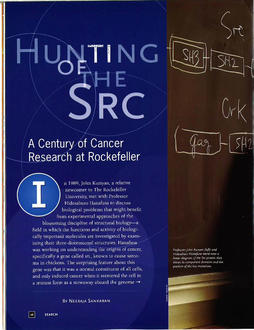
hunting of the src
TRANSCRIPT
of an infecting virus. Hanafusa
presented the following problem to Kuriyan: How did mutations in this Single gene change the functioning
of its protein product to induce a cell to start developing tumors?
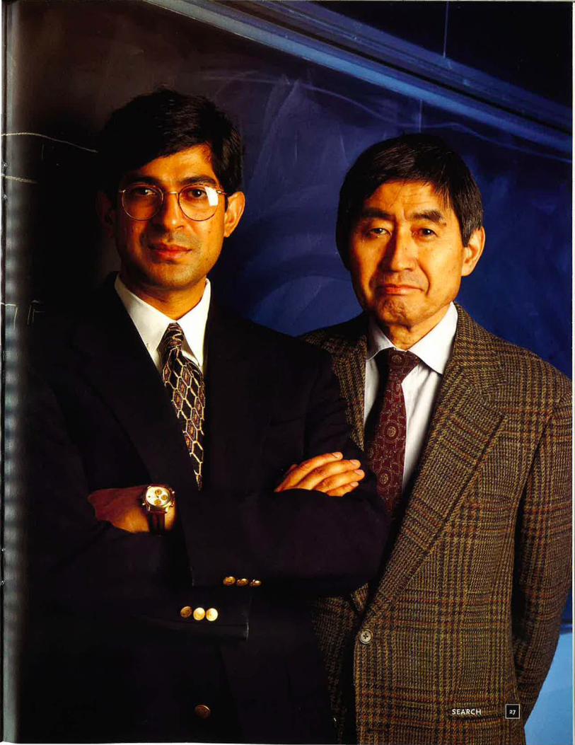
Kuriyan, now Patrick E. and Beatrice M. Haggerty Professor, head of the Laboratory of Molecular Biophysics and a Howard Hughes Medical Institute investigator, has a vivid memory of Hanafusa introducing him to the Src problem by drawi.ng a straight line on the blackboard and blocking out different regions, representing different parts of the molecule. Based on biochemical and genetic analyses, Hanafusa knew
Increasing the Magnification of Discovery Technology led the way to discovery of the src gene.
.. SEARCH
about mutations in different parts of the src gene that resulted in the "transformation" of normal cells to cancerous types, but he did not have a precise picture of how these mutations altered the normal activity of
the Src protein. One of the most significant
breakthroughs toward answering this question came last year, when Kuriyan's group, as well as Stephen Harrison's laboratory at Harvard University, solved the crystal structures of two Src proteins. The achievement represents an important milestone, not only with respect to answering specific questions about Src activity, but also because
Peyton Rous Discovers tumor agent, later called Rous Sarcoma Virus (RSV), that can transmit cancer in chickens.
of Src's leading role in the history of
understanding the general mechanisms underlying cancer. It is a story with deep roots at Rockefeller, dating
back to the turn of the century when Peyton Rous discovered the first tumor-inducing virus, thereby providing future cancer researchers with an indispensable tool for probing cell behavior and its breakdown.
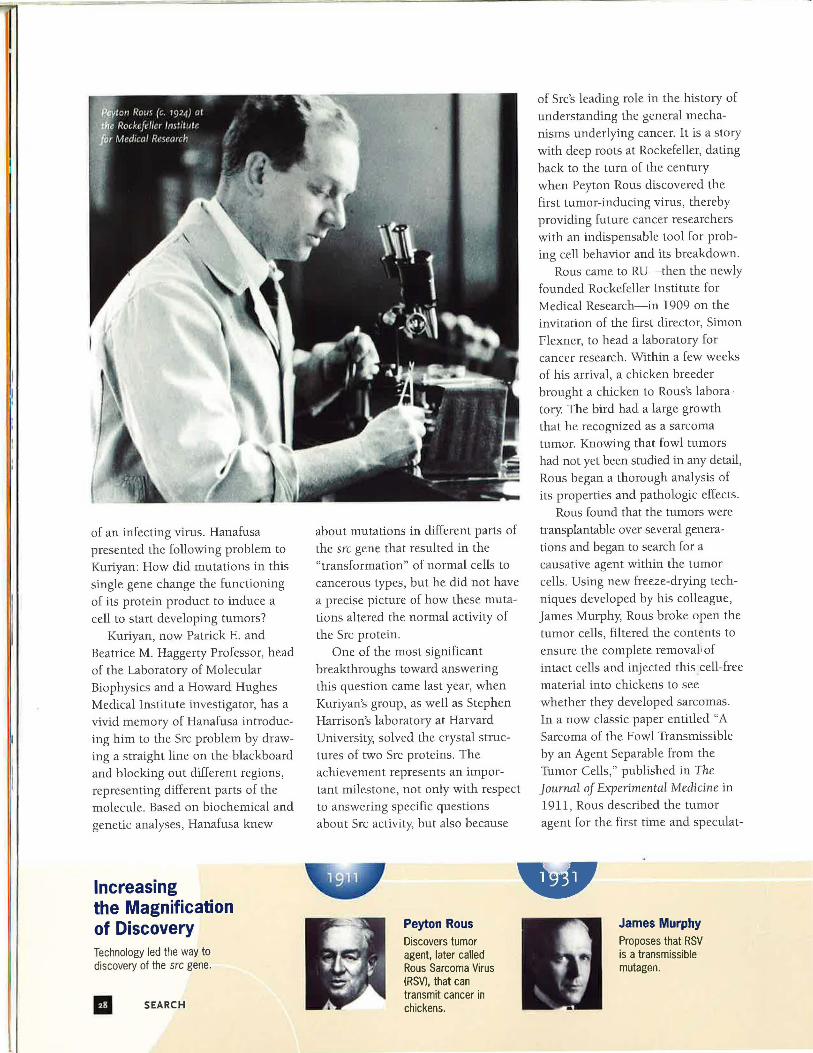
Rous came to RU-then the newly
founded Rockefeller Institute for Medical Research-in 1909 on the invitation of the first director, Simon Flexner, to head a laboratory for cancer research. Within a few weeks of his arrival, a chicken breeder brought a chicken to Rous's laboratory The bird had a large growth that he recognized as a sarcoma tumor. Knowing that fowl tumors had not yet been studied in any detail , Rous began a thorough analysiS of its properties and pathologic effects.
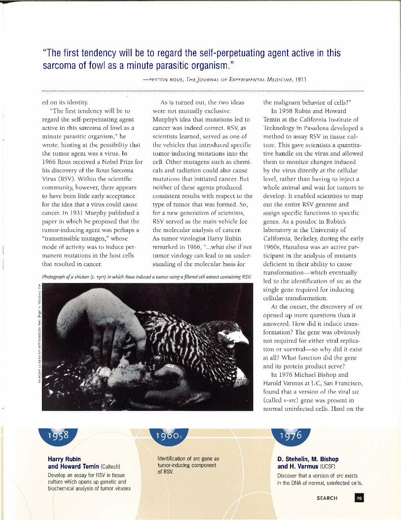
Rous found that the tumors were transplantable over several generations and began to search for a causative agent within the tumor cells. Using new freeze-drying techniques developed by his colleague, James Murphy, Rous broke open the tumor cells, filtered the contents to ensure the complete removal.: of intact cells and injected this cell-free material into chickens to see whether they developed sarcomas. In a now classic paper entitled "A Sarcoma of the Fowl Transmissible by an Agent Separable from the Tumor Cells," published in The Journal of Experimental Medicine in 1911, Rous described the tumor agent for the first time and speculat-
James Murphy Proposes that RSV is a transmissible mutagen.
"The first tendency will be to regard the self-perpetuating agent active in this sarcoma of fowl as a minute parasitic organism."
-PEYTON ROUS, THEJOURNAL OF EXPERIMENTAL MEDICINE, 1911
ed on its identity "The first tendency will be to
regard the self-perpetuating agent active in this sarcoma of fowl as a minute parasitic organism," he wrote, hinting at the possibility that the tumor agent was a virus. In 1966 Rous received a Nobel Prize for his discovery of the Rous Sarcoma Virus (RSV). Within the scientific
community, however, there appears to have been little early acceptance for the idea that a virus could cause
cancer. In 1931 Murphy published a paper in which he proposed that the tumor-inducing agent was perhaps a "transmissible mutagen," whose
mode of activity was to induce permanent mutations in the host cells that resulted in cancer.
As is turned out, the two ideas were not mutually exclusive. Murphy's idea that mutations led to cancer was indeed correct. RSY, as scientists learned, served as one of the vehicles that introduced specific tumor-inducing mutations into the cell. Other mutagens such as chemicals and radiation could also cause mutations that initiated cancer. But neither of these agents produced consistent results with respect to the type of tumor that was formed. So, for a new generation of scientists, RSV served as the main vehicle for
the molecular analysis of cancer. As tumor virologist Harry Rubin remarked in 1966, " ... what else if not tumor virology can lead to an understanding of the molecular basis for
Photograph of a chicken (c. 1911) in which Rous induced a tumor using a filtered cell extract containing RSV.
Harry Rubin and Howard Temin (Caltech)
Develop an assay for RSV in tissue culture which opens up genetic and biochemical analysis of tumor viruses
Identification of src gene as tumor·inducing component of RSV.
the malignant behavior of cells?" In 1958 Rubin and Howard
Temin at the California Institute of Technology in Pasadena developed a method to assay RSV in tissue culture. This gave scientists a quantitative handle on the virus and allowed them to monitor changes induced by the virus directly at the cellular level, rather than having to inject a whole animal and wait for tumors to develop. It enabled scientists to map out the entire RSV genome and assign specific functions to specific genes. As a postdoc in Rubin's laboratory at the University of California, Berkeley, during the early 1960s, Hanafusa was an active participant in the analysis of mutants deficient in their ability to cause transformation-which eventually led to the identification of src as the single gene required for inducing cellular transformation.
At the outset, the discovery of src opened up more questions than it answered. How did it induce trans
formation? The gene was obviously not required for either viral replication or survival-so why did it exist at all? What function did the gene and its protein product serve?
In 1976 Michael Bishop and Harold Varmus at UC, San Francisco, found that a version of the viral src (called v-src) gene was present in normal uninfected cells. Hard on the
D. Stehelin, M. Bishop and H. Varmus (UCSF)
Discover that a version of src exists in the DNA of normal, uninfected cells .
SEARCH IJ
", .. what else if not tumor virology can lead to an understanding of the molecular basis for the malignant behavior of cells?" -HARRY RUBIN, TUMOR VIROLOGIST, 1966
heels of this discovery, Hanafusa, who had moved to Rockefeller in 1973, furnished the genetic evidence that cellular src (c-src) sequences did in fact substitute for the transformation function missing in defective RSV mutants. To achieve this, he injected uninfected chickens with mutants of RSV that were known to have lost most of the DNA from their src gene and looked for the development of tumors.
"My prediction was that viruses containing partial deletions of the src sequence would undergo recombination with cellular sequences at a
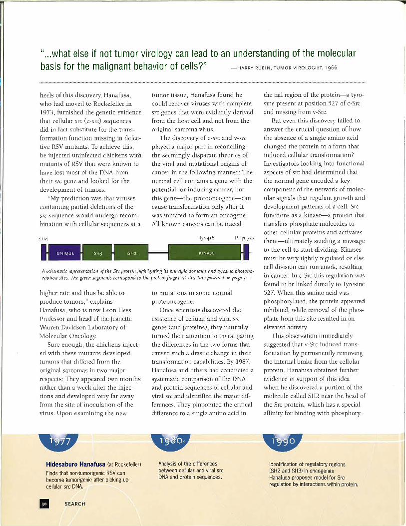
SH4
tumor tissue, Hanafusa found he could recover viruses with complete src genes that were evidently derived from the host cell and not from the original sarcoma virus.
The discovery of c-src and v-src played a major part in reconciling the seemingly disparate theories of the viral and mutational origins of cancer in the following manner: The normal cell contains a gene with the potential for inducing cancer, but this gene-the protooncogene-can cause transformation only after it was mutated to form an oncogene. All known cancers can be traced
Tyr·416 P-Tyr-S27
A schematic representation of the Src protein highlighting its principle domains and tyrosine phosphorylation sites. The green segments correspond to the protein fragment structure pictured on page 31.
higher rate and thus be able to
produce tumors," explains Hanafusa, who is now Leon Hess Professor and head of the Jeanette Warren Davidson Laboratory of Molecular Oncology.
Sure enough, the chickens injected with these mutants developed tumors that differed from the original sarcomas in two major respects: They appeared two months rather than a week after the injections and developed very far away from the site of inoculation of the virus. Upon examining the new
Hidesaburo Hanafusa (at Rockefeller)
Finds that non-tumorigenic RSV can become tumorigenic after picking up cellular src DNA.
II SEARCH
to mutations in some normal
proto oncogene. Once scientists discovered the
existence of cellular and viral src genes (and proteins), they naturally turned their attention to investigating the differences in the two forms that caused such a drastic change in their transformation capabilities. By 1987, Hanafusa and others had conducted a systematic comparison of the DNA and protein sequences of cellular and viral src and identified the major differences. They pinpointed the critical difference to a single amino acid in
Analysis of the differences between cellular and viral src DNA and protein sequences.
the tail region of the protein-a tyrosine present at position 527 of c-Src and missing from v-Src.
But even this discovery failed to answer the crucial question of how the absence of a single amino acid changed the protein to a form that induced cellular transformation? Investigators looking into functional aspects of src had determined that the normal gene encoded a key component of the network of molecular signals that regulate growth and development patterns of a cell. Src functions as a kinase-a protein that transfers phosphate molecules to other cellular proteins and activates them-ultimately sending a message to the cell to start dividing. Kinases must be very tightly regulated or else cell division can run amok, resulting in cancer. In c-Src this regulation was found to be linked directly to Tyrosine 527: When this amino acid was phosphorylated, the protein appeared inhibited, while removal of the phosphate from this site resulted in an
elevated activity This observation immediatdy
suggested that v-Src induced transformation by permanently removing the internal brake from the cellular protein. Hanafusa obtained further evidence in support of this idea when he discovered a portion of the molecule called SH2 near the head of
the Src protein, which has a special affinity for binding with phosphory-
Identification of regulatory regions (SH2 and SH3) in oncogenes Hanafusa proposes model for Src regulation by interactions within protein.
"The whole enzyme appears to be functioning like a Rube Goldberg machine, employing many complex parts to perform a simple task." -JOHN KURIYAN
lated tyrosine molecules. The dis<;overy had two exciting implications. First, it suggested that the SH2 region could bind to the phosphorylated Tyrosine 527, like a snake biting its own tail. An intramolecular contortion of this kind, Hanafusa imagined, would block Src's active site-its kinase domain-and thus
prevent it from phosphorylating other proteins. The finding also indicated that the SH2 domain played the role of a molecular postman, who recognized specific intracellular addresses-the tyrosines-where the kinase delivered the phosphate molecules. When attached to the internal tyrosine, the postman could no longer reach the other addresses, which resulted in the suppression of Src activity
This then was the model that
Hanafusa presented to Kuriyan in 1989: A linear diagram of the Src protein with its component domains and the position of the key mutations, superimposed with biochemical information on the possible areas of interaction.
"The question that Saburo posed, very simply and directly, was 'How is Src regulated?'" recounts Kuriyan.
"To proceed any further with understanding how it worked, one needed to know what it looked like in three dimensions."
The researchers had their first breakthrough when Kuriyan and his
Collaboration of several Rockefeller laboratories. Determination of the 3-D crystal structure of SH2 region of Src.
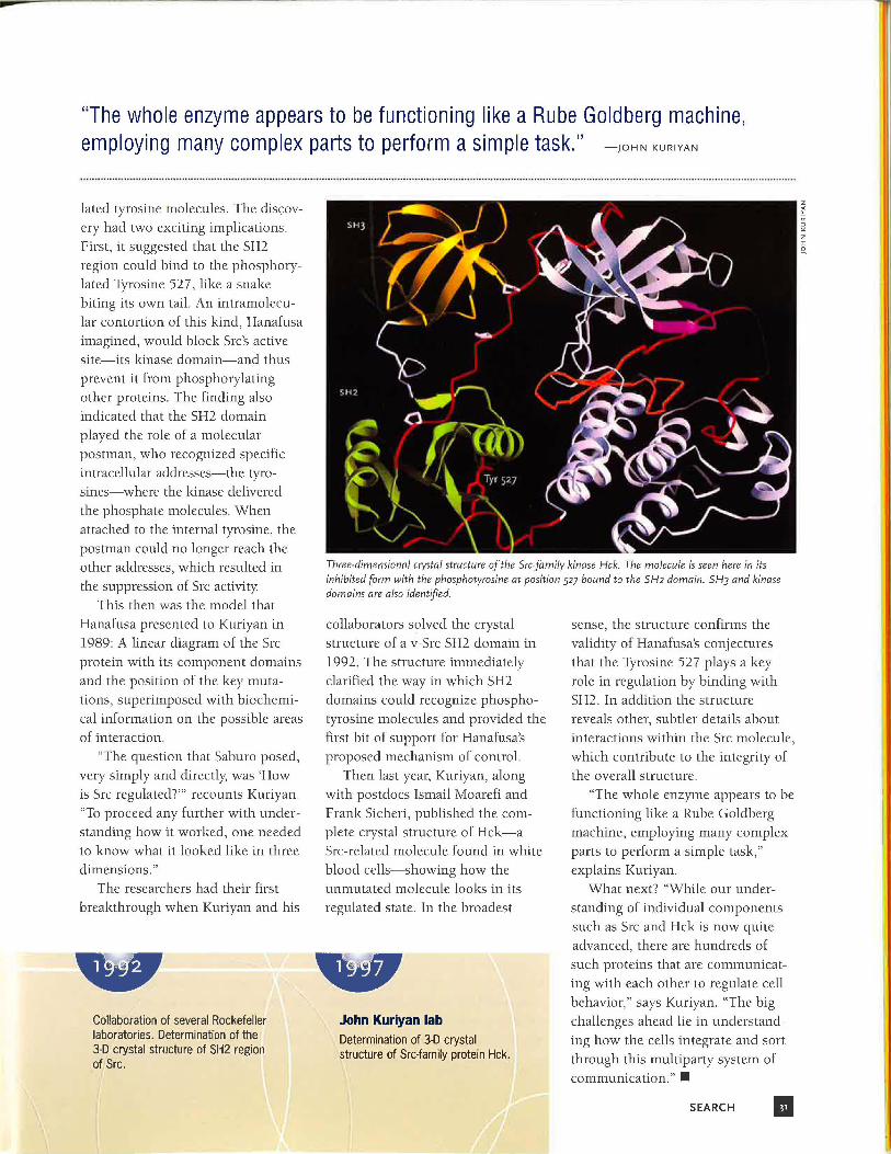
Three·dimensional crystal structure of the Srcfamily kinase Hck. The molecule is seen here in its inhibited form with the phosphotyrosine at position 527 bound to the SH2 domain. SH3 and kinase domains are also identified.
collaborators solved the crystal structure of a v-Src SH2 domain in 1992. The structure immediately clarified the way in which SH2 domains could recognize phosphotyrosine molecules and provided the first bit of support for Hanafusa's proposed mechanism of control.
Then last year, Kuriyan, along with postdocs Ismail Moarefi and Frank Sicheri, published the complete crystal structure of Hck-a Src-related molecule found in white blood cells-showing how the unmutated molecule looks in its regulated state. In the broadest
John Kuriyan lab Determination of JD crystal structure of Src-family protein Hck.
sense, the structure confirms the validity of Hanafusa's conjectures
that the Tyrosine 527 plays a key role in regulation by binding with SH2. In addition the structure reveals other, subtler details about interactions within the Src molecule, which contribute to the integrity of the overall structure.
"The whole enzyme appears to be functioning like a Rube Goldberg machine, employing many complex parts to perform a simple task," explains Kuriyan.
What next? "While our understanding of individual components such as Src and Hck is now quite advanced, there are hundreds of such proteins that are communicating with each other to regulate cell
behavior," says Kuriyan. "The big challenges ahead lie in understanding how the cells integrate and sort through this multiparty system of communication." •
SEARCH II