human metabolic response to systemic inflammation: assessment of the concordance between...
TRANSCRIPT
Kamisoglu et al. Critical Care (2015) 19:71 DOI 10.1186/s13054-015-0783-2
RESEARCH Open Access
Human metabolic response to systemicinflammation: assessment of the concordancebetween experimental endotoxemia and clinicalcases of sepsis/SIRSKubra Kamisoglu1, Beatrice Haimovich2, Steve E Calvano2, Susette M Coyle2, Siobhan A Corbett2,Raymond J Langley3, Stephen F Kingsmore4,5 and Ioannis P Androulakis1,2,6*
Abstract
Introduction: Two recent, independent, studies conducted novel metabolomics analyses relevant to human sepsisprogression; one was a human model of endotoxin (lipopolysaccharide (LPS)) challenge (experimental endotoxemia)and the other was community acquired pneumonia and sepsis outcome diagnostic study (CAPSOD). The purpose ofthe present study was to assess the concordance of metabolic responses to LPS and community-acquired sepsis.
Methods: We tested the hypothesis that the patterns of metabolic response elicited by endotoxin would agree withthose in clinical sepsis. Alterations in the plasma metabolome of the subjects challenged with LPS were comparedwith those of sepsis patients who had been stratified into two groups: sepsis patients with confirmed infection andnon-infected patients who exhibited systemic inflammatory response syndrome (SIRS) criteria. Common metabolitesbetween endotoxemia and both these groups were individually identified, together with their direction of changeand functional classifications.
Results: Response to endotoxemia at the metabolome level elicited characteristics that agree well with those observedin sepsis patients despite the high degree of variability in the response of these patients. Moreover, some distinct featuresof SIRS have been identified. Upon stratification of sepsis patients based on 28-day survival, the direction of change in 21of 23 metabolites was the same in endotoxemia and sepsis survival groups.
Conclusions: The observed concordance in plasma metabolomes of LPS-treated subjects and sepsis survivors strengthensthe relevance of endotoxemia to clinical research as a physiological model of community-acquired sepsis, and givesvaluable insights into the metabolic changes that constitute a homeostatic response. Furthermore, recapitulation ofmetabolic differences between sepsis non-survivors and survivors in LPS-treated subjects can enable further researchon the development and assessment of rational clinical therapies to prevent sepsis mortality. Compared with earlierstudies which focused exclusively on comparing transcriptional dynamics, the distinct metabolomic responses to systemicinflammation with or without confirmed infection, suggest that the metabolome is much better at differentiating thesepathophysiologies. Finally, the metabolic changes in the recovering patients shift towards the LPS-induced responsepattern strengthening the notion that the metabolic, as well as transcriptional responses, characteristic to theendotoxemia model represent necessary and “healthy” responses to infectious stimuli.
* Correspondence: [email protected] of Chemical and Biochemical Engineering, Rutgers University,Piscataway, NJ 08854, USA2Department of Surgery, Rutgers - Robert Wood Johnson Medical School,New Brunswick, NJ 08901, USAFull list of author information is available at the end of the article
© 2015 Kamisoglu et al.; licensee BioMed Central. This is an Open Access article distributed under the terms of the CreativeCommons Attribution License (http://creativecommons.org/licenses/by/4.0), which permits unrestricted use, distribution, andreproduction in any medium, provided the original work is properly credited. The Creative Commons Public DomainDedication waiver (http://creativecommons.org/publicdomain/zero/1.0/) applies to the data made available in this article,unless otherwise stated.
Kamisoglu et al. Critical Care (2015) 19:71 Page 2 of 10
IntroductionSepsis is defined as the combination of an infection withmultiple features of ‘systemic inflammatory responsesyndrome’ (SIRS) [1] and is one of the oldest and mostenigmatic conditions in medicine. There are more thana million cases of sepsis per year in the United States [2]and it is estimated that there are 19 million cases peryear worldwide [3,4]. According to the Centers forDisease Control, the cost of hospitalization is in theorder of $15 billion, with an anticipated further increasein the future [5]. Despite several decades of intensiveresearch and efforts to bring new therapies to the bedside,the number of cases and sepsis-associated deaths are stillsoaring [3,6]. Current treatment guidelines includecardiorespiratory resuscitation and non-specific protocolsaimed at mitigating immediate threats of uncontrolledinfection [3]. A significant barrier to progress is theperceived inadequacy of experimental models that canreproduce the pathophysiology of the disease in humans.The high degree of variability among patients and
multiple aspects of the disease, including patient gender,age and comorbidities complicate the design of relevantexperimental models and clinical studies. Moreover, theinitiating cause of infection and the physiologic responsesthat follow are also highly variable [7]. All these factorsexplain, at least in part, the difficulty in translating experi-mental results to the clinic and, consequently, the lack ofsuccess in the development of effective therapies [8].Endotoxemia, an experimental model in which healthy
volunteers are intravenously administered a form ofendotoxin (lipopolysaccharide, LPS, a major componentof Gram-negative bacteria outer membrane and a Toll-likereceptor 4 (TLR4) agonist) [9], has served as a valuableexperimental venue for more than six decades [10-12]. It isa model of systemic inflammation, rather than a truemimic of sepsis. Nonetheless, early transient physiochemicalchanges and biochemical pathway activation in this modelare strikingly similar to those observed during the earlyhyperdynamic phase of resuscitated injury and infection[13]. The LPS challenge triggers chills, myalgias, nausea,and an increase in core body temperature and heart rate,most of which begin to abate within six to eight hours[11,13,14]. Genome-wide analyses of circulating leukocytesrevealed transcriptional signatures indicative of changes inprotein translation and glycolysis [15], which shared similarcharacteristics with those observed in trauma patients [16].These studies helped elucidate the intricate regulatoryschemes governing the response to endotoxemia [16,17]and provided the foundations for in silico models ofsystemic inflammation [18-24]. More recently, we docu-mented the effects of LPS-induced inflammation on thewhole body metabolism in humans [25]. In contrast withother methods applied to the endotoxemia model,metabolomics reflects the combined output of all tissues
in the body [26]. In that study [25], plasma samples,collected from healthy subjects during 24-hours postchallenge with LPS, were subjected to non-targetedbiochemical profiling, revealing the temporal changesin the plasma metabolome. Unsupervised multivariateanalyses identified prominent changes in lipid andprotein metabolism, which peaked at six hours postLPS infusion. Subsequently, to understand better howthe inflammatory responses at the level of cells andwhole body correlate in humans, we integrated theanalysis of the plasma metabolome with that of theleukocyte transcriptome [27].In [28], an integrated analysis of clinical features,
plasma metabolome and proteome described the patternof metabolic perturbations in critically ill patientspresenting with symptoms of SIRS or sepsis. Thisstudy, the first of its kind, examined clinical featuresas well as the plasma metabolome, and proteome, ofpatients upon arrival at the emergency department (ED)and 24 hours later. An important and novel outcome ofthe study was the realization that metabolic differencescould ultimately be used as markers predicting survival.Since the endotoxemia model utilizes LPS, rather than
intact bacteria, there is an ongoing concern that dataderived from this model are of limited relevance toour understanding of sepsis-induced inflammatorymechanisms, although recent analyses of the leukocytetranscriptome seemed to argue otherwise [15]. The avail-ability of new metabolomic data [25,28] offered the oppor-tunity to compare responses detected in LPS-challengedsubjects to those of critically ill patients at the level of theentire organism. In this retrospective study we aimed toobjectively determine the relevance of the informationcontent gained by parallel analyses of LPS-challengedsubjects [25] and patients with or without community-acquired sepsis [28]. Our study identified a core responsethat was in agreement with what was observed in sepsispatients. Response to systemic inflammation withoutapparent infectious stimuli such as what is observed inSIRS was shown to have distinct features that may make ituniquely recognizable at the metabolomics level.Metabolic changes in the patients who are recoveringshifted towards an endotoxemia response pattern,strengthening the idea that the endotoxemia model repre-sents necessary and ‘healthy’ responses to an infectiousstimulus.
Material and methodsMetabolic dataThis is a retrospective analysis utilizing metabolomesobtained from subjects who participated in an experimen-tal endotoxemia study and from patients with or with-out community-acquired sepsis. In brief, as previouslydescribed [25], healthy volunteers participated in an
Kamisoglu et al. Critical Care (2015) 19:71 Page 3 of 10
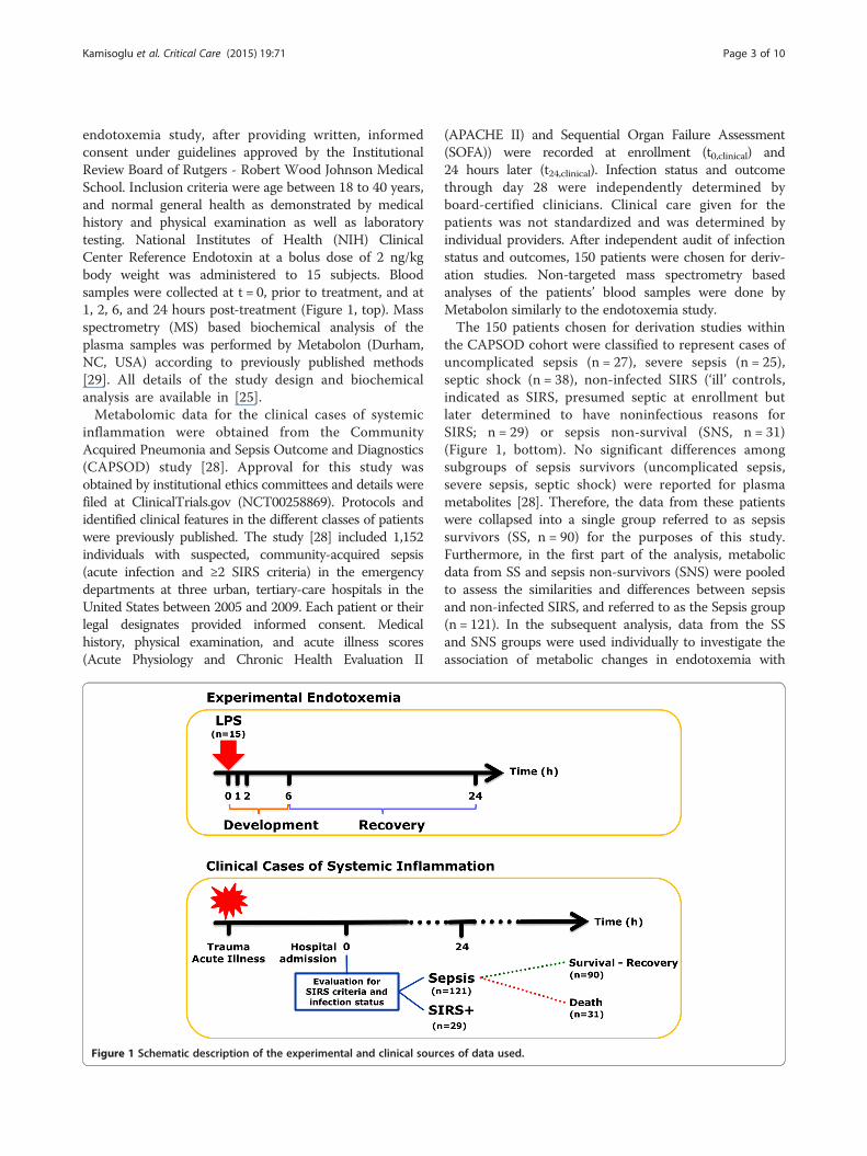
endotoxemia study, after providing written, informedconsent under guidelines approved by the InstitutionalReview Board of Rutgers - Robert Wood Johnson MedicalSchool. Inclusion criteria were age between 18 to 40 years,and normal general health as demonstrated by medicalhistory and physical examination as well as laboratorytesting. National Institutes of Health (NIH) ClinicalCenter Reference Endotoxin at a bolus dose of 2 ng/kgbody weight was administered to 15 subjects. Bloodsamples were collected at t = 0, prior to treatment, and at1, 2, 6, and 24 hours post-treatment (Figure 1, top). Massspectrometry (MS) based biochemical analysis of theplasma samples was performed by Metabolon (Durham,NC, USA) according to previously published methods[29]. All details of the study design and biochemicalanalysis are available in [25].Metabolomic data for the clinical cases of systemic
inflammation were obtained from the CommunityAcquired Pneumonia and Sepsis Outcome and Diagnostics(CAPSOD) study [28]. Approval for this study wasobtained by institutional ethics committees and details werefiled at ClinicalTrials.gov (NCT00258869). Protocols andidentified clinical features in the different classes of patientswere previously published. The study [28] included 1,152individuals with suspected, community-acquired sepsis(acute infection and ≥2 SIRS criteria) in the emergencydepartments at three urban, tertiary-care hospitals in theUnited States between 2005 and 2009. Each patient or theirlegal designates provided informed consent. Medicalhistory, physical examination, and acute illness scores(Acute Physiology and Chronic Health Evaluation II
Figure 1 Schematic description of the experimental and clinical sourc
(APACHE II) and Sequential Organ Failure Assessment(SOFA)) were recorded at enrollment (t0,clinical) and24 hours later (t24,clinical). Infection status and outcomethrough day 28 were independently determined byboard-certified clinicians. Clinical care given for thepatients was not standardized and was determined byindividual providers. After independent audit of infectionstatus and outcomes, 150 patients were chosen for deriv-ation studies. Non-targeted mass spectrometry basedanalyses of the patients’ blood samples were done byMetabolon similarly to the endotoxemia study.The 150 patients chosen for derivation studies within
the CAPSOD cohort were classified to represent cases ofuncomplicated sepsis (n = 27), severe sepsis (n = 25),septic shock (n = 38), non-infected SIRS (‘ill’ controls,indicated as SIRS, presumed septic at enrollment butlater determined to have noninfectious reasons forSIRS; n = 29) or sepsis non-survival (SNS, n = 31)(Figure 1, bottom). No significant differences amongsubgroups of sepsis survivors (uncomplicated sepsis,severe sepsis, septic shock) were reported for plasmametabolites [28]. Therefore, the data from these patientswere collapsed into a single group referred to as sepsissurvivors (SS, n = 90) for the purposes of this study.Furthermore, in the first part of the analysis, metabolicdata from SS and sepsis non-survivors (SNS) were pooledto assess the similarities and differences between sepsisand non-infected SIRS, and referred to as the Sepsis group(n = 121). In the subsequent analysis, data from the SSand SNS groups were used individually to investigate theassociation of metabolic changes in endotoxemia with
es of data used.
Kamisoglu et al. Critical Care (2015) 19:71 Page 4 of 10
those observed in either surviving or non-surviving sepsispatients.
Data analysisMS analysis of plasma samples from the human endo-toxemia study provided temporal information on 366metabolites at five time points. Previous results of theprincipal component analysis on this dataset showedthat the six hour time point (t6) was the most criticalsince the maximum difference between control andtreatment groups was observed at this time point [25]. Thisagreed well with prior transcriptional studies indicatingthat the maximal change in leukocyte gene expression wasobserved six hours after the LPS administration [16,30].Therefore, this data point was considered to represent thepeak of metabolic response to endotoxemia and used asreference for the assessment of concordance betweenexperimental and clinical data in this study. MS analysis ofthe samples from the CAPSOD study, on the other hand,had identified 370 metabolites at t0,clinical (time of hospitaladmission) and 401 metabolites at t24,clinical (24 hours afteradmission). In this study, both clinical and experimentaldatasets were individually normalized by setting themedian equal to 1. Missing values were imputed with theobserved minimums after normalization. The metabolitelists were consolidated. Only the metabolites commonlyidentified in the endotoxemia [25] and clinical [28] studieswere analyzed further. The final dataset included 177common metabolites from both studies [25,28]. Outlierswere removed using the median absolute deviation,MAD= 1.4826 × |(xi –Medianj(xj)|, of each metabolite, de-termined in each individual group [31,32]. Subsequently, the
score for each data point was calculated zi ¼ xi−Medianj xjð Þj jMAD
and data points with a score above 3 were removed fromthe dataset. The number of removed outliers for each groupis reported in Additional file 1: Table S1.The baseline of the human endotoxemia studies, that
is, samples collected before LPS administration (t0,LPS),defined the ‘baseline’ in this study. We identified the sixhour time point as the peak of the metabolic response inthe endotoxemia model in our previous metabolomicsstudy [25], as well as transcriptomic analysis [17,18], andhypothesized that this time point represents the point oftransition from the development and recovery phases ofthe response. For the clinical data collection, the startingpoint was the time of hospital admission (t0,clinical),whereas the second clinical time point was 24 hourslater (t24,clinical). Since the data obtained for the clinicalpatients lack internal controls, for obvious reasons,the responses of each group of patients, as well asthe endotoxemia subjects, were compared independentlyto the healthy baseline (t0,LPS). For comparing the meansof metabolites in each condition relative to the healthy
baseline, Welch’s t-test was used without assuming equalvariances (α = 0.05). The number of subjects in the experi-mental endotoxemia group (n = 15) was much smaller thanthe number of patients in the clinical groups to assumenormal distribution required for the t-test. However, at botht0,LPS and t6,LPS, the data passed the Kolmogorov-Smirnovtest for each metabolite allowing the application of thet-test. Q-values were calculated according to theBenjamini and Hochberg procedure [33] and metaboliteshaving a P- and a q-value less than 0.05 are called signifi-cant. We also evaluated how dispersed the data for eachmetabolite is in clinical cases with respect to those at thebaseline. Variances of the significant metabolites in eachcondition were also plotted relative to the variances at thebaseline and are shown in Additional file 2: Figure S1. Thedirection and magnitude of changes in plasma metaboliteconcentrations were determined based on log2 fold changesfrom t0,LPS for both clinical groups and plotted againstthose observed for endotoxemia as shown in Figure 2. Thefull list of the metabolites, their significance in each condi-tion, the direction and magnitude of the changes relative tothe baseline is provided in Additional file 3: Table S2.We lastly focused on changes in metabolites within
subpopulations of sepsis patients who ultimately survived,and those who did not. For this purpose, dynamics withinthe sepsis group were examined using the data from theSS and SNS groups. Metabolites that statistically differedbetween these groups at either time point were determinedby t-test as described earlier. The magnitude of changes inplasma metabolites relative to t0,LPS were calculated foreach group. These changes were compared betweenthe SS and SNS groups at both time points and withthe endotoxemia group at t6,LPS.
Results and discussionEndotoxemia induced by elective administration of LPSto healthy subjects has served as an invaluable toolfor obtaining mechanistic insight into homeostaticinflammatory responses. Previous studies comparedtranscript- and protein-expression patterns in immunecells obtained from LPS treated subjects and traumapatients, revealing significant overlap [15,34]. More re-cently, metabolomics analyses in both LPS-administeredsubjects [25] and patients with symptoms of systemicinflammation at time of presentation to emergencydepartments were published [28,35]. Building on theseprior studies [26,29], here we aimed to objectively com-pare metabolic indices obtained from experimental studiesand clinical sources.The inherent dynamics of a clinical and an experimental
study are obviously disparate although they focus on relatedphysiologic phenomena. The first time point in a clinicalstudy is generally at a point that had already deviated fromwhat can be called a ‘healthy state,’ whereas experimental
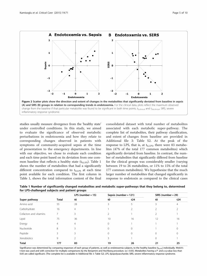
Figure 2 Scatter plots show the direction and extent of changes in the metabolites that significantly deviated from baseline in sepsis(A) and SIRS (B) groups in relation to corresponding trends in endotoxemia. For the clinical data, plots reflect the maximum observedchange from the baseline if that particular metabolite was found to be significant in both time points, t0,clinical and t24,clinical. SIRS, severeinflammatory response syndrome.
Kamisoglu et al. Critical Care (2015) 19:71 Page 5 of 10
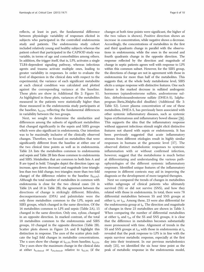
studies usually measure divergence from the ‘healthy state’under controlled conditions. In this study, we aimedto evaluate the significance of observed metabolicperturbations in endotoxemia and how they relate tocorresponding changes observed in patients withsymptoms of community-acquired sepsis at the timeof presentation to the emergency departments. In linewith our objective, we chose to evaluate each conditionand each time point based on its deviation from one com-mon baseline that reflects a healthy state (t0,LPS). Table 1shows the number of metabolites that had a significantlydifferent concentration compared to t0,LPS at each timepoint available for each condition. The first column inTable 1, shows the total information content of the final
Table 1 Number of significantly changed metabolites and mefor LPS-challenged subjects and patient groups
LPS (number = 15)
Super pathway Total t6
Amino acid 55 28
Carbohydrate 16 4
Cofactors and vitamins 7 3
Lipid 75 36
Energy 4 4
Nucleotide 8 3
Peptide 3 3
Xenobiotics 9 2
Total 177 83
Significance was determined by comparing responses of each group of patients, as wet-test was used and with correction for multiple comparisons by the Benjamini and Ho0.05 are called significant. (The complete list is available in Additional file 3: Table S2).
consolidated dataset with total number of metabolitesassociated with each metabolic super-pathway. Thecomplete list of metabolites, their pathway classification,and extent of changes from baseline are provided inAdditional file 3: Table S2. At the peak of theresponse to LPS, that is, at t6,LPS, there were 83 metabo-lites (47% of the total 177 common metabolites) whichsignificantly deviated from baseline. In contrast, the num-ber of metabolites that significantly differed from baselinefor the clinical groups was considerably smaller (varyingbetween 19 to 26 metabolites, or 11% to 15% of the total177 common metabolites). We hypothesize that the muchlarger number of metabolites that changed significantly inresponse to endotoxin as compared to the clinical cases
tabolic super-pathways that they belong to, determined
Sepsis (number = 121) SIRS (number = 29)
t0 t24 t0 t24
3 5 3 4
1 2 2 2
2 1 2 1
10 16 12 16
0 1 1 1
0 0 1 0
0 0 0 0
3 1 0 1
19 26 21 25
ll as endotoxemia subjects, to the healthy baseline (t0,LPS) individually. Welch’schberg procedure. (α = 0.05). Metabolites having a P- and a q-value less thanLPS, lipopolysaccharide; SIRS, severe inflammatory response syndrome.
Kamisoglu et al. Critical Care (2015) 19:71 Page 6 of 10
reflects, at least in part, the fundamental differencebetween physiologic variability of responses elicited insubjects who participated in the controlled endotoxemiastudy and patients. The endotoxemia study cohortincluded relatively young and healthy subjects whereas thepatient cohort that participated in the CAPSOD study wasvariable, in terms of age and comorbidities among others.In addition, the trigger itself, that is, LPS, activates a singleTLR4-dependent signaling pathway, whereas infectiousagents and trauma activate multiple ones, leading togreater variability in responses. In order to evaluate thelevel of dispersion in the clinical data with respect to theexperimental, the variance of each significant metaboliteat each clinical condition was calculated and plottedagainst the corresponding variance at the baseline.These plots are show in Additional file 2: Figure S1.As highlighted in these plots, variances of the metabolitesmeasured in the patients were statistically higher thanthose measured in the endotoxemia study participants atthe baseline, t0,LPS, reflecting the fundamental differencesin variability between the two groups.Next, we sought to determine the similarities and
differences among the subsets of significant metabolitesthat changed significantly in the sepsis and SIRS groupswhich were also significant in endotoxemia. Our intentionwas to be maximally inclusive of the clinically observedchanges. Therefore, we focused on metabolites that weresignificantly different from the baseline at either one ofthe two clinical time points as well as in endotoxemia.Table 2A lists the metabolites common to endotoxemiaand sepsis and Table 2B lists those common to endotoxemiaand SIRS. Metabolites that are common to both lists A andB are typed in bold. Triangles depict the direction (apex up:increase, apex down: decrease) and magnitude (one triangle:less than two fold change, two triangles: more than two foldchange) of the difference relative to the baseline (t0,LPS).Although the total number of metabolites in common withendotoxemia is close for the two clinical cases (16 inTable 2A and 18 in Table 2B), the agreement between thedirections of change is strikingly different. Bilirubin,docosapentaenoate (DPA) and palmitolate were theonly three metabolites common to the LPS, sepsis andSIRS groups, which changed in the same direction. Of the16 metabolites common to LPS and sepsis (Table 2A), 15changed in the same direction. Only one, xylose, changedin an opposite direction. In marked contrast, of the total18 metabolites common to the endotoxemia and SIRSgroups, 10 changed in the opposite direction (Table 2B).Scatter plots shown in Figure 2A and B highlight thisdistinction in response. The axes of the scatter plots indi-cate the log2 fold changes in metabolite concentrations.The x-axes show the change at t6,LPS from baseline, t0,LPS.The y-axes show the maximum change in the clinical dataat either t0,clinical or t24,clinical, relative to t0,LPS (if the
changes at both time points were significant, the higher ofthe two values is shown). Positive direction shows anincrease in concentration, while negative shows a decrease.Accordingly, the concentrations of metabolites in the firstand third quadrants change in parallel with the observa-tions in endotoxemia; while the ones in the second andfourth quadrants change in the opposite direction. Theresponse reflected by the direction and magnitude ofchange in septic patients agrees well with response to LPSwithin this common subset. However, for the SIRS group,the directions of change are not in agreement with those inendotoxemia for more than half of the metabolites. Thissuggests that, at the whole body metabolome level, SIRSelicits a unique response with distinctive features. One suchfeature is the marked decrease in sulfated androgenichormones (epiandrosterone sulfate, androsterone sul-fate, dehydroisoandrosterone sulfate (DHEA-S), 5alpha-pregnan-3beta,20alpha-diol disulfate) (Additional file 3:Table S2). Lower plasma concentration of one of thesemetabolites, DHEA-S, has previously been associated withother systemic inflammatory diseases, such as systemiclupus erythematosus and inflammatory bowel disease [36].This supports the idea that the inflammatory responsewithout apparent infectious stimuli might elicit distinctivefeatures not shared with sepsis or endotoxemia. It hasbeen previously suggested that acute inflammatorystresses from different etiologies result in highly similarresponses in humans at the genomic level [37]. Theobserved distinct metabolomic responses to systemicinflammation with or without confirmed infection,however, suggest that the metabolome is much betterat differentiating and understanding the various path-ophysiologies of the different systemic inflammatoryresponses. Identified unique features of the inflammatoryresponse in different contexts may aid in improving thediagnosis or the development of more targeted therapies.Next we compared the trends of changes in metabolites
within subgroups of clinical patients who ultimatelysurvived (SS) or did not survive (SNS), and how theyrelated with those in endotoxemia. In total, there were 78differential metabolites between SS and SNS groups ateither t0 or t24. Among these, 23 were also differential forthe endotoxemia group at t6. The direction and magnitudeof changes in these 23 metabolites are shown in Table 3.When comparing the number of differential metabolitesat either t0 and t24 of the SS and SNS groups, it is clearthat the difference in metabolites becomes substantiallymore pronounced with time. Alignment of trends in theSS and SNS groups at t24 with those in endotoxemia at t6revealed that the peak response to LPS is in line with thesepsis survivor metabolic response, especially at the firstday into their treatment. In our previous metabolomicsstudy [25], we identified the six hour time point as thepeak of metabolic response in the endotoxemia model,
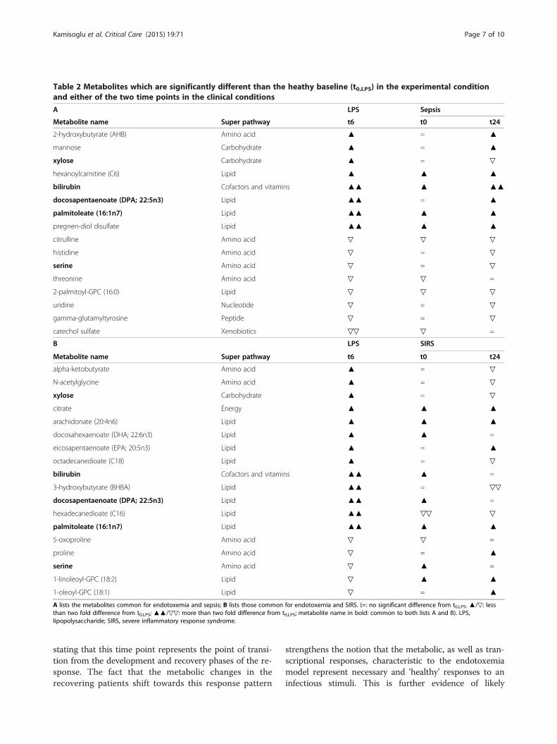
Table 2 Metabolites which are significantly different than the heathy baseline (t0,LPS) in the experimental conditionand either of the two time points in the clinical conditions
A LPS Sepsis
Metabolite name Super pathway t6 t0 t24
2-hydroxybutyrate (AHB) Amino acid ▲ = ▲
mannose Carbohydrate ▲ = ▲
xylose Carbohydrate ▲ = ▽
hexanoylcarnitine (C6) Lipid ▲ ▲ ▲
bilirubin Cofactors and vitamins ▲▲ ▲ ▲▲
docosapentaenoate (DPA; 22:5n3) Lipid ▲▲ = ▲
palmitoleate (16:1n7) Lipid ▲▲ ▲ ▲
pregnen-diol disulfate Lipid ▲▲ ▲ ▲
citrulline Amino acid ▽ ▽ ▽
histidine Amino acid ▽ = ▽
serine Amino acid ▽ = ▽
threonine Amino acid ▽ ▽ =
2-palmitoyl-GPC (16:0) Lipid ▽ ▽ ▽
uridine Nucleotide ▽ = ▽
gamma-glutamyltyrosine Peptide ▽ = ▽
catechol sulfate Xenobiotics ▽▽ ▽ =
B LPS SIRS
Metabolite name Super pathway t6 t0 t24
alpha-ketobutyrate Amino acid ▲ = ▽
N-acetylglycine Amino acid ▲ = ▽
xylose Carbohydrate ▲ = ▽
citrate Energy ▲ ▲ ▲
arachidonate (20:4n6) Lipid ▲ ▲ ▲
docosahexaenoate (DHA; 22:6n3) Lipid ▲ ▲ =
eicosapentaenoate (EPA; 20:5n3) Lipid ▲ = ▲
octadecanedioate (C18) Lipid ▲ = ▽
bilirubin Cofactors and vitamins ▲▲ ▲ =
3-hydroxybutyrate (BHBA) Lipid ▲▲ = ▽▽
docosapentaenoate (DPA; 22:5n3) Lipid ▲▲ ▲ =
hexadecanedioate (C16) Lipid ▲▲ ▽▽ ▽
palmitoleate (16:1n7) Lipid ▲▲ ▲ ▲
5-oxoproline Amino acid ▽ ▽ =
proline Amino acid ▽ = ▲
serine Amino acid ▽ ▲ =
1-linoleoyl-GPC (18:2) Lipid ▽ ▲ ▲
1-oleoyl-GPC (18:1) Lipid ▽ = ▲
A lists the metabolites common for endotoxemia and sepsis; B lists those common for endotoxemia and SIRS. (=: no significant difference from t0,LPS. ▲/▽: lessthan two fold difference from t0,LPS; ▲▲/▽▽: more than two fold difference from t0,LPS; metabolite name in bold: common to both lists A and B). LPS,lipopolysaccharide; SIRS, severe inflammatory response syndrome.
Kamisoglu et al. Critical Care (2015) 19:71 Page 7 of 10
stating that this time point represents the point of transi-tion from the development and recovery phases of the re-sponse. The fact that the metabolic changes in therecovering patients shift towards this response pattern
strengthens the notion that the metabolic, as well as tran-scriptional responses, characteristic to the endotoxemiamodel represent necessary and ‘healthy’ responses to aninfectious stimuli. This is further evidence of likely
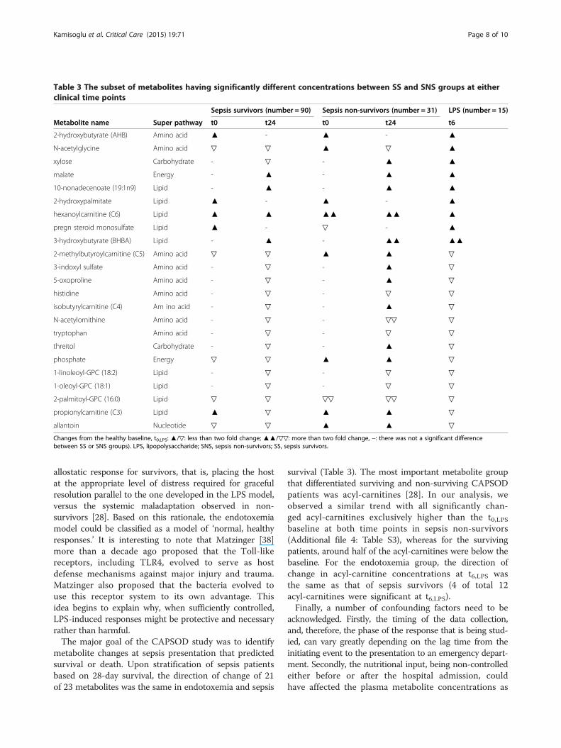
Table 3 The subset of metabolites having significantly different concentrations between SS and SNS groups at eitherclinical time points
Sepsis survivors (number = 90) Sepsis non-survivors (number = 31) LPS (number = 15)
Metabolite name Super pathway t0 t24 t0 t24 t6
2-hydroxybutyrate (AHB) Amino acid ▲ - ▲ - ▲
N-acetylglycine Amino acid ▽ ▽ ▲ ▽ ▲
xylose Carbohydrate - ▽ - ▲ ▲
malate Energy - ▲ - ▲ ▲
10-nonadecenoate (19:1n9) Lipid - ▲ - ▲ ▲
2-hydroxypalmitate Lipid ▲ - ▲ - ▲
hexanoylcarnitine (C6) Lipid ▲ ▲ ▲▲ ▲▲ ▲
pregn steroid monosulfate Lipid ▲ - ▽ - ▲
3-hydroxybutyrate (BHBA) Lipid - ▲ - ▲▲ ▲▲
2-methylbutyroylcarnitine (C5) Amino acid ▽ ▽ ▲ ▲ ▽
3-indoxyl sulfate Amino acid - ▽ - ▲ ▽
5-oxoproline Amino acid - ▽ - ▲ ▽
histidine Amino acid - ▽ - ▽ ▽
isobutyrylcarnitine (C4) Am ino acid - ▽ - ▲ ▽
N-acetylornithine Amino acid - ▽ - ▽▽ ▽
tryptophan Amino acid - ▽ - ▽ ▽
threitol Carbohydrate - ▽ - ▲ ▽
phosphate Energy ▽ ▽ ▲ ▲ ▽
1-linoleoyl-GPC (18:2) Lipid - ▽ - ▽ ▽
1-oleoyl-GPC (18:1) Lipid - ▽ - ▽ ▽
2-palmitoyl-GPC (16:0) Lipid ▽ ▽ ▽▽ ▽▽ ▽
propionylcarnitine (C3) Lipid ▲ ▽ ▲ ▲ ▽
allantoin Nucleotide ▽ ▽ ▲ ▲ ▽
Changes from the healthy baseline, t0,LPS: ▲/▽: less than two fold change; ▲▲/▽▽: more than two fold change, −: there was not a significant differencebetween SS or SNS groups). LPS, lipopolysaccharide; SNS, sepsis non-survivors; SS, sepsis survivors.
Kamisoglu et al. Critical Care (2015) 19:71 Page 8 of 10
allostatic response for survivors, that is, placing the hostat the appropriate level of distress required for gracefulresolution parallel to the one developed in the LPS model,versus the systemic maladaptation observed in non-survivors [28]. Based on this rationale, the endotoxemiamodel could be classified as a model of ‘normal, healthyresponses.’ It is interesting to note that Matzinger [38]more than a decade ago proposed that the Toll-likereceptors, including TLR4, evolved to serve as hostdefense mechanisms against major injury and trauma.Matzinger also proposed that the bacteria evolved touse this receptor system to its own advantage. Thisidea begins to explain why, when sufficiently controlled,LPS-induced responses might be protective and necessaryrather than harmful.The major goal of the CAPSOD study was to identify
metabolite changes at sepsis presentation that predictedsurvival or death. Upon stratification of sepsis patientsbased on 28-day survival, the direction of change of 21of 23 metabolites was the same in endotoxemia and sepsis
survival (Table 3). The most important metabolite groupthat differentiated surviving and non-surviving CAPSODpatients was acyl-carnitines [28]. In our analysis, weobserved a similar trend with all significantly chan-ged acyl-carnitines exclusively higher than the t0,LPSbaseline at both time points in sepsis non-survivors(Additional file 4: Table S3), whereas for the survivingpatients, around half of the acyl-carnitines were below thebaseline. For the endotoxemia group, the direction ofchange in acyl-carnitine concentrations at t6,LPS wasthe same as that of sepsis survivors (4 of total 12acyl-carnitines were significant at t6,LPS).Finally, a number of confounding factors need to be
acknowledged. Firstly, the timing of the data collection,and, therefore, the phase of the response that is being stud-ied, can vary greatly depending on the lag time from theinitiating event to the presentation to an emergency depart-ment. Secondly, the nutritional input, being non-controlledeither before or after the hospital admission, couldhave affected the plasma metabolite concentrations as
Kamisoglu et al. Critical Care (2015) 19:71 Page 9 of 10
an independent factor. Thirdly, some of the CAPSODpatients either had prior comorbidities that were likely toaffect the metabolome, such as diabetes mellitus, or werealso developing conditions which further exacerbated theresponse, including compromised renal function, a likelymajor contributor to the observed metabolome.
ConclusionsTherapeutic strategies that are successfully translatedinto the clinic are very few and mostly non-specific in thefield of critical care. This is due, in part, to the complex anddynamic physiological processes involved. Heterogeneity ofthe patient populations and consequent challenges inperforming insightful clinical studies also have contributedto the lack of progress in this realm of medicine [3,39].Emerging -omics tools that are capable of examiningphysiologic responses at the systems level are promising,especially for complex conditions, such as sepsis and SIRS[40]. The major caveat related to these tools is that sincethe biological processes are analyzed at a higher level,inter-species differences become as relevant to the responseas the sought-after question itself. Therefore, utility of theanimal models has been questioned recently in thescientific community [37,41].The human endotoxemia model has been serving as a
useful experimental platform for gaining insight into themechanisms governing systemic inflammation. It is arecognized fact that this model does not fully replicatethe magnitude of physiologic stress created by trauma orinfection [13,14]; however, it gives researchers theopportunity to study the mechanisms underlying theresponse to systemic inflammation and relevant therapyoptions without the inter-species differences obscuringthe interpretation of the results.Progression of response to systemic inflammation in-
duced by endotoxemia in immune cells has been describedat the genomic level [16,30]. Moreover, comparison of theresponse to experimental stimuli and traumatic/infectiousinsults revealed significant overlap of common featuresboth at the gene [15] and protein expression levels [34]. Inthe light of these observations, the current study aimed atan objective evaluation of the concordance between experi-mental and clinical cases of systemic inflammation andbenchmarked endotoxemia against sepsis of various originsat the level of metabolic response. The plasma metabolomecan be thought of as the metabolic fingerprint representa-tive of the state of the body at any given time and provideinformation on the dominant regulatory mechanisms atvarious levels of cellular processes including transcription,translation and signal transduction. For effective provisionof critical care, understanding the alterations in theplasma metabolome is crucial, because metabolitelevels impact the regulation of anti-inflammatory defenses,in turn, through steering critical cellular processes and
immune mechanisms. Therefore, we think that the assess-ment of the relevance of endotoxemia as an experimentalmodel representing critical illness is important.We believe that the observed concordance between the
responses of LPS-treated subjects and sepsis patients at themetabolome level, despite observed variability in clinicaldata, strengthens the relevance of endotoxemia to clinicalresearch as an elementary tool and gives valuableinsights into the metabolic changes necessary for properresponse to inflammatory stress at the systemic level.
Key messages
� We compared the metabolic response at the peak ofLPS-induced acute inflammation with those fromsepsis patients
� For the metabolites shown to change significantlyfrom the baseline, the direction and magnitude ofthe changes were in agreement with what wasobserved in sepsis patients.
� The metabolic response in SIRS patients was shownbe distinct from those in endotoxemia or sepsis.
� Metabolic changes in the surviving sepsis patientsshifted towards those observed in endotoxemia astheir recovery proceeded.
� These observations strengthened the relevance ofendotoxemia to clinical research as a valuableexperimental tool which can enable further researchon the development and assessment of rationalclinical therapies to prevent sepsis mortality.
Additional files
Additional file 1: Table S1. Number of outliers removed from the databefore any statistical analysis.
Additional file 2: Figure S1. Comparison of the variances of significantmetabolites in the clinical groups with respect to those in the baseline (t0,LPS).
Additional file 3: Table S2. Full list of the metabolites, theirsignificance in each condition, direction and magnitude of the changesrelative to the baseline (t0,LPS).
Additional file 4: Table S3. Results of t-test for acyl-GPCs andacyl-carnitines, between SS and SNS groups at each time point, andbetween t0,LPS and t6,LPS in endotoxemia group, together with theirdirection of change from the common baseline t0,LPS. (Changes fromthe healthy baseline, t0,LPS: ▲/▽: less than 2 fold change; ▲▲/▽▽:more than 2 fold change).
AbbreviationsAPACHE II: Acute Physiology and Chronic Health Evaluation II;CAPSOD: Community Acquired Pneumonia and Sepsis Outcome andDiagnostics study; LPS: lipopolysaccharide; MAD: median absolute deviation;MS: mass spectrometry; SIRS: systemic inflammatory response syndrome;SNS: sepsis non-survivors; SOFA: Sequential Organ Failure Assessment;SS: sepsis survivors; TLR4: Toll-like receptor 4.
Competing interestsThe authors declare that they have no competing interests.
Kamisoglu et al. Critical Care (2015) 19:71 Page 10 of 10
Authors’ contributionsKK performed the analysis and prepared the manuscript. BH, SEC, SMC,SAC assisted with the design of experimental endotoxemia and edited themanuscript. RJL and SFK provided the patient data and edited the manuscript.IPA designed and oversaw the analysis and edited the final manuscript.All authors read and approved the final manuscript.
AcknowledgementsThe authors greatly acknowledge the financial support from the NationalInstitutes of Health (NIH) (Grants: GM082974, GM34695, and U01AI066569),Pfizer Inc., and Roche Diagnostics Inc.
Author details1Department of Chemical and Biochemical Engineering, Rutgers University,Piscataway, NJ 08854, USA. 2Department of Surgery, Rutgers - Robert WoodJohnson Medical School, New Brunswick, NJ 08901, USA. 3Department ofRespiratory Immunology, Lovelace Respiratory Research Institute,Albuquerque, NM 87108, USA. 4Center for Pediatric Genomic Medicine,Children’s Mercy, Kansas City, MO 64108, USA. 5Departments of Pediatricsand Obstetrics/Gynecology, University of Missouri, Kansas City, MO 64108,USA. 6Department of Biomedical Engineering, Rutgers University, 599 TaylorRoad, Piscataway, NJ 08854, USA.
Received: 23 July 2014 Accepted: 3 February 2015
References1. Levy MM, Fink MP, Marshall JC, Abraham E, Angus D, Cook D, et al. 2001
SCCM/ESICM/ACCP/ATS/SIS International Sepsis Definitions Conference.Crit Care Med. 2003;31:1250–6.
2. Hall MJ, Williams SN, DeFrances CJ, Golosinskiy A. Inpatient care for septicemiaor sepsis: a challenge for patients and hospitals. NCHS Data Brief. 2011;62:1–8.
3. Angus DC, van der Poll T. Severe sepsis and septic shock. N Engl J Med.2013;369:840–51.
4. Lagu T, Rothberg MB, Shieh MS, Pekow PS, Steingrub JS, Lindenauer PK.Hospitalizations, costs, and outcomes of severe sepsis in the United States2003 to 2007. Crit Care Med. 2012;40:754–61.
5. Reinhart K, Bauer M, Riedemann NC, Hartog CS. New approaches to sepsis:molecular diagnostics and biomarkers. Clin Microbiol Rev. 2012;25:609–34.
6. Rittirsch D, Hoesel LM, Ward PA. The disconnect between animal models ofsepsis and human sepsis. J Leukoc Biol. 2007;81:137–43.
7. Deitch EA. Animal models of sepsis and shock: a review and lessonslearned. Shock. 1998;9:1–11.
8. Buras JA, Holzmann B, Sitkovsky M. Animal models of sepsis: setting thestage. Nat Rev Drug Discov. 2005;4:854–65.
9. Munford RS. Detoxifying endotoxin: time, place and person. J EndotoxinRes. 2005;11:69–84.
10. Wolff SM. Biological effects of bacterial endotoxins in man. J Infect Dis.1973;128:259–64.
11. Andreasen AS, Krabbe KS, Krogh-Madsen R, Taudorf S, Pedersen BK, MollerK. Human endotoxemia as a model of systemic inflammation. Curr MedChem. 2008;15:1697–705.
12. Lin E, Lowry S. The human response to endotoxin. Sepsis. 1998;2:255–62.13. Lowry SF. Human endotoxemia: a model for mechanistic insight and
therapeutic targeting. Shock. 2005;24:94–100.14. Calvano SE, Coyle SM. Experimental human endotoxemia: a model of the
systemic inflammatory response syndrome? Surg Infect (Larchmt). 2012;13:293–9.15. Haimovich B, Reddell MT, Calvano JE, Calvano SE, Macor MA, Coyle SM,
et al. A novel model of common Toll-like receptor 4- and injury-inducedtranscriptional themes in human leukocytes. Crit Care. 2010;14:R177.
16. Calvano SE, Xiao W, Richards DR, Felciano RM, Baker HV, Cho RJ, et al.A network-based analysis of systemic inflammation in humans.Nature. 2005;437:1032–7.
17. Nguyen TT, Foteinou PT, Calvano SE, Lowry SF, Androulakis IP.Computational identification of transcriptional regulators in humanendotoxemia. PLoS One. 2011;6:e18889.
18. Foteinou P, Calvano S, Lowry S, Androulakis I. Modeling endotoxin-inducedsystemic inflammation using an indirect response approach. Math Biosci.2009;217:27–42.
19. Foteinou PT, Calvano SE, Lowry SF, Androulakis IP. A physiological model forautonomic heart rate regulation in human endotoxemia. Shock. 2011;35:229.
20. Scheff JD, Mavroudis PD, Calvano SE, Lowry SF, Androulakis IP. Modelingautonomic regulation of cardiac function and heart rate variability inhuman endotoxemia. Physiol Genomics. 2011;43:951–64.
21. Foteinou PT, Calvano SE, Lowry SF, Androulakis IP. Multiscale model forthe assessment of autonomic dysfunction in human endotoxemia.Physiol Genomics. 2010;42:5–19.
22. Scheff JD, Mavroudis PD, Foteinou PT, Calvano SE, Androulakis IP. Modelingphysiologic variability in human endotoxemia. Crit Rev Biomed Eng.2012;40:313–22.
23. Scheff JD, Calvano SE, Lowry SF, Androulakis IP. Modeling the influence ofcircadian rhythms on the acute inflammatory response. J Theor Biol.2010;264:1068–76.
24. Scheff JD, Mavroudis PD, Calvano SE, Androulakis IP. Translationalapplications of evaluating physiologic variability in human endotoxemia.J Clin Monit Comput. 2013;27:405–15.
25. Kamisoglu K, Sleight KE, Calvano SE, Coyle SM, Corbett SA, Androulakis IP.Temporal metabolic profiling of plasma during endotoxemia in humans.Shock. 2013;40:519–26.
26. Kosmides AK, Kamisoglu K, Calvano SE, Corbett SA, Androulakis IP.Metabolomic fingerprinting: challenges and opportunities. Crit Rev BiomedEng. 2013;41:205–21.
27. Kamisoglu K, Calvano SE, Coyle S, Corbett SA, Androulakis IP. Integratedtranscriptional and metabolic profiling in human endotoxemia. Shock.2014;42:499–508.
28. Langley RJ, Tsalik EL, van Velkinburgh JC, Glickman SW, Rice BJ, Wang C,et al. An integrated clinico-metabolomic model improves prediction ofdeath in sepsis. Sci Transl Med. 2013;5:195ra195.
29. Evans AM, DeHaven CD, Barrett T, Mitchell M, Milgram E. Integrated,nontargeted ultrahigh performance liquid chromatography/electrosprayionization tandem mass spectrometry platform for the identification andrelative quantification of the small-molecule complement of biologicalsystems. Anal Chem. 2009;81:6656–67.
30. Talwar S, Munson PJ, Barb J, Fiuza C, Cintron AP, Logun C, et al. Geneexpression profiles of peripheral blood leukocytes after endotoxin challengein humans. Physiol Genomics. 2006;25:203–15.
31. Leys C, Ley C, Klein O, Bernard P, Licata L. Detecting outliers: do not usestandard deviation around the mean, use absolute deviation around themedian. J Exp Soc Psychol. 2013;49:764–6.
32. Hampel FR. The influence curve and its role in robust estimation. J Am StatAssoc. 1974;69:383–93.
33. Benjamini Y, Hochberg Y. Controlling the false discovery rate: a practicaland powerful approach to multiple testing. J R Stat Soc Ser B Methodol.1995;57:289–300.
34. Visser T, Pillay J, Pickkers P, Leenen LP, Koenderman L. Homology insystemic neutrophil response induced by human experimentalendotoxemia and by trauma. Shock. 2012;37:145–51.
35. Glickman SW, Cairns CB, Otero RM, Woods CW, Tsalik EL, Langley RJ, et al.Disease progression in hemodynamically stable patients presenting to theemergency department with sepsis. Acad Emerg Med. 2010;17:383–90.
36. Straub RH, Vogl D, Gross V, Lang B, Scholmerich J, Andus T. Association ofhumoral markers of inflammation and dehydroepiandrosterone sulfate orcortisol serum levels in patients with chronic inflammatory bowel disease.Am J Gastroenterol. 1998;93:2197–202.
37. Seok J, Warren HS, Cuenca AG, Mindrinos MN, Baker HV, Xu W, et al.Genomic responses in mouse models poorly mimic human inflammatorydiseases. Proc Natl Acad Sci U S A. 2013;110:3507–12.
38. Matzinger P. The danger model: a renewed sense of self. Science.2002;296:301–5.
39. Cain D, del Arroyo A, Ackland G. Uncontrolled sepsis: a systematic review oftranslational immunology studies in intensive care medicine. ICMx. 2014;2:1–25.
40. Maslove DM, Wong HR. Gene expression profiling in sepsis: timing, tissue,and translational considerations. Trends Mol Med. 2014;20:204–13.
41. Osuchowski MF, Remick DG, Lederer JA, Lang CH, Aasen AO, Aibiki M, et al.Abandon the mouse research ship? Not just yet! Shock. 2014;41:463–75.