human diabetic corneas preserve wound healing, basement membrane, integrin and mmp-10 differences...
TRANSCRIPT
Human diabetic corneas preserve wound healing, basementmembrane, integrin and MMP-10 differences from normalcorneas in organ culture
Andrea Kabosovaa, Andrei A. Kramerova, Annette M. Aokia, Gillian Murphyc, James D.Zieskeb, and Alexander V. Ljubimova,*aOphthalmology Research Laboratories, Cedars-Sinai Medical Center, Burns and Allen ResearchInstitute, Los Angeles, CA, USAbSchepens Eye Research Institute and Department of Ophthalmology, Harvard Medical School,Boston, MA, USAcSchool of Biological Sciences, University of East Anglia, Norwich, UK
AbstractThe authors have previously documented decreased epithelial basement membrane (BM)components and α3β1 epithelial integrin, and increased expression of matrix metalloproteinase(MMP)-10 in corneas of patients with diabetic retinopathy (DR) compared to normal corneas. Thepurpose of this study was to examine if organ-cultured DR corneas exhibited the same alterationsin wound healing and diabetic marker distribution as the autopsy DR corneas. Twenty normal and17 DR corneas were organ-cultured in serum-free medium over agar–collagen gel at the air–liquidinterface for up to 45 days. Circular 5 mm central epithelial wounds were made with n-heptanol,the procedure that will preserve fragile diabetic corneal BM. Wound healing was monitoredmicroscopically every 12 hr. Distribution of diabetic corneal epithelial markers includinglaminin-10 α5 chain, nidogen-1/entactin, integrin α3β1, and MMP-10, was examined byimmunofluorescence. Normal corneas healed the central epithelial defect within 3 days (mean=2.3days), whereas DR corneas on average healed about two times slower (mean=4.5 days). Inwounded and completely healed organ-cultured corneas, the patterns of studied markers were thesame as in the unwounded organ-cultured corneas. This concerned both normal and DR corneas.As in vivo, normal organ-cultured corneas had continuous staining for laminin-10 and nidogen-1/entactin in the epithelial BM, strong and homogeneous staining for both chains of α3β1 integrin inepithelial cells, and little if any staining for MMP-10. Organ-cultured DR corneas also had markerpatterns specific for in vivo DR corneas: interrupted to no staining for laminin-10 and nidogen-1/entactin in the epithelial BM, areas of weak or disorganized α3β1 integrin in epithelial cells, andsignificant MMP-10 staining in the epithelium and keratocytes. Fibrotic extracellular matrix andmyofibroblast markers were largely absent. Thus, epithelial wound healing was much slower inorgan-cultured DR corneas than in normal corneas, in complete accordance with clinical data indiabetic patients. DR corneas in organ culture preserved the same marker abnormalities as in vivo.The marker distribution was unchanged in wounded and healed organ-cultured corneas, comparedto unwounded corneas. The established corneal organ culture provides an adequate system forelucidating mechanisms of epithelial alterations in human DR corneas.
© 2003 Elsevier Ltd. All rights reserved.* Corresponding author. Ophthalmology Research Laboratories, Cedars-Sinai Medical Center, Davis Building, Room 2025, 8700Beverly Boulevard, Los Angeles, CA 90048, USA. [email protected] (A.V. Ljubimov). .
NIH Public AccessAuthor ManuscriptExp Eye Res. Author manuscript; available in PMC 2010 July 26.
Published in final edited form as:Exp Eye Res. 2003 August ; 77(2): 211–217.
NIH
-PA Author Manuscript
NIH
-PA Author Manuscript
NIH
-PA Author Manuscript
Keywordsdiabetic retinopathy; cornea; organ culture; basement membrane; integrin; laminin; nidogen;stromelysin; matrix metalloproteinase; MMP-10; tenascin-C; fibrillin-1; α-enolase; keratin 3
1. IntroductionDiabetic retinopathy (DR) is the leading cause of legal blindness in elderly people in theWestern world (Aiello et al., 1998). It is a severe vision-impairing diabetic complicationmainly affecting retinal vasculature. However, diabetes mellitus, both insulin-dependent(IDDM) and noninsulin-dependent (NIDDM), damages not only retina and lens, but alsocornea, eyelids, iris, ciliary body and cranial nerves (Herse, 1988; Lim and Murphy, 1991;Pickup and Williams, 1994). Corneal abnormalities are found in more than 70% of diabeticpatients (Didenko et al., 1999). It was recommended that routine eye examination in diabeticpatients should include assessment of cornea (Aiello et al., 1998). Corneal epithelialalterations are very frequent in diabetes and are referred to as diabetic keratopathy (Herse,1988; Ohashi, 1997; Aiello et al., 1998; Sánchez-Thorin, 1998; Didenko et al., 1999).Clinically observed corneal diabetic alterations include epithelial defects, fragility andrecurrent erosions, ulcers, edema, decreased sensitivity, abnormal wound repair, increasedautofluorescence and susceptibility to injury (Herse, 1988; Cavallerano, 1992; Chang et al.,1995; Saini and Khandalavla, 1995; Saini and Mittal, 1996; Ohashi, 1997; Sánchez-Thorin,1998; Van Schaik et al., 1998–1999; Didenko et al., 1999; Zagon et al., 2002). Experimentalstudies have detected abnormal epithelial basement membrane (BM), decreased number ofhemidesmosomes, functional impairment of the endothelium (Tabatabay et al., 1988; Azarand Gipson, 1989; Azar et al., 1989; 1992; Ljubimov et al., 1996; Meller et al., 1996; Sainiand Mittal, 1996; Sato et al., 1999) in diabetic corneas. Increased autofluorescence andepithelial fragility are augmented in DR patients (Chang et al., 1995; Saini and Mittal, 1996;Van Schaik et al., 1998–1999). Diabetic patients account for more than 80% of cases withcorneal complications after vitrectomy for vitreous hemorrhage (Sánchez-Thorin, 1998).Diabetes also presents a contra-indication to refractive surgery (Sánchez-Thorin, 1998).Diabetic corneal neuropathy accompanies epitheliopathy/keratopathy and is more advancedin DR patients (Saini and Mittal, 1996). Treatment of diabetic corneal problems remainssymptomatic (Cavallerano, 1992). In preliminary studies, aldose reductase inhibitors had apositive effect but they are still at the clinical trial stage (Cavallerano, 1992; Sánchez-Thorin, 1998). In a rat model of diabetes, impaired corneal wound healing could besignificantly improved by treatment with naltrexone, an opioid antagonist (Zagon et al.,2002). Overall, diabetic corneal disease is a significant clinical problem. Its efficienttreatment is hampered by lack of information about molecular changes in diabetic corneasand underlying mechanisms.
Since many of the diabetic corneal abnormalities are apparently related to changes in celladhesion and tissue repair, they are likely to be due to alterations of adhesive molecules ofthe extracellular matrix (ECM) and BM. Our previous data (Ljubimov et al., 1996;1998a)showed that DR corneas had a significant decrease in immunostaining for major epithelialBM components, nidogen-1/entactin, laminin-10 (α5β1γ1), and of their binding integrin,α3β1. These alterations, especially of α3β1 integrin, may be specific for diabetic corneassince they were not pronounced in corneas from patients with a common corneal disease,bullous keratopathy (Ljubimov et al., 1998a). Most recently, we demonstrated a specificupregulation of matrix metallo-proteinase (MMP)-10/stromelysin-2 in the epithelium andstroma of DR corneas (Saghizadeh et al., 2001). We have proposed that MMP-10overexpressed in diabetic corneas might cause degradation of specific corneal BM and cell
Kabosova et al. Page 2
Exp Eye Res. Author manuscript; available in PMC 2010 July 26.
NIH
-PA Author Manuscript
NIH
-PA Author Manuscript
NIH
-PA Author Manuscript
surface components, which could be the mechanism underlying diabetic corneal epithelialabnormalities.
To further test this hypothesis, a dynamic system was needed to study corneal diabeticalterations in time. Since animal models do not reproduce human proliferative DR (Kern andEngerman, 1996; Kern et al., 2000), our attention was turned to the tissue culture. Recently,a new organ culture system has been developed that allowed to easily and reproduciblyculture corneas at the air–liquid interface on top of the collagen–agar layer (Foreman et al.,1996; Xu et al., 2000; Zieske et al., 2000). This system was chosen here because (1) itreproduced well the process of normal wound healing, and (2) corneas could be successfullytransplanted to patients after long-term culture with this technique (Harper et al., 1998). It isshown here that normal and DR organ-cultured corneas preserve their in vivo differences inrespect to rates of wound healing and diabetic corneal markers distribution.
2. Methods2.1. Tissue
Age-matched autopsy corneas from 20 individuals without eye disease and diabetes(referred to as normal-healthy; mean age 65.9 ± 3.7 years) and from 17 patients withclinically diagnosed DR (mean age 66.4 ± 2.7 years; 15 with IDDM and two with NIDDM)were obtained in chilled Optisol solution within 48 hr after death from the National DiseaseResearch Interchange (NDRI, Philadelphia, PA, USA). NDRI has a human tissue collectionprotocol approved by the managerial committee and subject to National Institutes of Healthoversight. Upon arrival, corneas were immediately processed for organ culture.
2.2. Organ cultureCorneas were organ-cultured over agar–collagen gel essentially as described (Foreman et al.,1996; Xu et al., 2000; Zieske et al., 2000). Corneas were first washed in antibiotic–antimycotic mixture (ABAM, Invitrogen Life Technologies, Carlsbad, CA, USA), and thenplaced epithelial side down in sterile Chiron’s corneal transportation vials with a smallvolume of medium to prevent drying. Corneal concavity was filled with serum-freeminimum essential medium (MEM, Invitrogen Life Technologies) containing ABAM, 1 mgml−1 calf skin collagen (made from stock solution of 10 mg ml−1 in 0.1N acetic acid), and1% agar (both from Sigma Chemical Co. St Louis, MO, USA). This mixture wasmicrowaved to boiling to sterilize it and to dissolve agar and then cooled down to 37–39°C.After the addition to corneas, it solidified within 2–3 min. Corneas were then placed agarside down on a sterile 60 mm dish. Serum-free MEM containing ABAM and insulin–transferrin–sodium selenite (Sigma) was then added dropwise to the central corneal partuntil it reached the limbus. Corneas were kept in a humidified CO2 incubator at 35°C and100 μl medium was added one to two times a day to moisten the epithelium. Cornealmorphology and cell viability were monitored microscopically using Olympus BH-2microscope (Olympus USA, Inc. Melville, NY, USA) with a 4 × objective. Cultures weresuccessfully kept for up to 45 days but presented results concern corneas cultured for 10–20days.
2.3. Epithelial wound healingTo perform central epithelial debridement (Chung et al., 1998), a 5 mm filter paper discsoaked in n-heptanol (Sigma) was placed on the central corneal anterior surface for 60–90sec before applying agar–collagen mixture. The filter was removed, cornea washed incomplete serum-free medium and prepared for organ culture as above. This procedureremoves the epithelium but leaves behind an intact BM both in normal and DR corneas.Mechanical debridement was not used because it will disrupt fragile diabetic epithelial BM
Kabosova et al. Page 3
Exp Eye Res. Author manuscript; available in PMC 2010 July 26.
NIH
-PA Author Manuscript
NIH
-PA Author Manuscript
NIH
-PA Author Manuscript
(Hatchell et al., 1983). After n-heptanol debridement, residual epithelial cells usually diedand sloughed off the next day leaving behind microscopically intact and homogeneous BM,so there was no need for additional mechanical cell removal. Live healing and fellowunwounded (control) corneas were photographed every 12 hr until the epithelial defect wascompletely healed. After 10–45 days in culture, corneas were cut in half, embedded in OCTcompound (Ted Pella, Inc. Redding, CA, USA) and processed for indirectimmunofluorescence on cryostat sections (Ljubimov et al., 1995).
2.4. ImmunofluorescenceThis was done as we have described previously (Ljubimov et al., 1995,1998a;Saghizadeh etal., 2001). Monoclonal antibodies to α5 laminin chain (clone 4C7), fibrillin-1 (clone11C1.3), α3 integrin subunit (clone P1B5), and β1 integrin subunit (clone HB1.1) were fromChemicon International (Temecula, CA, USA). A monoclonal antibody to MMP-10/stromelysin 2 (clone 117239) was from R&D Systems (Minneapolis, MN, USA). Amonoclonal antibody to α-smooth muscle actin (clone 1A4) was from Sigma Chemical Co.Monoclonal antibodies to tenascin-C (clone BC-8, a gift from Dr L. Zardi, Istituto Nazionaleper la Ricerca sul Cancro, Genoa, Italy), nidogen-1/entactin (clone A9, a gift from Dr R.J.Butkowski, INCSTAR Corporation, Stillwater, MN, USA), keratin 3 (clone AE5, a gift fromDr T.-T. Sun, New York University Medical School, New York, NY, USA), α-enolase(clone 4G10), and a polyclonal antibody to MMP-10 have been described previously (Zieskeet al., 1992; Ljubimov et al., 1995, 1998b,c;Saghizadeh et al., 2001). A monoclonal and apolyclonal antibody to MMP-10 reacted similarly. Another monoclonal antibody toMMP-10 (clone 110304, R&D Systems) gave a strong nonspecific staining of keratocytes inpreliminary experiments and was not further used.
Each antibody was analyzed at least twice on most cases, with the same results. Routinespecificity controls were negative. Cryostat sections of normal and diseased corneas wereexposed to the same dilutions of antibodies simultaneously. Monoclonal antibodies wereused as straight hybridoma supernatants or at 20–50 μg/ml when purified, and polyclonalantibodies were diluted according to supplier’s recommendations.
2.5. Statistical analysisPatient age and wound healing data were analyzed by the unpaired two-tailed Student t-testusing InStat software program (GraphPad Software, San Diego, CA, USA). Data areexpressed as mean ± standard error of mean (S.E.M.). Immunostaining results were analyzedusing a double-sided Fisher’s exact test. P-value less than 0.05 was considered significantfor both tests.
3. Results3.1. Wound healing
Wounded normal/healthy corneas in organ culture (n = 13) completely closed n-heptanol-induced epithelial defects on average in 2.3 ± 0.2 days (Fig. 1), in accordance with earlierdata (Foreman et al., 1996). Wounded DR corneas in organ culture (n = 12) healedsignificantly slower on average (Fig. 1), with a mean healing time of 4.5 ± 0.7 days (p <0.005 vs. normal group). There was more heterogeneity in healing times between individualcorneas in the DR group compared to normal group, possibly related to differences in theseverity and/or duration of disease. These data were consistent with previous findings onimpaired diabetic corneal wound healing (Hallberg et al., 1996;Rosenberg et al., 2000).Control normal and DR corneas retained intact epithelial layer in culture; the longest follow-up so far was for 45 days.
Kabosova et al. Page 4
Exp Eye Res. Author manuscript; available in PMC 2010 July 26.
NIH
-PA Author Manuscript
NIH
-PA Author Manuscript
NIH
-PA Author Manuscript
3.2. Distribution of DR markers in organ-cultured corneasThe next step was to determine whether the markers (Ljubimov et al.,1996,1998a;Saghizadeh et al., 2001) altered in the in vivo DR corneas (laminin-10,nidogen-1/entactin, integrin α3β1, and MMP-10) retained their expression patterns in organ-cultured normal and DR corneas. After 10–20 days in organ culture, control and woundednormal and DR corneas were stained by immunofluorescence for α5 chain of laminin-10, α3and β1 subunits of integrin α3β1, nidogen-1/entactin, and MMP-10. As shown in Fig. 2 top,normal patterns of all markers were typical for organ-cultured normal corneas: (1) strongcontinuous staining for laminin-10 (and nidogen-1/entactin, not shown here) in the epithelialBM, (2) strong staining for α3β1 integrin in epithelial cells, mostly in the basal cells (shownfor α3 subunit, with similar results for the β1 subunit), and (3) little or no staining forMMP-10. In organ-cultured DR corneas (Fig. 2 bottom), the marker patterns specific forintact DR corneas were also evident: (1) discontinuous or little to no staining for laminin-10and nidogen-1/entactin in the epithelial BM, (2) areas of weak or disorganized staining forα3β1 integrin chains in epithelial cells, and (3) pronounced staining for MMP-10 in theepithelium and some keratocytes. Differences in marker distribution between normal andDR organ-cultured corneas were statistically significant, with p < 0.04 for laminin α5 chain,nidogen-1/entactin, α3 and β1 integrin subunits, and p < 0.005 for MMP-10.
To find out whether organ-cultured corneas would show nonspecific alterations in theepithelium or stroma compared to the in vivo corneas, stainings were performed for keratin3 (corneal epithelial marker), α-enolase (limbal epithelial marker), α-smooth muscle actin(myofibroblast marker), fibrillin-1, and tenascin-C (stromal fibrotic components). Bothcentral normal and DR corneal epithelium was usually positive in all layers for keratin 3(Fig. 3). Only about 25% of cases showed pronounced staining exclusively in the suprabasalcells as in limbus. All these corneas were also usually negative for α-enolase (Fig. 3), sameas normal central epithelium in vivo (again, only 25% cases had significant staining of basalepithelial cells). As in the in vivo corneas, tenascin-C (Fig. 3) and fibrillin-1 (not shownhere) were generally absent from both normal and DR organ-cultured corneas. α-smoothmuscle actin was not seen in any of the corneas (not shown here) indicating the absence ofmyofibroblasts and, therefore, no active tissue remodeling in organ-cultured corneas.
In normal or DR wounded corneas after 10–20 days in organ culture when the healing of theepithelial defect was complete, all markers had the same patterns as in the respectiveunwounded corneas (Fig. 4). Moreover, keratin 3, α-enolase, α-smooth muscle actin,tenascin-C, and fibrillin-1 also did not change their distribution in wounded and healedcorneas compared to the unwounded corneas (not shown here).
4. DiscussionCorneal organ cultures have been used to study wound healing, cell proliferation, and theexpression of MMPs, growth factors and integrins (Fini and Girard, 1990; Stepp et al., 1993;Moller-Pedersen and Moller, 1996; Gan et al., 1998; Redbrake et al., 1999; Messent et al.,2000; Shi et al., 2000; Zagon et al., 2000; Zieske et al., 2000). Most studies concluded thatdifferent corneal organ culture systems adequately represented the processes going on invivo. However, certain organ cultures were shown to change protein expression patternsfrom what was observed in the in vivo corneas. One such example is MMP-9 that is notexpressed in intact rabbit cornea but can be readily detected either in monolayer cultures ofcorneal cells or in organ-cultured corneas (Fini and Girard, 1990). Potentially, this couldoccur only with certain culture systems, since most recent papers reported good preservationof in vivo parameters in corneal organ culture including the system used here (Redbrake etal., 1999; Liminga and Oliw, 2000; Ma and Bazan, 2000; Messent et al., 2000; Zagon et al.,2000; Zieske et al., 2000; Crewe and Armitage, 2001). In general, however, the usage of
Kabosova et al. Page 5
Exp Eye Res. Author manuscript; available in PMC 2010 July 26.
NIH
-PA Author Manuscript
NIH
-PA Author Manuscript
NIH
-PA Author Manuscript
organ cultures to study the mechanisms of corneal disease requires a prior accuratecomparison of cultured corneas with in vivo corneas for the expression patterns of proteinsof interest.
There are only a few reports about diabetic organ-cultured corneas. In animal diabeticorgan-cultured corneas, wound healing rates and metabolism were similar to the in vivocorneas (Shimazaki et al., 1995; Hallberg et al., 1996). The only report to date on culturedhuman diabetic corneas described metabolic recovery of corneas during culture (Redbrake etal., 1997). There was no information available on the wound healing rates of human diabeticcorneas compared to normal in organ culture, or on the influence of organ culture onspecific diabetic corneal protein markers. This paper thus provides the first description inorgan-cultured human normal and DR corneas of the patterns of specific markers altered inthe in vivo DR corneas and of the epithelial wound healing.
It is reported here that epithelial wound healing was impaired in organ-cultured human DRcorneas. This result is in agreement with previous clinical observations of abnormal cornealwound healing in diabetic patients (Azar et al., 1989,1992;Sánchez-Thorin, 1998;Didenko etal., 1999;Rosenberg et al., 2000) and animals (Takahashi et al., 2000b;Zagon et al., 2002).Our current hypothesis is that the corneal epithelial BM, which is altered in diabetes byproteolysis (Takahashi et al., 2000a;Saghizadeh et al., 2001) and/or accumulation ofadvanced glycation end products (Kaji et al., 2000), does not support the migration ofepithelial cells that is essential (Zieske, 2001;Suzuki et al., 2003) for wound healing. Thismight result in slow closing of the epithelial defects and recurrent erosions observedclinically (Sánchez-Thorin, 1998;Didenko et al., 1999).
We have shown previously that diabetic, especially DR corneas have altered distribution ofepithelial BM components and α3β1 epithelial integrin, and increased levels of MMP-10(Ljubimov et al., 1996,1998a;Saghizadeh et al., 2001). In organ-cultured corneas, theexpression patterns of all these DR corneal markers were identical to those seen inrespective corneas in vivo (Fig. 2). Moreover, organ-cultured corneas did not showsignificant nonspecific epithelial alterations as judged by the staining for corneal epithelialdifferentiation markers, keratin 3 and α-enolase. Fibrotic markers, tenascin-C and fibrillin-1(Ljubimov et al., 1998b,c), were not observed in normal or DR organ-cultured corneas.Moreover, α-smooth muscle actin-positive myofibroblasts were also absent indicating noactive tissue remodeling. These results show that organ culture does not significantly changespecific protein expression patterns in normal or DR corneas.
The differences in marker expression between normal and DR corneas were also maintainedin wounded corneas after the healing was complete (Fig. 4). This finding may be importantfor future studies of the efficacy and toxicity of novel therapeutics aimed at restoring normalwound healing to diabetic corneas.
Previous thorough work in mice by Stepp’s group has shown that BM components did notchange their distribution during healing of small epithelial wounds (up to 1.5 mm), nor wasepithelial BM fragmented as judged by electron microscopy (Sta. Iglesia and Stepp, 2000).In a limbus to limbus debridement wounds, however, discontinuous staining was seen forlaminin-5, nidogen-1/entactin and perlecan, which reverted back to normal within 3 days.The 5 mm human corneal epithelial wounds studied here are more similar to the smallwounds than to large wounds in Stepp’s work in terms of their surface area compared tototal epithelial surface area. We, therefore, assume that no major changes in BM structureand component distribution occurred during healing of these wounds in human cornealorgan culture. A comparison of BM changes in normal and DR organ-cultured corneasduring healing of larger wounds would present interest for further studies.
Kabosova et al. Page 6
Exp Eye Res. Author manuscript; available in PMC 2010 July 26.
NIH
-PA Author Manuscript
NIH
-PA Author Manuscript
NIH
-PA Author Manuscript
Thus, the established corneal organ culture provides an adequate system for the studies ofnormal and diabetic human corneas. Organ cultures retain in vivo-like parameters of normaland diabetic intact corneas (wound healing dynamics and distribution of markers). It remainsto be established whether the mechanisms of the observed diabetes-related alterations are thesame in vitro and in vivo. Overall, this system would help unravel mechanisms of impairedwound healing and altered BM marker expression in DR corneas and develop means toprevent or slow down diabetic keratopathy.
AcknowledgmentsWe are grateful to Drs L. Zardi (Istituto Nazionale per la Ricerca sul Cancro, Genoa, Italy), R.J. Butkowski(INCSTAR Corporation, Stillwater, MN), and T.-T. Sun (New York University Medical School, New York, NY)for their generous gift of antibodies. We thank Dr F.X. Yu and A.E.K. Hutcheon, BS, for helpful suggestionsconcerning corneal organ culture. Supported by NIH grants EY12605 and EY13431 to A.V.L.
ReferencesAiello LP, Gardner TW, King GL, Blankenship G, Cavallerano JD, Ferris FL III, Klein R. Diabetic
retinopathy. Diabetes Care 1998;21:143–156. [PubMed: 9538986]Azar DT, Gipson IK. Repair of the corneal epithelial adhesion structures following keratectomy
wounds in diabetic rabbits. Acta Ophthalmol. Suppl 1989;192:72–79. [PubMed: 2554660]Azar DT, Spurr-Michaud SJ, Tisdale AS, Gipson IK. Decreased penetration of anchoring fibrils into
the diabetic stroma. A morphometric analysis. Arch. Ophthalmol 1989;107:1520–1523. [PubMed:2803103]
Azar DT, Spurr-Michaud SJ, Tisdale AS, Gipson IK. Altered epithelial-basement membraneinteractions in diabetic corneas. Arch. Ophthalmol 1992;110:537–540. [PubMed: 1532888]
Cavallerano J. Ocular manifestations of diabetes mellitus. Optom. Clin 1992;2:93–116. [PubMed:1504481]
Chang SW, Hsu HC, Hu FR, Chen MS. Corneal autofluorescence and epithelial barrier function indiabetic patients. Ophthal. Res 1995;27:74–79.
Chung JH, Kim WK, Lee JS, Pae YS, Kim HJ. Effect of topical Na-hyaluronan on hemidesmosomeformation in n-heptanol-induced corneal injury. Ophthal. Res 1998;30:96–100.
Crewe JM, Armitage WJ. Integrity of epithelium and endothelium in organ-cultured human corneas.Invest. Ophthalmol. Vis. Sci 2001;42:1757–1761. [PubMed: 11431439]
Didenko TN, Smoliakova GP, Sorokin EL, Egorov VV. Clinical and pathogenetic features ofneurotrophic corneal disorders in diabetes. Vestn. Oftalmol 1999;115:7–11. [PubMed: 10665277]
Fini ME, Girard MT. Expression of collagenolytic/gelatinolytic metalloproteinases by normal cornea.Invest. Ophthalmol. Vis. Sci 1990;31:1779–1788. [PubMed: 2170294]
Foreman DM, Pancholi S, Jarvis-Evans J, McLeod D, Boulton ME. A simple organ culture model forassessing the effects of growth factors on corneal re-epithelialization. Exp. Eye Res 1996;62:555–564. [PubMed: 8759523]
Gan L, Fagerholm P, Ekenbark S. Expression of proliferating cell nuclear antigen in corneas kept inlong term culture. Acta Ophthalmol. Scand 1998;76:308–313. [PubMed: 9686843]
Hallberg CK, Trocme SD, Ansari NH. Acceleration of corneal wound healing in diabetic rats by theantioxidant trolox. Res. Commun. Mol. Pathol. Pharmacol 1996;93:3–12. [PubMed: 8865365]
Harper CL, Boulton ME, Marcyniuk B, Tullo AB, Ridgway AE. Endothelial viability of organ-cultured corneas following penetrating keratoplasty. Eye 1998;12:834–838. [PubMed: 10070520]
Hatchell DL, Magolan JJ Jr, Besson MJ, Goldman AI, Pederson HJ, Schultz KJ. Damage to theepithelial basement membrane in the corneas of diabetic rabbits. Arch. Ophthalmol 1983;101:469–471. [PubMed: 6830506]
Herse PR. A review of manifestations of diabetes mellitus in the anterior eye and cornea. Am. J.Optom. Physiol. Opt 1988;65:224–230. [PubMed: 3284372]
Kabosova et al. Page 7
Exp Eye Res. Author manuscript; available in PMC 2010 July 26.
NIH
-PA Author Manuscript
NIH
-PA Author Manuscript
NIH
-PA Author Manuscript
Kaji Y, Usui T, Oshika T, Matsubara M, Yamashita H, Araie M, Murata T, Ishibashi T, Nagai R,Horiuchi S, Amano S. Advanced glycation end products in diabetic corneas. Invest. Ophthalmol.Vis. Sci 2000;41:362–368. [PubMed: 10670463]
Kern TS, Engerman RL. A mouse model of diabetic retinopathy. Arch. Ophthalmol 1996;114:986–990. [PubMed: 8694735]
Kern TS, Tang J, Mizutani M, Kowluru RA, Nagaraj RH, Romeo G, Podesta F, Lorenzi M. Responseof capillary cell death to aminoguanidine predicts the development of retinopathy: comparison ofdiabetes and galactosemia. Invest. Ophthalmol. Vis. Sci 2000;41:3972–3978. [PubMed:11053301]
Lim JI, Murphy RP. Review of diabetic retinopathy. Curr. Opin. Ophthalmol 1991;2:315–323.Liminga M, Oliw EH. Studies of lipoxygenases in the epithelium of cultured bovine cornea using an
air interface model. Exp. Eye Res 2000;71:57–67. [PubMed: 10880276]Ljubimov AV, Burgeson RE, Butkowski RJ, Couchman JR, Zardi L, Ninomiya Y, Sado Y, Huang Z,
Nesburn AB, Kenney MC. Basement membrane abnormalities in human eyes with diabeticretinopathy. J. Histochem. Cytochem 1996;44:1469–1479. [PubMed: 8985139]
Ljubimov AV, Burgeson RE, Butkowski RJ, Michael AF, Sun T-T, Kenney MC. Human cornealbasement membrane heterogeneity: topographical differences in the expression of type IVcollagen and laminin isoforms. Lab. Invest 1995;72:461–473. [PubMed: 7723285]
Ljubimov AV, Huang Z, Huang GH, Burgeson RE, Gullberg D, Miner JH, Ninomiya Y, Sado Y,Kenney MC. Human corneal epithelial basement membrane and integrin alterations in diabetesand diabetic retinopathy. J. Histochem. Cytochem 1998a;46:1033–1041. [PubMed: 9705969]
Ljubimov AV, Saghizadeh M, Spirin KS, Khin HL, Lewin SL, Zardi L, Bourdon MA, Kenney MC.Expression of tenascin-C splice variants in normal and bullous keratopathy human corneas. Invest.Ophthalmol. Vis. Sci 1998b;39:1135–1142. [PubMed: 9620072]
Ljubimov AV, Saghizadeh M, Spirin KS, Mecham RP, Sakai LY, Kenney MC. Increased expressionof fibrillin-1 in human corneas with bullous keratopathy. Cornea 1998c;17:309–314. [PubMed:9603388]
Ma X, Bazan HE. Increased platelet-activating factor receptor gene expression by corneal epithelialwound healing. Invest. Ophthalmol. Vis. Sci 2000;41:1696–1702. [PubMed: 10845588]
Meller D, Augustin AJ, Koch FH. A modified technique of impression cytology to study the finestructure of corneal epithelium. Ophthal. Res 1996;28:71–79.
Messent AJ, Blissett MJ, Smith GL, North AJ, Magee A, Foreman D, Garrod DR, Boulton M.Expression of a single pair of desmosomal glycoproteins renders the corneal epithelium uniqueamongst stratified epithelia. Invest. Ophthalmol. Vis. Sci 2000;41:8–15. [PubMed: 10634593]
Moller-Pedersen T, Moller HJ. Viability of human corneal keratocytes during organ culture. ActaOphthalmol. Scand 1996;74:449–455. [PubMed: 8950392]
Ohashi Y. Diabetic keratopathy. Nippon Ganka Gakkai Zasshi 1997;101:105–110. [PubMed:9124089]
Pickup, JC.; Williams, G., editors. Chronic Complications of Diabetes. Blackwell Scientific; Oxford:1994. p. 313
Redbrake C, Salla S, Frantz A, Reim M. Metabolic changes of the human donor cornea during organ-culture. Acta Ophthalmol. Scand 1999;77:266–272. [PubMed: 10406143]
Redbrake C, Salla S, Vonderhecken M, Sieben P, Reim M. Gewebezustand humaner hornhäute vorund nach organkultur einfluβ der todesursache des spenders. Ophthalmologe 1997;94:573–577.[PubMed: 9376696]
Rosenberg ME, Tervo TM, Immonen IJ, Muller LJ, Gronhagen-Riska C, Vesaluoma MH. Cornealstructure and sensitivity in type 1 diabetes mellitus. Invest. Ophthalmol. Vis. Sci 2000;41:2915–2921. [PubMed: 10967045]
Saghizadeh M, Brown DJ, Castellon R, Chwa M, Huang GH, Ljubimova JY, Rosenberg S, Spirin KS,Stolitenko RB, Adachi W, Kinoshita S, Murphy G, Windsor LJ, Kenney MC, Ljubimov AV.Overexpression of matrix metalloproteinase-10 and matrix metalloproteinase-3 in human diabeticcorneas. A possible mechanism of basement membrane and integrin alterations. Am. J. Pathol2001;158:723–734. [PubMed: 11159210]
Kabosova et al. Page 8
Exp Eye Res. Author manuscript; available in PMC 2010 July 26.
NIH
-PA Author Manuscript
NIH
-PA Author Manuscript
NIH
-PA Author Manuscript
Saini JS, Khandalavla B. Corneal epithelial fragility in diabetes mellitus. Can. J. Ophthalmol1995;30:142–146. [PubMed: 7627899]
Saini JS, Mittal S. Graded corneal sensitivity for screening of diabetic retinopathy. Indian J.Ophthalmol 1996;44:219–223. [PubMed: 9251266]
Sánchez-Thorin JC. The cornea in diabetes mellitus. Int. Ophthalmol. Clin 1998;38:19–36. [PubMed:9604736]
Sato N, Nakamura M, Chikama T, Nishida T. Abnormal deposition of laminin and type IV collagen atcorneal epithelial basement membrane during wound healing in diabetic rats. Jpn. J. Ophthalmol1999;43:343–347. [PubMed: 10580654]
Shi Y, Tabesh M, Sugrue SP. Role of cell adhesion-associated protein, pinin (DRS/memA), in cornealepithelial migration. Invest. Ophthalmol. Vis. Sci 2000;41:1337–1345. [PubMed: 10798648]
Shimazaki J, Tsubota K, Yoshida A, Tornheim K, Laing RA. Changes of corneal redox state indiabetic animal models. Cornea 1995;14:196–201. [PubMed: 7743804]
Sta. Iglesia DD, Stepp MA. Disruption of the basement membrane after corneal debridement. Invest.Ophthalmol. Vis. Sci 2000;41:1045–1053. [PubMed: 10752940]
Stepp MA, Spurr-Michaud S, Gipson IK. Integrins in the wounded and unwounded stratifiedsquamous epithelium of the cornea. Invest. Ophthalmol. Vis. Sci 1993;34:1829–1844. [PubMed:8473121]
Suzuki K, Saito J, Yanai R, Yamada N, Chikama T, Seki K, Nishida T. Cell–matrix and cell–cellinteractions during corneal epithelial wound healing. Prog. Retin. Eye Res 2003;22:113–133.[PubMed: 12604055]
Tabatabay CA, Bumbacher M, Baumgartner B, Leuenberger PM. Reduced number ofhemidesmosomes in the corneal epithelium of diabetics with proliferative vitreoretinopathy.Graefes Arch. Clin. Exp. Ophthalmol 1988;226:389–392. [PubMed: 3169590]
Takahashi H, Akiba K, Noguchi T, Ohmura T, Takahashi R, Ezure Y, Ohara K, Zieske JD. Matrixmetalloproteinase activity is enhanced during corneal wound repair in high glucose condition.Curr. Eye Res 2000a;21:608–615. [PubMed: 11148597]
Takahashi H, Ohara K, Ohmura T, Takahashi R, Zieske JD. Glucose transporter 1 expression incorneal wound repair under high serum glucose level. Jpn. J. Ophthalmol 2000b;44:470–474.[PubMed: 11033123]
Van Schaik HJ, del Castillo J.M. Benitez, Caubergh MJ, Gobert A, Leite E, Moldow B, Rosas V, VanBest JA. Evaluation of diabetic retinopathy by fluorophotometry. European concerted action onocular fluorometry. Int. Ophthalmol 1998–1999;22:97–104.
Xu K-P, Li X-F, Yu FX. Corneal organ culture model for assessing epithelial responses to surfactants.Toxicol. Sci 2000;58:306–314. [PubMed: 11099643]
Zagon IS, Jenkins JB, Sassani JW, Wylie JD, Ruth TB, Fry JL, Lang CM, McLaughlin PJ. Naltrexone,an opioid antagonist, facilitates reepithelialization of the cornea in diabetic rat. Diabetes2002;51:3055–3062. [PubMed: 12351447]
Zagon IS, Sassani JW, McLaughlin PJ. Reepithelialization of the human cornea is regulated byendogenous opioids. Invest. Ophthalmol. Vis. Sci 2000;41:73–81. [PubMed: 10634604]
Zieske JD. Extracellular matrix and wound healing. Curr. Opin. Ophthalmol 2001;12:237–241.[PubMed: 11507335]
Zieske JD, Bukusoglu G, Yankauckas MA, Wasson ME, Keutmann HT. Alpha-enolase is restricted tobasal cells of stratified squamous epithelium. Dev. Biol 1992;151:18–26. [PubMed: 1577187]
Zieske JD, Takahashi H, Hutcheon AEK, Dalbone AC. Activation of epidermal growth factor receptorduring corneal epithelial migration. Invest. Ophthalmol. Vis. Sci 2000;41:1346–1355. [PubMed:10798649]
Kabosova et al. Page 9
Exp Eye Res. Author manuscript; available in PMC 2010 July 26.
NIH
-PA Author Manuscript
NIH
-PA Author Manuscript
NIH
-PA Author Manuscript
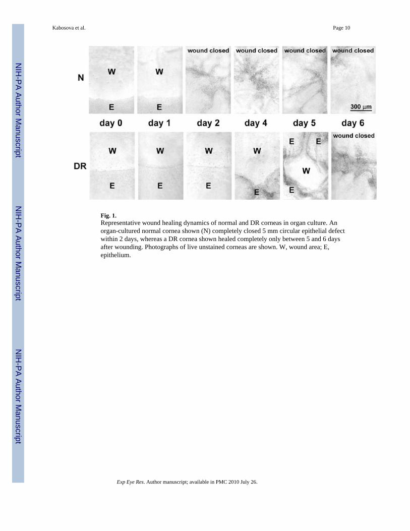
Fig. 1.Representative wound healing dynamics of normal and DR corneas in organ culture. Anorgan-cultured normal cornea shown (N) completely closed 5 mm circular epithelial defectwithin 2 days, whereas a DR cornea shown healed completely only between 5 and 6 daysafter wounding. Photographs of live unstained corneas are shown. W, wound area; E,epithelium.
Kabosova et al. Page 10
Exp Eye Res. Author manuscript; available in PMC 2010 July 26.
NIH
-PA Author Manuscript
NIH
-PA Author Manuscript
NIH
-PA Author Manuscript
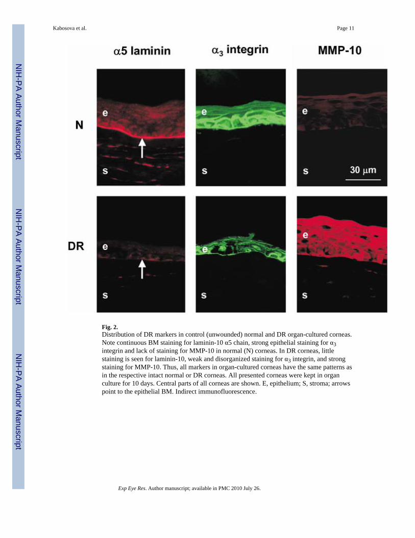
Fig. 2.Distribution of DR markers in control (unwounded) normal and DR organ-cultured corneas.Note continuous BM staining for laminin-10 α5 chain, strong epithelial staining for α3integrin and lack of staining for MMP-10 in normal (N) corneas. In DR corneas, littlestaining is seen for laminin-10, weak and disorganized staining for α3 integrin, and strongstaining for MMP-10. Thus, all markers in organ-cultured corneas have the same patterns asin the respective intact normal or DR corneas. All presented corneas were kept in organculture for 10 days. Central parts of all corneas are shown. E, epithelium; S, stroma; arrowspoint to the epithelial BM. Indirect immunofluorescence.
Kabosova et al. Page 11
Exp Eye Res. Author manuscript; available in PMC 2010 July 26.
NIH
-PA Author Manuscript
NIH
-PA Author Manuscript
NIH
-PA Author Manuscript
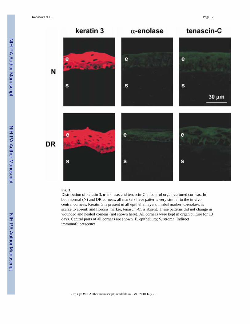
Fig. 3.Distribution of keratin 3, α-enolase, and tenascin-C in control organ-cultured corneas. Inboth normal (N) and DR corneas, all markers have patterns very similar to the in vivocentral corneas. Keratin 3 is present in all epithelial layers, limbal marker, α-enolase, isscarce to absent, and fibrosis marker, tenascin-C, is absent. These patterns did not change inwounded and healed corneas (not shown here). All corneas were kept in organ culture for 13days. Central parts of all corneas are shown. E, epithelium; S, stroma. Indirectimmunofluorescence.
Kabosova et al. Page 12
Exp Eye Res. Author manuscript; available in PMC 2010 July 26.
NIH
-PA Author Manuscript
NIH
-PA Author Manuscript
NIH
-PA Author Manuscript
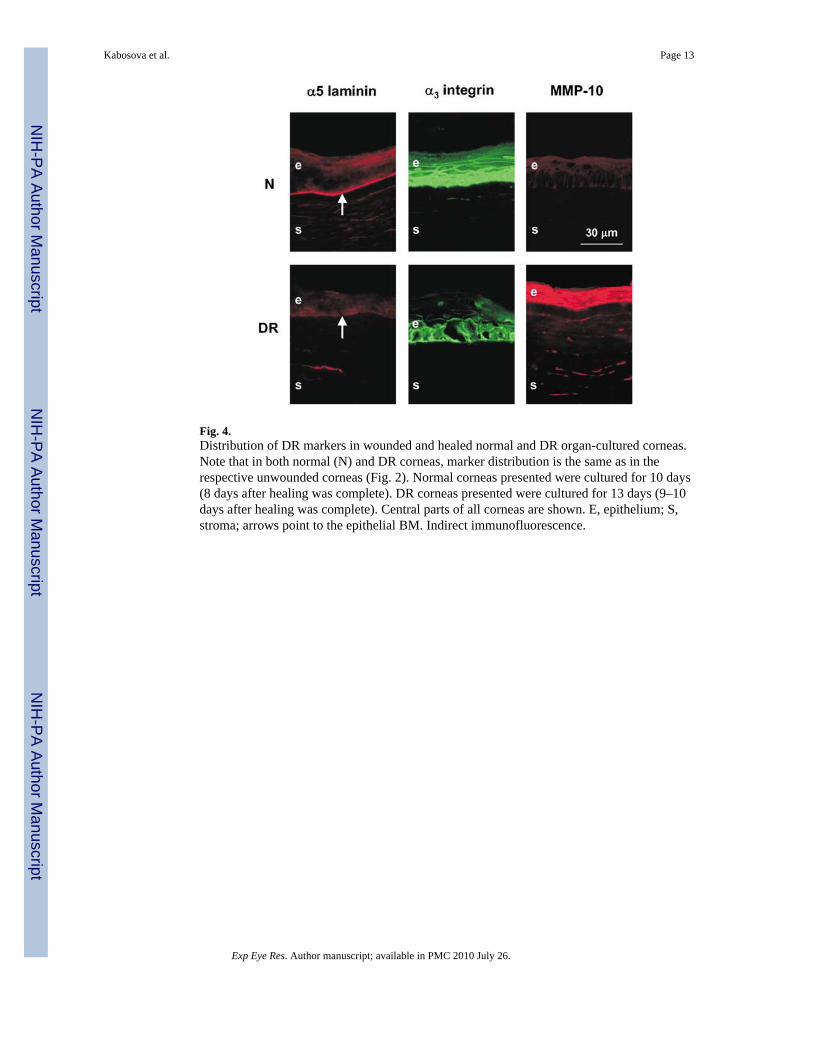
Fig. 4.Distribution of DR markers in wounded and healed normal and DR organ-cultured corneas.Note that in both normal (N) and DR corneas, marker distribution is the same as in therespective unwounded corneas (Fig. 2). Normal corneas presented were cultured for 10 days(8 days after healing was complete). DR corneas presented were cultured for 13 days (9–10days after healing was complete). Central parts of all corneas are shown. E, epithelium; S,stroma; arrows point to the epithelial BM. Indirect immunofluorescence.
Kabosova et al. Page 13
Exp Eye Res. Author manuscript; available in PMC 2010 July 26.
NIH
-PA Author Manuscript
NIH
-PA Author Manuscript
NIH
-PA Author Manuscript