hssb1 interacts directly with the mrn complex stimulating its recruitment to dna double-strand...
TRANSCRIPT
hSSB1 interacts directly with the MRN complexstimulating its recruitment to DNA double-strandbreaks and its endo-nuclease activityDerek J. Richard1,*, Liza Cubeddu2, Aaron J. Urquhart1, Amanda Bain1,
Emma Bolderson1, Dinoop Menon1, Malcolm F. White3 and Kum Kum Khanna1
1Signal Transduction Laboratory, Queensland Institute of Medical Research, Brisbane, Queensland 4006,2School of Molecular and Microbial Biosciences, University of Sydney, Sydney, NSW 2006, Australia and3Biomedical Sciences Research Complex, University of St. Andrews, North Haugh, St Andrews,Fife KY16 9ST, UK
Received November 5, 2010; Revised December 9, 2010; Accepted December 20, 2010
ABSTRACT
hSSB1 is a recently discovered single-stranded DNAbinding protein that is essential for efficient repair ofDNA double-strand breaks (DSBs) by the homolo-gous recombination pathway. hSSB1 is requiredfor the efficient recruitment of the MRN complexto sites of DSBs and for the efficient initiation ofATM dependent signalling. Here we explore theinterplay between hSSB1 and MRN. We demon-strate that hSSB1 binds directly to NBS1, a compo-nent of the MRN complex, in a DNA damageindependent manner. Consistent with the directinteraction, we observe that hSSB1 greatly stimu-lates the endo-nuclease activity of the MRNcomplex, a process that requires the C-terminaltail of hSSB1. Interestingly, analysis of two pointmutations in NBS1, associated with Nijmegenbreakage syndrome, revealed weaker binding tohSSB1, suggesting a possible disease mechanism.
INTRODUCTION
Cells frequently encounter DNA damage; with DNAdouble-strand breaks (DSBs) being among the most cyto-toxic of these lesions. Chromosomal instability may occureven from a single DSB, if it is repaired incorrectly, andthis may ultimately lead to cell death. It is essential thatDSBs in human cells are detected, signalled and repairedefficiently in order to prevent the accumulation of damage,which can lead to chromosomal instability or malignanttransformation. DSBs may be induced by a number offactors including ionizing radiation (IR), reactivechemical species and via normal cellular processes suchas DNA replication. Once a DSB is detected, DNA
repair proteins are recruited to the site of the DSB and amulti-faceted DSB pathway is activated. This complexsignalling network includes altered transcriptional andtranslational regulation and the induction of DSB repairand cell cycle arrest via the activation of checkpoints.While non-homologous end joining (NHEJ) may beused to repair DSBs in any phase of the cell cycle, hom-ologous recombination (HR) may be used to repair DSBsthat specifically occur in the S or G2 phases of the cellcycle (1–3).One of the first events in the process of HR is the re-
cruitment of the MRN repair complex to the DSB site.Once located at the DSB, MRN activates the ATM kinaseand tethers the DNA ends (4). When ATM is activated itin turn initiates signalling cascades, which leads to theresection of the DSBs to produce single-stranded DNA(ssDNA) (5). The resulting ssDNA acts as a substratefor Rad51-mediated strand exchange (4,6). Recentstudies have shown that MRN also has a role in classicaland alternative NHEJ (7,8).The ssDNA binding (SSB) protein family have a funda-
mental role in the repair of DNA damage in all threedomains of life. The simple SSBs and the replicationprotein A (RPA)-like SSBs form the sub-groups of thisfamily of proteins (9). RPA is the most widely studiedmember of the SSB family in humans and is believed tobe a pivotal factor of both DNA replication and DNArepair pathways (10–12). However, human RPA has acomplex oligomeric structure not seen in the simple bac-terial SSBs (13). The simple SSBs were believed to berestricted to the bacterial and archaeal domains of life,however, recently we have identified two new membersof the SSB family in humans: hSSB1 and hSSB2 (14).hSSB1 and hSSB2 are structurally much more closelyrelated to the bacterial and archaeal SSBs than to RPA(9). Both hSSBs consist of a ssDNA oligonucleotidebinding (OB) fold, a divergent spacer domain, followed
*To whom correspondence should be addressed. Tel: +61 7 33620339; Fax: +61 7 33620105; Email: [email protected]
Published online 11 January 2011 Nucleic Acids Research, 2011, Vol. 39, No. 9 3643–3651doi:10.1093/nar/gkq1340
� The Author(s) 2011. Published by Oxford University Press.This is an Open Access article distributed under the terms of the Creative Commons Attribution Non-Commercial License (http://creativecommons.org/licenses/by-nc/2.5), which permits unrestricted non-commercial use, distribution, and reproduction in any medium, provided the original work is properly cited.

by a conserved C-terminal tail predicted to be requiredfor protein–protein interactions (14). The crenarchaealSSB, from Sulfolobus solfataricus, also has a flexiblespacer followed by basic and acidic regions near theC-terminus; this region plays no part in DNA bind-ing but is known to modulate protein–proteininteractions (15).We have previously shown that hSSB1 is required for
the efficient signalling of DSBs following exposure to IR(14). Recently, we have also shown that hSSB1 is rapidlyrecruited to sites of DSBs and its presence is required forthe efficient recruitment of the MRN complex and subse-quent downstream partners (16). Furthermore, hSSB1 de-ficient cells fail to generate the ssDNA tracts associatedwith the nuclease activity of MRN and CtIP (16). We, andothers, have also demonstrated that hSSB1 is a compo-nent of a complex containing IntS3 (17,18).In this study, we demonstrate that hSSB1 forms a DNA
damage-independent complex with Mre11:Rad50:NBS1(MRN) which is distinct from the hSSB1:IntS3 complex.We show that hSSB1 plays an essential role in the recruit-ment and function of MRN at sites of DSBs. The MRNcomplex is believed to be the primary sensor of DSBs andpromotes the activation of ATM kinase, which in turninitiates downstream DSB signalling. MRN also functionsin the resection of the DSB, a process required for ATRsignalling and preparation of DNA for HR in particularRad51 mediated strand invasion (6,19,20). Our data nowdemonstrate how hSSB1 functions to directly recruit theMRN complex to DSBs, a process that also stimulatesMRN nuclease activity. These data implicate hSSB1 as apivotal member of the DNA damage response, cementingits role in maintaining genomic stability in metazoans.
MATERIALS AND METHODS
Cell lines, plasmids and siRNA
HeLa and HEK293T cells were maintained in DMEMsupplemented with 10% fetal bovine serum (Gibco).Transfection of plasmids was performed usingLipofectamine 2000 (Invitrogen) as per manufacturer’s in-structions. Full-length hSSB1 and truncations were clonedinto bacterial expression vectors encoding a His-tag(pET28c). GFP-hSSB1 tail (153–211amino acids) wasexpressed from pEGFP-C1. FLAG tagged Mre11 inpFastBac1 (TP813) and NBS1 in pFastBac1 (TP28) werekindly supplied by Tanya Paull. The HisRad50 wasexcised from the pVL1392 vector (TP11) (gift fromTanya Paull) using EcoR1 restriction enzyme and thencloned directly into an EcoR1 digested pFastBac1vector. Clones of the correct orientation were selected fol-lowing a diagnostic digest with EcoRV. Virion phiX174was supplied by New England Biolabs.
Antibodies
The following antibodies were used in the study: Rad50(Calbiochem), Mre11, NBS1 (Sigma), IntS3 (Bethyllaboratories) and Alexa secondary antibodies(Invitrogen). Sheep antiserum to hSSB1 has beendescribed previously (14).
Immunoprecipitation experiments
Co-immunoprecipitations were performed using theice-cold NP40 buffer; 20mM HEPES pH 8, 150mMKCl, 10mM MgCl2, 0.5mM EDTA, 0.2% NP40,0.5mM DTT, 5% glycerol, 1mM NaF, 1mM NaVO4,protease inhibitor cocktail (Sigma). Assays were per-formed at 5�C for 2 h using the antibodies as indicatedin the ‘Results’ section. Antibodies were captured usingmagnetic protein G agarose beads (Invitrogen), washedthree times in NP40 buffer before being analysed byimmunoblot.
hSSB1 pull-down assays
Recombinant hSSB1 was bound to cyanogen bromideagarose as per manufacturer’s instructions (GEHealthcare) with bound protein being quantified bycoomassie blue staining. hSSB1 pull-downs were per-formed in NP40 buffer (described above for immunopre-cipitations) and the washed beads were analysed byimmunoblot.
DNA pull-down assays
Annealed double-stranded oligonucleotides with six bpoverhangs were generated from the following two se-quences: oligo 1, 50-GATCCACTGGGTTAACGCCGGACATTGCCCGGAT; oligo 2, 50-TCCATGATCCGGGCAATGTCCGGCGTTAACCCAGTGGATC. Oligo 1was modified with a 50 biotin label. Oligonucleotideswere annealed and bound to streptavidin agarose priorto assay. Each assay consisted of 10 ng oligo bound tobeads with 20 ng (or 80 ng) of purified MRN, and a finalconcentration of 2 mM hSSB1, RPA or hSSB1-T1 (aminoacids 1–114). Binding was performed for 15min at roomtemperature in DNA pull-down buffer: 20mM HEPESpH 8, 150mM KCl, 5mM MgCl2, 5% glycerol, 0.05%NP40, prior to analysis on a NUPAGE 4–12% SDS gel.
Preparation of nuclear extract
Nuclear extract was prepared as described previously (21).
Protein purification
Recombinant hSSB1 was expressed in Escherichia coliRosetta 2 strain and purified by a three-step purificationmethod. Clarified soluble E. coli cell lysate treated withDNaseI (10mg/ml final concentration) was applied to aHi-trap metal chelating column loaded with Nickel in ahigh-salt buffer containing 20mM Tris pH 8.0, 500mMNaCl and 30mM imidazole at a flow rate of 1ml/min.hSSB1 was eluted using a gradient of 0–100% 500mMimidazole (1ml/min) on a BioLogic System (GE).Protein fractions were collected from the middle of thebroad protein peak and diluted 1:10 in heparin buffer con-sisting of 20mM Tris pH 7.6, 0.5mM EDTA, 1mM DTT.This allowed the salt concentration to be reduced to50mM NaCl when protein was loaded onto heparincolumn. Protein was eluted with a 0–100% gradient of1M NaCl. Protein was concentrated, then subjected tosize-exclusion chromatography on a Superdex75 column(in 20mM Tris pH 7.6, 150mM NaCl, 1mM DTT) at a
3644 Nucleic Acids Research, 2011, Vol. 39, No. 9

flow rate of 1ml/min to purify protein on the basis of size.Protein eluted as one sharp peak. Purified hSSB1 waselectrophoresed through a NuPAGE protein gel andstained with Sypro Ruby (GE Healthcare) to further de-termine the purity of the preparation. GST fusion proteinswere purified as described previously (22).
Nuclear protein fractionation
Pre-filtered nuclear extract (3mg) prepared fromHEK293T, as described previously (21), cells was passedthrough a Hi-load Superdex 16/60 size-exclusion column(GE Healthcare) at a flow rate of 1ml/min in buffer D.Samples were collected during the entire run.
MRN nuclease activity
Nuclease assays were performed essentially as previouslydescribed (23). Assays were performed for 40min (to dem-onstrate MRN endonuclease activity) or 5min (assayswith addition of hSSB1) as indicated. All assays were per-formed in the presence of 5mM MnCl2 unless otherwiseindicated.
RESULTS
hSSB1 interacts with the MRN complex and is requiredfor MRN recruitment to DSBs
We first sought to identify functional protein complexescontaining hSSB1. An N-terminal GST-hSSB1 fusionprotein was covalently linked to a cyanogen bromidesepharose column as the bait. Nuclear extract (10mg)was passed over the hSSB1 affinity column, containing10 mg/ml ethidium bromide to dissociate protein–DNAinteractions. The column was washed extensively andinteracting proteins eluted with 1M NaCl. Elutedproteins were then lyophilized along with the eluatefrom a GST-only control column and both samplesanalysed by tandem mass spectrometry (MS/MS). Weidentified the IntS3 complex, which has previously beenreported to interact with hSSB1 (17,18,24). We alsoidentified strong sequence coverage for Rad50, a compo-nent of the MRN complex (Supplementary Figure S1). Toconfirm this interaction we performed reciprocalco-immunoprecipitation experiments with hSSB1 andMre11 from nuclear extracts. This revealed that hSSB1interacts with Mre11 and NBS1 in a DNA damage-independent manner (±IR) (Figure 1a and b), suggestingthat hSSB1 is in complex with MRN.
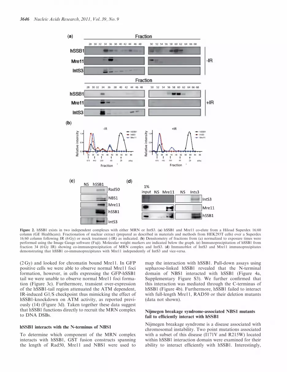
We next fractionated nuclear extract from HEK293Tcells stably expressing Flag-tagged hSSB1 using size-exclusion chromatography. This method allows for thedetection of stable protein complexes in solution. hSSB1and Mre11 co-elute in fraction 36 (smaller complex)before IR (Figure 2a and b). Interestingly following IR(6Gy, 1 h recovery), Mre11 shifts to be predominantly infraction 34 (larger complex). IntS3 also elutes in thesesame fractions, raising the possibility of a commoncomplex. To investigate this further we performedco-immunoprecipitation experiments using Fraction 34(IR) and found that although hSSB1 precipitated with
MRN components and IntS3; Mre11 failed to precipitateIntS3 and IntS3 failed to co-precipitate Mre11 (Figure 2cand d). This suggests that the hSSB1:MRN complex isdistinct from the hSSB1:IntS3 complex. We performedextensive immunoprecipitation experiments but wereunable to detect any interaction between MRN andIntS3 from nuclear or whole cell lysates under our experi-mental conditions (data not shown). Interestingly, theinteraction between MRN and IntS3 observed by Huanget al. (24) required the over-expression of all componentsof MRN and IntS3 in insect or HEK 293 cells, suggestingthe interaction may represent a small pool, or specific con-ditions which are not easily observed with endogenousprotein in asynchronous cells.
hSSB1 interacts directly with the MRN complex via itsC-terminal tail
We next sought to determine if hSSB1 interacted directlywith the MRN complex. Purified MRN was incubatedwith recombinant hSSB1 (Supplementary Figure S2a–c),and truncations of hSSB1 that were chemically crosslinkedto sepharose beads. These data confirmed the interactionwas direct and like other SSB interactions (9,22), wasmediated through the C-terminal tail of hSSB1, sincedeletion of this region in hSSB1 abrogated the interaction(Figure 3a). To further confirm this interaction weectopically expressed the hSSB1 C-terminal tail (aminoacids 154–211) as an N-terminal GFP fusion in HeLacells. Co-immunoprecipitation using GFP antibodiesfrom nuclear extract, demonstrated that the hSSB1 tailwas sufficient to mediate this MRN interaction(Figure 3b). Since the GFP-hSSB1 tail binds MRN wereasoned that this would compete with endogenoushSSB1 binding to MRN, thus preventing MRN recruit-ment to sites of DSBs. To confirm this, we irradiated cells
Figure 1. hSSB1 interacts with the MRN complex. (a and b)Co-immunoprecipitation of hSSB1 with NBS1 and or Mre11 usingantibodies as indicated or NS (nonspecific serum). Immunoprecipitatesobtained from cells with or without exposure to IR (6Gy) wereimmunoblotted with indicated antibodies.
Nucleic Acids Research, 2011, Vol. 39, No. 9 3645
(2Gy) and looked for chromatin bound Mre11. In GFPpositive cells we were able to observe normal Mre11 fociformation, however, in cells expressing the GFP-hSSB1tail we were unable to observe normal Mre11 foci forma-tion (Figure 3c). Furthermore, transient over-expressionof the hSSB1-tail region attenuated the ATM dependent,IR-induced G1/S checkpoint thus mimicking the effect ofhSSB1-knockdown on ATM activity, as reported previ-ously (14) (Figure 3d). Taken together these data suggestthat hSSB1 functions directly to recruit the MRN complexto DNA DSBs.
hSSB1 interacts with the N-terminus of NBS1
To determine which component of the MRN complexinteracts with hSSB1, GST fusion constructs spanningthe length of Rad50, Mre11 and NBS1 were used to
map the interaction with hSSB1. Pull-down assays usingsepharose-linked hSSB1 revealed that the N-terminaldomain of NBS1 interacted with hSSB1 (Figure 4a,Supplementary Figure S3). We further confirmed thatthis interaction was mediated through the C-terminus ofhSSB1 (Figure 4b). Furthermore, hSSB1 failed to interactwith full-length Mre11, RAD50 or their deletion mutants(data not shown).
Nijmegen breakage syndrome-associated NBS1 mutantsfail to efficiently interact with hSSB1
Nijmegen breakage syndrome is a disease associated withchromosomal instability. Two point mutations associatedwith a subset of this disease (I171V and R215W) locatedwithin hSSB1 interaction domain were examined for theirability to interact efficiently with hSSB1. Interestingly,
Figure 2. hSSB1 exists in two independent complexes with either MRN or IntS3. (a) hSSB1 and Mre11 co-elute from a Hiload Superdex 16/60column (GE Healthcare). Fractionation of nuclear extract (prepared as described in materials and methods from HEK293T cells) over a Superdex16/60 column following IR (6Gy) or mock treatment (-IR) as indicated. (b) Densitometry of fractions from (a) normalized to exposure times wereperformed using the Image Gauge software (Fuji). Molecular weight markers are indicated below the graph. (c) Immunoprecipitation of hSSB1 fromfraction 34 (6Gy IR) showing co-immunoprecipitation of MRN complex and IntS3. (d) Immunoblot of IntS3 and Mre11 immunoprecipitatesdemonstrating that hSSB1 co-immunoprecipitates with Mre11 independently of IntS3 and vice-versa.
3646 Nucleic Acids Research, 2011, Vol. 39, No. 9

neither mutant efficiently interacted with hSSB1(Figure 5a and b, Supplementary Figure S4). This maybe due to the mutations having a specific effect on thebinding site for hSSB1 or that altered folding ormisfolding of these mutants inhibits the hSSB1 inter-action. However, a loss in the efficiency of hSSB1binding to NBS1 would explain the chromosomal instabil-ity associated with these mutations.
hSSB1 stimulates the endonuclease activity of the MRNcomplex by facilitating its recruitment to DSBs withshort ss DNA overhangs
To determine if hSSB1 functions to directly recruit MRNto DSBs, we performed DNA pull-down assays. Since
most endogenously generated DSBs (e.g. collapsed repli-cation forks) possess short ssDNA overhangs, which havebeen shown to enhance ATM activation (25), we utilized adouble-stranded DNA (dsDNA) substrate with a short6 bp overhang. While MRN itself bound weakly in theexperimental conditions used, the addition of hSSB1resulted in a significant increase in MRN binding(Figure 6a), suggesting that hSSB1 was functioning tostimulate MRN recruitment to the DNA substrate. Incontrast, recombinant RPA was unable to significantlystimulate binding of the MRN complex directly(Figure 6a, Supplementary Figure S5). Interestingly,although RPA could not bind this DNA substrate alone,addition of MRN allowed binding to occur. This is likely
Figure 3. Mapping of region in hSSB1 required to pull-down MRN complex. (a) Pull-down reactions were performed by incubating full-lengthhSSB1 or C-terminal truncations of hSSB1 (T1 1–153, T2 1–114) bound to sepharose beads with purified recombinant MRN (20 ng) complex. Afterwashing, beads were subjected to immunoblotting with indicated antibodies. Coomassie stained gel shows the relative input of hSSB1 protein.(b) hSSB1 tail (amino acids 153–211) is sufficient for interaction with Mre11 in nuclear extracts. Cells were transfected with GFP-tagged hSSB1-tailor GFP vector alone and anti-GFP immunoprecipitates were immunoblotted with indicated antibodies. (c) Overexpression of GFP-hSSB1-tail (MRNinteracting domain) results in impaired Mre11 foci formation after IR. HeLa cells were transfected with GFP or GFP-hSSB1-tail. Forty-eight hoursafter transfection cells were treated with IR (6Gy, 30min) and immunostained with indicated antibodies. Overexpression of GFP-hSSB1 tail (MRNinteracting domain) results in impaired Mre11 foci formation after IR. HeLa cells were transfected with GFP or GFP-hSSB1-tail. Forty-eight hoursafter transfection cells were treated with IR (6Gy, 30min) and immunostained with indicated antibodies. (d) Overexpression of GFP-hSSB1 tail(MRN interacting domain) abrogates the IR induced G1/S checkpoint. HeLa cells were transfected with GFP or GFP-hSSB1-tail (GFP-CT).Forty-eight hours after transfection cells were treated with IR (6Gy, 30min) and allowed to recover for 16 h after which cells were pulse labeledwith with BrdUrd (30min, 10 mg/ml). Cells were subsequently stained with anti-BrdUrd followed by Alexa 488 secondary antibodies and propidiumiodide before being analysed by flow cytometry. The box illustrating BrdUrd positive cells indicates that GFP-transfected cells effectively arrest at theG1/S boundary compared to GFP-CT transfected cells, which continue to enter S-phase.
Nucleic Acids Research, 2011, Vol. 39, No. 9 3647

due to the end processing activity of MRN in these assays,generating ssDNA to which RPA could bind. We alsoobserved that DNA binding of MRN was furtherenhanced when both hSSB1 and RPA were added. Onepossible explanation for this is that once hSSB1 stimulatesbinding, RPA may function to stabilize the bound MRN.To confirm that hSSB1 directly recruited MRN to theDNA substrate we next examined whether truncatedhSSB1 (T1), (which binds ssDNA with a similar affinityto full-length hSSB1 (KD ’15 nM), but is unable tointeract with MRN could stimulate MRN binding.hSSB1-T1 failed to enhance MRN binding under normalassay conditions, however, with higher concentrations ofMRN (where MRN binding could normally be observedin the absence of full-length hSSB1) hSSB1-T1 inhibitedMRN binding (Figure 6b). This inhibition in the presenceof hSSB1-T1 is likely due to competition for the DNAsubstrate. In contrast, the full-length hSSB1 functionedto further enhance MRN binding under the sameconditions.Since both hSSB1 and MRN are needed for effective
DNA resection, and hSSB1 is directly required for recruit-ment of MRN to DSBs, we analysed the effect of hSSB1on the nuclease activity associated with MRN. WhileMRN has both exo- and endonuclease activities, itsssDNA endonuclease activity is important for DSB resec-tion (26). We tested this activity using a closed circular ssphiX174 DNA as a substrate (23). As shown previously(23), MRN exhibited nuclease activity in the presence ofmanganese but not magnesium chloride (SupplementaryFigure S6). The addition of hSSB1 to the assay drastically
increased the MRN nuclease activity while the additionof RPA had no effect on MRN activity (Figure 6c,Supplementary Figure S7). To discount co-purificationof any potential nuclease activity with recombinanthSSB1 we incubated the same substrate with hSSB1 for1 h. Under our experimental conditions we were unable toobserve nuclease activity (Supplementary Figure S8).Addition of a truncated hSSB1 missing the carboxyterminal tail (T1:1–153 amino acids), which is unable tointeract with the MRN complex, failed to activate theMRN nuclease activity (Figure 6c). These resultsindicate that hSSB1 interacts directly with the MRNcomplex and enhances its nuclease activity.
DISCUSSION
We have previously shown that hSSB1 is rapidly recruitedto sites of DSBs (16); this process is required for the effi-cient recruitment of MRN and activation of ATM. Morerecently, we have shown that hSSB1 is rapidly recruited tosites of DSBs and is required for generation/stability ofssDNA at the sites of damage (16). We now demonstratethat hSSB1 forms a complex with MRN, and that thiscomplex is required for the recruitment of MRN toDSBs with short ssDNA overhangs. Interestingly RPAcannot substitute for hSSB1 in these assays, providingfurther evidence that these two members of the SSBfamily have distinct roles in the repair of DSBs in humans.
We have demonstrated that hSSB1 directly interactswith NBS1 via its C-terminal tail. Interestingly, hSSB1interacts with the N-terminal domain of NBS1, a regionknown to also interact with MDC1 (27). The N-terminaldomain of NBS1 contains both the FHA and BRCT
Figure 4. Mapping of region within NBS1 required for interaction withhSSB1. (a and b) hSSB1 c-terminal tail interacts directly with theN-terminal fragment of NBS1. hSSB1 pull-down experiments were per-formed with purified recombinant hSSB1 bound to sepharose andpurified GST-tagged NBS1 fragments (NBS1A 1-221, NBS1B 199-473and NBS1C 454-754).
Figure 5. Nijmegen breakage syndrome mutants fail to interact effi-ciently with hSSB1. (a) Pull-down reactions were performed usinghSSB1 bound to sepharose incubated with NBS1A, or NBS1A withthe mutations I171V or R215W in the BRCT domain and immuno-blotted with indicated antibodies. (b) Quantification of data presentedin (a), densitometry was performed using Image Gauge software (Fuji).
3648 Nucleic Acids Research, 2011, Vol. 39, No. 9

domains; strikingly, mutations in this region severelyimpair HR, a process also dependent on hSSB1 (14,28).We also demonstrate, using size-exclusion fractionationand co-immunoprecipitation, that hSSB1 exists in twodistinct complexes with IntS3 and MRN. This is incontrast with Huang et al. (24) who suggested thatover-expression of NBS1 and IntS3 in insect and HEK293 cells allows an IntS3: NBS1 complex to form. After
repeated attempts, we have been unable to observe aninteraction between endogenous IntS3 and MRN compo-nents in cell lysates. This may suggest that over-expressionof both components forces the interaction kinetics toallow a rare, but potentially real, event to be observed.We have also shown that by inhibiting the hSSB1:MRNinteraction using dominant negative hSSB1 proteins,MRN recruitment to DSBs is abolished, resulting in a
Figure 6. hSSB1 directly facilitates MRN DNA binding and enhances the nuclease activity of the MRN complex. (a) Immunoblot of DNApull-down assays using Rad50 (as a marker of the MRN complex), RPA and hSSB1 as indicated. Proteins, MRN 20ng, 2 mM hSSB1 or2 mM RPA, as indicated were precipitated with a biotinylated-double-stranded DNA oligo with a 6 bp overhang bound to streptavidin agarosebeads. (b) Immunoblot of DNA pull-down assays using Rad50 as a marker of the MRN complex. Proteins, MRN 80ng, 2 mM hSSB1 or 2 mMhSSB1-T1, as indicated were precipitated with a biotinylated-double-stranded DNA oligo with a 6 bp overhang bound to streptavidin agarose beads.(c) hSSB1 stimulates MRN nuclease activity. MRN nuclease activity assays contained phiX DNA, 20 ng MRN, 2 mM hSSB1, 2 mM hSSB1 truncatedhSSB1 (1–153 amino acids, T1) or 2 mM RPA as indicated. Assays were incubated for 5min before being resolved on a 1% agarose gel and stainedwith ethidium bromide.
Nucleic Acids Research, 2011, Vol. 39, No. 9 3649
failure to activate DSB induced cell cycle checkpoints.Finally, we have demonstrated that hSSB1 can facilitatebinding of MRN to DSBs with short overhangs (6 bp),and that hSSB1 stimulates Mre11 nuclease activity.These data provide a plausible mechanism throughwhich hSSB1 could facilitate MRN dependent DSBresection.It has been well established that depletion of hSSB1
results in dramatic reduction in ATM activation (14,17).Our data, taken together with previous studies on MRNdependent ATM activation and DNA end resection, nowprovides a model for the role of hSSB1 in mediating initialsteps of DNA end resection that are essential for HR.MRN is proposed to tether together broken DNA endsand promote ATM activation via mediating the recruit-ment of ATM to sites of DNA damage (6,19,20). ATM inturn stimulates nuclease activity of Mre11 in response toDSBs and Mre11 in cooperation with CtIP carries outearly limited resection at the sites of DSBs (5,23,26).Our data suggests that hSSB1 is a crucial component ofMRN mediated resection. We find that hSSB1 exists in astable DNA damage-independent complex with MRN. Inresponse to DNA damage, hSSB1 recruits the MRNcomplex to DSBs with short ssDNA overhangs, or to nat-urally breathing DNA ends at DSBs. hSSB1-mediated re-cruitment of the MRN complex to DSBs then facilitatesATM activation and DNA end resection. This togetherwith our recent data demonstrating rapid recruitment ofhSSB1 to sites of DSBs (16) suggests that free hSSB1 maybind to DSBs causing localized duplex melting andopening of the DNA end, as has been shown previouslyfor S. solfataricus SSB (29). This in turn may provide thesubstrate required for hSSB1: MRN binding. Therefore, itis possible that the recruitment of hSSB1 to the initialDSB functions to protect the DSB from incorrect process-ing by nucleases. It may also then function to present theDSB to the hSSB1: MRN complex. In addition, we havedemonstrated that hSSB1-dependent MRN recruitmentstimulates/facilitates Mre11 endo-nuclease activity. Thiswould then lead to MRN-dependent generation ofssDNA, thereby providing a substrate for RPA to bind,allowing subsequent Rad51 loading and sister chromatidstrand invasion. This model is consistent with the datapresented and previous findings in which we havedemonstrated a loss of RPA and Rad51 foci formationand sister chromatid exchange upon hSSB1 depletion(14,16).The development of many anti-cancer drugs is
now focusing on the inhibition of DNA repair processes.Therefore, further studies into the mechanism ofthe hSSB1:MRN interaction, particularly as it actsat the earliest stages of the DNA damage response,could provide valuable information to aid drugdevelopment.
SUPPLEMENTARY DATA
Supplementary Data are available at NAR Online.
ACKNOWLEDGEMENTS
We would like to thank all colleagues in the Khannalaboratory for discussion and Stephen Miles for technicalassistance.
FUNDING
Cancer Council Queensland Project Grant (to D.J.R.);Program Grant from National Health and MedicalResearch Council of Australia (to K.K.K.). Funding foropen access charge: Queensland Institute of MedicalResearch.
Conflict of interest statement. None declared.
REFERENCES
1. Lieber,M.R. (2008) The mechanism of human nonhomologousDNA end joining. J. Biol. Chem., 283, 1–5.
2. Saleh-Gohari,N. and Helleday,T. (2004) Conservative homologousrecombination preferentially repairs DNA double-strand breaks inthe S phase of the cell cycle in human cells. Nucleic Acids Res.,32, 3683–3688.
3. Saintigny,Y., Delacote,F., Boucher,D., Averbeck,D. andLopez,B.S. (2007) XRCC4 in G1 suppresses homologousrecombination in S/G2, in G1 checkpoint-defective cells.Oncogene, 26, 2769–2780.
4. D’Amours,D. and Jackson,S.P. (2002) The Mre11 complex: at thecrossroads of dna repair and checkpoint signalling. Nat. Rev.Mol. Cell Biol., 3, 317–327.
5. Jazayeri,A., Balestrini,A., Garner,E., Haber,J.E. and Costanzo,V.(2008) Mre11-Rad50-Nbs1-dependent processing of DNA breaksgenerates oligonucleotides that stimulate ATM activity. Embo J.,27, 1953–1962.
6. Lee,J.H. and Paull,T.T. (2005) ATM activation by DNAdouble-strand breaks through the Mre11-Rad50-Nbs1 complex.Science, 308, 551–554.
7. Rass,E., Grabarz,A., Plo,I., Gautier,J., Bertrand,P. andLopez,B.S. (2009) Role of Mre11 in chromosomalnonhomologous end joining in mammalian cells. Nat. Struct.Mol. Biol., 16, 819–824.
8. Xie,A., Kwok,A. and Scully,R. (2009) Role of mammalian Mre11in classical and alternative nonhomologous end joining. Nat.Struct. Mol. Biol., 16, 814–818.
9. Richard,D.J., Bolderson,E. and Khanna,K.K. (2009) Multiplehuman single-stranded DNA binding proteins function in genomemaintenance: structural, biochemical and functional analysis.Crit. Rev. Biochem. Mol. Biol., 14, 1–19.
10. Iftode,C., Daniely,Y. and Borowiec,J.A. (1999) Replicationprotein A (RPA): the eukaryotic SSB. Crit. Rev. Biochem. Mol.Biol., 34, 141–180.
11. Wold,M.S. (1997) Replication protein A: a heterotrimeric,single-stranded DNA-binding protein required for eukaryoticDNA metabolism. Annu. Rev. Biochem., 66, 61–92.
12. Zou,Y., Liu,Y., Wu,X. and Shell,S.M. (2006) Functions of humanreplication protein A (RPA): from DNA replication to DNAdamage and stress responses. J. Cell Physiol., 208, 267–273.
13. Bochkarev,A., Pfuetzner,R.A., Edwards,A.M. and Frappier,L.(1997) Structure of the single-stranded-DNA-binding domain ofreplication protein A bound to DNA. Nature, 385, 176–181.
14. Richard,D.J., Bolderson,E., Cubeddu,L., Wadsworth,R.I.,Savage,K., Sharma,G.G., Nicolette,M.L., Tsvetanov,S.,McIlwraith,M.J., Pandita,R.K. et al. (2008) Single-strandedDNA-binding protein hSSB1 is critical for genomic stability.Nature, 453, 677–681.
15. Wadsworth,R.I. and White,M.F. (2001) Identification andproperties of the crenarchaeal single-stranded DNA bindingprotein from Sulfolobus solfataricus. Nucleic Acids Res., 29,914–920.
3650 Nucleic Acids Research, 2011, Vol. 39, No. 9

16. Richard,D.J., Kienan,S., Emma,B., Liza,C., Sairei,S.O.,Mihaela,G., David,J.C., Malcolm,F.W., Kerry,R. and Kevin,M.P.(2010) hSSB1 rapidly binds at the sites of DNA double-strandbreaks and is required for the efficient recruitment of the MRNcomplex. Nucleic Acids Res., 2010 [doi:10.1093/nar/gkq1098;Epub ahead of print 3 November 2010].
17. Skaar,J.R., Richard,D.J., Saraf,A., Toschi,A., Bolderson,E.,Florens,L., Washburn,M.P., Khanna,K.K. and Pagano,M. (2009)INTS3 controls the hSSB1-mediated DNA damage response.J. Cell Biol., 187, 25–32.
18. Li,Y., Bolderson,E., Kumar,R., Muniandy,P.A., Xue,Y.,Richard,D., Seidman,M., Pandita,T.K., Khanna,K.K. andWang,W. (2009) hSSB1 and hSSB2 form similar multi-proteincomplexes that participate in DNA damage response. J. Biol.Chem, 284, 23525–23531.
19. Dupre,A., Boyer-Chatenet,L. and Gautier,J. (2006) Two-stepactivation of ATM by DNA and the Mre11-Rad50-Nbs1complex. Nat. Struct. Mol. Biol., 13, 451–457.
20. Lee,J.H. and Paull,T.T. (2004) Direct activation of the ATM proteinkinase by the Mre11/Rad50/Nbs1 complex. Science, 304, 93–96.
21. Richard,D.J., Schumacher,V., Royer-Pokora,B. and Roberts,S.G.(2001) Par4 is a coactivator for a splice isoform-specifictranscriptional activation domain in WT1. Genes Dev, 15, 328–339.
22. Richard,D.J., Bell,S.D. and White,M.F. (2004) Physical andfunctional interaction of the archaeal single-strandedDNA-binding protein SSB with RNA polymerase. Nucleic AcidsRes., 32, 1065–1074.
23. Sartori,A.A., Lukas,C., Coates,J., Mistrik,M., Fu,S., Bartek,J.,Baer,R., Lukas,J. and Jackson,S.P. (2007) Human CtIP promotesDNA end resection. Nature, 450, 509–514.
24. Huang,J., Gong,Z., Ghosal,G. and Chen,J. (2009) SOSScomplexes participate in the maintenance of genomic stability.Mol. Cell, 35, 384–393.
25. Shiotani,B. and Zou,L. (2009) Single-stranded DNA orchestratesan ATM-to-ATR switch at DNA breaks. Mol. Cell, 33, 547–558.
26. Williams,R.S., Moncalian,G., Williams,J.S., Yamada,Y.,Limbo,O., Shin,D.S., Groocock,L.M., Cahill,D., Hitomi,C.,Guenther,G. et al. (2008) Mre11 dimers coordinate DNA endbridging and nuclease processing in double-strand-break repair.Cell, 135, 97–109.
27. Wu,L., Luo,K., Lou,Z. and Chen,J. (2008) MDC1 regulatesintra-S-phase checkpoint by targeting NBS1 to DNAdouble-strand breaks. Proc. Natl Acad. Sci. USA, 105,11200–11205.
28. Sakamoto,S., Iijima,K., Mochizuki,D., Nakamura,K.,Teshigawara,K., Kobayashi,J., Matsuura,S., Tauchi,H. andKomatsu,K. (2007) Homologous recombination repair is regulatedby domains at the N- and C-terminus of NBS1 and is dissociatedwith ATM functions. Oncogene, 26, 6002–6009.
29. Cubeddu,L. and White,M.F. (2005) DNA damage detection byan archaeal single-stranded DNA-binding protein. J. Mol. Biol.,353, 507–516.
Nucleic Acids Research, 2011, Vol. 39, No. 9 3651