hpv in situ hybridization: impact of different protocols on the detection of integrated hpv
TRANSCRIPT
HPV in situ hybridization: impact of different protocols on the detection
of integrated HPV
Anton H.N. Hopman 1*, Miriam A. Kamps1, Frank Smedts2, Ernst-Jan M. Speel1, C. Simon Herrington3
and Frans C.S. Ramaekers1
1Department of Molecular Cell Biology, Research Institute Growth and Development (GROW),University of Maastricht, The Netherlands2Department of Pathology, Foundation of Collaborating Hospitals of Eastern Groningen (SSZOG), Winschoten, The Netherlands3Bute Medical School, University of St. Andrews, St. Andrews, United Kingdom
Although there is consensus that HPV integration is common ininvasive cervical carcinomas and uncommon or absent in low-grade uterine cervical intraepithelial neoplasia (CIN I), estimatesfor HPV integration in CIN II/III range from 5 to 100% using dif-ferent PCR-based and in situ hybridization (ISH) approaches. Ithas been suggested that HPV integration can be identified usingISH by scoring of punctate signals. The increased sensitivity of flu-orescence ISH (FISH) methods, allowing the detection of singlecopies of HPV, complicates the distinction between integrated andepisomal HPV. Recently it has been suggested that, in such assays,the signals originating from integrated virus can be hidden in abackground of episomal HPV. We therefore compared 2 differentFISH protocols for the detection of integrated HPV in a series ofCIN II/III lesions: 1) a mild protocol in which episomal HPV andRNA is retained and 2) a harsh protocol that extensively extractsproteins and RNA, and which promotes the partial loss of episo-mal HPV but not integrated HPV. A series of 28 HPV 16/18 posi-tive CIN II/III lesions (17 solitary lesions and 11 lesions adjacentto microinvasive carcinoma) were studied. A punctate signal pat-tern was identified in 7 of these lesions with both protocols. Punc-tate signal was also present in control samples from lesions thatare known to be associated with HPV integration (invasive squa-mous cell carcinoma (n ¼ 3), adenocarcinoma in situ (n ¼ 3), andinvasive adenocarcinoma (n ¼ 1). HPV RNA contributed signifi-cantly to the intensity of punctate FISH signal, especially whenapplying the mild protocol, as shown by omitting DNA denatura-tion, including RNase pretreatment steps and measuring the fluo-rescence signal intensity. Also, HPV RNA was frequently detectedin addition to episomal/integrated HPV DNA in the majority ofthe other 21 CIN II/III lesions; this resulted in intense granular/diffuse FISH signals throughout the epithelium. However, in 7 ofthese lesions, the harsh protocol gave a more consistent punctatepattern in cells throughout the full thickness of the epithelium.This supports the hypothesis that the harsh protocol unmasks inte-grated HPV more efficiently by extracting RNA and episomalHPV. Overall, with this harsh protocol, a clonally expanded popu-lation of cells containing punctate HPV signals was found in 5 of17 (29%) solitary CIN II/III lesions and in 9 of 11 (88%) CIN II/III lesions associated with microinvasive carcinoma. Combiningthese data with the results from our previous study, with the harshprotocol in 7 of 40 (18%) solitary CIN II/III lesions and 19/21(90%) CIN II/III lesions associated with microinvasive carcinoma(p < 0.001), this pattern was found. This indicates that, whenrobustly defined, a punctate HPV pattern in CIN II/III lesions isassociated with the presence of an invasive carcinoma.' 2005 Wiley-Liss, Inc.
Key words: uterine cervix; dysplasia; SIL; CIN; HPV 16/18; HPVintegration; chromosomal aberrations
Several premalignant stages can be distinguished in the devel-opment of carcinoma of the uterine cervix. These include cervicalintraepithelial neoplasia grades I, II and III (CIN I–III), also desig-nated low-grade squamous intraepithelial lesions (LSIL, compris-ing CIN I) and high grade SIL (HSIL, comprising CIN II-III).1–4
Nearly all invasive cervical carcinomas (ICCs) and CIN grade II/III lesions contain human papillomavirus (HPV) DNA. Epidemio-logical studies indicate that HPV is without doubt the most impor-tant factor in the carcinogenic process of the uterine cervix. It hasbeen estimated that 80% of women acquire an HPV infection at
some point during their lifetime, but that the majority of theseinfections are transient, with only a minority ever resulting in recog-nizable CIN III. The vast majority of CIN I lesions regress sponta-neously, and only very few lesions persist or progress to CIN II–III.One factor considered to be of key importance for the progressionof intraepithelial lesions to invasive disease is integration of HPVinto the host cell genome.2,7–9 The majority of CIN II/III lesionsand all ICCs are infected with high risk (oncogenic) HPV types,particularly HPV 16 and 18. The E2 region of the HPV genome,which is involved in the regulation of the oncogenic HPV E6 andE7 proteins, is frequently disrupted when the virus is inte-grated.2,4,10,11 In the majority of cases this results in constant sup-pression of key cell-cycle control proteins p53 and pRB by theHPV E6 and E7 gene products, respectively. There is a consensusthat integration is common in ICC and most studies indicate thatintegration is uncommon or absent in CIN I.
In reviewing the literature, Evans and Cooper12 commented onthe wide range of estimates for integration in CINII/III, i.e., 5 to100% as determined by means of Southern blot assays, differenttypes of PCR analyses, as well as chromogenic and fluorescencein situ hybridization methods (FISH).13–18 They concluded that,although questions remain regarding the prevalence of integrationin preinvasive lesions, ISH approaches may be a viable alternativeto PCR in determining both infection with high risk HPV as wellas its physical status.
The first assays to detect integrated HPV were based on restric-tion digestion/Southern blot hybridization.19 Comparison of ISHsignal patterns with these assays demonstrated that a punctate pat-tern, consisting of 1 or a few discrete signal(s) in the nucleus, indi-cates HPV integration into the cellular genome. A diffuse nuclearpattern on the other hand represents multiple copies of episomalHPV and correlates with viral replication.19 These early ISHmethods were based on moderately sensitive approaches but, inthe last decade, numerous more sensitive (F)ISH assays havebeen developed that allow the detection of a single-copy ofHPV.17,20–24 Applying these sensitive methods to CIN II/III lesionsresults, however, in an increase in the number of FISH signalswithin individual nuclei. The inherent problem of this approach isthat signals originating from integrated HPV can be hidden in abackground of episomal copies. Furthermore, HPV can also bepresent as concatamers, which makes the recognition of integratedHPV even more difficult.12 Moreover, HPV RNA can contribute tothe signals observed in these (F)ISH protocols.25,26
In our study, we determined the impact of different protocolsfor the identification of HPV by FISH with respect to signal distri-bution throughout the epithelium, signal pattern (diffuse/punctate/
*Correspondence to: Department of Molecular Cell Biology (Box 17),University of Maastricht, PO Box 616, 6200 MD Maastricht, TheNetherlands. Fax: þ31-43-3884151.E-mail: [email protected] 24 August 2004; Accepted after revision 28 October 2004DOI 10.1002/ijc.20862Published online 1 February 2005 in Wiley InterScience (www.interscience.
wiley.com).
Int. J. Cancer: 115, 419–428 (2005)' 2005 Wiley-Liss, Inc.
Publication of the International Union Against Cancer

granular) and contribution of HPV RNA to the final hybridizationsignal. For this purpose we compared a frequently used HPVdetection method that employs a mild pretreatment protocol to aprotocol that we developed for the visualization of chromosomaltargets.27,28 These chromosomal targets need harsh pretreatmentsteps to open chromosomal sites for efficient hybridization andimmunocytochemical detection. The latter protocol has beenshown to remove most of the cytoplasmic as well as nuclear pro-teins and has been hypothesized to extract episomal HPV copies.12
Here we report that application of this protocol results in the par-tial extraction of episomal HPV as well as HPV RNA, resulting inbetter recognition of a punctate pattern indicative of integratedHPV.
Materials and methods
Tissue material
Formalin-fixed and paraffin wax-embedded endo/ectocervicalbiopsies, diathermy loop excisions and cold knife cervical coniza-tion samples were selected from the files of the Department ofPathology, Foundation of Collaborating Hospitals of Eastern Gro-ningen, Winschoten, The Netherlands. The cervical biopsies wereclassified independently according to WHO criteria by 2 patholo-gists (FS and CSH), after which all cases were reviewed togetherand cases with discrepancy discussed until consensus was reached.Invasion was classified according to FIGO staging criteria. Thirty-two CIN II/III lesions were selected; 19 lesions showed no evi-dence of an invasive carcinoma (consecutive cases of CIN II/III),while 13 CIN III lesions were associated with a microinvasive car-cinoma (consecutive cases of CIN II/III&mCA). The lesions wereprocessed in such a way that they were gathered on 3 slides. Thissmall tissue micro array allowed a comparison of hybridisationresults (quantified by measuring the fluorescence intensity)because all lesions were simultaneously processed including theenzymatic (peroxidase) signal amplification reaction. A small ser-ies of controls was selected that were predicted to containintegrated HPV; these included 3 squamous carcinomas, 1 adeno-carcinoma and 3 adenocarcinomas in situ (ACIS). All ACIS wereHPV 18 positive: HPV DNA (notably HPV 18) is usually presentin the integrated form in endocervical neoplasia.29
Ki-67 and p16ink4a immunohistochemistry
Four micrometer thick tissue sections were dewaxed andimmuno stained as described previously. Briefly, after deparaffini-zation sections were pretreated with 0.3% H2O2 in methanol toquench endogenous peroxidase activity, followed by antigenretrieval using microwave heating at 1008C in 0.01 M citrate buf-fer (pH 6.0) for 30 min (interval micro waving). The Ki-67(Immunotech, Marseille, France) and p16ink4a (E6H4, DAKO A/S,Denmark) were detected by incubating the slides subsequentlywith the monoclonal antibodies (according to the instructions ofthe supplier), biotinylated rabbit anti-mouse IgG (1:200, VectorLaboratories, Burlingame, CA) and an avidin-biotinylated peroxi-dase complex (ABC) (Vectastain PK4000, Vector Laboratories,Burlingame, CA). Peroxidase activity was visualized using diami-nobenzidine (DAB)/H2O2 and sections were counterstained withhaematoxylin and mounted in Entellan (Merck, Darmstadt,Germany).
Fluorescence in situ hybridization
Probe selection and labeling procedures. Digoxigenin-labeledHPV 16 and HPV 18 probes were purchased from PanPath,Amsterdam, The Netherlands and used as a mixture (HPV 16/18)for primary screening. Probes for chromosomes 1 (1q12),30 and 17(17 alphoid centromere sequence)31 were selected to determinehybridisation efficiency and reproducibility on chromosomal tar-gets, and used to determine chromosomal aneusomy. The probeswere labeled by standard nick translation with biotin (Bio)- ordigoxigenin (Dig)-dUTPs. For multiple target analyses the follow-ing probe combination was used: chrom 1-Bio and chrom 17-Dig.
Pretreatment protocols. Two different pretreatment protocolswere used. The mild procedure includes a simple pepsin incuba-tion step in 0.2 N HCl combined with high temperature denatura-tion.13 The harsh procedure includes 2 chemical (acid and proteindenaturation) soaking steps and a pepsin incubation step in 0.02 NHCl.28,32 The sensitivity and reliability of the harsh protocol forHPV was described previously by testing HPV positive cell lines(SiHa, Hela and Caski) and screening of routine fixed head andneck lesions and cervical preneoplasia.17,21
Mild pretreatment. Four micrometer thick paraffin wax tissuesections were dewaxed and digested with 8 mg/ml pepsin (800–1,200 U/mg protein porcine stomach mucosa; Sigma Chemical Co.,St. Louis, MO) in 0.2 M HCl for 10 min at 378C. The slides wererinsed 3 times in H2O and dehydrated in an ascending ethanolseries. After air drying the HPV probe set was applied under a cov-erslip (see below). Probe and target DNA were denatured simulta-neously for 6 min at 908C prior to hybridisation. When targetdenaturation was omitted, the probe was denatured separately in aneppendorf tube for 10 min at 808C and chilled on ice prior to appli-cation to the tissue section under a coverslip. For RNase treatment,the slides were dewaxed and incubated for 10 min at 808C in 2 �SSC, rinsed in H2O at room temperature and dehydrated in anascending ethanol series. They were then incubated in 4 mg/mlDNase free RNase (Roche, Mannheim, Germany) in 10 mMTrisHCl, 1 mM EDTA pH 7.5 for 60 min at 378C.
Harsh pretreatment. Four micrometer thick paraffin wax tis-sue sections were dewaxed, pre-treated with 85% formic acid/0.3% H2O2 for 20 min at room temperature, and subsequentlydehydrated with 70% ethanol containing 0.01 M HCl (acid dehy-dration), 90% ethanol and 100% ethanol for 3 min each prior toair drying. The slides were incubated in 1 M NaSCN for 10 min at808C, followed by acid dehydration and digestion with 4 mg/mlpepsin (800–1,200 U/mg protein porcine stomach mucosa: fromSigma Chemical Co., St. Louis, MO) in 0.02 M HCl for 15 min at378C. The slides were rinsed 3 times in 0.01 M HCl and aciddehydrated. After air drying sections were post-fixed in 1% form-aldehyde in PBS for 15 min at room temperature, rinsed 3 times inPBS and dehydrated in an ascending ethanol series.
The chemical pretreatment steps (formic acid/hydrogen perox-ide, and thiocyanate at 808C) in combination with pepsin treat-ments remove nearly all nuclear and cytoplasmic proteins. Thisprocedure strongly improves the hybridisation efficiency, main-tains nuclear morphology during the FISH procedure and reducesthe need for optimization of the digestion time for each sample.32
The chemical pretreatment steps in the harsh protocol lead to par-tial denaturation of DNA (thiocyanate is a DNA denaturingagent); application of this pretreatment step therefore does notenable a valid evaluation of the contribution of RNA hybridizationwhen applying this pretreatment step.
The low level of autofluorescence of cytoplasmic and nuclearprotein remnants after application of the harsh protocol indicatesthat these proteins have been efficiently removed. In the mild pro-tocol, nuclear and cytoplasmic autofluorescence is frequentlygreater, indicating that these proteins are at least partially retained.In particular, the latter will have impact on the retention of RNAin both cytoplasm and nucleus.
The probe sets were applied under a coverslip at a concentrationof 1 ng/ml in 60% formamide, 2 � SSC, 10% dextran sulfate and50 � excess of carrier DNA (salmon sperm DNA). Probe and tar-get DNA were denatured simultaneously for 5 min at 808C priorto hybridization overnight at 378C. After hybridization the prepa-rations were washed stringently in 50% formamide, 2 � SSC at428C (2 times 5 min) or 0.1 � SSC at 618C (2 times 5 min).
Probe detection and microscopic imaging. The digoxigenin(Dig)-labeled HPV probes were detected using the tyramide signalamplification (TSA) procedure as previously described for singletarget hybridisation using rhodamine-labeled tyramide.33 In short,the Dig-labeled probe was detected by peroxidase-conjugatedsheep anti-digoxigenin Fab fragments (SHaDIG-PO, 1:100; Roche
420 HOPMAN ET AL.

Molecular Chemicals, Basel, Switzerland) or first mouse anti-digoxigenin (MaDig, 1:2,000 Sigma Chemical Co.), then a peroxi-dase-conjugated rabbit anti-mouse (RaM-PO, 1:100 DAKO A/SGlostrup, Denmark) and finally a peroxidase-conjugated swineanti-rabbit (SwaR-PO, 1:100 DAKO) all for 30 min incubatoons at378C, and washed in PBS/0.05% Tween-20. Thereafter, the TSAamplification reaction was carried out under a coverslip by apply-ing 50 ml (1:500 diluted from a 1 mg/ml stock solution in ethanol)rhodamine-labeled tyramide in PBS containing 0.1 M imidazole,pH 7.6, and 0.001% H2O2 for 10 min at 378C. The slides werewashed in PBS containing 0.05% Tween-20 (Janssen Chimica,Beerse, Belgium), dehydrated in an ascending ethanol series andmounted in Vectashield (Vector Laboratories) containing 40,6-dia-midino-2-phenylindole (DAPI; Sigma Chemical Co.: 0.5 ng/ml).Comparison of conventional cytochemical detection systems and
TSA systems have shown that, especially with the latter system,both specific and nonspecific (background) ISH signals may begreatly amplified. It is essential to keep nonspecific probe bindingand detection at a minimum for routine application of the method.Many parameters have been shown to have impact on the signal-to-noise (S/N) ratio when applying the TSA method including, e.g.,probe and detection conjugate concentration, type of tyramide,amplification buffer, reaction time and reaction temperature.34
The biotin (Bio) labeled centromeric probe was detected withsequential incubations with fluorescein isothiocyanate (FITC)conjugated avidin (AvFITC, 1:100; Vector, Brunswig Chemie,Amsterdam, The Netherlands), biotin conjugated goat anti-avidin
(BioGaA, 1:100 Vector Laboratories) and FITC-conjugated avidin1:100, all for 20 min at 378C , diluted and washed in 4 � SSC/0.05% Tween-20 (Janssen Chimica, Beerse, Belgium). The follow-ing series of incubations were used to detect the Dig-labeled centro-meric probe: first mouse anti-digoxigenin (MaDig, 1:2,000; SigmaChemical Co.), then rhodamine (TRITC) conjugated rabbit anti-mouse (RaMTRITC, 1:1,000 Sigma Chemical Co.) and finallyTRITC-conjugated swine anti-rabbit (SwaRTRITC, 1:100 DAKOA/S Glostrup, Denmark), all for 30 min at 378C, diluted and washedin PBS/0.05% Tween-20. After detection, the slides were dehy-drated in an ascending ethanol series and mounted in Vectashield(Vector Laboratories) containing 40,6-diamidino-2-phenylindole(DAPI; Sigma Chemical Co.: 0.5 ng/ml).Imaging. Images were recorded with the Metasystems Image
Pro System (black and white CCD camera; Sandhausen, Germany)mounted on a Leica DM-RE fluorescence microscope equippedwith FITC, TRITC, DAPI and SpectrumGold single bandpass fil-ters for single color analysis and a triple bandpass filter set (FITC,TRITC and DAPI) for simultaneous dual- and triple-color analy-sis. Images were recorded using an automatic integration timeallowing quantitative measurements (using the full dynamic rangeof the camera without signal intensity saturation; TIF 8 bitsimage). Furthermore, fixed integration times were used to comparefluorescence intensity between different preparations.35 FISH fluo-rescence was measured per area or per individual nucleus usingImage/J (http://www.nih.gov, public domain), after color separa-tion (RGB split), thresholding and selection of regions of interest.
TABLE I – SUMMARY OF SEMI-QUANTITIVE EVALUATION OF IN SITU HPV SIGNALS USING A X40 DRY OBJECTIVE1
Group Case Age CIN lesion HPV tested
Staining patterns
mild protocol harsh protocol
Diffuse Granular Punctate Diffuse Granular Punctate
A 1 38 III 16/18 n �2 32 III&mCA 16/18 n &3 36 III 16/18 n n &4 24 III 16/18 n n �5 29 III 16/18 n n &6 28 III 16/18 n n � �7 35 III 16/18 n n n8 41 III 16/18 n n �9 37 III 16/18 n n n �10 31 II 16/18 n �11 22 III 16/18 n & &12 34 II/III 16/18 n n13 31 III 16/18 n & �14 36 II&mCA 16/18 n &
B 15 38 III&mCA 16/18 n & & &16 34 III&mCA 16/18 n �17 41 III&mCA 16/18 n n �18 n.a III 16/18 n n n n19 40 III&mCA 16/18 � n & �20 39 III 16/18 n �21 47 III&mCA 16/18 n �
C 22 30 III 16/18 n n � n �23 38 III&mCA 16/18 n � �24 43 III&mCA 16/18 � n25 37 III 16/18 n �26 30 III 16/18 n �27 48 III&mCA 16/18 n n28 30 III&mCA 16/18 n n29 35 squam. CARC 16/18 n n30 n.a squam. CARC 16/18 n.d. n31 n.a squam. CARC 16/18 n.d. n32 35 ACIS 18 n �33 n.a ACIS 18 n.d. n34 32 ACIS 18 n.d. n35 40 adeno CARC 18 n �
1Highest intensity indicated by n, intermediate intensity by �, and lowest intensity by &. The criteria for assessment of signal type are givenin Material and methods. n.a: not available, n.d: not done.
421HPV IN SITU HYBRIDIZATION

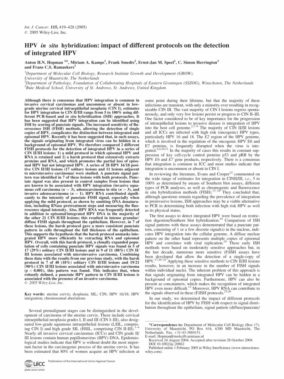
FIGURE 1.
422 HOPMAN ET AL.
Signal intensity (spot or nucleus) was calculated as mean signalintensity (per pixel) times area.
To compare the impact of the different protocols and/or pre-treatment steps on FISH signal distribution throughout the epithe-lium, only samples that were processed simultaneously werecompared. Furthermore lesions were collected on small tissuearrays that enabled identical processing of different lesions.
Controls and evaluation of FISH results. Controls includedHPV-16 and -18 hybridization on tissue sections of formalin fixedand paraffin wax-embedded HPV positive cell lines (SiHa, CaSkiand HeLa). Evaluation of nuclear hybridization signals was per-formed by 3 investigators (A.H., M.K. and C.H). Signal morphol-ogy was categorized as follows: 1) When nuclei were completelyand homogeneously stained, the signal was classified as diffuse.Often, the other nuclei exhibited multiple fluorescent signals withvariation in spot number between individual cells within thelesion; 2) When nuclear signals varied significantly in size andintensity this pattern was termed granular13 and 3) Discretenuclear signals (1–3 per nucleus) in a clean background were clas-sified as punctate, in line with the criteria of Cooper et al.19
The method was tested on 4 mm thick formalin fixed and paraf-fin wax-embedded CaSki and SiHa cell lines that contain appr-oximately 500 and 2 copies of integrated HPV 16, respectively.17
The FISH procedure as described above, guarantees an optimalsignal-to-noise ratio.17,33,36 In particular with the fluorescent TSAmethod, noise is often present as a result of background reactionswhich could be misclassified as viral particles. Histological HPVnegative areas (e.g., lymphocytes) served as internal controls toestimate specific and nonspecific signal.
Results
Ki-67 and p16INK4a immunohistochemistry
The Ki-67 index was mainly utilized for verification of the dif-ferent histological entities and served as a landmark in brightfieldmicroscopy to guide evaluation of the FISH patterns. All cervicallesions positive for p16INK4a exhibited strong nuclear and cyto-plasmic immuno staining in the premalignant areas. FISH HPV-positivity correlated strongly with p16INK4a accumulation, but notall p16INK4a positive cells showed a FISH signal for HPV 16/18DNA.
Distribution of HPV 16/18 FISH signals in (pre)malignant lesions
The results of the HPV FISH reactions for the 2 protocols aresummarized in Table I, and typical examples are depicted in
Figure 1. In total, 32 high grade CIN II/III lesions were examinedfor the FISH distribution patterns of HPV after applying the 2 dif-ferent protocols. HPV 16/18 positivity was detected in 17 CIN II/III lesions and in 11 CIN II/III lesions adjacent to microinvasivecarcinomas. Four lesions were HPV 16/18 FISH negative (in bothprotocols) and most likely contain other HPV types (not deter-mined). In the control group, cases were compared after applyingthe 2 different protocols.
The results were obtained by visual evaluation of the FISH sig-nal intensity. No imaging by means of the CCD camera wasneeded for this classification. Intensity differences obtained forboth protocols were compared for individual lesions within identi-cal areas on serial sections. The absolute fluorescence intensity ofthe signals varied within a relatively wide range between individ-ual lesions, which results in a range of capture times (automatedintegration time, range 0.1–3 sec, average about 0.3 sec) using theCCD camera for imaging. This variation does not influence theclassification upon visual inspection.
In general the mild protocol resulted in HPV signals of higherfluorescence intensity than the harsh protocol. The lesions weregrouped according to the major types of HPV distribution pattern.In lesions 1–14 (group A), only diffuse and/or granular patternswere recognized when applying both pretreatment protocols. Inlesions 15–21 (group B), the patterns obtained with the 2 protocolswere clearly discordant. Only punctate patterns were recognizedwith the harsh protocol, while the mild protocol showed a granularpattern in all cases. In half of these cases, this granular pattern wascombined with a diffuse pattern. In cases 22–35 (group C), themajority of the lesions showed the typical punctate pattern withboth protocols.
Cases 1–14 (group A). In this group, the nuclei with a diffusepattern were predominantly found in the superficial layers. A dif-fuse pattern is illustrated for the mild and harsh protocol in Figure1b,c (case 2). In this case, strongly fluorescent signals werepresent in individual cells of the basal part of the epithelium withthe mild protocol (compare Fig. 1e,g). In Figure 1b,c, these signalsin the lower compartment cannot be seen because of their smallsize, which is below the resolution of this image. A typical diffuseand granular pattern from this group after application of the mildprotocol is depicted in Figure 1j.
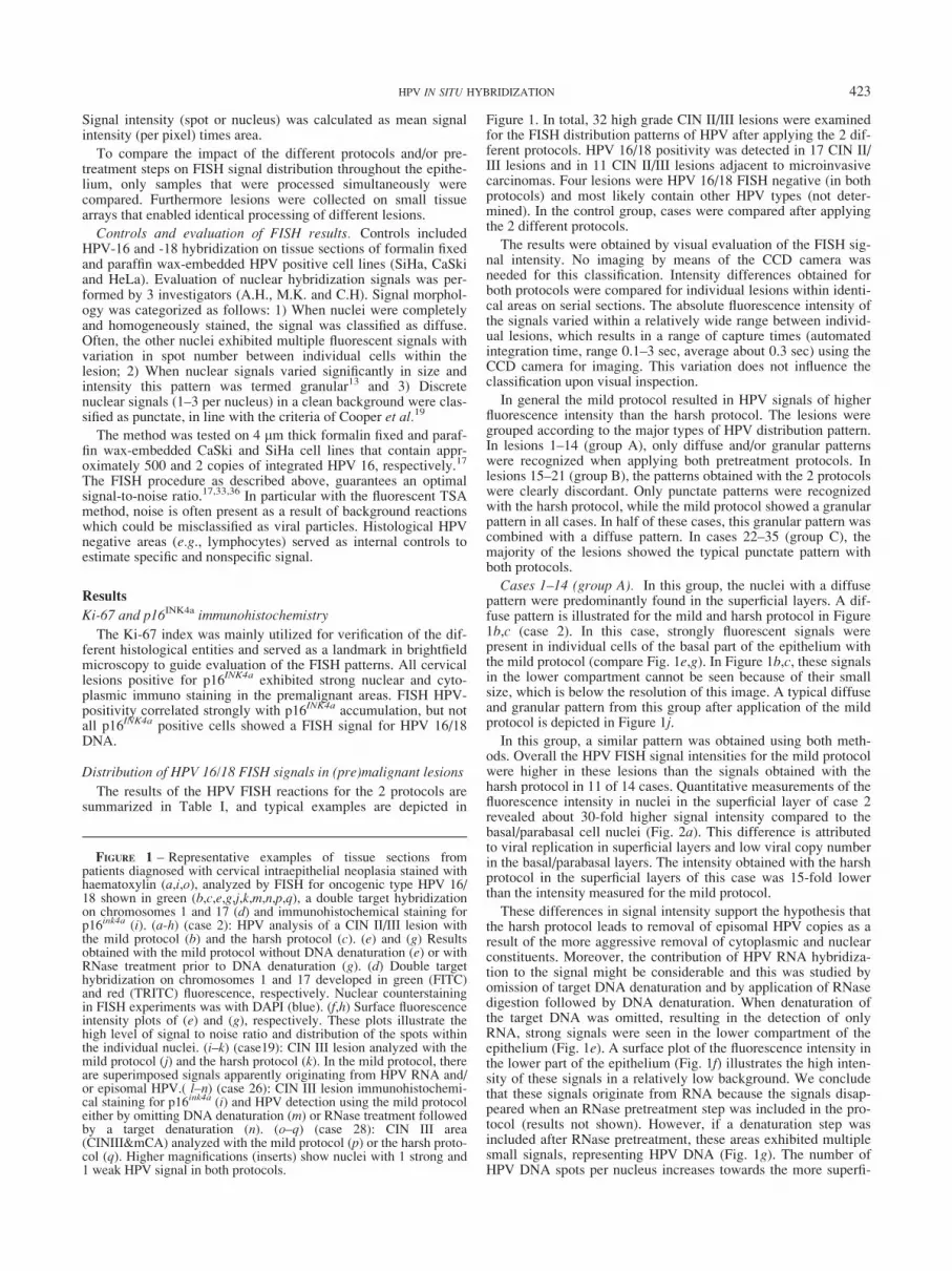
In this group, a similar pattern was obtained using both meth-ods. Overall the HPV FISH signal intensities for the mild protocolwere higher in these lesions than the signals obtained with theharsh protocol in 11 of 14 cases. Quantitative measurements of thefluorescence intensity in nuclei in the superficial layer of case 2revealed about 30-fold higher signal intensity compared to thebasal/parabasal cell nuclei (Fig. 2a). This difference is attributedto viral replication in superficial layers and low viral copy numberin the basal/parabasal layers. The intensity obtained with the harshprotocol in the superficial layers of this case was 15-fold lowerthan the intensity measured for the mild protocol.
These differences in signal intensity support the hypothesis thatthe harsh protocol leads to removal of episomal HPV copies as aresult of the more aggressive removal of cytoplasmic and nuclearconstituents. Moreover, the contribution of HPV RNA hybridiza-tion to the signal might be considerable and this was studied byomission of target DNA denaturation and by application of RNasedigestion followed by DNA denaturation. When denaturation ofthe target DNA was omitted, resulting in the detection of onlyRNA, strong signals were seen in the lower compartment of theepithelium (Fig. 1e). A surface plot of the fluorescence intensity inthe lower part of the epithelium (Fig. 1f) illustrates the high inten-sity of these signals in a relatively low background. We concludethat these signals originate from RNA because the signals disap-peared when an RNase pretreatment step was included in the pro-tocol (results not shown). However, if a denaturation step wasincluded after RNase pretreatment, these areas exhibited multiplesmall signals, representing HPV DNA (Fig. 1g). The number ofHPV DNA spots per nucleus increases towards the more superfi-
FIGURE 1 – Representative examples of tissue sections frompatients diagnosed with cervical intraepithelial neoplasia stained withhaematoxylin (a,i,o), analyzed by FISH for oncogenic type HPV 16/18 shown in green (b,c,e,g,j,k,m,n,p,q), a double target hybridizationon chromosomes 1 and 17 (d) and immunohistochemical staining forp16ink4a (i). (a-h) (case 2): HPV analysis of a CIN II/III lesion withthe mild protocol (b) and the harsh protocol (c). (e) and (g) Resultsobtained with the mild protocol without DNA denaturation (e) or withRNase treatment prior to DNA denaturation (g). (d) Double targethybridization on chromosomes 1 and 17 developed in green (FITC)and red (TRITC) fluorescence, respectively. Nuclear counterstainingin FISH experiments was with DAPI (blue). (f,h) Surface fluorescenceintensity plots of (e) and (g), respectively. These plots illustrate thehigh level of signal to noise ratio and distribution of the spots withinthe individual nuclei. (i–k) (case19): CIN III lesion analyzed with themild protocol (j) and the harsh protocol (k). In the mild protocol, thereare superimposed signals apparently originating from HPV RNA and/or episomal HPV.( l–n) (case 26): CIN III lesion immunohistochemi-cal staining for p16ink4a (i) and HPV detection using the mild protocoleither by omitting DNA denaturation (m) or RNase treatment followedby a target denaturation (n). (o–q) (case 28): CIN III area(CINIII&mCA) analyzed with the mild protocol (p) or the harsh proto-col (q). Higher magnifications (inserts) show nuclei with 1 strong and1 weak HPV signal in both protocols.
423HPV IN SITU HYBRIDIZATION

cial layers of the epithelium and so does the fluorescence intensityper individual nucleus (Fig. 1h). The majority of the other lesionsdid not show the clear-cut diffuse pattern in the superficial layer.Utilizing the mild protocol, a granular pattern was often seen inthese cases, signals being frequently heterogeneous in size andintensity (Fig, 1j). Sometimes these granular patterns were local-ized to the lower compartment of the epithelium, but in other casesthey were scattered throughout the whole of the epithelium. Thisgranular pattern correlated with RNA synthesis as shown by omis-sion of DNA denaturation or by including the RNAse digestionstep. Further analysis demonstrated that these RNA signals weresuperimposed onto multiple spots of HPV DNA. These findingsindicate that, in these cases, the contribution of HPV RNA to sig-nal intensity is considerable with the mild protocol. The sameholds true for the retention of episomal copies of HPV in the mildprotocol.
Cases 15–21 (group B). In this group, we recognized with theharsh protocol a punctate pattern that extended from basal tosuperficial layers. Figure 1k shows an example of the results withthe harsh protocol, with the number of signals per nucleus varyingwithin a small range (mean 1.3) and with a single discrete signal
throughout the entire thickness of the epithelium in 80% of thenuclei. The mild protocol resulted in a more granular/diffuse pat-tern with very heterogeneous staining throughout the epithelium(see Fig. 1j). In these nuclei, the number of signals varied over awide range (1 to >10, mean 4). Quantification of the fluorescenceintensity showed that the intensity of the individual spots in bothmethods was of comparable intensity and showed no significantdifference in average intensity (Fig. 2b). The distribution of theintensities of fluorescent HPV signals measured in a high powerfield is plotted, with the signals being ranked according to increas-ing fluorescence intensity. More signals can be identified using themild protocol, but the average intensity per signal differed onlyslightly when comparing the mild with the harsh protocol, i.e.,5,000 vs. 4,500 a.u., respectively.
The contribution of HPV RNA hybridization to the signalwas considerable in this group. This resulted in a granularpattern that was seen throughout the epithelial lesion but innone of these cases did positive cells extend from basal tosuperficial layer. Sometimes several layers within the epitheliumshowed strong nuclear RNA signals, while in others the distri-bution was more random.
FIGURE 2 – Quantitative fluorescence measurements of FISH signals. Fluorescence signal intensity per individual nucleus or FISH signals ismeasured per high power field (�400)and ranked according to increasing fluorescence signal intensity. For measurements of fluorescence inten-sity see Material and methods. (a) Histograms illustrating HPV integrated fluorescence (IF, arbitrary units) in selected nuclei present in basal/parabasal and superficial cell layers processed following either the mild or the harsh protocol (case 2). For representative high power fields, inwhich the IF was measured, see Figure 1b (capture time 0.2 sec) and 1c (capture time 3 sec) for the mild and harsh pretreatment methods,respectively. (b) Plot for HPV hybridization signals using the mild and harsh protocols in 2 corresponding areas of serial tissue sections (case19). For representative high power fields, in which the fluorescence signal intensity was measured, see Figure 1j and k for the mild and harsh pre-treatment methods, respectively. (c) Plot for HPV hybridization signals in 2 corresponding areas in serial tissue sections pretreated with the mildand harsh protocol exhibiting a typical punctate pattern (case 28). Many nuclei exhibited 2 discrete punctate signals, 1 large and 1 small fluores-cent signal. For representative high power field, in which the signals were measured, see figures 1p and q for the mild and harsh pretreatmentmethods, respectively. (d) Plot for centromere repeat sequence (1q12) hybridization spots in 2 lesions, within 2 different high power fields (case2: area 1 and 2, case 28: area 3 and 4). For a representative high power field, in which the signals were measured, see Figure 1d.
424 HOPMAN ET AL.

Cases 22–35 (group C). Typical examples of cases with punc-tate HPV FISH patterns are shown in Figure 1m,n. These imagesare illustrative for the majority of lesions in which both the mildand the harsh protocol resulted in a punctate pattern. In all theselesions, the positive cells were found throughout the entire thick-ness of the epithelium (basal to superficial) or occasionally assmall homogeneous foci. In 11 of 14 lesions that exhibited a punc-tate pattern, only 1 HPV signal was seen in each nucleus. In case28 the nuclei contained additional small FISH signals (1 or 2) nextto 1 strong signal in all nuclei throughout the full epithelial layer(see inserts Figure 1p,q).
The contribution of RNA to the HPV signal is more easilyassessed in this group than in the lesions in groups A and Bbecause these lesions exhibited a discrete single FISH signal. Case26 exhibited 1 strong FISH signal per nucleus with the mild proto-col (not shown). Omitting target denaturation resulted in intensestaining as depicted in Figure 1m. After RNase treatment andsimultaneous denaturation, 1 signal per nucleus was left (Fig. 1n)but with a significantly lower intensity than without RNase treat-ment. In this case, approximately 80% of the signal intensity stemsfrom hybridization to RNA. In 3 squamous lesions (cases 25, 26and 32) and 1 adenocarcinoma (case 35), RNA contributed signifi-cantly to the final signal using the mild procedure, again up to80%. In these cases the mild protocol produced higher signalintensity than the harsh protocol. It is noteworthy that 3 of these 4lesions were HPV 18 positive. In the other cases, the differencewas not so evident.
In Figure 2c (case 28), quantification of the fluorescence inten-sity showed that the intensities in both methods are comparable,about 280 and 390, respectively. It is difficult to conclude that thesomewhat higher intensity using the mild protocol is the directeffect of hybridization to RNA since hybridization to integratedHPV DNA will contribute to the hybridization signal.
Overal we found that RNA contributes significantly to thehybridization signal obtained with the mild protocol and that theharsh protocol removes RNA as a result of the aggressive extrac-tion of nuclear constituents.
Efficiency of chromosomal (DNA) target detection
To show that the harsh protocol does not influence the effi-ciency of chromosomal target detection, we applied both the mildand the harsh protocol for the detection of chromosome centro-meric DNA targets. For this we hybridized DNA centromericprobes to all of the lesions (for example, see Fig. 1d) and used thevariation in signal intensity between the different lesions as anindicator of the efficacy of both protocols. Comparable reactivitywas seen in all lesions with the harsh protocol, with nearly allnuclei reactive and showing strong FISH signals. The autofluores-cent background was low, which is an indicator of the efficientremoval of proteins from the cytoplasm and nucleus. Theseexperiments confirmed that the harsh protocol in principle is anefficient method to open chromososomal sites. The mild protocolon the other hand was suboptimal for hybridization to chromoso-mal targets since a comparable intensity to that obtained with theharsh protocol was identified in only about 25% of cases. In theother lesions, the reactivity was strongly variable throughout thelesion and many lesions showed a high level of autofluorescence.
Two lesions were analyzed in detail by measuring the intensityof individual signals. One case was known to show a typical dif-fuse HPV pattern while the other showed a punctate HPV pattern.About 700 chromosomal centromere FISH signal intensities weremeasured and the average intensity was not significantly differentin these 2 lesions (see Fig. 2d).
Frequency of punctate signal pattern in CIN II/III withand without an adjacent microinvasive carcinoma
Although the study was not intended to determine the frequencyof a punctate signal pattern in CIN II/III and CIN II/III adjacent toa microinvasive carcinoma, we describe these results here because
they add value to our earlier report.17 In the previous study, 45other cases were studied using only the harsh protocol. In thepresent study a punctate pattern was found in 5 of 17 (29%) soli-tary CIN II/III lesions and in 9 of 11 (82%) lesions with an adja-cent microinvasive carcinoma. This shows that the punctatepattern, which is assumed to be an indication of integration of thevirus into the genome, correlates strongly with the transition of apremalignant lesion to microinvasive carcinoma (p < 0.01). Themild protocol did not discriminate since a punctate pattern wasfound in 3 of 17 (17%) CIN II/III lesions and 4 of 11 (36%)CIN&mCA.
When the data from our previous study are included, a punctatepattern was seen in 7 of 40 (18%) CIN II/III lesions and in 19 of21 (90%) CIN&mCA lesions (p < 0.001).
Discussion
In our study, we determined the impact of 2 protocols on therecognition of punctate signal pattern by FISH. It is generallyaccepted that diffuse signals that represent (replicating) episomalHPV can be distinguished from (strong) punctate signals that indi-cate integrated HPV. The diffuse pattern is found commonly inlow grade intraepithelial lesions and the punctate pattern is charac-teristically present in invasive carcinomas. However, more com-plex patterns are found in lesions of intermediate grade (CIN II/III) since the associated HPV infections are generally abortive andthe full life cycle of the virus is not supported.2,9 Furthermore thesensitive tyramide amplification methods that allow the detectionof single copy HPV particles complicate these patterns. In additionto describing the morphological patterns of HPV FISH signalobtained with tyramide signal amplification systems, our studyquantified HPV signals for the first time in different types of highgrade CIN lesions and in invasive carcinomas and determined thecontribution of HPV RNA hybridization to signal intensity, aswell as intensity variations within single nuclei/cells throughoutthe epithelium. Two different protocols for HPV detection wereused: a mild protocol that retained episomal viral copies and RNAmore efficiently, and a harsh protocol that was hypothesized toremove protein and RNA extensively and to partly extract episo-mal HPV copies.
This comparison allowed us 1) to assess the contributionof RNA hybridization to FISH signal intensity and 2) to identifyclonally expanded cells harboring a punctate signal (indicativeof HPV integration) present throughout the thickness of theepithelium.
Impact of RNA hybridization on HPV signal
The contribution of RNA hybridisation to HPV signal was bestillustrated in the cases that exhibited a punctate pattern. In particu-lar, in those lesions where HPV is known to be integrated, as isthe case in ACIS and adenocarcinomas, it was noticed that, usingthe mild protocol, hybridization to HPV RNA was the major con-tributor to the HPV signal. That this RNA was extracted by theharsh protocol was deduced from experiments on these lesionsbecause the signal intensity for the harsh protocol was comparablewith the mild protocol in which RNase treatment had been per-formed prior to denaturation. In these cases, it cannot be excludedthat the harsh protocol detects only integrated HPV DNA, while,in the mild protocol, residual RNA is the major target for hybrid-ization. This is because many reports have shown that additionalsteps are needed in combination with the mild protocol to openchromosomal sites for efficient hybridization and immunocyto-chemical detection.37 So, loss of hybridization efficiency on DNAcould be compensated for by hybridization to RNA when the mildprotocol is used. The strong RNA signal is consistent with appa-rent disruption of the E2 gene, resulting in unregulated overex-pression of viral E6 and E7 gene products, although this cannot bedetermined with certainty as we used a total HPV DNA probe.9
425HPV IN SITU HYBRIDIZATION

When analyzed using the mild procedure, the majority of CINII/III lesions exhibited a mixed diffuse/granular pattern and RNAalso contributed significantly to the signal intensity. This resultedin patterns that were best described as granular. The RNA signalswere superimposed on multiple small HPV DNA signals: theseRNA patterns were inhomogeneous throughout the lesion andwere predominantly seen as punctate signals in the lower compart-ment of the epithelium. This is consistent with the synthesis of E6and E7 in this part of the epithelium.9 Whether or not these pat-terns represent cells in which the virus is integrated can not bedetermined by FISH12. We can argue either that this indicatesintegration, although the scattered distribution pattern of thesecells is not indicative of clonal outgrowth, or that, in these cells,there is temporary upregulation of RNA synthesis.
Impact of episomal HPV on the recognition of integrated HPV
With respect to the signal patterns, our study, when applying themild protocol, confirms the findings of Evans et al.13 who appliedthe mild protocol in combination with a sensitive bright field TSAISH method. They observed small punctate signals throughout allepithelial layers in nearly all CIN III lesions. In most cases thiswas combined with a diffuse pattern.
Quantification of the FISH signals in our study showed that,although the patterns were the same in both protocols, the signalintensity in the mild protocol was frequently higher compared tothe signal intensities obtained with the harsh protocol. In thelesions with a punctate pattern (group C), this could be attributedto the contribution of RNA (see above). In the other groups oflesions (group A and B), this difference can be ascribed to bothextraction of RNA and partial loss of episomal copies in the harshprotocol. It could be argued that this difference can be entirelyattributed to RNA hybridisation. However, the observations that1) after denaturation of a RNase-treated lesion, analyzed using themild protocol, the intensity was still higher compared to the harshprotocol and 2) chromosomal targets are more efficiently hybri-dized in the harsh protocol than in the mild protocol, indicatingthat integrated HPV is unlikely to have been lost in the harsh pro-tocol, argue in our opinion for loss of episomal copies in the harshprotocol. This partial loss of episomal copies is however very dif-ficult to measure because we do not know how many copies percell are present within the lesion and we have no denominator forthe effectiveness of removal of episomal copies. Furthermore, thepresence of concatamers of HPV (with a high molecular weight)in these cells, which are likely to be more strongly anchored in thenucleus, complicates this estimation. Evans et al.12 interpreted thepunctate signals as integration in all CIN 11/III lesions. Althoughthey emphasize that not all signals represent integrated HPV, allthe lesions were classified as HPV integrated lesions. This impliesthat, in nearly all CIN III lesions, the integrated HPV is hidden ina background of episomal copies.
Our data suggest that this shielding of integrated HPV doesoccur. However, this was only seen in 7 of 21 CIN II/III lesionsand was identified when the lesions were processed following theharsh protocol: the mild protocol was inconclusive in these cases.Furthermore the fact that the punctate pattern was seen throughoutthe full thickness of the epithelium in these lesions strengthens theassumption that these cells have undergone clonal expansion. Itwas surprising to see that 5 of these lesions were associated with amicroinvasive carcinoma.
One could argue that it is unlikely that these signals representintegrated HPV because more prominent expression of RNAshould have been identified in these cases and that this shouldhave resulted, in particular using the mild protocol, in strong punc-tate signals (as discussed above). A possible explanation for thereduced expression could be that RNA expression is variable orthat detection is affected by methodological issues such as, e.g.,tissue fixation and reproducibility of the pretreatment. An alterna-tive explanation is that the E2 gene is still intact, suppressing E6/E7 transcription, or that E2 synthesized from episomal copies sup-
presses the E6/E7 from integrated virus.38 The latter has beendemonstrated by transfection of E2 into cell lines containing inte-grated HPV.
Frequency of integration in CIN II/III
In a previous study, we found that the punctate signal pattern,and by implication integration of HPV, in CIN II/III lesions, asassessed by FISH, correlated with the presence of a microinvasivecarcinoma (p < 0.001)17. Although the present study was notdesigned to validate this observation further, the same significantcorrelation was found (p < 0.01) when utilizing the harsh protocolin the newly selected cases. Combining the data from both studies,7 of 40 (18%) solitary CIN II/III lesions showed a clonallyexpanded population of cells containing integrated HPV. In theseries of CIN III lesions adjacent to a microinvasive carcinoma,integration dominated and was shown in 9 of 11 cases. Consistentwith these findings, several earlier studies state that integration ofHPV, detected by PCR or FISH, occurs preferentially in associa-tion with invasion and is relatively uncommon in noninvasiveintraepithelial neoplasms.19,39,40
Recently Wentzensen et al.39–43 reviewed the genomic integra-tion sites of HPV in epithelial dysplasia and invasive cancer asdetermined by means of direct HPV DNA integration methods aswell as the amplification of papillomavirus oncogene transcripts(APOT) assay. By means of these assays, a total of 192 individualHPV integration sites have been reported; approximately 180 ofthese sites were determined in invasive carcinomas, while the siteof HPV integration was determine in only 10 CIN III lesions. Inthese CIN III lesions, the APOT assay was used in combinationwith an extensive comparative database search to identify thegenomic location. In these cases, there is direct (biochemical)proof of integration into the genome determined by the presenceof a RNA fusion product. Our observation that the cells with apunctate pattern (after application of the harsh protocol) arepresent throughout the whole epithelial layer supports the viewthat cervical epithelia expressing chromosomally integrated HPVoncogenes have a strong selective growth advantage.41,44
The frequency at which these punctate staining patterns aredetected in our study in solitary CIN II/III is in line with the data(frequencies) obtained by the group of von Knebel Doeberitz,40,45
who detected integration of HPV by means of PCR amplificationof viral transcripts (APOT assay: 4.5–5% CIN II; 14–15.6% CINIII) and Kalantari et al.39 who applied an inverse polymerase chainreaction (5% CIN III, rliPCR) assay. These studies point to themarked difference between the prevalence of integration in CINII/III lesions and invasive carcinomas, and confirm the dataobtained using Southern blot analysis.46.
In contrast, several other studies report that viral integration iscommon in CIN II/III. By use of a PCR protocol, measuring E2/E6 DNA ratio, Peitsaro et al.47 measured ratios between episomal/integrated HPV copies of about 1.0 (or lower) in nearly all CIN II/III lesions. There is no obvious methodological reason for the dis-crepancy between these data and those obtained using APOT andrliPCR, although, e.g., choice of primer, length of amplicon andquality of material (frozen vs. paraffin embedded material) willinfluence the efficiency of the assay. The duration of the disease,and hence the frequency of integration, may also be different invarious studies. Furthermore, in HPV DNA concatamers, E2 DNAsequences could be lost, while no RNA is synthesized. The latterwould result in an apparent discrepancy because loss of E2 wouldlead to classification as ‘‘integrated’’ HPV using the E2/E6 assay,while the absence of RNA (fusion product) would lead to classifi-cation as ‘‘nonintegrated’’ using the APOT assay.
Taken together, the exact frequency of viral integration in CINII/III on the basis of PCR and (F)ISH data is open to question.Comparison of data obtained using several of the DNA integrationmethods described above, targeting HPV DNA and RNA in thesame tissue material, is required to answer this ‘‘frequency’’ ques-tion in CIN II/III. We believe, however, that integrated HPV can
426 HOPMAN ET AL.

be detected efficiently using the harsh protocol and scoring ofpunctate signals. For the identification of HPV, and HPV typing,the mild protocol is beneficial since this protocol detects HPVRNA and most probably most of the episomal HPV copies.
We conclude that scoring of punctate HPV FISH signals is asuitable molecular marker for detection of CIN II/III at risk forprogression to invasive carcinoma. Since punctate signal, and byimplication viral integration, correlates strongly with the presenceof invasive carcinoma, the screening of cytological specimensfrom high-grade lesions by HPV FISH with determination of thesignal pattern could represent an auxiliary strategy to standard
screening methods. However, the pretreatment procedure used forISH is extremely important because episomal copies, as well asRNA, will produce morphologically localized signals that can bemisinterpreted as integrated virus. The concepts presented in ourstudy could help to design an optimal procedure and help classifi-cation of HPV patterns.
Acknowledgements
The authors thank F. Kwaspen for providing the HPV probes.
References
1. Crum CP. Contemporary theories of cervical carcinogenesis: thevirus, the host, and the stem cell. Mod Pathol 2000;13:243–51.
2. zur Hausen H. Papillomaviruses and cancer: from basic studies toclinical application. Nat Rev Cancer 2002;2:342–50.
3. Ferenczy A, Franco E. Persistent human papillomavirus infection andcervical neoplasia. Lancet Oncol 2002;3:11–6.
4. von Knebel Doeberitz M. New markers for cervical dysplasia to visu-alise the genomic chaos created by aberrant oncogenic papillomavirusinfections. Eur J Cancer 2002;38:2229–42.
5. Ostor AG, Mulvany N. The pathology of cervical neoplasia. CurrOpin Obstet Gynecol 1996;8:69–73.
6. Nobbenhuis MA, Walboomers JM, Helmerhorst TJ, Rozendaal L,Remmink AJ, Risse EK, van der Linden HC, Voorhorst FJ, KenemansP, Meijer CJ. Relation of human papillomavirus status to cervicallesions and consequences for cervical-cancer screening: a prospectivestudy. Lancet 1999;354:20–5.
7. Ziegert C, Wentzensen N, Vinokurova S, Kisseljov F, Einenkel J,Hoeckel M, von Knebel Doeberitz M. A comprehensive analysis ofHPV integration loci in anogenital lesions combining transcript andgenome-based amplification techniques. Oncogene 2003;22:3977–84.
8. Wentzensen N, Ridder R, Klaes R, Vinokurova S, Schaefer U,Doeberitz MK. Characterization of viral-cellular fusion transcriptsin a large series of HPV16 and 18 positive anogenital lesions. Onco-gene 2002;21:419–26.
9. Middleton K, Peh W, Southern S, Griffin H, Sotlar K, Nakahara T,El-Sherif A, Morris L, Seth R, Hibma M, Jenkins D, Lambert P, et al.Organization of human papillomavirus productive cycle duringneoplastic progression provides a basis for selection of diagnosticmarkers. J Virol 2003;77:10186–201.
10. Riley RR, Duensing S, Brake T, Munger K, Lambert PF, Arbeit JM.Dissection of human papillomavirus E6 and E7 function in transgenicmouse models of cervical carcinogenesis. Cancer Res 2003;63:4862–71.
11. Schaeffer AJ, Nguyen M, Liem A, Lee D, Montagna C, Lambert PF,Ried T, Difilippantonio MJ. E6 and E7 oncoproteins induce distinctpatterns of chromosomal aneuploidy in skin tumors from transgenicmice. Cancer Res 2004;64:538–46.
12. Evans MF, Cooper K. Human papillomavirus integration: detectionby in situ hybridization and potential clinical application. J Pathol2004;202:1–4.
13. Evans MF, Mount SL, Beatty BG, Cooper K. Biotinyl-tyramide-basedin situ hybridization signal patterns distinguish human papillomavirustype and grade of cervical intraepithelial neoplasia. Mod Pathol 2002;15:1339–47.
14. Rihet S, Lorenzato M, Clavel C. Oncogenic human papillomavirusesand ploidy in cervical lesions. J Clin Pathol 1996;49:892–6.
15. Lizard G, Chignol MC, Souchier C, Schmitt D, Chardonnet Y. Laserscanning confocal microscopy and quantitative microscopy with acharge coupled device camera improve detection of human papillo-mavirus DNA revealed by fluorescence in situ hybridization. Histo-chemistry 1994;101:303–10.
16. Ziol M, Di Tomaso C, Biaggi A, Tepper M, Piquet P, Carbillon L,Uzan M, Guettier C. Virological and biological characteristics of cer-vical intraepithelial neoplasia grade I with marked koilocytotic atypia.Hum Pathol 1998;29:1068–73.
17. Hopman AH, Smedts F, Dignef W, Ummelen M, Sonke G, MravunacM, Vooijs GP, Speel EJ, Ramaekers FC. Transition of high-grade cervi-cal intraepithelial neoplasia to micro-invasive carcinoma is character-ized by integration of HPV 16/18 and numerical chromosome abnor-malities. J Pathol 2004;202:23–33.
18. Sano T, Hikino T, Niwa Y, Kashiwabara K, Oyama T, Fukuda T,Nakajima T. In situ hybridization with biotinylated tyramide amplifi-cation: detection of human papillomavirus DNA in cervical neoplasticlesions. Mod Pathol 1998;11:19–23.
19. Cooper K, Herrington CS, Stickland JE, Evans MF, McGee JO. Epi-somal and integrated human papillomavirus in cervical neoplasiashown by non-isotopic in situ hybridisation. J Clin Pathol 1991;44:990–6.
20. Adler K, Erickson T, Bobrow M. High sensitivity detection of HPV-16 in SiHa and CaSki cells utilizing FISH enhanced by TSA. Histo-chem Cell Biol 1997;108:321–4.
21. Hafkamp HC, Speel EJ, Haesevoets A, Bot FJ, Dinjens WN, Ram-aekers FC, Hopman AH, Manni JJ. A subset of head and neck squa-mous cell carcinomas exhibits integration of HPV 16/18 DNA andoverexpression of p16INK4A and p53 in the absence of mutations inp53 exons 5–8. Int J Cancer 2003;107:394–400.
22. Kerstens H, Poddighe P, Hanselaar A. A novel in situ hybridizationsignal amplification method based on the deposition of biotinylatedtyramine. J Histochem Cytochem 1995;43:347–52.
23. Player AN, Shen LP, Kenny D, Antao VP, Kolberg JA. Single-copygene detection using branched DNA (bDNA) in situ hybridization.J Histochem Cytochem 2001;49:603–12.
24. Plummer TB, Sperry AC, Xu HS, Lloyd RV. In situ hybridizationdetection of low copy nucleic acid sequences using catalyzed reporterdeposition and its usefulness in clinical human papillomavirus typing.Diagn Mol Pathol 1998;7:76–84.
25. Kenny D, Shen LP, Kolberg JA. Detection of viral infection and geneexpression in clinical tissue specimens using branched DNA (bDNA)in situ hybridization. J Histochem Cytochem 2002;50:1219–27.
26. Stoler MH, Broker TR. In situ hybridization detection of human papil-lomavirus DNAs and messenger RNAs in genital condylomas and acervical carcinoma. Hum Pathol 1986;17:1250–8.
27. Hopman AH, Kamps MA, Speel EJ, Schapers RF, Sauter G, RamaekersFC. Identification of chromosome 9 alterations and p53 accumulation inisolated carcinoma in situ of the urinary bladder versus carcinoma insitu associated with carcinoma. Am J Pathol 2002;161:1119–25.
28. Veltman JA, Bot FJ, Huynen FC, Ramaekers FC, Manni JJ, HopmanAH. Chromosome instability as an indicator of malignant progressionin laryngeal mucosa. J Clin Oncol 2000;18:1644–51.
29. Park JS, Hwang ES, Park SN, Ahn HK, Um SJ, Kim CJ, Kim SJ,Namkoong SE. Physical status and expression of HPV genes in cervi-cal cancers. Gynecol Oncol 1997;65:121–9.
30. Cooke HJ, Hindley J. Cloning of human satellite III DNA: differentcomponents are on different chromosomes. Nucleic Acids Res1979;6:3177–97.
31. Waye JS, Willard HF. Molecular analysis of a deletion polymorphismin alpha satellite of human chromosome 17: evidence for homologousunequal crossing-over and subsequent fixation. Nucleic Acids Res1986;14:6915–27.
32. Hopman AHN, FCS R. Processing and staining of cell and tissuematerial for interphase cytogenetics. New York: John Wiley & Sons,Inc., 1998.
33. Speel EJ, Ramaekers FC, Hopman AH. Sensitive multicolor fluores-cence in situ hybridization using catalyzed reporter deposition(CARD) amplification. J Histochem Cytochem 1997;45:1439–46.
34. Speel EJ, Hopman AH, Komminoth P. Amplification methods toincrease the sensitivity of in situ hybridization: play card(s). J Histo-chem Cytochem 1999;47:281–8.
35. Nederlof PM, van der Flier S, Raap AK, Tanke HJ. Quantification ofinter- and intra-nuclear variation of fluorescence in situ hybridizationsignals. Cytometry 1992;13:831–8.
36. Hopman AH, Ramaekers FC, Speel EJ. Rapid synthesis of biotin-,digoxigenin-, trinitrophenyl-, and fluorochrome-labeled tyramides andtheir application for in situ hybridization using CARD amplification.J Histochem Cytochem 1998;46:771–7.
37. Evans MF, Aliesky HA, Cooper K. Optimization of biotinyl-tyramide-based in situ hybridization for sensitive background-freeapplications on formalin-fixed, paraffin-embedded tissue specimens.BMC Clin Pathol 2003;3:2.
38. Bechtold V, Beard P, Raj K. Human papillomavirus type 16 E2 pro-tein has no effect on transcription from episomal viral DNA. J Virol2003;77:2021–8.
39. Kalantari M, Blennow E, Hagmar B, Johansson B. Physical stateof HPV16 and chromosomal mapping of the integrated form incervical carcinomas. Diagn Mol Pathol 2001;10:46–54.
427HPV IN SITU HYBRIDIZATION

40. Klaes R, Woerner SM, Ridder R, Wentzensen N, Duerst M, SchneiderA, Lotz B, Melsheimer P, von Knebel Doeberitz M. Detection ofhigh-risk cervical intraepithelial neoplasia and cervical cancer byamplification of transcripts derived from integrated papillomavirusoncogenes. Cancer Res 1999;59:6132–6.
41. Wentzensen N, Vinokurova S, von Knebel Doeberitz M. Systematicreview of genomic integration sites of human papillomavirus genomesin epithelial dysplasia and invasive cancer of the female lower genitaltract. Cancer Res 2004;64:3878–84.
42. Thorland EC, Myers SL, Persing DH, Sarkar G, McGovern RM,Gostout BS, Smith DI. Human papillomavirus type 16 integrationsin cervical tumors frequently occur in common fragile sites. CancerRes 2000;60:5916–21.
43. Luft F, Klaes R, Nees M, Durst M, Heilmann V, Melsheimer P, vonKnebel Doeberitz M. Detection of integrated papillomavirus sequen-ces by ligation-mediated PCR (DIPS-PCR) and molecular character-ization in cervical cancer cells. Int J Cancer 2001;92:9–17.
44. Jeon S, Allen-Hoffmann BL, Lambert PF. Integration of human papil-lomavirus type 16 into the human genome correlates with a selectivegrowth advantage of cells. J Virol 1995;69:2989–97.
45. Melsheimer P, Vinokurova S, Wentzensen N, Bastert G, von KnebelDoeberitz M. DNA aneuploidy and integration of human papillomavi-rus type 16 e6/e7 oncogenes in intraepithelial neoplasia and invasivesquamous cell carcinoma of the cervix uteri. Clin Cancer Res 2004;10:3059–63.
46. Cullen AP, Reid R, Campion M, Lorincz AT. Analysis of the physicalstate of different human papillomavirus DNAs in intraepithelial andinvasive cervical neoplasm. J Virol 1991;65:606–12.
47. Peitsaro P, Johansson B, Syrjanen S. Integrated human papillomavirustype 16 is frequently found in cervical cancer precursors as demon-strated by a novel quantitative real-time PCR technique. J Clin Micro-biol 2002;40:886–91.
428 HOPMAN ET AL.