housestaff manual july 2019 - june 2020 - github pages
TRANSCRIPT
Housestaff Manual July 2019 - June 2020
Department of Medicine Massachusetts General Hospital
Harvard Medical School Boston, MA
Editors Melissa Lumish, MD Shilpa Sharma, MD
MGH Housestaff Manual Preface It is an honor to present the 25th edition of the MGH Department of Medicine Housestaff Manual. “The White Book” is a trusted resource for medical residents and other clinicians at MGH and a great tradition of the Department of Medicine Residency Program. It exemplifies the rigor, autonomy, and pride with which MGH medical residents approach their work and their training.
The White Book is comprised of a collective of clinical experiences on the medical services as well as an annual review of the literature. This book is a product of diligent work of many resident contributors (listed on the bottom of each page) as well as past generations of authors and editors.
We extend our sincere gratitude to those junior and senior residents who contributed significant time and energy in editing entire sections of this manual:
Cardiology: Rachel Frank, Avanthi Raghavan, Samuel Slavin Endocrinology: Alexandra Wick Pulmonology & Critical Care: Shelsey Johnson, Sneha Kannan Allergy & Immunology: Tiara Forsyth Gastroenterology: Raaj Mehta, Eric Przybyszewski Neurology: Jeffrey Gluckstein, Meabh O’Hare, Pavan Vaswani Nephrology: Dana Larsen, Kate Takvorian Psychiatry: Fiona Gispen, John Weems Infectious Disease: Ali Castle, Christian Larsen Primary Care: Andrew Hoekzema, Margaret Threadgill Hematology: Jackie Henson, Vinayak Venkataraman Consultants: Melissa Lumish Oncology: Lauren Banks, David Qualls Radiology: Craig Audin, Reece Goiffon Geriatrics & Palliative Care: Patrick Malecha, Jay Miller Procedures: Chris Kearney, Paige McLean Rheumatology: Louise Xu
In addition, we would like to thank the many faculty who assisted with this book.
Multiple sections have had significant updates and there are many new articles including: Cardiology – Mechanical Support & Transplant, Peripheral Artery Disease, Cardio-Oncology; Infectious Disease – Head & Neck Infections, Sexually Transmitted Infections, Travel Medicine; Geriatrics & Palliative Care – Non Pain Symptom Management, Advanced Care Planning; Endocrinology – Osteoporosis; Allergy & Immunology – Common Allergic Disorders; Psychiatry – Agitation, Psychosis; Primary Care – Decision Aids.
Our work would not be possible without the countless hours of work by the previous editors of the MGH Department of Medicine Housestaff Manual. We hope we have lived up to their example:
1994 Albert Shaw & Ravi Thadhani 1995 Barry Kitch 1996 Sam Hahn 1998 Marc Sabatine 2000 Sherri-Ann Burnett & Bill Lester 2001 Jose Florez 2003 Andrew Yee 2004 Ishir Bhan 2005 Aaron Baggish & Yi-Bin Chen 2006 Bobby Yeh & Eugene Rhee 2007 Rajeev Malhotra
2008 Maha Farhat & W. Steve Sigler 2009 David Dudzinski & Elizabeth Guancial 2010 Roby Bhattacharya & Paul Cremer 2011 Kerry Massman & Vilas Patwardhan 2012 Michelle Long & Mihir Parikh 2013 Molly Paras & David Sallman 2014 Zaven Sargsyan & George Anesi 2015 Ang Li & Jehan Alladina 2016 Nino Mihatov & Tessa Steel 2017 Michael Abers & C. Charles Jain 2018 Kelsey Lau-Min & Jonathan Salik
And of course, none of this would be possible without the guidance and support of so many amazing people that make up the Department of Medicine. In particular, we extend special thanks to Gabby Mills, Libby Cunningham, and Paula Prout for supporting this project. In addition, we would like to thank our Chief Residents – Emily Walsh, Daniel Restrepo, Nino Mihatov, and Nancy Haff for their undying support and sage wisdom. Finally, we are very grateful to Jay Vyas, Hasan Bazari, and Katrina Armstrong for their endless devotion to housestaff education.
It has been an incredible honor to edit The White Book. We look forward to the contributions of future generations of authors and editors in the years to come.
Melissa Lumish, MD & Shilpa Sharma, MD Department of Medicine, Massachusetts General Hospital June 2019
As with any other medical reference, this manual is NOT intended to provide specific clinical care decisions in an individual case, and should NOT substitute for clinical judgment. Every clinical care decision must be made by the exercise of professional judgment by the individual responsible for the care of a patient based on the facts of that individual case, which may differ from the facts upon which entries in this manual are based. You should consult other references and your fellow residents, fellows, and attendings whenever possible. We have carefully inspected every page, but errors may exist. If you find any errors, we would appreciate it if you would inform next year’s editors to make sure these errors are corrected.
MGH Housestaff Manual Table of Contents CARDIOLOGY 1 Acid-Base Disorders 95 Calcium Disorders 176
1 Sodium Disorders 97 Thyroid Disorders 177 3 Potassium Disorders 98 ALLERGY & IMMUNOLOGY 178 4 Magnesium & Phosphorus Disorders 99 Drug & Contrast Allergies 178 5 IV Fluids & Electrolyte Repletion 100 Angioedema & Anaphylaxis 180 6 Urinalysis 101 Delayed Rash & Organ Injury 181 8 The Nephron 102 Primary Immunodeficiency 182
10 INFECTIOUS DISEASE 103 NEUROLOGY 183 13 Empiric Antibiotics 103 Altered Mental Status 183 14 Gram Stain Interpretation 104 Delirium 184 15 Multi-Drug Resistant Organisms 105 Dementia 185 19 Community Acquired Pneumonia 106 Headache & Vertigo 186 21 HAP/VAP & Aspiration Pneumonia 107 Stroke & TIA 187 22 Viral Respiratory / Head & Neck Infections 108 CNS Emergencies 189 24 Urinary Tract Infections 109 Seizures 190 25 Skin & Soft Tissue Infections 110 Weakness & Neuromuscular Disorders 191 28 Osteomyelitis 111 Neuroprognostication 192 29 Bacteremia & Endocarditis 112 PSYCHIATRY 193 30 Meningitis & Encephalitis 113 Mental Status, Psychosis, & Agitation 193 31 C. Difficile Infection 114 Consent, Capacity, & Legal 194 32 Invasive Fungal Infections 115 Catatonia, NMS, & Serotonin Syndrome 195 34 Tuberculosis 116 Depression & Anxiety 196 35 HIV/AIDS & Opportunistic Infections 117 Alcohol Withdrawal 197 37 Transplant ID 118 Opioid Use Disorder & Withdrawal 198 38 STIs & Travel Medicine 119 Substance Use Disorders 199 39 Tick-Borne Diseases 120 PRIMARY CARE 200 40 Fever of Unknown Origin 121 Health Screening & Maintenance 200
ACLS: Arrest & Cooling ACLS: Bradycardia ACLS: Tachycardia ACLS: Defibrillation, Cardioversion, Pacing EKG Interpretation Narrow & Wide Complex Tachycardia Atrial Fibrillation & Flutter QTc Prolongation Chest Pain Acute Coronary Syndrome MI Complications Cardiac Anatomy & Catheterization Non-Invasive Cardiac Testing Echocardiography Inpatient Heart Failure Mechanical Support & Transplant Right Ventricular Failure Pulmonary Artery Catheterization Cardiac Devices: PPM & ICD Valvular Heart Disease Pericardial Disease Aortic Disease Syncope Hypertensive Urgency & Emergency Peripheral Artery Disease & Cardio-Onc Outpatient CV Health Less Common Cardiac Meds 42 Rare Diseases 122 Women’s Health 202 PULMONOLOGY & CRITICAL CARE 44 Infectious Precautions 123 Musculoskeletal Pain 204 Respiratory Distress 44 Antimicrobial Dosing 124 LGBTQ Health 206 Hypoxemia & Hypercarbia 45 HEMATOLOGY 125 Respiratory Complaints 207 Noninvasive Oxygenation & Ventilation 46 Anemia & Pancytopenia 125 Nodules 208 Mechanical Ventilation 47 Thrombocytopenia 127 Immigrant & Refugee Health 209 Sedation 49 Eosinophilia 128 Outpatient Disease Management Index 210 ARDS 50 Coagulation Disorders 129 Decision Aids 210 ECMO 52 Parenteral Anticoagulation 130 CONSULTANTS 211 Asthma 53 Oral Anticoagulation 131 Calling Consults 211 COPD 54 Transfusion Medicine 132 Perioperative Medicine 212 Bronchiectasis & Hemoptysis 55 Transfusion Reactions 134 Dermatology 214 Interstitial Lung Disease 56 ONCOLOGY 135 Surgery 217 VTE Diagnostics 57 Acute Leukemia 135 Urology 218 VTE Management 58 Lymphoma 137 ENT 219 Pulmonary Hypertension 59 Plasma Cell Disorders 138 Ophthalmology 220 Shock 60 MDS & MPN 139 RADIOLOGY 221 Sepsis 61 Stem Cell Transplantation 140 Contact Information & Life Images 221 Vasopressors 63 CAR T-Cell Therapy 143 Radiology Basics 222 Toxicology 64 Solid Organ Malignancies 144 Contrast 223 GASTROENTEROLOGY 66 Chemotherapy & Toxicities 146 Protocols 224 Upper GI Bleeding 66 Immune Checkpoint Inhibitors 148 Interpretation of Common Studies 226 Lower GI Bleeding 67 Oncologic Emergencies 150 PROCEDURES 228 GERD & Peptic Ulcer Disease 68 Febrile Neutropenia 151 Ultrasound Basics 228 Nausea & Vomiting 69 GERIATRICS & PALLIATIVE CARE 152 Ultrasound-Guided Peripheral IV 230 Diarrhea 70 Frailty & Polypharmacy 152 Central Line Placement 231 Constipation & Colonic Disorders 71 Pain Management 153 Arterial Line Placement 233 Motility Disorders 73 Non-Pain Symptom Management 155 Intraosseous Line Placement 234 Inflammatory Bowel Disease 74 Adv Care Planning & Code Status 156 Paracentesis 235 Intestinal Ischemia 75 Death Management & Pronouncement 158 Arthrocentesis 236 Nutrition & Feeding 76 Organ Donation 159 Lumbar Puncture 237 Pancreatitis 77 RHEUMATOLOGY 160 Thoracentesis 238 Liver Chemistry Tests 78 Approach to Rheumatologic Disease 160 Pericardial Drain 239 Biliary Disease 79 Autoantibodies 161 Fluid Analysis 240 Acute Liver Injury & Failure 80 Arthritis 162 Tube Management 241 Viral Hepatitis 81 Connective Tissue Diseases 164 Exposures & Needle Sticks 244 Alcohol Related Liver Disease 82 Vasculitis 165 LOGISTICS 245 End Stage Liver Disease 83 Miscellaneous Rheumatologic Diseases 167 Intern Swing Tasks 245 Hepatorenal Syndrome 87 Rheumatologic Medications 168 Senior On Encounters 246 Liver Transplant Evaluation 88 ENDOCRINOLOGY 169 Formulas 247 NEPHROLOGY 89 Outpatient Type 2 Diabetes Management 169 Post-Acute Care 249 Acute Kidney Injury 89 Inpatient Diabetes Management 171 Discharge Summaries 250 Glomerular Disease 91 DKA & HHS 172 MGH Directory 251 Chronic Kidney Disease 92 Adrenal Insufficiency 173 NWH Directory 253 Renal Replacement Therapy 93 Pituitary Disorders 174 Advanced Diuresis 94 Osteoporosis 174
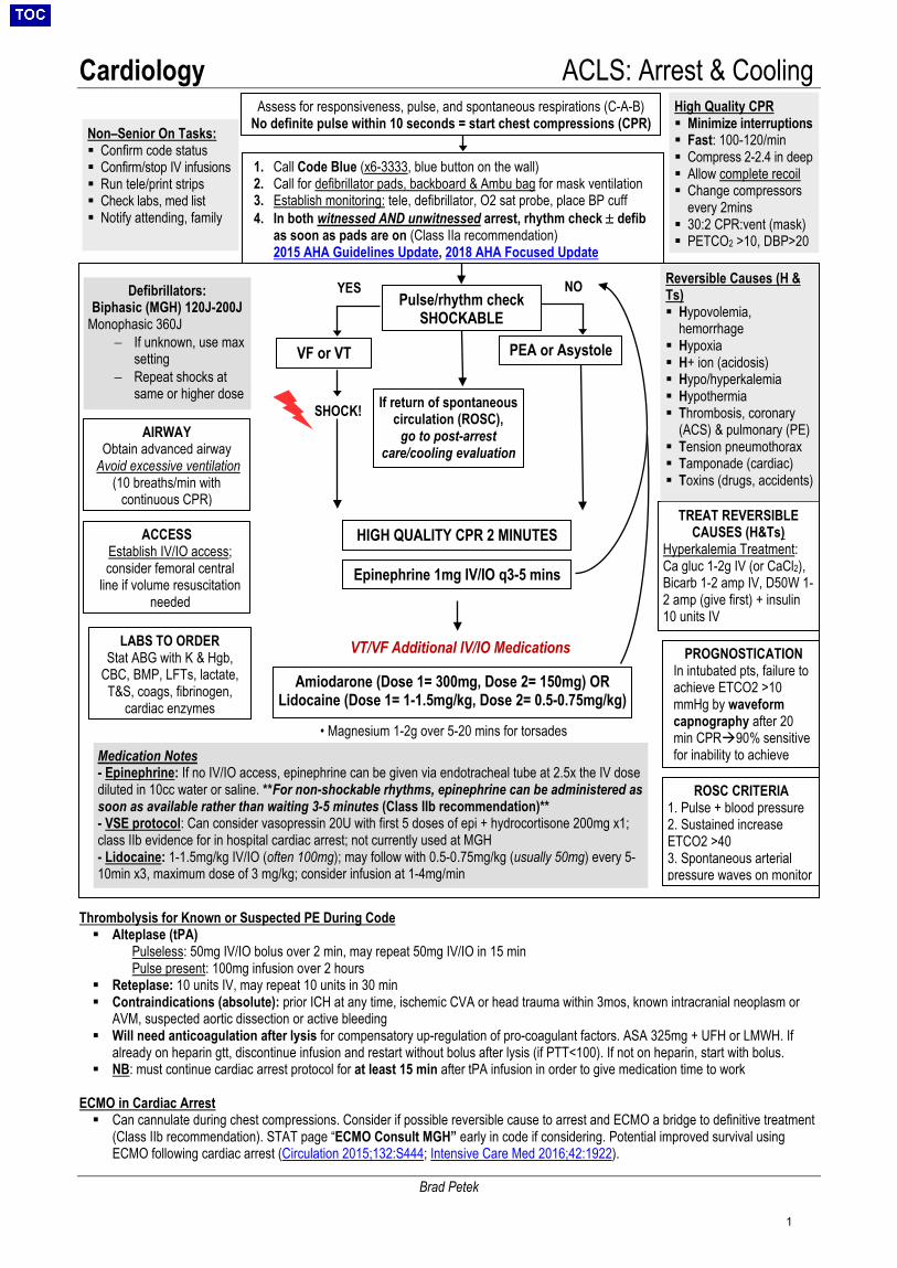
Cardiology ACLS: Arrest & Cooling
Brad Petek
Epinephrine 1mg IV/IO q3-5 mins
LABS TO ORDER Stat ABG with K & Hgb,
CBC, BMP, LFTs, lactate, T&S, coags, fibrinogen,
cardiac enzymes
TREAT REVERSIBLE CAUSES (H&Ts)
Hyperkalemia Treatment: Ca gluc 1-2g IV (or CaCl2), Bicarb 1-2 amp IV, D50W 1-2 amp (give first) + insulin 10 units IV
AIRWAY Obtain advanced airway
Avoid excessive ventilation (10 breaths/min with
continuous CPR)
HIGH QUALITY CPR 2 MINUTES ACCESS Establish IV/IO access; consider femoral central
line if volume resuscitation needed
ROSC CRITERIA 1. Pulse + blood pressure2. Sustained increaseETCO2 >403. Spontaneous arterialpressure waves on monitor
VT/VF Additional IV/IO Medications
Thrombolysis for Known or Suspected PE During Code Alteplase (tPA)
Pulseless: 50mg IV/IO bolus over 2 min, may repeat 50mg IV/IO in 15 min Pulse present: 100mg infusion over 2 hours
Reteplase: 10 units IV, may repeat 10 units in 30 min Contraindications (absolute): prior ICH at any time, ischemic CVA or head trauma within 3mos, known intracranial neoplasm or
AVM, suspected aortic dissection or active bleeding Will need anticoagulation after lysis for compensatory up-regulation of pro-coagulant factors. ASA 325mg + UFH or LMWH. If
already on heparin gtt, discontinue infusion and restart without bolus after lysis (if PTT<100). If not on heparin, start with bolus. NB: must continue cardiac arrest protocol for at least 15 min after tPA infusion in order to give medication time to work
ECMO in Cardiac Arrest Can cannulate during chest compressions. Consider if possible reversible cause to arrest and ECMO a bridge to definitive treatment
(Class IIb recommendation). STAT page “ECMO Consult MGH” early in code if considering. Potential improved survival usingECMO following cardiac arrest (Circulation 2015;132:S444; Intensive Care Med 2016;42:1922).
Assess for responsiveness, pulse, and spontaneous respirations (C-A-B) No definite pulse within 10 seconds = start chest compressions (CPR)
1. Call Code Blue (x6-3333, blue button on the wall)2. Call for defibrillator pads, backboard & Ambu bag for mask ventilation3. Establish monitoring: tele, defibrillator, O2 sat probe, place BP cuff4. In both witnessed AND unwitnessed arrest, rhythm check ± defib
as soon as pads are on (Class IIa recommendation)2015 AHA Guidelines Update, 2018 AHA Focused Update
Pulse/rhythm check SHOCKABLE
VF or VT PEA or Asystole
If return of spontaneous circulation (ROSC),
go to post-arrest care/cooling evaluation
Defibrillators: Biphasic (MGH) 120J-200J
Monophasic 360J − If unknown, use max
setting− Repeat shocks at
same or higher dose
High Quality CPR Minimize interruptions Fast: 100-120/min Compress 2-2.4 in deep Allow complete recoil Change compressors
every 2mins 30:2 CPR:vent (mask) PETCO2 >10, DBP>20
Non–Senior On Tasks: Confirm code status Confirm/stop IV infusions Run tele/print strips Check labs, med list Notify attending, family
YES NO
SHOCK!
Reversible Causes (H & Ts) Hypovolemia,
hemorrhage Hypoxia H+ ion (acidosis) Hypo/hyperkalemia Hypothermia Thrombosis, coronary
(ACS) & pulmonary (PE) Tension pneumothorax Tamponade (cardiac) Toxins (drugs, accidents)
Amiodarone (Dose 1= 300mg, Dose 2= 150mg) OR Lidocaine (Dose 1= 1-1.5mg/kg, Dose 2= 0.5-0.75mg/kg)
PROGNOSTICATION In intubated pts, failure to achieve ETCO2 >10 mmHg by waveform capnography after 20 min CPR90% sensitive for inability to achieve
Medication Notes - Epinephrine: If no IV/IO access, epinephrine can be given via endotracheal tube at 2.5x the IV dosediluted in 10cc water or saline. **For non-shockable rhythms, epinephrine can be administered assoon as available rather than waiting 3-5 minutes (Class IIb recommendation)**- VSE protocol: Can consider vasopressin 20U with first 5 doses of epi + hydrocortisone 200mg x1;class IIb evidence for in hospital cardiac arrest; not currently used at MGH- Lidocaine: 1-1.5mg/kg IV/IO (often 100mg); may follow with 0.5-0.75mg/kg (usually 50mg) every 5-10min x3, maximum dose of 3 mg/kg; consider infusion at 1-4mg/min
• Magnesium 1-2g over 5-20 mins for torsades
1
Cardiology ACLS: Arrest & Cooling
Brad Petek
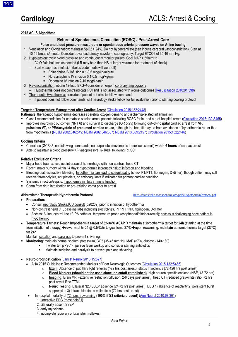
2015 ACLS Algorithms
Targeted Temperature Management after Cardiac Arrest (Circulation 2015;132:2448) Rationale: therapeutic hypothermia decreases cerebral oxygen demand and ischemia-related inflammation Class I recommendation for comatose cardiac arrest patients following ROSC for in- and out-of-hospital arrest (Circulation 2015;132:S465) Improves neurologic outcomes (NNT 6) and survival to discharge (OR 5.25) following out-of-hospital cardiac arrest from VF,
pulseless VT, or PEA/asystole of presumed cardiac cause, although the benefit may be from avoidance of hyperthermia rather thanfrom hypothermia (NEJM 2002;346:549; NEJM 2002;346:557; NEJM 2013;369:2197; Circulation 2015;132;2146)
Cooling Criteria Comatose (GCS<8, not following commands, no purposeful movements to noxious stimuli) within 6 hours of cardiac arrest Able to maintain a blood pressure +/- vasopressors +/- IABP following ROSC
Relative Exclusion Criteria Major head trauma: rule out intracranial hemorrhage with non-contrast head CT Recent major surgery within 14 days: hypothermia increases risk of infection and bleeding Bleeding diathesis/active bleeding: hypothermia can lead to coagulopathy (check PT/PTT, fibrinogen, D-dimer), though patient may still
receive thrombolytics, antiplatelets, or anticoagulants if indicated for primary cardiac condition Systemic infection/sepsis: hypothermia inhibits immune function Coma from drug intoxication or pre-existing coma prior to arrest
Abbreviated Therapeutic Hypothermia Protocol https://stopstroke.massgeneral.org/pdfs/HypothermiaProtocol.pdf • Preparation:
• Consult neurology Stroke/ICU consult (p20202) prior to initiation of hypothermia• Non-contrast head CT, baseline labs including electrolytes, PT/PTT/INR, fibrinogen, D-dimer• Access: A-line, central line +/- PA catheter, temperature probe (esophageal/bladder/rectal); access is challenging once patient is
hypothermic• Temperature Targets: Reach hypothermia target of 32-34ºC ASAPmaintain at hypothermia target for 24h (starting at the time
from initiation of therapy)rewarm at hr 24 @ 0.5ºC/hr to goal temp 37ºCupon rewarming, maintain at normothermia target (37ºC)for 24hMaintain sedation and paralysis to prevent shivering.
Monitoring: maintain normal sodium, potassium, CO2 (35-45 mmHg), MAP (>70), glucose (140-180) If water temp <70ºF, pursue fever workup and consider starting antibiotics Maintain sedation and paralysis to prevent pain and shivering
• Neuro-prognostication (Lancet Neurol 2016;15:597)• AHA 2015 Guidelines: Recommended Markers of Poor Neurologic Outcomes (Circulation 2015;132:S465)
o Exam: Absence of pupillary light reflexes (>72 hrs post arrest), status myoclonus (72-120 hrs post arrest)o Blood Markers (should not be used alone, no cutoff established): High neuron specific enolase (NSE, 48-72 hrs)o Imaging: Brain MRI (extensive restriction/diffusion, 2-6 days post arrest), head CT (reduced gray-white ratio, <2 hrs
post arrest if no TTM)o Neuro Testing: Bilateral N20 SSEP absence (24-72 hrs post arrest), EEG 1) absence of reactivity 2) persistent burst
suppression 3) intractable status epilepticus (72 hrs post arrest)• In-hospital mortality at 72h post-rewarming (100% if ≥2 criteria present) (Ann Neurol 2010;67:301)
1. unreactive EEG (most helpful)2. bilaterally absent SSEP3. early myoclonus4. incomplete recovery of brainstem reflexes
Return of Spontaneous Circulation (ROSC) / Post-Arrest Care Pulse and blood pressure measurable or spontaneous arterial pressure waves on A-line tracing
1. Ventilation and Oxygenation: maintain SpO2 > 94%. Do not hyperventilate (can induce cerebral vasoconstriction). Start at10-12 breaths/minute. Consider advanced airway waveform capnography. Target ETCO2 of 35-40 mm Hg.
2. Hypotension: cycle blood pressure and continuously monitor pulses. Goal MAP > 65mmHg.− IV/IO fluid boluses as needed (LR may be > than NS at larger volumes for treatment of shock)− Start vasopressor infusion (bolus code meds will wear off)
Epinephrine IV infusion 0.1-0.5 mcg/kg/minute Norepinephrine IV infusion 0.1-0.5 mcg/kg/min Dopamine IV infusion 2-10 mcg/kg/min
3. Revascularization: obtain 12-lead EKGconsider emergent coronary angiography− Hypothermia does not contraindicate PCI and is not associated with worse outcomes (Resuscitation 2010;81:398)
4. Therapeutic Hypothermia: consider if patient not able to follow commands− If patient does not follow commands, call neurology stroke fellow for full evaluation prior to starting cooling protocol
2
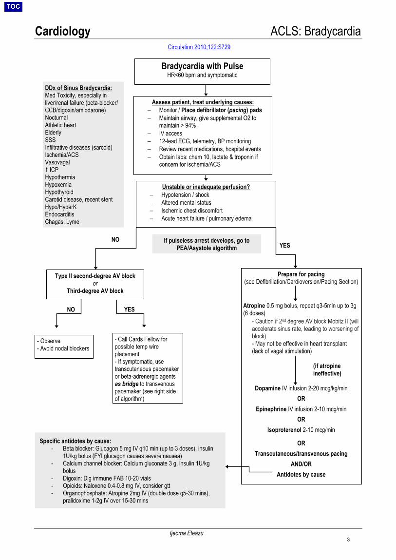
Cardiology ACLS: Bradycardia
Ijeoma Eleazu
Bradycardia with Pulse HR<60 bpm and symptomatic
Assess patient, treat underlying causes: − Monitor / Place defibrillator (pacing) pads− Maintain airway, give supplemental O2 to
maintain > 94%− IV access− 12-lead ECG, telemetry, BP monitoring− Review recent medications, hospital events− Obtain labs: chem 10, lactate & troponin if
concern for ischemia/ACS
Unstable or inadequate perfusion? − Hypotension / shock− Altered mental status− Ischemic chest discomfort− Acute heart failure / pulmonary edema
Type II second-degree AV block or
Third-degree AV block
DDx of Sinus Bradycardia: Med Toxicity, especially in liver/renal failure (beta-blocker/ CCB/digoxin/amiodarone) Nocturnal Athletic heart Elderly SSS Infiltrative diseases (sarcoid) Ischemia/ACS Vasovagal ICPHypothermiaHypoxemiaHypothyroidCarotid disease, recent stentHypo/HyperKEndocarditisChagas, Lyme
Prepare for pacing (see Defibrillation/Cardioversion/Pacing Section)
Atropine 0.5 mg bolus, repeat q3-5min up to 3g (6 doses)
- Caution if 2nd degree AV block Mobitz II (willaccelerate sinus rate, leading to worsening ofblock)- May not be effective in heart transplant(lack of vagal stimulation)
(if atropine ineffective)
Dopamine IV infusion 2-20 mcg/kg/min OR
Epinephrine IV infusion 2-10 mcg/min OR
Isoproterenol 2-10 mcg/min
OR Transcutaneous/transvenous pacing
AND/OR Antidotes by cause
- Observe- Avoid nodal blockers
- Call Cards Fellow forpossible temp wireplacement- If symptomatic, usetranscutaneous pacemakeror beta-adrenergic agentsas bridge to transvenouspacemaker (see right sideof algorithm)
If pulseless arrest develops, go to PEA/Asystole algorithm
YES
YES NO
NO
Specific antidotes by cause: - Beta blocker: Glucagon 5 mg IV q10 min (up to 3 doses), insulin
1U/kg bolus (FYI glucagon causes severe nausea)- Calcium channel blocker: Calcium gluconate 3 g, insulin 1U/kg
bolus- Digoxin: Dig immune FAB 10-20 vials- Opioids: Naloxone 0.4-0.8 mg IV, consider gtt- Organophosphate: Atropine 2mg IV (double dose q5-30 mins),
pralidoxime 1-2g IV over 15-30 mins
Circulation 2010;122:S729
3
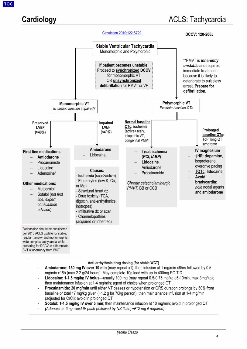
Cardiology ACLS: Tachycardia
Ijeoma Eleazu
DCCV: 120-200J
Polymorphic VT Evaluate baseline QTc
Monomorphic VT Is cardiac function impaired?
If patient becomes unstable: Proceed to synchronized DCCV
for monomorphic VT OR unsynchronized
defibrillation for PMVT or VF
Stable Ventricular Tachycardia Monomorphic and Polymorphic
First line medications: − Amiodarone− Procainamide− Lidocaine− Adenosine*
Other medications: − Metoprolol− Sotalol (not first
line; expertconsultationadvised)
− Amiodarone− Lidocaine
− IV magnesium− ↑HR: dopamine,
isoproterenol,overdrive pacing
− QTc: lidocaine− Avoid
bradycardia:hold nodal agentsand amiodarone
− Treat ischemia(PCI, IABP)
− Lidocaine− Amiodarone− Procainamide
Chronic catecholaminergic PMVT: BB or CCB
**PMVT is inherently unstable and requires immediate treatment because it is likely to deteriorate to pulseless arrest. Prepare for defibrillation.
Preserved LVEF
(>40%)
Impaired LVEF
(<40%)
Normal baseline QTc: ischemia (active>scar), idiopathic VT, congenital PMVT
Prolonged baseline QTc: TdP, long QT syndrome
*Adenosine should be consideredper 2015 ACLS update for stable,regular narrow- and monomorphicwide-complex tachycardia whilepreparing for DCCV to differentiateSVT w aberrancy from WCT
Anti-arrhythmic drug dosing (for stable WCT) - Amiodarone: 150 mg IV over 10 min (may repeat x1); then infusion at 1 mg/min x6hrs followed by 0.5
mg/min x18h (max 2.2 g/24 hours). May complete 10g load with up to 400mg PO TID.- Lidocaine: 1-1.5 mg/kg IV bolus—usually 100 mg (may repeat 0.5-0.75 mg/kg q5-10min, max 3mg/kg);
then maintenance infusion at 1-4 mg/min; agent of choice when prolonged QT- Procainamide: 20 mg/min until either VT ceases or hypotension or QRS duration prolongs by 50% from
baseline or total 17 mg/kg given (~1.2 g for 70kg person); then maintenance infusion at 1-4 mg/min(adjusted for CrCl); avoid in prolonged QT
- Sotalol: 1-1.5 mg/kg IV over 5 min; then maintenance infusion at 10 mg/min; avoid in prolonged QT- (Adenosine: 6mg rapid IV push (followed by NS flush)12 mg if required)
Causes: - Ischemia (scar>active)- Electrolytes (low K, Ca,or Mg)- Structural heart dz- Drug toxicity (TCA,digoxin, anti-arrhythmics,inotropes)- Infiltrative dz or scar- Channelopathies(acquired or inherited)
Circulation 2010;122:S729
4
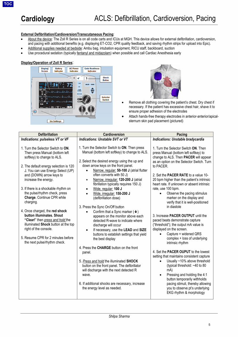
Cardiology ACLS: Defibrillation, Cardioversion, Pacing
Shilpa Sharma
External Defibrillation/Cardioversion/Transcutaneous Pacing: • About the device: The Zoll R Series is on all code carts and ICUs at MGH. This device allows for external defibrillation, cardioversion,
and pacing with additional benefits (e.g. displaying ET-CO2, CPR quality feedback, and saving rhythm strips for upload into Epic).• Additional supplies needed at bedside: Ambu bag, intubation equipment, RICU staff, backboard, suction• Use procedural sedation (typically fentanyl and midazolam) when possible and call Cardiac Anesthesia early
Display/Operation of Zoll R Series:
Defibrillation Cardioversion Pacing Indications: pulseless VT or VF
1. Turn the Selector Switch to ON.Then press Manual (bottom leftsoftkey) to change to ALS.
2. The default energy selection is 120J. You can use Energy Select (UP)and (DOWN) arrow keys toincrease the energy.
3. If there is a shockable rhythm onthe pulse/rhythm check, pressCharge. Continue CPR whilecharging.
4. Once charged, the red shockbutton illuminates. Shout“Clear!” then press and hold theilluminated Shock button at the topright of the console.
5. Resume CPR for 2 minutes beforethe next pulse/rhythm check.
Indications: Unstable SVT or VT
1. Turn the Selector Switch to ON. Then pressManual (bottom left softkey) to change to ALS.
2. Select the desired energy using the up anddown arrow keys on the front panel.• Narrow, regular: 50-100 J (atrial flutter
often converts with 50 J)• Narrow, irregular: 120-200 J (atrial
fibrillation typically requires 150 J)• Wide, regular: 100 J• Wide, irregular: 150-200 J
(defibrillation dose)
3. Press the Sync On/Off button• Confirm that a Sync marker ()
appears on the monitor above eachdetected R-wave to indicate wheredischarge will occur
• If necessary, use the LEAD and SIZEbuttons to establish settings that yieldthe best display
4. Press the CHARGE button on the frontpanel.
5. Press and hold the illuminated SHOCKbutton on the front panel. The defibrillatorwill discharge with the next detected Rwave.
6. If additional shocks are necessary, increasethe energy level as needed.
Indications: Unstable bradycardia
1. Turn the Selector Switch ON. Thenpress Manual (bottom left softkey) tochange to ALS. Then PACER will appearas an option on the Selector Switch. Turnto PACER.
2. Set the PACER RATE to a value 10-20 bpm higher than the patient’s intrinsicheart rate. If unknown or absent intrinsicrate, use 100 bpm.
• Observe the pacing stimulusmarker on the display andverify that it is well-positionedin diastole
3. Increase PACER OUTPUT until thepaced beats demonstrate capture(“threshold”); the output mA value isdisplayed on the screen.
• Capture = widened QRScomplex + loss of underlyingintrinsic rhythm
4. Set the PACER OUPUT to the lowestsetting that maintains consistent capture
• Usually ~10% above threshold(typical threshold: ~40 to 80mA)
• Pressing and holding the 4:1button temporarily withholdspacing stimuli, thereby allowingyou to observe pt’s underlyingEKG rhythm & morphology
• Remove all clothing covering the patient’s chest. Dry chest ifnecessary. If the patient has excessive chest hair, shave it toensure proper adhesion of the electrodes
• Attach hands-free therapy electrodes in anterior-anterior/apical-sternum skin pad placement (pictured)
5
Cardiology EKG Interpretation
Shawn Li, Nora Abo-Sido
Approach all EKGs systemically. Always note: rate, rhythm, QRS axis, complexes and intervals, chamber enlargement, ischemia/infarction, compare with prior EKG
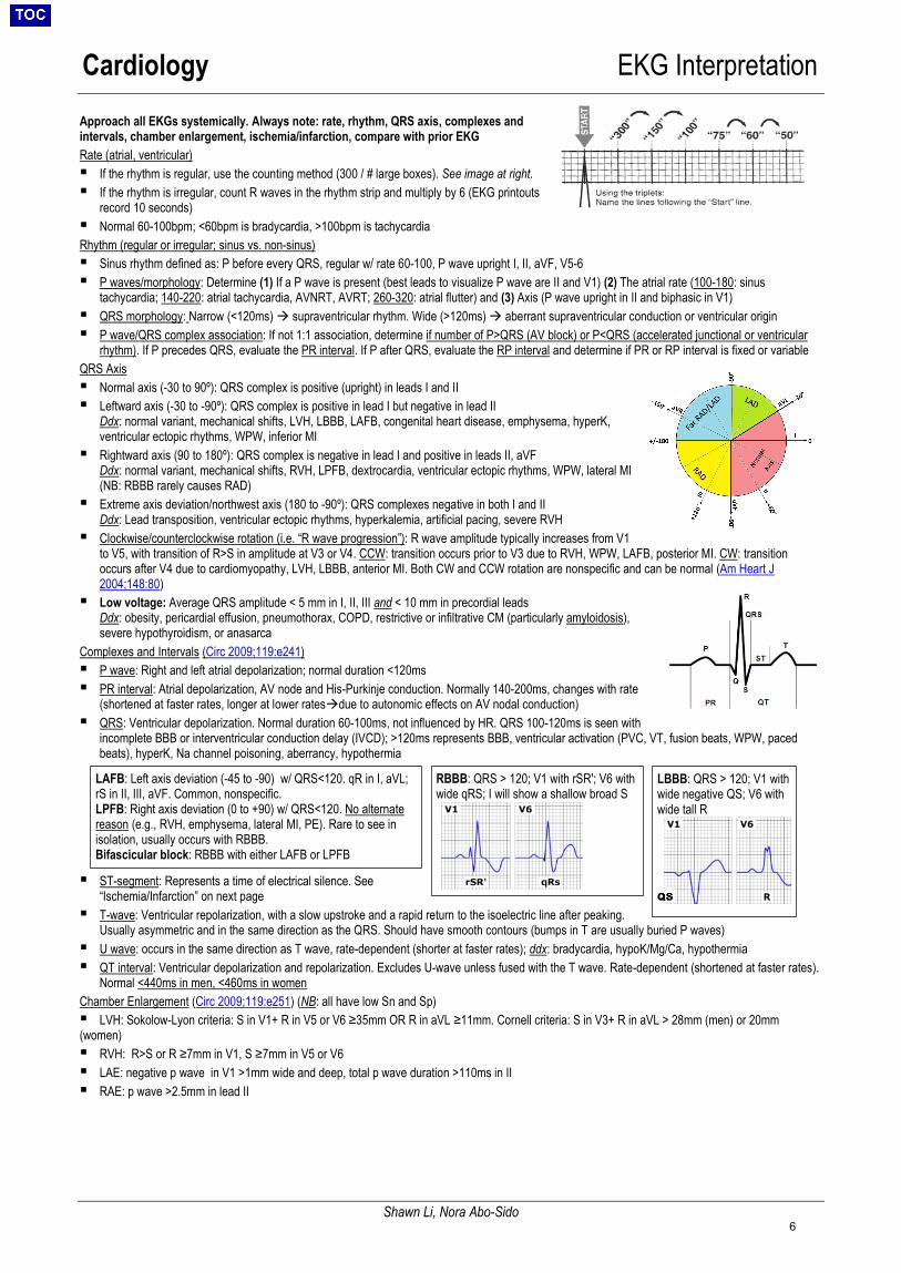
Rate (atrial, ventricular) If the rhythm is regular, use the counting method (300 / # large boxes). See image at right. If the rhythm is irregular, count R waves in the rhythm strip and multiply by 6 (EKG printouts
record 10 seconds) Normal 60-100bpm; <60bpm is bradycardia, >100bpm is tachycardia
Rhythm (regular or irregular; sinus vs. non-sinus) Sinus rhythm defined as: P before every QRS, regular w/ rate 60-100, P wave upright I, II, aVF, V5-6 P waves/morphology: Determine (1) If a P wave is present (best leads to visualize P wave are II and V1) (2) The atrial rate (100-180: sinus
tachycardia; 140-220: atrial tachycardia, AVNRT, AVRT; 260-320: atrial flutter) and (3) Axis (P wave upright in II and biphasic in V1) QRS morphology: Narrow (<120ms) supraventricular rhythm. Wide (>120ms) aberrant supraventricular conduction or ventricular origin P wave/QRS complex association: If not 1:1 association, determine if number of P>QRS (AV block) or P<QRS (accelerated junctional or ventricular
rhythm). If P precedes QRS, evaluate the PR interval. If P after QRS, evaluate the RP interval and determine if PR or RP interval is fixed or variable
QRS Axis Normal axis (-30 to 90º): QRS complex is positive (upright) in leads I and II Leftward axis (-30 to -90º): QRS complex is positive in lead I but negative in lead II
Ddx: normal variant, mechanical shifts, LVH, LBBB, LAFB, congenital heart disease, emphysema, hyperK,ventricular ectopic rhythms, WPW, inferior MI
Rightward axis (90 to 180º): QRS complex is negative in lead I and positive in leads II, aVFDdx: normal variant, mechanical shifts, RVH, LPFB, dextrocardia, ventricular ectopic rhythms, WPW, lateral MI(NB: RBBB rarely causes RAD)
Extreme axis deviation/northwest axis (180 to -90º): QRS complexes negative in both I and IIDdx: Lead transposition, ventricular ectopic rhythms, hyperkalemia, artificial pacing, severe RVH
Clockwise/counterclockwise rotation (i.e. “R wave progression”): R wave amplitude typically increases from V1to V5, with transition of R>S in amplitude at V3 or V4. CCW: transition occurs prior to V3 due to RVH, WPW, LAFB, posterior MI. CW: transitionoccurs after V4 due to cardiomyopathy, LVH, LBBB, anterior MI. Both CW and CCW rotation are nonspecific and can be normal (Am Heart J2004;148:80)
Low voltage: Average QRS amplitude < 5 mm in I, II, III and < 10 mm in precordial leadsDdx: obesity, pericardial effusion, pneumothorax, COPD, restrictive or infiltrative CM (particularly amyloidosis),severe hypothyroidism, or anasarca
Complexes and Intervals (Circ 2009;119:e241) P wave: Right and left atrial depolarization; normal duration <120ms PR interval: Atrial depolarization, AV node and His-Purkinje conduction. Normally 140-200ms, changes with rate
(shortened at faster rates, longer at lower ratesdue to autonomic effects on AV nodal conduction) QRS: Ventricular depolarization. Normal duration 60-100ms, not influenced by HR. QRS 100-120ms is seen with
incomplete BBB or interventricular conduction delay (IVCD); >120ms represents BBB, ventricular activation (PVC, VT, fusion beats, WPW, pacedbeats), hyperK, Na channel poisoning, aberrancy, hypothermia
ST-segment: Represents a time of electrical silence. See“Ischemia/Infarction” on next page
T-wave: Ventricular repolarization, with a slow upstroke and a rapid return to the isoelectric line after peaking.Usually asymmetric and in the same direction as the QRS. Should have smooth contours (bumps in T are usually buried P waves)
U wave: occurs in the same direction as T wave, rate-dependent (shorter at faster rates); ddx: bradycardia, hypoK/Mg/Ca, hypothermia QT interval: Ventricular depolarization and repolarization. Excludes U-wave unless fused with the T wave. Rate-dependent (shortened at faster rates).
Normal <440ms in men, <460ms in women
Chamber Enlargement (Circ 2009;119:e251) (NB: all have low Sn and Sp) LVH: Sokolow-Lyon criteria: S in V1+ R in V5 or V6 ≥35mm OR R in aVL ≥11mm. Cornell criteria: S in V3+ R in aVL > 28mm (men) or 20mm (women) RVH: R>S or R ≥7mm in V1, S ≥7mm in V5 or V6 LAE: negative p wave in V1 >1mm wide and deep, total p wave duration >110ms in II RAE: p wave >2.5mm in lead II
LBBB: QRS > 120; V1 with wide negative QS; V6 with wide tall R
RBBB: QRS > 120; V1 with rSR'; V6 with wide qRS; I will show a shallow broad S
LAFB: Left axis deviation (-45 to -90) w/ QRS<120. qR in I, aVL; rS in II, III, aVF. Common, nonspecific. LPFB: Right axis deviation (0 to +90) w/ QRS<120. No alternate reason (e.g., RVH, emphysema, lateral MI, PE). Rare to see in isolation, usually occurs with RBBB. Bifascicular block: RBBB with either LAFB or LPFB
6
Cardiology EKG Interpretation
Shawn Li, Nora Abo-Sido
Ischemia/Infarction (JACC 2009;53:1003) Analyze abnormalities along the vectors of ventricular depolarization and repolarization (QRS-ST-T) T-wave abnormalities: Hyperacute, symmetric T-waves can be found within minutes; followed by T wave inversions (≥0.1 mV in 2 contiguous leads) ST depression: Suggests subendocardial injury; ≥0.05 mV below the baseline (PR segment), measured at the J point, in two contiguous leads,
downsloping or horizontal = more ominous. ST depressions do not localize to territories (Circ Res 1998;82;957) NB: always look for ST elevations torule out reciprocal ST depression. Digoxin toxicity: scooping ST depressions.
ST elevation: Suggests transmural ischemia; ≥0.1 mV, except for leads V2 to V3 (≥0.2 mV in men ≥40yo and ≥0.15 mV in women), measured at theJ point. PR segment is the isoelectric interval on the ECG and can be used to assess ST segment elevation/depression.
Q-wave: Usually a marker of scar; must be deep (>1 mm) and broad (>0.04 seconds), more likely 2/2 prior MI if inverted T wave in same lead.Pathologic Q wave defined by 40ms duration (1 box wide), 25% height of QRS. “Isolated Q in III is free” (non-pathologic).
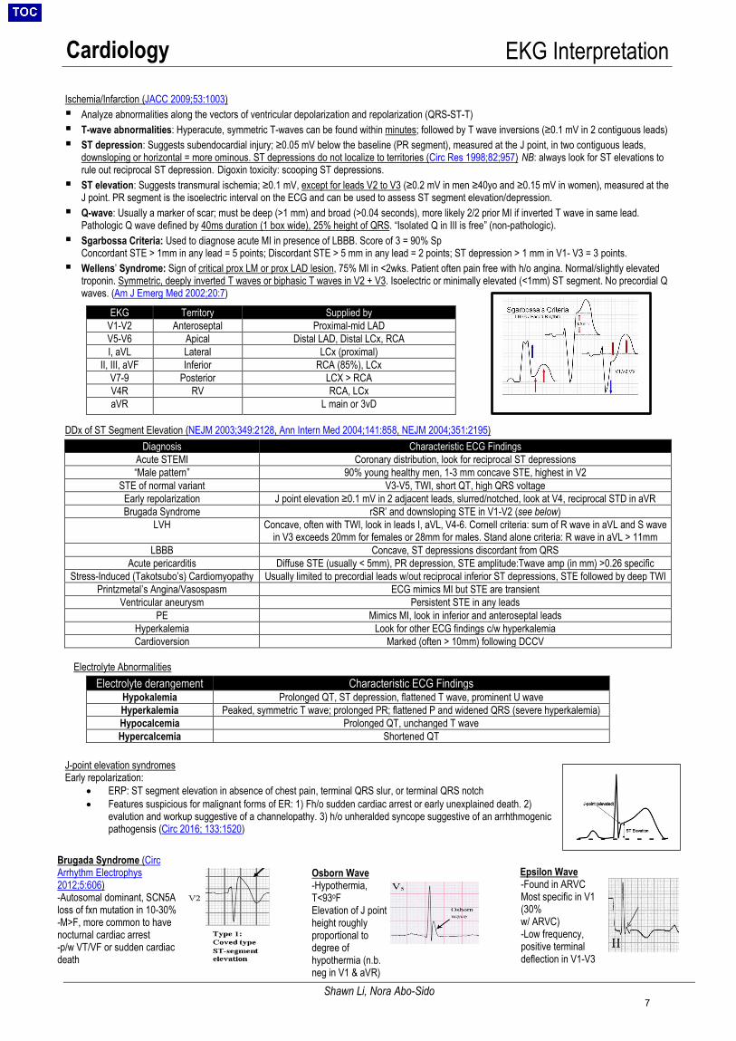
Sgarbossa Criteria: Used to diagnose acute MI in presence of LBBB. Score of 3 = 90% SpConcordant STE > 1mm in any lead = 5 points; Discordant STE > 5 mm in any lead = 2 points; ST depression > 1 mm in V1- V3 = 3 points.
Wellens’ Syndrome: Sign of critical prox LM or prox LAD lesion, 75% MI in <2wks. Patient often pain free with h/o angina. Normal/slightly elevatedtroponin. Symmetric, deeply inverted T waves or biphasic T waves in V2 + V3. Isoelectric or minimally elevated (<1mm) ST segment. No precordial Qwaves. (Am J Emerg Med 2002;20:7)
DDx of ST Segment Elevation (NEJM 2003;349:2128, Ann Intern Med 2004;141:858, NEJM 2004;351:2195)
Electrolyte Abnormalities
J-point elevation syndromesEarly repolarization:
• ERP: ST segment elevation in absence of chest pain, terminal QRS slur, or terminal QRS notch• Features suspicious for malignant forms of ER: 1) Fh/o sudden cardiac arrest or early unexplained death. 2)
evalution and workup suggestive of a channelopathy. 3) h/o unheralded syncope suggestive of an arrhthmogenicpathogensis (Circ 2016; 133:1520)
EKG Territory Supplied by V1-V2 Anteroseptal Proximal-mid LAD V5-V6 Apical Distal LAD, Distal LCx, RCA I, aVL Lateral LCx (proximal)
II, III, aVF Inferior RCA (85%), LCx V7-9 Posterior LCX > RCA V4R RV RCA, LCx aVR L main or 3vD
Diagnosis Characteristic ECG Findings Acute STEMI Coronary distribution, look for reciprocal ST depressions “Male pattern” 90% young healthy men, 1-3 mm concave STE, highest in V2
STE of normal variant V3-V5, TWI, short QT, high QRS voltage Early repolarization J point elevation ≥0.1 mV in 2 adjacent leads, slurred/notched, look at V4, reciprocal STD in aVR Brugada Syndrome rSR’ and downsloping STE in V1-V2 (see below)
LVH Concave, often with TWI, look in leads I, aVL, V4-6. Cornell criteria: sum of R wave in aVL and S wave in V3 exceeds 20mm for females or 28mm for males. Stand alone criteria: R wave in aVL > 11mm
LBBB Concave, ST depressions discordant from QRS Acute pericarditis Diffuse STE (usually < 5mm), PR depression, STE amplitude:Twave amp (in mm) >0.26 specific
Stress-Induced (Takotsubo’s) Cardiomyopathy Usually limited to precordial leads w/out reciprocal inferior ST depressions, STE followed by deep TWI Printzmetal’s Angina/Vasospasm ECG mimics MI but STE are transient
Ventricular aneurysm Persistent STE in any leads PE Mimics MI, look in inferior and anteroseptal leads
Hyperkalemia Look for other ECG findings c/w hyperkalemia Cardioversion Marked (often > 10mm) following DCCV
Electrolyte derangement Characteristic ECG Findings Hypokalemia Prolonged QT, ST depression, flattened T wave, prominent U wave Hyperkalemia Peaked, symmetric T wave; prolonged PR; flattened P and widened QRS (severe hyperkalemia) Hypocalcemia Prolonged QT, unchanged T wave Hypercalcemia Shortened QT
Brugada Syndrome (Circ Arrhythm Electrophys 2012;5:606) -Autosomal dominant, SCN5Aloss of fxn mutation in 10-30% pts-M>F, more common to havenocturnal cardiac arrest-p/w VT/VF or sudden cardiacdeath
Epsilon Wave -Found in ARVCMost specific in V1 (30% w/ ARVC)-Low frequency,positive terminaldeflection in V1-V3
Osborn Wave -Hypothermia,T<93oFElevation of J pointheight roughlyproportional todegree ofhypothermia (n.b.neg in V1 & aVR)
7
Cardiology Narrow & Wide Complex Tachycardia
Usama Abbasi and Raymond Parrish
Narrow Complex Tachycardia (QRS < 120 ms) (NEJM 2012;367:1438)
Diagnostic approach & general principles: o if unstable synchronized cardioversiono vagal maneuvers/carotid massage/adenosine can resolve diagnostic dilemmas and treat AVNRT and AVRTo acute treatment for all others is BB, CCB or amiodarone (but consider risk of pharmacologic cardioversion if pt is not anticoagulated)
Sinus Tachycardiao Gradual in onset (if not consider SANRT, which is similar to AVRT and terminates with adenoside or vagal maneuvers)o Consider: hypovolaemia, haemorrhage, withdrawal (EtOH, BZD, opiate, BB), intoxication, fever/infection, pain, hypoxaemia, PE, anaemia,
tamponade, dissection, hormonal (hyperthyroidism, adrenal insufficiency, pheochromocytoma)
Atrial Tachycardia (AT)o Long RP, single P morphology, non-sinus P wave axiso Arises from increased automaticity at single atrial focuso Classic digoxin toxicity is AT w/ variable AV block
Multifocal Atrial Tachycardia (MAT)o Long RP, 3 or more P wave morphologieso Irregular due to varying PP, PR and RR intervalso COPD, pHTN, CAD, electrolytes, theophylline
Atrial Fibrillation (AF)o No coordinated atrial activity (P wave absent), irregular o Arises from numerous re-entrant tracts in atria or
pulmonary veins
Atrial Flutter (AFL) o Arises from true (isthmus-dependent, typical) or functional
(isthmus-independent, atypical) re-entry w/in R atriumo PP interval constant but RR may vary (variable AV block)o Counterclockwise: negative flutter waves in II, III and aVFo Clockwise: positive flutter waves in II, III, aVFo Signature: no isoelectric baseline, atrial rate ~300, always
> 250, usually with 1:2 conduction
Atrioventricular Nodal Re-entrant Tachycardia (AVNRT)o Usually no RP (slow-fast), uncommon short RP (fast-
slow), rarely long RP (slow-slow)o Arises from functional re-entry w/in AV nodeo Trigger PAC (slow-fast) > PVC (fast-slow) o Young adults, F > M
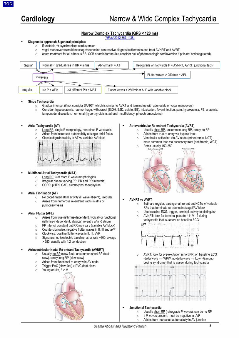
Atrioventricular Re-entrant Tachycardia (AVRT)o Usually short RP, uncommon long RP, rarely no RPo Arises from true re-entry via bypass tracto Ventricular activation via AV node (orthodromic, NCT)
more common than via accessory tract (antidromic, WCT)o Rates usually 150-250
AVNRT vs AVRTo Both are regular, paroxysmal, re-entrant NCTs w/ variable
RPs that terminate w/ adenosine/vagal/AV blocko Use baseline ECG, trigger, terminal activity to distinguish o AVNRT: look for terminal pseudo-r’ in V1-2 during
tachycardia that is absent on baseline ECG
o AVRT: look for pre-excitation (short PR) on baseline ECG(delta wave → WPW; no delta wave → Lown-Ganong-Levine syndrome) that is absent during tachycardia
Junctional Tachycardiao Usually short RP (retrograde P waves), can be no RPo If P waves present, must be negative in aVFo Arises from increased automaticity in AV junction
P-waves?
Regular
Irregular ≥3 different P’s = MAT No P = AFib Flutter waves > 250/min = ALF with variable block
Normal P, gradual rise in HR = sinus Abnormal P = AT Retrograde or not visible P = AVNRT, AVRT, junctional tach
Flutter waves > 250/min = AFL
8
Cardiology Narrow & Wide Complex Tachycardia
Usama Abbasi and Raymond Parrish
Wide Complex Tachycardia (QRS ≥ 120 ms) Goal: determine VT or SVT with aberrant conduction SVT w/ aberrant conduction includes: functional/rate-dependent BBB iso encroachment on bundle refractory period; RBBB > LBBB, SVT w/ pre-
existing BBB, antidromic AVRT, antiarrhythmic drugs (digoxin, class IA or IC, amiodarone), hyperkalemia, TCA overdose, pacemaker/endless looptachycardia (retrograde VA conduction of V-paced beat misidentified as native A-beat leading to additional V-pacing)
As majority of VT is due to re-entry (true about scar vs functional iso heterogeneous conduction), history is crucial. MI, cardiomyopathy, reducedLVEF and infiltrative disease all increase pre-test probability of VT
QRS w/ sharp initial deflection (some His-Purkinje conduction present) followed by broad terminal deflection favours SVT w/ aberrancy ECG factors that favor VT:
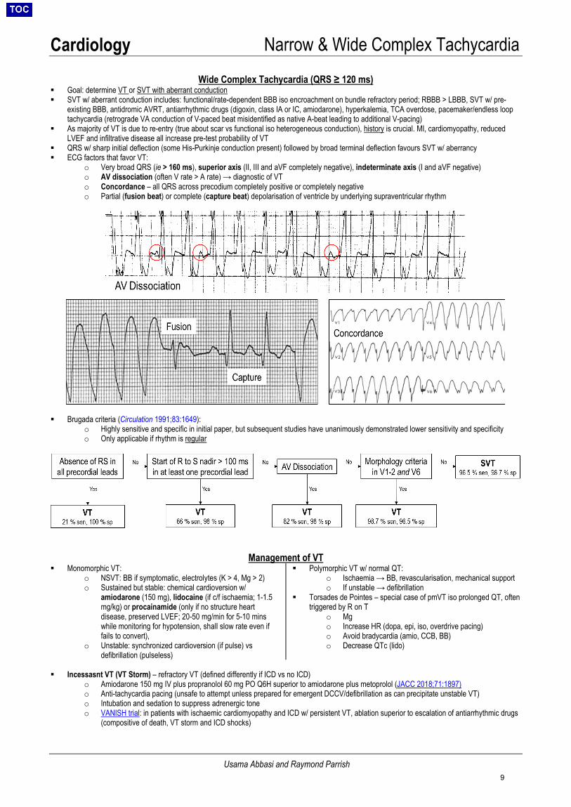
o Very broad QRS (ie > 160 ms), superior axis (II, III and aVF completely negative), indeterminate axis (I and aVF negative)o AV dissociation (often V rate > A rate) → diagnostic of VTo Concordance – all QRS across precodium completely positive or completely negativeo Partial (fusion beat) or complete (capture beat) depolarisation of ventricle by underlying supraventricular rhythm
Brugada criteria (Circulation 1991;83:1649):o Highly sensitive and specific in initial paper, but subsequent studies have unanimously demonstrated lower sensitivity and specificity o Only applicable if rhythm is regular
Management of VT Monomorphic VT:
o NSVT: BB if symptomatic, electrolytes (K > 4, Mg > 2)o Sustained but stable: chemical cardioversion w/
amiodarone (150 mg), lidocaine (if c/f ischaemia; 1-1.5mg/kg) or procainamide (only if no structure heartdisease, preserved LVEF; 20-50 mg/min for 5-10 minswhile monitoring for hypotension, shall slow rate even iffails to convert),
o Unstable: synchronized cardioversion (if pulse) vsdefibrillation (pulseless)
Polymorphic VT w/ normal QT:o Ischaemia → BB, revascularisation, mechanical supporto If unstable → defibrillation
Torsades de Pointes – special case of pmVT iso prolonged QT, oftentriggered by R on T
o Mgo Increase HR (dopa, epi, iso, overdrive pacing)o Avoid bradycardia (amio, CCB, BB)o Decrease QTc (lido)
Incessasnt VT (VT Storm) – refractory VT (defined differently if ICD vs no ICD)o Amiodarone 150 mg IV plus propranolol 60 mg PO Q6H superior to amiodarone plus metoprolol (JACC 2018;71:1897)o Anti-tachycardia pacing (unsafe to attempt unless prepared for emergent DCCV/defibrillation as can precipitate unstable VT)o Intubation and sedation to suppress adrenergic toneo VANISH trial: in patients with ischaemic cardiomyopathy and ICD w/ persistent VT, ablation superior to escalation of antiarrhythmic drugs
(compositive of death, VT storm and ICD shocks)
9
Cardiology Atrial Fibrillation & Flutter
David Olshan
Atrial Fibrillation Epidemiology and Classification (Heart Rhythm 2012;9:632) Prevalence increases with age; <0.1% for age<55 vs 9% for age>80. Reoccurs in majority of cases due to secondary precipitant (surgery, infection, MI, thyrotoxicosis, acute alcohol, PE) Often co-exists with atrial flutter (Chest 1992;101:34, Circ Arrythmia EP 2009;2:393) Classification:
• First diagnosed: not previously diagnosed irrespective of duration• Paroxysmal: self-termination within 7 days (includes those cardioverted within 7 days)• Persistent: continuous afib lasting >7 days• Long-standing persistent: continuous afib lasting >12 months• Permanent: term used when decision is made to stop further attempts to restore and/or maintain sinus rhythm
Clinical Evaluation of New-Onset Atrial Fibrillation History/Exam: presence and timing of symptoms, HTN, DM, valvular disease, CHF, angina, congenital heart disease,
OSA, family hx of AF, acute precipitants (e.g., EtOH, thyrotoxicosis, sympathomimetic drugs, surgery, myocardialischemia, myocarditis, PE, acute pulmonary disease, infection)
ECG: absence of discernible p waves, irregularly irregular R-R intervals TTE: LV function, LA/RA size, valve function, pulmonary HTN, LA thrombus (low sensitivity, better with TEE) CXR: evaluate for pulmonary parenchymal processes and pulmonary vasculature/edema Labs: TFTs, LFTs, BUN/Cr, CBC, NT-proBNP Additional testing: Zio patch, Holter monitor, implantable loop recorder, exercise testing (to assess rate control with
activity or as part of ischemic evaluation) Five “domains” of initial assessment: hemodynamic stability, precipitating factors, stroke risk and need for AC, HR and
need for rate control, symptom assessment and need for rhythm control
Cardioversion (ALWAYS consider high risk of embolic stroke if any breaks in AC for one month prior) Indications
• Urgent situations: ischemia, end-organ hypoperfusion, symptomatic hypotension, severe pulmonary edema• Elective: new-onset AF or unacceptable symptoms from persistent AF
Electrical Cardioversion (DCCV)• Synchronized DCCV at 150J (biphasic); increase energy in stepwise fashion if SR not achieved• Use procedural sedation if possible (consult cardiac anesthesia). If elective, should be performed in ICU or EP lab.• Consider anti-arrhythmic drugs as adjunct (e.g., amiodarone)
Chemical Cardioversion• Success rate significantly higher for acute (<7d) compared with longer-duration AF• Agents: pill-in-pocket (flecainide, propafenone), dofetilide, ibutilide, amiodarone
o Amiodarone: IV infusion weakly effective for conversion; PO load over 3-4 wk has 27% rate of conversion AC in Patients Undergoing Cardioversion (applies to BOTH chemical and electrical)
• Pre-procedure:o Definitive new onset <48 hours: may proceed without anticoagulationo Onset >48 hours: must anticoagulate for 3 weeks prior to DCCV or obtain TEE immediately prior to DCCV
(NEJM 2001;344:1411)• Post-procedure: anticoagulate for at least 4 weeks after DCCV (due to myocardial stunning)• NB: if obtaining TEE and pt is not anticoagulated, start UFH/LMWH on day of DCCV (or apixaban 2d before DCCV)
Acute Management of Atrial Fibrillation with Rapid Ventricular Response Step 1: Confirm atrial fibrillation or flutter with ECG Step 2: Determine hemodynamic stability: Stable: SBP>90 Usually if HR > 130 or symptomatic prefer IV, otherwise can consider starting PO / increasing current PO dose Beta-blocker: metoprolol (others: labetalol, propranolol, esmolol)
• IV: bolus 2.5-10 mg over 2 minutes; repeat as required q10-15 min• PO: up to 400mg total daily dose (although doses >200mg usually not effective)• Contraindicated: acute decompensated heart failure, history of severe bronchospasm
Calcium channel blocker: diltiazem (others: verapamil)• IV: bolus of 0.25 mg/kg (average adult dose 10-25 mg) over 2 minutes; repeat as required q10-15 min• PO: up to 360 mg total daily dose
10
Cardiology Atrial Fibrillation & Flutter
David Olshan
• Contraindicated: LV failure with pulmonary congestion, LVEF <40%• Reduce dose with hepatic impairment and renal impairment
Once rates are controlled with IV medication, ALWAYS chase with PO for sustained effectPeri-stable: SBP 80-90 If borderline BP, carefully attempt low-dose BB / CCA (attempt concomitant IVF if pulmonary edema not a concern) Consider BP-sparing agents:
• Digoxin load (0.5mg IV/PO followed by 0.25mg IV/PO q6hrs x2, total load 1mg), can lead to toxicity with renalimpairment, contraindicated if accessory pathways
• Amiodarone (150mg IV over 10 min followed by gtt; requires transfer to ICU or SDU) [NB: bolus x1 can be done onfloor w/ nursing supervisor]
o Consider risk of pharmacologic cardioversion and consequent embolization of LA thrombusUnstable: SBP<80 (usually with HR >150); signs of shock (AMS, cool extremities); refractory pulmonary edema or angina. Call for early back-up / Senior On for medication administration, cardioversion, and uptriage. Syncronized cardioversion (DCCV); usually start with 150J. If pressors are required, phenylephrine (neosynephrine) is first-line given reflex bradycardia NB: higher HRs (>140) more likely to cause HoTN alone; lower HRs (<140) may cause HoTN if systolic/diastolic dysfxn or
decreased preload (i.e., “loss of atrial kick”).Step 3: correct underlying causes or precipitants whenever possible (e.g. IVF).
Long-Term Rate vs. Rhythm Control Overall, rate control noninferior to rhythm control for AF symptoms, CV mortality, and stroke risk. (AFFIRM, RACE, PIAF,
. STAF, HOT CAFÉ, AF-CHF). • Exceptions: consider rhythm control if persistent AF sx impairing QoL, also if age <65 or comorbid HF (esp if
systolic dysfxn). Restoration of NSR may also lead to increased QoL and exercise performance (NEJM2005;352:1861, JACC 2004;43:241).
BB more successful than CCA in achieving rate control (70% vs. 54%), either alone or in combination with digoxin. Digoxin alone is moderately effective in controlling V-rate at rest, ineffective during exertion or high adrenergic tone. Long-term digoxin independently associated with increased mortality in AF patients (JACC 2018;71:1063). Rate Targets: Lenient rate control (resting HR <110) non-inferior to strict rate control (HR <80); similar outcomes in CV
death, stroke, bleeding, arrhythmia and hospitalization for HF (RACE II). Stricter HR (or rhythm control) may be beneficialin younger pts or pts w/ HF.
Contraindications/Warnings: Evidence of pre-excitation on ECG (in these patients, IV procainamide is 1st line), cautioususe in high-degree AVB. CCA should not be used in pts with EF<40% given negative inotropy.
“Pill-in-Pocket”: For pts w recent pAFib w infrequent and well-tolerated episodes, ppx may have risk>benefit, and thusprn flecainide or propafenone at sx onset is safe and effective (NEJM 2004;351:2384).
Long-Term Rhythm Control: Overview (Circulation 2012;125:381) Choice of Agents:
o No structural heart disease: pill-in-pocket (flecainide/propafenone), dofetilide, dronedarone, sotalol, amiodaroneo Structural: CAD: dofetilide, dronedarone, sotalol, amio | HF: amio, dofetilide | LVH: amio, dofetilide
Catheter ablation (pulmonary vein isolation): associated with a lower long-term AF recurrence rate vs. antiarrhythmicagents in both paroxysmal (MANTRA-PAF, RAAFT-2) and persistent AF (Eur Heart J 2014;35:501). CASTLE-AF trialshowed catheter ablation in pts w AFib and HF lowered morbidity/mortality 2/2 HF compared to medical therapy.
AV nodal ablation with PPM: indicated when pharm rate/rhythm control not achievable (JACC 2014;64:2246); considerCRT for EF<40%.
Antithrombotic Therapy (Stroke 2010;41:2731)
Treatment recommended for all pts except those with CHADS2-VASc 0, lone AF episode, or contraindications to therapy. LA appendage is the source of at least 90% of thrombi in pts with CVA and AF. Subclinical AF still associated with increased stroke/systemic embolism (ASSERT). Patients at relatively low risk for thromboembolism may be maintained on ASA alone (see below), but no reliable data
exist to guide decision between 81mg vs. 325mg ASA dose
Risk assessment CHA2DS2-VASc: [1pt for CHF, HTN, Age 65-74, DM, Female Sex, Vascular disease; 2pt for Age≥75, Stroke/TIA].
Preferred over CHADS2 in 2014 ACC/AHA/HRS guidelines (Ib). CHA2DS2-VASc > CHADS2 “truly low risk” subjects(Thromb Haemostasis 2012;107:1172).
11

Cardiology Atrial Fibrillation & Flutter
David Olshan
• Score 0 = no AC or ASA; Score 1 = no AC vs. ASA vs. oral AC based on clinical judgmenthow high is risk fromspecified risk factor? ex: HTN, DM, age bring greater risk compared to female sex, vascular dz; Score ≥2 = oral AC
CHA2DS2-VASc Score 0 1 2 3 4 5 6 7 8 9 Adjusted stroke rate (%/yr) 0 1.3 2.2 3.2 4 6.7 9.8 9.6 6.7 15.2
HAS-BLED: [HTN (SBP>160); Abnl renal function (CrCl<50); Liver disease (Cirrhosis or Bilis 2x ULN orAST/ALT/AlkPhos 3x ULN); Stroke; Bleeding history; Labile INR (<60% in Rx range); Elderly (>65y); Antiplatelet meds(ASA, NSAID); Alcohol (>8 drinks/wk) or other drug use]. Risk stratification of bleeding risk w/ oral AC. Score ≥3 suggestscaution and regular follow-up.
http://www.sparctool.com/ can aid in risk assessment and choice of anticoagulation
Choice of Antithrombotic Agent
Options for Oral Antiocoagulation Medication Action Dosing Avoid Effect
Warfarin Vitamin K antagonist Variable Pregnancy
(X) Annual RR of 68% for stroke (Arch Intern Med 1994;154:1449).
Dabigatran Direct
thrombin inhibitor
150mg BID CrCl <15 35% reduction in stroke compared to warfarin with no increase in major bleeding (RE-LY).
Rivaroxaban
Direct factor Xa inhibitor
20mg QD (15mg if CrCl 15-50)
Severe hepatic
impairment, CrCl <15
Non-inferior to warfarin for prevention of stroke in non-valvular AF; no difference in major bleeding, ↓ICH, and fatal bleeding (ROCKET-AF). For cryptogenic stroke prevention, both non superior and associated w increased bleeding when compared to ASA (NEJM 2018;378;2191). Can be completely and quickly reversed w Idarucizumab. (NEJM 2015;373:511)
Apixaban 5mg BID (2.5mg if Cr >1.5 AND age > 80 OR wt <60kg)
Severe hepatic
impairment
Superior to warfarin or aspirin alone in preventing stroke and systemic embolism w/o increasing the risk of major bleeding or ICH (ARISTOTLE).
Edoxaban 60mg QD CrCl <30 Noninferior to warfarin for prevention of stroke with lower rates of major bleeding and death (ENGAGE-AF).
NOACs vs Warfarin: In meta-analysis, NOACs shown to have lower risk of stroke or systemic embolic events (RR 0.81),all-cause mortality (RR 0.9), and ICH (RR 0.48) but higher risk of GI bleeding (RR 1.25) compared to warfarin (Lancet2014;383:955). In new AF, start a NOAC unless valvular afib (see below) (Eur Heart J 206;37:2893).
Bridging AC: Consider bridging with heparin or LMWH for CHADS2 scores >5 (Blood 2011;117:5044, NEJM2015;373:823).
Valvular Afib: AF in the setting of rheumatic mitral stenosis, mechanical or bioprosthetic heart valve, mitral valve repair(Circulation 2014;130:2071).
LAA closure (Watchman device): In non-valvular AFib, device placement provides comparable stroke prevention towarfarin with reduced bleeding risk and improved mortality (JACC 2017;70:2964).
NB: After cryptogenic embolic stroke, ambulatory ECG monitoring for 30 days significantly increased AFib detection when compared to shorter duration of monitoring (NEJM 2014;370:2467). If AF not detected, ASA non-inferior to NOAC.
Atrial Flutter Overview Less prevalent but often coexists or precedes AF. Type 1 (typical): Reentrant loop in RA via cavo-tricuspid isthmus (CTI). Divided based on
direction of circuit:• Counterclockwise (more common, inverted flutter waves in II, III, aVF + upright flutterwaves in V1)• Clockwise (less common, upright flutter waves in II, III, aVF + inverted flutter waves in V1)
Type 2 (atypical): does not meet criteria for Type 1; is typically faster and often refractory toablation
Anticoagulation: Risk of thromboembolism lower than AF (J Stroke Cerebrovasc 2018;27:839) but these are small studies – management should be similar to AF (Chest 2012;141:e531S). Rate control: Similar strategies (BB,CCA) to AF, but more difficult to successfully rate-control. Rhythm control: CTI ablation for typical flutter > 90% effective at 1yr (Circ Arrhythmia EP 2009;2:393).
12
Cardiology QTc Prolongation
Sumeet Khetarpal
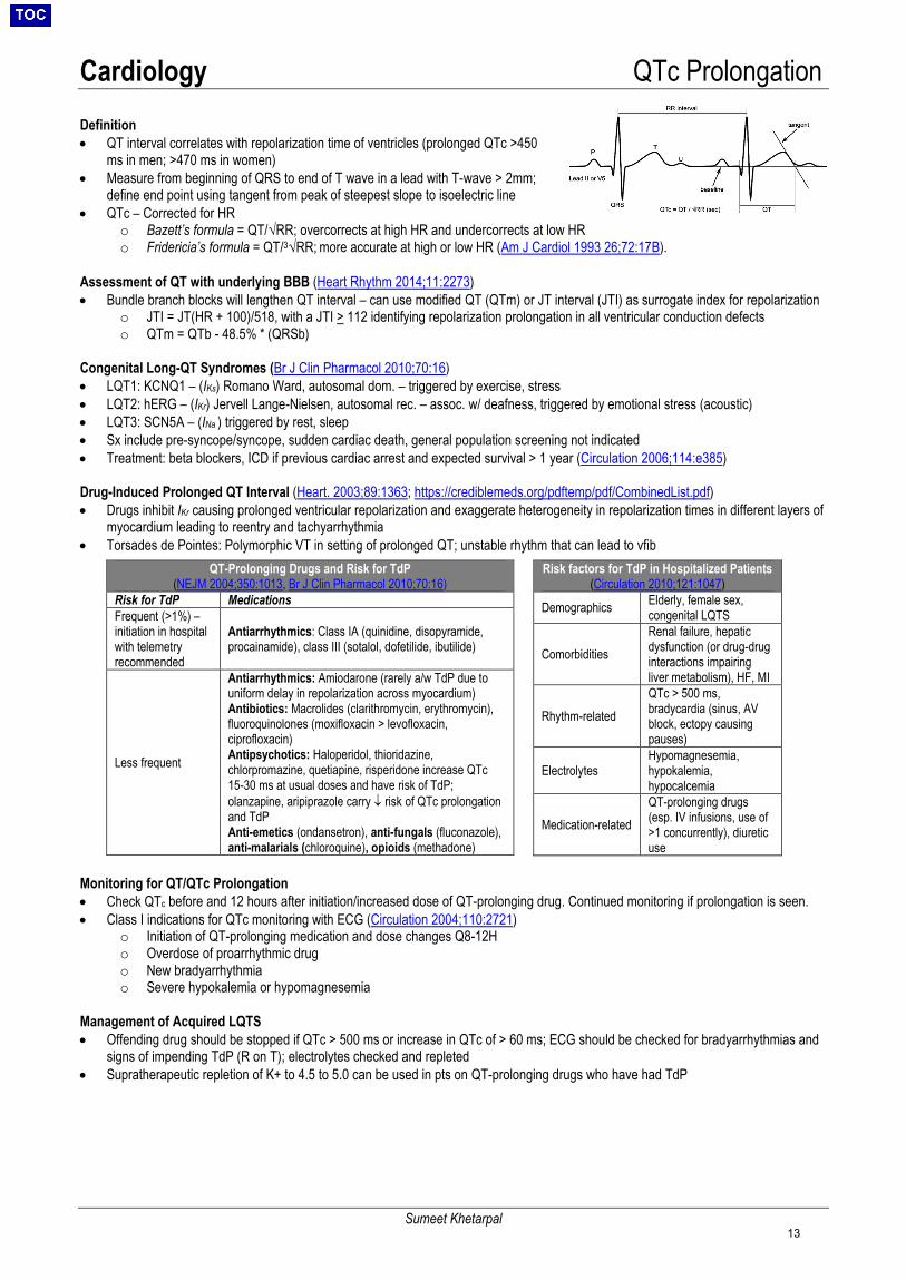
Definition • QT interval correlates with repolarization time of ventricles (prolonged QTc >450
ms in men; >470 ms in women)• Measure from beginning of QRS to end of T wave in a lead with T-wave > 2mm;
define end point using tangent from peak of steepest slope to isoelectric line• QTc – Corrected for HR
o Bazett’s formula = QT/√RR; overcorrects at high HR and undercorrects at low HRo Fridericia’s formula = QT/3√RR; more accurate at high or low HR (Am J Cardiol 1993 26;72:17B).
Assessment of QT with underlying BBB (Heart Rhythm 2014;11:2273)• Bundle branch blocks will lengthen QT interval – can use modified QT (QTm) or JT interval (JTI) as surrogate index for repolarization
o JTI = JT(HR + 100)/518, with a JTI > 112 identifying repolarization prolongation in all ventricular conduction defectso QTm = QTb - 48.5% * (QRSb)
Congenital Long-QT Syndromes (Br J Clin Pharmacol 2010;70:16)• LQT1: KCNQ1 – (IKs) Romano Ward, autosomal dom. – triggered by exercise, stress• LQT2: hERG – (IKr) Jervell Lange-Nielsen, autosomal rec. – assoc. w/ deafness, triggered by emotional stress (acoustic)• LQT3: SCN5A – (INa ) triggered by rest, sleep• Sx include pre-syncope/syncope, sudden cardiac death, general population screening not indicated• Treatment: beta blockers, ICD if previous cardiac arrest and expected survival > 1 year (Circulation 2006;114:e385)
Drug-Induced Prolonged QT Interval (Heart. 2003;89:1363; https://crediblemeds.org/pdftemp/pdf/CombinedList.pdf) • Drugs inhibit IKr causing prolonged ventricular repolarization and exaggerate heterogeneity in repolarization times in different layers of
myocardium leading to reentry and tachyarrhythmia• Torsades de Pointes: Polymorphic VT in setting of prolonged QT; unstable rhythm that can lead to vfib
Monitoring for QT/QTc Prolongation • Check QTc before and 12 hours after initiation/increased dose of QT-prolonging drug. Continued monitoring if prolongation is seen.• Class I indications for QTc monitoring with ECG (Circulation 2004;110:2721)
o Initiation of QT-prolonging medication and dose changes Q8-12Ho Overdose of proarrhythmic drugo New bradyarrhythmiao Severe hypokalemia or hypomagnesemia
Management of Acquired LQTS • Offending drug should be stopped if QTc > 500 ms or increase in QTc of > 60 ms; ECG should be checked for bradyarrhythmias and
signs of impending TdP (R on T); electrolytes checked and repleted• Supratherapeutic repletion of K+ to 4.5 to 5.0 can be used in pts on QT-prolonging drugs who have had TdP
QT-Prolonging Drugs and Risk for TdP (NEJM 2004;350:1013, Br J Clin Pharmacol 2010;70:16)
Risk for TdP Medications Frequent (>1%) – initiation in hospital with telemetry recommended
Antiarrhythmics: Class IA (quinidine, disopyramide, procainamide), class III (sotalol, dofetilide, ibutilide)
Less frequent
Antiarrhythmics: Amiodarone (rarely a/w TdP due to uniform delay in repolarization across myocardium) Antibiotics: Macrolides (clarithromycin, erythromycin), fluoroquinolones (moxifloxacin > levofloxacin, ciprofloxacin) Antipsychotics: Haloperidol, thioridazine, chlorpromazine, quetiapine, risperidone increase QTc 15-30 ms at usual doses and have risk of TdP;olanzapine, aripiprazole carry ↓ risk of QTc prolongationand TdPAnti-emetics (ondansetron), anti-fungals (fluconazole),anti-malarials (chloroquine), opioids (methadone)
Risk factors for TdP in Hospitalized Patients (Circulation 2010;121:1047)
Demographics Elderly, female sex, congenital LQTS
Comorbidities Renal failure, hepatic dysfunction (or drug-drug interactions impairing liver metabolism), HF, MI
Rhythm-related QTc > 500 ms, bradycardia (sinus, AV block, ectopy causing pauses)
Electrolytes Hypomagnesemia, hypokalemia, hypocalcemia
Medication-related QT-prolonging drugs (esp. IV infusions, use of >1 concurrently), diureticuse
13
Cardiology Chest Pain
David Olshan
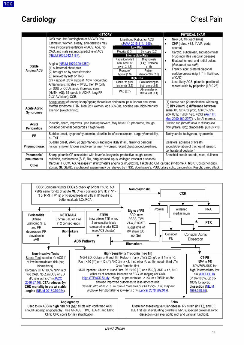
HISTORY PHYSICAL EXAM
Stable Angina/ACS
CVD risk: Use Framingham or ASCVD Risk Estimator. Women, elderly, and diabetics may have atypical presentations of ACS. Age, h/o CAD, and male sex most predictive of ACS (NEJM 2000;342:1187).
Angina (NEJM 1979:300:1350): (1) substernal chest pain(2) brought on by stress/exertion(3) relieved by rest or TNG3/3 = typical, 2/3 = atypical, 1/3 = noncardiacAntianginals: nitrates – 1st SL, then IV (onlyon SDU or CCU), avoid if preload sens(HoTN, AS); BB (avoid in ADHF, long PR,2˚/3˚ AV block); CCB.
Likelihood Ratios for ACS (JAMA 2015;314:1955).
Low Risk Pleuritic (0.3) Syncope (0.5)
Intermediate Risk Radiation to left
arm, neck, or jaw (1.3-1.5)
Diaphoresis (1.4); Exertional
(1.5) Pressure/
typical (1.9) Pattern
change/24h (2.0) High Risk
Similar to prior ischemia (2.2)
Pain radiating to both arms (2.6)
PAD (2.7) Abnormal prior stress test (3.1)
• New S4, MR (ischemia)• CHF (rales, +S3, ↑JVP, pedal
edema)• Carotid, subclavian, and abdominal
bruit (indicates vascular disease)• Bilateral femoral and radial pulses
(document pre-cath)• Frank’s sign: bilateral diagonal
earlobe crease (slight ↑ in likelihoodof CAD)
• Less likely ACS: pleuritic, positional,reproducible by palpation (LR 0.28)
Acute Aortic Syndromes
Abrupt onset of tearing/sharp/ripping thoracic or abdominal pain, known aneurysm, Marfan syndrome, HTN. Men 2x > women, age 60s-80s, cocaine use, high-intensity exertion (weight lifting).
(1) classic pain (2) mediastinal widening,(3) BP>20mmHg difference betweenarms: 0/3 Sx =7% prob, 1/3=31-39%,2/3= 83%; If ∆BP >20, >83% (Arch IntMed 2000;160:2977). √ for AI murmur.
Acute Pericarditis
Pleuritic, sharp, improves upon leaning forward. May have URI prodrome, though consider bacterial pericarditis if high fevers.
Friction rub (breath hold to distinguish from pleural rub); tamponade: pulsus >10.
PE Sudden onset, dyspnea/hypoxemia, pleuritic, hx of cancer/recent surgery/immobility, +/- TnT.
Tachycardia, tachypnea, hypoxemia
Pneumothorax Sudden onset, 20-40 yo (spontaneous and more likely if tall), family or personal history, smoker, known emphysema, men > women, recent chest procedures/lines.
Ipsilateral absence of breath sounds/deviation of trachea (if tension, contralateral deviation)
Pneumonia/ pneumonitis
Sharp, pleuritic CP associated with fever/leukocytosis, productive cough, recent radiation, autoimmune (SLE, RA, drug-induced lupus, collagen vascular diseases).
Bronchial breath sounds, rales, dullness
Other Cardiac: HOCM, AS, vasospasm (Prinzmetal’s angina or drug/toxin), Takotsubo CM, cardiac syndrome X; MSK: Costochondritis, Zoster; GI: GERD, esophageal spasm (may be relieved by TNG), Boerhaave’s, PUD, biliary colic, pancreatitis; Psych: panic attack
Pericarditis Diffuse
upsloping STE and PR
depression, PR elevation in
aVR
ECG: Compare w/prior ECGs & check q10-15m if susp, but <50% sens for dx of acute MI. Check posterior (if STD in V1-
3 or R>S in V1-2) or R-sided leads (if STE in II/III/avF) to better evaluate LCx/RCA
PTX
NSTEMI/UA 0.5mm STD or TWI in 2 consec leads
STEMI New ≥1mm STE in any
2 consecutive leads compared to prior ECG
(see ACS chapter)
Signs of PE RAD, new
RBBB, TWI V1-4, S1Q3T3 suggestive of RV strain (Sp,
not Sn)
CXR
Widened mediastinum
PNA
Consider PE
ACS Pathway
Biomarkers
Non-diagnostic
Normal
Consider Aortic Dissection
Angiography Used to r/o ACS in high risk pts (NB: all pts with confirmed ACS
should undergo angiography). Use GRACE, TIMI, HEART and Mayo Clinic CPC score for risk stratification.
High-Sensitivity Troponin (hs-cTn) MGH ED: Obtain at 0 and 1hr. Rules-in if any cTn ≥52 ng/L or if 1hr ∆ >5;
R/o if <10 (♀) or <12 (♂) AND 3hr ∆ <3. If no r/i or r/o at 1hr, obtain third cTn 3hrs from the first.
MGH Inpatient: Obtain at 0 and 3hrs. R/i if >10 (♀) or >15 (♂), AND ∆ >7, AND either sx of ischemia, ischemia on ECG, or imaging c/w CAD.
High-STEACS Study: ≥5 ng/L at presentation, ∆ ≥3, or >99%ile at 3hr showed improved outcomes vs less-strict criteria.
Caveat: intro of hs-cTn, w/ rule-in threshold of cTn ≥99% ULN, may not improve 1-yr mortality vs low-sens cTn (Lancet 2018;392:919).
Non-Invasive Tests Stress Test: used to r/o ACS if pt low-intermideiate risk (neg
biomarkers). Coronary CTA: 100% NPV in pt w/o CAD. No ∆ in LOS or ED
d/c rate vs hs-cTn (JACC 2016;67:16). CTA reduces 5yr CHD mortality in pts w/ stable angina (NEJM 2018;379:924).
Echo Useful for assessing valvular disease, RV strain (in PE), and EF.
TEE first test if evaluating prosthetic MV, suspected proximal aortic dissection (can eval aortic root and valvular function).
CT-PE NPV in PE
60%/89%/96% for high/ intermediate/ low
risk (PIOPED II).Sn 97-100%, Sp 83-100% for aortic dissection (NEJM 1993;328:35).
Biomarkers
14
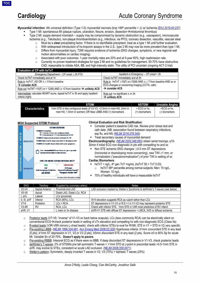
Cardiology Acute Coronary Syndrome
Anna O’Kelly, Leslie Chang, Cian McCarthy, Jonathon Salik
Myocardial infarction: 4th universal definition (Type 1-5): myocardial necrosis (trop >99th percentile + ∆) w/ ischemia (EHJ 2019;40:237) Type 1 MI: spontaneous MI (plaque rupture, ulceration, fissure, erosion, dissectionintraluminal thrombus) Type 2 MI: supply-demand mismatch – supply may be compromised by dynamic obstruction (e.g., vasospasm), microvascular
ischemia (e.g., Takotsubo), non-plaque thromboembolism (e.g., infectious, via PFO), coronary dissection, vasculitis, vascular stealo Must be a clear precipitating factor. If there is no identifiable precipitant, treat as a type 1 MI until further evaluation.o With widespread introduction of hs-troponin assays in the U.S., type 2 MI may now be more prevalent than type 1 MI.o Differs from myocardial injury. T2MI requires evidence of ischemia (EKG changes, symptoms, or new regional wall
motion abnormalities on cardiac imaging).o Associated with poor outcomes: 1-year mortality rates are 20% and at 5-year 60%, high readmission rate.o Currently no proven treatment strategies for type 2 MI and no guidelines for management. 50-70% have obstructive
CAD; reasonable to initiate ASA, BB, and high-intensity statin. The utility of PCI uncertain (ongoing ACT-2 trial)
STEMI NSTEMI Unstable Angina Characteristics 1mm STE in two contiguous leads (if V2-V3: >2.5mm in men<40, 2mm in
men>40, 1.5mm in women) OR New LBBB AND (+) biomarkers + ECG or hx,
(+) biomarkers+ECG or hx,
(-) biomarkers
MGH Suspected STEMI Protocol
EKG Territory Supplied by (coronary artery) Notes V2-V4 Septal-Anterior Proximal-mid LAD LAD occlusion implied by Wellen’s Syndrome & deWinter’s T-waves (see below) V5-V6 Apical Distal LAD, Distal LCx, RCA I, aVL Lateral LCx (proximal) II, III, aVF Inferior RCA (85%), LCx III>II elevation suggests RCA as culprit rather than LCxV7-9 Posterior LCx > RCA ST depressions in V1-V3 or R:S >1 in V1-V2 may represent posterior STE V3-4R RV RCA, LCx Check with inferior STE. 1mm STE in V4R most predictive of RV infarctaVR, V1 L main or 3v disease aVR>V1 STE with diffuse ST depressions = LMCA, 3VD or diffuse ischemia
o Posterior leads (V7-V9, “inverse” of V1-V3 on back below scapula); LCx (less commonly RCA) can be electrically silent onconventional ECGcheck posterior leads in setting of cTn elevation and compelling hx with non-diagnostic ECG (Class IIa)
o R-sided leads (V3R-V6R mirrors L-chest leads): check with inferior STEs to eval for RVMI; STE in V1 + STD in V2 very specifico Pre-existing LBBB: (NEJM 1996;334:481, Ann Emerg Med 2008;52:329) Sgarbossa criteria: ≥1mm concordant STE in any lead
(5 pts); ≥1mm ST depression in V1, V2 or V3 (3 pts); ≥5mm discordant STE in any lead (2 pts). Score ≥3 is 90% Sp for acuteMI. Variable Sn of 20-79%. Doesn’t apply to pacers.
o Pre-existing RBBB: Interpret ECG as if there were no BBB. If deep discordant ST depressions in V1-V3, check posterior leadso deWinter’s T-waves: 2% of STEMIs p/w tall symmetric T-waves + >1mm STD at J-point in precordial leads +0.5-1mm STE in
aVR, may evolve to STEs, consistent w/ acute LAD occlusion (NEJM 2008;359:2071)o Wellen’s pattern: Symmetric, deeply inverted T waves in V2, V3 (75%) > biphasic T waves (25%)
Evaluation of CP with hsTnT Emergency Department – CP onset ≥ 3h PTA Inpatient or Emergency – CP onset < 3h
Check hsTNT immediately and at 1h Check hsTNT immediately and at 3h Rule in: hsTnT ≥52 OR ∆ ≥ 5 from baseline consider ACS
Rule in: hsTnT ≥10(F) or≥15(M) AND ∆ ≥ 7 from baseline AND sx or ECG changes or concerning imaging (CCTA, cath) consider ACSRule out: hsTNT <10(F) or < 12(M) AND ∆ <3 from baseline unlikely ACS
Intermediate: calculate HEART score, repeat hsTnT in 3h and apply inpatient criteria (right)
Rule out: no significant ∆ in 3h unlikely ACS
Clinical Evaluation and Risk Stratification: Consider patient’s baseline CAD risk. Review prior stress test and
cath data. (NB: association found between respiratory infections,esp flu, and MI) (NEJM 2018;378:345)
Treat secondary causes of myocardial demandElectrocardiography: (NEJM 2003;348:993) obtain serial tracings; q15-30min if initial ECG non-diagnostic in pts with compelling hx and sx Non STE ischemic EKG changes: ≥0.5 mm ST depressions
(horizontal or downsloping more concerning), new TWI ≥1 mm, ornormalization (“pseudonormalization”) of prior TWI in setting of sx
Cardiac Biomarkers: hsTnT = ng/L, 4th gen TnT =ng/mL (hsTnT 50 = TnT 0.03)
o hsTnT 99th percentile among normal subjects: Men: 15 ng/L,Women: 10 ng/L
75% of healthy individuals will have a measurable hsTnT
15
Cardiology Acute Coronary Syndrome
Anna O’Kelly, Leslie Chang, Cian McCarthy, Jonathon Salik
o Wellen’s syndrome: (Am Heart J 1982;103:730) Wellen’s pattern in pts presenting w/ CP w/ resolutionindicates reperfusion ofmyocardium indicative of LM or proximal LAD stenosis. 75% of pts will have ant MI in days-weeks if not treated. Proceed to cathlab w/o stress testing. DDx: apical HCM, elevated ICP (w/ long QTc & brady), MI, PE, post-tachy/pacing, BBB, WPW, idiopathic
Risk Stratification for PCI Timing in NSTEMI/UA: Multiple risk models: GRACE, TIMI, PURSUIT, AMIS GRACE score, based on predictors of 6 mo mortality in pts w/ ACS – age, HR, SBP, Cr, cardiac arrest ?, ST deviation, elevated
trop (BMJ 2006;333:1091) 4 subgroups for urgency to get to the cath lab (JACC 2014;64:e139) Very high risk (“immediate invasive,” within 2 hrs): Refractory/recurrent angina, hemodynamic or electrical instability High risk (“early invasive,” within 24 hrs): temporal change in troponin, EKG changes (STD, TWI), high risk pt (GRACE>140)
Conflicting results between TIMAC (NEJM 2009;360:2165) and VERDICT (Circ 2018;138:2741) trials about outcome benefitof early cath. However both show improved outcomes with early cath in patients with GRACE >140.
Intermediate risk (“delayed invasive,” within 72 hrs): none of above but risk factors at baseline (ie EF <40%, GFR <60) Low risk (“ischemia guided,” no cath): no risk factors, GRACE <109, TIMI 0-1
Therapeutic Interventions: Thrombolytic indications: STEMI only (worse outcomes in NSTEMI), w/ angina >30min and <12h (no benefit after 12h), when
projected time to PCI>120min. Rescue PCI indicated for failed lysis of STEMI (persistent symptoms, STE <50% resolved at 60minpost-lysis) (NEJM 2005;353:2758)
PCI Indications: Recommended over fibrinolysis when at a PCI-capable center (1A). PCI with 20% lower rate of combined cardiacendpoint, 65% ↓ rate CVA vs. lysis when performed at high-volume (>400/yr) center (JAMA 1997;278:2093, Lancet 2003;361:13) STEMI: PCI if <12h sx onset, goal to PCI ideally <60min at PCI centers. PCI regardless of time from onset for cardiogenic shock,
malignant arrhythmia, persistent STE and/or CP. Late PCI (>48h post-event) generally not indicated in stable pts. (NEJM2006;355:2395)
NSTEMI/UA: See Risk Stratification in NSTEMI/UA above above PCI Strategies:
Primary stenting (culprit lesion): current standard of care: radial > fem access, DES > BMS (NEJM 2016;375:1242) Non-culprit lesions: Pts with MI + cardiogenic shock had decreased risk of death/RRT with culprit-lesion-only PCI than immediate
multi-vessel PCI (NEJM 2018; 379:1699). Consider PCI of non-infarct artery in STEMI prior to hospital discharge, data showsreduction in composite endpoints (NEJM 2013;69:1115, NEJM 2017;373:13, JACC 2015;65;963,), data on NSTE-ACS less clear(JACC 2016;67:3).
Chronic Total Occlusions (CTO): PCI only if severe ischemia despite OMT with non-3VD equivalent and viable tissue (Eur Heart J2018;39:2484), no difference in LVEF at 4 months in STEMI non-culprit CTO intervention (JACC 2016;68:1622).
CABG: preferred for 3VD (NEJM 2009;360:961, NEJM 2008;358:331), left main disease (similar composite outcome with PCI butreduced future PCI) (Lancet 2016;388:2743, NEJM 2016;375:2223), 2VD with proximal LAD stenosis or EF<50%; large area of viablemyocardium or high risk (all class I). Consider if DM + 2VD (NEJM 2012;367:2375)
Adjuncts to Reperfusion Therapy: ASA: Established mortality benefit, give to all patients in an immediate load/maintenance (325mg/81mg) strategy (Lancet 1988;2:8607) P2Y12 Inhibitors: (NB: Controversial if pre-cath load is beneficial, may delay CABG by 5-7 days. Consult with fellow before loading).
• Ticagrelor: Decreased mortality compared to clopidogrel w/o increasing major bleeding. Not a prodrug, reversible with platelettxfsn. Common side effect is mild dyspnea on initiation. Avoid in liver disease, previous stroke, oral AC (NEJM 2009; 361:1045)
• Prasugrel: Prodrug, lower rates of MI and stent thrombosis but ↑ bleeding compared to clopidogrel in PCI (NEJM 2007; 357:20) Ifmedical mgmt. only for UA/NSTEMI, no difference in outcome compared to clopidogrel (NEJM 2012; 367:1297) Contraindicated ifprior TIA/CVA, wt<60 kg, or >75 yo.
• Clopidogrel: Reduces neg outcomes (death+repeat MI) when load/maintenance w/ PCI (Lancet 2001;358:527I) or fibrinolysis(NEJM 2005;352:1179). Prodrug, metabolized by CYP219, less effective in those with LOF allele (NEJM 2009;360:354)
• Cangrelor: IV reversible inhibitor w/ immediate onset and return of nl plt function in 1h. Improved outcomes compared toclopidogrel (NEJM2013;368:14)
TNG: SL x 3, transition to gtt if refractory CP. Nitropaste and gtt have shorter half-life than SL if c/f hypotension. No mortality benefit;caution in IMI/RVMI, SBP<100 or PDE inhibitor use in last 48h. If continued CP despite ↑ dose of TNG, indication for earlier cath.
Anticoagulation:• UFH: trend towards mortality benefit in meta-analyses, optimal duration undefined. Usually stopped after 48h if ECG changes
improving and concern for ongoing ischemia resolved (BMJ 1996;313:652) Start gtt w/o bolus if giving lytics or if on coumadin andINR<2 (hold if INR>2). Use low intensity PTT goal (63-83s).
• LWMH: possible reduction in death w/ min evidence for ↑ major bleeding, trials vs. UFH largely null (IIa) (BMJ 2012; 344:e553)• Fondaparinux: preferred to UFH/LMWH if medically managed. Contraindicated in PCI 2/2 ↑ catheter thrombosis/complications
(JAMA 2006; 295:1519)• IIb/IIIa Inhibitors: Eptifibatide (Integrilin) used at MGH. Consider at PCI if high-risk (extensive thrombus). Usually initiated in cath
lab as upstream tx provides no mortality benefit and ↑ bleeding risk. Can consider in place of P2Y12 inhibitor if possible CABG(NEJM 2009;360:2176)
16
Cardiology Acute Coronary Syndrome
Anna O’Kelly, Leslie Chang, Cian McCarthy, Jonathon Salik
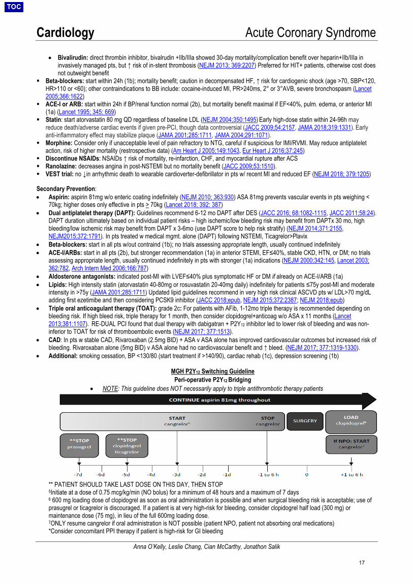
• Bivalirudin: direct thrombin inhibitor, bivalrudin +IIb/IIIa showed 30-day mortality/complication benefit over heparin+IIb/IIIa ininvasively managed pts, but ↑ risk of in-stent thrombosis (NEJM 2013; 369:2207) Preferred for HIT+ patients, otherwise cost doesnot outweight benefit
Beta-blockers: start within 24h (1b); mortality benefit; caution in decompensated HF, ↑ risk for cardiogenic shock (age >70, SBP<120,HR>110 or <60); other contraindications to BB include: cocaine-induced MI, PR>240ms, 2° or 3°AVB, severe bronchospasm (Lancet2005;366:1622)
ACE-I or ARB: start within 24h if BP/renal function normal (2b), but mortality benefit maximal if EF<40%, pulm. edema, or anterior MI(1a) (Lancet 1995; 345: 669)
Statin: start atorvastatin 80 mg QD regardless of baseline LDL (NEJM 2004;350:1495) Early high-dose statin within 24-96h mayreduce death/adverse cardiac events if given pre-PCI, though data controversial (JACC 2009;54:2157, JAMA 2018;319:1331). Earlyanti-inflammatory effect may stabilize plaque (JAMA 2001;285:1711, JAMA 2004;291:1071).
Morphine: Consider only if unacceptable level of pain refractory to NTG, careful if suspicious for IMI/RVMI. May reduce antiplateletaction, risk of higher mortality (restrospective data) (Am Heart J 2005;149:1043, Eur Heart J 2016;37:245)
Discontinue NSAIDs: NSAIDs ↑ risk of mortality, re-infarction, CHF, and myocardial rupture after ACS Ranolazine: decreases angina in post-NSTEMI but no mortality benefit (JACC 2009;53:1510). VEST trial: no ↓in arrhythmic death to wearable cardioverter-defibrillator in pts w/ recent MI and reduced EF (NEJM 2018; 379:1205)
Secondary Prevention: • Aspirin: aspirin 81mg w/o enteric coating indefinitely (NEJM 2010; 363:930) ASA 81mg prevents vascular events in pts weighing <
70kg; higher doses only effective in pts > 70kg (Lancet 2018; 392: 387)• Dual antiplatelet therapy (DAPT): Guidelines recommend 6-12 mo DAPT after DES (JACC 2016; 68:1082-1115, JACC 2011;58:24).
DAPT duration ultimately based on individual patient risks – high ischemic/low bleeding risk may benefit from DAPTx 30 mo, highbleeding/low ischemic risk may benefit from DAPT x 3-6mo (use DAPT score to help risk stratify) (NEJM 2014;371:2155,NEJM2015;372:1791). In pts treated w medical mgmt. alone (DAPT) following NSTEMI, Ticagrelor>Plavix
• Beta-blockers: start in all pts w/out contraind (1b); no trials assessing appropriate length, usually continued indefinitely• ACE-I/ARBs: start in all pts (2b), but stronger recommendation (1a) in anterior STEMI, EF≤40%, stable CKD, HTN, or DM; no trials
assessing appropriate length, usually continued indefinitely in pts with stronger (1a) indications (NEJM 2000;342:145, Lancet 2003;362:782, Arch Intern Med 2006;166:787)
• Aldosterone antagonists: indicated post-MI with LVEF≤40% plus symptomatic HF or DM if already on ACE-I/ARB (1a)• Lipids: High intensity statin (atorvastatin 40-80mg or rosuvastatin 20-40mg daily) indefinitely for patients ≤75y post-MI and moderate
intensity in >75y (JAMA 2001;285:1711) Updated lipid guidelines recommend in very high risk clinical ASCVD pts w/ LDL>70 mg/dLadding first ezetimibe and then considering PCSK9 inhibitor (JACC 2018;epub, NEJM 2015;372:2387; NEJM 2018;epub)
• Triple oral anticoagulant therapy (TOAT): grade 2c: For patients with AFib, 1-12mo triple therapy is recommended depending onbleeding risk. If high bleed risk, triple therapy for 1 month, then consider clopidogrel+anticoag w/o ASA x 11 months (Lancet2013;381:1107). RE-DUAL PCI found that dual therapy with dabigatran + P2Y12 inhibitor led to lower risk of bleeding and was non-inferior to TOAT for risk of thromboembolic events (NEJM 2017; 377:1513).
• CAD: In pts w stable CAD, Rivaroxaban (2.5mg BID) + ASA v ASA alone has improved cardiovascular outcomes but increased risk ofbleeding. Rivaroxaban alone (5mg BID) v ASA alone had no cardiovascular benefit and ↑ bleed. (NEJM 2017; 377:1319-1330).
• Additional: smoking cessation, BP <130/80 (start treatment if >140/90), cardiac rehab (1c), depression screening (1b)
MGH P2Y12 Switching Guideline Peri-operative P2Y12 Bridging
• NOTE: This guideline does NOT necessarily apply to triple antithrombotic therapy patients
** PATIENT SHOULD TAKE LAST DOSE ON THIS DAY, THEN STOP 5Initiate at a dose of 0.75 mcg/kg/min (NO bolus) for a minimum of 48 hours and a maximum of 7 days 6 600 mg loading dose of clopidogrel as soon as oral administration is possible and when surgical bleeding risk is acceptable; use of prasugrel or ticagrelor is discouraged. If a patient is at very high-risk for bleeding, consider clopidogrel half load (300 mg) or maintenance dose (75 mg), in lieu of the full 600mg loading dose. 7ONLY resume cangrelor if oral administration is NOT possible (patient NPO, patient not absorbing oral medications) *Consider concomitant PPI therapy if patient is high-risk for GI bleeding
17
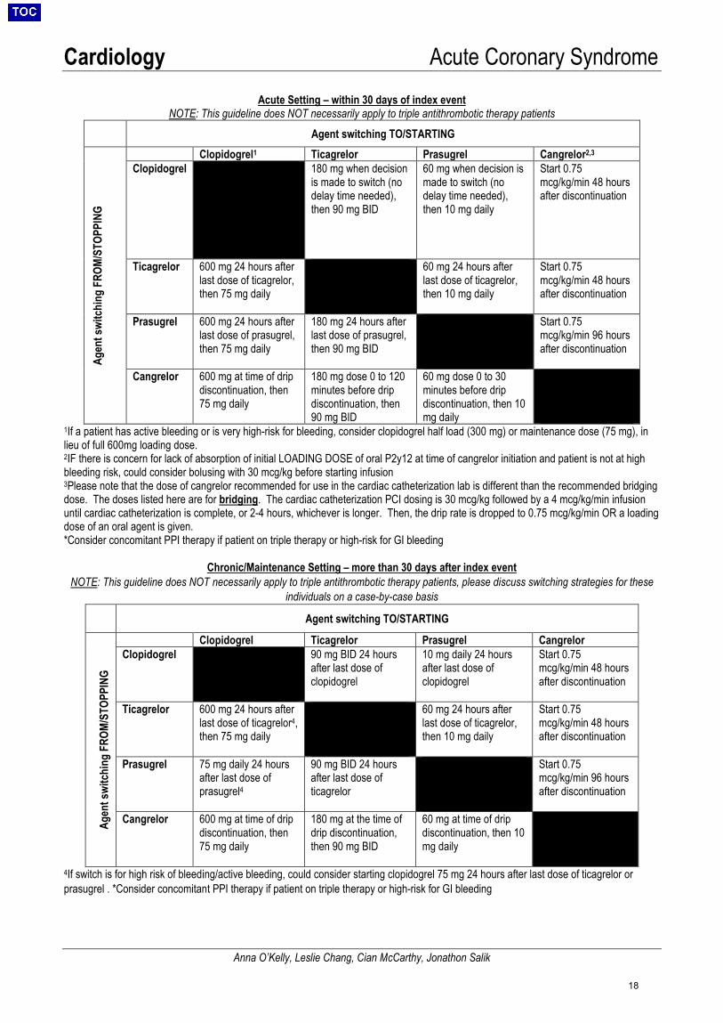
Cardiology Acute Coronary Syndrome
Anna O’Kelly, Leslie Chang, Cian McCarthy, Jonathon Salik
Acute Setting – within 30 days of index event NOTE: This guideline does NOT necessarily apply to triple antithrombotic therapy patients
Agent switching TO/STARTING
Agen
t swi
tchi
ng F
ROM/
STOP
PING
Clopidogrel1 Ticagrelor Prasugrel Cangrelor2,3
Clopidogrel 180 mg when decision is made to switch (no delay time needed), then 90 mg BID
60 mg when decision is made to switch (no delay time needed), then 10 mg daily
Start 0.75 mcg/kg/min 48 hours after discontinuation
Ticagrelor 600 mg 24 hours after last dose of ticagrelor, then 75 mg daily
60 mg 24 hours after last dose of ticagrelor, then 10 mg daily
Start 0.75 mcg/kg/min 48 hours after discontinuation
Prasugrel 600 mg 24 hours after last dose of prasugrel, then 75 mg daily
180 mg 24 hours after last dose of prasugrel, then 90 mg BID
Start 0.75 mcg/kg/min 96 hours after discontinuation
Cangrelor 600 mg at time of drip discontinuation, then 75 mg daily
180 mg dose 0 to 120 minutes before drip discontinuation, then 90 mg BID
60 mg dose 0 to 30 minutes before drip discontinuation, then 10 mg daily
1If a patient has active bleeding or is very high-risk for bleeding, consider clopidogrel half load (300 mg) or maintenance dose (75 mg), in lieu of full 600mg loading dose. 2IF there is concern for lack of absorption of initial LOADING DOSE of oral P2y12 at time of cangrelor initiation and patient is not at high bleeding risk, could consider bolusing with 30 mcg/kg before starting infusion 3Please note that the dose of cangrelor recommended for use in the cardiac catheterization lab is different than the recommended bridging dose. The doses listed here are for bridging. The cardiac catheterization PCI dosing is 30 mcg/kg followed by a 4 mcg/kg/min infusion until cardiac catheterization is complete, or 2-4 hours, whichever is longer. Then, the drip rate is dropped to 0.75 mcg/kg/min OR a loading dose of an oral agent is given. *Consider concomitant PPI therapy if patient on triple therapy or high-risk for GI bleeding
Chronic/Maintenance Setting – more than 30 days after index event NOTE: This guideline does NOT necessarily apply to triple antithrombotic therapy patients, please discuss switching strategies for these
individuals on a case-by-case basis
Agent switching TO/STARTING
Agen
t swi
tchi
ng F
ROM/
STOP
PING
Clopidogrel Ticagrelor Prasugrel Cangrelor Clopidogrel 90 mg BID 24 hours
after last dose of clopidogrel
10 mg daily 24 hours after last dose of clopidogrel
Start 0.75 mcg/kg/min 48 hours after discontinuation
Ticagrelor 600 mg 24 hours after last dose of ticagrelor4,then 75 mg daily
60 mg 24 hours after last dose of ticagrelor, then 10 mg daily
Start 0.75 mcg/kg/min 48 hours after discontinuation
Prasugrel 75 mg daily 24 hours after last dose of prasugrel4
90 mg BID 24 hours after last dose of ticagrelor
Start 0.75 mcg/kg/min 96 hours after discontinuation
Cangrelor 600 mg at time of drip discontinuation, then 75 mg daily
180 mg at the time of drip discontinuation, then 90 mg BID
60 mg at time of drip discontinuation, then 10 mg daily
4If switch is for high risk of bleeding/active bleeding, could consider starting clopidogrel 75 mg 24 hours after last dose of ticagrelor or prasugrel . *Consider concomitant PPI therapy if patient on triple therapy or high-risk for GI bleeding
18
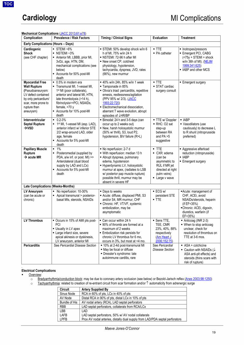
Cardiology MI Complications
Maeve Jones-O’Connor
Mechanical Complications (JACC 2013;61:e78) Complication Prevalence / Risk Factors Timing / Clinical Signs Evaluation Treatment Early Complications (Hours – Days) Cardiogenic Shock (see CHF chapter)
STEMI ~6% NSTEMI ~3% Anterior MI, LBBB, prior MI,
3vDz, age, HTN, DM,mechanical complications (seebelow)
Accounts for 50% post-MIdeath
STEMI: 50% develop shock w/in 6h of MI, 75% w/in 24 h NSTEMI: 72-96 h after MI New onset CP, cold/wet
physiology, hypotension,tachycardia, dyspnea, JVD, rales(66%), new murmur
TTE PA catheter
Inotropes/pressors Emergent PCI, CABG
(<75y + STEMI + shockw/in 36h of MI). (NEJM1999;341:625) IABP and other MCS
Myocardial Free Wall Rupture (Pseudoaneurysm: LV defect contained by only pericardium/ scar, more prone to rupture than aneurysm)
0.5% in modern era Transmural MI, 1-vessel MI,
1st MI (poor collaterals),anterior and lateral MI, HTN,late thrombolysis (>14 h),fibrinolysis>>PCI, NSAIDs,female, >70 y
Accounts for 10% post-MIdeath
40% w/in 24h, 85% w/in 1 week Tamponade in 85% Olivia’s triad: pericarditis, repetitive
emesis, restlessness/agitation(PPV 95% w/ 2/3). (JACC1993;22:720) Electromechanical dissociation,
aberrant T wave evolution, abruptepisodes of ↓HR/BP
TTE STAT cardiac
surgery consult
Emergent surgery
Interventricular Septal Rupture VSD
0.2-3% 1st MI, 1-vessel MI (esp. LAD),
anterior infarct w/ inferior STE2/2 wrap-around LAD, older age, female
Accounts for 5% post-MIdeath
Bimodal: 24 h and 3-5 days (can occur up to 2 weeks out) New, harsh holosystolic murmur
(50% w/ thrill), S3, loud P2,hypotension, BiV failure (R>L)
TTE w/ Doppler RHC: O2 sat
step-upbetween RAand PA >5suggestive
IABP Vasodilators (use
cautiously) to decrease Lto R shunt (nitroprussidepreferred)
Papillary Muscle Rupture acute MR
1% Posteromedial (supplied by
PDA, a/w inf. or post. MI) >>Anterolateral (dual bloodsupply by LAD and LCx)
Accounts for 5% post-MIdeath
No reperfusion: 2-7 d With reperfusion: median 13 h Abrupt dyspnea, pulmonary
edema, hypotension Hyperdynamic LV, holosystolic
murmur at apex, (radiates to LSBw/ posterior pap muscle rupture) possible thrill, murmur may beabsent in severe HF
TTE CXR: edema
(can beasymmetric toRUL if MR jetdirected at rightpulm veins)
Large v wave
Aggressive afterloadreduction (nitroprusside) IABP Emergent surgery
Late Complications (Weeks-Months) LV Aneurysm (can be acute or chronic)
No reperfusion: 10-30% Apical transmural > posterior-
basal MIs, steroids, NSAIDs
Days to weeks Acute: diffuse, displaced PMI, S3
and/or S4, MR murmur, CHF Chronic: HF, VT/VF, systemic
embolization, may beasymptomatic
ECG w/persistent STE TTE
Acute: management ofCHF, ACEi, avoidNSAIDs/steroids, heparin(if EF<35%) Chronic: ACEi, digoxin,
diuretics, warfarin (ifEF<35%)
LV Thrombus Occurs in 15% of AMI pts post-PCI Usually in LV apex Large infarct size, severe
apical akinesis or dyskinesis,LV aneurysm, anterior MI
Can occur within 24 h 90% of thrombi are formed at a
maximum of 2 weeks Embolization risk persists for
chronic LV thrombus for 6 mo,occurs in 3%, but most at <4 mo.
Sens TTE,TEE, CMR23%, 40%, 88% respectively(Am Heart J2006;152:75)
Anticoag (INR 2-3) When to stop anticoag
unclear, check for resolution of thrombus onTTE at 3-6 mos.
Pericarditis See Pericardial Disease Section 10% at 2-4d post-transmural MI May be focal or diffuse Dressler’s syndrome: late
autoimmune carditis, rare
See Pericardial Disease Section
ASA + colchicine Caution with NSAIDs (↓
ASA anti-plt effects) andsteroids (thins scars withrisk of rupture)
. Electrical Complications Overview:
o Bradyarrhythmia/conduction block: may be due to coronary artery occlusion (see below) or Bezold-Jarisch reflex (Anes 2003;98:1250)o Tachyarrhythmia: related to creation of re-entrant circuit from scar formation and/or ↑ automaticity from adrenergic surge
Circuit Artery Supplied By Sinus Node RCA in 60% of pts, LCx in 40% of pts AV Node Distal RCA in 90% of pts, distal LCx in 10% of pts Bundle of His AV nodal artery (RCA), LAD septal perforators RBB LAD septal perforators, collaterals from RCA/LCx LBB LAFB LPFB
LAD LAD septal perforators, 50% w/ AV nodal collaterals Prox AV nodal arteries, distally dual supply from LAD/PDA septal perforators
19
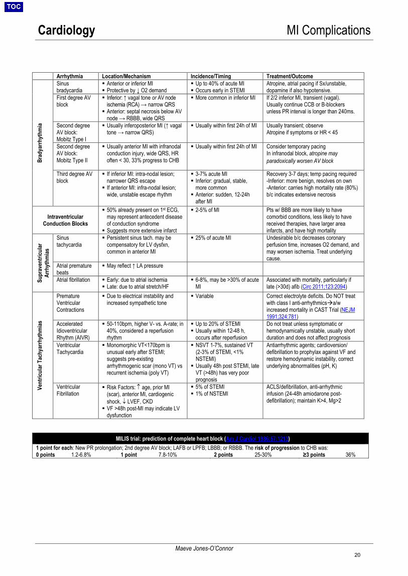
Cardiology MI Complications
Maeve Jones-O’Connor
Arrhythmia Location/Mechanism Incidence/Timing Treatment/Outcome
Brad
yarrh
ythm
ia
Sinus bradycardia
Anterior or inferior MI Protective by ↓ O2 demand
Up to 40% of acute MI Occurs early in STEMI
Atropine, atrial pacing if Sx/unstable, dopamine if also hypotensive.
First degree AV block
Inferior: ↑ vagal tone or AV node ischemia (RCA) → narrow QRS Anterior: septal necrosis below AV
node → RBBB, wide QRS
More common in inferior MI If 2/2 inferior MI, transient (vagal). Usually continue CCB or B-blockers unless PR interval is longer than 240ms.
Second degree AV block: Mobitz Type I
Usually inferoposterior MI (↑ vagaltone → narrow QRS)
Usually within first 24h of MI Usually transient; observe Atropine if symptoms or HR < 45
Second degree AV block: Mobitz Type II
Usually anterior MI with infranodalconduction injury, wide QRS, HR often < 30, 33% progress to CHB
Usually within first 24h of MI Consider temporary pacing In infranodal block, atropine may paradoxically worsen AV block
Third degree AV block
If inferior MI: intra-nodal lesion;narrower QRS escape If anterior MI: infra-nodal lesion;
wide, unstable escape rhythm
3-7% acute MI Inferior: gradual, stable,
more common Anterior: sudden, 12-24h
after MI
Recovery 3-7 days; temp pacing required -Inferior: more benign, resolves on own-Anterior: carries high mortality rate (80%) b/c indicates extensive necrosis
Intraventricular Conduction Blocks
50% already present on 1st ECG,may represent antecedent diseaseof conduction syndrome Suggests more extensive infarct
2-5% of MI Pts w/ BBB are more likely to have comorbid conditions, less likely to have received therapies, have larger area infarcts, and have high mortality
Supr
aven
tricu
lar
Arrh
ythm
ias
Sinus tachycardia
Persistent sinus tach. may becompensatory for LV dysfxn,common in anterior MI
25% of acute MI Undesirable b/c decreases coronary perfusion time, increases O2 demand, and may worsen ischemia. Treat underlying cause.
Atrial premature beats
May reflect ↑ LA pressure
Atrial fibrillation Early: due to atrial ischemia Late: due to atrial stretch/HF
6-8%, may be >30% of acuteMI
Associated with mortality, particularly if late (>30d) afib (Circ 2011;123:2094)
Vent
ricul
ar T
achy
arrh
ythm
ias
Premature Ventricular Contractions
Due to electrical instability andincreased sympathetic tone
Variable Correct electrolyte deficits. Do NOT treat with class I anti-arrhythmicsa/w increased mortality in CAST Trial (NEJM 1991;324:781)
Accelerated Idioventricular Rhythm (AIVR)
50-110bpm, higher V- vs. A-rate; in40%, considered a reperfusionrhythm
Up to 20% of STEMI Usually within 12-48 h,
occurs after reperfusion
Do not treat unless symptomatic or hemodynamically unstable, usually short duration and does not affect prognosis
Ventricular Tachycardia
Monomorphic VT<170bpm isunusual early after STEMI;suggests pre-existingarrhythmogenic scar (mono VT) vsrecurrent ischemia (poly VT)
NSVT 1-7%, sustained VT(2-3% of STEMI, <1% NSTEMI) Usually 48h post STEMI, late
VT (>48h) has very poor prognosis
Antiarrhythmic agents; cardioversion/ defibrillation to prophylax against VF and restore hemodynamic instability, correct underlying abnormalities (pH, K)
Ventricular Fibrillation
Risk Factors: ↑ age, prior MI(scar), anterior MI, cardiogenicshock, ↓ LVEF, CKD VF >48h post-MI may indicate LV
dysfunction
5% of STEMI 1% of NSTEMI
ACLS/defibrillation, anti-arrhythmic infusion (24-48h amiodarone post-defibrillation); maintain K>4, Mg>2
MILIS trial: prediction of complete heart block (Am J Cardiol 1986;57:1213)1 point for each: New PR prolongation; 2nd degree AV block; LAFB or LPFB; LBBB; or RBBB. The risk of progression to CHB was: 0 points 1.2-6.8% 1 point 7.8-10% 2 points 25-30% ≥3 points 36%
20

Cardiology Cardiac Catherization & Coronary Anatomy
Shawn Li, Sumeet Khetarpal
Anatomy LCA and RCA w/ their branches create two rings around the heart: RCA + LCX in AV plane; LAD + PDA in IV plane (see above) 80% of PDA arises from RCA (right dominant), thus inferior MI more likely from RCA lesion
Preparation for Catheterization NPO pMN; INR<2; monitor Cr and eGFR closely pre-procedure, no ppx ABX. Continue ASA, statin, BB, heparin. Hold metformin
(usually 1 day pre-proc, 2 days post-proc). May need to hold or delay initiating ACE-I. Document bilateral radial, femoral, popliteal, DP pulses, and Allen’s test prior to cath. Note bruit, hx of: HIT, PVD, Ao
aneurysm/dissection Pre-hydration w/ crystalloids and NAC/Bicarb have not shown to prevent CIN in most patients with moderate CKD (Lancet 2017;
389:1312; NEJM 2018; 378: 603); CIN risk calculator; Diagnostic cath = 25 cc contrast (CT-PE = 190 cc) Contrast allergy: Pre-treatment with steroids, H1, and/or H2 blockers if patient has documented allergy, see MGH 13h protocol.
Consult allergy service for expedited protocol if the cath is required emergently. Respiratory distress: Patient will need to lie flat; consider intubation if prohibitive hypoxemia/pulmonary edema
Percutaneous Coronary Intervention Considerations Access: Fewer bleeding/vascular complications if radial (vs. femoral), possible decreased death in ACS. (JACC 2018;71:1167) BMS vs. DES: DES have↓ in-stent thrombosis → subsequent ↓ revascularization; however, ↑ risk of late stent re-stenosis →
requires longer duration of DAPT Contraindications to stents: predicted DAPT non-adherence, anticipated major surgery within treatment time, elevated bleeding risk Antiplatelet Tx: ASA indefinitely (JACC 2016;03.513)
o No high bleeding risk:▫ BMS: add P2Y12 inhibitor (“DAPT”) for at least 1 month (stable ischemic heart disease), 12 months (ACS)▫ DES: add P2Y12 inhibitor (“DAPT”) for at least 6 months (stabile ischemic heart disease), 12 months (ACS)
o High bleeding risk:▫ BMS: add P2Y12 inhibitor (“DAPT”) for at least 1 month (stable ischemic heart disease), 6 months (ACS)▫ DES: add P2Y12 inhibitor (“DAPT”) for at least 3 months (stabile ischemic heart disease), 6 months (ACS)
o Triple therapy is generally tailored based on individual patient risk and de-escalated as soon as possible to dual therapy (NEJM2016;375:2423, NEJM 2017;377:1513)
Post-Procedure Care Groin access: 4-6 hrs bedrest after procedure. Closure devices decrease time needed for bedrest.
o Groin checks immediately post- and 6h, 8h post-procedure: check b/l pulses, palpate for pulsatile masses, auscultate forbruits
o Sheaths: during pass-off, ask interventional fellow about timing of arterial removal; only cardiology fellows remove sheaths Radial access: TR band x 4-6h
Post-Catherization Complications Access site complications: always inform the interventional fellow who performed the procedure, diagnose by U/S and exam
o Hematoma: Mass w/out bruit. Apply compression. If unable to control, may require Fem-Stop device to apply external pressure.o Pseudoaneurysm: presents as pulsatile mass with bruit at access site. Treat with compression if <2 cm, may require thrombin
injection or surgery if >2 cm. Urgent U/S and vascular surgery consult.o AV fistula: Presents as continuous bruit with no mass. Evaluate w/ U/S. Surgical repair is usually necessary.o Limb ischemia: From thrombus, dissection, or malpositioned closure device. Evaluate pulses, limb warmth, and PVRs.o Retroperitoneal bleed: presents within hours post-cath, often with hemodynamic instability +/- flank pain +/- ecchymoses. STAT
CT A/P if stable. Transfuse, IV fluids, discussion with attending re: stopping/reversing anticoagulation. Non-access complications:
o Infection: more common in setting of vascular closure deviceso Atheroembolism: eosinophilia; livedo reticularis; blue toes; mesenteric ischemia; acute, subacute, or chronic renal dysfunctiono CIN: peak ↑Cr 2-5d post contrast load, risk correlated with contrast load and initial GFRo Tamponade: post-cath hypotension from coronary or cardiac perforation. Check pulsus paradoxus (SBP Δ >10mmHg w/
inspiration), STAT TTE, alert cath fellow. Give IVF.o MI/CVA: due to in-stent thrombosis (MI) or distal embolization post-cath (CVA). Discuss all CP/neuro changes with cath fellow
21

Cardiology Non-Invasive Cardiac Testing
Sean Mendez
General Considerations • Indications:
Diagnose CAD: Sx of stable angina in patients w/ intermediate or high risk of CAD. Not indicated for low risk or asymp. pts. Known CAD: Stratify prognosis in new or changing sx c/f ischemia or post-MI prior to discharge (i.e., submaximal stress) Post-revascularization: Pts w/ angina; asymptomatic pt if incomplete revasc or >2 years post-PCI / >5 years post-CABG Pre-op risk assessment: Not routinely indicated; no evidence that revasc ↓ mortality or post-op MI for non-cardiac surgery.
(NEJM 2004;351:2795) Other: Newly dx HF/cardiomyopathy likely due to ischemia; functional capacity (for exercise prescription); viability testing
• Contraindications: untreated ACS, MI within 2d, high-risk or LM CAD, uncontrolled arrhythmia, acute CHF, severe AS or HOCM,recent DVT/PE, acute myo-/peri-/endocarditis, aortic dissection, uncontrolled HTN
• Patient Preparation: NPO 3h prior, longer if imaging oradenosine. Must reverse DNR/DNI for the test. If the question is “Does this patient have CAD?
hold BB and nitrates If the question is “How well are meds working in
known CAD?” continue BB and nitrates Hold caffeine >12h for adenosine. Hold BB >24h for dobutamine (>48h for atenolol).
• Caveats: Majority of vulnerable plaques are angiographically insignificant (<70% stenosis)stress testing unable to identify the presence
of these plaques (CTA more sensitive) Angiographically significant (>70% stenosis) 3vD may produce false-negative vasodilator stress test”balanced ischemia”
• Positive Test Results: Optimize medical tx. Decision re: angiography/revascularization varies by patient (degree of sx, known stenosis, current meds).
Schematic Approach to Noninvasive Cardiac Testing (adapted from UpToDate)
Stress Modality Imaging Modalities Exercise (treadmill) EKG, TTE, SPECT Vasodilator (adenosine, regadenoson) TTE, SPECT, PET, MRI Inotropy (dobutamine) TTE, SPECT, PET, MRI Ph
arm
22
Cardiology Non-Invasive Cardiac Testing
Sean Mendez
Exercise Tolerance Test (ETT) EKG or imaging (TTE, SPECT) • ETT preferred over pharmacologic testing if pt is able to reach goal exertion• Additional information obtained during ETT: exercise duration, METs, BP/HR response, HR recovery, double product (HR x SBP), Duke
Treadmill Score (estimates risk of CHD in patients w/chest pain undergoing treadmill stress testing)o Duke Treadmill Score = Exercise time (minutes based on the Bruce protocol) - (5 x maximum ST segment deviation in mm) - (4 x
exercise angina [0 = none; 1 = nonlimiting; 2 = exercise limiting]) (NEJM 1991;325:849, Circulation 1998;98:1622) Low risk: score ≥ +5; Moderate risk: score from -10 to +4; High risk: score ≤ -11
• Protocols: Bruce (large changes in workload between stages); modified Bruce (for less fit ptsadds stages of lower workload)• Data:
o Diagnostic if: >85% max-predict HR (220-age); peak double product (HRxBP) >20K; HR recovery (HRpeak – HR1min post-exercise) >12o Prognosis worse with: failure to achieve 5 METs, increased degree of STD, degree of symptoms, Duke score ≤ -11, LVEF<35%,
transient LV dilatation, radionuclide testing showing lung uptake or 1 large (or 2 moderate) reversible defectso Increased probability of ischemia: increased # leads with STD; increased degree of max STD; decreased METs when EKG changes
occur; ventricular ectopy during recovery; increased time to recovery of EKG; failure of SBP to rise with exercise
Pharmacologic Stress Test imaging only (TTE, SPECT, PET, MRI) a) Adenosine/Regadenoson• Detects ischemia by coronary steal (vasodilation via cAMP)based on the principle that stenosed coronary arteries are unable to further
dilate to adenosine and therefore have limited flow reserve to areas distal to the stenosis, producing a relative perfusion deficit• May cause wheezing and bradycardiacaution with ACTIVE bronchospasm, high-grade AVB, SSS, bradycardia, severe AS• Regadenoson: may have decreased respiratory or conduction side-effects, more cost-effective in obese patients. Caution with seizure hx
as reversal agent used with Regadenoson (aminophylline) has increased risk of seizure.• Balanced ischemia: False negative adenosine stress test may occur in 3vD because no relative perfusion deficit exists since as all three
vessels are affected equally
b) Dobutamine• Workload induced by positive inotropy and chronotropy via beta-1 receptor agonism• May cause tachyarrhythmias: Caution with MI<48h, hx of malignant arrhythmia, severe AS, HOCM, severe HTN, severe PAH
Cardiac CTA (CCTA) • 2010 CTA Guidelines (JACC 2010;55:2663)• Requires cardiac gating (goal HR 60-70, may need to give BB) and respiratory gating (for spatial resolution, breath hold for 5+ seconds)• General Use/Indications:
o Screening: CCTA should NOT be used to screen asymptomatic patientso Low Risk: CT has a high NPV (99%) in low-risk patients for CAD rule-out (JACC 2008;52:1724)o Moderate Risk: CCTA is reasonable for further risk stratification in patients at “intermediate” risk of CAD or patients with equivocal
stress test results• In large prospective trial, 2-year ACS risk significantly elevated in pts with high-risk plaque (16%) and/or stenotic disease (6%)
compared to patients with low-risk plaque/non-stenotic disease (0.6-1.4%) or no plaque (0%) (JACC 2015 28:337)• Several RCTs of CCTA vs. standard of care in pts with CAD (or acute chest pain with neg. EKG/troponin) showed similar 90d
outcomes, cost of care, and length of stay between groups. Higher mean but decreased median radiation exposure. PROMISE(NEJM 2015;372:1291), ROMICAT-II (NEJM 2012;367:299), BEACON (JACC 2016;67:16)
• CT less useful in patients with extensive calcifications or stented vessels due to “blooming” artifact (can’t evaluate patency)• Fractional Flow Reserve (FFR) derived from CCTA closely approximates invasive FFR, providing possible functional data that
may be used in decisions to revascularize (Am J Cardiol 2015;116;1469)• CTA in stable chest pain reduces non-fatal MIs and deaths from CAD (2.3% vs 3.9%) at 5 years significantly without resulting in
a significantly higher rate of coronary angiography or coronary revascularization (NEJM 2018;379:924)• NB: unlike stress imaging, CCTA (and cMRI) are NOT functional tests (i.e., they cannot discern ischemia) they can only identify degree of
stenosis within coronary vessels
Coronary MRI (cMRI) • CCTA has higher Sn/Sp (85%/95%) than cMRI (72%/87%) for coronary stenosis (>50%) (Ann Intern Med 2010;152:167)• cMRI is preferred for post-CABG vessel imaging Sn/Sp 96%/92% for >70% graft stenosis. (Circulation 2003;107:1502)• cMRI is also preferred for evaluation of suspected or known congenital or acquired coronary anomalies• cMRI w/ stress detects significant stenosis (>50%) with Sn/Sp 83%/83% greater than stress echo (Circulation 1999;100:1676)
Viability Testing • Utility: to determine the viability of ischemic myocardial tissue “hibernating myocardium”• Imaging Modalities: SPECT (thallium or sestamibi), PET, TTE, MRI
o NB: SPECT performed using exercise or pharmacologic stress; PET/TTE/MRI performed using pharmacologic stress only
23
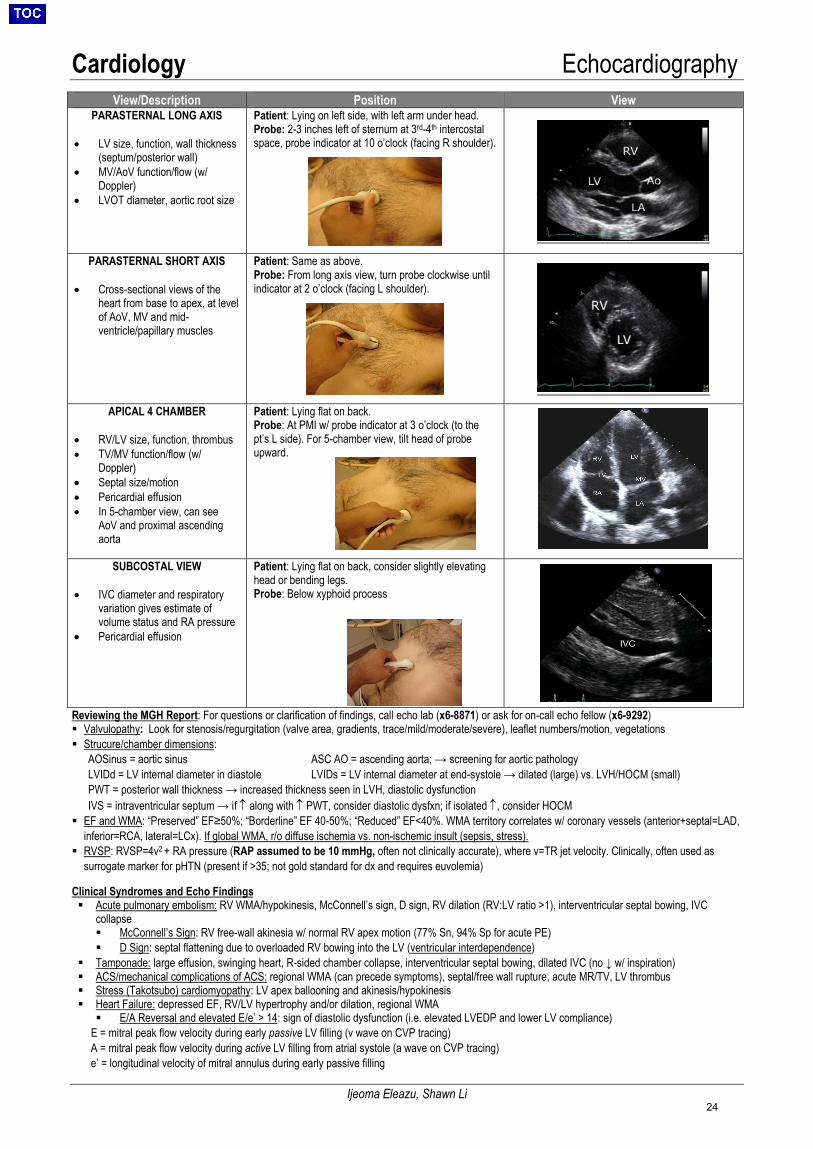
Cardiology Echocardiography
Ijeoma Eleazu, Shawn Li
Reviewing the MGH Report: For questions or clarification of findings, call echo lab (x6-8871) or ask for on-call echo fellow (x6-9292) Valvulopathy: Look for stenosis/regurgitation (valve area, gradients, trace/mild/moderate/severe), leaflet numbers/motion, vegetations Strucure/chamber dimensions: AOSinus = aortic sinus ASC AO = ascending aorta; → screening for aortic pathology LVIDd = LV internal diameter in diastole LVIDs = LV internal diameter at end-systole → dilated (large) vs. LVH/HOCM (small) PWT = posterior wall thickness → increased thickness seen in LVH, diastolic dysfunction IVS = intraventricular septum → if ↑ along with ↑ PWT, consider diastolic dysfxn; if isolated ↑, consider HOCM EF and WMA: “Preserved” EF≥50%; “Borderline” EF 40-50%; “Reduced” EF<40%. WMA territory correlates w/ coronary vessels (anterior+septal=LAD,
inferior=RCA, lateral=LCx). If global WMA, r/o diffuse ischemia vs. non-ischemic insult (sepsis, stress). RVSP: RVSP=4v2 + RA pressure (RAP assumed to be 10 mmHg, often not clinically accurate), where v=TR jet velocity. Clinically, often used as
surrogate marker for pHTN (present if >35; not gold standard for dx and requires euvolemia)
Clinical Syndromes and Echo Findings Acute pulmonary embolism: RV WMA/hypokinesis, McConnell’s sign, D sign, RV dilation (RV:LV ratio >1), interventricular septal bowing, IVC
collapse McConnell’s Sign: RV free-wall akinesia w/ normal RV apex motion (77% Sn, 94% Sp for acute PE) D Sign: septal flattening due to overloaded RV bowing into the LV (ventricular interdependence)
Tamponade: large effusion, swinging heart, R-sided chamber collapse, interventricular septal bowing, dilated IVC (no ↓ w/ inspiration) ACS/mechanical complications of ACS: regional WMA (can precede symptoms), septal/free wall rupture, acute MR/TV, LV thrombus Stress (Takotsubo) cardiomyopathy: LV apex ballooning and akinesis/hypokinesis Heart Failure: depressed EF, RV/LV hypertrophy and/or dilation, regional WMA E/A Reversal and elevated E/e’ > 14: sign of diastolic dysfunction (i.e. elevated LVEDP and lower LV compliance)
E = mitral peak flow velocity during early passive LV filling (v wave on CVP tracing)A = mitral peak flow velocity during active LV filling from atrial systole (a wave on CVP tracing)e’ = longitudinal velocity of mitral annulus during early passive filling
View/Description Position View PARASTERNAL LONG AXIS
• LV size, function, wall thickness(septum/posterior wall)
• MV/AoV function/flow (w/Doppler)
• LVOT diameter, aortic root size
Patient: Lying on left side, with left arm under head. Probe: 2-3 inches left of sternum at 3rd-4th intercostal space, probe indicator at 10 o’clock (facing R shoulder).
PARASTERNAL SHORT AXIS
• Cross-sectional views of theheart from base to apex, at levelof AoV, MV and mid-ventricle/papillary muscles
Patient: Same as above. Probe: From long axis view, turn probe clockwise until indicator at 2 o’clock (facing L shoulder).
APICAL 4 CHAMBER
• RV/LV size, function, thrombus• TV/MV function/flow (w/
Doppler)• Septal size/motion• Pericardial effusion• In 5-chamber view, can see
AoV and proximal ascendingaorta
Patient: Lying flat on back. Probe: At PMI w/ probe indicator at 3 o’clock (to the pt’s L side). For 5-chamber view, tilt head of probe upward.
SUBCOSTAL VIEW
• IVC diameter and respiratoryvariation gives estimate ofvolume status and RA pressure
• Pericardial effusion
Patient: Lying flat on back, consider slightly elevating head or bending legs. Probe: Below xyphoid process
24
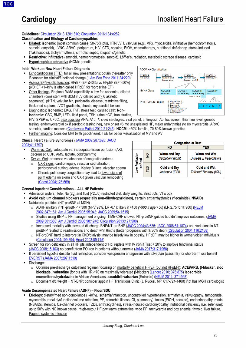
Cardiology Inpatient Heart Failure
Jeremy Feng, Charlotte Lee
Guidelines: Circulation 2013;128:1810; Circulation 2016:134:e282 Classification and Etiology of Cardiomyopathies Dilated: ischemic (most common cause, 50-75% pts), HTN/LVH, valvular (e.g., MR), myocarditis, infiltrative (hemochromatosis,
sarcoid, amyloid), LVNC, ARVC, peripartum, HIV, CTD, cocaine, EtOH, chemotherapy, nutritional deficiency, stress-induced(Takatsubo’s), tachyarrhythmia, cirrhotic, septic, idiopathic/genetic
Restrictive: infiltrative (amyloid, hemochromotosis, sarcoid), Löffler’s, radiation, metabolic storage disease, carcinoid Hypertrophic obstructive (HCM): genetic
Initial Workup: New Heart Failure Diagnosis
Echocardiogram (TTE): for all new presentations; obtain thereafter onlyif concern for clinical/functional change (J Am Soc Echo 2011;24:229)
Assess EF/systolic function: HFrEF (EF ≤40%) vs HFpEF (EF >50%)(NB: EF 41-49% is often called HFbEF for ‘borderline EF’)
Other findings: Regional WMA (specificity is low for ischemia), dilatedchambers (consistent with dCM if LV dilated and > 6 akineticsegments), pHTN, valvular fxn, pericardial disease, restrictive filling,thickened septum, LVOT gradients, shunts, myocardial texture
Diagnostics: Ischemic: EKG, TnT, stress test, cardiac cath; Non-ischemic: CBC, BMP, LFTs, lipid panel, TSH, urine hCG, iron studies,HIV, SPEP w/ UFLC; also consider ANA, A1c, T. cruzi serologies, viral panel, antimyosin Ab, tox screen, thiamine level, genetictesting, endomyocardial bx if serologic testing neg, new onset <6 mo unexplained HF, major arrhythmias (to r/o myocarditis, ARVC,sarcoid), cardiac masses (Cardiovasc Pathol 2012;21:245); HOCM: >50% familial, 70-80% known genetics
Further imaging: Consider MRI (with gadolinium); TEE for better visualization of MV and AVClinical Heart Failure Syndromes (JAMA 2002;287:628; JACC 2003;41:1797) Warm vs. Cold: adequate vs. inadequate tissue perfusion (AKI,
decreased UOP, AMS, lactate, cold/clammy) Dry vs. Wet: presence vs. absence of congestion/edema
o CXR signs: cardiomegaly, vascular cephalization,peribronchial cuffing, edema, Kerley B lines, alveolar edema
o Chronic pulmonary congestion may lead to fewer signs ofpulm edema on exam and CXR given vascular remodeling(Chest 2004;125:669)
General Inpatient Considerations – ALL HF Patients: Admission orders: Tele, Na (2g) and fluid (<2L/d) restricted diet, daily weights, strict I/Os, VTE ppx Avoid calcium channel blockers (especially non-dihydropyridines), certain antiarrhythmics (flecainide), NSAIDs Natriuretic peptides (NT-proBNP at MGH)
o ADHF unlikely if NT-proBNP < 300 (NPV 98%, LR -0.1); likely if >450 (>900 if age >50) (LR 2.75 for ≥ 900) (NEJM2002;347:161; Am J Cardiol 2005;95:948; JACC 2009;54:1515)
o Studies using BNP to HF management ongoing; TIME-CHF showed NT-proBNP guided tx didn’t improve outcomes, (JAMA2009;301:383; Am J Cardiol 2006;98:1248; Circulation 2013;127:500)
o Increased mortality with elevated discharge BNP/NT-proBNP (JACC 2004;43:635; JACC 2008;51:1874) and variations in NT-proBNP related to readmissions and death w/in 6mths (better prognosis with ≥ 30% decr) (Circulation 2004;110:2168)
o NT-proBNP hard to interpret in CKD/dialysis; may be falsely low in obesity, HFpEF; may be higher in women/older individuals(Circulation 2004;109:594; Heart 2003;89:745)
Screen for iron deficiency in all HF pts independent of Hb; replete with IV iron if Tsat < 20% to improve functional status(JACC 2008;15:103) no benefit from PO iron in patients without anemia (JAMA 2017;317:1958)
If persistent hypoNa despite fluid restriction, consider vasopressin antagonism with tolvaptan (class IIB) for short-term sxs benefitEVERST (JAMA 2007;297:1319)
Discharge:o Optimize pre-discharge outpatient regimen focusing on mortality benefit in HFrEF (but not HFpEF!): ACEi/ARB, β-blocker, aldo
blockade, ivabradine (for pts with HR ≥70 on maximally tolerated β-blocker) (Lancet 2010; 376:875) isosorbidemononitrate/hydralazine in African-Americans, sacubitril-valsartan (Entresto) (NEJM 2014; 371:993)
o Document d/c weight + NT-BNP; consider appt in HF Transitions Clinic (J. Ruckel, NP; 617-724-1400) if pt has MGH cardiologist
Acute Decompensated Heart Failure (ADHF) – Floor/SDU: Etiology: dietary/med non-compliance (~40%), ischemia/infarction, uncontrolled hypertension, arrhythmia, valvulopathy, tamponade,
myocarditis, renal dysfunction/volume retention, PE, comorbid illness (GI, pulmonary), toxins (EtOH, cocaine), endocrinopathy, meds(NSAIDs, steroids, Ca-channel blockers, TZDs, anthracyclines), stress-induced cardiomyopathy, nutritional deficiency (i.e. selenium);up to 50% with NO known cause. *high-output HF p/w warm extremities, wide PP, tachycardia and ddx anemia, thyroid, liver failure,Pagets, systemic infection
25
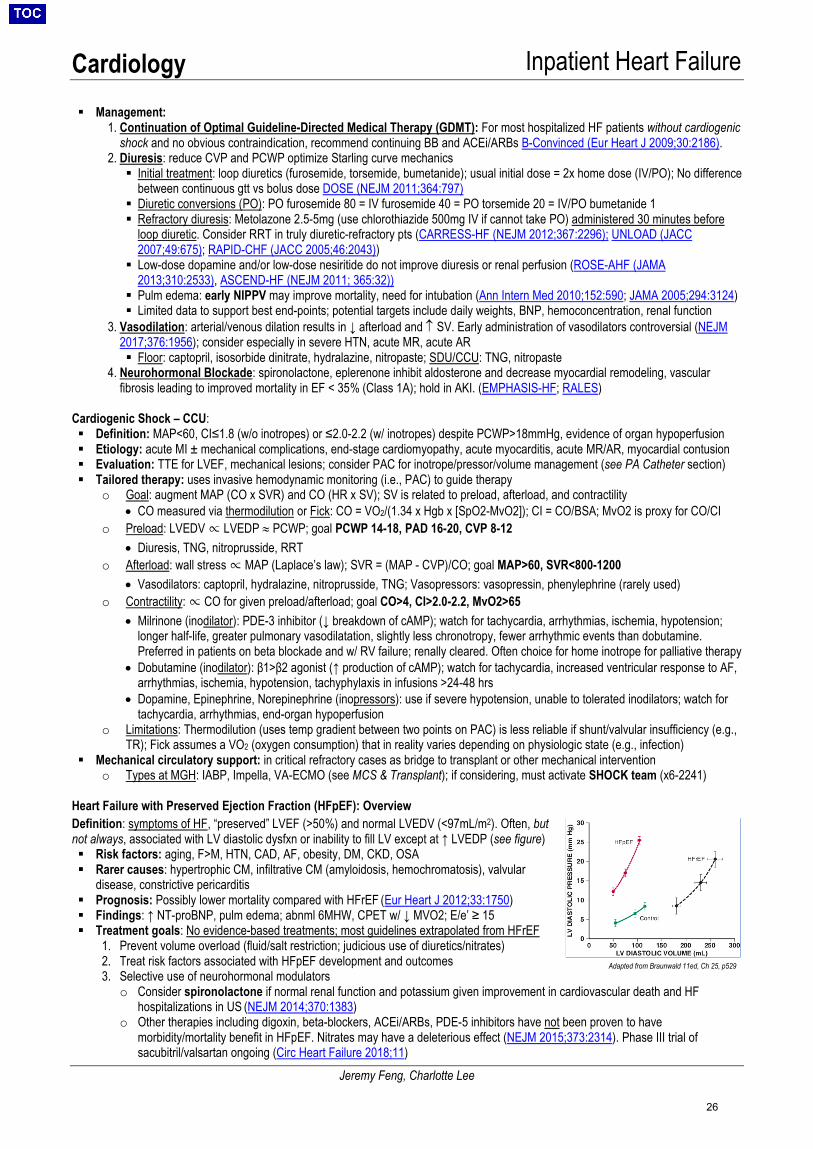
Cardiology Inpatient Heart Failure
Jeremy Feng, Charlotte Lee
Management:1. Continuation of Optimal Guideline-Directed Medical Therapy (GDMT): For most hospitalized HF patients without cardiogenic
shock and no obvious contraindication, recommend continuing BB and ACEi/ARBs B-Convinced (Eur Heart J 2009;30:2186).2. Diuresis: reduce CVP and PCWP optimize Starling curve mechanics Initial treatment: loop diuretics (furosemide, torsemide, bumetanide); usual initial dose = 2x home dose (IV/PO); No difference
between continuous gtt vs bolus dose DOSE (NEJM 2011;364:797) Diuretic conversions (PO): PO furosemide 80 = IV furosemide 40 = PO torsemide 20 = IV/PO bumetanide 1 Refractory diuresis: Metolazone 2.5-5mg (use chlorothiazide 500mg IV if cannot take PO) administered 30 minutes before
loop diuretic. Consider RRT in truly diuretic-refractory pts (CARRESS-HF (NEJM 2012;367:2296); UNLOAD (JACC2007;49:675); RAPID-CHF (JACC 2005;46:2043))
Low-dose dopamine and/or low-dose nesiritide do not improve diuresis or renal perfusion (ROSE-AHF (JAMA2013;310:2533), ASCEND-HF (NEJM 2011; 365:32))
Pulm edema: early NIPPV may improve mortality, need for intubation (Ann Intern Med 2010;152:590; JAMA 2005;294:3124) Limited data to support best end-points; potential targets include daily weights, BNP, hemoconcentration, renal function
3. Vasodilation: arterial/venous dilation results in ↓ afterload and ↑ SV. Early administration of vasodilators controversial (NEJM2017;376:1956); consider especially in severe HTN, acute MR, acute AR Floor: captopril, isosorbide dinitrate, hydralazine, nitropaste; SDU/CCU: TNG, nitropaste
4. Neurohormonal Blockade: spironolactone, eplerenone inhibit aldosterone and decrease myocardial remodeling, vascularfibrosis leading to improved mortality in EF < 35% (Class 1A); hold in AKI. (EMPHASIS-HF; RALES)
Cardiogenic Shock – CCU: Definition: MAP<60, CI≤1.8 (w/o inotropes) or ≤2.0-2.2 (w/ inotropes) despite PCWP>18mmHg, evidence of organ hypoperfusion Etiology: acute MI ± mechanical complications, end-stage cardiomyopathy, acute myocarditis, acute MR/AR, myocardial contusion Evaluation: TTE for LVEF, mechanical lesions; consider PAC for inotrope/pressor/volume management (see PA Catheter section) Tailored therapy: uses invasive hemodynamic monitoring (i.e., PAC) to guide therapy
o Goal: augment MAP (CO x SVR) and CO (HR x SV); SV is related to preload, afterload, and contractility• CO measured via thermodilution or Fick: CO = VO2/(1.34 x Hgb x [SpO2-MvO2]); CI = CO/BSA; MvO2 is proxy for CO/CI
o Preload: LVEDV ∝ LVEDP ≈ PCWP; goal PCWP 14-18, PAD 16-20, CVP 8-12• Diuresis, TNG, nitroprusside, RRT
o Afterload: wall stress ∝ MAP (Laplace’s law); SVR = (MAP - CVP)/CO; goal MAP>60, SVR<800-1200• Vasodilators: captopril, hydralazine, nitroprusside, TNG; Vasopressors: vasopressin, phenylephrine (rarely used)
o Contractility: ∝ CO for given preload/afterload; goal CO>4, CI>2.0-2.2, MvO2>65• Milrinone (inodilator): PDE-3 inhibitor (↓ breakdown of cAMP); watch for tachycardia, arrhythmias, ischemia, hypotension;
longer half-life, greater pulmonary vasodilatation, slightly less chronotropy, fewer arrhythmic events than dobutamine.Preferred in patients on beta blockade and w/ RV failure; renally cleared. Often choice for home inotrope for palliative therapy
• Dobutamine (inodilator): β1>β2 agonist (↑ production of cAMP); watch for tachycardia, increased ventricular response to AF,arrhythmias, ischemia, hypotension, tachyphylaxis in infusions >24-48 hrs
• Dopamine, Epinephrine, Norepinephrine (inopressors): use if severe hypotension, unable to tolerated inodilators; watch fortachycardia, arrhythmias, end-organ hypoperfusion
o Limitations: Thermodilution (uses temp gradient between two points on PAC) is less reliable if shunt/valvular insufficiency (e.g.,TR); Fick assumes a VO2 (oxygen consumption) that in reality varies depending on physiologic state (e.g., infection)
Mechanical circulatory support: in critical refractory cases as bridge to transplant or other mechanical interventiono Types at MGH: IABP, Impella, VA-ECMO (see MCS & Transplant); if considering, must activate SHOCK team (x6-2241)
Heart Failure with Preserved Ejection Fraction (HFpEF): Overview Definition: symptoms of HF, “preserved” LVEF (>50%) and normal LVEDV (<97mL/m2). Often, but not always, associated with LV diastolic dysfxn or inability to fill LV except at ↑ LVEDP (see figure) Risk factors: aging, F>M, HTN, CAD, AF, obesity, DM, CKD, OSA Rarer causes: hypertrophic CM, infiltrative CM (amyloidosis, hemochromatosis), valvular
disease, constrictive pericarditis Prognosis: Possibly lower mortality compared with HFrEF (Eur Heart J 2012;33:1750) Findings: ↑ NT-proBNP, pulm edema; abnml 6MHW, CPET w/ ↓ MVO2; E/e’ ≥ 15 Treatment goals: No evidence-based treatments; most guidelines extrapolated from HFrEF
1. Prevent volume overload (fluid/salt restriction; judicious use of diuretics/nitrates)2. Treat risk factors associated with HFpEF development and outcomes3. Selective use of neurohormonal modulators
o Consider spironolactone if normal renal function and potassium given improvement in cardiovascular death and HFhospitalizations in US (NEJM 2014;370:1383)
o Other therapies including digoxin, beta-blockers, ACEi/ARBs, PDE-5 inhibitors have not been proven to havemorbidity/mortality benefit in HFpEF. Nitrates may have a deleterious effect (NEJM 2015;373:2314). Phase III trial ofsacubitril/valsartan ongoing (Circ Heart Failure 2018;11)
Adapted from Braunwald 11ed, Ch 25, p529
26
Cardiology Inpatient Heart Failure
Jeremy Feng, Charlotte Lee
Specific Causes of Cardiomyopathy • Hypertrophic Cardiomyopathy (HCM) (Circulation 2011;124:2761)
o LV and/or RV hypertrophy of various morphologies: ± LVOT dynamic obstruction (HOCM), diastolic dysfxn, ischemia, MRo Presentation: SOB, arrhythmias/palpitations, CHF, angina, pre-/syncope, SCD (most common in asymptomatic pts <35yo)o Physical Exam: LVOT obstruction – medium-pitch SEM at LLSB/apex that augments with Valsalva or on standing (due to ↓
preload); S2 paradox split, S4o Routine Diagnostics: EKG (abnl in 90% probands, 75% asymptomatic relatives -- prominent voltages w/ depolarization
abnormalities, large abnormal Q waves in inferior (II, III, avF) or lateral (I, aVL and V4-V6) leads, P wave abnormalities (atrialenlargement), LAD, giant negative T waves in V2-V4 (apical HCM variant“Yamaguchi’s syndrome”), TTE (unexplained LVH>15mm in any pattern, SAM of MV, outflow tract gradient), cMR (late gadolinium enhancement LGE = fibrosis, possibly earlydetection not seen on TTE, controversial role in decision making since no strong association with outcomes)
o Risk Stratification: (1) Ambulatory EKG monitoring 24-48hrs (2) ETT (failure to augment BP to exercise due to dynamic LVOTobstruction, symptoms, arrhythmias, ST depressions) + stress echo (increasing outflow tract gradient, worsening MR)
o Genetics: Clinical genetic testing (mutation in ~70% of cases) helpful for family screening; not useful for dx or risk stratificationo Treatment: Avoid volume depletion or high dose vasodilators (may worsen obstruction), activity restriction, medical Rx (BB >
verapamil). Use phenylephrine to treat hypotension in patients with HOCM who do not repond to fluid boluses (↑ afterload,stents open LVOT), septal ablation or surgical myectomy for medically refractory sx, ICD (for high SCD risk, risk factors below)
o Risk factors for SCD/VT in order of decreasing risk: (1) Prior VT/SCD/unexplained syncope; (2) FHx of SCD in 1° relative, (3)Massive LVH (>30 mm wall thickness), (4) NSVT on Holter, (5) abnormal BP response to exercise, (6) burden of LGE on cMR
• Non-ischemic Dilated Cardiomyopathy (DCM) (JACC 2016;67:2996)o Takotsubo (stress-induced) (NEJM 2015;373:929)
o Potential mechanisms: catecholamine surge from physical/emotional stress, coronary artery spasm, microvascular ACSo Presentation: may present like ACS; sxs include chest pain (76%), dyspnea (47%), shock (10%), syncope (7.7%). If in
shock, urgent TTE to assess for LVOT obstruction.o Diagnostic criteria (ALL needed): (1) Transient dysfunction (hypo-/dys-/akinesis) of LV mid-segments w/ or w/o apical
involvement. Regional WMA extend beyond a single coronary distribution (2) Rule out ACS/obstructive coronarydisease (via cath) (3) New EKG abnormalities (STE (44%) and/or TWI) OR ↑ troponin. (4) absence of pheo, myocarditis.WMA can be apical (82%) mid-ventricle (15%), basal (2%), focal (2%)
o Treatment: Remove stressor. ACEi (may improve survival), BB, diuretic. If non-obstructive CAD, add ASA+statin.o Prognosis: 4% in-hospital mortality,19% severe complications; most recover LV fxn in 1-4 wks
o Alcohol-induced:o Acquired DCM a/w >80g/day of EtOH over >5 years (toxic to myocytes via O2 free radicals + defects in protein synthesis)o Treatment: Abstinence + HF therapy
Prognosis: Better/equivalent to idiopathic CM if able to abstain/consume <20g/day, worse w/ continued EtOH abuseo Toxins: Chemotherapy (anthracyclines, cyclophosphamide, trastuzumab, 5-FU), antiretroviral drugs, phenothiazines,
chloroquine, clozapine, amphetamines, cocaine, carbon monoxide, cobalt, lead, mercury, lithiumo Infection: Viral (HIV, lyme, adenovirus, coxsackie A/B, CMV/EBV, HHV6, parvovirus B19, varicella), bacterial (brucellosis,
diphtheria, psittacosis, typhoid fever), protozoal/helminthic (chagas, malaria, toxo, schistosomiasis, strongyloidiasis)o Other causes of DCM: Pregnancy, nutritional deficiency (carnitine, thiamine, selenium, niacin), tachyarrhythmia, electrolyte
abnormality (hypocalcemia, hypophos, uremia), endocrine (Cushing’s disease, acromegaly, DM, hypo/hyperthyroidism,pheochromocytoma), cirrhotic, septic, CTD (SLE, RA, Scl, dermatomyositis), vasculitis (GPA, EGPA, Kawasaki, PAN),deposition (hemochromatosis, amyloid), hypersensitivity myocarditis, NM disease, genetic (~50% of “idiopathic” DCM)
• Restrictive Cardiomyopathy (JACC 2010;55:1769): (NB: these conditions may also manifest as DCM)Condition Presentation EKG Echo/MRI
Amyloidosis (AL, TTR)
HF with other findings of amyloid (renal, neurologic, hepatic disease)
Decreased voltage, pseudoinfarct pattern in inferolateral leads
Symmetric LV/RV wall thickness, speckled myocardium on TTE, LGE in subendocardium on cMR
Hemochomotosis If hereditary: M>30 yo; F> 40 yo If 2°: can present at any age LFT abnormalities, arthralgias, DM, hyperpigmented skin
SVT (ventricular conduction abnormalities are rare)
Dilated LV with global systolic dysfunction. cMR can detect iron overload with T2* protocol
Sarcoidosis Young adult w/ HF (more commonly presents as DCM)
Infrahisian block, atypical infarction pattern
Variable wall thickness. Focal/global hypokinesis, LV aneurysm. Patchy enhancement of basal and LV walls on cMR
Treatment • Treat underlying disease + HF guidelines based on underlying EF (HFrEF vs. HFpEF) as aboveo Amyloidosis: tafamidis (↓ TTR deposition) shown ↓ mortality, admissions, functional/QoL decline (NYHA I-III) [NEJM 2018;379:1007]; liver
transplant in familial disease
27
Cardiology Mechanical Support & Transplant
Jeremy Feng, Sean Murphy
Mechanical Circulatory Support (MCS) – If inotrope-refractory cardiogenic shock, call SHOCK team (x6-2241) Selected MCS Modalities
Device Indications Support Provided Considerations Management Complications
IABP (intra-aortic balloon bump)
Refractory heartfailure (bridge todurable MCS)
Cardiogenicshock/massive PE
Refractorymalignantarrhythmias
Support duringhigh-riskprocedureso Complex PCIo Ablation of
ventricular arrhythmias
o Perc valverepair
Acute allograftfailure
Minimal hemodynamic support ↓ LV afterload ↑ Coronary perfusion (possible mortality benefit for high risk lesions pre-CABG)
Bedside insertion Does not require AC
(when at 1:1) No ↓ mortality in
cardiogenic shock(IABP-SHOCK II,NEJM 2012;367:1287)
Least costly
Pt cannot sit up/bendlegs
✓ CXR daily (tip 1-4cmbelow Ao notch)
✓ Waveform daily Wean by ↓ ratio (then
return to 1:1, stop AC,pull)
Limb ischemia Vascular injury Thromboemoblism Bleeding Infection Balloon leak/rupture
(STAT vascular surgc/s)
Impella Partial LV support: Cath lab placement: Impella 2.5 (2.5 L/min), Impella CP (3.5 L/min), OR placement: Impella 5.0 (5 L/min)
Partial RV support Impella RP (4 L/min)
Ventricular decompression
Requires AC Allows pt mobilization
(if axillary placement) Longer-term support
(weeks)
P1 (lowest) to P9 (highest support)
✓ Urine color (hemolysis) ✓ Suction events (↓
preload, RV failure,position)
✓ Ventricular arrhythmias(device migration)
Infection Bleeding Limb ischemia Thromboemoblism Thrombocytopenia Vascular injury Position alarm
(reposition under fluoro/echo)
VA-ECMO
HD support (4-10 L/min), oxygenation & CO2 clearance (Often requires additional device for LV unloading, i.e. Impella)
Bedside insertionpossible
Short-term support(days/weeks)
See ECMO section
Durable VAD
Bridge totransplant
Destinationtherapy (DT)
“Bridge todecision” (ontransplant or DT)
Bridge to recovery(LV unloading can be therapeutic)
Full LV support (10 L/min) HeartMate II HeartMate 3 HeartWare HVAD
Mobility Long-term support
(years)
BP via manual cuff w/doppler (goal MAP 70-80)
If hypotensive, place a-line
If unconscious, w/o hum,and MAP<50: chestcompressions
TTE if any concern
Acquired vWF defic. Hemolysis (possible
pump thrombosis) Ventr. arrhythmias Thromboembolism RV failure AR Driveline infections
Heart Transplant 2018 UNOS Adult Heart Allocation Criteria: Status 1: VA-ECMO, MCS with life-threatening vent. arrhythmia; Status 2: non-dischargeable LVAD, MCS + device malfunction, IABP; Status 3: ≥2 inotropes or single high-dose + continuous hemodynamic monitoring, dischargeable LVAD for discretionary 30 days, VA-ECMO after 7d, IABP after 14d; Status 4: re-transplant, inotropes without hemodynamic monitoring, dischargeable LVAD without discretionary 30 days; Status 5: awaiting dual organ Tx; Status 6: all others; Status 7: inactive listing
Transplant evaluation at MGH: • Labs: blood typing (2 samples on separate days), second sample for PRA (check with tissue typing x63722), BMP, LFT,
amylase, CBC+diff, PT-INR/PTT, TFTs, lipids, PTH, 25-Vit-D, 1,25-Vit-D, HIV, CMV, Toxo Igs, EBV, VZV serology, MMR, RPR,hepatitis serologies, IGRA, UA, 24h urine CrCl (and 24h urine protein if diabetic)
• Vaccines: HBV, PPSV23, Tdap• Consults: Psychiatry (Dr John Purcell), SW (Kathryn Tsagronis), Tx coordinators (Sally Keck, Coral Haggan, Kerry Gaj, Karen
Turvey – they can all consent patient for Tx), Dental (panorex, inpatient consult), Nutrition, Endocrine, Pall Care• Diagnostics: RHC (eval for presence & reversibility of pHTN with vasodilator challenge; if unsuccessful vasodilator challenge,
note that PVR often declines after 24-48h of treatment [e.g. diuretics, inotropes, vasoactive agents]), +/- LHC, level 1 CPET (PaulPappas, x47825) (complementary ISHLT criteria for Tx: VO2 max ≤14ml/kg/min [or ≤12 if on βb]), abdominal US, carotid US,TTE, ECG, CXR, DXA, ABIs +/- angiography, cancer screening up-to-date
Post-transplant immunosuppression: steroids (typically tapered off over 6 months, 1st line for acute rejection), calcineurin inhibitors (cyclosporine/tacrolimus), anti-proliferatives (azathioprine/mycophenolate), mTOR inhibitors (sirolimus/everolimus – most effective for coronary allograft vasculopathy [CAV]; avoid in immediate post-tx phase as inhibit wound healing) Monitoring: protocolized schedule of RV biopsies to r/o cellular/humoral rejection, R/LHC and TTE to assess graft function and for CAV
28
Cardiology Right Ventricular Failure
Maeve Jones-O’Connor, Chitra Mosarla
(Chest 2005;128:1836)
RV “death spiral”
Right Ventricle Physiology (Circulation 2018;137:e578) • RV has thinner myocardium compared to LV↑ compliance compared to LV, so it does not adapt well to acute increases in pressure• RV and LV are interdependentfailure of RV leads to failure of LV through several mechanisms: (a) decreased LV preload (because
RV output = LV preload; (b) septal bowing into LV, causing diastolic impairment (“Bernheim effect”)Acute Changes in RV Hemodynamics • ↑ RV afterload (e.g., PE), ↑ RV preload (e.g., LR shunting through ASD/VSD), or ↓ RV contractility (e.g., MI) all lead to increased
RV wall stress and resultant ischemia• RV CO subsequently ↓ and RV dilates, precipitating RV “death spiral”• ↓ RV CO leads to ↓ MAP (and ↑ RVP), resulting in ↓ coronary perfusion pressure (CPPRV = MAP –RVP)• ↓ CPPRV leads to more RV ischemia, propagating “death spiral” furtherClinical Features and Workup• Exam: Elevated JVP, peripheral edema, RV heave, pulsatile liver. Less
common: Split S2, new tricuspid regurgitation (loudest: RLSB)• Imaging: CXRhard to evaluate RV 2/2 position, lateral film can help;
CTbest for RV size/septum position• Echocardiography: measure RV size/function to elucidate underlying
etiology. RVEF based on displacement of base towards apex (tricuspidannular plane systolic excursion [TAPSE], nl 2.4-2.7cm).
• RHC: gold standard for measurement of ventricular filling pressures, CO,PA pressureso RVSP: correlates w/ RHC but can vary up to 10mmHg (esp w/
chronic lung dz, pos pressure vent).• Labs: NT-proBNP and troponin are not specific but can indicate RV failure if
no L-sided disease.
Management (Am J Respir Crit Care Med 2011;184:1114) • Identify and treat triggers: infection, anemia, arrhythmia, PE, MI, hypoxia• Preload: Clinical assessment of optimal preload is challenging. Both hypo- and hypervolemia may ↓ CO.
o Acute: consider volume loading in pts with acute RVMI or PE in absence of marked CVP elevation (“preload-dependent”)o Subacute/chronic: consider diuresis to reduce RV filling pressures and improve RV CO
• Afterload:o Systemic: if pt hypotensive, start systemic pressors; no clinical data regarding pressor of choice, but often choose or
vasopressin or norepinephrine because it affects SVR>>PVRo Pulmonary: remove factors that ↑ pulm vasc tone (e.g., hypoxia). Consider pulm vasodilators (inhaled>oral in acute setting).
Types: prostacyclin agonists (e.g., epoprostenol), endothelin antagonists (e.g., bosentan, ambrisentan), nitric oxideenhancers (e.g., PDE-5 inhibitors: sildenafil, tadalafil; inhaled NO)
• Contractility: often use milrinone to enhance pulmonary vasodilation (vasodilates arteries in both systemic and pulmonary circulation)
Intubation and Mechanical Ventilation (Curr Heart Fail Rep 2012;9:228) • Intubation/NIPPV in RV failure precipitate risk for HD collapse and arrest• Drugs commonly used: BZDs, propofol, muscle relaxants → tendency towards vasodilation and negative inotropy → decreases
venous return → decreased LV preloado Consider RSI (etomidate >> propofol for induction) and push dose epinephrine (10-20mcg)/vasopressin (1-2U) if
emergent intubation anticipating hypotension• Positive pressure ventilation → increased pulmonary pressures and ∴ RV afterload → increased RV dilation → “death spiral”• Vent Management: cannot allow hypoxia and hypercarbia as drive up PVR, consider moderate TV (~8cc/kg) with low PEEP (<12
cm H2O) with moderate plateau pressure goal (<30 mmHg)
Right Ventricular Myocardial Infarction (Circulation 2012;127:e362) • EKG: Check R-sided EKG leads in pts with inferior STEMI (10-15% of pts with inf. STEMI have RV involvement)
o 1mm STE in V4R → 88% Sn, 78% Sp in inferior STEMI; STE III>II suggests RVMIo High-grade AV block seen in ~50% of pts with RVMI
• Management: pts with RVMI are initially “preload-dependent” and often benefit from fluid bolus; caution w/ TNG (↓ preload) and BB(↓HR) o If CVP >15mmHg and BP not improving w/ IVF, additional fluids may worsen RV failure/overload (Eur Heart J Acute
Cardiovasc Care 2013;2:226)
↓ MAP
Ventricular interdependence
▼
RV Volume Overload
↑ RV Afterload
RV Dilation
↓RV CO
↓ LV Preload
↓ LV CO
↑ RV Wall Stress
RV Pressure Overload
&
▼
▼
▼
29
Cardiology Pulmonary Artery Catheterization
Maeve James-O’Connor, Leslie Chang
Overview: Indications—7 primary indications for the placement of PA lines:
o (1) diagnose etiology of shock (e.g., cardiogenic vs. distributive) (2) diagnose cardiogenic vs. non-cardiogenic pulm. edema (3)diagnose PH (4) diagnose LR shunting (5) diagnose valve disease (6) diagnose pericardial disease (7) tailored therapy
Efficacy: No benefit in the ICU setting or peri-op for mortality, LOS, cost (JAMA 2005;294:1625, Lancet 2005;366:472). Line course: central vein (IJ/subclavian/femoral) → SVC/IVC → RA → RV → PA → distal pulmonary arteriole
Venous Waveforms (CVP/PCWP): a wave: atrial contraction; coincides with QRS complex (on CVP tracing) c wave: bowing of TV/MV into atrium during ventricular contraction; more visible in 1st degree
AV block. Often absent on PCWP. x descent: atrial relaxation (early x descent), downward mvmt. of TV/MV (late x descent) v wave: passive atrial filling (venous return) when TV/MV closed; coincides with T wave y descent: rapid atrial emptying following opening of the TV/MV (ventricular diastole)
Obtaining PA Line Numbers on AM Rounds: 1) Position patient supine with head-of-bed 0-60° elevation2) Check level of transducer with phlebostatic axis (4th intercostal
space and mid-axillary line)3) Zero transducer to air and assess waveform for dampness4) Record PA systolic, PA diastolic, PA mean, CVP, and line position5) Open the PA catheter balloon port and remove 1cc air6) Inject 1cc air until PCWP waveform observed (use minimum air
required to reduce risk of PA infarction/rupture) and record PCWP (limit balloon inflation to no more than 8-10 seconds)7) Release safety syringe and allow balloon to deflate passively. Verify balloon deflated by confirmation of PA waveform.8) Troubleshooting:
a. Arrhythmia: Catheter may be in RVOT. Talk to fellow/attending and consider repositioning catheter.b. Dampened waveform: Kinked tubing, air/thrombus, or catheter tip against vessel wall. Flush and/or withdraw catheter.c. No PCWP tracing: Catheter tip is not far enough, balloon has ruptured, or catheter coiled in the RV.
Calculating Hemodynamic Parameters: Normal: “rule of 5s”RA 5, RV 25/5, PA 25/10, PCWP 10, LV 125/10 Cardiac Output:
o Fick = VO2 / (13.4 * Hgb * [SpO2 – MvO2]) [nl: 4-7 L/min] VO2 ≈ 250 ml/min OR 3*wt(kg) OR 125*BSA
o Thermodilution: Temperature change (measured by thermistor inPA) is proportional to LV CO (inaccurate w/ TR, intracardiac shunt)
Cardiac index = CO/BSA [normal: 2.6-4.2 L/min/m2] SVR = (MAP-CVP) / CO x 80 [normal: 700-1200 dynes*s*cm5] PVR = (mPAP-PCWP) / CO x 80 [normal: 20-130 dynes*s*cm5]
Hemodynamic Considerations: All quantitative pressure measurements (especially PCWP) should be made at end-expiration (when intrathoracic pressure is zero)
o Spontaneous respiration: RA and PCWP ↑ with expirationmeasure from the higher a waves (“patient = peak”) o Positive pressure ventilation: RA and PCWP ↓ with expirationmeasure from the the lower a waves (“vent = valley”)
Measure RA and PCWP at end-diastole (i.e. just before the c wave) Correlate PCWP with PA diastolic pressure; if well correlated, can trend PAd as proxy for PCWP
o If no PA line in place, MvO2 may be used as a proxy for CO/CI [normal: >65-75%]
Clinical Considerations: Placement: Usually through RIJ Cordis. Advance ONLY with balloon inflated. Deflate balloon when withdrawing and at ALL other
times. Must have cardiology or pulmonary fellow present to place/advance at MGH.o Cath lab insertion if: Severe PH (PAP>70mmHg), large RV, LBBB, PPM/ICD, temp wire, severe TR, prosthetic TV/PV
Contraindications: RA/RV mass/thrombosis, mechanical TV/PV, endocarditis (TV/PV) Markings on PA catheter: Each thin line=10cm; each thick line=50cm. Position: On CXR: middle 1/3 of the chest bilaterally. Ability to wedge more important than CXR position. Complications: infection, bleeding, PTX, VT, RBBB, CHB, PA rupture (place patient on side with the catheter [“bleeding side down”],
order STAT CXR, CBC, coags, CT surgery consult), pulm infarct, PE Duration: No data defining maximum length of time; at MGH, standard is 7d; others suggest 4-5d
30
Cardiology Cardiac Devices: PPM&ICD
Chitra Mosarla, Usama Abbasi
Permanent Pacemakers (PPM), Implantable Cardioverter-Defibrillators (ICD), & Cardiac Resynchronization Therapy (CRT): ▪ Types: Single chamber (RA or RV lead) vs. dual chamber (RA + RV leads) vs. biventricular (+/- RA + RV + LV leads)▪ PPM: sense/pace the RA and RV to treat bradyarrhythmias▪ ICD: device with an RV lead capable of terminating re-entrant ventricular tachyarrhythmias via pacing, cardioversion or defibrillation▪ CRT: provides simultaneous RV+LV pacing in HFrEF pts w/ wide QRS to ↓ desynchrony → LV reverse remodeling and ↑ LVEF
o CRT-P = BiV +/- RA pacing; CRT-D = CRT-P w/ ICD functions
NASPE/BPEG Codes for Pacing Operating Modes: Common Mode Guide:
Hardware Overview: System consists of pulse generator + leads. Usually implanted SQ in upper chest (L>R) >> abdominal. ▪ Types: Traditional (SQ pulse generator + IV leads in ventricle) vs. leadless (pulse generator directly implanted into RV; no pocket
complications but unclear what to do when battery dies) vs. SQ ICD (no IV hardware; low risk for infection but NO pacing capabilities)▪ Placement: RA lead → RA appendage; RV lead → RV apex; LV lead → coronary sinus → branches of great cardiac vein▪ Interrogation: Page EP Technician (PPM, p16939) during normal business hours; EP fellow on call if after-hours/weekend.▪ MRI Compatibility: Not all devices are MRI compatible, however even non-MRI compatible devices may be safe to scan after re-
programming (NEJM 2017; 376: 755). Determined on case-by-case basis by radiology. Need to know device model.
Class I PPM Indications: below are highlights of most important indications (JACC 2013; 61: e6) Sinus Node Dysfunction: ▪ Symptomatic sinus bradycardia (± sinus pauses) or chronotropic incompetence▪ Symptomatic medication-induced bradycardia if medication (i.e., BB) is required for underlying medical conditionAV Block (AVB)/Conduction Disease:▪ Symptomatic 2° AVB or 3°AVB▪ Asymptomatic Mobitz II 2° AVB or 3° AVB with: asystole ≥ 3 sec (>5 seconds if in AF), escape rate ≤ 40 BPM (or >40 BPM if
cardiomegaly also present), or wide-complex escape rhythm▪ Permanent Mobitz II 2° AVB or intermittent 3° AVB (regardless of symptoms)▪ Alternating bundle branch blockNeurocardiogenic:▪ Recurrent syncope AND inducible asystole ≥ 3 sec with carotid massage
Class I ICD Indications: ↓mortality vs. optimal medical therapy (OMT); guidelines apply only to pts who meet eligibility while on OMT(JACC 2013; 61: e6)
Primary Prevention Secondary Prevention - iCM with EF ≤ 35% at least 40d s/p MI + NYHA Class II/III- iCM with EF ≤ 30% at least 40d s/p MI + NYHA Class I- Non-ischemic dCM with EF ≤ 35% + NYHA Class II/III*- iCM with EF ≤ 40% at least 40d s/p MI + NSVT + inducible VT/VF on EP study- Unexplained syncope w/ hemodynamically significant inducible VT/VF on EP study
- Prior episode of cardiac arrest (VF/pulseless VT) or sustained unstable VT if no reversible causefound- Structural heart disease with spontaneoussustained VT (stable or unstable)
*DANISH trial demonstrated that although ICD implantation in patients with non-ischemic systolic heart failure reduces risk of sudden cardiac death, itdoes not provide a mortality benefit
ACA/AHA/HRS 2012 CRT Indications: ↓mortality vs. OMT (JACC 2013; 61: e6)) NYHA I NYHA II NYHA III / NYHA IV
Class I None - LVEF ≤ 35%, QRS ≥ 150ms, LBBB, &
sinus rhythm- LVEF ≤ 35%, QRS ≥ 150ms, LBBB, & sinus rhythm
Class IIa None - LVEF ≤ 35%, QRS 120-149ms, LBBB,and sinus rhythm
- LVEF ≤ 35%, QRS 120-149ms, LBBB, & sinus rhythm- LVEF ≤ 35%, QRS ≥ 150ms, & non-LBBB
Class IIb - LVEF ≤ 30%, QRS ≥150ms, LBBB, & iCM
- LVEF ≤ 35%, QRS ≥ 150ms, non-LBBB pattern, & sinus rhythm
- LVEF ≤ 35%, QRS 120-149ms, non-LBBB pattern, & sinusrhythm
AOO/DOO Asynchronous pacing; Avoid sensing electrocautery, magnetic resonance or electromagnetic interference
AAI/AAI-R Isolated sinus node dysfunction, intact AV nodal conduction; isolated sinus node dysfunction, do not have to cross TV for placement +/- rate response
VVI/VVI-R Atrial arrhythmias (chronic AF) bypasses AV node in high grade blocks/pauses; does not track atrial arrhythmias +/- rate response
DDD/DDD-R Sinus node is intact but AV conduction issue; Allows synchronous pacing with coordination of A and V pacing +/- rate response for chronotropic incompetence
Position I (Chamber Paced)
O – none, A – atrium, V – ventricle, D – dual (A+V)
Position II (Chamber Sensed)
O, A, V, D (A +V)
Position III (Response to Sensing)
O, T – triggered, I – inhibited, D – dual (T+I)
Position IV (Rate Modulation)
O, R – rate modulation
Position V (Multisite Pacing)
O, A, V, Dual (A+V)
31
Cardiology Valvular Heart Disease
Chitra Mosarla, Nicky Singh
Aortic Stenosis Etiology: senile calcific (most common cause >70yo; associated with metabolic syndrome, CAD, CKD), bicuspid valve (most
common cause <70yo), rheumatic heart disease (leaflets fuse, often with concurrent MV disease) Clinical Manifestations: most important determinant of prognosis 50% mortality at 5y for angina, 3y for syncope, 2y for HF
• Angina: ↑ afterload/outflow obstruction ↑ LV pressuresLVH ↑ O2 demand and compression of coronary arteries• Syncope: exercise-induced vasodilationinability to augment CO due to obstructionhypotension• Heart failure (dyspnea): LVHdiastolic dysfunction (NB: systolic dysfunction is a rare and late finding)• Acquired vWF def.: 20% of severe AS, can expose bleeding from GI AVMs (Heyde’s syndrome) (NEJM 2012;367:1954)
Diagnosis:• Physical exam: harsh, mid-systolic crescendo-decrescendo murmur at RUSB radiating to carotids. If more severe: murmur
late-peaking, delayed carotid upstroke (pulsus parvus et tardus), soft S2 (Am Heart J 1999;137:298)• TTE: measure mean (not peak) gradient, valve area, and jet velocity; also important to assess EF (gradient can be
underestimated with reduced EFlow flow, low gradient AS)o Severe AS: peak aortic valve velocity >4m/s, mean aortic valve pressure gradient >40 mmHg, aortic valve area <1cm2,
for full staging reference (J Am Coll Cardiol 2014;63:2438)• EKG: LVH, LAE, LAFB, LBBB• Exercise stress testing: recommended in asymptomatic severe AS to assess for symptoms; do not perform in pts w/ sx
Natural History: variable, but on average, AVA ↓ ~ 0.1 cm2/yr and mean gradient ↑ 8 mmHg/yr (J Am Coll Cardiol 1989;13:545) Aortic Valve Replacement (AVR) (AUC Severe Aortic Stenosis 2017): Determining indication for valve replacement is based on
evaluating: 1) presence of symptoms 2) severity by TTE criteria 3) LV function (EF)• Symptomatic, severe AS: AVR is indicated• Asymptomatic, severe: intervention appropriate if abnormal stress test or EF < 50%• If low-flow (<50%) and low-gradient (<40mmHg) w/ AVA < 1cm2: dobutamine stress TTE to distinguish between low-flow,
low-gradient AS versus “pseudosevere AS” (Circ 2011;124:e739)o Low-flow, low-gradient severe AS: if dobutamine stress echo results in Vmax > 4 m/s or pressure gradient >
40mmHg while AVA remains < 1cm2, then AVR is warrantedo Pseudosevere AS: if dobutamine stress echo results in AVA > 1cm2, then AVR not warranted
• SAVR vs TAVR: depends on surgical risk (STS-PROM score) and/or concomitant heart/vascular disease that is amenableto surgery. TAVR is recommended for those at extreme surgical risk (compared to medical therapy; PARTNER). TAVR isnoninferior to SAVR in those at high (NEJM 2011;364:2187) or intermediate (PARTNER 2, SURTAVI) surgical risk withinterim data (NOTION) suggesting that TAVR may be non-inferior to SAVR in low-risk patients but longterm follow-uprequired. Valve-in-valve TAVR may additionally be beneficial in pts with surgical bioprosthetic AV failure (JACC2017;69:2283). If CABG is indicated based on cath, SAVR w/ CABG appropriate. If other valve/aortic disease can besimultaneously fixed by cardiac surgery, SAVR is indicated.
• TAVR Evaluation: Consult General Cardiology Direct to Structural/CT-surgery, Pre-TAVR ECHO, TAVR Protocoled CT• FDA Approved Valves: Sapien XT (balloon-expandable), Sapien 3 (balloon-expandable), CoreValve (self-expandable)• TAVR Complications: Valve embolization, valvular regurgitation (central or paravalvular), shock, coronary occlusion,
annular rupture, ventricular perforation, CHB requiring PPM, stroke (ischemic/hemorrhagic), bleeding/hemorrhage, accesssite complication
Medical Management:• Treat hypertension: reduces the “double load” on the ventricle; however, no optimal regimen exists because many anti-
hypertensives can lead to hemodynamic issues (diuretics reduce preload which lead to decrease CO, vasodilators canreduce coronary artery perfusion, BB can reduce needed contractility). Bottom line: start low and go slow.
• Control volume status; these patients operate within a narrow preload range, prone to both underfilling (“preload-dependent”and overfilling (volume overload)
Additional Valvular Disorders
Aortic Regurgitation Mitral Stenosis Mitral Regurgitation Tricuspid Regurgitation
Etiology
Acute: aortic dissection, valve perforation (usually due to MI or endocarditis), traumatic valve leaflet rupture
Chronic: leaflet abnormalities (bicuspid valve, endocarditis, RHD) or root dilation (HTN, CTD, dissection, syphilis)
80% due to RHD (only 50-70% report h/o rheumatic fever); endocarditis, annular calcification (rarely significant), congenital, autoimmune valvulitis (SLE), carcinoid, endomyocardial fibroelastosis, XRT-assoc (10-20 yrs after Hodgkin’s treatment)
Dilated annulus (“functional MR”), MVP, ischemic papillary muscle dysfunction, ruptured chordae, endocarditis, RHD, CTD
Dilated annulus, pulmonary hypertension (“functional TR”), Direct valve injury, endocarditis, RHD, carcinoid, ischemic papillary muscle dysfunction, CTD, drug-induced
Pathophys
Acute: diastolic regurgitant flow → sudden ↑LVEDP (w/o remodeling time) → dec. CO → pulm. edema
Chronic: diastolic regurgitant flow →↑LVEDV → initial maintenanceof SV/CO → progressive dilatation,eventual failure
Elevated LAP → pulmonary HTN, AFib (47%); demand for ↑ CO precipitates symptoms; valve narrows 0.1cm2/yr
LA/LV volume overload → LV dysfunction, progressive enlargement of LV dilated mitral annulus worsening MR
Similar to MR
32
Cardiology Valvular Heart Disease
Chitra Mosarla, Nicky Singh
Clinical
Cardiogenic shock (acute), angina, left-sided HF. Thirty-one eponyms for signs in chronic AI, most due to large initial SV (Int J Car 2006;107:421)
Dyspnea (most common symptom), pulmonary edema, hemoptysis, thromboembolism even w/o AFib (Ann Int Med 1998;128:885), RV failure
Acute: flash pulmonary edema, HTN, shock
Chronic: DOE, orthopnea, PND, edema, AF
Right-sided HF: Hepatosplenomegaly, ascites, edema
Exam
↑ pulse pressure (bounding pulses, bouncing head/uvula, nail bed capillary pulse, etc.). High-pitched, blowing diastolic decrescendo murmur along LSB. Longer = more severe/chronic. May also hear low-pitched diastolic murmur at apex due to regurgitant jet displacing anterior leaflet (Austin-Flint )
Loud S1, high-pitched opening snap (earlier more severe, indicating higher LAP), low-pitched diastolic rumble heard best at apex at end-expiration
Holosystolic murmur at apex radiating to axilla, S3, displaced PMI. Early diastolic rumble and S3 may be the only signs in acute MR.
Holosystolic murmur at left mid or lower sternal border that increases with inspiration, S3.
Treatment
Acute: usually needs urgent surgery. Nitroprusside to ↓ afterload, ino- and chronotropes to ↓ diastole time. Do not use vasoconstrictors or IABP (worsens regurg) or beta-blockers (blocks compensation, ↑ diastolic regurgitant time)
Chronic: ACE-I, nifedipine, or hydralazine/nitrates (to reduce LV afterload); Proceed to AVR if: symptomatic, LV systolic dysfunction (EF <50%), LV end-systolic dimension >50mm, need for CABG or other valve surgery
Medical: warfarin if LA thrombus, AF, prior embolism (Class I) or LA > 55mm (Class IIb); β-blocker if tachycardic or dyspneic; diuresis if pulm vasc congestion
Intervention: need to have severe MS + symptoms to be considered for surgery (unless noted below). Tx is perc balloon mitral commisurotomy (PBMC) if pt has favorable valve morphology (Wilkins Score based on TTE parameters). Proceed to MVR if: not PBMC candidate, PBMC fails, or undergoing another cardiac surgery (even if asymptomatic)
Acute: ↓ afterload (e.g., nitroprusside), inotropes (dobutamine), diuresis. If hemodynamically unstable (esp. post-MI or endocarditis), consider IABP and/or urgent surgical repair (NEJM 2012;366:2466). If ischemic, consider revascularization.
Chronic: MVR if: primary symptomatic severe MR and EF >30% OR asymptomatic but EF 30-60% or significant LV dilatation (LVEDD >40mm) (NEJM 2005;352:928, Circulation 2013;127:1870). If excessive surgical risk, percutaneous MV clip/repair or CRT are also options (EVEREST II). If severe functional MR, repair equivalent to chord-sparing replacement (NEJM 2014;370:23). Benefit of percutaneous clip if HF and secondary symptomatic failing GDMT including CRT (COAPT).
Medical: Diuresis, Management of underlying cause
Intervention: TVR if: undergoing left-sided valve surgery AND severe TR, tricuspid annular dilatation, or evidence of right heart failure (Circulation 2017;135:e1159). Isolated TV surgery associated with high mortality, though may be recommened for severe TR refractory to medical therapy. Numerous transcatheter therapies including copatation devices, heterotopic caval valve implant, transcatheter tricuspid valve replacement, suture annuloplasty, and ring annuloplasty are potential options but still lack long-term clinical outcome/performance data (JACC 2018;71:2935).
Anticoagulation after Valve Replacement • NOACs are not approved for valve replacement and may cause harm (RE-ALIGN).• Bridging UFH or SC LMWH if AC interrupted only in mechanical MV or mechanical AV with RFs* (Class I recommendation)• Mechanical valve bleeding risk > bioprosthetic valve (likely AC related), however bioprosthetic need for reoperation > mechanicalProsthesis Location Timing and Risk Factors* INR Class
Mechanical Mitral Indefinitely 2.5-3.5 (+ ASA 81) I
Aortic Indefinitely, (+) risk factors 2.5-3.5 (+ ASA 81) I Indefinitely, (-) risk factors† 2.0-3.0 (+ ASA 81) I
Bioprosthetic Mitral First 3 months after placement, regardless of RFs 2.0-3.0 (+ ASA 81) IIa
>3 months after placement ASA 81 IIa
Aortic First 3 months after placement, regardless of RFs 2.0-3.0 (+ ASA 81) IIb >3 months after placement ASA 81 IIa
TAVR Aortic
First 3 months after placement, low risk of bleeding 2.5-3.5§ (+ ASA 81) IIb First 6 months after placement Plavix 75 + ASA 81 IIb
>6 months after placement ASA 81 IIb *RFs: AFib, LV dysfunction, prev. VTE, hypercoagulable state, older-generation mech AVR (Star-Edwards valve or disc valve other than
Medtronic Hall) (Circ 2014:129:2440). †A lower target (INR 1.5-2.0) may be reasonable in pts with mechanical On-X AVR and no thromboembolic RFs (IIb) (Circ 2017;135:e1159).
§Recent data shows subclinical leaflet thrombosis may occur in patients receiving TAVR with aspirin alone but not with VKA (NEJM2015;373:2014, Heart 2017;103:1942).
33
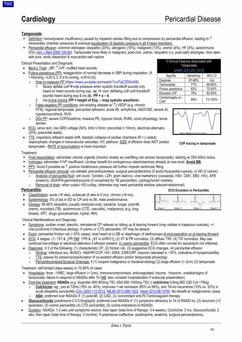
Cardiology Pericardial Disease
Erika J. Parisi
Tamponade: Definition: hemodynamic insufficiency caused by impaired cardiac filling due to compression by pericardial effusion, leading to ↑
intracardiac chamber pressures & eventual equalization of diastolic pressure in all 4 heart chambers. Pericardial effusion: common etiologies: idiopathic (20%), iatrogenic (16%), malignant (13%), uremic (6%), HF (5%), autoimmune
(5%) (Am J Med 2000;109:95). Tamponade more likely in malignant, post-viral, uremic, idiopathic (i.e. post-cath) etiologies. Also seenwith prox. aortic dissection & myocardial wall rupture.
Clinical Presentation and Diagnosis: • Beck’s Triad: ↓BP, ↑JVP, muffled heart sounds.• Pulsus paradoxus (PP): exaggeration of normal decrease in SBP during inspiration. (If
>10mmHg, +LR=3.3. If ≤10 mmHg, +LR=0.03).o How to measure PP (https://www.youtube.com/watch?v=jTsjCZ9QxW8)
1. Slowly deflate cuffnote pressure when systolic Korotkoff sounds onlyheard w/ heart sounds during exp. (a) cont. deflating cuff until Korotkoffsounds heard during exp & ins (b). PP = a – b
2. Via A-line tracing (PP = height of Exp. – Insp systolic waveform)o False-negative PP conditions: pre-existing disease w/ ↑LVEDP (e.g. chronic
HTN), regional tamponade, pericardial adhesion, acute MI, arrhythmia, ASD/VSD, severe AI,hypotension/shock, RVH.
o DDx PP: severe COPD/asthma, massive PE, hypovol shock, RVMI, const physiology, tenseascites.
• ECG: sinus tach, low QRS voltage (50%; limb ≤ 5mm; precordial ≤ 10mm), electrical alternans(20%; precordial leads).
• TTE: inspiratory leftward septal shift, diastolic collapse of cardiac chambers (R > L-sided),respirophasic changes in transvalvular velocities, IVC plethora. SIZE of effusion does NOT predicttamponade - RATE of accumulation is more important.
Treatment: Fluid resuscitation: administer volume urgently (monitor closely as overfilling can worsen tamponade), starting w/ 250-500cc bolus. Inotropes: administer if IVF insufficient. Unclear benefit b/c endogenous catecholamines already at max level. Avoid BB. PPV: Avoid if possible as ↑ positive intrathoracic pressure will further impede ventricular filling. Pericardial effusion removal: via catheter pericardiocentesis, surgical pericardiectomy (if aortic/myocardial rupture), or HD (if uremic)
o Analysis of pericardial fluid: cell count, Tprotein, LDH, gram stain/cx, viral markers/cx (coxsackie, HSV, CMV, EBV, HIV), AFBsmear/cx, ADA/IFN-gamma/lysozyme (if concerned for TB pericarditis), cytology/tumor markers.
o Removal of drain: when output <50 cc/day, otherwise may need pericardial window (pleural>abdominal).Pericarditis: • Classification: acute (<6 wks), subacute (6 wks to 6 mo), chronic (>6 mo).• Epidemiology: 5% of pts in ED w/ CP and no MI, male predominance.• Etiology: 85-90% idiopathic (usually viral/post-viral), bacterial, fungal, post-MI,
uremic, mycobact.(TB), autoimmune (CTD, vasculitis), malignancy (e.g. lung,breast), XRT, drugs (procainamide, hydral, INH)
Clinical Manifestations and Diagnosis: • Symptoms: sudden onset, pleuritic, retrosternal CP relieved w/ sitting up & leaning forward (may radiate to trapezius muscles), +/-
viral prodrome if infectious etiology. If uremic or CTD pericarditis: CP may be absent.• Exam: pericardial friction rub (~30% cases), best heard at LLSB w/ diaphragm of stethoscope at end-expiration w/ pt leaning forward• ECG: 4 stages: (1) ↑ST & ↓PR [NB: ↑PR & ↓ST in aVR/V1]; (2) ST & PR normalize; (3) diffuse TWI; (4) TW normalize. May see
continual low-voltage or electrical alternans if effusion present. In uremic pericarditis: ECG often normal b/c epicardium not inflamed.• Diagnosis: ≥ 2 of the following: (1) characteristic CP, (2) friction rub, (3) suggestive ECG changes, (4) pericardial effusion
o Workup: infectious w/u, BUN/Cr, ANA/RF/CCP, HIV, IGRA, ESR/CRP, troponin (elevated in ~30%, indicative of myopericarditis)o TTE: assess for presence/size/location of co-existent effusion and/or tamponade physiologyo Pericardiocentesis/Surgical Drainage: if (1) suspect malignancy or bacterial etiology (2) large effusion (> 2cm) (3) tamponade
Treatment: self-limited (days-weeks) in 70-90% of cases • Hospitalize: fever, ↑WBC, large effusion (> 2cm), immunocompromised, anticoagulated, trauma, ↑troponin, unstable/signs of
tamponade, failure to respond to NSAIDs after 7d (NB: also consider hospitalization if subacute presentation)• First-line treatment: NSAIDs (e.g. ibuprofen 600-800mg TID; ASA 650-1000mg TID) + colchicine 0.6mg BID (QD if pt <70kg)
o Colchicine: sig ↓sxs at 72hrs (19% vs. 40%), improves 1-wk remission (85% vs 58%), and 18-mo recurrence (10% vs. 32%) inacute idiopathic pericarditis (Circ 2005;112:2012, NEJM 2013;369:1522, Heart 2012;98:1078). No benefit w/ maligt/uremic cases
o ASA: preferred over NSAIDs if: (1) post-MI, (2) CAD, (3) concomitant anti-PLT/anticoagulant therapy• Glucocorticoids (prednisone 0.2-0.5mg/kg/d): preferred over NSAIDs if: (1) symptoms refractory to 7d of NSAID trx; (2) recurrent (>2
episodes); (3) uremic pericarditis (4) CTD pericarditis; (5) contra-indications to NSAIDs• Duration: NSAIDs: 1-2 wks until symptoms resolve, then taper (total time of therapy: 3-4 weeks); Colchicine: 3 mo; Glucocorticoids: 2
wks, then taper (total time of therapy: 3 months). If prednisone ineffective: azathioprine, anakinra, surgical pericardectomy.
5 Clinical Features Associated with Tamponade
(JAMA 2007;297:1810) Sign/Sx Sensitivity 95% CI
Dyspnea 87-88% n/a Tachycardia 77% 69-85%Pulsus paradoxus 82% 72-92%Elevated JVP 76% 62-90%Cardiomegaly on CXR 89% 73-100%
ECG Evolution in Pericarditis
CVP tracing in tamponade
34
Cardiology Aortic Disease
Sumeet Khetarpal
Aortic Aneurysms (JACC 2016;68:1054)
AAA TAA Epidemiology - 4-6:1 M:F ratio
- 4-8% if age >65- Most infrarenal
- 1.7:1 M:F ratio- Mostly 50s-70s- 50% Asc Ao, 40% Descending Ao, 10% Arch
Etiology - Usually due to atherosclerotic disease- Risk factors: smoking, male sex, age, pre-existingatherosclerosis, obesity, HLD, HTN, FHx
- Atherosclerotic: Majority of cases. Mostly indescending Ao. Risk factors: smoking, HLD, HTN- Structural/genetic: Mostly in root and ascendingaorta. Causes: CTD disease (Marfan, Ehlers-Danlos,Loeys-Dietz), Turner, bicuspid AoV, trauma- Infectious: 3° syphilis, mycotic aneurysm (mostcommon org: Staph spp., Salmonella spp.)- Inflammatory: GCA (~10% have TAA), Takayasu,RA, psoriasis, Behcet’s, Wegener’s, IgG4
Screening/ Surveillance
- ACC/AHA: One-time abd. ultrasound in all men >60 w/FHx of AAA (IIC) and all men >65 that have ever smoked(IA)- USPSTF: One-time abd. ultrasound for men age 65-75who have ever smoked (Grade B) and selective screeningfor male never smokers 65-75 (Grade C). Screeningwomen not recommended.- Surveillance:3-3.4 cm: U/S q3y3.5-4.4 cm: U/S or CT q12mo4.5-5.4 cm: U/S or CT q6mo
- General population: Not recommended- Indications: At time of dx of Marfan (IC), Turner (IC),Loeys-Dietz, Takayasu or GCA. 1st deg relatives of ptw/ TAA, dissection, bicuspid valve (IB/IC).- Surveillance: If aneurysm only, then same as AAA. Ifalso with dissection, image at 1, 3, 6, & 12 months thenannually. Image entire aorta (CT/MRI) if multipleaneurysms (~25% TAA will have AAA; ~25% AAA willhave TAA).
Imaging Modalities
- Abdominal U/S: screening and surveillance of infrarenal AAAs. High Se/Sp (>90%), operator-dependent- CT w/ contrast: high Se/Sp, better than U/S for suprarenal AAAs- MRI/MRA: good Se/Sp, preferred for aortic root imaging and for imaging tortuous aortas- CXR: “enlarged aorta” nonspecific (tortuous aorta vs. aneurysm)- TTE: useful for root and proximal thoracic aorta; TEE: will visualize entire thoracic aorta but rarely used.
Treatment Medical: - Smoking cessation (slows AAA growth by up to 25%)- Reduce BP in accordance with ACC/AHA standards- Meds: Statins (reduce all-cause mortality in pts s/psurgery); BBs (may slow expansion; IA for perioperativeuse); ACEi (controversial; may prevent rupture but mayspeed growth); low dose ASA (may slow growth);antibiotics (e.g., roxithromycin may reduce expansion rate,not mortality)
Surgical: - Men: >5.5cm OR growing at >0.5cm/yr ORsymptomatic Women: >4.5-5cm (controversial)- Open repair (~4-6% 30 day mortality) vs. EVAR (only~50% suitable, c/b endoleaks [continued blood flow intoaneurysmal cavity, ~1% 30 day mortality]).
Medical: - Reduce BP (<140/90 or <130/80 if DM or CKD; littleactual evidence, IB)- BBs proven to decrease TAA growth in Marfan- ARBs slow aortic root aneurysm expansion in Marfanpatients, likely via TGF-B inhibition (NEJM 2008;358:2787)- Statins LDL<70, some evidence- Smoking cessation; avoid straining- Stress test used to guide BP management (followSBP response to stress)
Surgical: - Root/ascending TAAs: usually concomitant aorticvalve replacement- Arch/descending TAAs: mostly open graft, (EVAR).Ischemic brain/spine injury most worrisomecomplication.
Complications - Rupture: Devastating mortality. AAA annual rupture rates are 4%, 7%, 20% at 5, 6, and 7cm, respectively. Riskfactors: size, rate of expansion, female gender
- Symptoms: Triad of abd/back pain + pulsatile abd mass + hypotension immediate OR (don’t image)- Dissection: pain (chest/abdomen/back), occlusion of aortic vessels, thromboembolism- Post-repair: EVAR: endoleak, graft failure, thrombosis. Open: MI, embolization, AKI, ischemic colitis
35
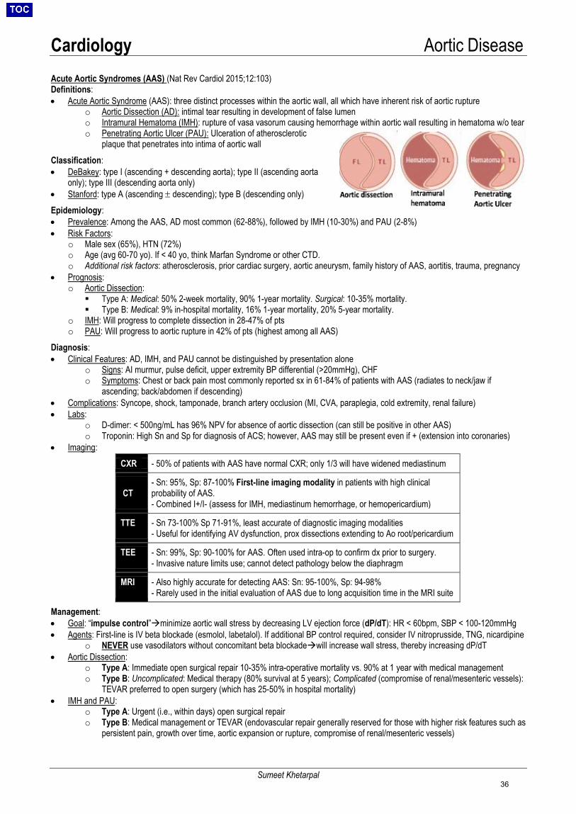
Cardiology Aortic Disease
Sumeet Khetarpal
Acute Aortic Syndromes (AAS) (Nat Rev Cardiol 2015;12:103) Definitions: • Acute Aortic Syndrome (AAS): three distinct processes within the aortic wall, all which have inherent risk of aortic rupture
o Aortic Dissection (AD): intimal tear resulting in development of false lumeno Intramural Hematoma (IMH): rupture of vasa vasorum causing hemorrhage within aortic wall resulting in hematoma w/o tearo Penetrating Aortic Ulcer (PAU): Ulceration of atherosclerotic
plaque that penetrates into intima of aortic wallClassification: • DeBakey: type I (ascending + descending aorta); type II (ascending aorta
only); type III (descending aorta only)• Stanford: type A (ascending ± descending); type B (descending only)Epidemiology:• Prevalence: Among the AAS, AD most common (62-88%), followed by IMH (10-30%) and PAU (2-8%)• Risk Factors:
o Male sex (65%), HTN (72%)o Age (avg 60-70 yo). If < 40 yo, think Marfan Syndrome or other CTD.o Additional risk factors: atherosclerosis, prior cardiac surgery, aortic aneurysm, family history of AAS, aortitis, trauma, pregnancy
• Prognosis:o Aortic Dissection:
Type A: Medical: 50% 2-week mortality, 90% 1-year mortality. Surgical: 10-35% mortality. Type B: Medical: 9% in-hospital mortality, 16% 1-year mortality, 20% 5-year mortality.
o IMH: Will progress to complete dissection in 28-47% of ptso PAU: Will progress to aortic rupture in 42% of pts (highest among all AAS)
Diagnosis: • Clinical Features: AD, IMH, and PAU cannot be distinguished by presentation alone
o Signs: AI murmur, pulse deficit, upper extremity BP differential (>20mmHg), CHFo Symptoms: Chest or back pain most commonly reported sx in 61-84% of patients with AAS (radiates to neck/jaw if
ascending; back/abdomen if descending)• Complications: Syncope, shock, tamponade, branch artery occlusion (MI, CVA, paraplegia, cold extremity, renal failure)• Labs:
o D-dimer: < 500ng/mL has 96% NPV for absence of aortic dissection (can still be positive in other AAS)o Troponin: High Sn and Sp for diagnosis of ACS; however, AAS may still be present even if + (extension into coronaries)
• Imaging:
Management: • Goal: “impulse control”minimize aortic wall stress by decreasing LV ejection force (dP/dT): HR < 60bpm, SBP < 100-120mmHg• Agents: First-line is IV beta blockade (esmolol, labetalol). If additional BP control required, consider IV nitroprusside, TNG, nicardipine
o NEVER use vasodilators without concomitant beta blockadewill increase wall stress, thereby increasing dP/dT• Aortic Dissection:
o Type A: Immediate open surgical repair 10-35% intra-operative mortality vs. 90% at 1 year with medical managemento Type B: Uncomplicated: Medical therapy (80% survival at 5 years); Complicated (compromise of renal/mesenteric vessels):
TEVAR preferred to open surgery (which has 25-50% in hospital mortality)• IMH and PAU:
o Type A: Urgent (i.e., within days) open surgical repairo Type B: Medical management or TEVAR (endovascular repair generally reserved for those with higher risk features such as
persistent pain, growth over time, aortic expansion or rupture, compromise of renal/mesenteric vessels)
CXR - 50% of patients with AAS have normal CXR; only 1/3 will have widened mediastinum
CT - Sn: 95%, Sp: 87-100% First-line imaging modality in patients with high clinicalprobability of AAS.- Combined I+/I- (assess for IMH, mediastinum hemorrhage, or hemopericardium)
TTE - Sn 73-100% Sp 71-91%, least accurate of diagnostic imaging modalities- Useful for identifying AV dysfunction, prox dissections extending to Ao root/pericardium
TEE - Sn: 99%, Sp: 90-100% for AAS. Often used intra-op to confirm dx prior to surgery.- Invasive nature limits use; cannot detect pathology below the diaphragm
MRI - Also highly accurate for detecting AAS: Sn: 95-100%, Sp: 94-98%- Rarely used in the initial evaluation of AAS due to long acquisition time in the MRI suite
36
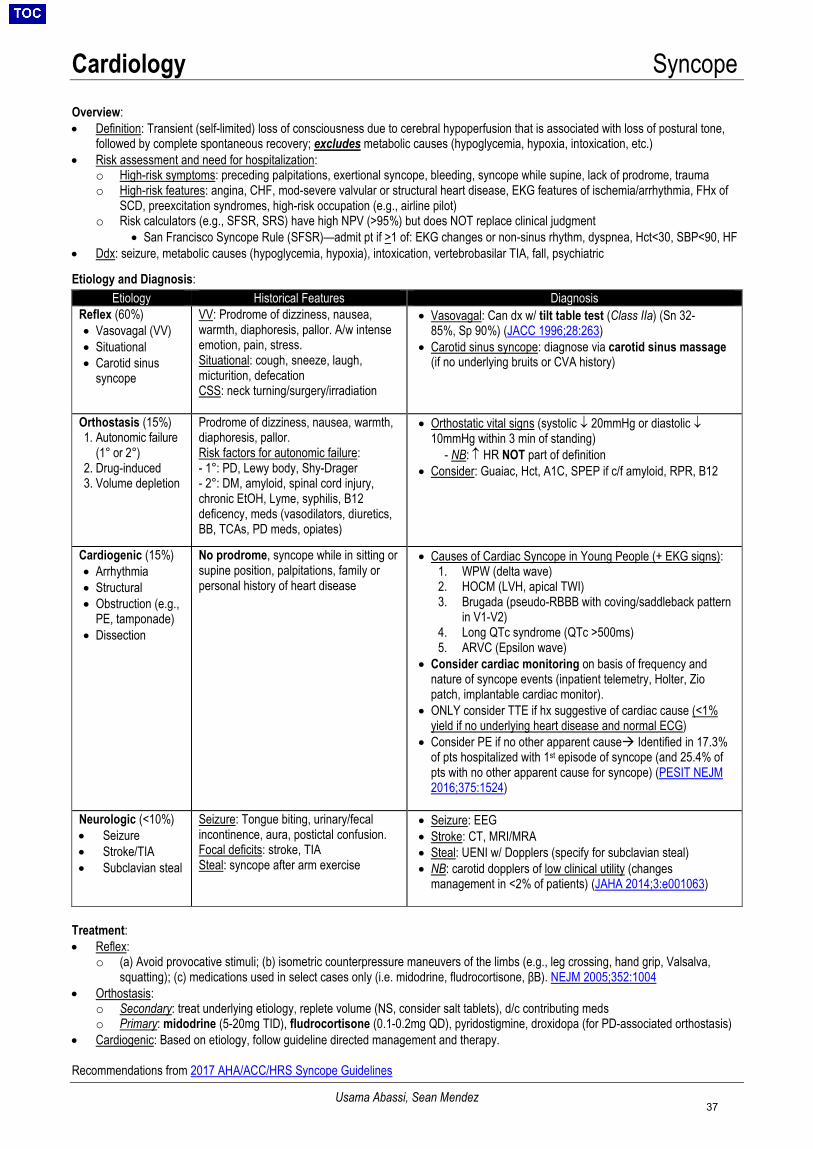
Cardiology Syncope
Usama Abassi, Sean Mendez
Overview: • Definition: Transient (self-limited) loss of consciousness due to cerebral hypoperfusion that is associated with loss of postural tone,
followed by complete spontaneous recovery; excludes metabolic causes (hypoglycemia, hypoxia, intoxication, etc.)• Risk assessment and need for hospitalization:
o High-risk symptoms: preceding palpitations, exertional syncope, bleeding, syncope while supine, lack of prodrome, traumao High-risk features: angina, CHF, mod-severe valvular or structural heart disease, EKG features of ischemia/arrhythmia, FHx of
SCD, preexcitation syndromes, high-risk occupation (e.g., airline pilot)o Risk calculators (e.g., SFSR, SRS) have high NPV (>95%) but does NOT replace clinical judgment
• San Francisco Syncope Rule (SFSR)—admit pt if >1 of: EKG changes or non-sinus rhythm, dyspnea, Hct<30, SBP<90, HF• Ddx: seizure, metabolic causes (hypoglycemia, hypoxia), intoxication, vertebrobasilar TIA, fall, psychiatric
Etiology and Diagnosis:
Etiology Historical Features Diagnosis Reflex (60%) • Vasovagal (VV)• Situational• Carotid sinus
syncope
VV: Prodrome of dizziness, nausea, warmth, diaphoresis, pallor. A/w intense emotion, pain, stress. Situational: cough, sneeze, laugh, micturition, defecation CSS: neck turning/surgery/irradiation
• Vasovagal: Can dx w/ tilt table test (Class IIa) (Sn 32-85%, Sp 90%) (JACC 1996;28:263)
• Carotid sinus syncope: diagnose via carotid sinus massage(if no underlying bruits or CVA history)
Orthostasis (15%) 1. Autonomic failure
(1° or 2°)2. Drug-induced3. Volume depletion
Prodrome of dizziness, nausea, warmth, diaphoresis, pallor. Risk factors for autonomic failure: - 1°: PD, Lewy body, Shy-Drager- 2°: DM, amyloid, spinal cord injury,chronic EtOH, Lyme, syphilis, B12deficency, meds (vasodilators, diuretics,BB, TCAs, PD meds, opiates)
• Orthostatic vital signs (systolic ↓ 20mmHg or diastolic ↓10mmHg within 3 min of standing)
- NB: ↑ HR NOT part of definition• Consider: Guaiac, Hct, A1C, SPEP if c/f amyloid, RPR, B12
Cardiogenic (15%) • Arrhythmia• Structural• Obstruction (e.g.,
PE, tamponade)• Dissection
No prodrome, syncope while in sitting or supine position, palpitations, family or personal history of heart disease
• Causes of Cardiac Syncope in Young People (+ EKG signs):1. WPW (delta wave)2. HOCM (LVH, apical TWI)3. Brugada (pseudo-RBBB with coving/saddleback pattern
in V1-V2)4. Long QTc syndrome (QTc >500ms)5. ARVC (Epsilon wave)
• Consider cardiac monitoring on basis of frequency andnature of syncope events (inpatient telemetry, Holter, Ziopatch, implantable cardiac monitor).
• ONLY consider TTE if hx suggestive of cardiac cause (<1%yield if no underlying heart disease and normal ECG)
• Consider PE if no other apparent cause Identified in 17.3%of pts hospitalized with 1st episode of syncope (and 25.4% ofpts with no other apparent cause for syncope) (PESIT NEJM2016;375:1524)
Neurologic (<10%) • Seizure• Stroke/TIA• Subclavian steal
Seizure: Tongue biting, urinary/fecal incontinence, aura, postictal confusion. Focal deficits: stroke, TIA Steal: syncope after arm exercise
• Seizure: EEG• Stroke: CT, MRI/MRA• Steal: UENI w/ Dopplers (specify for subclavian steal)• NB: carotid dopplers of low clinical utility (changes
management in <2% of patients) (JAHA 2014;3:e001063)
Treatment: • Reflex:
o (a) Avoid provocative stimuli; (b) isometric counterpressure maneuvers of the limbs (e.g., leg crossing, hand grip, Valsalva,squatting); (c) medications used in select cases only (i.e. midodrine, fludrocortisone, βB). NEJM 2005;352:1004
• Orthostasis:o Secondary: treat underlying etiology, replete volume (NS, consider salt tablets), d/c contributing medso Primary: midodrine (5-20mg TID), fludrocortisone (0.1-0.2mg QD), pyridostigmine, droxidopa (for PD-associated orthostasis)
• Cardiogenic: Based on etiology, follow guideline directed management and therapy.
Recommendations from 2017 AHA/ACC/HRS Syncope Guidelines
37
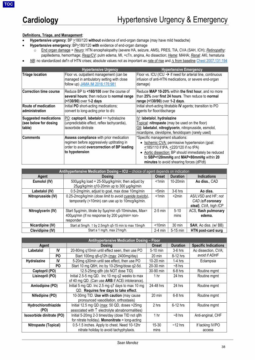
Cardiology Hypertensive Urgency & Emergency
Sean Mendez
Definitions, Triage, and Management: • Hypertensive urgency: BP >180/120 without evidence of end-organ damage (may have mild headache)• Hypertensive emergency: BP>180/120 with evidence of end-organ damage
o End organ damage = Neuro: HTN encephalopathy (severe HA, seizure, AMS), PRES, TIA, CVA (SAH, ICH); Retinopathy:papilledema, hemorrhage; Resp/CV: pulm edema, MI, +cTn, angina, Ao dissection; Heme: MAHA; Renal: AKI, hematuria
• NB: no standardized def’n of HTN crises; absolute values not as important as rate of rise and ∆ from baseline Chest 2007;131;194
Hypertensive Urgency Hypertensive Emergency Triage location Floor vs. outpatient management (can be
managed in ambulatory setting with close follow up) JAMA IM 2016;176:981
Floor vs. ICU (ICU if need for arterial line, continuous infusion of anti-HTN medications, or severe end-organ damage)
Correction time course Reduce BP to <160/100 over the course of several hours; then reduce to normal range (<130/90) over 1-2 days
Reduce MAP 10-20% within the first hour, and no more than 25% over first 24 hours. Then reduce to normal range (<130/90) over 1-2 days.
Route of medication administration
Initial PO short-acting medications; convert to long-acting prior to d/c
Initial short-acting titratable IV agents; transition to PO agents for floor/discharge
Suggested medications (see below for dosing table)
PO: captopril, labetalol >> hydralazine (unpredictable effect, reflex tachycardia), isosorbide dinitrate
IV: labetalol, hydralazine Topical: nitropaste (may be used on the floor) Gtt: labetalol, nitroglycerin, nitroprusside, esmolol, nicardipine, clevidipine, fenoldopam (rarely used)
Comments Assess compliance with prior medication regimen before aggressively uptitrating in order to avoid overcorrection of BP leading to hypotension
*Specific management situations:• Ischemic CVA: permissive hypertension (goal:
<185/110 if tPA, <220/120 if no tPA)• Aortic dissection: BP should immediately be reduced
to SBP<120mmHg and MAP<80mmHg within 20minutes to avoid shearing forces (dP/dt)
Antihypertensive Medication Dosing – ICU – choice of agent depends on indication Agent Dosing Onset Duration Indications
Esmolol (IV) 500µg/kg load + 25-50µg/kg/min; then adjust by 25µg/kg/min q10-20min up to 300 µg/kg/min
<1min 10-20min Ao diss., CAD
Labetalol (IV) 0.5-2mg/min, adjust to goal, max dose 10mg/min <5min 3-6 hrs Ao diss. Nitroprusside (IV) 0.25-2mcg/kg/min (dose limit to avoid cyanide toxicity),
temporarily (<10min) can use up to 10mcg/kg/min. <1min <2min AS/LVSD and HF; not
CAD (c/f coronary steal), CVA, high ICP
Nitroglycerin (IV) Start 5µg/min, titrate by 5µq/min q5-10minutes, Max= 400µq/min (If no response by 200 µq/min= non-responder
2-5 min 5-10mins
ACS, flash pulmonary edema.
Nicardipine (IV) Start at 5mg/h. ↑ by 2.5mg/h q5-15 min to max 15mg/h <10min 30 min SAH, Ao diss. (w/ BB) Clevidipine (IV) Start a 1 mg/h, max 21mg/h. 2-4 min 5-15 min HTN post-card surg
Antihypertensive Medication Dosing – Floor Agent Dosing Onset Duration Specific Indications
Labetalol IV 20-80mg q10min until effect seen, then use PO 5-10 min 3-6 hrs Ao dissection, CVA; avoid if ADHF PO Start 100mg q8-q12h (max: 2400mg/day) 20 min 8-12 hrs
Hydralazine IV 5-20mg q30min until see effect, then use PO 10-20 min 1-4 hrs Eclampsia PO Start 10 mg Q6H, inc by 10-25mg/dose q2-5d. 20-30 min ~8 hrs
Captopril (PO) 12.5-25mg q8h (do NOT dose TID) 30-90 min 6-8 hrs Routine mgmt Lisinopril (PO) Initial 2.5-5 mg QD. Inc 10 mg q2 weeks to max
of 40 mg QD. (Can use ARB if ACEi intolerance). 1 hr 24 hrs Routine mgmt
Amlodipine (PO) Initial 5 mg QD. Inc 2.5 mg q7 days to max 10 mg QD. Requires few days to take effect.
24-48 hrs 24 hrs Routine mgmt
Nifedipine (PO) 10-30mg TID. Use with caution (may causepronounced vasodilation, orthostasis)
20 min 6-8 hrs Routine mgmt
Hydrochlorothiazide (PO)
Initial 12.5 mg QD (max: 50 QD, doses >25mg associated with ↑ electrolyte abnabnormalities)
2 hrs 6-12 hrs Routine mgmt
Isosorbide dinitrate (PO) Initial 5-20mg 2-3 times/day (dose TID not q8h for nitrate holiday). Mononitrate = long-acting
1 hr ~8 hrs Anti-anginal, CHF
Nitropaste (Topical) 0.5-1.5 inches. Apply to chest. Need 10-12hr nitrate holiday to avoid tachyphylaxis.
15-30mins
~12 hrs If lacking IV/PO access
38
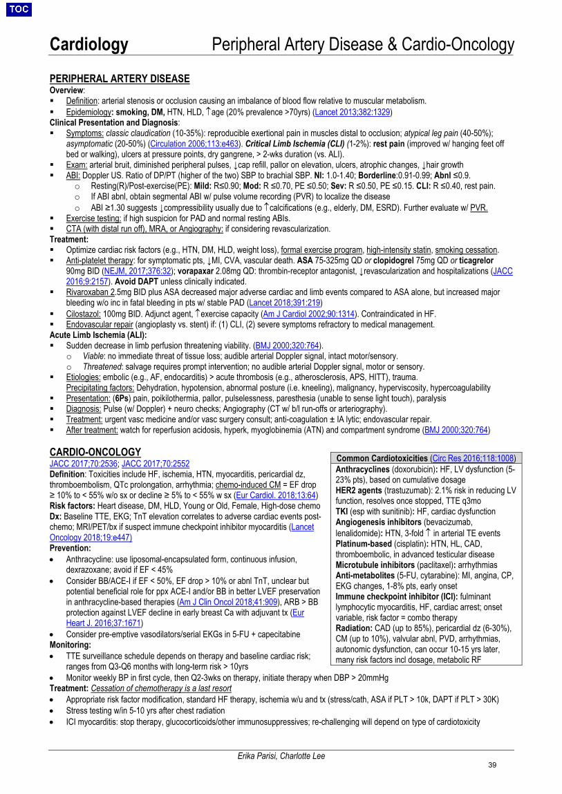
Cardiology Peripheral Artery Disease & Cardio-Oncology
Erika Parisi, Charlotte Lee
PERIPHERAL ARTERY DISEASE Overview: Definition: arterial stenosis or occlusion causing an imbalance of blood flow relative to muscular metabolism. Epidemiology: smoking, DM, HTN, HLD, ↑age (20% prevalence >70yrs) (Lancet 2013;382:1329) Clinical Presentation and Diagnosis: Symptoms: classic claudication (10-35%): reproducible exertional pain in muscles distal to occlusion; atypical leg pain (40-50%);
asymptomatic (20-50%) (Circulation 2006;113:e463). Critical Limb Ischemia (CLI) (1-2%): rest pain (improved w/ hanging feet offbed or walking), ulcers at pressure points, dry gangrene, > 2-wks duration (vs. ALI).
Exam: arterial bruit, diminished peripheral pulses, ↓cap refill, pallor on elevation, ulcers, atrophic changes, ↓hair growth ABI: Doppler US. Ratio of DP/PT (higher of the two) SBP to brachial SBP. Nl: 1.0-1.40; Borderline:0.91-0.99; Abnl ≤0.9.
o Resting(R)/Post-exercise(PE): Mild: R≤0.90; Mod: R ≤0.70, PE ≤0.50; Sev: R ≤0.50, PE ≤0.15. CLI: R ≤0.40, rest pain.o If ABI abnl, obtain segmental ABI w/ pulse volume recording (PVR) to localize the diseaseo ABI ≥1.30 suggests ↓compressibility usually due to ↑calcifications (e.g., elderly, DM, ESRD). Further evaluate w/ PVR.
Exercise testing: if high suspicion for PAD and normal resting ABIs. CTA (with distal run off), MRA, or Angiography: if considering revascularization.Treatment: Optimize cardiac risk factors (e.g., HTN, DM, HLD, weight loss), formal exercise program, high-intensity statin, smoking cessation. Anti-platelet therapy: for symptomatic pts, ↓MI, CVA, vascular death. ASA 75-325mg QD or clopidogrel 75mg QD or ticagrelor
90mg BID (NEJM, 2017;376:32); vorapaxar 2.08mg QD: thrombin-receptor antagonist, ↓revascularization and hospitalizations (JACC2016;9:2157). Avoid DAPT unless clinically indicated.
Rivaroxaban 2.5mg BID plus ASA decreased major adverse cardiac and limb events compared to ASA alone, but increased majorbleeding w/o inc in fatal bleeding in pts w/ stable PAD (Lancet 2018;391:219)
Cilostazol: 100mg BID. Adjunct agent, ↑exercise capacity (Am J Cardiol 2002;90:1314). Contraindicated in HF. Endovascular repair (angioplasty vs. stent) if: (1) CLI, (2) severe symptoms refractory to medical management.Acute Limb Ischemia (ALI): Sudden decrease in limb perfusion threatening viability. (BMJ 2000;320:764).
o Viable: no immediate threat of tissue loss; audible arterial Doppler signal, intact motor/sensory.o Threatened: salvage requires prompt intervention; no audible arterial Doppler signal, motor or sensory.
Etiologies: embolic (e.g., AF, endocarditis) > acute thrombosis (e.g., atherosclerosis, APS, HITT), trauma.Precipitating factors: Dehydration, hypotension, abnormal posture (i.e. kneeling), malignancy, hyperviscosity, hypercoagulability
Presentation: (6Ps) pain, poikilothermia, pallor, pulselessness, paresthesia (unable to sense light touch), paralysis Diagnosis: Pulse (w/ Doppler) + neuro checks; Angiography (CT w/ b/l run-offs or arteriography). Treatment: urgent vasc medicine and/or vasc surgery consult; anti-coagulation ± IA lytic; endovascular repair. After treatment: watch for reperfusion acidosis, hyperk, myoglobinemia (ATN) and compartment syndrome (BMJ 2000;320:764)
CARDIO-ONCOLOGY JACC 2017;70:2536; JACC 2017;70:2552 Definition: Toxicities include HF, ischemia, HTN, myocarditis, pericardial dz, thromboembolism, QTc prolongation, arrhythmia; chemo-induced CM = EF drop ≥ 10% to < 55% w/o sx or decline ≥ 5% to < 55% w sx (Eur Cardiol. 2018;13:64) Risk factors: Heart disease, DM, HLD, Young or Old, Female, High-dose chemo Dx: Baseline TTE, EKG; TnT elevation correlates to adverse cardiac events post-chemo; MRI/PET/bx if suspect immune checkpoint inhibitor myocarditis (Lancet Oncology 2018;19:e447) Prevention: • Anthracycline: use liposomal-encapsulated form, continuous infusion,
dexrazoxane; avoid if EF < 45%• Consider BB/ACE-I if EF < 50%, EF drop > 10% or abnl TnT, unclear but
potential beneficial role for ppx ACE-I and/or BB in better LVEF preservationin anthracycline-based therapies (Am J Clin Oncol 2018;41:909), ARB > BBprotection against LVEF decline in early breast Ca with adjuvant tx (EurHeart J. 2016;37:1671)
• Consider pre-emptive vasodilators/serial EKGs in 5-FU + capecitabineMonitoring:• TTE surveillance schedule depends on therapy and baseline cardiac risk;
ranges from Q3-Q6 months with long-term risk > 10yrs• Monitor weekly BP in first cycle, then Q2-3wks on therapy, initiate therapy when DBP > 20mmHgTreatment: Cessation of chemotherapy is a last resort• Appropriate risk factor modification, standard HF therapy, ischemia w/u and tx (stress/cath, ASA if PLT > 10k, DAPT if PLT > 30K)• Stress testing w/in 5-10 yrs after chest radiation• ICI myocarditis: stop therapy, glucocorticoids/other immunosuppressives; re-challenging will depend on type of cardiotoxicity
Common Cardiotoxicities (Circ Res 2016;118:1008) Anthracyclines (doxorubicin): HF, LV dysfunction (5-23% pts), based on cumulative dosage HER2 agents (trastuzumab): 2.1% risk in reducing LV function, resolves once stopped, TTE q3mo TKI (esp with sunitinib): HF, cardiac dysfunction Angiogenesis inhibitors (bevacizumab, lenalidomide): HTN, 3-fold ↑ in arterial TE events Platinum-based (cisplatin): HTN, HL, CAD, thromboembolic, in advanced testicular disease Microtubule inhibitors (paclitaxel): arrhythmias Anti-metabolites (5-FU, cytarabine): MI, angina, CP, EKG changes, 1-8% pts, early onset Immune checkpoint inhibitor (ICI): fulminant lymphocytic myocarditis, HF, cardiac arrest; onset variable, risk factor = combo therapy Radiation: CAD (up to 85%), pericardial dz (6-30%), CM (up to 10%), valvular abnl, PVD, arrhythmias, autonomic dysfunction, can occur 10-15 yrs later, many risk factors incl dosage, metabolic RF
39
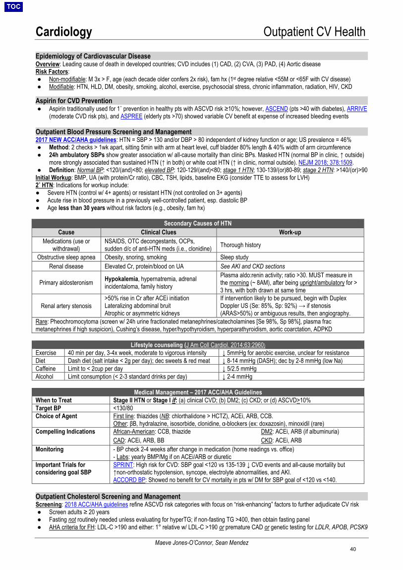
Cardiology Outpatient CV Health
Maeve Jones-O’Connor, Sean Mendez
Epidemiology of Cardiovascular Disease Overview: Leading cause of death in developed countries; CVD includes (1) CAD, (2) CVA, (3) PAD, (4) Aortic disease Risk Factors: ● Non-modifiable: M 3x > F, age (each decade older confers 2x risk), fam hx (1st degree relative <55M or <65F with CV disease)● Modifiable: HTN, HLD, DM, obesity, smoking, alcohol, exercise, psychosocial stress, chronic inflammation, radiation, HIV, CKD
Aspirin for CVD Prevention● Aspirin traditionally used for 1˚ prevention in healthy pts with ASCVD risk ≥10%; however, ASCEND (pts >40 with diabetes), ARRIVE
(moderate CVD risk pts), and ASPREE (elderly pts >70) showed variable CV benefit at expense of increased bleeding events
Outpatient Blood Pressure Screening and Management 2017 NEW ACC/AHA guidelines: HTN = SBP > 130 and/or DBP > 80 independent of kidney function or age; US prevalence = 46% ● Method: 2 checks > 1wk apart, sitting 5min with arm at heart level, cuff bladder 80% length & 40% width of arm circumference● 24h ambulatory SBPs show greater association w/ all-cause mortality than clinic BPs. Masked HTN (normal BP in clinic, ↑ outside)
more strongly associated than sustained HTN (↑ in both) or white coat HTN (↑ in clinic, normal outside). NEJM 2018; 378:1509.● Definition: Normal BP: <120/(and)<80; elevated BP: 120-129/(and)<80; stage 1 HTN; 130-139/(or)80-89; stage 2 HTN: >140/(or)>90
Initial Workup: BMP, UA (with protein/Cr ratio), CBC, TSH, lipids, baseline EKG (consider TTE to assess for LVH)2˚ HTN: Indications for workup include:● Severe HTN (control w/ 4+ agents) or resistant HTN (not controlled on 3+ agents)● Acute rise in blood pressure in a previously well-controlled patient, esp. diastolic BP● Age less than 30 years without risk factors (e.g., obesity, fam hx)
Secondary Causes of HTN Cause Clinical Clues Work-up
Medications (use or withdrawal)
NSAIDS, OTC decongestants, OCPs, sudden d/c of anti-HTN meds (i.e., clonidine) Thorough history
Obstructive sleep apnea Obesity, snoring, smoking Sleep study Renal disease Elevated Cr, protein/blood on UA See AKI and CKD sections
Primary aldosteronism Hypokalemia, hypernatremia, adrenal incidentaloma, family history
Plasma aldo:renin activity; ratio >30. MUST measure in the morning (~ 8AM), after being upright/ambulatory for > 3 hrs, with both drawn at same time
Renal artery stenosis >50% rise in Cr after ACEi initiationLateralizing abdominal bruitAtrophic or asymmetric kidneys
If intervention likely to be pursued, begin with Duplex Doppler US (Se: 85%, Sp: 92%) → if stenosis (ARAS>50%) or ambiguous results, then angiography.
Rare: Pheochromocytoma (screen w/ 24h urine fractionated metanephrines/catecholamines [Se 98%, Sp 98%], plasma frac metanephrines if high suspicion), Cushing’s disease, hyper/hypothyroidism, hyperparathyroidism, aortic coarctation, ADPKD
Lifestyle counseling (J Am Coll Cardiol. 2014;63:2960) Exercise 40 min per day, 3-4x week, moderate to vigorous intensity ↓ 5mmHg for aerobic exercise, unclear for resistance Diet Dash diet (salt intake < 2g per day); dec sweets & red meat ↓ 8-14 mmHg (DASH); dec by 2-8 mmHg (low Na) Caffeine Limit to < 2cup per day ↓ 5/2.5 mmHg Alcohol Limit consumption (< 2-3 standard drinks per day) ↓ 2-4 mmHg
Medical Management – 2017 ACC/AHA GuidelinesWhen to Treat Stage II HTN or Stage I if: (a) clinical CVD; (b) DM2; (c) CKD; or (d) ASCVD>10% Target BP <130/80 Choice of Agent First line: thiazides (NB: chlorthalidone > HCTZ), ACEi, ARB, CCB.
Other: βB, hydralazine, isosorbide, clonidine, α-blockers (ex: doxazosin), minoxidil (rare) Compelling Indications African-American: CCB, thiazide DM2: ACEi, ARB (if albuminuria)
CAD: ACEi, ARB, BB CKD: ACEi, ARB Monitoring - BP check 2-4 weeks after change in medication (home readings vs. office)
- Labs: yearly BMP/Mg if on ACEi/ARB or diureticImportant Trials for considering goal SBP
SPRINT: High risk for CVD: SBP goal <120 vs 135-139 ↓ CVD events and all-cause mortality but ↑non-orthostatic hypotension, syncope, electrolyte abnormalities, and AKI. ACCORD BP: Showed no benefit for CV mortality in pts w/ DM for SBP goal of <120 vs <140.
Outpatient Cholesterol Screening and Management Screening: 2018 ACC/AHA guidelines refine ASCVD risk categories with focus on “risk-enhancing” factors to further adjudicate CV risk ● Screen adults ≥ 20 years● Fasting not routinely needed unless evaluating for hyperTG; if non-fasting TG >400, then obtain fasting panel● AHA criteria for FH: LDL-C >190 and either: 1° relative w/ LDL-C >190 or premature CAD or genetic testing for LDLR, APOB, PCSK9
40
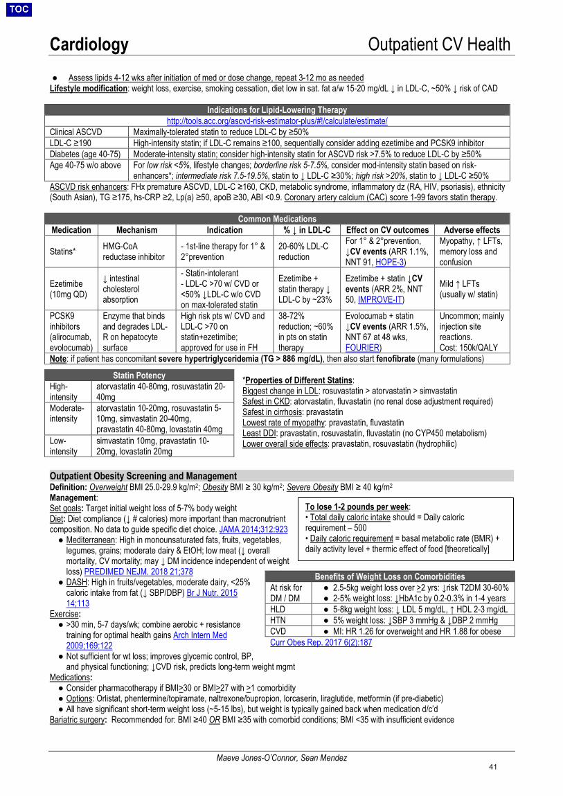
Cardiology Outpatient CV Health
Maeve Jones-O’Connor, Sean Mendez
● Assess lipids 4-12 wks after initiation of med or dose change, repeat 3-12 mo as neededLifestyle modification: weight loss, exercise, smoking cessation, diet low in sat. fat a/w 15-20 mg/dL ↓ in LDL-C, ~50% ↓ risk of CAD
Indications for Lipid-Lowering Therapy http://tools.acc.org/ascvd-risk-estimator-plus/#!/calculate/estimate/
Clinical ASCVD Maximally-tolerated statin to reduce LDL-C by ≥50% LDL-C ≥190 High-intensity statin; if LDL-C remains ≥100, sequentially consider adding ezetimibe and PCSK9 inhibitor Diabetes (age 40-75) Moderate-intensity statin; consider high-intensity statin for ASCVD risk >7.5% to reduce LDL-C by ≥50% Age 40-75 w/o above For low risk <5%, lifestyle changes; borderline risk 5-7.5%, consider mod-intensity statin based on risk-
enhancers*; intermediate risk 7.5-19.5%, statin to ↓ LDL-C ≥30%; high risk >20%, statin to ↓ LDL-C ≥50% ASCVD risk enhancers: FHx premature ASCVD, LDL-C ≥160, CKD, metabolic syndrome, inflammatory dz (RA, HIV, psoriasis), ethnicity (South Asian), TG ≥175, hs-CRP ≥2, Lp(a) ≥50, apoB ≥30, ABI <0.9. Coronary artery calcium (CAC) score 1-99 favors statin therapy.
Common Medications Medication Mechanism Indication % ↓ in LDL-C Effect on CV outcomes Adverse effects
Statins* HMG-CoA reductase inhibitor
- 1st-line therapy for 1° &2°prevention
20-60% LDL-Creduction
For 1° & 2°prevention, ↓CV events (ARR 1.1%, NNT 91, HOPE-3)
Myopathy, ↑ LFTs, memory loss and confusion
Ezetimibe (10mg QD)
↓ intestinal cholesterol absorption
- Statin-intolerant- LDL-C >70 w/ CVD or<50% ↓LDL-C w/o CVDon max-tolerated statin
Ezetimibe + statin therapy ↓ LDL-C by ~23%
Ezetimibe + statin ↓CV events (ARR 2%, NNT 50, IMPROVE-IT)
Mild ↑ LFTs (usually w/ statin)
PCSK9 inhibitors (alirocumab, evolocumab)
Enzyme that binds and degrades LDL-R on hepatocyte surface
High risk pts w/ CVD and LDL-C >70 on statin+ezetimibe; approved for use in FH
38-72% reduction; ~60%in pts on statintherapy
Evolocumab + statin ↓CV events (ARR 1.5%, NNT 67 at 48 wks, FOURIER)
Uncommon; mainly injection site reactions. Cost: 150k/QALY
Note: if patient has concomitant severe hypertriglyceridemia (TG > 886 mg/dL), then also start fenofibrate (many formulations)
*Properties of Different Statins:Biggest change in LDL: rosuvastatin > atorvastatin > simvastatinSafest in CKD: atorvastatin, fluvastatin (no renal dose adjustment required)Safest in cirrhosis: pravastatinLowest rate of myopathy: pravastatin, fluvastatinLeast DDI: pravastatin, rosuvastatin, fluvastatin (no CYP450 metabolism)Lower overall side effects: pravastatin, rosuvastatin (hydrophilic)
Outpatient Obesity Screening and Management Definition: Overweight BMI 25.0-29.9 kg/m2; Obesity BMI ≥ 30 kg/m2; Severe Obesity BMI ≥ 40 kg/m2 Management: Set goals: Target initial weight loss of 5-7% body weight Diet: Diet compliance (↓ # calories) more important than macronutrient composition. No data to guide specific diet choice. JAMA 2014;312:923● Mediterranean: High in monounsaturated fats, fruits, vegetables,
legumes, grains; moderate dairy & EtOH; low meat (↓ overallmortality, CV mortality; may ↓ DM incidence independent of weightloss) PREDIMED NEJM. 2018 21;378
● DASH: High in fruits/vegetables, moderate dairy, <25% caloric intake from fat (↓ SBP/DBP) Br J Nutr. 2015 14;113
Exercise: ● >30 min, 5-7 days/wk; combine aerobic + resistance
training for optimal health gains Arch Intern Med2009;169:122
● Not sufficient for wt loss; improves glycemic control, BP,and physical functioning; ↓CVD risk, predicts long-term weight mgmt
Medications: ● Consider pharmacotherapy if BMI>30 or BMI>27 with >1 comorbidity● Options: Orlistat, phentermine/topiramate, naltrexone/bupropion, lorcaserin, liraglutide, metformin (if pre-diabetic)● All have significant short-term weight loss (~5-15 lbs), but weight is typically gained back when medication d/c’d
Bariatric surgery: Recommended for: BMI ≥40 OR BMI ≥35 with comorbid conditions; BMI <35 with insufficient evidence
Statin Potency High-intensity
atorvastatin 40-80mg, rosuvastatin 20-40mg
Moderate-intensity
atorvastatin 10-20mg, rosuvastatin 5-10mg, simvastatin 20-40mg, pravastatin 40-80mg, lovastatin 40mg
Low-intensity
simvastatin 10mg, pravastatin 10-20mg, lovastatin 20mg
Benefits of Weight Loss on Comorbidities
At risk for DM / DM
● 2.5-5kg weight loss over >2 yrs: ↓risk T2DM 30-60%● 2-5% weight loss: ↓HbA1c by 0.2-0.3% in 1-4 years
HLD ● 5-8kg weight loss: ↓ LDL 5 mg/dL, ↑ HDL 2-3 mg/dLHTN ● 5% weight loss: ↓SBP 3 mmHg & ↓DBP 2 mmHgCVD ● MI: HR 1.26 for overweight and HR 1.88 for obeseCurr Obes Rep. 2017 6(2):187
To lose 1-2 pounds per week: • Total daily caloric intake should = Daily caloricrequirement – 500• Daily caloric requirement = basal metabolic rate (BMR) +daily activity level + thermic effect of food [theoretically]
41
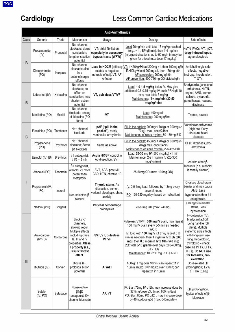
Cardiology Less Common Cardiac Medications
Chitra Mosarla, Usama Abbasi
Anti-Arrhythmics
Class Generic Trade Mechanism Usage Dosing Side effects
IA
Procainamide (IV) Pronestyl
Na+ channel blockade; slows
conduction; lengthens action
potential
VT; atrial fibrillation, especially in accessory
bypass tracts (WPW)
Load 20mg/min until total 17 mg/kg reached (e.g., ~1h, BP q5 min); then 1-4 mg/min
(in urgent situations, up to 50 mg/min may be given for a total max dose 17 mg/kg)
HoTN, PVCs, VT, ↑QT, drug-induced lupus,
agranulocytosis
Disopyramide (PO) Norpace
Na+ channel blockade; also
has anticholinergic
effects
Used in HOCM (efficacy relates to negative
inotropic effect), VT, AF, A-flutter
VT: If <50kgload 200mg x1, then 100mg q6h If >50kgload 200mg x1, then 150mg q6h
AF conversion: 200mg q4-6h AF prevention: 400-750mg QD divided q6h
Anticholinergic side effects, negative
inotropy, hypotension, ↑ QTc
IB
Lidocaine (IV) Xylocaine
Na+ channel blockade; no
effect on conduction; may shorten action
potential
VT, pulseless VT/VF
Load: 1.0-1.5 mg/kg bolus IV. May give additional 0.5-0.75 mg/kg IV push PRN q5-10
min; max total: 3 mg/kg Maintenance: 1-4 mg/min (30-50
mcg/kg/min)
Bradycardia, junctional arrhythmia, HoTN,
angina, AMS, tremor, seizure, dysarthria,
paresthesias, nausea, dizziness
Mexiletine (PO) Mexitil Na+ channel
blockade; analog of lidocaine (PO
form) VT Load: 400mg x1
Maintenance: 200mg q8hrs Tremor, nausea
IC Flecainide (PO) Tambocor Na+ channel
blockade pAF (“pill in the pocket”), rarely
ventricular arrhythmia
Pill in the pocket: 200mg(< 70kg) or 300mg (> 70kg) max. once/24hrs
Maintenance of sinus rhythm: 50-150mg BID
Ventricular arrhythmia (high risk if any structural heart
disease)
Propafenone (PO) Rhythmol
Na+ channel blockade; Some
β1 blockade Same as above
Pill in the pocket: 450mg(< 70kg) or 600mg (> 70kg) max. once/24hrs
Maintenance of sinus rhythm: 225-425 BID GI sx, dizziness, pro-
arrhythmia
II
Esmolol (IV) [Br Brevibloc β1 antagonist. t 1/2 = 9 min
Acute HR/BP control in Ao dissection, SVT
Load: 20-30 mg IV (500 mcg/kg) x1 min Maintenance: 2-21 mg/min IV (25-300
mcg/kg/min) As with other β-blockers (n.b. atenolol
is renally cleared) Atenolol (PO) Tenormin β1 antagonist,
atenolol 2x more potent than metoprolol
SVT, ACS, post-MI, CAD, HTN, chronic HF 25-50mg QD (max: 100mg QD)
Propranolol (IV, PO) Inderal
Non-selective β-blocker
Thyroid storm, Ao dissection, tremor,
variceal bleed ppx, pheo, anxiety
IV: 0.5-1mg load, followed by 1-3mg every several hours
PO: 120-320 mg/day (based on indication)
Crosses blood-brain barrier and may cause
AMS. Less hypotension than β1
antagonists.
Nadolol (PO) Corgard Variceal hemorrhage prophylaxis 20-80mg QD (max: 240mg)
Changes in mental status. Less hypotension
III
Amiodarone (IV/PO) Cordarone
Blocks K+ channels,
slowing repol. Multiple effects including class
Ia, II, and IV properties. Class II property (i.e., BB) is fastest
effect.
SVT, VT, pulseless VT/VF
Pulseless VT/VF: 300 mg IV push, may repeat 150 mg IV push every 3-5 min as needed
WCT: IV: load with 150 mg IV x1 (may repeat q10 min as needed), then 1 mg/min IV x 6h (360
mg), then 0.5 mg/min IV x 18h (540 mg) PO: total 8-10 grams over days (200-400mg,
BID-TID) Maintenance: 100-200 mg PO QD-BID
Hypotension (IV), bradycardia,↑QT. Long half-life (58 days). Multiple
systemic side effects with long-term use (lung, hepatotoxic, thyrotoxic – check
baseline PFTs, LFTs, TFTs). Do NOT use for torsades, pre-
excitation.
Ibutilide (IV) Corvert Blocks K+,
prolongs action potential
AF/AFl >60kg: 1 mg over 10min; can repeat x1 in10min <60kg: 0.01mg/kg over 10min; can
repeat x1 in 10min
Dose-related QT prolongation; 1.7%
TdP, HA (3.6%)
Sotalol (IV, PO) Betapace
Nonselective β1/β2
antagonist, K+ channel blockade
AF, VT IV: Start 75mg IV q12h, may increase dose by
37.5mg/dose q3d (max: 600mg/day) PO: Start 80mg PO q12h, may increase dose
by 40mg/dose q3d (max: 640mg/day)
QT prolongation, typical effects of β-
blockade
42
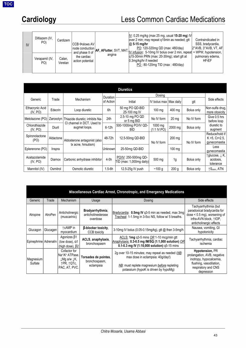
Cardiology Less Common Cardiac Medications
Chitra Mosarla, Usama Abbasi
Diltiazem (IV, PO) Cardizem
CCBslows AV node conduction and phase II of
the cardiac action potential
AF, AFlutter, SVT, MAT, angina
IV: 0.25 mg/kg (max 25 mg, usual 15-20 mg) IV over 2 min; may repeat q15min as needed; gtt @ 5-15 mg/hr
PO: 120-320mg QD (max: 480/day) IV infusion: 5-10mg IV bolus over 2 min, repeat q15-30min PRN (max: 20-30mg); start gtt at 0.3mg/kg/hr if needed
PO : 80-120mg TID (max : 480/day)
Contraindicated in SSS, bradycardia,
2°AVB, 3°AVB, VT, AF + WPW, hypotension,
pulmonary edema, HFrEF
IV
Verapamil (IV, PO)
Calan, Verelan
Diuretics
Generic Trade Mechanism Duration of Action
Dosing Side effects Initial IV bolus max Max daily gtt
Ethacrynic Acid (IV, PO) Edecrin Loop diuretic 6h 50 mg PO QD-BID
25-100 mg IV 100 mg 400 mg Bolus only Non-sulfa drug, more otoxicity
Metolazone (PO) Zaroxolyn Thiazide diuretic; inhibits Na-Cl channel in DCT. Used to
augmet loops.
24h 2.5-10 mg PO QD or 5 mg BID No IV form 20 mg No IV form Give 0.5 hrs
before loop diuretic to augment
Chlorothiazide (IV, PO) Diuril 6-12h 500-1000mg PO/IV QD-
BID 1000 mg
(1:1 IV:PO) 2000 mg Bolus only
Spironolactone (PO) Aldactone Aldosterone antagonist (also
tx acne, hirsutism)
48-72h 12.5-50mg QD-BID No IV form
200 mg No IV form
Reduce/hold if K >5, Cr>2.5, gynecomastia
Eplerenone (PO) Inspra Unknown 25-50mg QD-BID 100 mg Less gynecomastia
Acetazolamide (IV, PO) Diamox Carbonic anhydrase inhibitor 4-5h PO/IV: 250-500mg QD-
TID (max: 1,000mg daily) 500 mg 1g Bolus only ↑glucose, ↓ K,
acidosis, tolerance
Mannitol (IV) Osmitrol Osmotic diuretic 1.5-6h 12.5-25g IV push ~100 g 200 g Bolus only ↑Sosm, ATN
Miscellaneous Cardiac Arrest, Chronotropic, and Emergency Medications
Generic Trade Mechanism Usage Dosing Side effects
Atropine AtroPen Anticholinergic (muscarinic)
Bradyarrhythmia, anticholinesterase
overdose Bradycardia: 0.5mg IV q3-5 min as needed, max 3mg
Tracheal: 1-1.5mg in 3-5cc NS, follow w/ 5 breaths.
Tachyarrhythmia (but paradoxical bradycardia for
dose < 0.5 mg), worsening of infra-AVN block, ↑IOP, anticholinergic effects
Glucagon Glucagen ↑cAMP in myocardium
β-blocker toxicity, CCB toxicity 3-10mg IV bolus (0.05-0.15mg/kg), gtt @ then 3-5mg/h Nausea, vomiting, GI
hypotonicity
Epinephrine Adrenalin Agonizes β1
(low dose), α1 (high dose), β2
ACLS, anaphylaxis, bronchospasm
ACLS: 1mg q3-5 mins OR 1-10 mcg/min gtt Anaphylaxis: 0.3-0.5 mg IM/SQ (1:1,000 solution) OR
0.1-0.3 mg IV (1:10,000 solution) q5-15 mins Tachyarrhythmia, cardiac
ischemia
Magnesium Sulfate
Cofactor for Na+/K+ ATPase.
↓Mg a/w ↓K, ↑PR, ↑QTc,
PAC, AT, PVC.
Torsades de pointes, bronchospasm,
eclampsia
2g over 10-15 minutes; may repeat as needed (NB: max dose in eclampsia: 40g/day!)
NB: must replete magnesium before repleting potassium (hypoK is driven by hypoMg)
Hypotension, PR prolangation, AVB, negative
inotropy, hypocalcemia, flushing, vasodilation, respiratory and CNS
depression
43
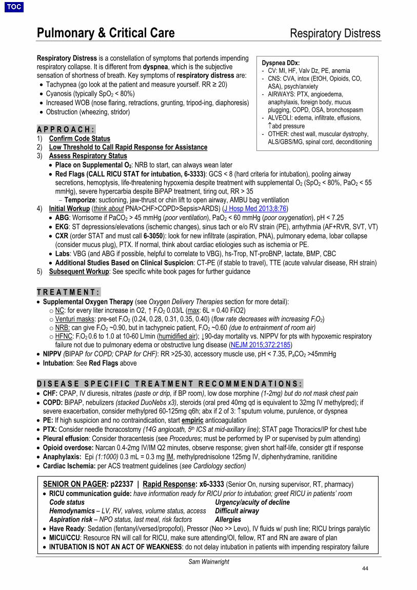
Pulmonary & Critical Care Respiratory Distress
Sam Wainwright
Dyspnea DDx: - CV: MI, HF, Valv Dz, PE, anemia- CNS: CVA, intox (EtOH, Opioids, CO,
ASA), psych/anxiety- AIRWAYS: PTX, angioedema,
anaphylaxis, foreign body, mucusplugging, COPD, OSA, bronchospasm
- ALVEOLI: edema, infiltrate, effusions,↑abd pressure
- OTHER: chest wall, muscular dystrophy,ALS/GBS/MG, spinal cord, deconditioning
Respiratory Distress is a constellation of symptoms that portends impending respiratory collapse. It is different from dyspnea, which is the subjective sensation of shortness of breath. Key symptoms of respiratory distress are: • Tachypnea (go look at the patient and measure yourself. RR ≥ 20)• Cyanosis (typically SpO2 < 80%)• Increased WOB (nose flaring, retractions, grunting, tripod-ing, diaphoresis)• Obstruction (wheezing, stridor)
A P P R O A C H :1) Confirm Code Status2) Low Threshold to Call Rapid Response for Assistance3) Assess Respiratory Status
• Place on Supplemental O2: NRB to start, can always wean later
• Red Flags (CALL RICU STAT for intubation, 6-3333): GCS < 8 (hard criteria for intubation), pooling airwaysecretions, hemoptysis, life-threatening hypoxemia despite treatment with supplemental O2 (SpO2 < 80%, PaO2 < 55mmHg), severe hypercarbia despite BiPAP treatment, tiring out, RR > 35– Temporize: suctioning, jaw-thrust or chin lift to open airway, AMBU bag ventilation
4) Initial Workup (think about PNA>CHF>COPD>Sepsis>ARDS) (J Hosp Med 2013;8:76)• ABG: Worrisome if PaCO2 > 45 mmHg (poor ventilation), PaO2 < 60 mmHg (poor oxygenation), pH < 7.25• EKG: ST depressions/elevations (ischemic changes), sinus tach or e/o RV strain (PE), arrhythmia (AF+RVR, SVT, VT)• CXR (order STAT and must call 6-3050): look for new infiltrate (aspiration, PNA), pulmonary edema, lobar collapse
(consider mucus plug), PTX. If normal, think about cardiac etiologies such as ischemia or PE.• Labs: VBG (and ABG if possible, helpful to correlate to VBG), hs-Trop, NT-proBNP, lactate, BMP, CBC• Additional Studies Based on Clinical Suspicion: CT-PE (if stable to travel), TTE (acute valvular disease, RH strain)
5) Subsequent Workup: See specific white book pages for further guidance
T R E A T M E N T :• Supplemental Oxygen Therapy (see Oxygen Delivery Therapies section for more detail):
o NC: for every liter increase in O2, ↑ FiO2 0.03/L (max: 6L = 0.40 FiO2)o Venturi masks: pre-set FiO2 (0.24, 0.28, 0.31, 0.35, 0.40) (flow rate decreases with increasing FiO2)o NRB: can give FiO2 ~0.90, but in tachypneic patient, FiO2 ~0.60 (due to entrainment of room air)o HFNC: FiO2 0.6 to 1.0 at 10-60 L/min (humidified air); ↓90-day mortality vs. NIPPV for pts with hypoxemic respiratory
failure not due to pulmonary edema or obstructive lung disease (NEJM 2015;372:2185)• NIPPV (BIPAP for COPD; CPAP for CHF): RR >25-30, accessory muscle use, pH < 7.35, PaCO2 >45mmHg• Intubation: See Red Flags above
D I S E A S E S P E C I F I C T R E A T M E N T R E C O M M E N D A T I O N S :• CHF: CPAP, IV diuresis, nitrates (paste or drip, if BP room), low dose morphine (1-2mg) but do not mask chest pain• COPD: BiPAP, nebulizers (stacked DuoNebs x3), steroids (oral pred 40mg qd is equivalent to 32mg IV methylpred); if
severe exacerbation, consider methylpred 60-125mg q6h; abx if 2 of 3: ↑sputum volume, purulence, or dyspnea• PE: If high suspicion and no contraindication, start empiric anticoagulation• PTX: Consider needle thoracostomy (14G angiocath, 5th ICS at mid-axillary line); STAT page Thoracics/IP for chest tube• Pleural effusion: Consider thoracentesis (see Procedures; must be performed by IP or supervised by pulm attending)• Opioid overdose: Narcan 0.4-2mg IV/IM Q2 minutes, observe response; given short half-life, consider gtt if response• Anaphylaxis: Epi (1:1000) 0.3 mL = 0.3 mg IM, methylprednisolone 125mg IV, diphenhydramine, ranitidine• Cardiac Ischemia: per ACS treatment guidelines (see Cardiology section)
SENIOR ON PAGER: p22337 | Rapid Response: x6-3333 (Senior On, nursing supervisor, RT, pharmacy) • RICU communication guide: have information ready for RICU prior to intubation; greet RICU in patients’ room
Code status Hemodynamics – LV, RV, valves, volume status, access Aspiration risk – NPO status, last meal, risk factors
Urgency/acuity of decline Difficult airway Allergies
• Have Ready: Sedation (fentanyl/versed/propofol), Pressor (Neo >> Levo), IV fluids w/ push line; RICU brings paralytic• MICU/CCU: Resource RN will call for RICU, make sure attending/OI, fellow, RT and RN are aware of plan• INTUBATION IS NOT AN ACT OF WEAKNESS: do not delay intubation in patients with impending respiratory failure
44
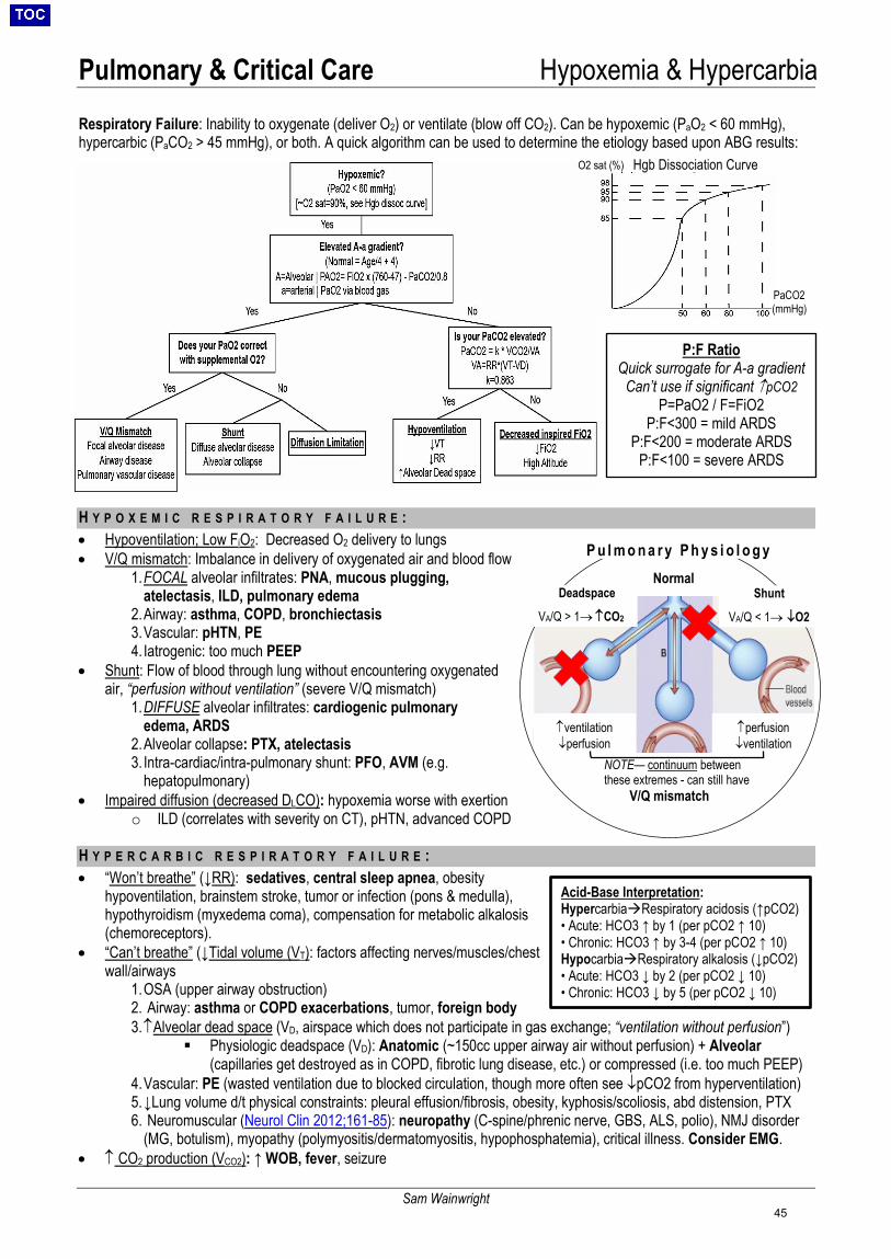
Pulmonary & Critical Care Hypoxemia & Hypercarbia
Sam Wainwright
Respiratory Failure: Inability to oxygenate (deliver O2) or ventilate (blow off CO2). Can be hypoxemic (PaO2 < 60 mmHg), hypercarbic (PaCO2 > 45 mmHg), or both. A quick algorithm can be used to determine the etiology based upon ABG results:
H Y P O X E M I C R E S P I R A T O R Y F A I L U R E :• Hypoventilation; Low FiO2: Decreased O2 delivery to lungs• V/Q mismatch: Imbalance in delivery of oxygenated air and blood flow
1. FOCAL alveolar infiltrates: PNA, mucous plugging,atelectasis, ILD, pulmonary edema
2. Airway: asthma, COPD, bronchiectasis3. Vascular: pHTN, PE4. Iatrogenic: too much PEEP
• Shunt: Flow of blood through lung without encountering oxygenatedair, “perfusion without ventilation” (severe V/Q mismatch)
1. DIFFUSE alveolar infiltrates: cardiogenic pulmonaryedema, ARDS
2. Alveolar collapse: PTX, atelectasis3. Intra-cardiac/intra-pulmonary shunt: PFO, AVM (e.g.
hepatopulmonary) • Impaired diffusion (decreased DLCO): hypoxemia worse with exertion
o ILD (correlates with severity on CT), pHTN, advanced COPD
H Y P E R C A R B I C R E S P I R A T O R Y F A I L U R E :• “Won’t breathe” (↓RR): sedatives, central sleep apnea, obesity
hypoventilation, brainstem stroke, tumor or infection (pons & medulla),hypothyroidism (myxedema coma), compensation for metabolic alkalosis(chemoreceptors).
• “Can’t breathe” (↓Tidal volume (VT): factors affecting nerves/muscles/chestwall/airways
1. OSA (upper airway obstruction)2. Airway: asthma or COPD exacerbations, tumor, foreign body3. ↑Alveolar dead space (VD, airspace which does not participate in gas exchange; “ventilation without perfusion”)
Physiologic deadspace (VD): Anatomic (~150cc upper airway air without perfusion) + Alveolar(capillaries get destroyed as in COPD, fibrotic lung disease, etc.) or compressed (i.e. too much PEEP)
4. Vascular: PE (wasted ventilation due to blocked circulation, though more often see ↓pCO2 from hyperventilation)5. ↓Lung volume d/t physical constraints: pleural effusion/fibrosis, obesity, kyphosis/scoliosis, abd distension, PTX6. Neuromuscular (Neurol Clin 2012;161-85): neuropathy (C-spine/phrenic nerve, GBS, ALS, polio), NMJ disorder
(MG, botulism), myopathy (polymyositis/dermatomyositis, hypophosphatemia), critical illness. Consider EMG.• ↑ CO2 production (VCO2): ↑ WOB, fever, seizure
Normal
↑ventilation ↓perfusion
↑perfusion ↓ventilation
NOTE— continuum between these extremes - can still have
V/Q mismatch
P u l m o n a r y P h y s i o l o g y
Shunt
VA/Q < 1→ ↓O2
Deadspace
VA/Q > 1→ ↑CO2
P:F Ratio Quick surrogate for A-a gradient
Can’t use if significant ↑pCO2 P=PaO2 / F=FiO2
P:F<300 = mild ARDS P:F<200 = moderate ARDS
P:F<100 = severe ARDS
Acid-Base Interpretation: HypercarbiaRespiratory acidosis (↑pCO2) • Acute: HCO3 ↑ by 1 (per pCO2 ↑ 10)• Chronic: HCO3 ↑ by 3-4 (per pCO2 ↑ 10)HypocarbiaRespiratory alkalosis (↓pCO2)• Acute: HCO3 ↓ by 2 (per pCO2 ↓ 10)• Chronic: HCO3 ↓ by 5 (per pCO2 ↓ 10)
Hgb Dissociation Curve O2 sat (%)
PaCO2 (mmHg)
45
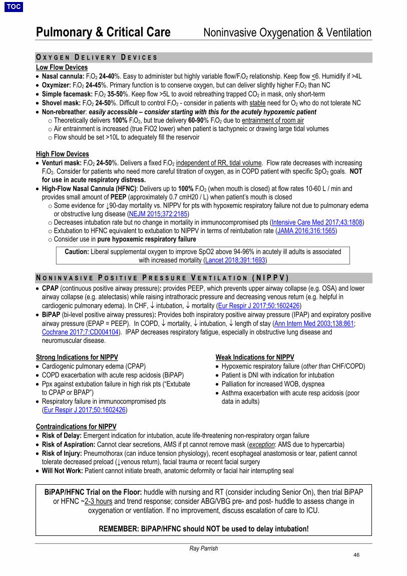
Pulmonary & Critical Care Noninvasive Oxygenation & Ventilation
Ray Parrish
O X Y G E N D E L I V E R Y D E V I C E SLow Flow Devices • Nasal cannula: FiO2 24-40%. Easy to administer but highly variable flow/FiO2 relationship. Keep flow <6. Humidify if >4L• Oxymizer: FiO2 24-45%. Primary function is to conserve oxygen, but can deliver slightly higher FiO2 than NC• Simple facemask: FiO2 35-50%. Keep flow >5L to avoid rebreathing trapped CO2 in mask, only short-term• Shovel mask: FiO2 24-50%. Difficult to control FiO2 - consider in patients with stable need for O2 who do not tolerate NC• Non-rebreather: easily accessible – consider starting with this for the acutely hypoxemic patient
o Theoretically delivers 100% FiO2, but true delivery 60-90% FiO2 due to entrainment of room airo Air entrainment is increased (true FiO2 lower) when patient is tachypneic or drawing large tidal volumeso Flow should be set >10L to adequately fill the reservoir
High Flow Devices • Venturi mask: FiO2 24-50%. Delivers a fixed FiO2 independent of RR, tidal volume. Flow rate decreases with increasing
FiO2. Consider for patients who need more careful titration of oxygen, as in COPD patient with specific SpO2 goals. NOTfor use in acute respiratory distress.
• High-Flow Nasal Cannula (HFNC): Delivers up to 100% FiO2 (when mouth is closed) at flow rates 10-60 L / min andprovides small amount of PEEP (approximately 0.7 cmH20 / L) when patient’s mouth is closedo Some evidence for ↓90-day mortality vs. NIPPV for pts with hypoxemic respiratory failure not due to pulmonary edema
or obstructive lung disease (NEJM 2015;372:2185)o Decreases intubation rate but no change in mortality in immunocompromised pts (Intensive Care Med 2017;43:1808)o Extubation to HFNC equivalent to extubation to NIPPV in terms of reintubation rate (JAMA 2016;316:1565)o Consider use in pure hypoxemic respiratory failure
Caution: Liberal supplemental oxygen to improve SpO2 above 94-96% in acutely ill adults is associated with increased mortality (Lancet 2018;391:1693)
N O N I N V A S I V E P O S I T I V E P R E S S U R E V E N T I L A T I O N ( N I P P V )• CPAP (continuous positive airway pressure): provides PEEP, which prevents upper airway collapse (e.g. OSA) and lower
airway collapse (e.g. atelectasis) while raising intrathoracic pressure and decreasing venous return (e.g. helpful incardiogenic pulmonary edema). In CHF, ↓ intubation, ↓ mortality (Eur Respir J 2017;50:1602426)
• BiPAP (bi-level positive airway pressures): Provides both inspiratory positive airway pressure (IPAP) and expiratory positiveairway pressure (EPAP = PEEP). In COPD, ↓ mortality, ↓ intubation, ↓ length of stay (Ann Intern Med 2003;138:861;Cochrane 2017;7:CD004104). IPAP decreases respiratory fatigue, especially in obstructive lung disease andneuromuscular disease.
Strong Indications for NIPPV
• Cardiogenic pulmonary edema (CPAP)Weak Indications for NIPPV • Hypoxemic respiratory failure (other than CHF/COPD)
• COPD exacerbation with acute resp acidosis (BiPAP)• Ppx against extubation failure in high risk pts (“Extubate
to CPAP or BPAP”)• Respiratory failure in immunocompromised pts
(Eur Respir J 2017;50:1602426)
• Patient is DNI with indication for intubation• Palliation for increased WOB, dyspnea• Asthma exacerbation with acute resp acidosis (poor
data in adults)
Contraindications for NIPPV • Risk of Delay: Emergent indication for intubation, acute life-threatening non-respiratory organ failure• Risk of Aspiration: Cannot clear secretions, AMS if pt cannot remove mask (exception: AMS due to hypercarbia)• Risk of Injury: Pneumothorax (can induce tension physiology), recent esophageal anastomosis or tear, patient cannot
tolerate decreased preload (↓venous return), facial trauma or recent facial surgery• Will Not Work: Patient cannot initiate breath, anatomic deformity or facial hair interrupting seal
BiPAP/HFNC Trial on the Floor: huddle with nursing and RT (consider including Senior On), then trial BiPAP or HFNC ~2-3 hours and trend response; consider ABG/VBG pre- and post- huddle to assess change in
oxygenation or ventilation. If no improvement, discuss escalation of care to ICU.
REMEMBER: BiPAP/HFNC should NOT be used to delay intubation!
46
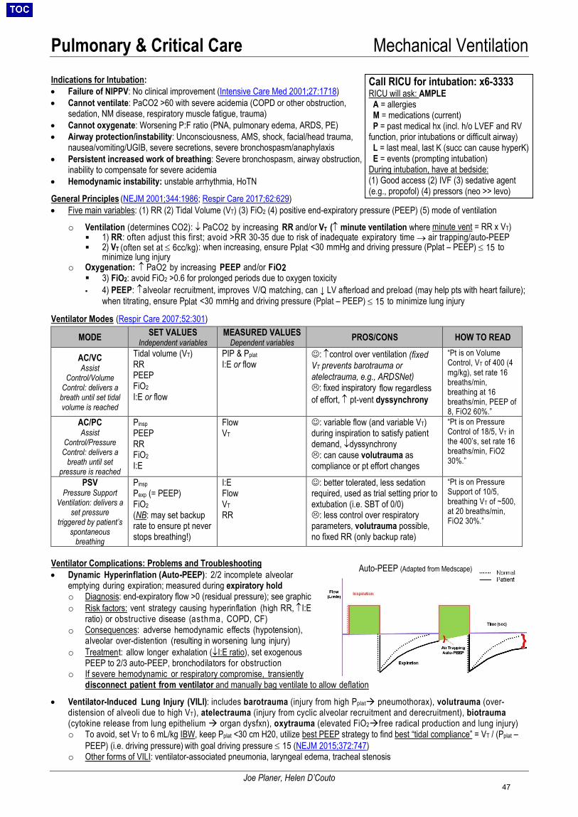
Pulmonary & Critical Care Mechanical Ventilation
Joe Planer, Helen D’Couto
Indications for Intubation: • Failure of NIPPV: No clinical improvement (Intensive Care Med 2001;27:1718)• Cannot ventilate: PaCO2 >60 with severe acidemia (COPD or other obstruction,
sedation, NM disease, respiratory muscle fatigue, trauma) • Cannot oxygenate: Worsening P:F ratio (PNA, pulmonary edema, ARDS, PE)• Airway protection/instability: Unconsciousness, AMS, shock, facial/head trauma,
nausea/vomiting/UGIB, severe secretions, severe bronchospasm/anaphylaxis• Persistent increased work of breathing: Severe bronchospasm, airway obstruction,
inability to compensate for severe acidemia• Hemodynamic instability: unstable arrhythmia, HoTN
General Principles (NEJM 2001;344:1986; Respir Care 2017;62:629)• Five main variables: (1) RR (2) Tidal Volume (VT) (3) FiO2 (4) positive end-expiratory pressure (PEEP) (5) mode of ventilation
o Ventilation (determines CO2): ↓ PaCO2 by increasing RR and/or VT (↑ minute ventilation where minute vent = RR x VT) 1) RR: often adjust this first; avoid >RR 30-35 due to risk of inadequate expiratory time → air trapping/auto-PEEP 2) VT (often set at ≤ 6cc/kg): when increasing, ensure Pplat <30 mmHg and driving pressure (Pplat – PEEP) ≤ 15 to
minimize lung injuryo Oxygenation: ↑ PaO2 by increasing PEEP and/or FiO2
3) FiO2: avoid FiO2 >0.6 for prolonged periods due to oxygen toxicity 4) PEEP: ↑alveolar recruitment, improves V/Q matching, can ↓ LV afterload and preload (may help pts with heart failure);
when titrating, ensure Pplat <30 mmHg and driving pressure (Pplat – PEEP) ≤ 15 to minimize lung injury
Ventilator Modes (Respir Care 2007;52:301)
MODE SET VALUES Independent variables
MEASURED VALUES Dependent variables PROS/CONS HOW TO READ
AC/VC Assist
Control/Volume Control: delivers a
breath until set tidal volume is reached
Tidal volume (VT) RR PEEP FiO2I:E or flow
PIP & Pplat I:E or flow
: ↑control over ventilation (fixedVT prevents barotrauma oratelectrauma, e.g., ARDSNet): fixed inspiratory flow regardlessof effort, ↑ pt-vent dyssynchrony
“Pt is on Volume Control, VT of 400 (4 mg/kg), set rate 16 breaths/min, breathing at 16 breaths/min, PEEP of 8, FiO2 60%.”
AC/PC Assist
Control/Pressure Control: delivers a
breath until set pressure is reached
Pinsp PEEP RR FiO2 I:E
Flow VT
: variable flow (and variable VT)during inspiration to satisfy patientdemand, ↓dyssynchrony: can cause volutrauma ascompliance or pt effort changes
“Pt is on Pressure Control of 18/5, VT in the 400’s, set rate 16 breaths/min, FiO2 30%.”
PSV Pressure Support
Ventilation: delivers a set pressure
triggered by patient’s spontaneous
breathing
Pinsp Pexp (= PEEP) FiO2 (NB: may set backup rate to ensure pt never stops breathing!)
I:E Flow VT RR
: better tolerated, less sedationrequired, used as trial setting prior toextubation (i.e. SBT of 0/0): less control over respiratoryparameters, volutrauma possible,no fixed RR (only backup rate)
“Pt is on Pressure Support of 10/5, breathing VT of ~500, at 20 breaths/min, FiO2 30%.”
Ventilator Complications: Problems and Troubleshooting • Dynamic Hyperinflation (Auto-PEEP): 2/2 incomplete alveolar
emptying during expiration; measured during expiratory holdo Diagnosis: end-expiratory flow >0 (residual pressure); see graphico Risk factors: vent strategy causing hyperinflation (high RR, ↑I:E
ratio) or obstructive disease (asthma, COPD, CF)o Consequences: adverse hemodynamic effects (hypotension),
alveolar over-distention (resulting in worsening lung injury)o Treatment: allow longer exhalation (↓I:E ratio), set exogenous
PEEP to 2/3 auto-PEEP, bronchodilators for obstructiono If severe hemodynamic or respiratory compromise, transiently
disconnect patient from ventilator and manually bag ventilate to allow deflation
• Ventilator-Induced Lung Injury (VILI): includes barotrauma (injury from high Pplat pneumothorax), volutrauma (over-distension of alveoli due to high VT), atelectrauma (injury from cyclic alveolar recruitment and derecruitment), biotrauma(cytokine release from lung epithelium organ dysfxn), oxytrauma (elevated FiO2free radical production and lung injury)o To avoid, set VT to 6 mL/kg IBW, keep Pplat <30 cm H20, utilize best PEEP strategy to find best “tidal compliance” = VT / (Pplat –
PEEP) (i.e. driving pressure) with goal driving pressure ≤ 15 (NEJM 2015;372:747)o Other forms of VILI: ventilator-associated pneumonia, laryngeal edema, tracheal stenosis
Call RICU for intubation: x6-3333 RICU will ask: AMPLE A = allergies M = medications (current) P = past medical hx (incl. h/o LVEF and RV function, prior intubations or difficult airway) L = last meal, last K (succ can cause hyperK) E = events (prompting intubation) During intubation, have at bedside: (1) Good access (2) IVF (3) sedative agent(e.g., propofol) (4) pressors (neo >> levo)
Auto-PEEP (Adapted from Medscape)
47
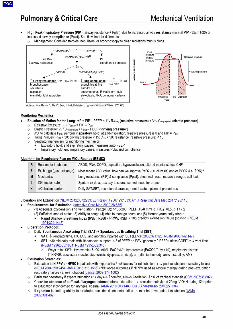
Pulmonary & Critical Care Mechanical Ventilation
Joe Planer, Helen D’Couto
• High Peak-Inspiratory Pressure (PIP = airway resistance + Pplat): due to increased airway resistance (normal PIP <35cm H20) or increased airway compliance (Pplat). See flowchart for differential. o Management: Consider steroids, nebulizers, or bronchoscopy to clear secretions/mucus plugs
Monitoring Mechanics • Equation of Motion for the Lung: ∆P = PIP – PEEP = V̇ x Rairway (resistive pressure) + VT / Cresp system (elastic pressure)
o Resistive Pressure: V̇ x Rairway = PIP – Pplat o Elastic Pressure: VT / Cresp system = Pplat – PEEP (“driving pressure”) o NB: to calculate Pplat, perform inspiratory hold; at end-inspiration, resistive pressure is 0 and PIP = Pplat o Target Values: Pplat < 30; driving pressure < 15; CRS > 50; resistance (resistive pressure) < 10 o Ventilator maneuvers for monitoring mechanics:
Expiratory hold: end expiratory pause; measures auto-PEEP Inspiratory hold: end inspiratory pause; measures Pplat and compliance
Algorithm for Respiratory Plan on MICU Rounds (REMIX)
Liberation and Extubation (NEJM 2012;367:2233; Eur Respir J 2007;29:1033; Am J Resp Crit Care Med 2017;195:115) • Requirements for Extubation: (Intensive Care Med 2002;28:535)
o (1) Adequate oxygenation and ventilation: PaO2/FiO2 >150-200, PEEP ≤5-8 mmHg, FiO2 <0.5, pH >7.2 (2) Sufficient mental status (3) Ability to cough (4) Able to manage secretions (5) Hemodynamically stable Rapid Shallow Breathing Index (RSBI) RSBI = RR/VT; RSBI > 105 predicts extubation failure (sp>>sn) (NEJM
1991;324:1445) • Liberation Protocol:
o Daily Spontaneous Awakening Trial (SAT) + Spontaneous Breathing Trial (SBT) SAT: ↓ ventilator time, ICU LOS, and mortality if paired with SBT (Lancet 2008;371:126; NEJM 2000;342:147) SBT: ~30 min daily trials with little/no vent support (≤ 5 of PEEP on PSV, generally 0 PEEP unless COPD) = ↓ vent time
(NEJM 1996;335:1864; NEJM 1995;332:345)o Ways to fail SBT: Hypoxemia (SaO2 <90%, PaO2<60), hypercarbia (PaCO2 ↑ by >10), respiratory distress
(↑HR/RR, accessory muscle, diaphoresis, dyspnea, anxiety), arrhythmia, hemodynamic instability, AMS• Extubation Strategies:
o Extubation to NIPPV or HFNC in patients with hypercarbia / risk factors for reintubation→ ↓ post-extubation respiratory failure(NEJM 2004;350:2454; JAMA 2016;316:1565) (NB: worse outcomes if NIPPV used as rescue therapy during post-extubationrespiratory failure vs. re-intubation) (Lancet 2009;374:1082)
o Early tracheostomy if expect intubation >14 days→ ↑comfort, allows ↓sedation, ↓risk of tracheal stenosis (CCM 2007;35:802)o Check for absence of cuff leak / laryngeal edema before extubation → consider methylpred 20mg IV Q4H during 12hr prior
to extubation if concerned for laryngeal edema (JAMA 2010;303:1483; Eur J Anaesthesiol 2010;27:534)o If agitation is limiting ability to extubate, consider dexmedetomidine → may improve odds of extubation (JAMA
2009;301:489)
R Reason for intubation ARDS, PNA, COPD, aspiration, hypoventilation, altered mental status, CHF E Exchange (gas exchange) Most recent ABG value; how can we improve PaO2 (i.e. diuresis) and/or PCO2 (i.e. ↑RR)? M Mechanics Lung resistance (PIP) & compliance (Pplat), chest wall, resp. muscle strength, cuff leak I ID/infection (abx) Sputum cx data, abx day #, source control, need for bronch X eXtubation barriers Daily SAT/SBT, secretion clearance, mental status, planned procedures
48
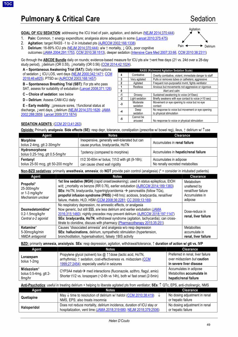
Pulmonary & Critical Care Sedation
Helen D’Couto
GOAL OF ICU SEDATION: addressing the ICU triad of pain, agitation, and delirium (NEJM 2014;370:444) 1. Pain: Common, ↑ energy expenditure; analgesia alone adequate in some (Lancet 2010;375:475)2. Agitation: target RASS -1 to -2 in intubated pts (AJRCCM 2002;166:1338)3. Delirium: 16-89% ICU pts (NEJM 2014;370:444); a/w ↑ mortality, ↓ QOL, poor cognitive
outcomes (JAMA 2004;291:1753, CCM 2010;38:1513), deeper sedation (Intensive Care Med 2007;33:66, CCM 2010:38:2311)
Go through the ABCDE Bundle daily on rounds; evidence-based measure for ICU pts a/w ↑vent free days (21 vs. 24d over a 28-day study period), ↓delirium (OR 0.55), ↓mortality (OR 0.56) (CCM 2014;42:1024).
A – Spontaneous Awakening Trial (SAT): Daily interruptions of sedation ↓ ICU LOS, vent days (NEJM 2000;342:1471; CCM 2018;46:e825); PTSD sx (AJRCCM 2003;168:1457) B – Spontaneous Breathing Trial (SBT): For pts who pass SAT, assess for suitability of extubation (Lancet 2008;371:126) C – Choice of sedation: see below D – Delirium: Assess CAM-ICU daily
E – Early mobility: ↓pressure sores, ↑functional status at discharge, ↓vent days, ↓delirium (NEJM 2014;370:1626; JAMA 2002;288:2859; Lancet 2009;373:1874)
SEDATION AGENTS: (CCM 2013;41:263)Opioids: Primarily analgesia. Side effects (SE): resp depr, tolerance, constipation (prescribe w/ bowel reg), ileus, ↑ delirium w/ ↑use
Agent Notes Clearance Morphine bolus 2-4mg, gtt 2-30mg/hr
Inexpensive, generally well-tolerated but can cause pruritus, bradycardia, HoTN Accumulates in renal failure
Hydromorphone bolus 0.25-1mg, gtt 0.5-5mg/hr ↑potency (compared to morphine) Accumulates in hepatic/renal failure
Fentanyl bolus 25-50 mcg, gtt 50-200 mcg/hr
t1/2 30-60m w/ bolus; ↑t1/2 with gtt (9-16h); can cause chest wall rigidity
Accumulates in adipose No renally excreted metabolites
Non-BZD sedatives: primarily anesthesia, amnesia; do NOT provide pain control (analgesia) (* = consider in intubated patients) Agent Notes Clearance
Propofol* 25-300mg/hror 1-3 mg/kg/hrMechanism unclear
1st line sedative (MGH) (rapid onset/awakening); used in status epilepticus, EtOH w/d; ↓mortality vs benzos (RR 0.76), earlier extubation (AJRCCM 2014;189:1383) SEs: HoTN, bradycardia, hypertriglyceridemia pancreatitis (follow TGs), propofol infusion syndrome (PRIS) (>48 hrs): acidosis, bradycardia, renal/liver failure, rhabdo, HLD, HSM (CCM 2008;36:2281; CC 2009;13:169)
Metabolism unaltered by renal/liver failure Accumulates in adipose
Dexmedetomidine* 0.2-1.5mcg/kg/hr Central α-2 agonist
No respiratory depression, no amnestic effects, or analgesia Now generic, but still $$$, a/w less delirium and earlier extubation (JAMA 2016;315:1460); nightly precedex may prevent delirium (AJRCCM 2018;197:1147) SEs: bradycardia, HoTN, withdrawal syndrome (agitation, tachycardia); can cross-titrate to clonidine, discuss with pharmacy (Pharmacotherapy 2015;35:251)
Dose-reduce in renal, liver failure
Ketamine* 5-30mcg/kg/minNMDA antagonist
Causes “dissociated amnesia” and analgesia w/o resp depression SEs: hallucinations, delirium, sympathetic stimulation (hypertension, bronchodilation, hypersalivation), falsely ↑BIS activity
Metabolites accumulate in renal, liver failure
BZD: primarily amnesia, anxiolysis. SEs: resp depression, agitation, withdrawal/tolerance, ↑ duration of action w/ gtt vs. IVP
Anti-Psychotics: useful in treating delirium + helping to liberate agitated pts from ventilator; SEs: ↑ QTc, EPS, anti-cholinergic, NMS
RASS (Richmond Agitation Sedation Scale) 4 Combative Overtly combative, violent, immediate danger to staff 3 Very agitated Pulls or removes tubes or catheters; aggressive 2 Agitated Frequent non-purposeful mvmt, fights ventilator 1 Restless Anxious but movements not aggressive or vigorous 0 Alert and calm -1 Drowsy Sustained awakening to voice (≥10sec) -2 Light sedation Briefly awakens with eye contact to voice (<10 sec)
-3 Moderate sedation
Movement or eye opening to voice but no eye contact
-4 Deep sedation
No response to voice but movement or eye opening to physical stimulation
-5 Cannot be aroused No response to voice or physical stimulation
Agent Notes Clearance
Lorazepam bolus 1-2mg
Propylene glycol (solvent) tox @ ↑↑dose (lactic acid, HoTN, arrhythmia); ↑ sedation, cost-effectiveness vs. midazolam (CCM 1999;27:2454); especially useful in seizures
Preferred in renal, liver failure over midazolam but caution in severe liver disease
Midazolam* bolus 0.5-4mg, gtt 2-8mg/hr
CYP3A4 metab med interactions (fluconazole, azithro, flagyl, amio) Shorter t1/2 vs. lorazepam (~2-6h vs 14h), both w/ fast onset (2-5min)
Accumulates in adipose Metabolites accumulate in hepatic/renal failure
Agent Notes Clearance
Quetiapine May ↓ time to resolution of delirium w/ haldol (CCM 2010;38:419) ↓ NMS, EPS; also treats insomnia
No dosing adjustment in renal or hepatic failure
Haloperidol Does not reduce mortality, delirium incidence, duration of ICU stay or hospitalization, vent time (JAMA 2018;319:680; NEJM 2018;379:2506)
No dosing adjustment in renal or hepatic failure
Agitation
Pain Delirium
49
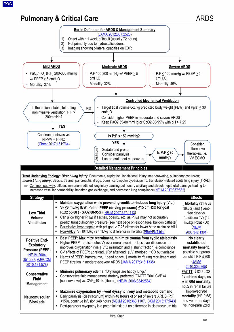
Pulmonary & Critical Care ARDS
Viral Shah
Detailed Management Principles
Treat Underlying Etiology: Direct lung injury: Pneumonia, aspiration, inhalational injury, near drowning, pulmonary contusion; Indirect lung injury: Sepsis, trauma, pancreatitis, drugs, burns, cardiopulm bypass/pump, transfusion-related acute lung injury (TRALI) ⇒ Common pathway: diffuse, immune-mediated lung injury causing pulmonary capillary and alveolar epithelial damage leading to
increased vascular permeability, impaired gas exchange, and decreased lung compliance (NEJM 2017;377:562)
Strategy Effects
Low Tidal Volume
Ventilation
Maintain oxygenation while preventing ventilator-induced lung injury (VILI) VT <6 mL/kg IBW, Pplat - PEEP (driving pressure) <15 cmH2O for goal
PaO2 55-80 (~ SpO2 88-95%) (NEJM 2007;357:1113) Can allow higher Pplat if ascites, obesity, etc. as Pplat may not accurately
predict transpulmonary pressure (see next page on esophageal balloon catheter) Permissive hypercapnia with pH goal > 7.25 allows for lower VT to minimize VILI Non-ARDS: VT 10mL/kg vs 4mL/kg no difference in mortality (PReVENT trial)
↓ Mortality (31% vs 39.8%) and ↑vent-
free days vs. “traditional” VT (12 mL/kg, Pplat <50)
(NEJM 2000;342:1301)
Positive End-Expiratory
Pressure (PEEP) (NEJM 2004;
351:327; AJRCCM 2010;181:578)
Best PEEP: Maximize recruitment, minimize trauma from cyclic atelectasis Higher PEEP → distributes VT over more alveoli → less over-distension →
improves oxygenation (via ↓ V/Q mismatch and ↓ shunt fraction) & compliance CV effects of PEEP: ↓preload, ↑RV afterload, ↓LV afterload, ↑CO but variable Harms of PEEP: barotrauma, ↑ dead space, ↑ mortality r/t lung recruitment and
PEEP titration in moderate/severe ARDS (JAMA 2017;318:1335)
No clearly established
mortality benefit, possible subgroup benefit if P:F ≤200
(JAMA 2010;303:865)
Conservative Fluid
Management
Minimize pulmonary edema: “Dry lungs are happy lungs” Conservative fluid management strategy preferred (FACTT Trial: CVP<4
[conservative] vs. CVP<10-14 [liberal]) (NEJM 2006;354:2564)
FACTT: ↓ICU LOS, ↑vent-free days, no ∆ in 60d mortality, no ∆ in renal failure
Neuromuscular Blockade
Maximize oxygenation by ↓vent dyssynchrony and metabolic demand Early paralysis (cisatracurium) within 48 hours of onset of severe ARDS (P:F
<150), continue infusion x48 hours (NEJM 2010;363:1107, CCM 2013;17:R43) Post-paralysis myopathy is a potential risk but no difference in cisatracurium trial
Improved 90d mortality (HR 0.68) and vent-free days vs. non-paralyzed
Berlin Definition for ARDS & Management Summary (JAMA 2012;307:2526)
1) Onset within 1 week of insult (usually 72 hours)2) Not primarily due to hydrostatic edema3) Imaging showing bilateral opacities on CXR
Is the patient stable, tolerating noninvasive ventilation, P:F >
200mmHg?
Controlled Mechanical Ventilation
- Target tidal volume 6cc/kg predicted body weight (PBW) and Pplat < 30cmH2O
- Consider higher PEEP in moderate and severe ARDS- Keep PaO2 55-80 mmHg or SpO2 88-95% with pH > 7.25
1) Sedate and prone2) Consider paralysis3) Lung recruitment maneuvers
Consider alternative
therapies, i.e. VV ECMO
Continue noninvasive: NIPPV = HFNC
(Chest 2017;151:764)
Mild ARDS
- PaO2/FiO2 (P:F) 200-300 mmHgw/ PEEP > 5 cmH2O
- Mortality: 27%
Moderate ARDS
- P:F 100-200 mmHg w/ PEEP > 5cmH2O
- Mortality: 32%
Severe ARDS - P:F < 100 mmHg w/ PEEP > 5
cmH2O- Mortality: 45%
Is P:F < 150 mmHg?
Is P:F < 80 mmHg?
YES
NO
YES
50
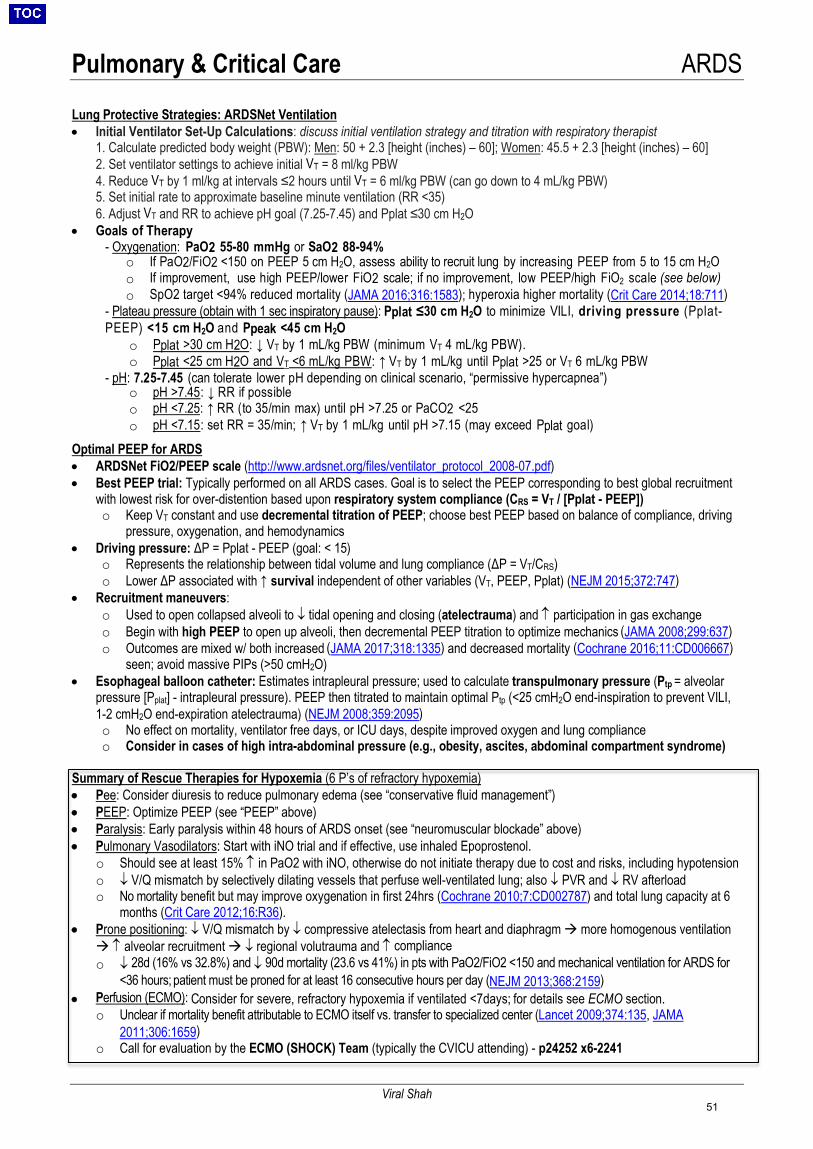
Pulmonary & Critical Care ARDS
Viral Shah
Lung Protective Strategies: ARDSNet Ventilation • Initial Ventilator Set-Up Calculations: discuss initial ventilation strategy and titration with respiratory therapist
1. Calculate predicted body weight (PBW): Men: 50 + 2.3 [height (inches) – 60]; Women: 45.5 + 2.3 [height (inches) – 60]2. Set ventilator settings to achieve initial VT = 8 ml/kg PBW4. Reduce VT by 1 ml/kg at intervals ≤2 hours until VT = 6 ml/kg PBW (can go down to 4 mL/kg PBW)5. Set initial rate to approximate baseline minute ventilation (RR <35)6. Adjust VT and RR to achieve pH goal (7.25-7.45) and Pplat ≤30 cm H2O
• Goals of Therapy- Oxygenation: PaO2 55-80 mmHg or SaO2 88-94%
o If PaO2/FiO2 <150 on PEEP 5 cm H2O, assess ability to recruit lung by increasing PEEP from 5 to 15 cm H2Oo If improvement, use high PEEP/lower FiO2 scale; if no improvement, low PEEP/high FiO2 scale (see below)o SpO2 target <94% reduced mortality (JAMA 2016;316:1583); hyperoxia higher mortality (Crit Care 2014;18:711)
- Plateau pressure (obtain with 1 sec inspiratory pause): Pplat ≤30 cm H2O to minimize VILI, driving pressure (Pplat-PEEP) <15 cm H2O and Ppeak <45 cm H2O
o Pplat >30 cm H2O: ↓ VT by 1 mL/kg PBW (minimum VT 4 mL/kg PBW).o Pplat <25 cm H2O and VT <6 mL/kg PBW: ↑ VT by 1 mL/kg until Pplat >25 or VT 6 mL/kg PBW
- pH: 7.25-7.45 (can tolerate lower pH depending on clinical scenario, “permissive hypercapnea”)o pH >7.45: ↓ RR if possibleo pH <7.25: ↑ RR (to 35/min max) until pH >7.25 or PaCO2 <25o pH <7.15: set RR = 35/min; ↑ VT by 1 mL/kg until pH >7.15 (may exceed Pplat goal)
Optimal PEEP for ARDS • ARDSNet FiO2/PEEP scale (http://www.ardsnet.org/files/ventilator_protocol_2008-07.pdf)• Best PEEP trial: Typically performed on all ARDS cases. Goal is to select the PEEP corresponding to best global recruitment
with lowest risk for over-distention based upon respiratory system compliance (CRS = VT / [Pplat - PEEP])o Keep VT constant and use decremental titration of PEEP; choose best PEEP based on balance of compliance, driving
pressure, oxygenation, and hemodynamics• Driving pressure: ΔP = Pplat - PEEP (goal: < 15)
o Represents the relationship between tidal volume and lung compliance (ΔP = VT/CRS)o Lower ΔP associated with ↑ survival independent of other variables (VT, PEEP, Pplat) (NEJM 2015;372:747)
• Recruitment maneuvers:o Used to open collapsed alveoli to ↓ tidal opening and closing (atelectrauma) and ↑ participation in gas exchangeo Begin with high PEEP to open up alveoli, then decremental PEEP titration to optimize mechanics (JAMA 2008;299:637)o Outcomes are mixed w/ both increased (JAMA 2017;318:1335) and decreased mortality (Cochrane 2016;11:CD006667)
seen; avoid massive PIPs (>50 cmH2O)• Esophageal balloon catheter: Estimates intrapleural pressure; used to calculate transpulmonary pressure (Ptp = alveolar
pressure [Pplat] - intrapleural pressure). PEEP then titrated to maintain optimal Ptp (<25 cmH2O end-inspiration to prevent VILI,1-2 cmH2O end-expiration atelectrauma) (NEJM 2008;359:2095)o No effect on mortality, ventilator free days, or ICU days, despite improved oxygen and lung compliance
o Consider in cases of high intra-abdominal pressure (e.g., obesity, ascites, abdominal compartment syndrome)
Summary of Rescue Therapies for Hypoxemia (6 P’s of refractory hypoxemia) • Pee: Consider diuresis to reduce pulmonary edema (see “conservative fluid management”)• PEEP: Optimize PEEP (see “PEEP” above)• Paralysis: Early paralysis within 48 hours of ARDS onset (see “neuromuscular blockade” above)• Pulmonary Vasodilators: Start with iNO trial and if effective, use inhaled Epoprostenol.
o Should see at least 15% ↑ in PaO2 with iNO, otherwise do not initiate therapy due to cost and risks, including hypotensiono ↓ V/Q mismatch by selectively dilating vessels that perfuse well-ventilated lung; also ↓ PVR and ↓ RV afterloado No mortality benefit but may improve oxygenation in first 24hrs (Cochrane 2010;7:CD002787) and total lung capacity at 6
months (Crit Care 2012;16:R36).• Prone positioning: ↓ V/Q mismatch by ↓ compressive atelectasis from heart and diaphragm more homogenous ventilation ↑ alveolar recruitment ↓ regional volutrauma and ↑ compliance o ↓ 28d (16% vs 32.8%) and ↓ 90d mortality (23.6 vs 41%) in pts with PaO2/FiO2 <150 and mechanical ventilation for ARDS for
<36 hours; patient must be proned for at least 16 consecutive hours per day (NEJM 2013;368:2159)• Perfusion (ECMO): Consider for severe, refractory hypoxemia if ventilated <7days; for details see ECMO section.
o Unclear if mortality benefit attributable to ECMO itself vs. transfer to specialized center (Lancet 2009;374:135, JAMA2011;306:1659)
o Call for evaluation by the ECMO (SHOCK) Team (typically the CVICU attending) - p24252 x6-2241
51
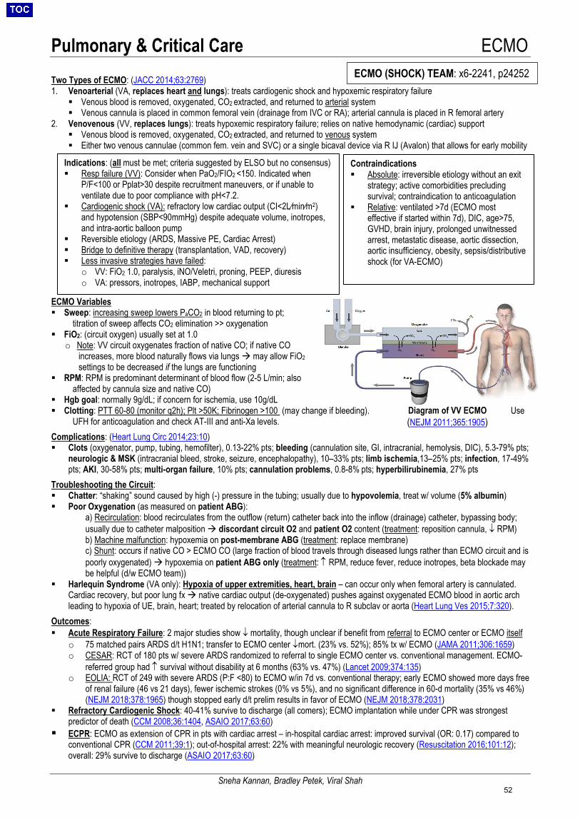
Pulmonary & Critical Care ECMO
Sneha Kannan, Bradley Petek, Viral Shah
Diagram of VV ECMO (NEJM 2011;365:1905)
Two Types of ECMO: (JACC 2014;63:2769) 1. Venoarterial (VA, replaces heart and lungs): treats cardiogenic shock and hypoxemic respiratory failure
Venous blood is removed, oxygenated, CO2 extracted, and returned to arterial system Venous cannula is placed in common femoral vein (drainage from IVC or RA); arterial cannula is placed in R femoral artery
2. Venovenous (VV, replaces lungs): treats hypoxemic respiratory failure; relies on native hemodynamic (cardiac) support Venous blood is removed, oxygenated, CO2 extracted, and returned to venous system Either two venous cannulae (common fem. vein and SVC) or a single bicaval device via R IJ (Avalon) that allows for early mobility
ECMO Variables Sweep: increasing sweep lowers PaCO2 in blood returning to pt;
titration of sweep affects CO2 elimination >> oxygenation FiO2: (circuit oxygen) usually set at 1.0
o Note: VV circuit oxygenates fraction of native CO; if native COincreases, more blood naturally flows via lungs may allow FiO2settings to be decreased if the lungs are functioning
RPM: RPM is predominant determinant of blood flow (2-5 L/min; alsoaffected by cannula size and native CO)
Hgb goal: normally 9g/dL; if concern for ischemia, use 10g/dL Clotting: PTT 60-80 (monitor q2h); Plt >50K; Fibrinogen >100 (may change if bleeding). Use
UFH for anticoagulation and check AT-III and anti-Xa levels. Complications: (Heart Lung Circ 2014;23:10) Clots (oxygenator, pump, tubing, hemofilter), 0.13-22% pts; bleeding (cannulation site, GI, intracranial, hemolysis, DIC), 5.3-79% pts;
neurologic & MSK (intracranial bleed, stroke, seizure, encephalopathy), 10–33% pts; limb ischemia,13–25% pts; infection, 17-49%pts; AKI, 30-58% pts; multi-organ failure, 10% pts; cannulation problems, 0.8-8% pts; hyperbilirubinemia, 27% pts
Troubleshooting the Circuit: Chatter: “shaking” sound caused by high (-) pressure in the tubing; usually due to hypovolemia, treat w/ volume (5% albumin) Poor Oxygenation (as measured on patient ABG):
a) Recirculation: blood recirculates from the outflow (return) catheter back into the inflow (drainage) catheter, bypassing body;usually due to catheter malposition discordant circuit O2 and patient O2 content (treatment: reposition cannula, ↓ RPM)b) Machine malfunction: hypoxemia on post-membrane ABG (treatment: replace membrane)c) Shunt: occurs if native CO > ECMO CO (large fraction of blood travels through diseased lungs rather than ECMO circuit and ispoorly oxygenated) hypoxemia on patient ABG only (treatment: ↑ RPM, reduce fever, reduce inotropes, beta blockade maybe helpful (d/w ECMO team))
Harlequin Syndrome (VA only): Hypoxia of upper extremities, heart, brain – can occur only when femoral artery is cannulated.Cardiac recovery, but poor lung fx native cardiac output (de-oxygenated) pushes against oxygenated ECMO blood in aortic archleading to hypoxia of UE, brain, heart; treated by relocation of arterial cannula to R subclav or aorta (Heart Lung Ves 2015;7:320).
Outcomes: Acute Respiratory Failure: 2 major studies show ↓ mortality, though unclear if benefit from referral to ECMO center or ECMO itself
o 75 matched pairs ARDS d/t H1N1; transfer to ECMO center ↓mort. (23% vs. 52%); 85% tx w/ ECMO (JAMA 2011;306:1659)o CESAR: RCT of 180 pts w/ severe ARDS randomized to referral to single ECMO center vs. conventional management. ECMO-
referred group had ↑ survival without disability at 6 months (63% vs. 47%) (Lancet 2009;374:135)o EOLIA: RCT of 249 with severe ARDS (P:F <80) to ECMO w/in 7d vs. conventional therapy; early ECMO showed more days free
of renal failure (46 vs 21 days), fewer ischemic strokes (0% vs 5%), and no significant difference in 60-d mortality (35% vs 46%)(NEJM 2018;378:1965) though stopped early d/t prelim results in favor of ECMO (NEJM 2018;378:2031)
Refractory Cardiogenic Shock: 40-41% survive to discharge (all comers); ECMO implantation while under CPR was strongestpredictor of death (CCM 2008;36:1404, ASAIO 2017;63:60)
ECPR: ECMO as extension of CPR in pts with cardiac arrest – in-hospital cardiac arrest: improved survival (OR: 0.17) compared toconventional CPR (CCM 2011;39:1); out-of-hospital arrest: 22% with meaningful neurologic recovery (Resuscitation 2016;101:12);overall: 29% survive to discharge (ASAIO 2017;63:60)
ECMO (SHOCK) TEAM: x6-2241, p24252
Contraindications Absolute: irreversible etiology without an exit
strategy; active comorbidities precludingsurvival; contraindication to anticoagulation
Relative: ventilated >7d (ECMO mosteffective if started within 7d), DIC, age>75,GVHD, brain injury, prolonged unwitnessedarrest, metastatic disease, aortic dissection,aortic insufficiency, obesity, sepsis/distributiveshock (for VA-ECMO)
Indications: (all must be met; criteria suggested by ELSO but no consensus) Resp failure (VV): Consider when PaO2/FIO2 <150. Indicated when
P/F<100 or Pplat>30 despite recruitment maneuvers, or if unable toventilate due to poor compliance with pH<7.2.
Cardiogenic shock (VA): refractory low cardiac output (CI<2L⁄min⁄m2)and hypotension (SBP<90mmHg) despite adequate volume, inotropes,and intra-aortic balloon pump
Reversible etiology (ARDS, Massive PE, Cardiac Arrest) Bridge to definitive therapy (transplantation, VAD, recovery) Less invasive strategies have failed:
o VV: FiO2 1.0, paralysis, iNO/Veletri, proning, PEEP, diuresiso VA: pressors, inotropes, IABP, mechanical support
52
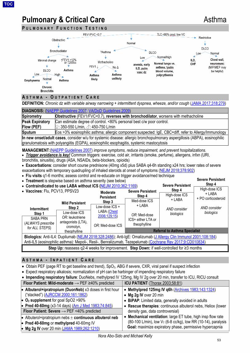
Pulmonary & Critical Care Asthma
Nora Abo-Sido and Michael Kelly
P U L M O N A R Y F U N C T I O N T E S T I N G
A S T H M A - O U T P A T I E N T C A R EDEFINITION: Chronic dz with variable airway narrowing + intermittent dyspnea, wheeze, and/or cough (JAMA 2017;318:279)
DIAGNOSIS: (NAEPP Guidelines 2007; VA/DoD Guidelines 2009) Spirometry Obstructive (FEV1/FVC<0.7), reverses with bronchodilator, worsens with methacholine Peak Expiratory Flow (PEF)
Can estimate degree of control. <80% personal best c/w poor control. ♀: 350-550 L/min, ♂: 450-750 L/min
Sputum Eos >3% eosinophilic asthma; allergic component suspected: IgE, CBC+diff, refer to Allergy/Immunology In new onset/adult cases, consider w/u for systemic disease: allergic bronchopulmonary aspergillosis (ABPA), eosinophilic granulomatosis with polyangiitis (EGPA), eosinophilic esophagitis, systemic mastocytosis
MANAGEMENT:(NAEPP Guidelines 2007) improve symptoms, reduce impairment, and prevent hospitalizations.
Severe Persistent Step 4
Severe Persistent Step 4
Severe Persistent Step 4
Moderate Persistent
Step 3
High-dose ICS + LABA
+ PO corticosteroid
AND considerbiologics
Mild Persistent Step 2
High-dose ICS + LABA
AND consider biologics
Intermittent Step 1
Med-dose ICS + LABA
OR: Med-dose ICS+ either LTA or
theophylline
Low-dose ICS + LABA (Chest 2006;129;15)
OR: Med-dose ICS
Low-dose ICS OR: leukotriene
antagonists (LTA), cromolyn,
theophylline
SABA PRN (ALWAYS prescribe
for ALL STEPS) Referral to Asthma Specialist
Biologics: Anti-IL4: Dupilumab (NEJM 2018;328:2486); Anti-IgE: Omalizumab (J Allergy Clin Immunol 2001;108:184) Anti-IL5 (eosinophilic asthma): Mepoli-, Resli-, Benralizumab, Tezepelumab (Cochrane Rev 2017;9:CD010834)
Step Up: reassess q2-4 weeks for improvement. Step Down: if well-controlled for ≥3 months
A S T H M A - I N P A T I E N T C A R E• Obtain PEF (page RT to get baseline and trend), SpO2, ABG if severe, CXR, viral panel if suspect infection• Expect respiratory alkalosis; normalization of pH can be harbinger of impending respiratory failure• Impending respiratory failure: DuoNebs, methylpred IV 125mg, Mg IV 2g over 20 min, transfer to ICU, RICU consultFloor Patient: Mild-moderate → PEF ≥40% predicted ICU PATIENT (Thorax 2003;58:81) • Albuterol+ipratropium (DuonNeb) x3 doses in first hour
(“stacked”) (AJRCCM 2000;161:1862)• O2 supplement for goal SpO2 >90%• Pred 40-60mg (x3-14 days) (Am J Med 1983;74:845)
• Methylpred 125mg IV q6h (Archives 1983;143:1324)• Mg 2g IV over 20 min• BiPAP: Limited data, generally avoided in adults• Rescue therapies: continuous albuterol nebs, Heliox (lower
density gas, data controversial) • Mechanical ventilation: large ET tube, high insp flow rate
(80-100 L/min), low VT (6-8 cc/kg), low RR (10-14), paralysisGoal: maximize expiratory phase, permissive hypercapnia
Floor Patient: Severe → PEF <40% predicted • Albuterol+ipratropium nebs ± continuous albuterol neb• Pred 40-60mg or methylpred 40-60mg IV• Mg 2g IV over 20 min (JAMA 1989;262:1210)
• Trigger avoidance is key! Common triggers: exercise, cold air, irritants (smoke, perfume), allergens, infxn (URI,bronchitis, sinusitis), drugs (ASA, NSAIDs, beta-blockers, opioids)• Exacerbations: consider short course prednisone (40mg x5d) plus SABA q4-6h standing x24 hrs; lower rates of severeexacerbations with temporary quadrupling of inhaled steroids at onset of symptoms (NEJM 2018;378:902)• F/u visits q1-6 months; assess control and re-educate on trigger avoidance/med technique• Treatment is stepwise based on asthma severity (see below)• Contraindicated to use LABA without ICS (NEJM 2010;362:1169)• Vaccines: Flu, PCV13, PPSV23
or ↑200cc
53
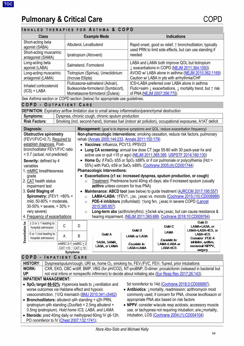
Pulmonary & Critical Care COPD
Nora Abo-Sido and Michael Kelly
I N H A L E D T H E R A P I E S F O R A S T H M A & C O P D
See Asthma section or COPD section (below) for appropriate use guidelines. C O P D - O U T P A T I E N T C A R EDEFINITION: Expiratory airflow limitation due to small airway inflammation/parenchymal destruction
Symptoms: Dyspnea, chronic cough, chronic sputum production Risk Factors: Smoking (incl. second-hand), biomass fuel (indoor air pollution), occupational exposures, A1AT deficit Diagnosis: Management: (goal is to improve symptoms and QOL, reduce exacerbation frequency) Obstructive spirometry (FEV1/FVC<0.7), Required to establish diagnosis. Post-bronchodilator FEV1/FVC ratio < 0.7 (actual, not predicted)
Severity: defined by 4 variables 1. mMRC breathlessness
grade 2. CAT health status
impairment test3. Gold Staging of
Spirometry: (FEV1: >80% =mild, 50-80% = moderate,30-50% = severe, < 30% =very severe)
4. Frequency of exacerbations
Non-pharmacologic interventions: smoking cessation, reduce risk factors, pulmonary rehab (Annals 2005;144:233, Annals 2011;155:179) • Vaccines: influenza, PCV13, PPSV23• Lung CA screening: annual low dose CT (age 55-80 with 30 pack-year hx and
active use or quit <15 yr ago) (NEJM 2011;365:395, USPSTF 2014;160:130)• Home O2: if PaO2 ≤55 or SpO2 ≤88% or if cor pulmonale or polycythemia (Hct >
55%) with PaO2 ≤59 or SaO2 ≤89% (Cochrane 2005;(4):CD001744)Phamacologic interventions: • Exacerbations (≥1 sx: increased dyspnea, sputum production, or cough)o Treatment: Prednisone burst 40mg x5 days; abx if increased sputum (usually
azithro unless concern for true PNA)• Maintenance: ABCD tool (see below) to guide treatment (AJRCCM 2017;195:557)o LAMA+LABA: ↑FEV1, ↓sx, ↓exac vs. monotx (Cochrane 2015;(10):CD008989)o PDE-4 inhibitors (roflumilast): ↑lung fxn, ↓exac in severe COPD (Lancet
2015;385:857)o Long-term abx (azithro/erythro): >3x/wk a/w↓exac, but can cause resistance &
hearing impairment. (NEJM 2011;365:689, Cochrane 2018;10:CD009764)
\
C O P D - I N P A T I E N T C A R EHISTORY: Dyspnea/sputum/cough, URI sx, home O2, smoking hx, FEV1/FVC, FEV1 %pred, prior intubations WORK-UP:
CXR, EKG, CBC w/diff, BMP, VBG (for pH/CO2), NT-proBNP, D-dimer; procalcitonin (released in bacterial but not viral infxns or nonspecific inflmmxn) to decide about initiating abx (Eur Resp Rev 2017;26:143).
INPATIENT MANAGEMENT: • SpO2 target 88-92%: Hyperoxia leads to ↓ventilation and
worse outcomes via Haldane effect and hypoxicvasoconstriction, ↑V/Q mismatch (BMJ 2010;341:c5462)
• Bronchodilators: albuterol q4h standing + q2h PRN,ipratropium q4h standing (DuoNeb = 2.5mg albuterol +0.5mg ipratropium). Hold home ICS, LABA, and LAMA.
• Steroids: pred 40mg daily or methylpred 60mg IV q6-12h.PO noninferior to IV (Chest 2007;132:1741);
5d noninferior to 14d (Cochrane 2018;3:CD006897). • Antibiotics: ↓mortality, readmission; azithromycin most
commonly used; if concern for PNA, choose levofloxacin orappropriate PNA abx based on risk factors
• NPPV: consider w/acute resp acidosis, accessory muscleuse, or tachypnea not requiring intubation; a/w↓mortality,intubation, LOS (Cochrane 2004;(1):CD004104)
Class Example Meds Indications Short-acting beta agonist (SABA) Albuterol, Levalbuterol Rapid onset, good sx relief, ↑ bronchodilation; typically
used PRN to limit side effects, but can use standing if needed Short-acting muscarinic
antagonist (SAMA) Ipratropium (Atrovent)
Long-acting beta agonist (LABA) Salmeterol, Formoterol LABA and LAMA both improve QOL but tiotropium
↓ exacerbations in COPD (NEJM 2011;364:1093)AVOID w/ LABA alone in asthma (NEJM 2010;362:1169) Caution w/ LABA in pts with arrhythmia/CHF
Long-acting muscarinic antagonist (LAMA)
Tiotropium (Spiriva), Umeclidinium (Incruse Ellipta)
Inhaled corticosteroid (ICS) + LABA
Fluticasone-salmeterol (Advair), Budesonide-formoterol (Symbicort), Mometasone-formoterol (Dulera)
ICS+LABA preferred over LABA alone in asthma Flutic+salm ↓ exacerbations, ↓ mortality trend, but ↑ risk of PNA (NEJM 2007;356:775)
C D
A B
mMRC 0-1 CAT <10
mMRC > 2 CAT > 10
> 2 or > 1 leading to hospital admission
0 or 1 (not leading to hospital admission)
Exac
erba
tion
hx
Symptoms
54
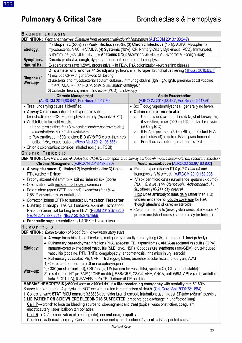
Pulmonary & Critical Care Bronchiectasis & Hemoptysis
Michael Kelly
B R O N C H I E C T A S I SDEFINITION: Permanent airway dilatation from recurrent infection/inflammation (AJRCCM 2013;188:647)
Etiology: (1) Idiopathic (50%), (2) Post-Infectious (20%), (3) Chronic Infectious (15%): ABPA, Mycoplasma,mycobacteria, MAC, HIV/AIDS, (4) Systemic (10%): CF, Primary Ciliary Dyskinesis (PCD), Immunodef,Autoimmune (RA, SLE, IBD), (5) Anatomic (5%): Aspiration/GERD, RML Syndrome, Foreign Body
Symptoms: Chronic productive cough, dyspnea, recurrent pneumonia, hemoptysis Natural Hx: Exacerbations (avg 1.5/yr), progressive ↓ in FEV1, PsA colonization→worsening disease
Diagnosis/Work-up:
CT diameter of bronchus >1.5x adj artery: bronchi fail to taper, bronchial thickening (Thorax 2010;65;1) 1) Exclude CF with gene/sweat Cl- testing2) Bacterial and mycobacterial sputum cultures, immunoglobulins (IgG, IgA, IgM), pneumococcal vaccine
titers, ANA, RF, anti-CCP, SSA, SSB, alpha1-antitrypsin3) Consider bronch, nasal nitric oxide (PCD), Endoscopy
Chronic Management (AJRCCM 2014;88:647, Eur Resp J 2017;50)
Acute Exacerbation (AJRCCM 2014;88:647, Eur Resp J 2017;50)
• Treat underlying cause if identified• Airway Clearance: inhaled tx (hypertonic saline,
bronchodilators, ICS) + chest physiotherapy (Acapella + PT)• Antibiotics in bronchiectasis:
o Long-term azithro for >3 exacerbations/yr: controversial; ↓exacerbations but c/f abx resistance
o PsA eradication: 500mg cipro BID (IVPO cipro, then nebcolistin)↓ exacerbations (Resp Med 2012;106:356)
• Chronic colonization: consider inhaled abx (i.e., TOBI)
• Sx: ↑ cough/sputum/dyspnea - generally no fevers• Obtain resp cx prior to abx:o Use previous cx data; if no data, start Levaquin;
if sensitive, amox (500mg TID) or clarithromycin(500mg BID)
o If PsA, cipro (500-750mg BID); if resistant PsA(or history of), requires IV antipseudomonal
o For all exacerbations, treatment is 14d
C Y S T I C F I B R O S I SDEFINITION: CFTR mutation Defective Cl-/HCO3- transport onto airway surface mucus accumulation, recurrent infection
Chronic Management (AJRCCM 2013;187:680) Acute Exacerbation (AJRCCM 2009;180:802) • Airway clearance: 1) albuterol 2) hypertonic saline 3) Chest
PT/exercise + DNase• Prophy abx/anti-inflamm tx = azithro+inhaled abx (tobra)• Colonization with resistant pathogens common• Potentiators (open CFTR channel): Ivacaftor (for 4% w/
G551D or similar class mutations)• Corrector (brings CFTR to surface): Lumacaftor, Tezacaftor• Dual/triple therapy (Tez/Iva, Luma/Iva, VX-659–Tezacaftor–
Ivacaftor) beneficial for long term FEV1 (NEJM 2015;373;220,NEJM 2017;377:2013, NEJM 2018;379:1599)
• Pancreatic supplementation: vit ADEK + lipase + insulin
• Rule out spontaneous PTX (0.7% annual) andhemoptysis (1% annual) (AJRCCM 2010;182:298)
• IV abx per micro data (surveillance sputum cx q3mo),PsA + S. aureus >> Stenotroph., Achromobact., H.flu, others (10-21+ day course)Tips: Dose aminoglycosides daily rather than TID;unclear evidence for double coverage for PsA,though standard of care; no steroids
• Continue chronic tx (airway clearance, etc) + nebs +/-prednisone (short course steroids may be helpful)
H E M O P T Y S I SDEFINITION: Expectoration of blood from lower respiratory tract
Etiology:
• Airway: bronchitis, bronchiectasis, malignancy (usually primary lung CA), trauma (incl. foreign body)• Pulmonary parenchyma: infection (PNA, abscess, TB, aspergilloma), ANCA-associated vasculitis (GPA),
immune-complex mediated vasculitis (SLE, cryo, HSP), Goodpasture syndrome (anti-GBM), drug-induced vasculitis (cocaine, PTU, TNFi), coagulopathy, endometriosis, inhalation injury, sarcoid
• Pulmonary vascular: PE, CHF, mitral regurgitation, bronchovascular fistula, aneurysm, AVM
Work-up: 1) Consider other sources (GI or nasopharyngeal)2) CXR (most important), CBC/coags, UA (screen for vascultits), sputum Cx, CT chest (if stable)3) In select pts: NT-proBNP (if CHF on ddx), ESR/CRP, C3/C4, ANA, ANCA, anti-GBM, APLA (anti-cardiolipin,
beta-2 GP1, LA), IGRA/AFB to r/o TB, D-dimer (if PE on ddx)MASSIVE HEMOPTYSIS (>500mL/day or >100mL/hr) is a life-threatening emergency with mortality rate 50-80%. Source is often arterial. Asphyxiation NOT exsanguination is mechanism of death. (Crit Care Med 2000;28:1684) 1) Control airway: STAT RICU consult (x63333); consider bronchoscopic intubation; use largest ET-tube (>8mm) possible2) LIE PATIENT ON SIDE WHERE BLEEDING IS SUSPECTED (preserve gas exchange in unaffected lung)
Call IP→bronch to localize bleeding source to lobe/segment and treat (topical vasoconstriction, coagulant,electrocautery, laser, balloon tamponade);Call IR→CTA (embolization of bleeding site); correct coagulopathyConsider c/s thoracic surgery. Consider pulse dose methylprednisolone if vasculitis is suspected cause.
55
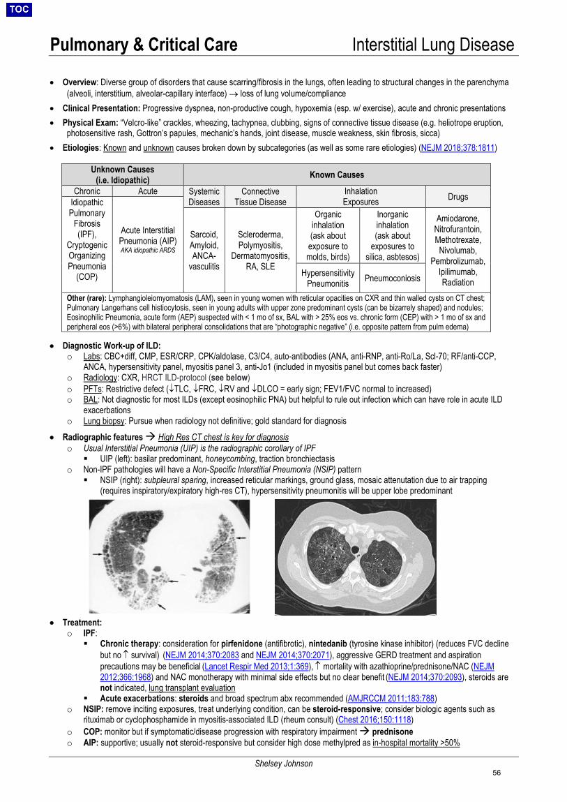
Pulmonary & Critical Care Interstitial Lung Disease
Shelsey Johnson
• Overview: Diverse group of disorders that cause scarring/fibrosis in the lungs, often leading to structural changes in the parenchyma(alveoli, interstitium, alveolar-capillary interface) → loss of lung volume/compliance
• Clinical Presentation: Progressive dyspnea, non-productive cough, hypoxemia (esp. w/ exercise), acute and chronic presentations• Physical Exam: “Velcro-like” crackles, wheezing, tachypnea, clubbing, signs of connective tissue disease (e.g. heliotrope eruption,
photosensitive rash, Gottron’s papules, mechanic’s hands, joint disease, muscle weakness, skin fibrosis, sicca)• Etiologies: Known and unknown causes broken down by subcategories (as well as some rare etiologies) (NEJM 2018;378:1811)
Unknown Causes (i.e. Idiopathic) Known Causes
Chronic Acute Systemic Diseases
Connective Tissue Disease
Inhalation Exposures Drugs Idiopathic
Pulmonary Fibrosis (IPF),
Cryptogenic Organizing Pneumonia
(COP)
Acute Interstitial Pneumonia (AIP) AKA idiopathic ARDS
Sarcoid, Amyloid, ANCA-
vasculitis
Scleroderma, Polymyositis,
Dermatomyositis, RA, SLE
Organic inhalation (ask about
exposure to molds, birds)
Inorganic inhalation (ask about
exposures to silica, asbtesos)
Amiodarone, Nitrofurantoin, Methotrexate,
Nivolumab, Pembrolizumab,
Ipilimumab, Radiation
Hypersensitivity Pneumonitis Pneumoconiosis
Other (rare): Lymphangioleiomyomatosis (LAM), seen in young women with reticular opacities on CXR and thin walled cysts on CT chest; Pulmonary Langerhans cell histiocytosis, seen in young adults with upper zone predominant cysts (can be bizarrely shaped) and nodules; Eosinophilic Pneumonia, acute form (AEP) suspected with < 1 mo of sx, BAL with > 25% eos vs. chronic form (CEP) with > 1 mo of sx and peripheral eos (>6%) with bilateral peripheral consolidations that are “photographic negative” (i.e. opposite pattern from pulm edema)
• Diagnostic Work-up of ILD:o Labs: CBC+diff, CMP, ESR/CRP, CPK/aldolase, C3/C4, auto-antibodies (ANA, anti-RNP, anti-Ro/La, Scl-70; RF/anti-CCP,
ANCA, hypersensitivity panel, myositis panel 3, anti-Jo1 (included in myositis panel but comes back faster)o Radiology: CXR, HRCT ILD-protocol (see below)o PFTs: Restrictive defect (↓TLC, ↓FRC, ↓RV and ↓DLCO = early sign; FEV1/FVC normal to increased)o BAL: Not diagnostic for most ILDs (except eosinophilic PNA) but helpful to rule out infection which can have role in acute ILD
exacerbationso Lung biopsy: Pursue when radiology not definitive; gold standard for diagnosis
• Radiographic features High Res CT chest is key for diagnosiso Usual Interstitial Pneumonia (UIP) is the radiographic corollary of IPF
UIP (left): basilar predominant, honeycombing, traction bronchiectasiso Non-IPF pathologies will have a Non-Specific Interstitial Pneumonia (NSIP) pattern
NSIP (right): subpleural sparing, increased reticular markings, ground glass, mosaic attenutation due to air trapping(requires inspiratory/expiratory high-res CT), hypersensitivity pneumonitis will be upper lobe predominant
• Treatment:o IPF:
Chronic therapy: consideration for pirfenidone (antifibrotic), nintedanib (tyrosine kinase inhibitor) (reduces FVC declinebut no ↑ survival) (NEJM 2014;370:2083 and NEJM 2014;370:2071), aggressive GERD treatment and aspirationprecautions may be beneficial (Lancet Respir Med 2013;1:369), ↑ mortality with azathioprine/prednisone/NAC (NEJM2012;366:1968) and NAC monotherapy with minimal side effects but no clear benefit (NEJM 2014;370:2093), steroids arenot indicated, lung transplant evaluation
Acute exacerbations: steroids and broad spectrum abx recommended (AMJRCCM 2011;183:788)o NSIP: remove inciting exposures, treat underlying condition, can be steroid-responsive; consider biologic agents such as
rituximab or cyclophosphamide in myositis-associated ILD (rheum consult) (Chest 2016;150:1118)o COP: monitor but if symptomatic/disease progression with respiratory impairment prednisoneo AIP: supportive; usually not steroid-responsive but consider high dose methylpred as in-hospital mortality >50%
56
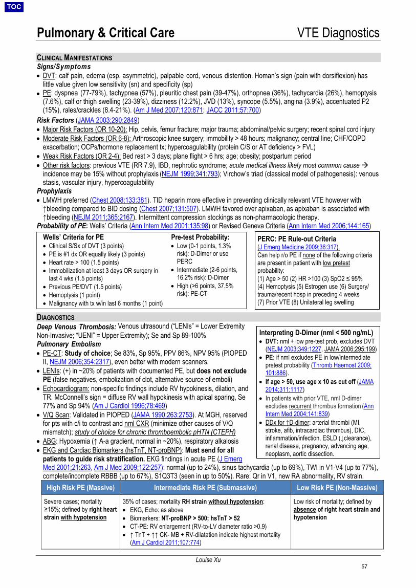
Pulmonary & Critical Care VTE Diagnostics
Louise Xu
PERC: PE Rule-out Criteria (J Emerg Medicine 2009;36:317). Can help r/o PE if none of the following criteria are present in patient with low pretest probability: (1) Age > 50 (2) HR >100 (3) SpO2 ≤ 95%(4) Hemoptysis (5) Estrogen use (6) Surgery/trauma/recent hosp in preceding 4 weeks(7) Prior VTE (8) Unilateral leg swelling
CLINICAL MANIFESTATIONS Signs/Symptoms • DVT: calf pain, edema (esp. asymmetric), palpable cord, venous distention. Homan’s sign (pain with dorsiflexion) has
little value given low sensitivity (sn) and specificity (sp)• PE: dyspnea (77-79%), tachypnea (57%), pleuritic chest pain (39-47%), orthopnea (36%), tachycardia (26%), hemoptysis
(7.6%), calf or thigh swelling (23-39%), dizziness (12.2%), JVD (13%), syncope (5.5%), angina (3.9%), accentuated P2(15%), rales/crackles (8.4-21%). (Am J Med 2007;120:871; JACC 2011;57:700)
Risk Factors (JAMA 2003;290:2849) • Major Risk Factors (OR 10-20): Hip, pelvis, femur fracture; major trauma; abdominal/pelvic surgery; recent spinal cord injury• Moderate Risk Factors (OR 6-8): Arthroscopic knee surgery; immobility > 48 hours; malignancy; central line; CHF/COPD
exacerbation; OCPs/hormone replacement tx; hypercoagulability (protein C/S or AT deficiency > FVL)• Weak Risk Factors (OR 2-4): Bed rest > 3 days; plane flight > 6 hrs; age; obesity; postpartum period• Other risk factors: previous VTE (RR 7.9), IBD, nephrotic syndrome; acute medical illness likely most common cause
incidence may be 15% without prophylaxis (NEJM 1999;341:793); Virchow’s triad (classical model of pathogenesis): venousstasis, vascular injury, hypercoagulability
Prophylaxis • LMWH preferred (Chest 2008;133:381). TID heparin more effective in preventing clinically relevant VTE however with↑bleeding compared to BID dosing (Chest 2007;131:507). LMWH favored over apixaban, as apixaban is associated with↑bleeding (NEJM 2011;365:2167). Intermittent compression stockings as non-pharmacologic therapy.
Probability of PE: Wells’ Criteria (Ann Intern Med 2001;135:98) or Revised Geneva Criteria (Ann Intern Med 2006;144:165) Wells’ Criteria for PE • Clinical S/Sx of DVT (3 points)• PE is #1 dx OR equally likely (3 points)• Heart rate > 100 (1.5 points)• Immobilization at least 3 days OR surgery in
last 4 wks (1.5 points)• Previous PE/DVT (1.5 points)• Hemoptysis (1 point)• Malignancy with tx w/in last 6 months (1 point)
Pre-test Probability: • Low (0-1 points, 1.3%
risk): D-Dimer or usePERC
• Intermediate (2-6 points,16.2% risk): D-Dimer
• High (>6 points, 37.5%risk): PE-CT
DIAGNOSTICS Deep Venous Thrombosis: Venous ultrasound (“LENIs” = Lower Extremity Non-Invasive; “UENI” = Upper Extremity); Se and Sp 89-100% Pulmonary Embolism • PE-CT: Study of choice; Se 83%, Sp 95%, PPV 86%, NPV 95% (PIOPED
II, NEJM 2006;354:2317), even better with modern scanners.• LENIs: (+) in ~20% of patients with documented PE, but does not exclude
PE (false negatives, embolization of clot, alternative source of emboli)• Echocardiogram: non-specific findings include RV hypokinesis, dilation, and
TR. McConnell’s sign = diffuse RV wall hypokinesis with apical sparing, Se77% and Sp 94% (Am J Cardiol 1996;78:469)
• V/Q Scan: Validated in PIOPED (JAMA 1990;263:2753). At MGH, reservedfor pts with c/i to contrast and nml CXR (minimize other causes of V/Qmismatch); study of choice for chronic thromboembolic pHTN (CTEPH)
• ABG: Hypoxemia (↑ A-a gradient, normal in ~20%), respiratory alkalosis• EKG and Cardiac Biomarkers (hsTnT, NT-proBNP): Must send for all
patients to guide risk stratification. EKG findings in acute PE (J Emerg Med 2001;21:263, Am J Med 2009;122:257): normal (up to 24%), sinus tachycardia (up to 69%), TWI in V1-V4 (up to 77%), complete/incomplete RBBB (up to 67%), S1Q3T3 (seen in up to 50%). Rare: Qr in V1, new RA abnormality, RV strain.
High Risk PE (Massive) Intermediate Risk PE (Submassive) Low Risk PE (Non-Massive)
Severe cases; mortality ≥15%; defined by right heart strain with hypotension
35% of cases; mortality RH strain without hypotension: • EKG, Echo: as above• Biomarkers: NT-proBNP > 500; hsTnT > 52• CT-PE: RV enlargement (RV-to-LV diameter ratio >0.9)• ↑ TnT + ↑↑ CK- MB + RV-dilatation indicate highest mortality
(Am J Cardiol 2011;107:774)
Low risk of mortality; defined by absence of right heart strain and hypotension
Interpreting D-Dimer (nml < 500 ng/mL) • DVT: nml + low pre-test prob, excludes DVT
(NEJM 2003;349:1227, JAMA 2006;295:199)• PE: if nml excludes PE in low/intermediate
pretest probability (Thromb Haemost 2009;101:886).
• If age > 50, use age x 10 as cut off (JAMA2014;311:1117)
• In patients with prior VTE, nml D-dimerexcludes recurrent thrombus formation (AnnIntern Med 2004;141:839)
• DDx for ↑D-dimer: arterial thrombi (MI,stroke, afib, intracardiac thrombus), DIC,inflammation/infection, ESLD (↓clearance),renal disease, pregnancy, advancing age, neoplasm, aortic dissection.
57
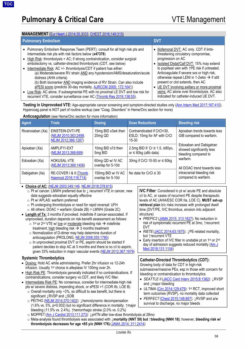
Pulmonary & Critical Care VTE Management
Louise Xu
MANAGEMENT (Eur Heart J 2014;35,3033, CHEST 2016;149:315) Pulmonary Embolism DVT
• Pulmonary Embolism Response Team (PERT): consult for all high risk pts andintermediate risk pts with risk factors below (x47378)
• High Risk: thrombolysis + AC; if strong contraindication, consider surgicalembolectomy vs. catheter-directed thrombolysis (CDT, see below)
• Intermediate Risk: AC +/- thrombolysis/CDT if patient has either:(a) Moderate/severe RV strain AND any hypotension/AMS/desaturation/acutedistress (AHA criteria)(b) Both biomarker AND imaging evidence of RV Strain. Can also includesPESI score (predicts 30-day mortality, AJRCCM 2005; 172:1041)
• Low Risk: AC alone. If subsegmental PE with no proximal LE DVT and low risk forrecurrent VTE, consider surveillance over AC (Thromb Res 2016;138:55).
• Iliofemoral DVT: AC only. CDT if limb-threatening circulatory compromise,progression on AC
• Isolated Distal/Calf DVT: 15% may extendto popliteal vein with ↑PE risk if untreated.Anticoagulate if severe sxs or high risk,otherwise repeat LENI in 1-2wks if stillpresent or clot extends, then AC
• UE DVT involving axillary or more proximalveins: AC alone over thrombolysis. AC alsoindicated for catheter-induced UE DVT.
Testing in Unprovoked VTE: Age-appropriate cancer screening and symptom-directed studies only (Ann Intern Med 2017;167:410). Hypercoag panel is NOT part of routine workup (see “Coag. Disorders” in Heme/Onc section for more) Anticoagulation (see Heme/Onc section for more information)
Agent Trials Dosing Dose Reductions Bleeding risk
Rivaroxaban (Xa) EINSTEIN-DVT/-PE (NEJM 2010;363:2499, NEJM 2012;366:1287)
15mg BID x3wk then 20mg QD
Contraindicated if CrCl<30, ESLD; 15mg for AF with CrCl 15-30
Apixaban trends towards less GIB compared to warfarin.
Edoxaban and Dabigatran showed significantly less bleeding compared to warfarin.
All DOAC trend towards less intracranial bleeding risk compared to warfarin.
Apixaban (Xa) AMPLIFY/-EXT(NEJM 2013;368:699)
10mg BID x7d then 5mg BID
2.5mg BID if: Cr ≥ 1.5, ≥80yo, or ≤ 60kg (afib data)
Edoxaban (Xa) HOKUSAL-VTE (NEJM 2013;369:1406)
60mg QD w/ IV AC overlap for 5-10d
30mg if CrCl 15-50 or ≤ 60kg
Dabigatran (IIa) RE-COVER I & II (Thomb Haemost 2016;116:714)
150mg BID w/ IV AC overlap for 5-10d
No data for CrCl ≤ 30
• Choice of AC: (NEJM 2003;349:146, NEJM 2018;378:615)o Pt w/ cancer: LMWH preferred due to ↓ recurrent VTE in cancer, new
data suggests edoxaban equally effectiveo Pt w/ APLAS: warfarin preferredo Pt undergoing thrombolysis or need for rapid reversal: UFHo All others: DOAC > warfarin (Grade 2B) > LMWH (Grade 2C)
• Length of Tx: 3 months if provoked. Indefinite if cancer-associated. Ifunprovoked, duration depends on risk-benefit assessment as follows:o 1st or 2nd VTE w/ low or moderate bleeding risk indefinite
treatment; high bleeding risk 3 months treatmento Normalization of D-dimer may help determine duration of
anticoagulation (PROLONG, NEJM 2006;355:1780)o In unprovoked proximal DVT or PE, aspirin should be started if
patient decides to stop AC at 3 months and there is no c/i to aspirin,given 33% reduction in major vascular events (NEJM 2012;367:1979)
Systemic Thrombolytics • Dosing: Hold AC while administering. Prefer 2hr infusion vs 12-24h
infusion. Usually 1st choice is alteplase IV 100mg over 2h.• High Risk PE: Thrombolysis generally indicated if no contraindications. If
contraindications, consider surgery vs CDT, and likely IVC filter.• Intermediate Risk PE: No consensus, consider for intermediate-high risk
pts w/ severe distress, impending shock, or sPESI >1 (COR IIb, LOE B)o Overall mortality only ~3%, so difficult to see benefit, but there is
significant ↓RVSP and ↓SOB o PEITHO (NEJM 2014;370:1402): ↓hemodynamic decompensation
(1.6% vs. 5%, p=0.002) but no significant difference in mortality. ↑majorbleeding (11.5% vs. 2.4%), ↑hemorrhagic stroke (2.0% vs. 0.2%)
o MOPPET (Am J Cardiol 2013;111:273): ↓pHTN after low-dose thrombolysis at 28moo Meta-analysis found thrombolysis was associated with ↓mortality (NNT 59) but ↑bleeding (NNH 18); however, bleeding risk w/
thrombolysis decreases for age <65 y/o (NNH 176) (JAMA 2014; 311:2414)
Catheter-Directed Thrombolytics (CDT): Growing body of data for CDT in high-risk submassive/massive PEs, esp in those with concern for bleeding or contraindication to thrombolytics • SEATTLE II (JACC Card Interv 2015;8:1382): ↓RVSP
and ↓major bleeding• ULTIMA (Circ 2014;129:479): 1st RCT, improved short
term outcomes (RVSP), no mortality data collected • PERFECT (Chest 2015;148:667): ↓RVSP and a/w
survival to discharge, no major bleeds
IVC Filter: Considered in pt w/ acute PE and absolute c/i to AC, or cases of recurrent PE despite therapeutic levels of AC (AHA/ESC COR IIa, LOE C). MUST set-up retrieval plan b/c risks increase with prolonged dwell time (DVT/PE, IVC thrombus, erosion into adjacent structure) • PREPIC2 (JAMA 2015; 313:1627): No reduction in
risk of symptomatic recurrent PE at 3mo, ↑recurrentDVT
• RIETE (JACC 2014;63:1675): ↓PE-related mortality,but ↑recurrent VTE
• Early insertion of IVC filter in unstable pt on 1st or 2nd
day of admission suggests reduced mortality (Am JMed 2018;131:1104)
58

Pulmonary & Critical Care Pulmonary Hypertension
Louise Xu
RV Failure (refer to “RV Failure” in Cardiology section) • Assess for reversible causes: PE, arrhythmia, ischemia, etc• Acute: ↑ preload to ↑ CO; avoid intubation and PPV if possible• Chronic: ↓ preload (improves RCA perfusion, reduces
ventricular interdependence, improves LV diastolic filling)• Maintenance of sinus rhythm and atrioventricular synchrony is
especially important given preload-dependent state.• ↑RV contractility, ↓RV afterload: reverse hypoxia,
hypercapnia, and acidemia; IV/inhaled pulm vasodilators;milrinone or dobutamine for inotropic support with pulmvasodilation (use norepi/phenylephrine/vaso if hypotensive)
• ECMO for reversible causes, pre-lung tx (Circ 2008;117:1717)
WHO Classification: (JACC 2013;62:D34) Pre-capillary (PCWP ≤ 15) Post-capillary (PCWP > 15)
Group 1: Pulmonary arterial hypertension Group 3: Lung disease/hypoxia
Group 4: CTEPH
Group 2: L heart disease
Idiopathic (F>M); Genetic (BMPR2, ALK1, ENG, SMAD9, CAV1, KCNK3); Drug/toxin (e.g., anorexigens, rapeseed oil, dasatanib, cocaine, amphetamines, SSRIs); CTD (MCTD, scleroderma, SLE); HIV; portopulmonary HTN; congenital heart diseases; schistosomiasis Subgroups: 1’ PVOD and/or pulmonary capillary hemangiomatosis; 1’’ persistent pulm HTN of newborn
COPD; ILD; OSA; Alveolar hypoventilation disorders; Developmental lung disease, among others
Chronic thromboembolic (occurs after ~ 4% of PEs)
(NEJM 2004;350:2257)
HFrEF; HFpEF; Valvular disease; Congenital/acquired left heart inflow/outflow tract obstruction and congenital cardiomyopathies
Group 5: Miscellaneous — Chronic hemolytic anemia; Sickle cell disease; Myeloproliferative; Splenectomy; Sarcoidosis; Metabolic, etc Diagnostic Workup: • Labs: HIV, ANA, RF/CCP, ANCA, Scl-70, Ro/La, BNP, LFTs• EKG: R-axis deviation, R/S >1 (V1), RBBB, ↑ P wave (II)• CXR: ↑PA, ↑R heart border, ↓retrosternal space (↑RV),
vascular pruning• CT: ↑PA, PA:Ao > 1, RV:LV >1, septal flattening, segmental
artery:bronchus >1• PFTs: isolated ↓DLCO (PAH), TLC <50%, FEV/FVC <70%• V/Q: 97% Se, 95% Sp for CTEPH (J Nucl Med 2007;48:680)• Exercise testing: 6-minute walk test, stress echo, CPET• Polysomnography: OSA evaluation• RHC (gold standard): R-sided P, PCWP, CO, vasoreactivity to
inhaled nitric oxide (guides treatment in idiopathic PAH)• TTE: ↑RA, ↑RV, TR, septum flattening, mid-systolic notch (PA doppler), RVOT AT <0.1s, E/A ratio < 1; PH likely if PASP >50 & TRV
>3.4; PASP differs from invasive testing by +/- 10 mmHg in 48% cases (Chest 2005;127:1836, AJRCCM 2009;179:615)Management: Close monitoring and specialist involvement recommended before starting these medications 1. Primary therapy to treat underlying etiology: CTD, CHF, hypoxemia (O2 therapy), VTE, etc.2. Advanced therapy (see table below): Reserved for WHO functional class II-IV despite adequate primary therapy. Most evidence inGroup 1. Usually not recommended in Group 2. Ongoing investigation for Group 3 and Group 5.
Note: ↓mPAP ≥ 10 mmHg and to ≤ 40 mmHg w/o ↓CO = positive vasodilator response (<12% pts) 3. Surgery: lung transplant (group 1), pulmonary thromboendarterectomy (group 4), atrial septostomy (R→ L shunt for severe RHF)
Medication Indication Side effects
CC BNifedipine, diltiazem Positive vasoreactivity test ↓BP, palpitations, LE edema, flushing, nausea, dizziness, headache, MI, CHF, hepatotoxicity
ET-1
Endothelin R antagonist: bosentan, ambrisentan, macitentan
Group 1: 1st line in PAH or SSc (Lancet 2001;358:1119, NEJM 2002;346:896) Macitentan ↓morbidity, mortality (NEJM 2013;369:809)
Bosentan/ambrisentan: anemia, PNA, edema, hepatitis Macitentan: as above + flu, HA, UTI, bronchitis
NO
Inahled NO (iNO) Group 1: remains investigational; used primarily for vasoreactivity testing
Hypotension, methemoglobinemia, rebound pHTN if discontinued
PDE5 inhibitors: sildenafil, tadalafil
Group 1: ↑exercise capacity, hemodynamics (NEJM 2005;353:2148) Group 3: improves hemodynamics (NEJM 2013;369:330)
Erythema, flushing, indigestion, HA, insomnia, epistaxis, rhinitis, retinal hemorrhage
sGC stimulator: riociguat Group 1, Group 4 (NEJM 2013;369:330) ↓BP, constipation, diarrhea, GERD, vomiting, anemia, dizziness, headache, hemorrhage
PGI2
Analogues: epoprostenol, treprostinil, iloprost
Groups 1, 3-5: reserved for sickest patients (NEJM 1996;334:296)
CP, ↓BP, ↑HR, flushing, ab pain, anorexia, n/v/d, jaw pain, MSK pain, dizziness, HA, hemorrhage
R Agonist: selexipag Group 1: 40% ↓ hospitalization; no Δ mortality (NEJM 2015;373:2522)
Diarrhea, nausea, jaw pain, headache, anemia
mean PA pressure (mPAP) = (PVR x CO) + PCWP Definition of PH: mPAP ≥ 25 mmg Hg at rest Measure: mPAP from RHC Calculate: PVR = (mPAP – PCWP) / CO (Fick’s or TD) Pre-Cap PH: PCWP <15, DPG >7, PVR > 3 Post-Cap PH: PCWP >15, DPG <7, PVR <3 Mixed PH: PCWP >15, DPG >7, PVR >3 **Diastolic pulm. gradient (DPG) = PAd - PCWP (NB: may also use transpulm. gradient [TPG] > 12)**
Presentation (nonspecific/insidious sxn; ~2 year delay to diagnosis, Chest 2011;140:19): • Early: dyspnea on exertion, lethargy, fatigue• Late: exertional chest pain, syncope, edema, hepatic
congestion• Rare: cough, hemoptysis, hoarseness (Ortner’s syndrome)• Exam: ↑JVP, edema, ascites, loud P2, prominent a wave,
TR, PR, L parasternal heave
59
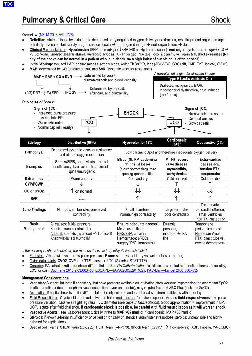
Pulmonary & Critical Care Shock
Ray Parrish, Joe Planer
Overview: (NEJM 2013;369:1726) • Definition: state of tissue hypoxia due to decreased or dysregulated oxygen delivery or extraction, resulting in end-organ damage
o Initially reversible, but rapidly progresses: cell death end-organ damage multiorgan failure death• Clinical Manifestations: Hypotension (SBP <90mmHg or ↓SBP >40mmHg from baseline); end organ dysfunction: oliguria (UOP
<0.5cc/kg/hr), altered mental status, metabolic acidosis (+/- anion gap, ↑lactate); cool & clammy vs. warm & flushed extremities (Nb.any of the above can be normal in a patient who is in shock, so a high index of suspicion is often needed)
• Initial Workup: focused H&P, ensure access, review meds, order EKG/CXR, labs (ABG/VBG, CBC+diff, CMP, TnT, lactate, CVO2)• MAP: determined by CO (cardiac output) and SVR (systemic vascular resistance)
Alternative etiologies for elevated lactate:
Etiologies of Shock:
Etiology Distributive (66%) Hypovolemic (16%) Cardiogenic (16%) Obstructive (2%)
Pathophys. Decreased systemic vascular resistance and altered oxygen extraction Low cardiac output and therefore inadequate oxygen delivery
Examples Sepsis/SIRS, anaphylaxis, adrenal
insufficiency, liver failure, toxins/meds, spinal/neurogenic
Bleed (GI, RP, abdominal, thigh), GI losses
(diarrhea/vomiting), third spacing (pancreatitis),
MI, HF, severe valve disease, myocarditis, arrhythmias
Extra-cardiac causes (PE, tension PTX, tamponade)
Extremities Warm and dry Cold and dry Cold and wet Cold and dry CVP/PCWP ↓ ↓ ↑ ↑ CO or CVO2 ↑ or normal ↓↓ ↓↓ ↓↓
SVR ↓↓ ↑ ↑ ↑
Echo Findings Normal chamber size, preserved contractility
Small chambers, normal/high contractility
Large ventricles, poor contractility
Tamponade: pericardial effusion,
small ventricles PE/PTX: dilated RV
Basic Management
All causes: fluids, pressors Sepsis: source control, abx Adrenal: steroids (hydrocort +/- fludricort) Anaphylaxis: epi 0.3mg IM
Ensure adequate access! Most cases: fluids HRS/SBP: albumin Hemorrhage: pRBCs, surgery/IR/GI hemostasis
Diuresis, pressors, inotrope, +/- PA line
Tamponade: pericardiocentesis PE: heparin/lysis PTX: chest tube vs. needle decompress
If the etiology of shock is unclear, the most useful ways to quickly distinguish include: • First step: Vitals: wide vs. narrow pulse pressure; Exam: warm vs. cold, dry vs. wet, rashes or mottling• Quick data points: CVO2, CVP, and TTE (consider POCUS and/or STAT TTE)• Consider: PA catheterization for shock differentiation. See PA Catheterization for full discussion, but no benefit in terms of mortality,
LOS, or cost (Cochrane 2013;2:CD003408, ESCAPE—JAMA 2005;294:1625, PAC-Man—Lancet 2005;366:472)
Management Considerations • Ventilatory Support: intubate if necessary, but have pressors available as intubation often worsens hypotension; be aware that SpO2
is often unreliable due to peripheral vasoconstriction (even on earlobe), may require frequent ABG Plus (includes SaO2)• Antibiotics: If septic shock is on the differential, get early cultures and start broad spectrum antibiotics without delay• Fluid Resuscitation: Crystalloid or albumin given as bolus (not infusion) for quick response. Assess fluid responsiveness by: pulse
pressure variation, passive straight leg raise, IVC diameter (see Sepsis: Resuscitation). Good approximation = improvement in BP,UOP, lactate after fluid challenge. If cardiogenic shock is possible, be careful with fluid resuscitation as it will worsen shock.
• Vasoactive Agents: (see Vasopressors); typically titrate to MAP >65 mmHg (if cardiogenic, MAP >60 mmHg)• Steroids: if known adrenal insufficiency or patient chronically on steroids, administer stress-dose steroids; unclear role and highly
debated for septic shock• Specialized Teams: STEMI team (x6-8282), PERT team (x4-7378), Shock team (p29151 if considering IABP, Impella, VA-ECMO)
MAP = RAP + CO x SVR
Signs of ↑CO: - Increased pulse pressure- Low diastolic BP- Warm extremities- Normal cap refill (early)
(2/3) DBP + (1/3) SBP HR x SV
Determined by vessel diameter/length and blood viscosity
Determined by preload, afterload, and contractility
SHOCK
↑CO ↓CO
Signs of ↓CO: - Narrow pulse pressure- Cold extremities- Slow cap refill
Diabetes, malignancy, EtOH, mitochondrial dysfunction, drug induced (metformin)
Type B Lactic Acidosis Ddx
60
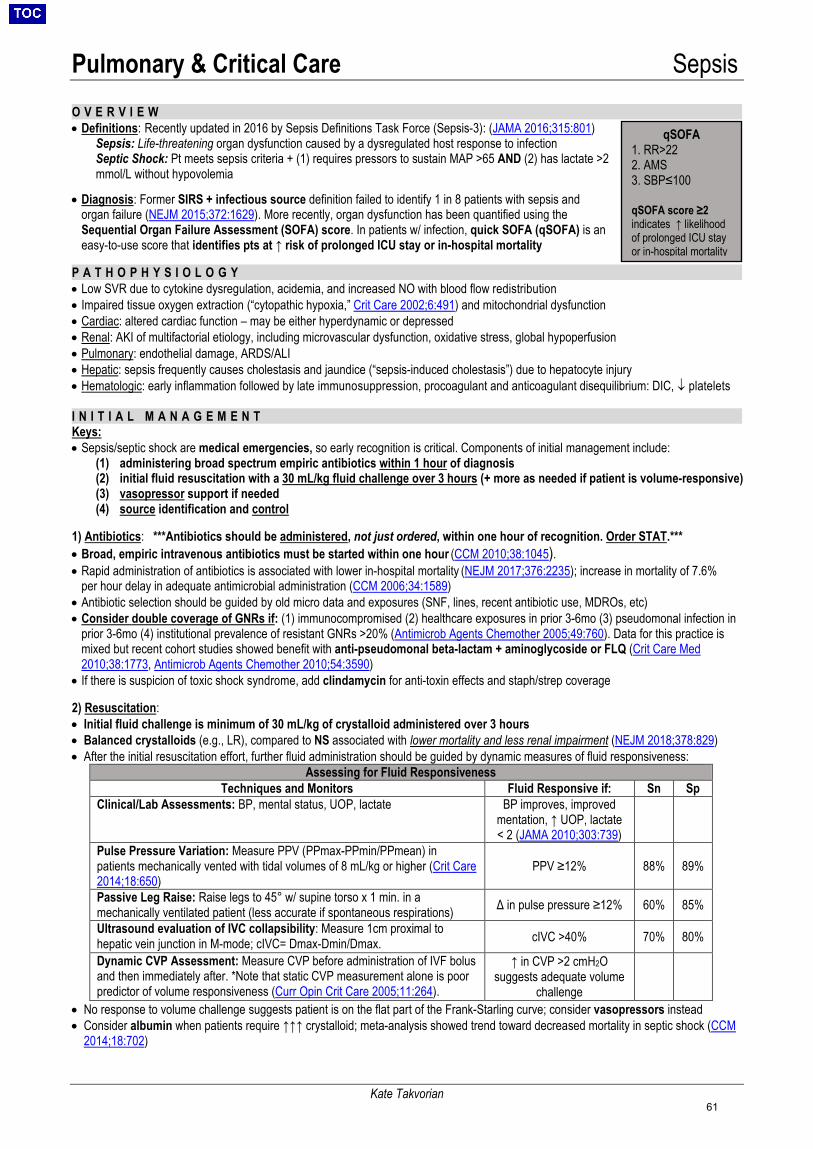
Pulmonary & Critical Care Sepsis
Kate Takvorian
O V E R V I E W• Definitions: Recently updated in 2016 by Sepsis Definitions Task Force (Sepsis-3): (JAMA 2016;315:801)
Sepsis: Life-threatening organ dysfunction caused by a dysregulated host response to infection Septic Shock: Pt meets sepsis criteria + (1) requires pressors to sustain MAP >65 AND (2) has lactate >2 mmol/L without hypovolemia
• Diagnosis: Former SIRS + infectious source definition failed to identify 1 in 8 patients with sepsis andorgan failure (NEJM 2015;372:1629). More recently, organ dysfunction has been quantified using theSequential Organ Failure Assessment (SOFA) score. In patients w/ infection, quick SOFA (qSOFA) is aneasy-to-use score that identifies pts at ↑ risk of prolonged ICU stay or in-hospital mortality
P A T H O P H Y S I O L O G Y• Low SVR due to cytokine dysregulation, acidemia, and increased NO with blood flow redistribution• Impaired tissue oxygen extraction (“cytopathic hypoxia,” Crit Care 2002;6:491) and mitochondrial dysfunction• Cardiac: altered cardiac function – may be either hyperdynamic or depressed• Renal: AKI of multifactorial etiology, including microvascular dysfunction, oxidative stress, global hypoperfusion• Pulmonary: endothelial damage, ARDS/ALI• Hepatic: sepsis frequently causes cholestasis and jaundice (“sepsis-induced cholestasis”) due to hepatocyte injury• Hematologic: early inflammation followed by late immunosuppression, procoagulant and anticoagulant disequilibrium: DIC, ↓ platelets
I N I T I A L M A N A G E M E N TKeys: • Sepsis/septic shock are medical emergencies, so early recognition is critical. Components of initial management include:
(1) administering broad spectrum empiric antibiotics within 1 hour of diagnosis(2) initial fluid resuscitation with a 30 mL/kg fluid challenge over 3 hours (+ more as needed if patient is volume-responsive)(3) vasopressor support if needed(4) source identification and control
1) Antibiotics: ***Antibiotics should be administered, not just ordered, within one hour of recognition. Order STAT.***• Broad, empiric intravenous antibiotics must be started within one hour (CCM 2010;38:1045).• Rapid administration of antibiotics is associated with lower in-hospital mortality (NEJM 2017;376:2235); increase in mortality of 7.6%
per hour delay in adequate antimicrobial administration (CCM 2006;34:1589)• Antibiotic selection should be guided by old micro data and exposures (SNF, lines, recent antibiotic use, MDROs, etc)• Consider double coverage of GNRs if: (1) immunocompromised (2) healthcare exposures in prior 3-6mo (3) pseudomonal infection in
prior 3-6mo (4) institutional prevalence of resistant GNRs >20% (Antimicrob Agents Chemother 2005;49:760). Data for this practice ismixed but recent cohort studies showed benefit with anti-pseudomonal beta-lactam + aminoglycoside or FLQ (Crit Care Med2010;38:1773, Antimicrob Agents Chemother 2010;54:3590)
• If there is suspicion of toxic shock syndrome, add clindamycin for anti-toxin effects and staph/strep coverage
2) Resuscitation:• Initial fluid challenge is minimum of 30 mL/kg of crystalloid administered over 3 hours• Balanced crystalloids (e.g., LR), compared to NS associated with lower mortality and less renal impairment (NEJM 2018;378:829)• After the initial resuscitation effort, further fluid administration should be guided by dynamic measures of fluid responsiveness:
Assessing for Fluid Responsiveness Techniques and Monitors Fluid Responsive if: Sn Sp
Clinical/Lab Assessments: BP, mental status, UOP, lactate BP improves, improved mentation, ↑ UOP, lactate < 2 (JAMA 2010;303:739)
Pulse Pressure Variation: Measure PPV (PPmax-PPmin/PPmean) in patients mechanically vented with tidal volumes of 8 mL/kg or higher (Crit Care 2014;18:650)
PPV ≥12% 88% 89%
Passive Leg Raise: Raise legs to 45° w/ supine torso x 1 min. in a mechanically ventilated patient (less accurate if spontaneous respirations) Δ in pulse pressure ≥12% 60% 85%
Ultrasound evaluation of IVC collapsibility: Measure 1cm proximal to hepatic vein junction in M-mode; cIVC= Dmax-Dmin/Dmax. cIVC >40% 70% 80%
Dynamic CVP Assessment: Measure CVP before administration of IVF bolus and then immediately after. *Note that static CVP measurement alone is poor predictor of volume responsiveness (Curr Opin Crit Care 2005;11:264).
↑ in CVP >2 cmH2O suggests adequate volume
challenge • No response to volume challenge suggests patient is on the flat part of the Frank-Starling curve; consider vasopressors instead• Consider albumin when patients require ↑↑↑ crystalloid; meta-analysis showed trend toward decreased mortality in septic shock (CCM
2014;18:702)
qSOFA 1. RR>222. AMS3. SBP≤100
qSOFA score ≥2 indicates ↑ likelihood of prolonged ICU stay or in-hospital mortality
61
Pulmonary & Critical Care Sepsis
Kate Takvorian
3) Vasopressors: (see Vasopressors page for full details):• Target a mean arterial pressure (MAP) of >65 mm Hg (SEPSISPAM trial, NEJM 2014;370:1583)• Per MGH MICU guidelines, arterial lines are needed for BP monitoring only in pts on moderate doses of pressors (e.g., 10 -15 of NE)
o Norepinephrine (NE, Levophed): Often first choice vasopressoro Vasopressin: Can be added to NE with intent of either raising MAP or decreasing NE dosage (Circulation 1997;95:1122)o Epinephrine: Recommended when additional agent is needed to maintain adequate blood pressure; can be used instead of vasoo Phenylephrine (Neo): Recommended primarily when: (a) NE is associated with serious arrhythmias, (b) CO is high and BP
persistently low, (c) as salvage therapy when norepinephrine + vasopressin have failed to achieve MAP target (d) hypotension a/wAFRVR
o Dopamine: reserved for highly selective patient population with bradycardia and low risk of tachyarrhythmia, associated w/increased risk of arrhythmias/mortality in all-comers (Cochrane 2011;5:CD003709)
o Methylene blue: Uncommonly used, pressor of last resort when NO-mediated vasoplegia is suspectedo Angiotensin II: not currently available in MGH MICU; anticipate eventual availability given ATHOS-3 trial (NEJM 2019;377:419)
4) Source Control:• Cultures: Obtain cultures prior to antimicrobials (unless this will significantly
delay administration). Get at least 2 sets of BCx (both aerobic and anaerobic bottles), with at least one drawn percutaneously.
• Evaluate early for conditions that would require emergent source control (e.g.,necrotizing soft tissue infections, peritonitis, cholangitis, intestinal infarctions,abscess, obstructive renal stone w/ hydro)
• Routine blood cultures will grow Candida, Trichosporon, Fusarium andCryptococcus. Consider 1,3 beta-D-glucan assay and/or cryptococcal Ag ifconcerned for fungemia.
• Imaging studies: Failure to improve on broad spectrum antibiotics should prompt evaluation for an occult source
C O N T I N U I N G M A N A G E M E N TAntibiotics: • Attempt to narrow within 48 hours, ideally guided by culture data + sensitivities and clinical improvement• Duration of therapy should typically be 7-10 days; longer courses in patients w/ slow clinical response, undrainable foci, bacteremia, S.
aureus, some fungal or viral infections, or immunologic deficiencies (e.g., neutropenia)• Procalcitonin levels can inform decision to discontinue antibiotics or shorten course (Lancet Inf Dis 2016;16:819; PRORATA trial, Lancet
2010;375:463)Resuscitation: Overall conservative fluid management results in faster recovery in ARDS (FACTT trial, NEJM 2006;354:2564) Transfusion: No evidence to support transfusion goal higher than hgb 7 g/dl unless cardiac ischemia, hemorrhage, or severe hypoxemia
is present (TRISS trial, NEJM 2014;371:1381) Ventilation: see ARDS page Renal Dysfunction: • CVVH and HD are largely equivalent for treating AKI, but CVVH can help minimize fluid shifts in hemodynamically unstable patients.• Can consider early renal consult for RRT in patients with refractory acidosis, though recent data suggests no mortality benefit to early
(<12h) versus delayed (>48h) initiation of RRT in patients with septic shock and severe AKI (NEJM 2018;379:1431)• Caution: ampules of bicarbonate cause intracellular acidosis and pH- and calcium-dependent reductions in cardiac contractility (NEJM
2014;371:2309)• Traditionally, bicarbonate is not recommended for treatment of lactic acidosis (CHEST 2000;117:260)• In patients with AKI and severe metabolic academia (pH<7.20, PaCO2 <45mmHg, bicarb <20mmol/L), recent BICAR-ICU trial
showed mortality benefit with infusion of 125-250ml 2% IV bicarb over 30 min with max of 1000ml in 24h (Lancet 2018;392:31), goalpH >7.30
C O N T R O V E R S I A L M A N A G E M E N T S T R A T E G I E SCorticosteroids: An initial study suggested a mortality benefit for corticosteroids in patients with septic shock (“Annane” or French trial, JAMA 2002;288:862). Patients were randomized to placebo or hydrocortisone + fludrocortisone x 7d. ICU mortality, 28-day mortality, and in-hospital mortality were significantly improved compared to placebo. The same author showed similar results in a recent study (NEJM 2018;378:809). Controversy: Findings were not replicated in recent trials by other groups (CORTICUS, HYPRESS, and ADRENAL). • Key points: Consider IV hydrocortisone for patients in whom fluids and pressors do not restore hemodynamic stability. Note that cortisol
measurements are not adequate predictors of response to steroids in ICU patients, and that etomidate (used by RICU for rapid sequenceintubation) directly inhibits adrenal steroid biosynthesis.
Esmolol: RCT demonstrated significant mortality difference in pts w/ septic shock treated w/ esmolol (49.4% v. 80.5% in control group, JAMA 2013;310:1683). Controversy: Single-center trial performed in Italy; control group had a significantly higher mortality rate (80.5%) than that expected for septic patients. No subsequent studies have confirmed these findings. • Key points: Esmolol not routinely used for pts in septic shock
Where to draw blood cultures? Drawing cultures from vascular access devices can lead to high rates of false positives. Obtain cultures from vascular access devices only if concerned for CRBSI (rigors with infusion, erythema/induration around line site); otherwise obtain only peripheral blood cultures.
62
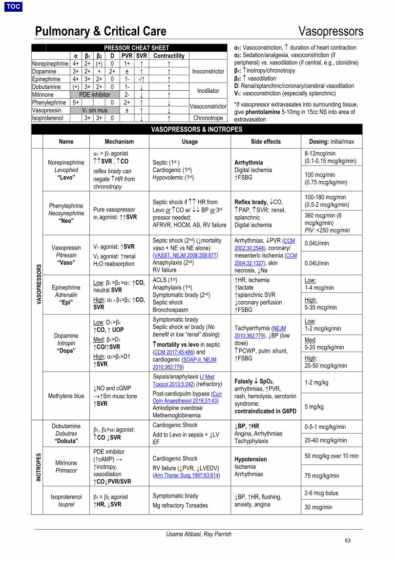
Pulmonary & Critical Care Vasopressors
Usama Abbasi, Ray Parrish
\
PRESSOR CHEAT SHEET α β1 β2 D PVR SVR Contractility
Norepinephrine 4+ 2+ (+) 0 1+ ↑ ↑ Inoconstrictor Dopamine 3+ 2+ + 2+ ± ↑ ↑
Epinephrine 4+ 3+ 2+ 0 1- -/↑ ↑ Dobutamine (+) 3+ 2+ 0 1- ↓ ↑ Inodilator Milrinone PDE inhibitor 2- ↓ ↑ Phenylephrine 5+ 0 2+ ↑ ↓ Vasoconstrictor Vasopressin V1 sm mus ± ↑ ↓ Isoproterenol 3+ 3+ 0 ↓ ↑ Chronotrope
VASOPRESSORS & INOTROPES Name Mechanism Usage Side effects Dosing: initial/max
VASO
PRES
SORS
Norepinephrine Levophed “Levo”
α1 > β1 agonist ↑↑SVR , ↑CO reflex brady can negate ↑HR from chronotropy
Septic (1st ) Cardiogenic (1st) Hypovolemic (1st)
Arrhythmia Digital Ischemia ↑FSBG
8-12mcg/min(0.1-0.15 mcg/kg/min)
100 mcg/min (0.75 mcg/kg/min)
Phenylephrine Neosynephrine
“Neo” Pure vasopressor α1 agonist: ↑↑SVR
Septic shock if ↑↑ HR from Levo or ↑CO w/ ↓↓ BP or 3rd pressor needed; AFRVR, HOCM, AS, RV failure
Reflex brady, ↓CO, ↑PAP, ↑SVR: renal, splanchnic Digital ischemia
100-180 mcg/min(0.5-2 mcg/kg/min)360 mcg/min (6 mcg/kg/min) PIV: <250 mcg/min
Vasopressin Pitressin “Vaso”
V1 agonist: ↑SVR V2 agonist: ↑renal H2O reabsorption
Septic shock (2nd) (↓mortality vaso + NE vs NE alone) (VASST, NEJM 2008;358:877) Anaphylaxis (2nd) RV failure
Arrhythmias, ↓PVR (CCM 2002;30:2548), coronary/ mesenteric ischemia (CCM 2004;32:1327), skin necrosis, ↓Na
0.04U/min
0.04U/min
Epinephrine Adrenalin
“Epi”
Low: β1 >β2 >α1: ↑CO, neutral SVR High: α1 > β1>β2: ↑CO, SVR
ACLS (1st) Anaphylaxis (1st) Symptomatic brady (2nd) Septic shock Bronchospasm
↑HR, ischemia ↑lactate ↑splanchnic SVR ↓coronary perfusion ↑FSBG
Low: 1-4 mcg/min
High: 5-35 mcg/min
Dopamine Intropin “Dopa”
Low: D1 >β1 ↑CO, ↑ UOP Med: β1>D1 ↑CO/↑SVR High: α1>β1>D1 ↑SVR
Symptomatic brady Septic shock w/ brady (No benefit in low “renal” dosing) ↑mortality vs levo in septic (CCM 2017;45:486) and cardiogenic (SOAP-II, NEJM 2010;362:779)
Tachyarrhymia (NEJM 2010;362:779), ↓BP (low dose) ↑PCWP, pulm shunt, ↑FSBG
Low: 1-2 mcg/kg/min
Med: 5-20 mcg/kg/minHigh: 20-50 mcg/kg/min
Methylene blue ↓NO and cGMP →↑Sm musc tone ↑SVR
Sepsis/anaphylaxis (J Med Toxicol 2013;3:242) (refractory) Post-cardiopulm bypass (Curr Opin Anaesthesiol 2018;31:43) Amlodipine overdose Methemoglobinemia
Falsely ↓ SpO2, arrhythmias, ↑PVR, rash, hemolysis, serotonin syndrome; contraindicated in G6PD
1-2 mg/kg
5 mg/kg
INOT
ROPE
S
Dobutamine Dobutrex “Dobuta”
β1, β2>α1 agonist: ↑CO ↓SVR
Cardiogenic Shock Add to Levo in sepsis + ↓LV EF
↓BP, ↑HR Angina, Arrhythmias Tachyphylaxis
0-5-1 mcg/kg/min
20-40 mcg/kg/min
Milrinone Primacor
PDE inhibitor (↑cAMP) → ↑inotropy, vasodilation ↑CO↓PVR/SVR
Cardiogenic Shock RV failure (↓PVR, ↓LVEDV) (Ann Thorac Surg 1997;63:814)
Hypotension Ischemia Arrhythmias
50 mcg/kg over 10 min
75 mcg/kg/min
Isoproterenol Isuprel
β1 = β2 agonist ↑HR, ↓SVR
Symptomatic brady Mg refractory Torsades
↓BP, ↑HR, flushing, anxiety, angina
2-6 mcg bolus
30 mcg/min
α1: Vasoconstriction, ↑ duration of heart contraction α2: Sedation/analgesia, vasoconstriction (if peripheral) vs. vasodilation (if central, e.g., clonidine) β1: ↑inotropy/chronotropy
β2: ↑ vasodilation D: Renal/splanchnic/coronary/cerebral vasodilation V1: vasoconstriction (especially splanchnic) *If vasopressor extravasates into surrounding tissue,give phentolamine 5-10mg in 15cc NS into area ofextravasation
63
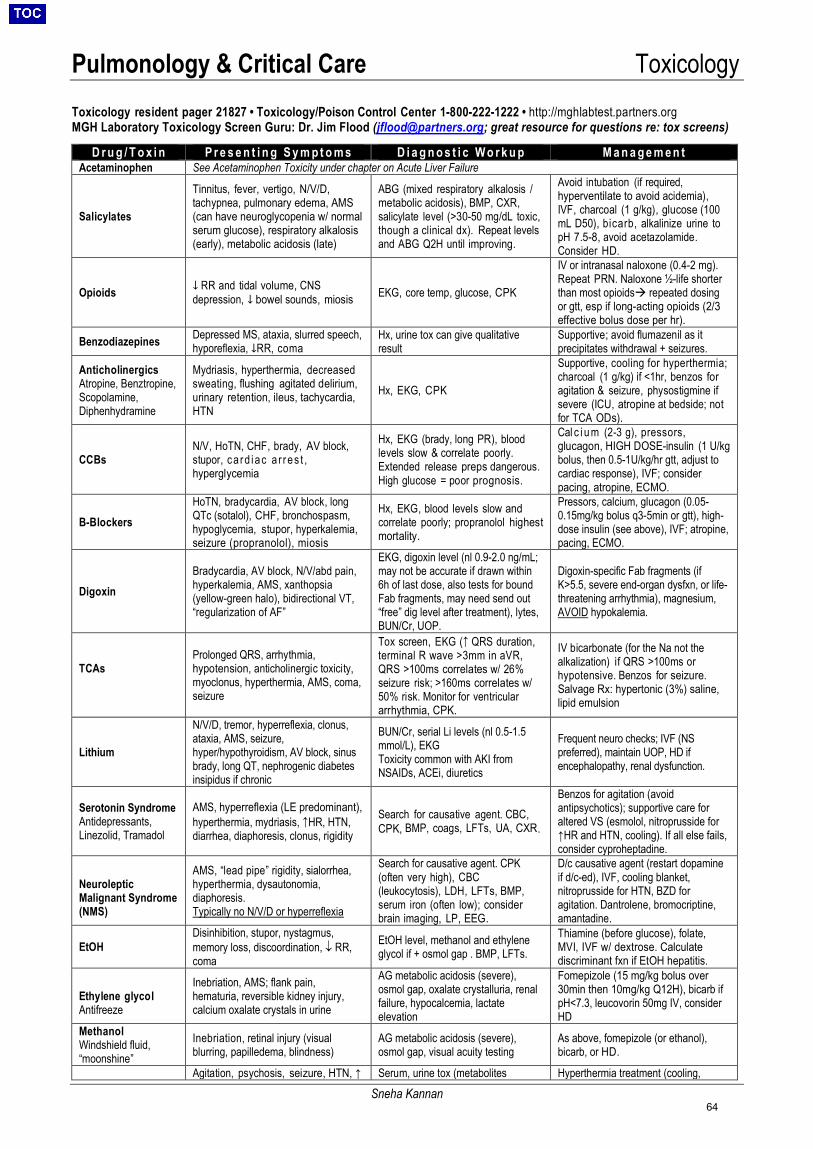
Pulmonology & Critical Care Toxicology
Sneha Kannan
Toxicology resident pager 21827 • Toxicology/Poison Control Center 1-800-222-1222 • http://mghlabtest.partners.org MGH Laboratory Toxicology Screen Guru: Dr. Jim Flood ([email protected]; great resource for questions re: tox screens)
D r u g / T o x i n P r e s e n t i n g S y m p t o m s D i a g n o s t i c W o r k u p M a n a g e m e n t Acetaminophen See Acetaminophen Toxicity under chapter on Acute Liver Failure
Salicylates
Tinnitus, fever, vertigo, N/V/D, tachypnea, pulmonary edema, AMS (can have neuroglycopenia w/ normal serum glucose), respiratory alkalosis (early), metabolic acidosis (late)
ABG (mixed respiratory alkalosis / metabolic acidosis), BMP, CXR, salicylate level (>30-50 mg/dL toxic, though a clinical dx). Repeat levels and ABG Q2H until improving.
Avoid intubation (if required, hyperventilate to avoid acidemia), IVF, charcoal (1 g/kg), glucose (100 mL D50), bicarb, alkalinize urine to pH 7.5-8, avoid acetazolamide. Consider HD.
Opioids ↓ RR and tidal volume, CNS depression, ↓ bowel sounds, miosis EKG, core temp, glucose, CPK
IV or intranasal naloxone (0.4-2 mg). Repeat PRN. Naloxone ½-life shorter than most opioids repeated dosing or gtt, esp if long-acting opioids (2/3 effective bolus dose per hr).
Benzodiazepines Depressed MS, ataxia, slurred speech, hyporeflexia, ↓RR, coma
Hx, urine tox can give qualitative result
Supportive; avoid flumazenil as it precipitates withdrawal + seizures.
Anticholinergics Atropine, Benztropine, Scopolamine, Diphenhydramine
Mydriasis, hyperthermia, decreased sweating, flushing agitated delirium, urinary retention, ileus, tachycardia, HTN
Hx, EKG, CPK
Supportive, cooling for hyperthermia; charcoal (1 g/kg) if <1hr, benzos for agitation & seizure, physostigmine if severe (ICU, atropine at bedside; not for TCA ODs).
CCBs N/V, HoTN, CHF, brady, AV block, stupor, ca rd iac a r r es t , hyperglycemia
Hx, EKG (brady, long PR), blood levels slow & correlate poorly. Extended release preps dangerous. High glucose = poor prognosis.
Cal c i u m (2-3 g), pressors, glucagon, HIGH DOSE-insulin (1 U/kg bolus, then 0.5-1U/kg/hr gtt, adjust to cardiac response), IVF; consider pacing, atropine, ECMO.
B-BlockersHoTN, bradycardia, AV block, long QTc (sotalol), CHF, bronchospasm, hypoglycemia, stupor, hyperkalemia, seizure (propranolol), miosis
Hx, EKG, blood levels slow and correlate poorly; propranolol highest mortality.
Pressors, calcium, glucagon (0.05-0.15mg/kg bolus q3-5min or gtt), high-dose insulin (see above), IVF; atropine, pacing, ECMO.
Digoxin Bradycardia, AV block, N/V/abd pain, hyperkalemia, AMS, xanthopsia (yellow-green halo), bidirectional VT, “regularization of AF”
EKG, digoxin level (nl 0.9-2.0 ng/mL; may not be accurate if drawn within 6h of last dose, also tests for bound Fab fragments, may need send out “free” dig level after treatment), lytes, BUN/Cr, UOP.
Digoxin-specific Fab fragments (if K>5.5, severe end-organ dysfxn, or life-threatening arrhythmia), magnesium, AVOID hypokalemia.
TCAs Prolonged QRS, arrhythmia, hypotension, anticholinergic toxicity, myoclonus, hyperthermia, AMS, coma, seizure
Tox screen, EKG (↑ QRS duration, terminal R wave >3mm in aVR, QRS >100ms correlates w/ 26% seizure risk; >160ms correlates w/ 50% risk. Monitor for ventricular arrhythmia, CPK.
IV bicarbonate (for the Na not the alkalization) if QRS >100ms or hypotensive. Benzos for seizure. Salvage Rx: hypertonic (3%) saline, lipid emulsion
Lithium
N/V/D, tremor, hyperreflexia, clonus, ataxia, AMS, seizure, hyper/hypothyroidism, AV block, sinus brady, long QT, nephrogenic diabetes insipidus if chronic
BUN/Cr, serial Li levels (nl 0.5-1.5 mmol/L), EKG Toxicity common with AKI from NSAIDs, ACEi, diuretics
Frequent neuro checks; IVF (NS preferred), maintain UOP, HD if encephalopathy, renal dysfunction.
Serotonin Syndrome Antidepressants, Linezolid, Tramadol
AMS, hyperreflexia (LE predominant), hyperthermia, mydriasis, ↑HR, HTN, diarrhea, diaphoresis, clonus, rigidity
Search for causative agent. CBC, CPK, BMP, coags, LFTs, UA, CXR.
Benzos for agitation (avoid antipsychotics); supportive care for altered VS (esmolol, nitroprusside for ↑HR and HTN, cooling). If all else fails, consider cyproheptadine.
Neuroleptic Malignant Syndrome (NMS)
AMS, “lead pipe” rigidity, sialorrhea, hyperthermia, dysautonomia, diaphoresis. Typically no N/V/D or hyperreflexia
Search for causative agent. CPK (often very high), CBC (leukocytosis), LDH, LFTs, BMP, serum iron (often low); consider brain imaging, LP, EEG.
D/c causative agent (restart dopamine if d/c-ed), IVF, cooling blanket, nitroprusside for HTN, BZD for agitation. Dantrolene, bromocriptine, amantadine.
EtOH Disinhibition, stupor, nystagmus, memory loss, discoordination, ↓ RR, coma
EtOH level, methanol and ethylene glycol if + osmol gap . BMP, LFTs.
Thiamine (before glucose), folate, MVI, IVF w/ dextrose. Calculate discriminant fxn if EtOH hepatitis.
Ethylene glycol Antifreeze
Inebriation, AMS; flank pain, hematuria, reversible kidney injury, calcium oxalate crystals in urine
AG metabolic acidosis (severe), osmol gap, oxalate crystalluria, renal failure, hypocalcemia, lactate elevation
Fomepizole (15 mg/kg bolus over 30min then 10mg/kg Q12H), bicarb if pH<7.3, leucovorin 50mg IV, consider HD
Methanol Windshield fluid, “moonshine”
Inebriation, retinal injury (visual blurring, papilledema, blindness)
AG metabolic acidosis (severe), osmol gap, visual acuity testing
As above, fomepizole (or ethanol), bicarb, or HD.
Agitation, psychosis, seizure, HTN, ↑ Serum, urine tox (metabolites Hyperthermia treatment (cooling,
64
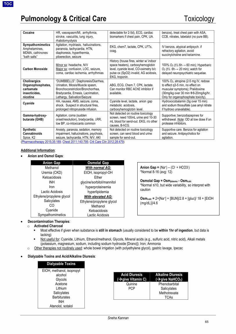
Pulmonology & Critical Care Toxicology
Sneha Kannan
Cocaine HR, vasospasm/MI, arrhythmia, stroke, vasculitis, lung injury, rhabdomyolysis
detectable for 2-5d), ECG, cardiac biomarkers if chest pain, CPK, UA.
benzos), treat chest pain with ASA, CCB, nitrates, labetalol (no pure BB).
Sympathomimetics Amphetamines, MDMA, cathinones “bath salts”
Agitation, mydriasis, hallucinations, paranoia, tachycardia, HTN, diaphoresis, hyperthermia, piloerection, seizure
EKG, chem7, lactate, CPK, LFTs, coag.
IV benzos, atypical antipsych. if refractory agitation, avoid succinylcholine and ketamine.
Carbon Monoxide Minor sx: headache, N/V Major sx: confusion, LOC, seizure, coma, cardiac ischemia, arrhythmias
History (house fires, winter w/ indoor space heaters), carboxyhemoglobin level, cyanide level, CO-oximetry b/c pulse ox (SpO2) invalid, AG acidosis, EKG, troponin.
100% O2 (t½ 6h→ 60 min); Hyperbaric O2 (t½ 6h→ 20 min); watch for delayed neuropsychiatric sequelae.
Cholinergics Organophosphates, carbamate insecticides, nicotine
“DUMBBELLS”: Diaphoresis/Diarrhea, Urination, Miosis/Muscle spasm, Bronchoconstriction/Bronchorrhea, Bradycardia, Emesis, Lacrimation, Lethargy, Salivation/Seizure
ABG, ECG, Chem 7, CPK, lactate. Can monitor RBC AChE inhibitor if available.
100% O2, atropine (2-5 mg IV, redose to effect q3-5 min, no effect on muscular symptoms); Pralidoxime (30mg/kg over 30 min8-20mg/kg/hr. Only for organophosphate toxicity).
Cyanide HA, nausea, AMS, seizure, coma, shock. Suspect in structural fires, prolonged nitroprusside infusion.
Cyanide level, lactate, anion gap metabolic acidosis, carboxyhemoglobin level.
Hydroxocobalamin (5g over 15 min) and sodium thiosulfate (use amyl nitrate if hydroxo unavailable).
Gamma-hydroxy-butarate (GHB)
Agitation, coma (sudden onset/resolution), bradycardia, ↓RR, low BP, co-intoxicants common
Not detected on routine toxicology screen, need 100mL urine and 10-30 mL blood for send-out. EKG, r/o other causes, B-hCG.
Supportive; benzodiazepines for withdrawal. Note: OD at low dose if on protease inhibitors.
Synthetic Cannabinoids Spice, K2
Anxiety, paranoia, sedation, memory impairment, hallucinations, psychosis, seizure, tachycardia, HTN, N/V, AKI
Not detected on routine toxicology screen, can send blood and urine sample for send-out.
Supportive care. Benzos for agitation and seizure. Antipsychotics for agitation.
(Pharmacotherapy 2015;35:189; Chest 2011;140:795; Crit Care Clin 2012;28:479)
Additional Information:
• Anion and Osmol Gaps:
• Decontamination Therapies:o Activated Charcoal Most effective if given when substance is still in stomach (usually considered to be within 1hr of ingestion, but data is
lacking) Not useful for: Cyanide, Lithium, Ethanol/methanol, Glycols, Mineral acids (e.g., sulfuric acid, nitric acid), Alkali metals
(potassium, magnesium, sodium, including sodium hydroxide [Drano]); Iron; Ammoniao Other therapies not routinely used: whole bowel irrigation (with polyethylene glycol), gastric lavage, Ipecac
• Dialyzable Toxins and Acid/Alkaline Diuresis:
Dialyzable Toxins
EtOH, methanol, isopropyl alcohol Glycols Acetone Lithium
Salicylates Barbiturates
INH Atenolol, sotalol
Anion Gap Osmolal Gap Methanol
Uremia (CKD) Ketoacidosis
INH Iron
Lactic Acidosis Ethylene/propylene glycol
Salicylates CO
Cyanide Sympathomimetics
With normal AG: EtOH, isopropyl-OH
Ether glycine/sorbitol/mannitol
hyperproteinemia hyperlipidemia
With elevated AG: Ethylene/propylene glycol
Methanol Ketoacidosis
Lactic Acidosis
Acid Diuresis (give Vitamin C)
Alkaline Diuresis (give NaHCO3)
Quinine PCP
Phenobarbital Salicylates
Methotrexate TCAs
Anion Gap = (Na+) – (Cl- + HCO3-) *Normal 8-16 (avg: 12)
Osmolal Gap = Osmplasma - Osmcalc *Normal ≤10, but wide variability, so interpret withcaution
Osmcalc = 2×[Na+] + [BUN]/2.8 + [gluc]/ 18 + [EtOH (mg/dL)]/4.6
65
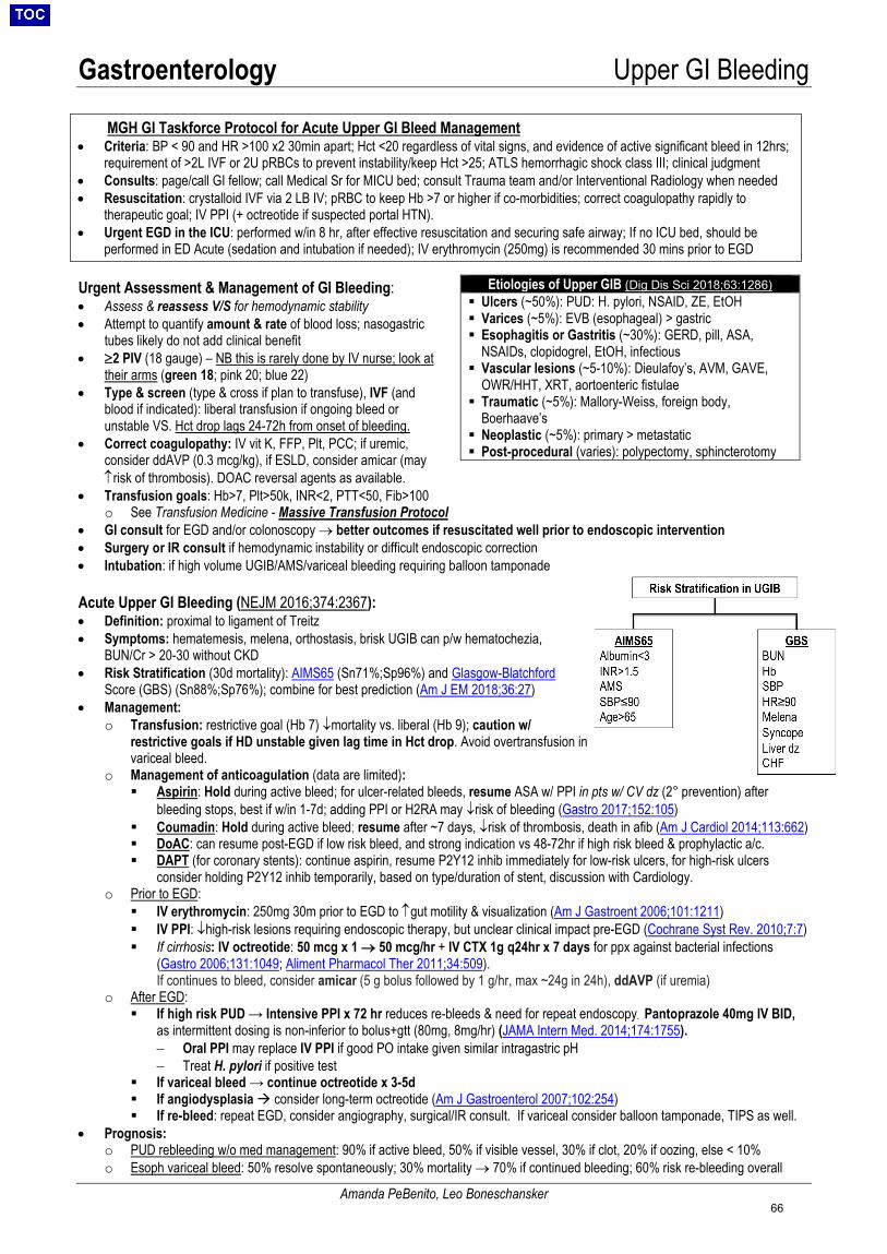
Gastroenterology Upper GI Bleeding
Amanda PeBenito, Leo Boneschansker
MGH GI Taskforce Protocol for Acute Upper GI Bleed Management • Criteria: BP < 90 and HR >100 x2 30min apart; Hct <20 regardless of vital signs, and evidence of active significant bleed in 12hrs;
requirement of >2L IVF or 2U pRBCs to prevent instability/keep Hct >25; ATLS hemorrhagic shock class III; clinical judgment• Consults: page/call GI fellow; call Medical Sr for MICU bed; consult Trauma team and/or Interventional Radiology when needed• Resuscitation: crystalloid IVF via 2 LB IV; pRBC to keep Hb >7 or higher if co-morbidities; correct coagulopathy rapidly to
therapeutic goal; IV PPI (+ octreotide if suspected portal HTN).• Urgent EGD in the ICU: performed w/in 8 hr, after effective resuscitation and securing safe airway; If no ICU bed, should be
performed in ED Acute (sedation and intubation if needed); IV erythromycin (250mg) is recommended 30 mins prior to EGD
Urgent Assessment & Management of GI Bleeding: • Assess & reassess V/S for hemodynamic stability• Attempt to quantify amount & rate of blood loss; nasogastric
tubes likely do not add clinical benefit• ≥2 PIV (18 gauge) – NB this is rarely done by IV nurse; look at
their arms (green 18; pink 20; blue 22)• Type & screen (type & cross if plan to transfuse), IVF (and
blood if indicated): liberal transfusion if ongoing bleed orunstable VS. Hct drop lags 24-72h from onset of bleeding.
• Correct coagulopathy: IV vit K, FFP, Plt, PCC; if uremic,consider ddAVP (0.3 mcg/kg), if ESLD, consider amicar (may↑risk of thrombosis). DOAC reversal agents as available.
• Transfusion goals: Hb>7, Plt>50k, INR<2, PTT<50, Fib>100o See Transfusion Medicine - Massive Transfusion Protocol
• GI consult for EGD and/or colonoscopy → better outcomes if resuscitated well prior to endoscopic intervention• Surgery or IR consult if hemodynamic instability or difficult endoscopic correction• Intubation: if high volume UGIB/AMS/variceal bleeding requiring balloon tamponade
Acute Upper GI Bleeding (NEJM 2016;374:2367): • Definition: proximal to ligament of Treitz• Symptoms: hematemesis, melena, orthostasis, brisk UGIB can p/w hematochezia,
BUN/Cr > 20-30 without CKD• Risk Stratification (30d mortality): AIMS65 (Sn71%;Sp96%) and Glasgow-Blatchford
Score (GBS) (Sn88%;Sp76%); combine for best prediction (Am J EM 2018;36:27)• Management:
o Transfusion: restrictive goal (Hb 7) ↓mortality vs. liberal (Hb 9); caution w/restrictive goals if HD unstable given lag time in Hct drop. Avoid overtransfusion invariceal bleed.
o Management of anticoagulation (data are limited): Aspirin: Hold during active bleed; for ulcer-related bleeds, resume ASA w/ PPI in pts w/ CV dz (2° prevention) after
bleeding stops, best if w/in 1-7d; adding PPI or H2RA may ↓risk of bleeding (Gastro 2017;152:105) Coumadin: Hold during active bleed; resume after ~7 days, ↓risk of thrombosis, death in afib (Am J Cardiol 2014;113:662) DoAC: can resume post-EGD if low risk bleed, and strong indication vs 48-72hr if high risk bleed & prophylactic a/c. DAPT (for coronary stents): continue aspirin, resume P2Y12 inhib immediately for low-risk ulcers, for high-risk ulcers
consider holding P2Y12 inhib temporarily, based on type/duration of stent, discussion with Cardiology.o Prior to EGD:
IV erythromycin: 250mg 30m prior to EGD to ↑gut motility & visualization (Am J Gastroent 2006;101:1211) IV PPI: ↓high-risk lesions requiring endoscopic therapy, but unclear clinical impact pre-EGD (Cochrane Syst Rev. 2010;7:7) If cirrhosis: IV octreotide: 50 mcg x 1 → 50 mcg/hr + IV CTX 1g q24hr x 7 days for ppx against bacterial infections
(Gastro 2006;131:1049; Aliment Pharmacol Ther 2011;34:509).If continues to bleed, consider amicar (5 g bolus followed by 1 g/hr, max ~24g in 24h), ddAVP (if uremia)
o After EGD: If high risk PUD → Intensive PPI x 72 hr reduces re-bleeds & need for repeat endoscopy. Pantoprazole 40mg IV BID,
as intermittent dosing is non-inferior to bolus+gtt (80mg, 8mg/hr) (JAMA Intern Med. 2014;174:1755).− Oral PPI may replace IV PPI if good PO intake given similar intragastric pH− Treat H. pylori if positive test
If variceal bleed → continue octreotide x 3-5d If angiodysplasia consider long-term octreotide (Am J Gastroenterol 2007;102:254) If re-bleed: repeat EGD, consider angiography, surgical/IR consult. If variceal consider balloon tamponade, TIPS as well.
• Prognosis:o PUD rebleeding w/o med management: 90% if active bleed, 50% if visible vessel, 30% if clot, 20% if oozing, else < 10%o Esoph variceal bleed: 50% resolve spontaneously; 30% mortality → 70% if continued bleeding; 60% risk re-bleeding overall
Etiologies of Upper GIB (Dig Dis Sci 2018;63:1286) Ulcers (~50%): PUD: H. pylori, NSAID, ZE, EtOH Varices (~5%): EVB (esophageal) > gastric Esophagitis or Gastritis (~30%): GERD, pill, ASA,
NSAIDs, clopidogrel, EtOH, infectious Vascular lesions (~5-10%): Dieulafoy’s, AVM, GAVE,
OWR/HHT, XRT, aortoenteric fistulae Traumatic (~5%): Mallory-Weiss, foreign body,
Boerhaave’s Neoplastic (~5%): primary > metastatic Post-procedural (varies): polypectomy, sphincterotomy
66
Gastroenterology Lower GI Bleeding
Leo Boneschansker
Acute Lower GI Bleeding (NEJM 2017;376:1054) • Definition: bleeding distal to ligament of Treitz resulting in
vital sign instability, anemia and/or need for bloodtransfusion
• Symptoms: hematochezia (maroon colored stools, brightred blood, or blood clots) or less commonly melena(generally requires that blood spends 14 hr in the GI tract)o Stool appearance is a poor indicator of GI bleeding
source - hematochezia can also be seen with briskUGIB (suspect if pt is hemodynamically unstable)
o Anorectal/L colon: Bright red bloodo R colon: Maroon colored stools, melena possible if slow
colonic transit o Bleeding will stop spontaneously in 80-85%
• History:o Painless hematochezia: diverticular bleed, angioectasia, hemorrhoido Abdominal pain: IBD, ischemic colitis, perforationo Weight loss: malignancy, IBDo Fever/Diarrhea: IBD, acute mesenteric ischemia, infectious colitiso Recent Colonoscopy: post-polypectomy bleedo Prior Abdominal/Pelvic XRT: radiation proctitis/colitiso Prior Abdominal Operations: anastamotic ulcerso History of AAA: aortoenteric fistulao AS, vWF, LVAD, ESRD: angioectasiao Atrial Fibrillation: acute mesenteric ischemiao Medications: NSAIDs (could precipitate diverticular bleed), anticoagulants, antiplatelets
• Diagnosis:o Exonerate UGIB source first (signs include orthostatic HoTN, hemodynamic instability, BUN:Cr >20-30 without CKD)o Consider NGT placement if there is moderate suspicion for UGIB (~15% of hematochezia is UGIB) Coffee-ground material, bright red blood → EGD No blood or bile seen: Indicates indeterminate source → consider EGD before Colonoscopy Bilious fluid: No active UGIB source → Colonoscopy
• Risk Stratification:o Several risk-factor models have been developed, but they are less well studied than models of upper GI bleeding. Overall
only limited ability to predict which patients will have poor outcomes. NOBLADS score: NSAID use, no diarrhea, no abdominal tenderness, SBP <100 mmHg, antiplatelet agent, albumin
<3.0 g/dL, ≥2 comorbidities, syncope (Clin Gastroenterol Hepatol 2016;14:1562) HR >100 bpm, SBP <115 mmHg, syncope, non tender abodomen, bleeding in <4 hr of eval, ASA use, >2
comorbidities (Am J Gastroenterol 2005;100:1821)• Management (Am J Gastroenterol 2016;111:459):
o Transfusion goals: Hgb >7 (consider >8 in active CAD), Plt >50k, INR <1.5 (INR 1.5-2.5 ok to perform endoscopichemostasis before reversing; INR >2.5 consider using reversal agent)
o Diverticular hemorrhage, angioectasia, post-polypectomy bleeding, and hemorrhoids amenable to endoscopic treatmento IF HEMODYNAMICALLY STABLE: Prep for colonoscopy (after discussion with GI) If ongoing bleeding or high risk perform within 24hr Use order set in EPIC OK to place NG tube for high-risk patients with ongoing bleeding who are intolerant of prep (if no known h/o varices) Urgent colonoscopy (within 12hr) improves localization but not mortality (Am J Gastroenterol 2010;105:2636) No data to suggest bowel prep increases or reactivates bleeding
o IF HEMODYNAMICALLY UNSTABLE: EGD to r/o UGIB, IR and surgical consult (“blind” surgery mortality ~29%),massive transfusion protocol
Unknown Source of GI Bleeding after Colonoscopy/EGD (“Occult GIB”) • Tagged RBC scan: (bleeding rate needs to be >0.5 mL/min): more sensitive for slow bleed, but poor localization• Video capsule study: allows visualization of full length of small bowel• Push enteroscopy: allows visualization of approximately the proximal 60cm of the jejunum• CT Angiography: (bleeding rate >1 mL/min) (ACR Radiologic Management of Lower Gastrointestinal Tract Bleeding)• IR Angiography: (bleeding rate needs to be >0.5 mL/min): success rate 25-70% in identifying cause, allows for intervention
(e.g. embolization, intra-arterial vasopressin) but incurs risk of arrhythmias, bowel ischemia, and vascular injury.
Etiologies of Lower GIB/Hematochezia Diverticulosis (30-65%) Ischemic colitis (5-20%) Hemorrhoids (5-20%) Brisk UGIB (~13%) Colorectal polyps or neoplasms (2-15%) Angioectasias (5-10%) Post-polypectomy (2-7%) IBD (3-5%) Infectious colitis (2-5%) Stercoral ulceration, colorectal varices, radiation
proctopathy, NSAID induced colopathy, Dieulafoy’slesion (0-5%)
67
Gastroenterology GERD & Peptic Ulcer Disease
Hawra Al Lawati
Gastroesophageal Reflux Disease (GERD) Signs & Sx: heartburn w/ food (i.e. spicy foods, coffee, soda, chocolate) or position (reclining), regurgitation, sour taste after awakening, sore throat, dysphagia, globus, chronic cough/throat clearing, hoarseness, asthma exacerbation, chest pain
Alarm symptoms: dysphagia/odynophagia, wt loss, GIB, Fe def. anemia, vomiting, persistent sx despite appropriate medical rx Complications:
Barrett’s Esophagus (BE): Squamous epithelium columnar intestinal epithelium. AdenoCa risk is 0.1-2%/year. Combined high dose PPIs and aspirin may ↓ mortality, cancer risk, and dysplasia (Lancet 2018;392:400)
• Screen w/ EGD only in: men w/ chronic GERD sx + 2 RFs (age>50, Caucasian, obesity, tobacco hx, FH of BE or adenoca)Esophageal stricture: p/w progressive solid food dysphagia. Endoscopy w/ biopsy can differentiate stricture from cancer.
Evaluation (Am J Gastro 2013;108:308): If sx’s suggestive of uncomplicated GERD, trial of empiric PPI QD for 4-8 wks is diagnostic test of choice. If alarm symptoms EGD w/ biopsy: detects tissue damage and/or complications, alternative DDx (i.e. EoE, malignancy) Ambulatory pH monitoring/impedence testing: indicated if endoscopy negative but persistent symptoms
Management: Mild/Intermittent symptoms -> see algorithm figure 1 in Gastro 2018;154:302
• Lifestyle rx: weight loss, head of bed elevation, tobacco cessation, reduce food triggers, no bedtime snacks• Antacids: Ca carbonate/Maalox for rapid relief. Gaviscon with NNT of 4. (A IM 2006;166:965, Ali PharmTher 2007;25:143)• PPIs: superior to antacids/H2RAs for sx relief in empiric treatment, and optimal for erosive esophagitis (Coch 2013)• Omeprazole/pantoprazole (initiate 20mg QD). 30min before meals. Reassess 2-4 weeks, uptitrate to 40mg QD then BID
if no relief. Maintain patients on lowest PPI dose that controls symptoms• Tapering off PPI: Recommended if asx > 3 months, no Barrett’s or severe EoE. Decrease dose by 50% per wk until d/c.• PPI risks (controversial): Probable association: Mg wasting (↑QTc), AIN. Possible association: ↑risk of osteoporosis,
dementia, CKD, C. diff/other enteric infx (Gastro 2017;152:706)• H2RAs (ranitidine, famotidine): can be given for nighttime sx prn w/ PPI, tachyphylaxis common after wksSevere/Persistent symptoms: (Am J Gastro 2013;108:308)• If there is no symptom relief after 2-4 weeks on high-dose twice daily PPI, refer for EGD and consider alternative dx• Consider gastric fundoplication (in general thought to be equivalent to PPI, reserved for severe & refractory cases).
Peptic Ulcer Disease (PUD) Signs & Sx: intermittent gnawing, dull, aching, or “hunger-like” epigastric pain relieved w/ antacids; duodenal ulcers p/w classic pain 2-5 hrs after meal (persistent acid secretion w/o buffer). Other sx: early satiety, nausea, vomiting, reflux. Etiology: 90% caused by H. pylori infxn or NSAIDs. Other: EtOH, smoking, meds (bisphosphonates, steroids, clopidogrel), ZES, mastocytosis, CMV, Crohn’s, sarcoid, lymphoma, CKD, carcinoid, cirrhosis. Evaluation: (Gastro Endosc 2010;71:663, Am J Gastro 2007;102:1808)
EGD: w/ biopsies of gastric ulcer margins to r/o malignancy. F/u EGD controversial in benign-appearing lesions. Repeat EGD ~12 wks if persistent, unclear etiology, or inadequate initial histology. H. pylori testing: Note false negative stool H.pylori test if on abx, PPIs, or bismuth. Urea breath test not available at MGH.• Serum Ab: Sn >90%, Sp 76-96%. For all other tests, must hold PPIs 7-14 days, abx/bismuth for 4 weeks, then Urea breath
test: Sn 88-95%, Sp >95%; Stool Ag: Sn > 90%, Sp 86-92%; useful for testing for active infection (Ab can be + even aftertx) and for confirming eradication.
ZES: check fasting serum gastrin (↑if on PPI, recheck 1 week s/p cessation), secretin stimulation test if non-diagnostic Management: anti-secretory therapy, H. pylori eradication if positive, removal of offending agents, optimization of comorbidities
Anti-secretory therapy: PPIs > H2RAs • H2RAs: Healing rates 70-80% at 4 wks, 87-94% at 8 wks (Gastro 1990;99:345)• PPIs: Healing rates 63-93% at 2wks, 80-100% at 4wks (Eur J Gastro Hepatol 1995;7:661)Mucosal barrier therapy: Sucralfate: use w/ anti-secretory therapy for duodenal ulcers 2/2 excess acid (i.e. not NSAID-induced or gastric ulcers). Improves mucosal barrier defense (forms complex by binding to protein exudates).H. pylori eradication therapy: (Gastro 2016;151:51)• First line = Quadruple Therapy: PPI BID, amoxicillin 1g BID, clarithromycin 500mg BID, metronidazole 500 BID x 14d.
If PCN allergic: PPI BID + metronidazole 500 BID + doxy 500 QID + bismuth QID x 14d. Combo pill: pylera (no PPI)• Complicated ulcers (see below): continue PPI for additional 2-4 wks if duodenal ulcer, or 4-6wks if gastric ulcer• Confirmation of eradication: breath test, stool Ag test, or upper endoscopy >4 wks after completion of abx and PPIFor patients with PUD 2/2 NSAIDs (other than low-dose aspirin): stop NSAIDs, consider starting a COX-2 inhibitor plus a PPI
Refractory symptoms Maintenance rx in recurrent/refractory ulcers: Can trial high dose PPI. Surgical rx include sectioning of vagus nerve, antrectomy, partial gastrectomy.
Complications and Management: Ulcer considered complicated if any of the following are present: Bleeding: IVF, transfusion, IV PPI, endoscopy (see GI Bleed management). If refractory, consider surgery. Perforation: Graham patch (omental piece covering ulcer) Gastric outlet obstruction: commonly due to pyloric channel/duodenal ulceration. Antrectomy/distal gastrectomy +/- vagotomy
68
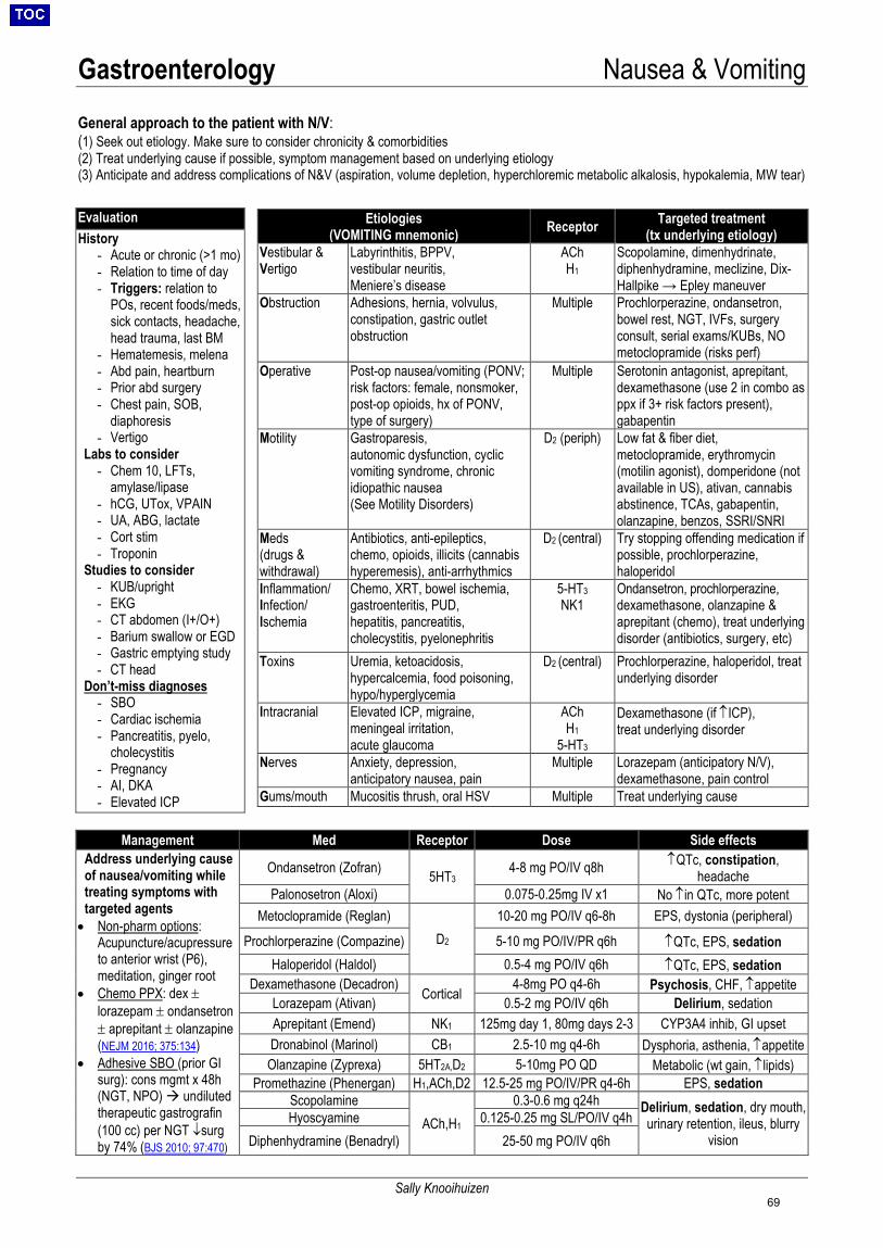
Gastroenterology Nausea & Vomiting
Sally Knooihuizen
General approach to the patient with N/V: (1) Seek out etiology. Make sure to consider chronicity & comorbidities(2) Treat underlying cause if possible, symptom management based on underlying etiology(3) Anticipate and address complications of N&V (aspiration, volume depletion, hyperchloremic metabolic alkalosis, hypokalemia, MW tear)
Evaluation History
- Acute or chronic (>1 mo)- Relation to time of day- Triggers: relation to
POs, recent foods/meds,sick contacts, headache,head trauma, last BM
- Hematemesis, melena- Abd pain, heartburn- Prior abd surgery- Chest pain, SOB,
diaphoresis- Vertigo
Labs to consider - Chem 10, LFTs,
amylase/lipase- hCG, UTox, VPAIN- UA, ABG, lactate- Cort stim- Troponin
Studies to consider - KUB/upright- EKG- CT abdomen (I+/O+)- Barium swallow or EGD- Gastric emptying study- CT head
Don’t-miss diagnoses - SBO- Cardiac ischemia- Pancreatitis, pyelo,
cholecystitis- Pregnancy- AI, DKA- Elevated ICP
Etiologies (VOMITING mnemonic) Receptor Targeted treatment
(tx underlying etiology) Vestibular & Vertigo
Labyrinthitis, BPPV, vestibular neuritis, Meniere’s disease
ACh H1
Scopolamine, dimenhydrinate, diphenhydramine, meclizine, Dix-Hallpike → Epley maneuver
Obstruction Adhesions, hernia, volvulus, constipation, gastric outlet obstruction
Multiple Prochlorperazine, ondansetron, bowel rest, NGT, IVFs, surgery consult, serial exams/KUBs, NO metoclopramide (risks perf)
Operative Post-op nausea/vomiting (PONV; risk factors: female, nonsmoker, post-op opioids, hx of PONV, type of surgery)
Multiple Serotonin antagonist, aprepitant, dexamethasone (use 2 in combo as ppx if 3+ risk factors present), gabapentin
Motility Gastroparesis, autonomic dysfunction, cyclic vomiting syndrome, chronic idiopathic nausea (See Motility Disorders)
D2 (periph) Low fat & fiber diet, metoclopramide, erythromycin (motilin agonist), domperidone (not available in US), ativan, cannabis abstinence, TCAs, gabapentin, olanzapine, benzos, SSRI/SNRI
Meds (drugs & withdrawal)
Antibiotics, anti-epileptics, chemo, opioids, illicits (cannabis hyperemesis), anti-arrhythmics
D2 (central) Try stopping offending medication if possible, prochlorperazine, haloperidol
Inflammation/ Infection/ Ischemia
Chemo, XRT, bowel ischemia, gastroenteritis, PUD, hepatitis, pancreatitis, cholecystitis, pyelonephritis
5-HT3NK1
Ondansetron, prochlorperazine, dexamethasone, olanzapine & aprepitant (chemo), treat underlying disorder (antibiotics, surgery, etc)
Toxins Uremia, ketoacidosis, hypercalcemia, food poisoning, hypo/hyperglycemia
D2 (central) Prochlorperazine, haloperidol, treat underlying disorder
Intracranial Elevated ICP, migraine, meningeal irritation, acute glaucoma
ACh H1
5-HT3
Dexamethasone (if ↑ICP), treat underlying disorder
Nerves Anxiety, depression, anticipatory nausea, pain
Multiple Lorazepam (anticipatory N/V), dexamethasone, pain control
Gums/mouth Mucositis thrush, oral HSV Multiple Treat underlying cause
Management Med Receptor Dose Side effects Address underlying cause of nausea/vomiting while treating symptoms with targeted agents • Non-pharm options:
Acupuncture/acupressureto anterior wrist (P6),meditation, ginger root
• Chemo PPX: dex ±lorazepam ± ondansetron± aprepitant ± olanzapine(NEJM 2016; 375:134)
• Adhesive SBO (prior GIsurg): cons mgmt x 48h(NGT, NPO) undilutedtherapeutic gastrografin(100 cc) per NGT ↓surgby 74% (BJS 2010; 97:470)
Ondansetron (Zofran) 5HT34-8 mg PO/IV q8h ↑QTc, constipation,
headache Palonosetron (Aloxi) 0.075-0.25mg IV x1 No ↑in QTc, more potent
Metoclopramide (Reglan) D2
10-20 mg PO/IV q6-8h EPS, dystonia (peripheral)
Prochlorperazine (Compazine) 5-10 mg PO/IV/PR q6h ↑QTc, EPS, sedation Haloperidol (Haldol) 0.5-4 mg PO/IV q6h ↑QTc, EPS, sedation
Dexamethasone (Decadron) Cortical 4-8mg PO q4-6h Psychosis, CHF, ↑appetite Lorazepam (Ativan) 0.5-2 mg PO/IV q6h Delirium, sedation Aprepitant (Emend) NK1 125mg day 1, 80mg days 2-3 CYP3A4 inhib, GI upset Dronabinol (Marinol) CB1 2.5-10 mg q4-6h Dysphoria, asthenia, ↑appetite
Olanzapine (Zyprexa) 5HT2A,D2 5-10mg PO QD Metabolic (wt gain, ↑lipids) Promethazine (Phenergan) H1,ACh,D2 12.5-25 mg PO/IV/PR q4-6h EPS, sedation
Scopolamine ACh,H1
0.3-0.6 mg q24h Delirium, sedation, dry mouth, urinary retention, ileus, blurry
vision Hyoscyamine 0.125-0.25 mg SL/PO/IV q4h
Diphenhydramine (Benadryl) 25-50 mg PO/IV q6h
69
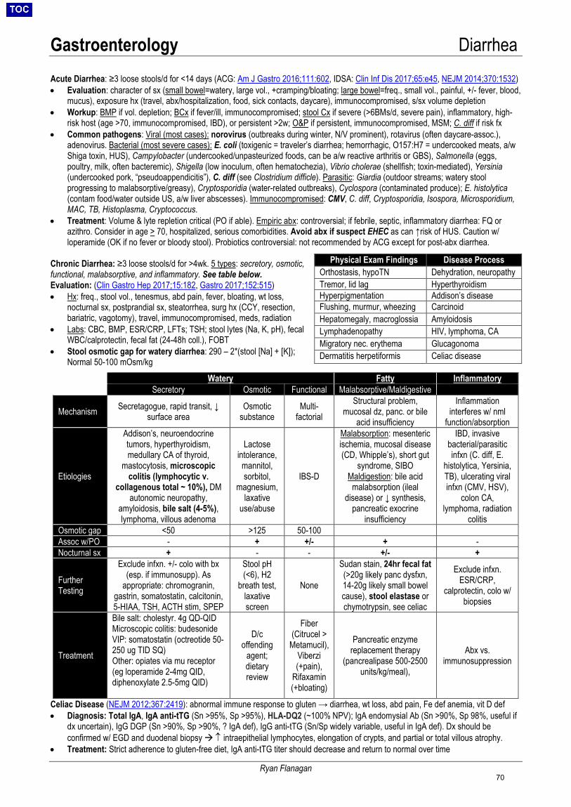
Gastroenterology Diarrhea
Ryan Flanagan
Acute Diarrhea: ≥3 loose stools/d for <14 days (ACG: Am J Gastro 2016;111:602, IDSA: Clin Inf Dis 2017;65:e45, NEJM 2014;370:1532) • Evaluation: character of sx (small bowel=watery, large vol., +cramping/bloating; large bowel=freq., small vol., painful, +/- fever, blood,
mucus), exposure hx (travel, abx/hospitalization, food, sick contacts, daycare), immunocompromised, s/sx volume depletion• Workup: BMP if vol. depletion; BCx if fever/ill, immunocompromised; stool Cx if severe (>6BMs/d, severe pain), inflammatory, high-
risk host (age >70, immunocompromised, IBD), or persistent >2w; O&P if persistent, immunocompromised, MSM; C. diff if risk fx• Common pathogens: Viral (most cases): norovirus (outbreaks during winter, N/V prominent), rotavirus (often daycare-assoc.),
adenovirus. Bacterial (most severe cases): E. coli (toxigenic = traveler’s diarrhea; hemorrhagic, O157:H7 = undercooked meats, a/wShiga toxin, HUS), Campylobacter (undercooked/unpasteurized foods, can be a/w reactive arthritis or GBS), Salmonella (eggs,poultry, milk, often bacteremic), Shigella (low inoculum, often hematochezia), Vibrio cholerae (shellfish; toxin-mediated), Yersinia(undercooked pork, “pseudoappendicitis”), C. diff (see Clostridium difficle). Parasitic: Giardia (outdoor streams; watery stoolprogressing to malabsorptive/greasy), Cryptosporidia (water-related outbreaks), Cyclospora (contaminated produce); E. histolytica(contam food/water outside US, a/w liver abscesses). Immunocompromised: CMV, C. diff, Cryptosporidia, Isospora, Microsporidium,MAC, TB, Histoplasma, Cryptococcus.
• Treatment: Volume & lyte repletion critical (PO if able). Empiric abx: controversial; if febrile, septic, inflammatory diarrhea: FQ orazithro. Consider in age > 70, hospitalized, serious comorbidities. Avoid abx if suspect EHEC as can ↑risk of HUS. Caution w/loperamide (OK if no fever or bloody stool). Probiotics controversial: not recommended by ACG except for post-abx diarrhea.
Chronic Diarrhea: ≥3 loose stools/d for >4wk. 5 types: secretory, osmotic, functional, malabsorptive, and inflammatory. See table below. Evaluation: (Clin Gastro Hep 2017;15:182, Gastro 2017;152:515) • Hx: freq., stool vol., tenesmus, abd pain, fever, bloating, wt loss,
nocturnal sx, postprandial sx, steatorrhea, surg hx (CCY, resection,bariatric, vagotomy), travel, immunocompromised, meds, radiation
• Labs: CBC, BMP, ESR/CRP, LFTs; TSH; stool lytes (Na, K, pH), fecalWBC/calprotectin, fecal fat (24-48h coll.), FOBT
• Stool osmotic gap for watery diarrhea: 290 – 2*(stool [Na] + [K]);Normal 50-100 mOsm/kg
Celiac Disease (NEJM 2012;367:2419): abnormal immune response to gluten → diarrhea, wt loss, abd pain, Fe def anemia, vit D def • Diagnosis: Total IgA, IgA anti-tTG (Sn >95%, Sp >95%), HLA-DQ2 (~100% NPV); IgA endomysial Ab (Sn >90%, Sp 98%, useful if
dx uncertain), IgG DGP (Sn >90%, Sp >90%, ? IgA def), IgG anti-tTG (Sn/Sp widely variable, useful in IgA def). Dx should beconfirmed w/ EGD and duodenal biopsy ↑ intraepithelial lymphocytes, elongation of crypts, and partial or total villous atrophy.
• Treatment: Strict adherence to gluten-free diet, IgA anti-tTG titer should decrease and return to normal over time
Physical Exam Findings Disease Process Orthostasis, hypoTN Dehydration, neuropathy Tremor, lid lag Hyperthyroidism Hyperpigmentation Addison’s disease Flushing, murmur, wheezing Carcinoid Hepatomegaly, macroglossia Amyloidosis Lymphadenopathy HIV, lymphoma, CA Migratory nec. erythema Glucagonoma Dermatitis herpetiformis Celiac disease
Watery Fatty Inflammatory Secretory Osmotic Functional Malabsorptive/Maldigestive
Mechanism Secretagogue, rapid transit, ↓ surface area
Osmotic substance
Multi- factorial
Structural problem, mucosal dz, panc. or bile
acid insufficiency
Inflammation interferes w/ nml
function/absorption
Etiologies
Addison’s, neuroendocrine tumors, hyperthyroidism, medullary CA of thyroid,
mastocytosis, microscopic colitis (lymphocytic v.
collagenous total ~ 10%), DM autonomic neuropathy,
amyloidosis, bile salt (4-5%), lymphoma, villous adenoma
Lactose intolerance,
mannitol, sorbitol,
magnesium, laxative
use/abuse
IBS-D
Malabsorption: mesenteric ischemia, mucosal disease (CD, Whipple’s), short gut
syndrome, SIBO Maldigestion: bile acid
malabsorption (ileal disease) or ↓ synthesis,
pancreatic exocrine insufficiency
IBD, invasive bacterial/parasitic infxn (C. diff, E.
histolytica, Yersinia, TB), ulcerating viral infxn (CMV, HSV),
colon CA, lymphoma, radiation
colitis Osmotic gap <50 >125 50-100Assoc w/PO - + +/- + - Nocturnal sx + - - +/- +
Further Testing
Exclude infxn. +/- colo with bx (esp. if immunosupp). As
appropriate: chromogranin, gastrin, somatostatin, calcitonin, 5-HIAA, TSH, ACTH stim, SPEP
Stool pH (<6), H2
breath test, laxative screen
None
Sudan stain, 24hr fecal fat (>20g likely panc dysfxn, 14-20g likely small bowelcause), stool elastase orchymotrypsin, see celiac
Exclude infxn. ESR/CRP,
calprotectin, colo w/ biopsies
Treatment
Bile salt: cholestyr. 4g QD-QID Microscopic colitis: budesonide VIP: somatostatin (octreotide 50-250 ug TID SQ) Other: opiates via mu receptor (eg loperamide 2-4mg QID, diphenoxylate 2.5-5mg QID)
D/c offending
agent; dietary review
Fiber (Citrucel >
Metamucil), Viberzi (+pain),
Rifaxamin (+bloating)
Pancreatic enzyme replacement therapy
(pancrealipase 500-2500 units/kg/meal),
Abx vs. immunosuppression
70
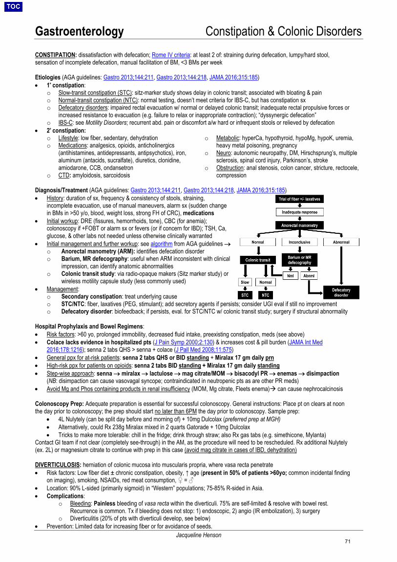
Gastroenterology Constipation & Colonic Disorders
Jacqueline Henson
CONSTIPATION: dissatisfaction with defecation; Rome IV criteria: at least 2 of: straining during defecation, lumpy/hard stool, sensation of incomplete defecation, manual facilitation of BM, <3 BMs per week
Etiologies (AGA guidelines: Gastro 2013;144:211, Gastro 2013;144:218, JAMA 2016;315:185) • 1º constipation:
o Slow-transit constipation (STC): sitz-marker study shows delay in colonic transit; associated with bloating & paino Normal-transit constipation (NTC): normal testing, doesn’t meet criteria for IBS-C, but has constipation sxo Defecatory disorders: impaired rectal evacuation w/ normal or delayed colonic transit; inadequate rectal propulsive forces or
increased resistance to evacuation (e.g. failure to relax or inappropriate contraction); “dyssynergic defecation”o IBS-C: see Motility Disorders; recurrent abd. pain or discomfort a/w hard or infrequent stools or relieved by defecation
• 2º constipation:o Lifestyle: low fiber, sedentary, dehydrationo Medications: analgesics, opioids, anticholinergics
(antihistamines, antidepressants, antipsychotics), iron,aluminum (antacids, sucralfate), diuretics, clonidine,amiodarone, CCB, ondansetron
o CTD: amyloidosis, sarcoidosis
o Metabolic: hyperCa, hypothyroid, hypoMg, hypoK, uremia,heavy metal poisoning, pregnancy
o Neuro: autonomic neuropathy, DM, Hirschsprung’s, multiplesclerosis, spinal cord injury, Parkinson’s, stroke
o Obstruction: anal stenosis, colon cancer, stricture, rectocele,compression
Diagnosis/Treatment (AGA guidelines: Gastro 2013;144:211, Gastro 2013;144:218, JAMA 2016;315:185) • History: duration of sx, frequency & consistency of stools, straining,
incomplete evacuation, use of manual maneuvers, alarm sx (sudden changein BMs in >50 y/o, blood, weight loss, strong FH of CRC), medications
• Initial workup: DRE (fissures, hemorrhoids, tone), CBC (for anemia);colonoscopy if +FOBT or alarm sx or fevers (or if concern for IBD); TSH, Ca,glucose, & other labs not needed unless otherwise clinically warranted
• Initial management and further workup: see algorithm from AGA guidelines →o Anorectal manometry (ARM): identifies defecation disordero Barium, MR defecography: useful when ARM inconsistent with clinical
impression, can identify anatomic abnormalitieso Colonic transit study: via radio-opaque makers (Sitz marker study) or
wireless motility capsule study (less commonly used)• Management:
o Secondary constipation: treat underlying causeo STC/NTC: fiber, laxatives (PEG, stimulant); add secretory agents if persists; consider UGI eval if still no improvemento Defecatory disorder: biofeedback; if persists, eval. for STC/NTC w/ colonic transit study; surgery if structural abnormality
Hospital Prophylaxis and Bowel Regimens: • Risk factors: >60 yo, prolonged immobility, decreased fluid intake, preexisting constipation, meds (see above)• Colace lacks evidence in hospitalized pts (J Pain Symp 2000;2:130) & increases cost & pill burden (JAMA Int Med
2016;178:1216); senna 2 tabs QHS > senna + colace (J Pall Med 2008;11:575)• General ppx for at-risk patients: senna 2 tabs QHS or BID standing + Miralax 17 gm daily prn• High-risk ppx for patients on opioids: senna 2 tabs BID standing + Miralax 17 gm daily standing• Step-wise approach: senna → miralax → lactulose → mag citrate/MOM → bisacodyl PR → enemas → disimpaction
(NB: disimpaction can cause vasovagal syncope; contraindicated in neutropenic pts as are other PR meds)• Avoid Mg and Phos containing products in renal insufficiency (MOM, Mg citrate, Fleets enema) can cause nephrocalcinosis
Colonoscopy Prep: Adequate preparation is essential for successful colonoscopy. General instructions: Place pt on clears at noon the day prior to colonoscopy; the prep should start no later than 6PM the day prior to colonoscopy. Sample prep:
• 4L Nulytely (can be split day before and morning of) + 10mg Dulcolax (preferred prep at MGH)• Alternatively, could Rx 238g Miralax mixed in 2 quarts Gatorade + 10mg Dulcolax• Tricks to make more tolerable: chill in the fridge; drink through straw; also Rx gas tabs (e.g. simethicone, Mylanta)
Contact GI team if not clear (completely see-through) in the AM, as the procedure will need to be rescheduled. Rx additional Nulytely (ex. 2L) or magnesium citrate to continue with prep in this case (avoid mag citrate in cases of IBD, dehydration)
DIVERTICULOSIS: herniation of colonic mucosa into muscularis propria, where vasa recta penetrate • Risk factors: Low fiber diet ± chronic constipation, obesity, ↑ age (present in 50% of patients >60yo; common incidental finding
on imaging), smoking, NSAIDs, red meat consumption, ♀ = ♂• Location: 90% L-sided (primarily sigmoid) in “Western” populations; 75-85% R-sided in Asia.• Complications:
o Bleeding: Painless bleeding of vasa recta within the diverticuli. 75% are self-limited & resolve with bowel rest.Recurrence is common. Tx if bleeding does not stop: 1) endoscopic, 2) angio (IR embolization), 3) surgery
o Diverticulitis (20% of pts with diverticuli develop, see below)• Prevention: Limited data for increasing fiber or for avoidance of seeds.
71
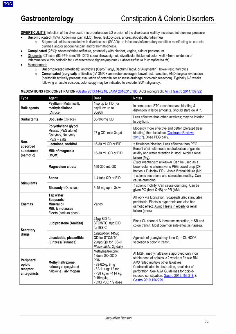
Gastroenterology Constipation & Colonic Disorders
Jacqueline Henson
DIVERTICULITIS: infection of the diverticuli: micro-perforation 2/2 erosion of the diverticular wall by increased intraluminal pressure• Uncomplicated (75%): Abdominal pain (LLQ), fever, leukocytosis, anorexia/obstipation/diarrhea
o Segmental colitis associated with diverticulosis (SCAD): an infectious/inflammatory condition manifesting as chronicdiarrhea and/or abdominal pain and/or hematochezia.
• Complicated (25%): Abscess/stricture/fistula, potentially with bladder, vagina, skin or peritoneum• Diagnosis: CT scan (93-97% sens/99-100% spec) shows sigmoid diverticula, thickened colon wall >4mm, evidence of
inflammation within pericolic fat + characteristic signs/symptoms (+ abscess/fistula in complicated dz)• Management:
o Uncomplicated (medical): antibiotics (Cipro/Flagyl, Bactrim/Flagyl, or Augmentin), bowel rest, narcoticso Complicated (surgical): antibiotics (IV GNR + anaerobe coverage), bowel rest, narcotics, AND surgical evaluation
(peritonitis typically present; evaluation of potential for abscess drainage or colonic resection). Typically 6-8 weeksfollowing an acute episode, colonscopy may be indicated to exclude IBD/malignancy.
MEDICATIONS FOR CONSTIPATION (Gastro 2013;144:218, JAMA 2016;315:185, ACG monograph: Am J Gastro 2014;109:S2)
Type Agent Dose Notes
Bulk agents Psyllium (Metamucil), methylcellulose (Citrucel)
1tsp up to TID (for psyllium: up to 30g/d)
In some (esp. STC), can increase bloating & distention in large amounts. Should start low & ↑.
Surfactants Docusate (Colace) 50-360mg QD Less effective than other laxatives; may be inferior to psyllium.
Non-absorbed substances (osmotic)
Polyethylene glycol Miralax (PEG alone) GoLytely, NuLytely (PEG + salts)
17 g QD; max 34g/d Modestly more effective and better tolerated (less bloating) than lactulose (Cochrane Reviews 2010;7). Dose PEG daily.
Lactulose, sorbitol 15-30 ml QD or BID ↑ flatulence/bloating. Less effective than PEG.
Milk of magnesia (MOM) 15-30 mL QD or BID
Benefit of simultaneous neutralization of gastric acidity and water retention in stool. Avoid if renal failure (Mg).
Magnesium citrate 150-300 mL QDExact mechanism unknown. Can be used as a lower-volume alternative to PEG bowel prep (2+ bottles + Dulcolax PR). Avoid if renal failure (Mg).
Stimulants Senna 1-4 tabs QD or BID ↑ colonic secretions and stimulates motility. Can
cause cramping.
Bisacodyl (Dulcolax) 5-15 mg up to 3x/w ↑ colonic motility. Can cause cramping. Can be given PO (best QHS) or PR (AM).
Enemas
Tap water Soapsuds Mineral oil Milk & molasses Fleets (sodium phos.)
Varies All work via lubrication. Soapsuds also stimulates peristalsis. Fleets is hypertonic and also has osmotic effect. Avoid Fleets in elderly or renal failure (phos).
Secretory drugs
Lubiprostone (Amitiza) 24μg BID for STC/NTC; 8μg BID for IBS-C
Binds Cl- channel & increases secretion, ↑ SB and colon transit. Most common side-effect is nausea.
Linaclotide, plecanitide (Linzess/Trulance)
Linaclotide: 145μg QD for STC/NTC; 290μg QD for IBS-C Plecanatide: 3g daily
Agonists of guanylate cyclase-C; ↑ Cl, HCO3 secretion & colonic transit.
Peripheral opioid receptor antagonists
Methylnaltrexone, naloxegol (pegylated naloxone), alvimopan
Methylnaltrexone: 1 dose SQ QOD PRN - 38-62kg: 8mg- 62-114kg: 12 mg- <38 kg or >114 kg:0.15mg/kg- CrCl <30: 1/2 dose
At MGH, methylnaltrexone approved only if on stable dose of opioids ≥ 2 weeks x 3d w/o BM AND failed multiple other laxatives. Contraindicated in obstruction, small risk of perforation. See AGA Guidelines for opioid-induced constipation: Gastro 2019;156:218 & Gastro 2019;156:229.
72

Gastroenterology Motility Disorders
Ryan Flanagan
Oropharyngeal Dysphagia Esophageal Dysphagia Symptoms Difficulty initiating swallowing, coughing, choking Difficulty seconds after initiation, food stuck in esophagus Neuro-muscular (solids & liq)
Central: tumor, stroke, PD, ALS, MS, polio Peripheral: neuropathy, myasthenia gravis Muscular: polymyositis, muscular dystrophy
Primary: achalasia, esophageal motility disorders (e.g. distal esophageal spasm, Jackhammer esophagus) Secondary: diabetes, scleroderma, Chagas (JAMA 2015;313:18)
Structural (solids at onset)
Intrinsic: tumor, XRT, trauma/surgical resection, Zenker’s Extrinsic: anterior mediastinal mass, goiter, cervical spondylosis
Intraluminal: food impaction Intrinsic: tumor, stricture, infxn (Cand., HSV, CMV), rings, EoE, webs, foreign body, pills (NSAIDs, tetracyclines, bisphosph) Extrinsic: vascular rings, aortic enlargement, LA compression, mediastinal, substernal thyroid, LAD (Gastro 2014;147:1238)
Work-up History: sx onset & duration, solids v. liquid dysphagia, underlying med conditions (e.g. CNS, malignancy, thyroid, DM, scleroderma), use of offending meds (pill esophagitis), immunocompromise (?HIV, chemotherapy ?infectious esophagitis), radiation, etc. Dysphagia in older adults is an alarm sx, should not be attributed to normal aging. PE: gen appearance (?systemic disease or CNS issue), HEENT exam (?evidence of LAD, tumor, asymmetry), FOBT Labs (consider): CBC, TFTs, ANA, α-Scl-70, α -centromere, α -RNP, α -Jo, HgbA1C, iron studies, HIV, AChR-Ab
Diagnostics 1) Modified barium swallow, ENT eval, +/- EGD toidentify obstructive structural problem2) Consider chest/neck CT to diagnose extrinsiccompression
1) EGD +/- barium swallow (mucosal pathology or structuralabnormality)2) if normal esoph. manometry to diagnose motility disorder3) Consider chest/neck CT to diagnose extrinsic compression
Selected conditions
Zenker’s diverticulum: p/w halitosis, regurgitation of food, cough. Tx w/ endoscopic surgery (rigid vs. flexible). Peptic strictures & rings: if lumen <13mm, dysphagia common. Tx PPI, dilation, intralesion steroid inj, stent Distal esophageal spasm: uncoordinated peristalsis a/w intermittent chest pain and regurgitation. Versus Jackhammer (hypercontractile) esophagus. Tx both w/ PPI, nitrates/CCB/PDEi, TCA/SSRI trial Eosinophilic esophagitis (EoE): dysphagia + hx allergies or atopy. EGD w/ stacked rings, strictures, Bx > 15 eos/hpf. Tx PPI, diet modification (culprits: dairy, wheat > soy, eggs, nuts, fish), use swallowed inhaler steroids; consider dilation. Achalasia: Progressive dysphagia solids/liquids, + regurgitation; barium swallow with bird’s beak appearance of distal esophagus; Manometry: absent distal peristalsis, incomplete LES relaxation; tx w/ pneumatic dilation, Heller myotomy, POEM, botox injections, CCBs (least effective) (World J Gastroenterol 2013;19:5806)
Gastroparesis Ileus Description: decreased gastric motility w/o obstruction, Sx: N/V, early satiety, postprandial fullness; rarely abdominal pain Causes: diabetes (vagus nerve damage 2/2 hyperglycemia), post-surgical (e.g. vagus nerve injury post-bariatic surgery), post-viral, systemic disease (thyroid disease, critical illness, Parkinson’s, connective tissue d/o), meds (opiates, CCB, anti-cholinergics) Exam: succussion splash (sloshing on abd ausc). Labs: TSH, A1c, tot protein, alb, CBC-diff. Studies: exclude mech obstruction w/ EGD; gastric emptying scintigraphy (gold std, hold motility meds 48 hrs prior); motility capsule, CO2 breath test. Treatment: Small meals, prokinetic agents (metoclopramide or erythromycin, consider domperidone), antiemetics, feeding tube if needed; pyloric botox is not recommended (Gastro Clinics of NA 2015;44:9; World J Gastro 2015;21:6842; ANZ J Surg 2015;85:709; Am J Gastroenterol 2013;108:18)
Description: slow motility of the gut w/o obstruction, often post-op, p/w nausea/vomiting, ↓BMs and ↓flatus, abd distention Post-operative paralytic ileus: Often post intra-abdominal surgery, KUB with dilated loops of small bowel w/o transition point. Treat with bowel rest, IVF, decompression (via NGT), avoid opioids Acute colonic pseudo-obstruction (Ogilvie’s): typically in elderly, hospitalized, ill patients. A/w severe illness (e.g. sepsis, pancreatitis, peritonitis), systemic disease (thyroid dis., DM, renal or liver failure), neuro problems (spinal cord compression or trauma, Parkinson’s, MS), meds (opiates, CCB, anticholinergics). Studies: KUB or CTAP with colonic dilatation Treatment: bowel rest, avoid opiates, replete lytes. PRN: rectal decompression, IV neostigmine (requires monitoring for bradycardia), methylnaltrexone.
FUNCTIONAL GI DISORDERS: GI disorders caused by aberrant neuronal signaling (dysfunction of the gut-brain axis) rather than structural or known molecular abnormality. Classification of >20 disorders per the Rome IV Criteria. (Gastroenterology 2016;150:1393) Functional Dyspepsia Early satiety, epigast pain/burning. Must r/o structural/organic cause. Tx: TCAs, metoclopramide Globus Sensation Sensation of obstruction in the throat when there is none, Tx: anti-reflux, antidepressants, CBT Cyclical Vomiting Syndrome Oscillation of intense N/V and no symptoms. More common in kids. Often trigger and prodrome.
Tx: avoid triggers, use benzos acutely to sedate, limited evidence for other tx (TCAs, Zofran) Cannabis Hyperemesis Syndrome Frequent cannabis use, N/V w/o normal periods, frequent hot bath/shower. Tx: MJ cessation Sphincter of Oddi Dysfxn Biliary pain, +/- pancreatitis 2/2 sphincter inability to relax, often post-CCY. Dx/Tx: ERCP
Irritable Bowel Syndrome (IBS): Definition (per Rome IV Criteria): recurrent abd discomfort ≥ 1x/wk on average for 3 months a/w 2+ of the following: (1) related to defecation, (2) change in stool frequency, (3) change in stool form. No nocturnal pain, weight loss, bleeding, elevated ESR/CRP. Epidemiology: ↑ risk w/ younger age, ♀ > ♂, psychosocial stressors, low QoL, hypochondriasis; bacterial gastroenteritis may be trigger. Types: IBS-C (constipation-predominant), IBS-D (diarrhea-predominant), IBS-M (mixed), IBS-U (unclassified), by Bristol Stool Score Treatment: exercise, diet modification, cognitive-behavioral therapy; laxatives (lubiprostone, linaclotide, PEG) for IBS-C; rifaximin, luxadoline,loperamide for IBS-D (limited data) (World J of Gastro 2014;20:12144)
73

Gastroenterology Inflammatory Bowel Disease
Amanda PeBenito, Sam Miller
When to Suspect IBD? (Crohn’s disease = CD, Ulcerative Colitis = UC) Epidemiology: Onset 15-40y, bimodal in CD w/ 2nd peak 50-80y. Genetic predisposition (up to 25% variance per GWAS studies; ↑incidence in Jews, Caucasians) + environment (↑risk w/ Western diet, abx exposure, NSAID use, smoking ↑ risk for CD & ↓ risk for UC) GI manifestations: Abd pain, diarrhea, bloody stools (UC>CD), incontinence/soiling, tenesmus, N/V, oral ulcers, perianal dz (CD) Extra-intestinal manifestations: Rheum (seronegative arthritis, sacroilitis), cutaneous (erythema nodosum, pyoderma gangrenosum), ophthalmic (uveitis, iritis, episcleritis), heme (DVT, AIHA), GI (PSC, B12 def), GU (Ca-Ox or UA stones), pulm (bronchiectasis, ILD) CD: Skip lesions, fibrosis/strictures, fistulae, transmural inflammation, noncaseating granulomas, cobblestoning UC: Continuous colonic mucosal inflammation spreading proximally from rectum, crypt abscesses, pseudopolyps Complications: Toxic megacolon (5% of UC), obstruction (2/2 strictures), abscesses, fistulae (CD), malabsorption, 2o amyloid, ↑risk CRC Inpatient Work-up and Management: H&P: baseline pain, BRBPR, #BM/consistency, #BM at night, surgical hx, date of onset, presenting sx, dz extent (fistulizing, stricturing, for CD), new meds (OTCs, NSAIDs, abx), smoking, nutrition/TPN, travel, extra-intest. sxs, current/past IBD meds compliance/efficacy Labs: CBC, Chem 10, LFTs (↑ALP?PSC), ESR/CRP, Mg, fecal calprotectin/lactoferrin, Stool Cx, O&P, C. diff, Fe/TIBC/B12 (if anemic) Imaging: if physical exam suggests peritonitis/obstruction/mass (abscess) KUB upright or CT A/P. Consider MRE to eval sm intestine.
Severity UC (True-Love Witts, Montreal Criteria)1,2 CD (CD Activity Index)3 Mild <4 stools (bloody or not), afebrile, nl ESR Ambulatory, tolerates PO/no dehydration, no pain/toxicity
Moderate 4-6 BM, bloody BM, low fever, ↑pain, mild anemia Failed 1st line tx, low fever, N/V, wt loss, pain, anemia Severe >6 BMs, Hb <10.5, fever, HR>90, wt loss, ESR >30 Failed advanced tx, toxic, abscess, obstruction, peritonitis, cachexia
Indications for surgery: CD: undilatable stricture, fistulae, abscess failing medical tx. UC: refractory disease, perforation, toxic megacolon Class Drug* Use Notes
Steroids Prednisone (PO) Budesonide (PO/PR) Methylprednisolone (IV)
Induction AE: Osteoporosis, AVN, infection, AI, weight gain, mood lability, delirium
Aminosali-cylates (UC>>CD)
Sulfasalazine Mesalamine (PO: Pentasa, Ascol, Lialda, Apriso. PR: Canasa, Rowasa)
Induction and maintenance
Sulfasalazine- pro-drug with more AEs, also systemic effects Mesalamine forms differ in gut penetration: Pentasa (ileum, R>L colon), Ascol (R>L colon), Lialda & Apriso (pancolon), Canasa & Rowasa (distal). AE: HA, nausea, fever, rash, diarrhea, pancreatitis, ↓sperm count, kidney injury
Thiopurines Azathioprine (pro-drug) 6-MP
Induction and maintenance
Labs: Need TPMT testing prior4, CBC, LFTs AE: N/V, hepatitis, BM suppression, pancreatitis, NHL, skin cancer
Anti-TNF Infliximab (Remicade) Adalimumab (Humira) Certolizumab (Cimzia) (for CD) Golimumab (Simponi) (for UC)
Induction and maintenance (Inflix and adal)
Maintenance (Certol, Golim)
Anti-TNFs are c/i if toxic megacolon, pyogenic infections, Labs: Need T-spot, hepatitis panel prior. Albumin (low albumin predictor of poor response in UC), CBC, LFTs. If flare during maintenance: measure trough (24hrs prior to dose) and antidrug Ab levels, determine if dose escalation or new drug is required.4 AE: site rxn, infxn, TB/HBV reactivation, neutropenia, demyelinating dz, HF.
Anti-integrin Vedolizumab5,6
(Entyvio)Induction and maintenance
2nd line, after Anti-TNF AE: Infusion reactions, nasopharyngitis
IL-12, -23 inhibitor
Uztekinumab7 (Stelara) (for CD)
Induction and maintenance
2nd line, after Anti-TNF, or for those >60, hx malignancy or infectious AE AE: Infection, HA, nasopharyngitis, nausea, abdominal pain, arthralgias
JAK inhibitor
Tofacitinib8 (Xeljanz) Induction and maintenance
AE: Infection, herpes zoster, HA, nasopharyngitis, arthralgias
Calcineurin inhib (IV)
Cyclosporine (Severe UC)
Induction only C/i in s/o toxic megacolon. Labs: troughs (q2-3d) Cr, Mg, lipids, LFTs AE: renal injury, ↑K, infxn, neurotoxicity/seizures (esp. if ↓Mg or cholesterol)
*For UC and CD unless otherwise notedMaintenance Treatment: “Step-up” therapy (least most toxic drug) preferred, but starting w/TNFα may be beneficial in severe dz9 Malignancy screening: colonoscopy after 8 years of active disease, repeat every 1-3 years w/ random 4-quadrant bx q10cm of colon
1. Nat Rev Gastro 2016;13:654; 2.Gut 2006;55:749; 3. Am J Gastroenterol. 2018;113:481; 4.Gastroenterology 2017;153:8275. NEJM 2013;369:699, 6. NEJM 2013;369:711 7. NEJM 2016;375:1946; 8. NEJM 2017; 376:1723; 9. Lancet 2015;386:1825
† Methylpred 20mg q8h IV ‡ ↑ risk of DVT/PE in IBD pts Δ In pts w/ fistula, abscess, peritonitis, pouchitis, perforation, high fever, leukocytosis, bandemia ϒ↑ risk of tox meg in UC * Bridge to maintenance txψ Anti-TNF, anti-integrin,etc.
Mild/Moderate (outpatient) Severe (inpatient)
IV steroids†, IV Fluids/lytes High-risk DVT ppx‡, +/- Cipro/FlagylΔTspot, Hep serologies No NSAIDs/opiods/antidiarrhealsϒ NPO for endoscopy (c/s IBD team, inform outpt GI), ADAT
Aminosalicylates (UC>>CD) Budesonide oral/topical (UC/CD) PO prednisone (UC/CD) Antibiotics (CD) Thiopurines (UC/CD)
IBD Flare Therapy
Resolution* Refractory disease/toxic Refractory after 3-5 days: Advance therapy,ψ, surgical consult
74
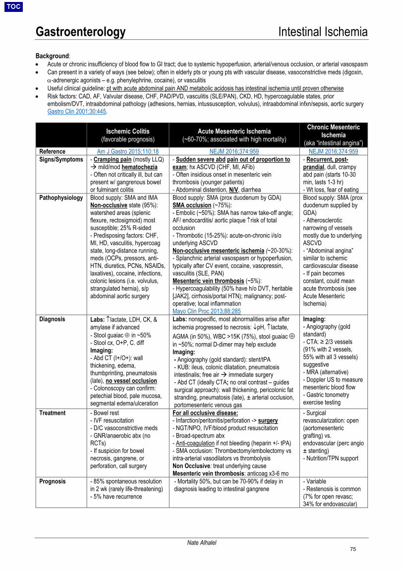
Gastroenterology Intestinal Ischemia
Nate Alhalel
Background: • Acute or chronic insufficiency of blood flow to GI tract; due to systemic hypoperfusion, arterial/venous occlusion, or arterial vasospasm• Can present in a variety of ways (see below); often in elderly pts or young pts with vascular disease, vasoconstrictive meds (digoxin,
α-adrenergic agonists – e.g. phenylephrine, cocaine), or vasculitis• Useful clinical guideline: pt with acute abdominal pain AND metabolic acidosis has intestinal ischemia until proven otherwise• Risk factors: CAD, AF, Valvular disease, CHF, PAD/PVD, vasculitis (SLE/PAN), CKD, HD, hypercoagulable states, prior
embolism/DVT, intraabdominal pathology (adhesions, hernias, intussusception, volvulus), intraabdominal infxn/sepsis, aortic surgeryGastro Clin 2001;30:445.
Ischemic Colitis (favorable prognosis)
Acute Mesenteric Ischemia (~60-70%; associated with high mortality)
Chronic Mesenteric Ischemia
(aka “intestinal angina”) Reference Am J Gastro 2015;110:18 NEJM 2016;374:959 NEJM 2016;374:959 Signs/Symptoms - Cramping pain (mostly LLQ)
mild/mod hematochezia- Often not critically ill, but canpresent w/ gangrenous bowelor fulminant colitis
- Sudden severe abd pain out of proportion toexam; hx ASCVD (CHF, MI, AFib)- Often insidious onset in mesenteric veinthrombosis (younger patients)- Abdominal distention, N/V, diarrhea
- Recurrent, post-prandial, dull, crampyabd pain (starts 10-30min, lasts 1-3 hr)- Wt loss, fear of eating
Pathophysiology Blood supply: SMA and IMA Non-occlusive state (95%): watershed areas (splenic flexure, rectosigmoid) most susceptible; 25% R-sided - Predisposing factors: CHF,MI, HD, vasculitis, hypercoagstate, long-distance running,meds (OCPs, pressors, anti-HTN, diuretics, PCNs, NSAIDs,laxatives), cocaine, infections,colonic lesions (i.e. volvulus,strangulated hernia), s/pabdominal aortic surgery
Blood supply: SMA (prox duodenum by GDA) SMA occlusion (~75%): - Embolic (~50%): SMA has narrow take-off angle;AF/ endocarditis/ aortic plaque ↑risk of totalocclusion- Thrombotic (15-25%): acute-on-chronic i/s/ounderlying ASCVDNon-occlusive mesenteric ischemia (~20-30%):- Splanchnic arterial vasospasm or hypoperfusion,typically after CV event, cocaine, vasopressin,vasculitis (SLE, PAN)Mesenteric vein thrombosis (~5%):- Hypercoagulability (50% have h/o DVT, heritable[JAK2], cirrhosis/portal HTN); malignancy; post-operative; local inflammationMayo Clin Proc 2013;88:285
Blood supply: SMA (prox duodenum supplied by GDA) - Atheroscleroticnarrowing of vesselsmostly due to underlyingASCVD- “Abdominal angina”similar to ischemiccardiovascular disease- If pain becomesconstant, could meanacute thrombosis (seeAcute MesentericIschemia)
Diagnosis Labs: ↑lactate, LDH, CK, & amylase if advanced - Stool guaiac ⊕ in ~50%- Stool cx, O+P, C. diffImaging:- Abd CT (I+/O+): wallthickening, edema,thumbprinting, pneumatosis(late), no vessel occlusion- Colonoscopy can confirm:petechial blood, pale mucosa,segmental edema/ulceration
Labs: nonspecific, most abnormalities arise after ischemia progressed to necrosis: ↓pH, ↑lactate, AGMA (in 50%), WBC >15K (75%), stool guaiac ⊕ in ~50%; normal D-dimer may help exclude Imaging: - Angiography (gold standard): stent/tPA- KUB: ileus, colonic dilatation, pneumatosisintestinalis; free air immediate surgery- Abd CT (ideally CTA; no oral contrast – guidessurgical approach): wall thickening, pericolonic fatstranding, pneumatosis (late), ± arterial occlusion,portomesenteric venous gas
Imaging: - Angiography (goldstandard)- CTA: ≥ 2/3 vessels(91% with 2 vessels,55% with all 3 vessels)suggestive- MRA (alternative)- Doppler US to measuremesenteric blood flow- Gastric tonometryexercise testing
Treatment - Bowel rest- IVF resuscitation- D/C vasoconstrictive meds- GNR/anaerobic abx (noRCTs)- If suspicion for bowelnecrosis, gangrene, orperforation, call surgery
For all occlusive disease: - Infarction/peritonitis/perforation -> surgery- NGT/NPO, IVF/blood product resuscitation- Broad-spectrum abx- Anti-coagulation if not bleeding (heparin +/- tPA)- SMA occlusion: Thrombectomy/embolectomy vsintra-arterial vasodilators vs thrombolysisNon Occlusive: treat underlying causeMesenteric vein thrombosis: anticoag x3-6 mo
- Surgicalrevascularization: open(aortomesentericgrafting) vs.endovascular (perc angio± stenting)- Nutrition/TPN support
Prognosis - 85% spontaneous resolutionin 2 wk (rarely life-threatening)- 5% have recurrence
- Mortality 50%, but can be 70-90% if delay indiagnosis leading to intestinal gangrene
- Variable- Restenosis is common(7% for open revasc;34% for endovascular)
75
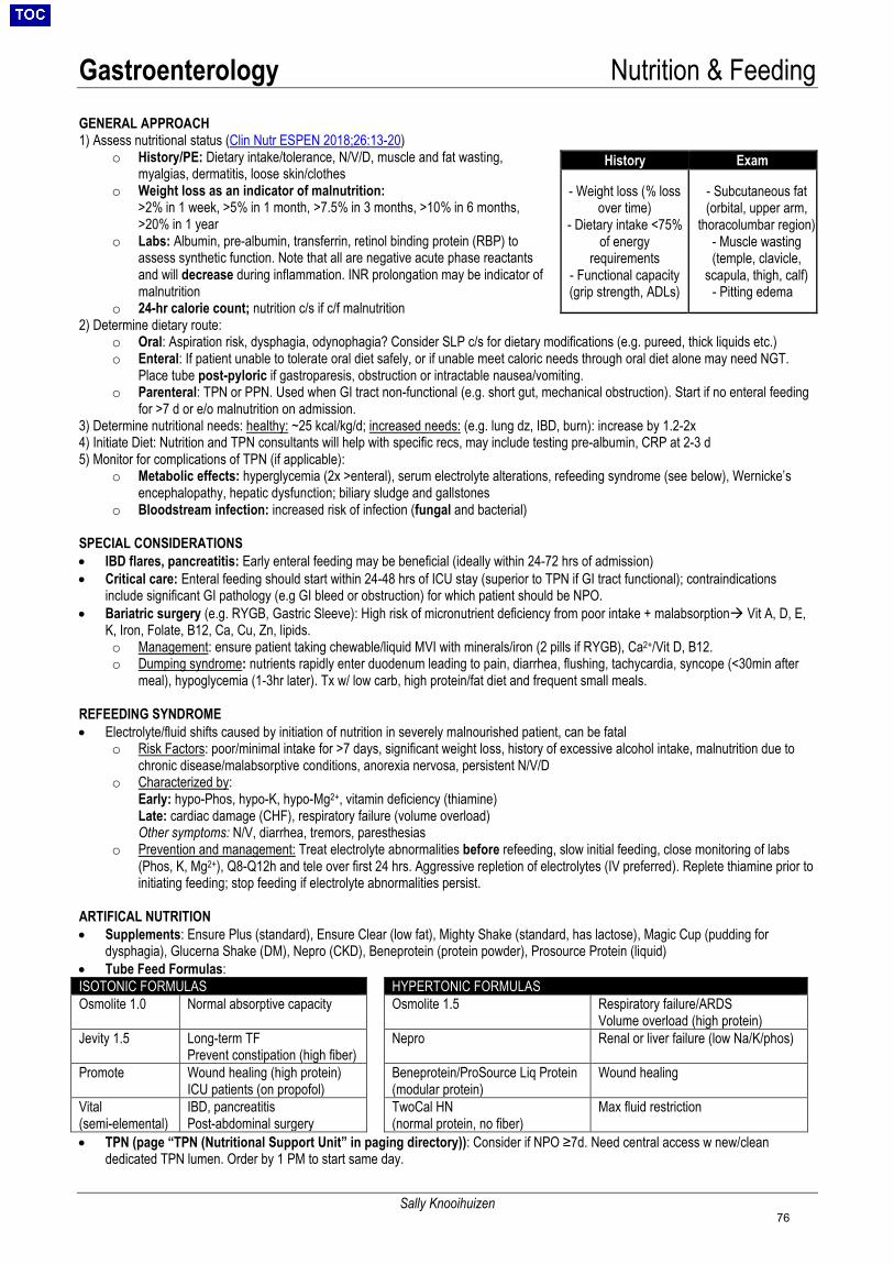
Gastroenterology Nutrition & Feeding
Sally Knooihuizen
GENERAL APPROACH 1) Assess nutritional status (Clin Nutr ESPEN 2018;26:13-20)
o History/PE: Dietary intake/tolerance, N/V/D, muscle and fat wasting,myalgias, dermatitis, loose skin/clothes
o Weight loss as an indicator of malnutrition:>2% in 1 week, >5% in 1 month, >7.5% in 3 months, >10% in 6 months,>20% in 1 year
o Labs: Albumin, pre-albumin, transferrin, retinol binding protein (RBP) toassess synthetic function. Note that all are negative acute phase reactantsand will decrease during inflammation. INR prolongation may be indicator ofmalnutrition
o 24-hr calorie count; nutrition c/s if c/f malnutrition2) Determine dietary route:
o Oral: Aspiration risk, dysphagia, odynophagia? Consider SLP c/s for dietary modifications (e.g. pureed, thick liquids etc.)o Enteral: If patient unable to tolerate oral diet safely, or if unable meet caloric needs through oral diet alone may need NGT.
Place tube post-pyloric if gastroparesis, obstruction or intractable nausea/vomiting.o Parenteral: TPN or PPN. Used when GI tract non-functional (e.g. short gut, mechanical obstruction). Start if no enteral feeding
for >7 d or e/o malnutrition on admission.3) Determine nutritional needs: healthy: ~25 kcal/kg/d; increased needs: (e.g. lung dz, IBD, burn): increase by 1.2-2x4) Initiate Diet: Nutrition and TPN consultants will help with specific recs, may include testing pre-albumin, CRP at 2-3 d5) Monitor for complications of TPN (if applicable):
o Metabolic effects: hyperglycemia (2x >enteral), serum electrolyte alterations, refeeding syndrome (see below), Wernicke’sencephalopathy, hepatic dysfunction; biliary sludge and gallstones
o Bloodstream infection: increased risk of infection (fungal and bacterial)
SPECIAL CONSIDERATIONS • IBD flares, pancreatitis: Early enteral feeding may be beneficial (ideally within 24-72 hrs of admission)• Critical care: Enteral feeding should start within 24-48 hrs of ICU stay (superior to TPN if GI tract functional); contraindications
include significant GI pathology (e.g GI bleed or obstruction) for which patient should be NPO.• Bariatric surgery (e.g. RYGB, Gastric Sleeve): High risk of micronutrient deficiency from poor intake + malabsorption Vit A, D, E,
K, Iron, Folate, B12, Ca, Cu, Zn, lipids.o Management: ensure patient taking chewable/liquid MVI with minerals/iron (2 pills if RYGB), Ca2+/Vit D, B12.o Dumping syndrome: nutrients rapidly enter duodenum leading to pain, diarrhea, flushing, tachycardia, syncope (<30min after
meal), hypoglycemia (1-3hr later). Tx w/ low carb, high protein/fat diet and frequent small meals.
REFEEDING SYNDROME • Electrolyte/fluid shifts caused by initiation of nutrition in severely malnourished patient, can be fatal
o Risk Factors: poor/minimal intake for >7 days, significant weight loss, history of excessive alcohol intake, malnutrition due tochronic disease/malabsorptive conditions, anorexia nervosa, persistent N/V/D
o Characterized by:Early: hypo-Phos, hypo-K, hypo-Mg2+, vitamin deficiency (thiamine)Late: cardiac damage (CHF), respiratory failure (volume overload)Other symptoms: N/V, diarrhea, tremors, paresthesias
o Prevention and management: Treat electrolyte abnormalities before refeeding, slow initial feeding, close monitoring of labs(Phos, K, Mg2+), Q8-Q12h and tele over first 24 hrs. Aggressive repletion of electrolytes (IV preferred). Replete thiamine prior toinitiating feeding; stop feeding if electrolyte abnormalities persist.
ARTIFICAL NUTRITION • Supplements: Ensure Plus (standard), Ensure Clear (low fat), Mighty Shake (standard, has lactose), Magic Cup (pudding for
dysphagia), Glucerna Shake (DM), Nepro (CKD), Beneprotein (protein powder), Prosource Protein (liquid)• Tube Feed Formulas:ISOTONIC FORMULAS HYPERTONIC FORMULAS Osmolite 1.0 Normal absorptive capacity Osmolite 1.5 Respiratory failure/ARDS
Volume overload (high protein) Jevity 1.5 Long-term TF
Prevent constipation (high fiber) Nepro Renal or liver failure (low Na/K/phos)
Promote Wound healing (high protein) ICU patients (on propofol)
Beneprotein/ProSource Liq Protein (modular protein)
Wound healing
Vital (semi-elemental)
IBD, pancreatitis Post-abdominal surgery
TwoCal HN (normal protein, no fiber)
Max fluid restriction
• TPN (page “TPN (Nutritional Support Unit” in paging directory)): Consider if NPO ≥7d. Need central access w new/cleandedicated TPN lumen. Order by 1 PM to start same day.
History Exam
- Weight loss (% lossover time)
- Dietary intake <75%of energy
requirements - Functional capacity(grip strength, ADLs)
- Subcutaneous fat(orbital, upper arm,
thoracolumbar region) - Muscle wasting(temple, clavicle,
scapula, thigh, calf)- Pitting edema
76
Gastroenterology Pancreatitis
Ryan Flanagan
ETIOLOGY (Clin Gastro Hepatol 2007;5:648) Gallstones/sludge: 40-75% of cases, #1 in women Alcohol: 30% of cases, #1 in men Hypertriglyceridemia: Typically pre-existing lipid abnormality with TG now >1000-2000 (risk starts when >500 mg/dL), lower amylase values, a/w genetic d/o, (+) FHx, #3 overall Idiopathic: 10-25% of cases Anatomic: ampullary diverticula/stenosis, duodenal stricture, tumor, divisum, parasites, foreign body Autoimmune: ↑IgG4, +ANA (rare) Genetic: cationic trypsinogen (PRSS1), SPINK1, CFTR, chymotrypsin C, calcium-sensing receptor, claudin-2 Post-ERCP: 3-5%. In high-risk pts, post-ERCP rectal NSAIDs reduce rate of pancreatitis (NEJM 2012;366:1414)
Drugs: <5%, Class Ia: ACEi, dapsone, lasix, flagyl, pentamidine, statins, sulfa, tetracycline, valproate, mesalamine; Class Ib: amiodarone, azathioprine/6-MP, dexamethasone; Class II: didanosine, estrogen, propofol, tamoxifen, hydrochlorothiazide Infections: Viral (Coxsackie, EBV, CMV, HIV, Mumps, VZV, HAV, HBV, HSV), Bacterial (Mycoplasma, Legionella, Salmonella), Fungal (Aspergillus), Parasitic (Toxoplasma, Crypto, Ascaris) Ischemia: vasculitis (SLE, PAN), hypotn/shock, cholesterol emboli Toxins: organophosphates, scorpion venom, methanol, smoking Trauma: blunt, especially s/p MVA Hypercalcemia: Ca activates pancreatic enzymes Tropical: Pt from low SES in SE Asia, first bout as child, central ductal stones, fibrocalcific diabetes
DIAGNOSIS (Lancet 2008;371:143; Am J Gastro 2013;108:1400)• 2/3: 1) Consistent clinical presentation, 2) Lipase or amylase > 3x ULN, 3) Suggestive cross-sectional abdominal imaging• Clinical: abd pain (90%) → band-like pain to back is specific (50%), N/V (90%), ileus, jaundice, flank/umbilical ecchymoses• Mild: absence of organ failure and local or systemic complications; 80% of cases w/ interstitial edema, focal fat necrosis• Mod-Severe: defined by local complications (pancreatic necrosis, peripancreatic fluid collections, gastric outlet obstruction, splenic
and PVT, colonic necrosis) or persistent organ failure (AKI, respiratory failure, shock, GIB) and SIRS. High rates of mortality.WORKUP • CBC, BMP, LFTs, albumin, lipids, lactate. ALT >3x ULN best predictor of gallstone pancreatitis (>95% PPV).• Lipase: early peak, specificity > sensitivity compared to amylase (DO NOT TREND). Higher baseline levels in DM and ESRD• Amylase: ↑ after 6-12hr, stays ↑ 3-5 days, >3x ULN has sens 67-83%, spec 85-98%. Normal on admission in 20% pts w/ alcoholic
pancreatitis and 50% pts w/ hypertriglyceridemia pancreatitis. No correlation between peak level and severity.• IgG4, ANA: consider in pt w/ recurrent, idiopathic AP w/ associated biliary stricture, Sjogrens syndrome, thyroiditis, IBD, nephritis• RUQUS: all patients on first attack to r/o gallstones. RUQ may miss distal CBD stone→ EUS more sensitive• CT/MRI w/ contrast useful to establish dx, exclude other dx, or after >48-72hr + decompensation to r/o complication (e.g. necrosis)MANAGEMENT (Clin. Gastro Hepatol 2011;9:710; BMJ 2004;328:1407; Cochrane 2010;1:CD002837)• Reverse precipitants: Treat ↑Ca or ↑TG, stop culprit meds, urgent (24-72H) ERCP for choledocholithiasis. CCY ideally prior to
discharge as ↑ biliary complications if CCY is delayed in non-necrotizing pancreatitis but no ∆ mortality (Surgery 2009;145:260)• IVF: Severe hypovolemia from 3rd spacing. LR>NS (↓SIRS, ↓CRP; avoid if ↑Ca). Bolus + gtt (150-250/hr). Goal: Reduce HR, BUN,
Hct, UOP >1cc/kg/hr. Stop aggressive resuscitation at 48hr. Monitor for abdominal compartment syndrome (bladder pressure>20).• Nutrition: Start PO (low fat) immediately once no n/v or abd pain. At 96h if PO not tolerated start TFs. Enteral feeding maintains
intestinal barrier, prevents gut flora translocation; NJ = NG in efficacy and safety. TPN → ↑ risk of infections, organ failure/death• HyperTG: Gemfibrozil 600mg BID, Insulin gtt (0.1-0.3U/kg/hr) + D5, √ q1h FSBG, q12h TG goal <500 (may take several days). C/S
GI and Heme for apheresis, esp if TG >1000. DC: Diet, exercise, lipid clinic referral.COMPLICATIONS (NEJM 2016;375:1972; Pancreas 2002;24:264) • SIRS, Thromboses (splenic, portal, SMV), Metabolic (↓Mg, Cal; ↑Glu), ARDS (phospholipase degradation of surfactant)• <4w: peripancreatic fluid collection, necrotic collection, infected necrosis. 1/3 necrosisinfected; suspect if 7-10d w/o improv or w/
decomp. CTX/flagyl (community), cefepime/flagyl or zosyn (recent procedure/hospitalized). C/S GI and surg for necrosectomy (ifstable, wait 4 wks while tx w/ abx).
• >4w: Pseudocyst: pain,↑amylase→drain if rapid enlarg. or local compression. Abscess: fever, pain,↑ amylase→usually needsdrainage. Walled-Off Pancreatic Necrosis: pancreatic necrosectomy (endoscopy vs surgery vs IR) (Gastro Endosc 2011;73:718)
• Pseudoanuerysm: Bleeding into pseudocyst. Suspect if ↓Hgb, expansion of walled off collection, hematochezia /melena/hematemesis. Dx: arterial phase CT, surgery/GI/IR c/s. Tx: IR embo prior to drainage, if severe may require surgery w/ high morbidity.
• Long term: (1) pancreatic exocrine/endocrine dysfxn (20-30%) (2) chronic pancreatitis (33-50%): p/w abd pain→back, wt loss,steatorrhea, bloating. Rad: calcifications, pancreatic ductal dilatation. Chronic pancreatitis→splanchnic venous thromboses→varices.Labs: fecal elastase, vit A,D,E, K, B12. Tx exocrine dysfunction w/ Creon.
PROGNOSIS (Am J Gastro 2009;104:966) • BISAP: w/in 24 hr 1) BUN >25, 2) ∆MS, 3) SIRS, 4) age >60, 5) pleural effusion; <3→ 1% mortality, ≥3→ 18% mortality• SIRS (practical): never→ 0% mortality, admission only→ 8% mortality, persistent→ 25% mortality. Ranson/APACHE less practical.PANCREATIC MASSES (Curr Gastro Rep 2013;15:347)• Solid: adenoCA (85-90%), autoimmune panc, neuroendocrine (1-5%), 1o lymphoma (<1%), mets (melanoma, RCC, etc)• Cystic: inflammatory (pseudocyst, paraduodenal wall cyst), IPMN (mucinous cystic or serous adenoma or adeno Ca)• Imaging: CT Abd pancreatic mass protocol; EUS with FNA allows biopsy (87% Se & 96% Sp); MRI useful in <2 cm lesions or
when vascular involvement needs to be delineated better); consider PET-CT, MRCP for malignancy in IPMN (70% Se, 92% Sp)• Serology: CA 19-9 (+ in 80% of panc ca, 86% Se, 87% Sp), CEA (mucinous lesions), ANA, IgG4 (if autoimmune panc suspected)
77
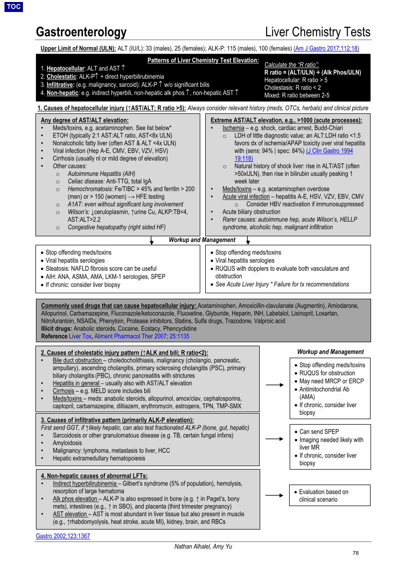
Gastroenterology Liver Chemistry Tests
Nathan Alhalel, Amy Yu Gastro 2002;123:1367
2. Causes of cholestatic injury pattern (↑ALK and bili; R ratio<2):• Bile duct obstruction – choledocholithiasis, malignancy (cholangio, pancreatic,
ampullary), ascending cholangitis, primary sclerosing cholangitis (PSC), primarybiliary cholangitis (PBC), chronic pancreatitis with strictures
• Hepatitis in general – usually also with AST/ALT elevation• Cirrhosis – e.g. MELD score includes bili• Meds/toxins – meds: anabolic steroids, allopurinol, amox/clav, cephalosporins,
captopril, carbamazepine, diltiazem, erythromycin, estrogens, TPN, TMP-SMX
Extreme AST/ALT elevation, e.g., >1000 (acute processes):
• Ischemia – e.g. shock, cardiac arrest, Budd-Chiario LDH of little diagnostic value; an ALT:LDH ratio <1.5
favors dx of ischemia/APAP toxicity over viral hepatitiswith (sens: 94% | spec: 84%) (J Clin Gastro 199419:118)
o Natural history of shock liver: rise in ALT/AST (often>50xULN), then rise in bilirubin usually peaking 1week later
• Meds/toxins – e.g. acetaminophen overdose• Acute viral infection – hepatitis A-E, HSV, VZV, EBV, CMV
o Consider HBV reactivation if immunosuppressed• Acute biliary obstruction• Rarer causes: autoimmune hep, acute Wilson’s, HELLP
syndrome, alcoholic hep, malignant infiltration
3. Causes of infiltrative pattern (primarily ALK-P elevation):First send GGT, if ↑likely hepatic, can also test fractionated ALK-P (bone, gut, hepatic) • Sarcoidosis or other granulomatous disease (e.g. TB, certain fungal infxns)• Amyloidosis• Malignancy: lymphoma, metastasis to liver, HCC• Hepatic extramedullary hematopoiesis
4. Non-hepatic causes of abnormal LFTs:• Indirect hyperbilirubinemia – Gilbert’s syndrome (5% of population), hemolysis,
resorption of large hematoma• Alk phos elevation – ALK-P is also expressed in bone (e.g. ↑ in Paget’s, bony
mets), intestines (e.g., ↑ in SBO), and placenta (third trimester pregnancy)• AST elevation – AST is most abundant in liver tissue but also present in muscle
(e.g., ↑rhabdomyolysis, heat stroke, acute MI), kidney, brain, and RBCs
• Stop offending meds/toxins• RUQUS for obstruction• May need MRCP or ERCP• Antimitochondrial Ab
(AMA)• If chronic, consider liver
biopsy
Commonly used drugs that can cause hepatocellular injury: Acetaminophen, Amoxicillin-clavulanate (Augmentin), Amiodarone, Allopurinol, Carbamazepine, Fluconazole/ketoconazole, Fluoxetine, Glyburide, Heparin, INH, Labetalol, Lisinopril, Losartan, Nitrofurantoin, NSAIDs, Phenytoin, Protease inhibitors, Statins, Sulfa drugs, Trazodone, Valproic acid Illicit drugs: Anabolic steroids, Cocaine, Ecstacy, Phencyclidine Reference Liver Tox, Aliment Pharmacol Ther 2007; 25:1135
1. Causes of hepatocellular injury (↑AST/ALT; R ratio >5): Always consider relevant history (meds, OTCs, herbals) and clinical picture
Any degree of AST/ALT elevation:
• Meds/toxins, e.g. acetaminophen. See list below*• ETOH (typically 2:1 AST:ALT ratio, AST<8x ULN)• Nonalcoholic fatty liver (often AST & ALT <4x ULN)• Viral infection (Hep A-E, CMV, EBV, VZV, HSV)• Cirrhosis (usually nl or mild degree of elevation)• Other causes:
o Autoimmune Hepatitis (AIH)o Celiac disease: Anti-TTG, total IgAo Hemochromatosis: Fe/TIBC > 45% and ferritin > 200
(men) or > 150 (women) → HFE testingo A1AT: even without significant lung involvemento Wilson’s: ↓ceruloplasmin, ↑urine Cu, ALKP:TB<4,
AST:ALT>2.2o Congestive hepatopathy (right sided HF)
• Can send SPEP• Imaging needed likely with
liver MR• If chronic, consider liver
biopsy
Patterns of Liver Chemistry Test Elevation: 1. Hepatocellular: ALT and AST ↑2. Cholestatic: ALK-P↑ + direct hyperbilirubinemia3. Infiltrative: (e.g. malignancy, sarcoid): ALK-P ↑ w/o significant bilis4. Non-hepatic: e.g. indirect hyperbili, non-hepatic alk phos ↑, non-hepatic AST ↑
Calculate the “R ratio”: R ratio = (ALT/ULN) ÷ (Alk Phos/ULN) Hepatocellular: R ratio > 5 Cholestasis: R ratio < 2 Mixed: R ratio between 2-5
Workup and Management
• Stop offending meds/toxins• Viral hepatitis serologies• Steatosis: NAFLD fibrosis score can be useful• AIH: ANA, ASMA, AMA, LKM-1 serologies, SPEP• If chronic: consider liver biopsy
• Evaluation based onclinical scenario
Upper Limit of Normal (ULN): ALT (IU/L): 33 (males), 25 (females); ALK-P: 115 (males), 100 (females) (Am J Gastro 2017;112:18)
• Stop offending meds/toxins• Viral hepatitis serologies• RUQUS with dopplers to evaluate both vasculature and
obstruction• See Acute Liver Injury * Failure for tx recommendations
Workup and Management
78
Gastroenterology Biliary Disease
Nate Alhalel, Sumeet Khetarpal
Gallstone Diseases: • Biliary colic: dull RUQ/epigastric pain, 30 min-6 hrs, caused by GB contracting around sludge/stone often postprandial• Cholelithiasis: presence of stones in the gallbladder (6% of men, 9% of women); labs typically normal
o Stone Types: Cholesterol (most common) -> 5 Fs: fat, female, forty, fertile (multiparous), fair (Caucasian); Pigment:Crohn’s/ileal disease, extravascular hemolysis, TPN
o Imaging: best test is RUQUS (sens 84%, spec 99%) showing stones in GB; CT has poor sensitivity (55-80%)o Treatment: Asymptomatic: observe; CCY only if at increased risk for gallbladder carcinoma (stone >3cm, porcelain
gallbladder, gallbladder adenoma); Symptomatic (“biliary colic”): elective CCY (67% recurrence rate if no CCY)• Cholecystitis: calculous (gallstone in cystic duct) or acalculous (10% of cases, usually critically ill pts, starts as bile stasis “sludge” or
gallbladder ischemia); often caused by sterile inflammation of gallbladder ± secondary infection. WBC↑↑, other labs WNLo Clinical Manifestations: RUQ pain, fever, Murphy’s sign; jaundice uncommon
Acalculous cholecystitis: Unexplained fever, leukocytosis, vague abd pain ± jaundice ± RUQ mass in ICU pt or jaundicein pt post-CCY. Risk factors: trauma, burns, TPN, critical illness, fasting, sepsis (Clin Gastro Hep 2010;8:15)
o Imaging: RUQUS (GB wall thickening, pericholecystic fluid, sonographic Murphy’s sign), HIDA scan if RUQUS negativeo Treatment: antibiotics—may not ↓mortality but often given empirically (Zosyn OR [ciprofloxacin/CTX AND
metronidazole]), consider stopping abx 1d after definitive intervention. Early (<7d) CCY during hospitalization ↓morbidity if↓surgical risk (Br J Surg 2015;102:1302); GB drainage (i.e. perc chole) if ↑risk and unimproved w/ abx+bowel rest
• Choledocholithiasis: gallstone in CBD; complications: acute pancreatitis, acute cholangitis; WBC - ; AST/ALT- / ↑; AlkP↑↑; Bilis ↑↑o Clinical Manifestations: RUQ pain, n/v, jaundice; sx may be intermittent if “ball-valve” effecto Imaging: RUQUS to look for CBD dilation >6mm (poor sensitivity for visualizing stones themselves), MRCP if equivocal.o Treatment: Endoscopic or surgical stone removal (ERCP ± CCY)
• Acute Cholangitis: asc biliary infx 2/2 obstruction (stone/stent/malignancy/post-ERCP/PSC) WBC ↑↑;AST/ALT - / ↑; AlkP ↑↑; Bilis ↑↑o Clinical Manifestations: Charcot’s triad (RUQ pain, fever, jaundice), Reynolds’ pentad (Charcot’s triad + AMS, shock)o Imaging: RUQUS (CBD >6mm); may proceed directly to ERCP (i.e., no US) if pt has Charcot’s triad + cholestasiso Treatment: antibiotics (Zosyn OR [ciprofloxacin/ceftriaxone AND metronidazole] x7-10d); CBD drainage (ERCP or
PCT if ERCP not feasible); CCY during hospitalization• Others: gallstone pancreatitis (obstrxn at Sphincter of Oddi), gallstone ileus (obstructing gallstone from cholecysto-enteric fistula)Autoimmune Biliary Diseases:• Primary Biliary Cholangitis (PBC): autoimmune destruction of intrahepatic bile ducts (Hepatology 2019;69:394)
o Clinical Manifestations: F>M, asymptomatic (50-60%), pruritus and fatigue, sicca syndrome, cirrhosis (late)o Diagnosis: >2 of the following: alk phos >1.5x upper limit of normal; AMA >1:40 titer (95% pts); biopsy findingso Other Labs: +ANA (70% pts), ↑total Igs, ↑IgM. NB: <10% of pts with +AMA develop PBCo Complications (some): hypothyroidism (20% pts), anemia, metabolic bone disease, overlap with Sjogren’s syndromeo Rx: ursodiol: first line, ↓cholestasis + improve LFTs; obeticholic acid: use as adjunctive or replacement for ursodiol,
fibrates: off label alternative; cholestyramine: for pruritus; modafinil: for fatigue; liver transplant: definitive treatmento Follow-up: LFTs q3-6mo, TFTs annually, Vit ADEK levels annually, DEXA scan at dx and q2-4yrs
• Primary Sclerosing Cholangitis (PSC): autoimmune destruction of intra + extrahepatic bile ducts (NEJM 2016;375:1161)o Clinical Manifestations: M>F, asymptomatic (50%), pruritus and fatigue (most common), cirrhosis (late)o Diagnosis: cholestatic LFTs, MRCP/ERCP (segmental strictures), ± biopsy; order AMA/IgG4 to exclude alternative dxo Other Labs: +p-ANCA (30-80%), ↑total Igs (30%), ↑IgM (40-50%)o Complications (some): IBD (>75% pts, UC>>Crohn’s), cholangiocarcinoma (10-15% pts), metabolic bone diseaseo Treatment: ursodiol: may ↓cholestasis + improve LFTs; cholestyramine: for pruritus; liver transplant: definitive treatmento Follow-up: RUQUS q1yr (for gallbladder Ca/cirrhosis), MRCP q6-12mo (cholangiocarcinoma), CA19-9/CEA q1mo (colon
cancer); colonoscopy q1yr if pt with IBD, q3-5yrs if no IBD; other: LFTs q3-6mo, Vit ADEK levels annually, DEXA q2-4yrsRare forms of intrahepatic cholestasis: Vanishing bile duct syndrome (s/p liver tx/BMT), AIDS cholangiopathy (CMV or cryptosporidia)Malignant Disease of the Biliary Tract: • Gallbladder Carcinoma: risk factors: gallstone disease (34x more likely to develop Ca), porcelain GB, GB polyps
o Clinical Manifestations: usually asymptomatic; sx may include N/V, weight loss, biliary colic, jaundice (if obstruction)o Diagnosis: LFTs usually normal, ↑CA19-9/CEA; RUQUS best screening test, then EUS + MRI/MRCP
• Cholangiocarcinoma: may be extrahepatic (90%) or intrahepatic (10%); risk factors: PSC, liver flukes, intrahepatic gallstoneso Clinical Manifestations: cholestasis (jaundice, pruritus, acholic stool, dark urine), RUQ pain, N/V, weight loss, fevero Diagnosis: ↑CA19-9/CEA, cholestatic LFTs; RUQUS best screening test, then ERCP + MRI/MRCP
Algorithm for radiographic assessment of suspected biliary pathology (Am J Roentgenol 2011;197:551):
79
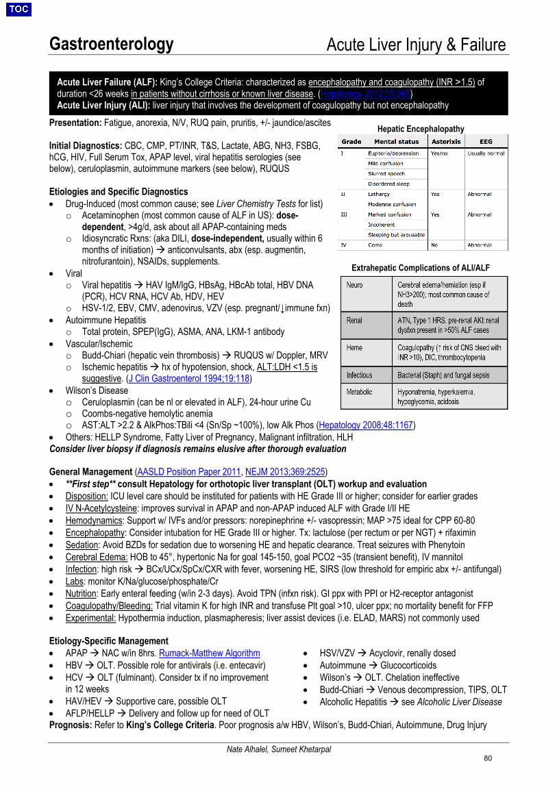
Gastroenterology Acute Liver Injury & Failure
Nate Alhalel, Sumeet Khetarpal
Presentation: Fatigue, anorexia, N/V, RUQ pain, pruritis, +/- jaundice/ascites
Initial Diagnostics: CBC, CMP, PT/INR, T&S, Lactate, ABG, NH3, FSBG, hCG, HIV, Full Serum Tox, APAP level, viral hepatitis serologies (see below), ceruloplasmin, autoimmune markers (see below), RUQUS
Etiologies and Specific Diagnostics • Drug-Induced (most common cause; see Liver Chemistry Tests for list)
o Acetaminophen (most common cause of ALF in US): dose-dependent, >4g/d, ask about all APAP-containing meds
o Idiosyncratic Rxns: (aka DILI, dose-independent, usually within 6months of initiation) anticonvulsants, abx (esp. augmentin,nitrofurantoin), NSAIDs, supplements.
• Viralo Viral hepatitis HAV IgM/IgG, HBsAg, HBcAb total, HBV DNA
(PCR), HCV RNA, HCV Ab, HDV, HEVo HSV-1/2, EBV, CMV, adenovirus, VZV (esp. pregnant/↓immune fxn)
• Autoimmune Hepatitiso Total protein, SPEP(IgG), ASMA, ANA, LKM-1 antibody
• Vascular/Ischemico Budd-Chiari (hepatic vein thrombosis) RUQUS w/ Doppler, MRVo Ischemic hepatitis hx of hypotension, shock, ALT:LDH <1.5 is
suggestive. (J Clin Gastroenterol 1994;19:118)• Wilson’s Disease
o Ceruloplasmin (can be nl or elevated in ALF), 24-hour urine Cuo Coombs-negative hemolytic anemiao AST:ALT >2.2 & AlkPhos:TBili <4 (Sn/Sp ~100%), low Alk Phos (Hepatology 2008;48:1167)
• Others: HELLP Syndrome, Fatty Liver of Pregnancy, Malignant infiltration, HLHConsider liver biopsy if diagnosis remains elusive after thorough evaluation
General Management (AASLD Position Paper 2011, NEJM 2013;369:2525)
• **First step** consult Hepatology for orthotopic liver transplant (OLT) workup and evaluation• Disposition: ICU level care should be instituted for patients with HE Grade III or higher; consider for earlier grades• IV N-Acetylcysteine: improves survival in APAP and non-APAP induced ALF with Grade I/II HE• Hemodynamics: Support w/ IVFs and/or pressors: norepinephrine +/- vasopressin; MAP >75 ideal for CPP 60-80• Encephalopathy: Consider intubation for HE Grade III or higher. Tx: lactulose (per rectum or per NGT) + rifaximin• Sedation: Avoid BZDs for sedation due to worsening HE and hepatic clearance. Treat seizures with Phenytoin• Cerebral Edema: HOB to 45°, hypertonic Na for goal 145-150, goal PCO2 ~35 (transient benefit), IV mannitol• Infection: high risk BCx/UCx/SpCx/CXR with fever, worsening HE, SIRS (low threshold for empiric abx +/- antifungal)• Labs: monitor K/Na/glucose/phosphate/Cr• Nutrition: Early enteral feeding (w/in 2-3 days). Avoid TPN (infxn risk). GI ppx with PPI or H2-receptor antagonist• Coagulopathy/Bleeding: Trial vitamin K for high INR and transfuse Plt goal >10, ulcer ppx; no mortality benefit for FFP• Experimental: Hypothermia induction, plasmapheresis; liver assist devices (i.e. ELAD, MARS) not commonly used
Etiology-Specific Management • APAP NAC w/in 8hrs. Rumack-Matthew Algorithm• HBV OLT. Possible role for antivirals (i.e. entecavir)• HCV OLT (fulminant). Consider tx if no improvement
in 12 weeks• HAV/HEV Supportive care, possible OLT• AFLP/HELLP Delivery and follow up for need of OLT
• HSV/VZV Acyclovir, renally dosed• Autoimmune Glucocorticoids• Wilson’s OLT. Chelation ineffective• Budd-Chiari Venous decompression, TIPS, OLT• Alcoholic Hepatitis see Alcoholic Liver Disease
Prognosis: Refer to King’s College Criteria. Poor prognosis a/w HBV, Wilson’s, Budd-Chiari, Autoimmune, Drug Injury
Acute Liver Failure (ALF): King’s College Criteria: characterized as encephalopathy and coagulopathy (INR >1.5) of duration <26 weeks in patients without cirrhosis or known liver disease. (Hepatology 2012;55:965) Acute Liver Injury (ALI): liver injury that involves the development of coagulopathy but not encephalopathy
Extrahepatic Complications of ALI/ALF
Hepatic Encephalopathy
80
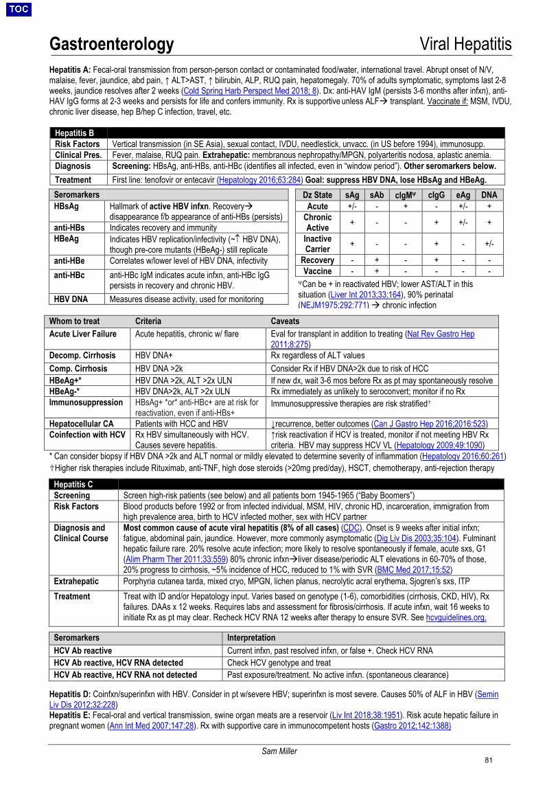
Gastroenterology Viral Hepatitis
Sam Miller
Hepatitis A: Fecal-oral transmission from person-person contact or contaminated food/water, international travel. Abrupt onset of N/V, malaise, fever, jaundice, abd pain, ↑ ALT>AST, ↑ bilirubin, ALP, RUQ pain, hepatomegaly. 70% of adults symptomatic, symptoms last 2-8 weeks, jaundice resolves after 2 weeks (Cold Spring Harb Perspect Med 2018; 8). Dx: anti-HAV IgM (persists 3-6 months after infxn), anti-HAV IgG forms at 2-3 weeks and persists for life and confers immunity. Rx is supportive unless ALF transplant. Vaccinate if: MSM, IVDU, chronic liver disease, hep B/hep C infection, travel, etc.
Hepatitis B Risk Factors Vertical transmission (in SE Asia), sexual contact, IVDU, needlestick, unvacc. (in US before 1994), immunosupp. Clinical Pres. Fever, malaise, RUQ pain. Extrahepatic: membranous nephropathy/MPGN, polyarteritis nodosa, aplastic anemia. Diagnosis Screening: HBsAg, anti-HBs, anti-HBc (identifies all infected, even in “window period”). Other seromarkers below. Treatment First line: tenofovir or entecavir (Hepatology 2016;63:284) Goal: suppress HBV DNA, lose HBsAg and HBeAg.
Whom to treat Criteria Caveats Acute Liver Failure Acute hepatitis, chronic w/ flare Eval for transplant in addition to treating (Nat Rev Gastro Hep
2011;8:275) Decomp. Cirrhosis HBV DNA+ Rx regardless of ALT values Comp. Cirrhosis HBV DNA >2k Consider Rx if HBV DNA>2k due to risk of HCC HBeAg+* HBV DNA >2k, ALT >2x ULN If new dx, wait 3-6 mos before Rx as pt may spontaneously resolve HBeAg-* HBV DNA>2k, ALT >2x ULN Rx immediately as unlikely to seroconvert; monitor if no Rx Immunosuppression HBsAg+ *or* anti-HBc+ are at risk for
reactivation, even if anti-HBs+ Immunosuppressive therapies are risk stratified♱
Hepatocellular CA Patients with HCC and HBV ↓recurrence, better outcomes (Can J Gastro Hep 2016;2016:523) Coinfection with HCV Rx HBV simultaneously with HCV.
Causes severe hepatitis. ↑risk reactivation if HCV is treated, monitor if not meeting HBV Rx criteria. HBV may suppress HCV VL (Hepatology 2009;49:1090)
* Can consider biopsy if HBV DNA >2k and ALT normal or mildly elevated to determine severity of inflammation (Hepatology 2016;60:261)♱Higher risk therapies include Rituximab, anti-TNF, high dose steroids (>20mg pred/day), HSCT, chemotherapy, anti-rejection therapy
Hepatitis C Screening Screen high-risk patients (see below) and all patients born 1945-1965 (“Baby Boomers”) Risk Factors Blood products before 1992 or from infected individual, MSM, HIV, chronic HD, incarceration, immigration from
high prevalence area, birth to HCV infected mother, sex with HCV partner Diagnosis and Clinical Course
Most common cause of acute viral hepatitis (8% of all cases) (CDC). Onset is 9 weeks after initial infxn; fatigue, abdominal pain, jaundice. However, more commonly asymptomatic (Dig Liv Dis 2003;35:104). Fulminant hepatic failure rare. 20% resolve acute infection; more likely to resolve spontaneously if female, acute sxs, G1 (Alim Pharm Ther 2011;33:559) 80% chronic infxnliver disease/periodic ALT elevations in 60-70% of those, 20% progress to cirrhosis, ~5% incidence of HCC, reduced to 1% with SVR (BMC Med 2017;15:52)
Extrahepatic Porphyria cutanea tarda, mixed cryo, MPGN, lichen planus, necrolytic acral erythema, Sjogren’s sxs, ITP Treatment Treat with ID and/or Hepatology input. Varies based on genotype (1-6), comorbidities (cirrhosis, CKD, HIV), Rx
failures. DAAs x 12 weeks. Requires labs and assessment for fibrosis/cirrhosis. If acute infxn, wait 16 weeks to initiate Rx as pt may clear. Recheck HCV RNA 12 weeks after therapy to ensure SVR. See hcvguidelines.org.
Hepatitis D: Coinfxn/superinfxn with HBV. Consider in pt w/severe HBV; superinfxn is most severe. Causes 50% of ALF in HBV (Semin Liv Dis 2012;32:228) Hepatitis E: Fecal-oral and vertical transmission, swine organ meats are a reservoir (Liv Int 2018;38:1951). Risk acute hepatic failure in pregnant women (Ann Int Med 2007;147:28). Rx with supportive care in immunocompetent hosts (Gastro 2012;142:1388)
Seromarkers HBsAg Hallmark of active HBV infxn. Recovery
disappearance f/b appearance of anti-HBs (persists) anti-HBs Indicates recovery and immunity HBeAg Indicates HBV replication/infectivity (~↑ HBV DNA),
though pre-core mutants (HBeAg-) still replicate anti-HBe Correlates w/lower level of HBV DNA, infectivity anti-HBc anti-HBc IgM indicates acute infxn, anti-HBc IgG
persists in recovery and chronic HBV. HBV DNA Measures disease activity, used for monitoring
Dz State sAg sAb cIgMψ cIgG eAg DNA Acute +/- - + - +/- +
Chronic Active + - - + +/- +
Inactive Carrier + - - + - +/-
Recovery - + - + - - Vaccine - + - - - -
Seromarkers Interpretation HCV Ab reactive Current infxn, past resolved infxn, or false +. Check HCV RNA HCV Ab reactive, HCV RNA detected Check HCV genotype and treat HCV Ab reactive, HCV RNA not detected Past exposure/treatment. No active infxn. (spontaneous clearance)
ψCan be + in reactivated HBV; lower AST/ALT in this situation (Liver Int 2013;33:164), 90% perinatal (NEJM1975;292:771) chronic infection
81
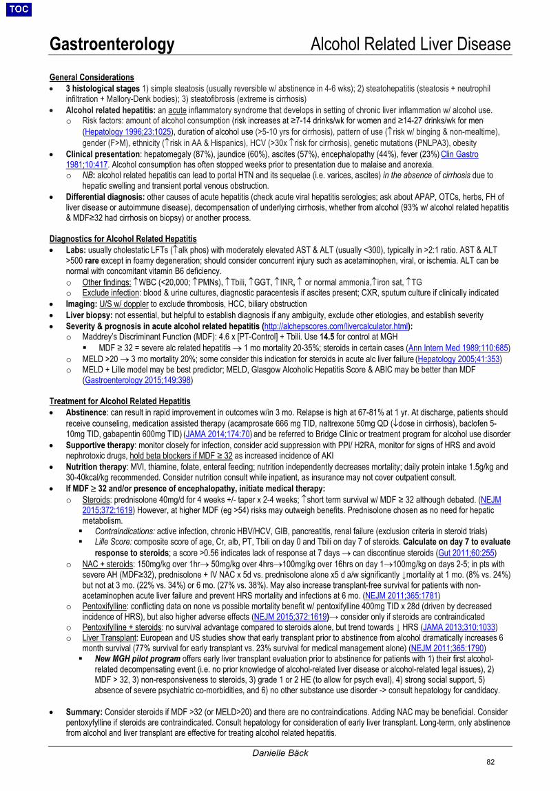
Gastroenterology Alcohol Related Liver Disease
Danielle Bäck
General Considerations • 3 histological stages 1) simple steatosis (usually reversible w/ abstinence in 4-6 wks); 2) steatohepatitis (steatosis + neutrophil
infiltration + Mallory-Denk bodies); 3) steatofibrosis (extreme is cirrhosis)• Alcohol related hepatitis: an acute inflammatory syndrome that develops in setting of chronic liver inflammation w/ alcohol use.
o Risk factors: amount of alcohol consumption (risk increases at ≥7-14 drinks/wk for women and ≥14-27 drinks/wk for men;
(Hepatology 1996;23:1025), duration of alcohol use (>5-10 yrs for cirrhosis), pattern of use (↑risk w/ binging & non-mealtime),gender (F>M), ethnicity (↑risk in AA & Hispanics), HCV (>30x ↑risk for cirrhosis), genetic mutations (PNLPA3), obesity
• Clinical presentation: hepatomegaly (87%), jaundice (60%), ascites (57%), encephalopathy (44%), fever (23%) Clin Gastro1981;10:417. Alcohol consumption has often stopped weeks prior to presentation due to malaise and anorexia.o NB: alcohol related hepatitis can lead to portal HTN and its sequelae (i.e. varices, ascites) in the absence of cirrhosis due to
hepatic swelling and transient portal venous obstruction.• Differential diagnosis: other causes of acute hepatitis (check acute viral hepatitis serologies; ask about APAP, OTCs, herbs, FH of
liver disease or autoimmune disease), decompensation of underlying cirrhosis, whether from alcohol (93% w/ alcohol related hepatitis& MDF≥32 had cirrhosis on biopsy) or another process.
Diagnostics for Alcohol Related Hepatitis • Labs: usually cholestatic LFTs (↑alk phos) with moderately elevated AST & ALT (usually <300), typically in >2:1 ratio. AST & ALT
>500 rare except in foamy degeneration; should consider concurrent injury such as acetaminophen, viral, or ischemia. ALT can benormal with concomitant vitamin B6 deficiency.o Other findings: ↑WBC (<20,000; ↑PMNs), ↑Tbili, ↑GGT, ↑INR, ↑ or normal ammonia,↑iron sat, ↑TGo Exclude infection: blood & urine cultures, diagnostic paracentesis if ascites present; CXR, sputum culture if clinically indicated
• Imaging: U/S w/ doppler to exclude thrombosis, HCC, biliary obstruction• Liver biopsy: not essential, but helpful to establish diagnosis if any ambiguity, exclude other etiologies, and establish severity• Severity & prognosis in acute alcohol related hepatitis (http://alchepscores.com/livercalculator.html):
o Maddrey’s Discriminant Function (MDF): 4.6 x [PT-Control] + Tbili. Use 14.5 for control at MGH MDF ≥ 32 = severe alc related hepatitis → 1 mo mortality 20-35%; steroids in certain cases (Ann Intern Med 1989;110:685)
o MELD >20 → 3 mo mortality 20%; some consider this indication for steroids in acute alc liver failure (Hepatology 2005;41:353)o MELD + Lille model may be best predictor; MELD, Glasgow Alcoholic Hepatitis Score & ABIC may be better than MDF
(Gastroenterology 2015;149:398)
Treatment for Alcohol Related Hepatitis • Abstinence: can result in rapid improvement in outcomes w/in 3 mo. Relapse is high at 67-81% at 1 yr. At discharge, patients should
receive counseling, medication assisted therapy (acamprosate 666 mg TID, naltrexone 50mg QD (↓dose in cirrhosis), baclofen 5-10mg TID, gabapentin 600mg TID) (JAMA 2014;174:70) and be referred to Bridge Clinic or treatment program for alcohol use disorder
• Supportive therapy: monitor closely for infection, consider acid suppression with PPI/ H2RA, monitor for signs of HRS and avoidnephrotoxic drugs, hold beta blockers if MDF ≥ 32 as increased incidence of AKI
• Nutrition therapy: MVI, thiamine, folate, enteral feeding; nutrition independently decreases mortality; daily protein intake 1.5g/kg and30-40kcal/kg recommended. Consider nutrition consult while inpatient, as insurance may not cover outpatient consult.
• If MDF ≥ 32 and/or presence of encephalopathy, initiate medical therapy:o Steroids: prednisolone 40mg/d for 4 weeks +/- taper x 2-4 weeks; ↑short term survival w/ MDF ≥ 32 although debated. (NEJM
2015;372:1619) However, at higher MDF (eg >54) risks may outweigh benefits. Prednisolone chosen as no need for hepaticmetabolism. Contraindications: active infection, chronic HBV/HCV, GIB, pancreatitis, renal failure (exclusion criteria in steroid trials) Lille Score: composite score of age, Cr, alb, PT, Tbili on day 0 and Tbili on day 7 of steroids. Calculate on day 7 to evaluate
response to steroids; a score >0.56 indicates lack of response at 7 days → can discontinue steroids (Gut 2011;60:255)o NAC + steroids: 150mg/kg over 1hr→ 50mg/kg over 4hrs→100mg/kg over 16hrs on day 1→100mg/kg on days 2-5; in pts with
severe AH (MDF≥32), prednisolone + IV NAC x 5d vs. prednisolone alone x5 d a/w significantly ↓mortality at 1 mo. (8% vs. 24%)but not at 3 mo. (22% vs. 34%) or 6 mo. (27% vs. 38%). May also increase transplant-free survival for patients with non-acetaminophen acute liver failure and prevent HRS mortality and infections at 6 mo. (NEJM 2011;365:1781)
o Pentoxifylline: conflicting data on none vs possible mortality benefit w/ pentoxifylline 400mg TID x 28d (driven by decreasedincidence of HRS), but also higher adverse effects (NEJM 2015;372:1619)→ consider only if steroids are contraindicated
o Pentoxifylline + steroids: no survival advantage compared to steroids alone, but trend towards ↓ HRS (JAMA 2013;310:1033)o Liver Transplant: European and US studies show that early transplant prior to abstinence from alcohol dramatically increases 6
month survival (77% survival for early transplant vs. 23% survival for medical management alone) (NEJM 2011;365:1790) New MGH pilot program offers early liver transplant evaluation prior to abstinence for patients with 1) their first alcohol-
related decompensating event (i.e. no prior knowledge of alcohol-related liver disease or alcohol-related legal issues), 2)MDF > 32, 3) non-responsiveness to steroids, 3) grade 1 or 2 HE (to allow for psych eval), 4) strong social support, 5)absence of severe psychiatric co-morbidities, and 6) no other substance use disorder -> consult hepatology for candidacy.
• Summary: Consider steroids if MDF >32 (or MELD>20) and there are no contraindications. Adding NAC may be beneficial. Considerpentoxyfylline if steroids are contraindicated. Consult hepatology for consideration of early liver transplant. Long-term, only abstinencefrom alcohol and liver transplant are effective for treating alcohol related hepatitis.
82
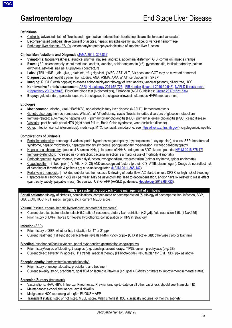
Gastroenterology End Stage Liver Disease
Jacqueline Henson, Amy Yu
Definitions • Cirrhosis: advanced state of fibrosis and regenerative nodules that distorts hepatic architecture and vasculature• Decompensated cirrhosis: development of ascites, hepatic encephalopathy, jaundice, or variceal hemorrhage• End-stage liver disease (ESLD): accompanying pathophysiologic state of impaired liver function
Clinical Manifestations and Diagnosis (JAMA 2012; 307:832) • Symptoms: fatigue/weakness, jaundice, pruritus, nausea, anorexia, abdominal distention, GIB, confusion, muscle cramps• Exam: ↓BP, splenomegaly, caput medusae, ascites, jaundice, spider angiomata (>3), gynecomastia, testicular atrophy, palmar
erythema, asterixis, nail Δs, Dupuytren’s contracture• Labs: ↑TBili, ↑INR, ↓Alb, ↓Na, ↓platelets, +/- ↓Hgb/Hct, ↓WBC; AST, ALT, Alk phos, and GGT may be elevated or normal• Diagnostics: viral hepatitis panel, iron studies, ANA, ASMA, AMA, α1AT, ceruloplasmin, SPEP• Imaging: RUQUS (with doppler) to assess echogenicity/morphology of liver, ascites, vascular patency, biliary tree, HCC• Non-invasive fibrosis assessment: APRI (Hepatology 2011;53:726), FIB-4 index (Liver Int 2010;30:546), NAFLD fibrosis score
(Hepatology 2007;45:846), FibroSure blood test (6 biomarkers), FibroScan (AGA Guidelines: Gastro 2017;152:1536)• Biopsy: gold standard (percutaneous vs. transjugular; transjugular allows simultaneous HVPG measurement)
Etiologies • Most common: alcohol, viral (HBV/HCV), non-alcoholic fatty liver disease (NAFLD), hemochromatosis• Genetic disorders: hemochromatosis, Wilson’s, α1AT deficiency, cystic fibrosis, inherited disorders of glucose metabolism• Immune-related: autoimmune hepatitis (AIH), primary biliary cholangitis (PBC), primary sclerosis cholangitis (PSC), celiac disease• Vascular: post-hepatic portal HTN (right heart failure, Budd-Chiari syndrome, veno-occlusive disease)• Other: infection (i.e. schistosomiasis), meds (e.g. MTX, isoniazid, amiodarone; see https://livertox.nlm.nih.gov/), cryptogenic/idiopathic
Complications of Cirrhosis • Portal hypertension: esophageal varices, portal hypertensive gastropathy, hypersplenism (→cytopenias), ascites, SBP, hepatorenal
syndrome, hepatic hydrothorax, hepatopulmonary syndrome, portopulmonary hypertension, cirrhotic cardiomyopathy• Hepatic encephalopathy: ↑mucosal & luminal NH3, ↓clearance of NH3 & endogenous BDZ-like compounds (NEJM 2016;375:17)• Immune dysfunction: increased risk of infection; bacterial infection is a major cause of morbidity & mortality• Endocrinopathies: hypoglycemia, thyroid dysfunction, hypogonadism, hyperestrinism (palmar erythema, spider angiomata)• Coagulopathy: ↓ in both pro- (II,V, VII, IX, X, XI) AND anticoagulant factors (protein C/S, ATIII, plasminogen). Coags do not reflect risk
of bleeding or thrombosis & patients not auto-anticoagulated (NEJM 2011;365:147).• Portal vein thrombosis: ↑ risk due unbalanced hemostasis & slowing of portal flow. AC started unless CPS C or high risk of bleeding.• Hepatocellular carcinoma: 1-8% risk per year. May be asymptomatic, lead to decompensation, and/or have sx related to mass effect
(pain, early satiety, palpable mass). Screen with US +/- AFP (AASLD guidelines: Hepatology 2018;68:723).
VIBES: a systematic approach to the management of cirrhosis For all patients: etiology of cirrhosis, complications, compensated or decompensated (& etiology of decompensation: infection, SBP, GIB, EtOH, HCC, PVT, meds, surgery, etc.), current MELD score
Volume (ascites, edema, hepatic hydrothorax, hepatorenal syndrome) • Current diuretics (spironolactone/lasix 5:2 ratio) & response; dietary Na+ restriction (<2 g/d), fluid restriction 1.5L (if Na<125)• Prior history of LVPs, thoras for hepatic hydrothorax, consideration of TIPS if refractory
Infection (SBP) • Prior history of SBP, whether has indication for 1° or 2° ppx• Current treatment (if diagnostic paracentesis reveals PMNs >250) or ppx (CTX if active GIB; otherwise cipro or Bactrim)
Bleeding (esophageal/gastric varices, portal hypertensive gastropathy, coagulopathy) • Prior history/source of bleeding, therapies (e.g. banding, sclerotherapy, TIPS), current prophylaxis (e.g. βB)• Current bleed: severity, IV access, H/H trends, medical therapy (PPI/octreotide), results/plan for EGD, SBP ppx as above
Encephalopathy (portosystemic encephalopathy) • Prior history of encephalopathy, precipitant, and treatment• Current severity, trend, precipitant, goal #BM on lactulose/rifaximin (eg: goal 4 BM/day or titrate to improvement in mental status)
Screening/Surgery (transplant) • Vaccinations: HAV, HBV, Influenza, Pneumovax, Prevnar (and up-to-date on all other vaccines), should see Transplant ID• Maintenance: alcohol abstinence, avoid NSAIDs• Malignancy: HCC screening with q6m RUQUS + AFP• Transplant status: listed or not listed, MELD score, Milan criteria if HCC, classically requires ~6 months sobriety
83
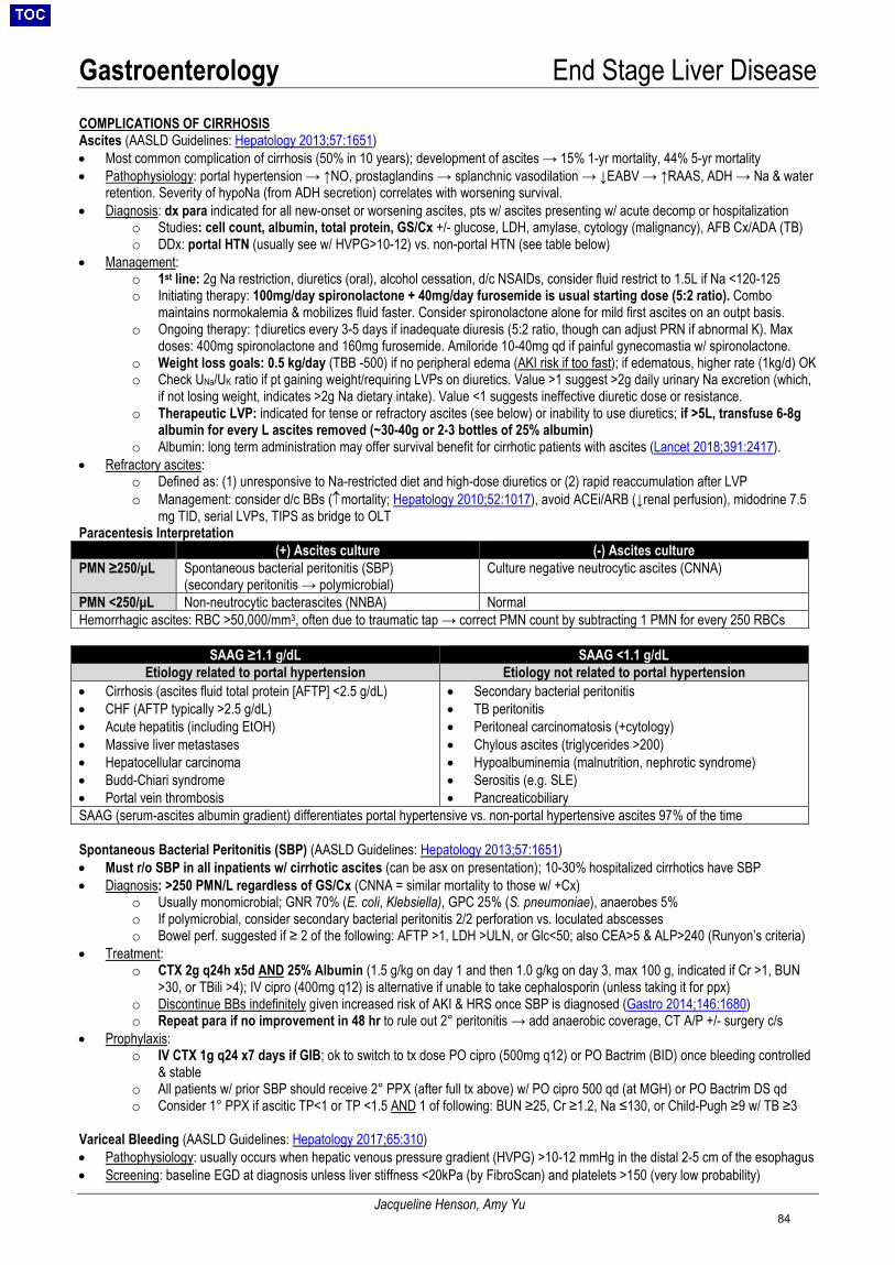
Gastroenterology End Stage Liver Disease
Jacqueline Henson, Amy Yu
COMPLICATIONS OF CIRRHOSIS Ascites (AASLD Guidelines: Hepatology 2013;57:1651) • Most common complication of cirrhosis (50% in 10 years); development of ascites → 15% 1-yr mortality, 44% 5-yr mortality• Pathophysiology: portal hypertension → ↑NO, prostaglandins → splanchnic vasodilation → ↓EABV → ↑RAAS, ADH → Na & water
retention. Severity of hypoNa (from ADH secretion) correlates with worsening survival.• Diagnosis: dx para indicated for all new-onset or worsening ascites, pts w/ ascites presenting w/ acute decomp or hospitalization
o Studies: cell count, albumin, total protein, GS/Cx +/- glucose, LDH, amylase, cytology (malignancy), AFB Cx/ADA (TB)o DDx: portal HTN (usually see w/ HVPG>10-12) vs. non-portal HTN (see table below)
• Management:o 1st line: 2g Na restriction, diuretics (oral), alcohol cessation, d/c NSAIDs, consider fluid restrict to 1.5L if Na <120-125o Initiating therapy: 100mg/day spironolactone + 40mg/day furosemide is usual starting dose (5:2 ratio). Combo
maintains normokalemia & mobilizes fluid faster. Consider spironolactone alone for mild first ascites on an outpt basis.o Ongoing therapy: ↑diuretics every 3-5 days if inadequate diuresis (5:2 ratio, though can adjust PRN if abnormal K). Max
doses: 400mg spironolactone and 160mg furosemide. Amiloride 10-40mg qd if painful gynecomastia w/ spironolactone.o Weight loss goals: 0.5 kg/day (TBB -500) if no peripheral edema (AKI risk if too fast); if edematous, higher rate (1kg/d) OKo Check UNa/UK ratio if pt gaining weight/requiring LVPs on diuretics. Value >1 suggest >2g daily urinary Na excretion (which,
if not losing weight, indicates >2g Na dietary intake). Value <1 suggests ineffective diuretic dose or resistance.o Therapeutic LVP: indicated for tense or refractory ascites (see below) or inability to use diuretics; if >5L, transfuse 6-8g
albumin for every L ascites removed (~30-40g or 2-3 bottles of 25% albumin)o Albumin: long term administration may offer survival benefit for cirrhotic patients with ascites (Lancet 2018;391:2417).
• Refractory ascites:o Defined as: (1) unresponsive to Na-restricted diet and high-dose diuretics or (2) rapid reaccumulation after LVPo Management: consider d/c BBs (↑mortality; Hepatology 2010;52:1017), avoid ACEi/ARB (↓renal perfusion), midodrine 7.5
mg TID, serial LVPs, TIPS as bridge to OLTParacentesis Interpretation
(+) Ascites culture (-) Ascites culture PMN ≥250/μL Spontaneous bacterial peritonitis (SBP)
(secondary peritonitis → polymicrobial) Culture negative neutrocytic ascites (CNNA)
PMN <250/μL Non-neutrocytic bacterascites (NNBA) Normal Hemorrhagic ascites: RBC >50,000/mm3, often due to traumatic tap → correct PMN count by subtracting 1 PMN for every 250 RBCs
SAAG ≥1.1 g/dL SAAG <1.1 g/dL Etiology related to portal hypertension Etiology not related to portal hypertension
• Cirrhosis (ascites fluid total protein [AFTP] <2.5 g/dL)• CHF (AFTP typically >2.5 g/dL)• Acute hepatitis (including EtOH)• Massive liver metastases• Hepatocellular carcinoma• Budd-Chiari syndrome• Portal vein thrombosis
• Secondary bacterial peritonitis• TB peritonitis• Peritoneal carcinomatosis (+cytology)• Chylous ascites (triglycerides >200)• Hypoalbuminemia (malnutrition, nephrotic syndrome)• Serositis (e.g. SLE)• Pancreaticobiliary
SAAG (serum-ascites albumin gradient) differentiates portal hypertensive vs. non-portal hypertensive ascites 97% of the time
Spontaneous Bacterial Peritonitis (SBP) (AASLD Guidelines: Hepatology 2013;57:1651) • Must r/o SBP in all inpatients w/ cirrhotic ascites (can be asx on presentation); 10-30% hospitalized cirrhotics have SBP• Diagnosis: >250 PMN/L regardless of GS/Cx (CNNA = similar mortality to those w/ +Cx)
o Usually monomicrobial; GNR 70% (E. coli, Klebsiella), GPC 25% (S. pneumoniae), anaerobes 5%o If polymicrobial, consider secondary bacterial peritonitis 2/2 perforation vs. loculated abscesseso Bowel perf. suggested if ≥ 2 of the following: AFTP >1, LDH >ULN, or Glc<50; also CEA>5 & ALP>240 (Runyon’s criteria)
• Treatment:o CTX 2g q24h x5d AND 25% Albumin (1.5 g/kg on day 1 and then 1.0 g/kg on day 3, max 100 g, indicated if Cr >1, BUN
>30, or TBili >4); IV cipro (400mg q12) is alternative if unable to take cephalosporin (unless taking it for ppx)o Discontinue BBs indefinitely given increased risk of AKI & HRS once SBP is diagnosed (Gastro 2014;146:1680)o Repeat para if no improvement in 48 hr to rule out 2° peritonitis → add anaerobic coverage, CT A/P +/- surgery c/s
• Prophylaxis:o IV CTX 1g q24 x7 days if GIB; ok to switch to tx dose PO cipro (500mg q12) or PO Bactrim (BID) once bleeding controlled
& stableo All patients w/ prior SBP should receive 2° PPX (after full tx above) w/ PO cipro 500 qd (at MGH) or PO Bactrim DS qdo Consider 1° PPX if ascitic TP<1 or TP <1.5 AND 1 of following: BUN ≥25, Cr ≥1.2, Na ≤130, or Child-Pugh ≥9 w/ TB ≥3
Variceal Bleeding (AASLD Guidelines: Hepatology 2017;65:310) • Pathophysiology: usually occurs when hepatic venous pressure gradient (HVPG) >10-12 mmHg in the distal 2-5 cm of the esophagus• Screening: baseline EGD at diagnosis unless liver stiffness <20kPa (by FibroScan) and platelets >150 (very low probability)
84
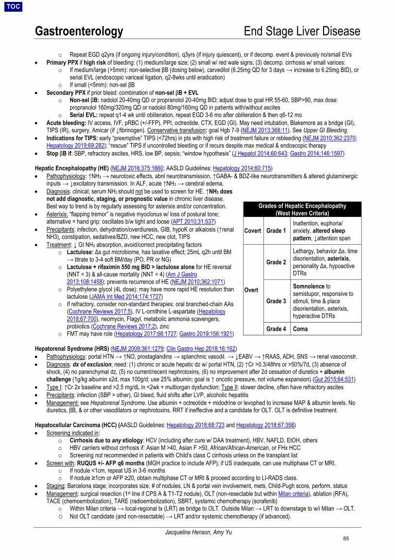
Gastroenterology End Stage Liver Disease
Jacqueline Henson, Amy Yu
o Repeat EGD q2yrs (if ongoing injury/condition), q3yrs (if injury quiescent), or if decomp. event & previously no/small EVs• Primary PPX if high risk of bleeding: (1) medium/large size; (2) small w/ red wale signs; (3) decomp. cirrhosis w/ small varices:
o If medium/large (>5mm): non-selective βB (dosing below), carvedilol (6.25mg QD for 3 days → increase to 6.25mg BID), orserial EVL (endoscopic variceal ligation, q2-8wks until eradication)
o If small (<5mm): non-sel βB• Secondary PPX if prior bleed: combination of non-sel βB + EVL
o Non-sel βB: nadolol 20-40mg QD or propranolol 20-40mg BID; adjust dose to goal HR 55-60, SBP>90, max dose:propranolol 160mg/320mg QD or nadolol 80mg/160mg QD in patients with/without ascites
o Serial EVL: repeat q1-4 wk until obliteration, repeat EGD 3-6 mo after obliteration & then q6-12 mo• Acute bleeding: IV access, IVF, pRBC (+/-FFP), PPI, octreotide, CTX, EGD (GI). May need intubation, Blakemore as a bridge (GI),
TIPS (IR), surgery, Amicar (if ↓fibrinogen). Conservative transfusion: goal Hgb 7-9 (NEJM 2013;368:11). See Upper GI Bleeding.• Indications for TIPS: early “preemptive” TIPS (<72hrs) in pts with high risk of treatment failure or rebleeding (NEJM 2010;362:2370;
Hepatology 2019;69:282); “rescue” TIPS if uncontrolled bleeding or if recurs despite max medical & endoscopic therapy• Stop βB if: SBP, refractory ascites, HRS, low BP, sepsis; “window hypothesis” (J Hepatol 2014;60:643; Gastro 2014;146:1597)
Hepatic Encephalopathy (HE) (NEJM 2016;375:1660; AASLD Guidelines: Hepatology 2014;60:715) • Pathophysiology: ↑NH3 → neurotoxic effects, abnl neurotransmission, ↑GABA- & BDZ-like neurotransmitters & altered glutaminergic
inputs → ↓excitatory transmission. In ALF, acute ↑NH3 → cerebral edema.• Diagnosis: clinical; serum NH3 should not be used to screen for HE. ↑NH3 does
not add diagnostic, staging, or prognostic value in chronic liver disease.Best way to trend is by regularly assessing for asterixis and/or concentration.
• Asterixis: “flapping tremor” is negative myoclonus w/ loss of postural tone;alternative = hand grip: oscillates b/w tight and loose (APT 2010;31:537)
• Precipitants: infection, dehydration/overdiuresis, GIB, hypoK or alkalosis (↑renalNH3), constipation, sedatives/BZD, new HCC, new clot, TIPS
• Treatment: ↓ GI NH3 absorption, avoid/correct precipitating factorso Lactulose: Δs gut microbiome, has laxative effect; 25mL q2h until BM
→ titrate to 3-4 soft BM/day (PO, PR or NG)o Lactulose + rifaximin 550 mg BID > lactulose alone for HE reversal
(NNT = 3) & all-cause mortality (NNT = 4) (Am J Gastro2013;108:1458); prevents recurrence of HE (NEJM 2010;362:1071)
o Polyethylene glycol (4L dose): may have more rapid HE resolution thanlactulose (JAMA Int Med 2014;174:1727)
o If refractory, consider non-standard therapies: oral branched-chain AAs(Cochrane Reviews 2017;5), IV L-ornithine L-aspartate (Hepatology 2018;67:700), neomycin, Flagyl, metabolic ammonia scavengers, probiotics (Cochrane Reviews 2017;2), zinc
o FMT may have role (Hepatology 2017;66:1727; Gastro 2019;156:1921)
Hepatorenal Syndrome (HRS) (NEJM 2009;361:1279; Clin Gastro Hep 2018;16:162) • Pathophysiology: portal HTN → ↑NO, prostaglandins → splanchnic vasodil. → ↓EABV → ↑RAAS, ADH, SNS → renal vasoconstr.• Diagnosis: dx of exclusion; need: (1) chronic or acute hepatic dz w/ portal HTN, (2) ↑Cr >0.3/48hrs or >50%/7d, (3) absence of
shock, (4) no parenchymal dz, (5) no current/recent nephrotoxins, (6) no improvement after 2d cessation of diuretics + albuminchallenge (1g/kg albumin x2d, max 100g/d; use 25% albumin; goal is ↑ oncotic pressure, not volume expansion) (Gut 2015;64:531)
• Type I: ↑Cr 2x baseline and >2.5 mg/dL in <2wk + multiorgan dysfunction; Type II: slower decline, often have refractory ascites• Precipitants: infection (SBP > other), GI bleed, fluid shifts after LVP, alcoholic hepatitis• Management: see Hepatorenal Syndrome. Use albumin + octreotide + midodrine or levophed to increase MAP & albumin levels. No
diuretics, βB, & or other vasodilators or nephrotoxins. RRT if ineffective and a candidate for OLT. OLT is definitive treatment.
Hepatocellular Carcinoma (HCC) (AASLD Guidelines: Hepatology 2018;68:723 and Hepatology 2018;67:358) • Screening indicated in:
o Cirrhosis due to any etiology: HCV (including after cure w/ DAA treatment), HBV, NAFLD, EtOH, otherso HBV carriers without cirrhosis if: Asian M >40, Asian F >50, African/African-American, or FHx HCCo Screening not recommended in patients with Child’s class C cirrhosis unless on the transplant list
• Screen with: RUQUS +/- AFP q6 months (MGH practice to include AFP); if US inadequate, can use multiphase CT or MRI.o If nodule <1cm, repeat US in 3-6 monthso If nodule ≥1cm or AFP ≥20, obtain multiphase CT or MRI & proceed according to LI-RADS class.
• Staging: Barcelona stage; incorporates size, # of nodules, LN & portal vein involvement, mets, Child-Pugh score, perform. status• Management: surgical resection (1st line if CPS A & T1-T2 nodule), OLT (non-resectable but within Milan criteria), ablation (RFA),
TACE (chemoembolization), TARE (radioembolization), SBRT, systemic chemotherapy (sorafenib)o Within Milan criteria → local-regional tx (LRT) as bridge to OLT. Outside Milan → LRT to downstage to w/i Milan → OLT.o Not OLT candidate (and non-resectable) → LRT and/or systemic chemotherapy (if advanced).
Grades of Hepatic Encephalopathy (West Haven Criteria)
Covert Grade 1 Inattention, euphoria/ anxiety, altered sleep pattern, ↓attention span
Overt
Grade 2 Lethargy, behavior Δs, time disorientation, asterixis, personality Δs, hypoactive DTRs
Grade 3
Somnolence to semistupor, responsive to stimuli, time & place disorientation, asterixis, hyperactive DTRs
Grade 4 Coma
85
Gastroenterology End Stage Liver Disease
Jacqueline Henson, Amy Yu
Hepatic Hydrothorax (AASLD Guidelines: Hepatology 2013;57:1651) • Transudative effusion due to shift of ascites into pleural space (due to neg. intrathoracic pressure) via small diaphragmatic defects.
Usually unilateral, R-sided. Can become infected (spontaneous bacterial empyema) even in the absence of SBP.• Diagnosis: exclude other causes of transudative effusion; can visualize w/ radioisotope injection into ascites• Treatment: same as for ascites (diuretics, <2g Na); therapeutic thora for dyspnea. TIPS if refractory. Chest tube and pleurodesis
not recommended.
Hepatopulmonary Syndrome (HPS) (NEJM 2008;358:2378; EASL Guidelines: J Hepatol 2018;69:406) • Syndrome of intrapulmonary shunting through vasodilation/AVMs; mechanism unclear, possibly due to circulating NO• Presentation: shunting tends to occur at lung bases → platypnea (dyspnea when upright, relieved when supine) & orthodeoxia
(upright hypoxemia, PaO2 ↓ by 4 mmHg or ≥5%), clubbing, cyanosis, hypoxemia (↓PaO2 <70-80)• Diagnosis: TTE with late bubbles (3-6 cardiac cycles after RA), ↑A-a gradient ≥15 (or >20 if age >64).
o 99mTc MAA scan is alternative to TTE but more invasive, less sensitive. May be useful in quantifying shunting if severehypoxemia and coexistent intrinsic lung disease.
o Pulmonary angiography performed if severe hypoxemia poorly responsive to 100% O2 & areas amenable to embolizationo PFTs can be performed to evaluate for intrinsic lung disease; ↓DLCO in HPS
• Management: O2; no effective medical therapies; OLT can significantly improve (and reverse) HPS
Portopulmonary Hypertension (PPHTN) (EASL Guidelines: J Hepatol 2018;69:406) • Rare cause of group 1 pulmonary hypertension in setting of portal HTN• Pathogenesis: unknown; possibly 2/2 humoral substances (ex. serotonin, interleukin-1, endothelin-1, normally cleared by liver) that
reach pulmonary circulation through portosystemic collaterals, resulting in PPHTN• Presentation: DOE, chest pain, fatigue, palpitations, syncope, hemoptysis, orthopnea; often w/ TR murmur, EKG w/ RVH, RAB, RBBB• Diagnosis: RHC w/ PAH (mPAP >25 mmHg, PCWP <15 mmHg) in pt with established portal hypertension in absence of other
etiology of PAH or venous hypertension• Management: may benefit from advanced therapies (epoprostenol, bosentan, sildenafil, iloprost); OLT can improve/normalize the
PAH; βB and TIPS may be harmful and should be avoided• Transplant: increased risk with mPAP ≥35; mPAP ≥45 is a contraindication
Cirrhotic Cardiomyopathy (EASL Guidelines: J Hepatol 2018;69:406) • Definition: chronic cardiac dysfunction in cirrhotic patients with no known cardiac disease; characterized by 1) impaired cardiac
contractility in response to stress, 2) altered diastolic relaxation, 3) electrophysiological abnormalities such as prolonged QTc• Prevalence: up to 50% of patients undergoing liver transplantation have signs of cardiac dysfunction• Diagnosis: echocardiography with dynamic stress testing w/ pharmacologics or exercise• Pathophysiology: myocardial dysfunction 2/2 systemic inflammation; shear stress from portal hypertension → mechanical force on
myocardial fibers; other possible mechanisms involve collagen configuration, sodium retention and activation of RAAS• Treatment: same as HF management in non-cirrhotic patients• Prognosis: largely subclinical and asymptomatic; however poses risk in the presence of stress such as infection, TIPS, or OLT; thus
detailed cardiac assessment required prior to interventions
Hematologic Abnormalities (NEJM 2011;365:147, Clin Gastro Hep 2013;11:1064, Thromb Haemost 2018;118:1491) • Cytopenias: multifactorial; thrombocytopenia (splenomegaly, ↓TPO), leukopenia (splenomegaly), anemia (bleeding, spur cell
anemia); also can have BM suppression by EtOH, nutritional deficiencies (e.g. folate), direct effect of HCV/HBV• Coagulation abnormalities: ↓ coagulation factors (except for VIII), ↓ anticoagulant proteins (C, S, ATIII), dysfibrinogenemia,
accelerated fibrinolysis (↑tPA) → ↑ risk of both clotting and bleeding & patients not auto-anticoagulated; balance tends to favorthrombosis in early stages and bleeding in late stages of cirrhosis
o Labs: ↑PT/INR, ↑PTT, ↑/nl fibrinogen (though does not function normally; ↓ if fulminant), ↑/nl D-dimer (vs. ↑↑ in DIC),↑factor VIII (vs. ↓ in DIC); note PT and PT/INR do NOT correlate with risk of bleeding or clotting
• Anticoagulation: VTE ppx should not be withheld unless high risk of bleeding, plts<50. Systemic AC ok unless decomp. CPS C orhigh risk of bleeding. EGD for EVs prior to starting. VKA, LMWH, or DOAC all options. VKA dosing can be c/b ↑baseline PT/INR;LMWH can be c/b ↓ATIII levels; DOACs not easily reversible & some are hepatically-cleared (J Hepatol 2017;66:1313, JACC2018;71:2162)
• Bleeding: consider role of coagulation factor deficiency, dysfibrinogenemia, hyperfibrinolysis, thrombocytopeniao If suspect vitamin K deficiency, give vitamin K 10mg x3 days to correct nutritional componento Transfuse pRBCs Hgb<7, platelets <50k, cryo for fibrinogen <150 (or if >150 but c/f dysfibrinogenemia)o Persistent bleeding despite cryo or requiring many pRBCs → can give FFP (though large volume → ↑ portal pressures)o Delayed bleeding or oozing from mucocutaneous sites → c/f hyperfibrinolysis → Amicar or TXA (topical and/or systemic)
• Procedures:o Platelets: >50k for surgery, TIPS, liver biopsy, or other procedure w/ high bleeding risk; TPO agonists can reduce need for
peri-procedural plt transfusions (NEJM 2012;367:716; Gastro 2018;155:705).o PT/INR: NO benefit to giving FFP pre-procedure to “correct” INR. ↑volume can ↑ bleeding risk by ↑ portal pressures.
86

Gastroenterology Hepatorenal Syndrome
Jacqueline Henson
MGH Algorithm for the Diagnosis and Treatment of Hepatorenal Syndrome(Gut 2015;64:531. Dig Dis Sci 2014;59:471. Dig Dis Sci 2015;60:1474. Nephron 2015;131:191. Clin Gastroenterol Hepatol
2018;16:162)
87
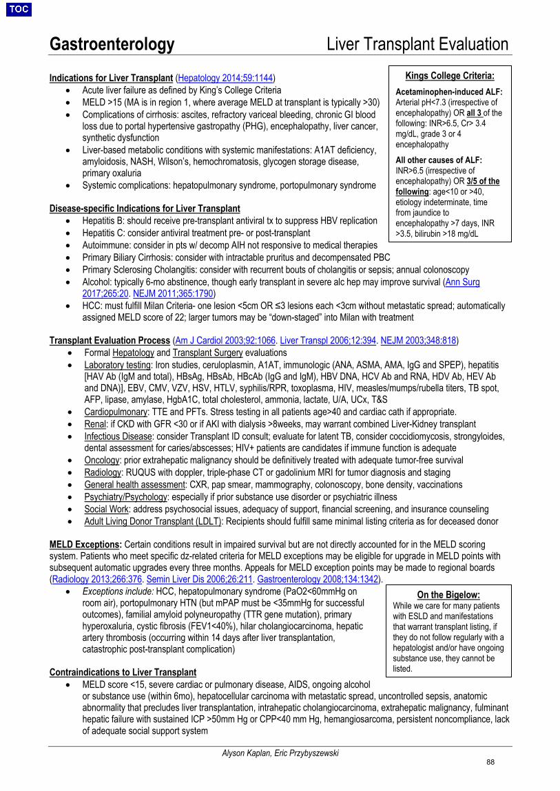
Gastroenterology Liver Transplant Evaluation
Alyson Kaplan, Eric Przybyszewski
Indications for Liver Transplant (Hepatology 2014;59:1144)
• Acute liver failure as defined by King’s College Criteria• MELD >15 (MA is in region 1, where average MELD at transplant is typically >30)• Complications of cirrhosis: ascites, refractory variceal bleeding, chronic GI blood
loss due to portal hypertensive gastropathy (PHG), encephalopathy, liver cancer,synthetic dysfunction
• Liver-based metabolic conditions with systemic manifestations: A1AT deficiency,amyloidosis, NASH, Wilson’s, hemochromatosis, glycogen storage disease,primary oxaluria
• Systemic complications: hepatopulmonary syndrome, portopulmonary syndrome
Disease-specific Indications for Liver Transplant • Hepatitis B: should receive pre-transplant antiviral tx to suppress HBV replication• Hepatitis C: consider antiviral treatment pre- or post-transplant• Autoimmune: consider in pts w/ decomp AIH not responsive to medical therapies• Primary Biliary Cirrhosis: consider with intractable pruritus and decompensated PBC• Primary Sclerosing Cholangitis: consider with recurrent bouts of cholangitis or sepsis; annual colonoscopy• Alcohol: typically 6-mo abstinence, though early transplant in severe alc hep may improve survival (Ann Surg
2017;265:20. NEJM 2011;365:1790)• HCC: must fulfill Milan Criteria- one lesion <5cm OR ≤3 lesions each <3cm without metastatic spread; automatically
assigned MELD score of 22; larger tumors may be “down-staged” into Milan with treatment
Transplant Evaluation Process (Am J Cardiol 2003;92:1066. Liver Transpl 2006;12:394. NEJM 2003;348:818) • Formal Hepatology and Transplant Surgery evaluations• Laboratory testing: Iron studies, ceruloplasmin, A1AT, immunologic (ANA, ASMA, AMA, IgG and SPEP), hepatitis
[HAV Ab (IgM and total), HBsAg, HBsAb, HBcAb (IgG and IgM), HBV DNA, HCV Ab and RNA, HDV Ab, HEV Aband DNA)], EBV, CMV, VZV, HSV, HTLV, syphilis/RPR, toxoplasma, HIV, measles/mumps/rubella titers, TB spot,AFP, lipase, amylase, HgbA1C, total cholesterol, ammonia, lactate, U/A, UCx, T&S
• Cardiopulmonary: TTE and PFTs. Stress testing in all patients age>40 and cardiac cath if appropriate.• Renal: if CKD with GFR <30 or if AKI with dialysis >8weeks, may warrant combined Liver-Kidney transplant• Infectious Disease: consider Transplant ID consult; evaluate for latent TB, consider coccidiomycosis, strongyloides,
dental assessment for caries/abscesses; HIV+ patients are candidates if immune function is adequate• Oncology: prior extrahepatic malignancy should be definitively treated with adequate tumor-free survival• Radiology: RUQUS with doppler, triple-phase CT or gadolinium MRI for tumor diagnosis and staging• General health assessment: CXR, pap smear, mammography, colonoscopy, bone density, vaccinations• Psychiatry/Psychology: especially if prior substance use disorder or psychiatric illness• Social Work: address psychosocial issues, adequacy of support, financial screening, and insurance counseling• Adult Living Donor Transplant (LDLT): Recipients should fulfill same minimal listing criteria as for deceased donor
MELD Exceptions: Certain conditions result in impaired survival but are not directly accounted for in the MELD scoring system. Patients who meet specific dz-related criteria for MELD exceptions may be eligible for upgrade in MELD points with subsequent automatic upgrades every three months. Appeals for MELD exception points may be made to regional boards(Radiology 2013;266:376. Semin Liver Dis 2006;26:211. Gastroenterology 2008;134:1342).
• Exceptions include: HCC, hepatopulmonary syndrome (PaO2<60mmHg onroom air), portopulmonary HTN (but mPAP must be <35mmHg for successfuloutcomes), familial amyloid polyneuropathy (TTR gene mutation), primaryhyperoxaluria, cystic fibrosis (FEV1<40%), hilar cholangiocarcinoma, hepaticartery thrombosis (occurring within 14 days after liver transplantation,catastrophic post-transplant complication)
Contraindications to Liver Transplant • MELD score <15, severe cardiac or pulmonary disease, AIDS, ongoing alcohol
or substance use (within 6mo), hepatocellular carcinoma with metastatic spread, uncontrolled sepsis, anatomicabnormality that precludes liver transplantation, intrahepatic cholangiocarcinoma, extrahepatic malignancy, fulminanthepatic failure with sustained ICP >50mm Hg or CPP<40 mm Hg, hemangiosarcoma, persistent noncompliance, lackof adequate social support system
On the Bigelow: While we care for many patients with ESLD and manifestations that warrant transplant listing, if they do not follow regularly with a hepatologist and/or have ongoing substance use, they cannot be listed.
Kings College Criteria: Acetaminophen-induced ALF: Arterial pH<7.3 (irrespective of encephalopathy) OR all 3 of the following: INR>6.5, Cr> 3.4 mg/dL, grade 3 or 4 encephalopathy All other causes of ALF: INR>6.5 (irrespective of encephalopathy) OR 3/5 of the following: age<10 or >40, etiology indeterminate, time from jaundice to encephalopathy >7 days, INR >3.5, bilirubin >18 mg/dL
88

Nephrology Acute Kidney Injury
Dana Larsen
MEDS Causing AKI (J Renal Inj Prev 2015;4:57; Clin Inf Dis 2017;64:116)Chronic/acute interstitial
nephritis Acute tubular
necrosis Crystal nephropathy Rhabdomyolysis Thrombotic microangiopathy
Lithium APAP Aminoglycosides Acyclovir/ Ganciclovir Amitriptyline Clopidogrel Methotrexate Allopurinol Amphotericin B Indinavir Benzodiazepine Cyclosporine Phenytoin NSAID Cephalosporins Glomerular Cocaine/methamphetamine Quinine Rifampin Beta lactams Contrast hemodynamics Diphenhydramine Ranitidine Cisplatin Tacrolimus ACE-I/ARB Haloperidol Sulfas Furosemide Tetracyclines Vanc (esp w/ Zosyn) Methadone
MANAGEMENT: “A Euvolemic Kidney is a Happy Kidney; Fluids are NOT always the answer” 1) Optimize hemodynamics: Stop NSAIDs, ACEi/ARBs; correct volume status: IVF if hypovolemic, diuretics if volume overloaded
• No evidence of benefit of dopamine (Ann Int Med 2005;142:510), empiric diuretics in oliguria (JAMA 2002;288:2547), or mannitol2) Avoid nephrotoxins: Iodinated contrast (see below), offending medications (see above)3) Renally dose meds: Antibiotics, narcotics, LMWH → UFH, Keppra (NB: remember Cr overestimates GFR in AKI)4) Manage complications: HyperK: calcium gluconate, insulin/dextrose→ kayexalate, bowel reg, furosemide; Hyperphos: sevelamer vsphoslo depending on calcium; Metabolic Acidosis: Sodium bicarb tabs; Bleeding with concern for uremic platelets: DDAVP 0.3 mcg/kg IV,onset 1hr, lasts 4-8hr5) Indications for HD (AEIOU): Acidosis (esp pH<7.1), Electrolytes (usually K+), Intoxication (lithium, ethylene glycol, metformin,salicylates, theophylline), Volume Overload, Uremia (encephalopathy, neuropathy, pericarditis)
SPECIFIC MANAGEMENT CONCERNS: • HRS: See hepatology section.• Rapidly progressive glomerulonephritis (RPGN): Urgent Nephrology consult for consideration of methylprednisolone 0.5-1g IV x
3d ± cyclophosphamide or mycophenolate mofetil ± plasmapheresis (should not be deciding this alone)• Scleroderma renal crisis: ACEi (captopril) at maximum tolerated dose. ARBs should theoretically have similar effect but no data.• Rhabdomyolysis: AKI unlikely unless CK >2000-5000; aggressive IVF for UOP >250/h with normal saline. Consider isotonic sodium
bicarbonate if marked acidosis (NEJM 2009;361:62), but no convincing evidence that isotonic bicarbonate is superior to normal saline.Continue aggressive IVF until 1) CK < 2000 or 2) volume overload
• Acute interstitial nephritis (AIN): Stop offending agent, consider prednisone 40mg-60mg qd for 1-2wk if biopsy-confirmed or highpre-test (Am J Med 1978;65:756) (poor quality data)
• Crystal deposition: discontinue drug; fomepizole/HD if ethylene glycol toxicity; rasburicase if TLS• Post-renal: Foley; α-antagonists; 5α-reductase inhibitor (effect not immediate); urology/IR if percutaneous nephrostomy tube needed
RENAL EMERGENCIES (when to page the renal fellow overnight):• Acidosis: Severe metabolic acidosis, unstable patient, usually in the ICU with pH < 7.1. Temporize with HCO3 pushes and isotonic
bicarb gtt, intubation and hyperventilation if unable to compensate by breathing off CO2. Likely CVVH.• Ingestions: Ethylene glycol, methanol (elevated osmolar gap) with end organ damage (i.e. renal failure, vision loss).• Hyperkalemia: Marked hyperkalemia leading to ECG changes or arrhythmia (K>6.5). Temporize with Ca gluconate, Lasix,
Insulin/D50, etc. Note HD much faster at clearing K than CVVH.• Hyponatremia: Call if severely symptomatic (AMS with low GCS, seizures, etc) requiring bolus hypertonic saline.• RPGN: when clinically suspected, urgent Nephrology consultation to consider pulse dose steroids +/- plasmapheresis (as above)
CONTRAST-INDUCED NEPHROPATHY (CIN): (Mayo 2009;84:170; Circ 2012;122:2451; NEJM 2006;354:379) Definition: ↑Cr ≥ 0.5 or 25% within 48-72h of contrast without other causes. Clinical syndrome: starts 24-48hr, peaks 3-5d, resolves 10d; FENa usually <1% but can be normal or high. Usually non-oliguric. Controversy: More recent controlled studies and meta-analyses have raised questions regarding the risk of AKI following contrast, which is probably lower than many previous studies indicated. For now, we operate under the assumption that contrast plays a role. Do not invoke contrast without full workup for other causes (Ann Em Med 2017;69:577). Risk factors include higher contrast load, intra-arterial injection, presence diabetes, proteinuria and multiple myeloma.Prophylaxis: For high risk pts receiving arterial or IV contrast, give NS at 1ml/kg/hr for 6-12hr pre, 6-12hr post. No added benefit for Na bicarb, NAC (arterial) (NEJM 2018;378:603), or prophylactic HD (IV) (Am J Med 2012;125:66).
CARDIORENAL SYNDROME (TYPE 1): Nat Rev Neph 2009;5:641; Nat Rev Neph 2013;9:99; CJASN 2013;8:1800) Refers to 5 categories of disease processes which impact the heart and kidneys with various causal relationships, but at MGH we use the term to refer to type 1, in which acute CHF leads to AKI. Pathophysiology: Decreased renal perfusion from low CO is one factor, but more importantly, RV failure and high CVP lead to a low trans-renal perfusion pressure. More of a problem with “underdraining” (congestion) than with “underfilling” (perfusion), though worsened by neurohumoral activation in setting of low EABV. Treatment: Relief of renal venous congestion. Trend creatinine against TBB to test hypothesis, but expect a lag effect. • Loop diuretics are first line for type 1 +/- addition of thiazide (metolazone / diuril).• ROSE trial (JAMA 2013;310:2533): No benefit of low dose dopamine or nesiritide to improve forward flow.• CARESS-HF (NEJM 2012;367:2296): Ultrafiltration showed similar outcomes in regard to weight loss and decompensated CHF
symptoms, but worsened renal function compared to pharmacologic therapy with loop/thiazide diuretics.
89
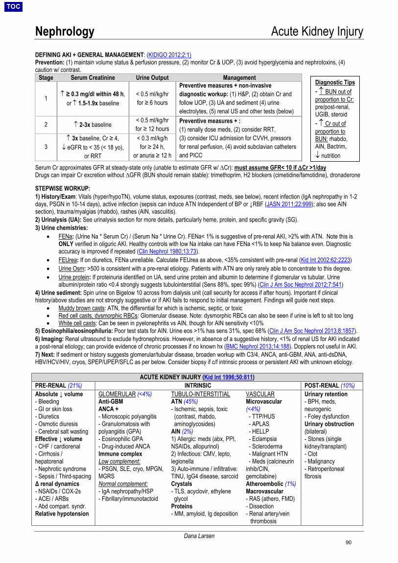
Nephrology Acute Kidney Injury
Dana Larsen
DEFINING AKI + GENERAL MANAGEMENT: (KIDIGO 2012;2:1)Prevention: (1) maintain volume status & perfusion pressure, (2) monitor Cr & UOP, (3) avoid hyperglycemia and nephrotoxins, (4) caution w/ contrast.
Serum Cr approximates GFR at steady-state only (unable to estimate GFR w/ ∆Cr): must assume GFR< 10 if ∆Cr >1/day Drugs can impair Cr excretion without ∆GFR (BUN should remain stable): trimethoprim, H2 blockers (cimetidine/famotidine), dronaderone
STEPWISE WORKUP: 1) History/Exam: Vitals (hyper/hypoTN), volume status, exposures (contrast, meds, see below), recent infection (IgA nephropathy in 1-2days, PSGN in 10-14 days), active infection (sepsis can induce ATN Independent of BP or ↓RBF (JASN 2011;22:999); also see AINsection), trauma/myalgias (rhabdo), rashes (AIN, vasculitis).2) Urinalysis (UA): See urinalysis section for more details, particularly heme, protein, and specific gravity (SG).3) Urine chemistries:
• FENa: (Urine Na * Serum Cr) / (Serum Na * Urine Cr). FENa< 1% is suggestive of pre-renal AKI, >2% with ATN. Note this isONLY verified in oliguric AKI. Healthy controls with low Na intake can have FENa <1% to keep Na balance even. Diagnosticaccuracy is improved if repeated (Clin Nephrol 1980;13:73).
• FEUrea: If on diuretics, FENa unreliable. Calculate FEUrea as above, <35% consistent with pre-renal (Kid Int 2002;62:2223)• Urine Osm: >500 is consistent with a pre-renal etiology. Patients with ATN are only rarely able to concentrate to this degree.• Urine protein: If proteinuria identified on UA, send urine protein and albumin to determine if glomerular vs tubular. Urine
albumin/protein ratio <0.4 strongly suggests tubulointerstitial (Sens 88%, spec 99%) (Clin J Am Soc Nephrol 2012;7:541)4) Urine sediment: Spin urine on Bigelow 10 across from dialysis unit (call security for access if after hours). Important if clinicalhistory/above studies are not strongly suggestive or if AKI fails to respond to initial management. Findings will guide next steps.
• Muddy brown casts: ATN, the differential for which is ischemic, septic, or toxic• Red cell casts, dysmorphic RBCs: Glomerular disease. Note: dysmorphic RBCs can also be seen if urine is left to sit too long• White cell casts: Can be seen in pyelonephritis vs AIN, though for AIN sensitivity <10%
5) Eosinophilia/eosinophiluria: Poor test stats for AIN. Urine eos >1% has sens 31%, spec 68% (Clin J Am Soc Nephrol 2013;8:1857).6) Imaging: Renal ultrasound to exclude hydronephrosis. However, in absence of a suggestive history, <1% of renal US for AKI indicateda post-renal etiology; can provide evidence of chronic processes if no known hx (BMC Nephrol 2013;14:188). Dopplers not useful in AKI.7) Next: If sediment or history suggests glomerular/tubular disease, broaden workup with C3/4, ANCA, anti-GBM, ANA, anti-dsDNA,HBV/HCV/HIV, cryos, SPEP/UPEP/SFLC as per below. Consider biopsy if c/f intrinsic process or persistent AKI with unknown etiology.
ACUTE KIDNEY INJURY (Kid Int 1996;50:811) PRE-RENAL (21%) INTRINSIC POST-RENAL (10%) Absolute ↓ volume - Bleeding- GI or skin loss- Diuretics- Osmotic diuresis- Cerebral salt wastingEffective ↓ volume- CHF / cardiorenal- Cirrhosis /hepatorenal- Nephrotic syndrome- Sepsis / Third-spacingΔ renal dynamics- NSAIDs / COX-2s- ACEi / ARBs- Abd compart. syndr.Relative hypotension
GLOMERULAR (<4%) Anti-GBM ANCA + - Microscopic polyangitis- Granulomatosis withpolyangiitis (GPA)- Eosinophilic GPA- Drug-induced ANCAImmune complexLow complement:- PSGN, SLE, cryo, MPGN,MGRSNormal complement:- IgA nephropathy/HSP- Fibrillary/immunotactoid
TUBULO-INTERSTITIAL ATN (45%) - Ischemic, sepsis, toxic
(contrast, rhabdo,aminoglycosides)
AIN (2%)1) Allergic: meds (abx, PPI,NSAIDs, allopurinol)2) Infectious: CMV, lepto,legionella3) Auto-immune / infiltrative:TINU, IgG4 disease, sarcoidCrystals- TLS, acyclovir, ethylene
glycolProteins- MM, amyloid, Ig deposition
VASCULAR Microvascular (<4%)
- TTP/HUS- APLAS- HELLP- Eclampsia- Scleroderma- Malignant HTN- Meds (calcineurin
inhib/CIN,gemcitabine)Atheroembolic (1%)Macrovascular- RAS (athero, FMD)- Dissection- Renal artery/vein
thrombosis
Urinary retention - BPH, meds,neurogenic- Foley dysfunctionUrinary obstruction(bilateral)- Stones (singlekidney/transplant)- Clot- Malignancy- Retroperitonealfibrosis
Stage Serum Creatinine Urine Output Management
1 ↑ ≥ 0.3 mg/dl within 48 h, or ↑ 1.5-1.9x baseline
< 0.5 ml/kg/hr for ≥ 6 hours
Preventive measures + non-invasive diagnostic workup: (1) H&P, (2) obtain Cr and follow UOP, (3) UA and sediment (4) urine electrolytes, (5) renal US and other tests (below)
2 ↑ 2-3x baseline < 0.5 ml/kg/hr for ≥ 12 hours
Preventive measures + : (1) renally dose meds, (2) consider RRT,(3) consider ICU admission for CVVH, pressorsfor renal perfusion, (4) avoid subclavian cathetersand PICC
3 ↑ 3x baseline, Cr ≥ 4,
↓ eGFR to < 35 (< 18 yo), or RRT
< 0.3 ml/kg/h for ≥ 24 h,
or anuria ≥ 12 h
Diagnostic Tips - ↑ BUN out ofproportion to Cr:pre/post-renal,UGIB, steroid- ↑ Cr out ofproportion toBUN: rhabdo,AIN, Bactrim,↓ nutrition
90
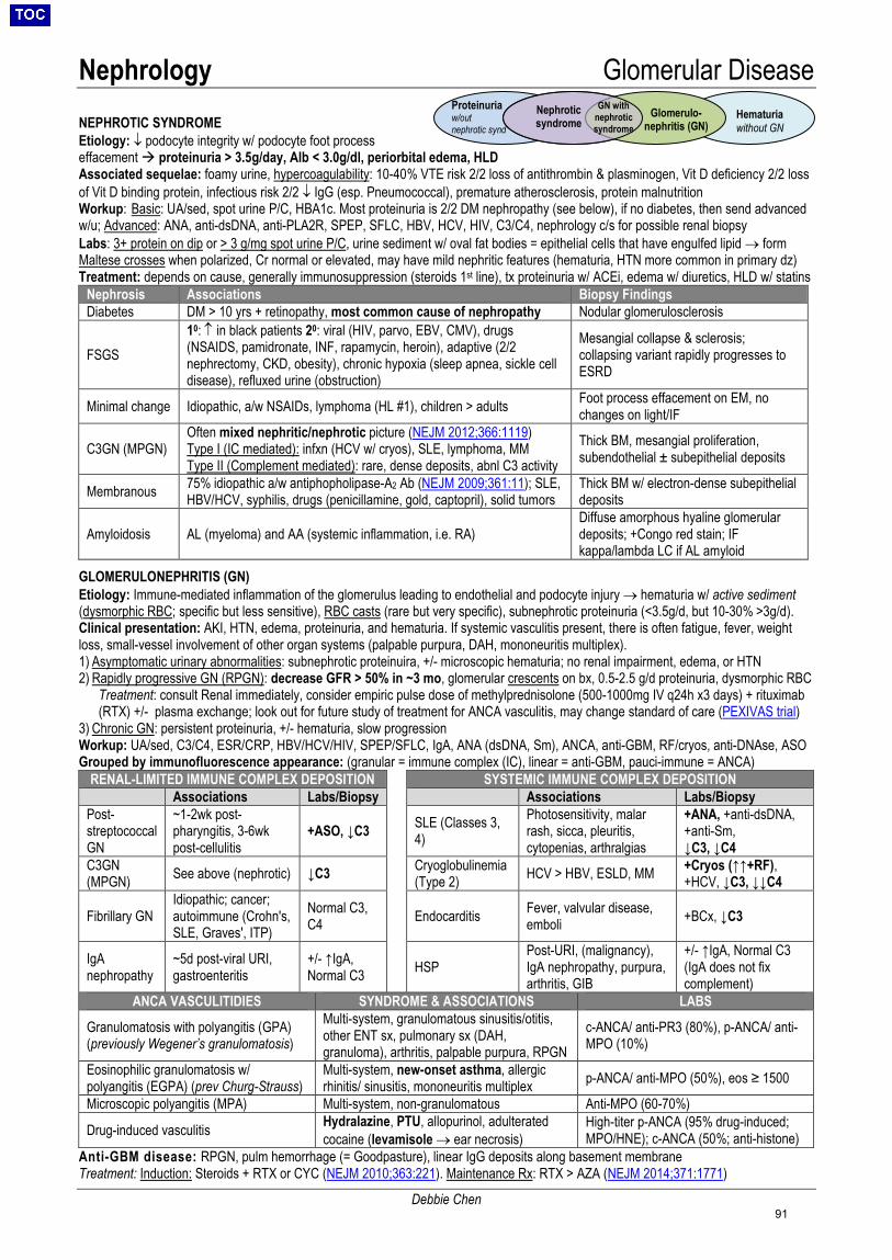
Nephrology Glomerular Disease
Debbie Chen
NEPHROTIC SYNDROME Etiology: ↓ podocyte integrity w/ podocyte foot process effacement proteinuria > 3.5g/day, Alb < 3.0g/dl, periorbital edema, HLD Associated sequelae: foamy urine, hypercoagulability: 10-40% VTE risk 2/2 loss of antithrombin & plasminogen, Vit D deficiency 2/2 loss of Vit D binding protein, infectious risk 2/2 ↓ IgG (esp. Pneumococcal), premature atherosclerosis, protein malnutrition Workup: Basic: UA/sed, spot urine P/C, HBA1c. Most proteinuria is 2/2 DM nephropathy (see below), if no diabetes, then send advanced w/u; Advanced: ANA, anti-dsDNA, anti-PLA2R, SPEP, SFLC, HBV, HCV, HIV, C3/C4, nephrology c/s for possible renal biopsy Labs: 3+ protein on dip or > 3 g/mg spot urine P/C, urine sediment w/ oval fat bodies = epithelial cells that have engulfed lipid → form Maltese crosses when polarized, Cr normal or elevated, may have mild nephritic features (hematuria, HTN more common in primary dz) Treatment: depends on cause, generally immunosuppression (steroids 1st line), tx proteinuria w/ ACEi, edema w/ diuretics, HLD w/ statins
Nephrosis Associations Biopsy Findings Diabetes DM > 10 yrs + retinopathy, most common cause of nephropathy Nodular glomerulosclerosis
FSGS 10: ↑ in black patients 20: viral (HIV, parvo, EBV, CMV), drugs (NSAIDS, pamidronate, INF, rapamycin, heroin), adaptive (2/2 nephrectomy, CKD, obesity), chronic hypoxia (sleep apnea, sickle cell disease), refluxed urine (obstruction)
Mesangial collapse & sclerosis; collapsing variant rapidly progresses to ESRD
Minimal change Idiopathic, a/w NSAIDs, lymphoma (HL #1), children > adults Foot process effacement on EM, no changes on light/IF
C3GN (MPGN) Often mixed nephritic/nephrotic picture (NEJM 2012;366:1119) Type I (IC mediated): infxn (HCV w/ cryos), SLE, lymphoma, MM Type II (Complement mediated): rare, dense deposits, abnl C3 activity
Thick BM, mesangial proliferation, subendothelial ± subepithelial deposits
Membranous 75% idiopathic a/w antiphopholipase-A2 Ab (NEJM 2009;361:11); SLE, HBV/HCV, syphilis, drugs (penicillamine, gold, captopril), solid tumors
Thick BM w/ electron-dense subepithelial deposits
Amyloidosis AL (myeloma) and AA (systemic inflammation, i.e. RA) Diffuse amorphous hyaline glomerular deposits; +Congo red stain; IF kappa/lambda LC if AL amyloid
GLOMERULONEPHRITIS (GN) Etiology: Immune-mediated inflammation of the glomerulus leading to endothelial and podocyte injury → hematuria w/ active sediment (dysmorphic RBC; specific but less sensitive), RBC casts (rare but very specific), subnephrotic proteinuria (<3.5g/d, but 10-30% >3g/d). Clinical presentation: AKI, HTN, edema, proteinuria, and hematuria. If systemic vasculitis present, there is often fatigue, fever, weight loss, small-vessel involvement of other organ systems (palpable purpura, DAH, mononeuritis multiplex). 1) Asymptomatic urinary abnormalities: subnephrotic proteinuira, +/- microscopic hematuria; no renal impairment, edema, or HTN2) Rapidly progressive GN (RPGN): decrease GFR > 50% in ~3 mo, glomerular crescents on bx, 0.5-2.5 g/d proteinuria, dysmorphic RBC
Treatment: consult Renal immediately, consider empiric pulse dose of methylprednisolone (500-1000mg IV q24h x3 days) + rituximab(RTX) +/- plasma exchange; look out for future study of treatment for ANCA vasculitis, may change standard of care (PEXIVAS trial)
3) Chronic GN: persistent proteinuria, +/- hematuria, slow progressionWorkup: UA/sed, C3/C4, ESR/CRP, HBV/HCV/HIV, SPEP/SFLC, IgA, ANA (dsDNA, Sm), ANCA, anti-GBM, RF/cryos, anti-DNAse, ASOGrouped by immunofluorescence appearance: (granular = immune complex (IC), linear = anti-GBM, pauci-immune = ANCA)
RENAL-LIMITED IMMUNE COMPLEX DEPOSITION SYSTEMIC IMMUNE COMPLEX DEPOSITION Associations Labs/Biopsy Associations Labs/Biopsy
Post-streptococcal GN
~1-2wk post-pharyngitis, 3-6wk post-cellulitis
+ASO, ↓C3 SLE (Classes 3, 4)
Photosensitivity, malar rash, sicca, pleuritis, cytopenias, arthralgias
+ANA, +anti-dsDNA,+anti-Sm,↓C3, ↓C4
C3GN (MPGN) See above (nephrotic) ↓C3 Cryoglobulinemia
(Type 2) HCV > HBV, ESLD, MM +Cryos (↑↑+RF),+HCV, ↓C3, ↓↓C4
Fibrillary GN Idiopathic; cancer; autoimmune (Crohn's, SLE, Graves', ITP)
Normal C3, C4 Endocarditis Fever, valvular disease,
emboli +BCx, ↓C3
IgA nephropathy
~5d post-viral URI, gastroenteritis
+/- ↑IgA, Normal C3 HSP
Post-URI, (malignancy), IgA nephropathy, purpura, arthritis, GIB
+/- ↑IgA, Normal C3 (IgA does not fix complement)
ANCA VASCULITIDIES SYNDROME & ASSOCIATIONS LABS
Granulomatosis with polyangitis (GPA) (previously Wegener’s granulomatosis)
Multi-system, granulomatous sinusitis/otitis, other ENT sx, pulmonary sx (DAH, granuloma), arthritis, palpable purpura, RPGN
c-ANCA/ anti-PR3 (80%), p-ANCA/ anti-MPO (10%)
Eosinophilic granulomatosis w/ polyangitis (EGPA) (prev Churg-Strauss)
Multi-system, new-onset asthma, allergic rhinitis/ sinusitis, mononeuritis multiplex p-ANCA/ anti-MPO (50%), eos ≥ 1500
Microscopic polyangitis (MPA) Multi-system, non-granulomatous Anti-MPO (60-70%)
Drug-induced vasculitis Hydralazine, PTU, allopurinol, adulterated cocaine (levamisole → ear necrosis)
High-titer p-ANCA (95% drug-induced; MPO/HNE); c-ANCA (50%; anti-histone)
Anti-GBM disease: RPGN, pulm hemorrhage (= Goodpasture), linear IgG deposits along basement membrane Treatment: Induction: Steroids + RTX or CYC (NEJM 2010;363:221). Maintenance Rx: RTX > AZA (NEJM 2014;371:1771)
Proteinuria w/out nephrotic synd
Nephrotic syndrome
GN with nephrotic syndrome
Glomerulo-nephritis (GN)
Hematuria without GN
91
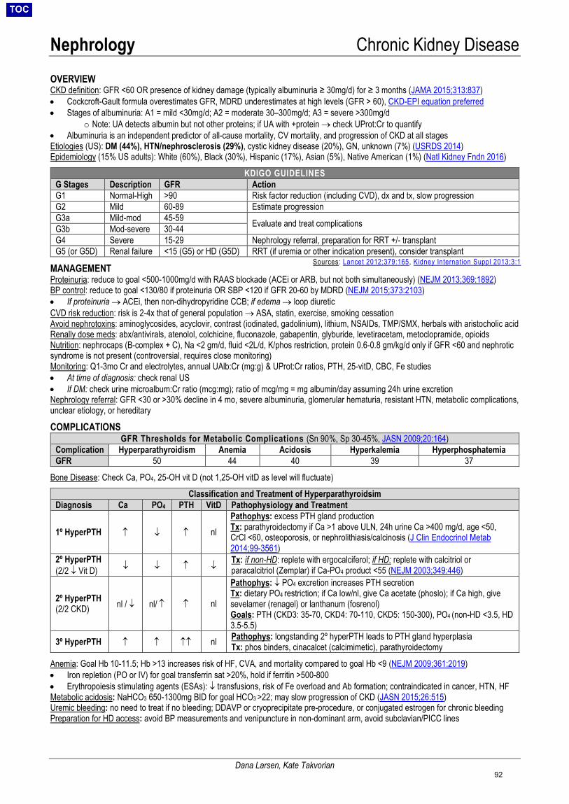
Nephrology Chronic Kidney Disease
Dana Larsen, Kate Takvorian
OVERVIEW CKD definition: GFR <60 OR presence of kidney damage (typically albuminuria ≥ 30mg/d) for ≥ 3 months (JAMA 2015;313:837)• Cockcroft-Gault formula overestimates GFR, MDRD underestimates at high levels (GFR > 60), CKD-EPI equation preferred• Stages of albuminuria: A1 = mild <30mg/d; A2 = moderate 30–300mg/d; A3 = severe >300mg/d
o Note: UA detects albumin but not other proteins; if UA with +protein → check UProt:Cr to quantify• Albuminuria is an independent predictor of all-cause mortality, CV mortality, and progression of CKD at all stagesEtiologies (US): DM (44%), HTN/nephrosclerosis (29%), cystic kidney disease (20%), GN, unknown (7%) (USRDS 2014)Epidemiology (15% US adults): White (60%), Black (30%), Hispanic (17%), Asian (5%), Native American (1%) (Natl Kidney Fndn 2016)
KDIGO GUIDELINES G Stages Description GFR Action G1 Normal-High >90 Risk factor reduction (including CVD), dx and tx, slow progression G2 Mild 60-89 Estimate progression G3a Mild-mod 45-59 Evaluate and treat complications G3b Mod-severe 30-44G4 Severe 15-29 Nephrology referral, preparation for RRT +/- transplant G5 (or G5D) Renal failure <15 (G5) or HD (G5D) RRT (if uremia or other indication present), consider transplant
MANAGEMENT Proteinuria: reduce to goal <500-1000mg/d with RAAS blockade (ACEi or ARB, but not both simultaneously) (NEJM 2013;369:1892)BP control: reduce to goal <130/80 if proteinuria OR SBP <120 if GFR 20-60 by MDRD (NEJM 2015;373:2103) • If proteinuria → ACEi, then non-dihydropyridine CCB; if edema → loop diureticCVD risk reduction: risk is 2-4x that of general population → ASA, statin, exercise, smoking cessationAvoid nephrotoxins: aminoglycosides, acyclovir, contrast (iodinated, gadolinium), lithium, NSAIDs, TMP/SMX, herbals with aristocholic acidRenally dose meds: abx/antivirals, atenolol, colchicine, fluconazole, gabapentin, glyburide, levetiracetam, metoclopramide, opioidsNutrition: nephrocaps (B-complex + C), Na <2 gm/d, fluid <2L/d, K/phos restriction, protein 0.6-0.8 gm/kg/d only if GFR <60 and nephroticsyndrome is not present (controversial, requires close monitoring)Monitoring: Q1-3mo Cr and electrolytes, annual UAlb:Cr (mg:g) & UProt:Cr ratios, PTH, 25-vitD, CBC, Fe studies• At time of diagnosis: check renal US• If DM: check urine microalbum:Cr ratio (mcg:mg); ratio of mcg/mg = mg albumin/day assuming 24h urine excretionNephrology referral: GFR <30 or >30% decline in 4 mo, severe albuminuria, glomerular hematuria, resistant HTN, metabolic complications,unclear etiology, or hereditary
COMPLICATIONS GFR Thresholds for Metabolic Complications (Sn 90%, Sp 30-45%, JASN 2009;20:164)
Complication Hyperparathyroidism Anemia Acidosis Hyperkalemia Hyperphosphatemia GFR 50 44 40 39 37
Bone Disease: Check Ca, PO4, 25-OH vit D (not 1,25-OH vitD as level will fluctuate)
Classification and Treatment of Hyperparathyroidsim Diagnosis Ca PO4 PTH VitD Pathophysiology and Treatment
1º HyperPTH ↑ ↓ ↑ nl Pathophys: excess PTH gland production Tx: parathyroidectomy if Ca >1 above ULN, 24h urine Ca >400 mg/d, age <50, CrCl <60, osteoporosis, or nephrolithiasis/calcinosis (J Clin Endocrinol Metab 2014;99-3561)
2º HyperPTH (2/2 ↓ Vit D) ↓ ↓ ↑ ↓ Tx: if non-HD: replete with ergocalciferol; if HD: replete with calcitriol or
paracalcitriol (Zemplar) if Ca-PO4 product <55 (NEJM 2003;349:446)
2º HyperPTH (2/2 CKD) nl / ↓ nl/ ↑ ↑ nl
Pathophys: ↓ PO4 excretion increases PTH secretion Tx: dietary PO4 restriction; if Ca low/nl, give Ca acetate (phoslo); if Ca high, give sevelamer (renagel) or lanthanum (fosrenol) Goals: PTH (CKD3: 35-70, CKD4: 70-110, CKD5: 150-300), PO4 (non-HD <3.5, HD 3.5-5.5)
3º HyperPTH ↑ ↑ ↑↑ nl Pathophys: longstanding 2º hyperPTH leads to PTH gland hyperplasia Tx: phos binders, cinacalcet (calcimimetic), parathyroidectomy
Anemia: Goal Hb 10-11.5; Hb >13 increases risk of HF, CVA, and mortality compared to goal Hb <9 (NEJM 2009;361:2019)• Iron repletion (PO or IV) for goal transferrin sat >20%, hold if ferritin >500-800• Erythropoiesis stimulating agents (ESAs): ↓ transfusions, risk of Fe overload and Ab formation; contraindicated in cancer, HTN, HFMetabolic acidosis: NaHCO3 650-1300mg BID for goal HCO3 >22; may slow progression of CKD (JASN 2015;26:515)Uremic bleeding: no need to treat if no bleeding; DDAVP or cryoprecipitate pre-procedure, or conjugated estrogen for chronic bleedingPreparation for HD access: avoid BP measurements and venipuncture in non-dominant arm, avoid subclavian/PICC lines
Sources: Lancet 2012;379:165 , Kidney Internation Suppl 2013;3:1
92
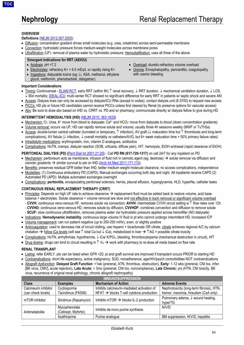
Nephrology Renal Replacement Therapy
Elizabeth Kurtz
OVERVIEW Definitions (NEJM 2012;367:2505) • Diffusion: concentration gradient drives small molecules (e.g. urea, creatinine) across semi-permeable membrane • Convection: hydrostatic pressure forces medium-weight molecules across membrane pores • Ultrafiltration (UF): removal of plasma water by hydrostatic pressure; Hemodiafiltration: uses all three of the above
Emergent Indications for RRT (AEIOU) • Acidosis: pH <7.2 • Electrolytes: refractory K+ > 6.0 mEq/L or rapidly rising K+ • Ingestions: dialyzable toxins (eg: Li, ASA, methanol, ethylene
glycol, metformin, phenobarbital, dabigatran)
• Overload: diuretic-refractory volume overload • Uremia: Encephalopathy, pericarditis, coagulopathy
with uremic bleeding
Important Considerations • Timing: Controversial - ELAIN RCT: early RRT (within 8h) ↑ renal recovery, ↓ RRT duration, ↓ mechanical ventilation duration, ↓ LOS, ↓ 90d mortality; IDEAL-ICU: multi-center RCT showed no significant difference for early RRT in patients w/ septic shock and severe AKI
• Access: Dialysis lines can only be accessed by dialysis/ICU RNs (except in codes); contact dialysis unit (6-3700) to request new access • PICCs: HD pts or future HD candidates cannot receive PICCs unless first cleared by Renal (to preserve options for vascular access) • Abx: Be sure to dose abx based on IHD vs. CRRT vs. PD and w/ pharmacy; communicate directly w/ dialysis fellow to give during HD
INTERMITTENT HEMODIALYSIS (IHD) (NEJM 2010; 363:1833) • Mechanism: Cr, Urea, K+ move from blood to dialysate; Ca2+ and HCO3- move from dialysate to blood (down concentration gradients) • Volume removal: occurs via UF; HD can rapidly remove solute and volume; usually three 4h sessions weekly (MWF or TuThSa) • Access: double-lumen central catheter (tunneled or temporary, ↑ infection); AV graft (↓ maturation time but ↑ thrombosis and long-term
complications); AV fistula (↓ infection, ↓ overall mortality vs catheters/AVG, but 6+ week maturation time + 50% primary failure rates) • Intradialytic medications: erythropoietin, iron, vitamin D analogues, antibiotics • Complications: HoTN, cramps, dialyzer reaction (SOB, urticaria, diffuse pain), HIT, hemolysis, EtOH w/drawal (rapid clearance of EtOH)
PERITONEAL DIALYSIS (PD) (Perit Dial Int 2001;21:25) - Call PD RN (617-720-1317) on call 24/7 for any inpatient on PD • Mechanism: peritoneum acts as membrane; infusion of fluid rich in osmotic agent (eg: dextrose) solute removal via diffusion and
osmotic gradients similar survival to pts on IHD (Arch Int Med 2011;171:110). • Benefits: preserves residual GFR better than IHD, better medium weight molecule clearance, no access complications, independence • Modalities: (1) Continuous ambulatory PD (CAPD): Manual exchanges occurring both day and night. All inpatients receive CAPD (2)
Automated PD (APD): Multiple automated exchanges overnight • Complications: peritonitis, encapsulating peritoneal sclerosis, hernia, pleural effusion, hyperglycemia, HLD, hyperNa, catheter leaks
CONTINUOUS RENAL REPLACEMENT THERAPY (CRRT) • Principles: Depends on high UF rate to achieve clearance replacement fluid must be added back to restore volume, acid base
balance + electrolytes. Solute clearance + volume removal are slow and not effective in toxin removal or significant volume overload - CVVH: continuous veno-venous HF, removes solute via convection; AVVH: intermediate CVVH circuit setting w ↑ flow rates over 12h - CVVHD: continuous veno-venous HD, removes solute by diffusion; CVVHDF: combines convection and diffusion to remove solute - SCUF: slow continuous ultrafiltration, removes plasma water via hydrostatic pressure applied across hemofilter (NO dialysate)
• Indications: Hemodynamic instability; continuous large volume IV fluid in pt who cannot undergo intermittent HD; increased ICP • Volume management: can run patient negative (up to 200-250 ml/hr), even, or slightly positive • Anticoagulation: used to decrease risk of circuit clotting, use heparin + bicarbonate OR citrate, citrate achieves regional A/C by calcium
chelation follow iCa levels (will see ↑ total Ca but ↓ iCa), metabolized in liver ↑AG = possible citrate toxicity • Complications: HoTN, arrhythmias, hypothermia, ↓ iCa/ K/PO4, bleeding, thrombocytopenia (mechanical destruction in circuit), HIT • Drug dosing: drugs can bind to circuit resulting in ↑ VD work with pharmacy to re-dose all meds based on flow rate
RENAL TRANSPLANT • Listing: refer EARLY, pts can be listed when GFR <20; pt and graft survival are improved if transplant occurs PRIOR to starting HD • Contraindications: short life expectancy, active malignancy, SUD, nonadherence; age/HIV/psych comorbidities NOT contraindications • Allograft dysfunction: Delayed Graft Function: <1wk (prerenal, ATN, thrombus, obstruction), Early: 1-12 wks (prerenal, CNI tox, infxn
[BK virus, CMV], acute rejection), Late Acute: > 3mo (prerenal, CNI tox, noncompliance), Late Chronic: yrs (HTN, CNI toxicity, BK virus, recurrence of original renal pathology, chronic allograft nephropathy)
IMMUNOSUPPRESSION Class Examples Mechanism of Action Adverse Events Calcineurin inhibitor (can check levels)
Cyclosporine Tacrolimus (FK506)
Inhibits calcineurin-mediated activation of NFAT blocks T-cell cytokine production
Nephrotoxicity (long-term fibrosis), HTN, tremor, insomnia, hirsutism (CsA only)
mTOR inhibitor Sirolimus (Rapamycin) Inhibits mTOR blocks IL-2 production Pulmonary edema, ↓ wound healing, hyperTG
Antimetabolite Mycophenolate (Cellcept, Myfortic) Inhibits de-novo purine synthesis N/V/D
Azathioprine Purine analogue BM suppression, N/V/D, hepatitis
93
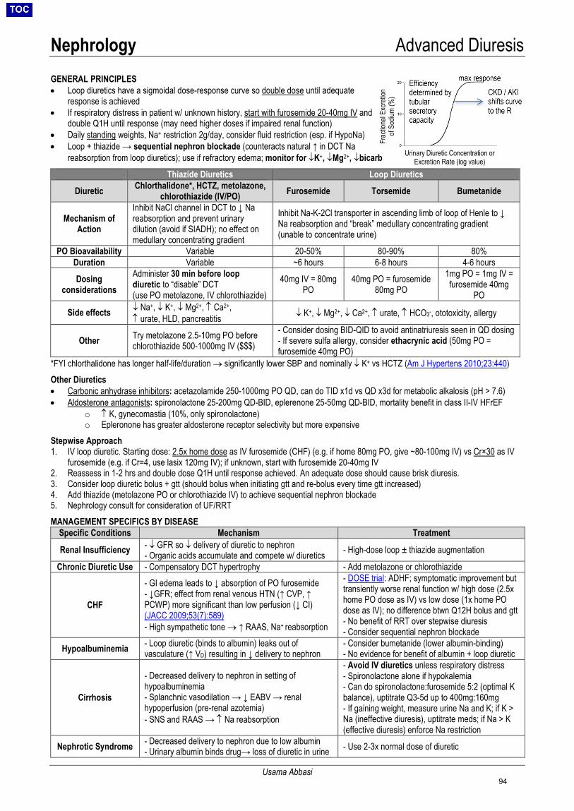
Nephrology Advanced Diuresis
Usama Abbasi
Frac
tiona
l Exc
retio
n of
Sodiu
m (%
)
Urinary Diuretic Concentration or Excretion Rate (log value)
GENERAL PRINCIPLES • Loop diuretics have a sigmoidal dose-response curve so double dose until adequate
response is achieved• If respiratory distress in patient w/ unknown history, start with furosemide 20-40mg IV and
double Q1H until response (may need higher doses if impaired renal function)• Daily standing weights, Na+ restriction 2g/day, consider fluid restriction (esp. if HypoNa)• Loop + thiazide → sequential nephron blockade (counteracts natural ↑ in DCT Na
reabsorption from loop diuretics); use if refractory edema; monitor for ↓K+, ↓Mg2+, ↓bicarb
Thiazide Diuretics Loop Diuretics
Diuretic Chlorthalidone*, HCTZ, metolazone, chlorothiazide (IV/PO) Furosemide Torsemide Bumetanide
Mechanism of Action
Inhibit NaCl channel in DCT to ↓ Na reabsorption and prevent urinary dilution (avoid if SIADH); no effect on medullary concentrating gradient
Inhibit Na-K-2Cl transporter in ascending limb of loop of Henle to ↓ Na reabsorption and “break” medullary concentrating gradient (unable to concentrate urine)
PO Bioavailability Variable 20-50% 80-90% 80% Duration Variable ~6 hours 6-8 hours 4-6 hours
Dosing considerations
Administer 30 min before loop diuretic to “disable” DCT (use PO metolazone, IV chlorothiazide)
40mg IV = 80mg PO
40mg PO = furosemide 80mg PO
1mg PO = 1mg IV = furosemide 40mg
PO
Side effects ↓ Na+, ↓ K+, ↓ Mg2+, ↑ Ca2+, ↑ urate, HLD, pancreatitis ↓ K+, ↓ Mg2+, ↓ Ca2+, ↑ urate, ↑ HCO3-, ototoxicity, allergy
Other Try metolazone 2.5-10mg PO before chlorothiazide 500-1000mg IV ($$$)
- Consider dosing BID-QID to avoid antinatriuresis seen in QD dosing- If severe sulfa allergy, consider ethacrynic acid (50mg PO =furosemide 40mg PO)
*FYI chlorthalidone has longer half-life/duration → significantly lower SBP and nominally ↓ K+ vs HCTZ (Am J Hypertens 2010;23:440)Other Diuretics• Carbonic anhydrase inhibitors: acetazolamide 250-1000mg PO QD, can do TID x1d vs QD x3d for metabolic alkalosis (pH > 7.6)• Aldosterone antagonists: spironolactone 25-200mg QD-BID, eplerenone 25-50mg QD-BID, mortality benefit in class II-IV HFrEF
o ↑ K, gynecomastia (10%, only spironolactone)o Epleronone has greater aldosterone receptor selectivity but more expensive
Stepwise Approach 1. IV loop diuretic. Starting dose: 2.5x home dose as IV furosemide (CHF) (e.g. if home 80mg PO, give ~80-100mg IV) vs Cr×30 as IV
furosemide (e.g. if Cr=4, use lasix 120mg IV); if unknown, start with furosemide 20-40mg IV2. Reassess in 1-2 hrs and double dose Q1H until response achieved. An adequate dose should cause brisk diuresis.3. Consider loop diuretic bolus + gtt (should bolus when initiating gtt and re-bolus every time gtt increased)4. Add thiazide (metolazone PO or chlorothiazide IV) to achieve sequential nephron blockade5. Nephrology consult for consideration of UF/RRTMANAGEMENT SPECIFICS BY DISEASE
Specific Conditions Mechanism Treatment
Renal Insufficiency - ↓ GFR so ↓ delivery of diuretic to nephron- Organic acids accumulate and compete w/ diuretics - High-dose loop ± thiazide augmentation
Chronic Diuretic Use - Compensatory DCT hypertrophy - Add metolazone or chlorothiazide
CHF
- GI edema leads to ↓ absorption of PO furosemide- ↓GFR; effect from renal venous HTN (↑ CVP, ↑PCWP) more significant than low perfusion (↓ CI)(JACC 2009;53(7):589)- High sympathetic tone → ↑ RAAS, Na+ reabsorption
- DOSE trial: ADHF; symptomatic improvement buttransiently worse renal function w/ high dose (2.5xhome PO dose as IV) vs low dose (1x home POdose as IV); no difference btwn Q12H bolus and gtt- No benefit of RRT over stepwise diuresis- Consider sequential nephron blockade
Hypoalbuminemia - Loop diuretic (binds to albumin) leaks out ofvasculature (↑ VD) resulting in ↓ delivery to nephron
- Consider bumetanide (lower albumin-binding)- No evidence for benefit of albumin + loop diuretic
Cirrhosis
- Decreased delivery to nephron in setting ofhypoalbuminemia- Splanchnic vasodilation → ↓ EABV → renalhypoperfusion (pre-renal azotemia)- SNS and RAAS → ↑ Na reabsorption
- Avoid IV diuretics unless respiratory distress- Spironolactone alone if hypokalemia- Can do spironolactone:furosemide 5:2 (optimal Kbalance), uptitrate Q3-5d up to 400mg:160mg- If gaining weight, measure urine Na and K; if K >Na (ineffective diuresis), uptitrate meds; if Na > K(effective diuresis) enforce Na restriction
Nephrotic Syndrome - Decreased delivery to nephron due to low albumin- Urinary albumin binds drug→ loss of diuretic in urine - Use 2-3x normal dose of diuretic
94
Nephrology Acid-Base Disorders
Alexander Blair, Sarah Street, Elizabeth Kurtz
TREATMENT OF ACID-BASE DISORDERS: treat underlying cause Metabolic acidosis: • Severity of metabolic acidosis reflects disease severity but does not contribute to mortality (Sci World J 2014;2014:627673)• On giving bicarb: BICAR-ICU multicenter RCT of patients with metabolic acidosis (pH < 7.2) treated with 4.2% sodium HCO3 for goal
pH > 7.3 → general population had no change in overall mortality, but ↓ RRT initiation. A subset of pts with AKIN stages 2-3 hadimproved mortality at 28-days (63% v. 46%, p=0.017) (Lancet 2018;392:31)
• When HCO3 loss is primary cause: (i.e., RTA, diarrhea), can replace by administration of NaHCO3o If pH < 7.2 or HCO3- < 6, administer 1-2 mEq/kg as IV bolus → re-dose prn targeting pHo Caveats: HCO3 generates CO2 and provides Na load → hypercarbia, hypernatremia, hypocalcemia, hypertonicity,
hypervolemia, overshoot alkalosis• Methanol or ethylene glycol intoxication: oral charcoal, HCO3-, fomepizole, or HD (if level >50 mg/dL, vision Δ, AKI)• Salicylate poisoning: NaHCO3 to urine pH >6.5 or HD (if level > 80 mg/dL, coma, AKI, hypervolemia)• Consider HD or CVVH in patients with volume overload, catabolism, ethylene glycol >300 mg/dL, isopropanol >500 mg/dLMetabolic alkalosis: replete volume, K, and Cl• Treat both (1) underlying cause of metabolic alkalosis and (2) cause of renal retention of HCO3-
• If saline responsive: NS w/ KCl until urine pH >7. For patients w/ CHF/cirrhosis and alkalosis 2/2 diuresis, consider K+ sparing diuretic• If saline resistant: For mineralocorticoid excess use K-sparing diuretic (amiloride) and consider surgical removal of adenoma• If pH > 7.6 and persistent volume overload, give acetazolamide vs. KCl + loop diuretic with close K+ monitoringRespiratory acidosis:• NaHCO3 unlikely to be helpful, theoretically harmful if unable to blow off CO2 produced by conservation of mass (CO2 + H2O
H2CO3 HCO3 + H+ ); for every 100mEq HCO3 administered, 2.2 L CO2 must be exhaled (~10 min of normal body production)Respiratory alkalosis: Address underlying cause (correct hypoxemia, treat pain/anxiety/fever); adjust vent settings if intubated
RENAL TUBULAR ACIDOSIS (RTA) (Int J Clin Pract 2011;65:350; JASN 2002;13:2160, JASN 2009;20;251) Consider in any patient with non-AG metabolic acidosis or hyperK (Type IV). Pathophysiology: inappropriate net retention of acid or inadequate excretion of bicarb • In acidemia, kidney should ↑ NH4+ excretion; urine pH should be < 5.3; this process is defective in RTAs• Caveat: CKD of any etiology is associated with ↓ NH4+ production and acidosisEtiologies:Distal RTA (Type I): ↓ distal acidification• Primary: genetic loss of H+ or HCO3 transporters (basolateral Cl/HCO3 exchanger or luminal H+-ATPase in intercalated cells)• Acquired: Autoimmune dz (RA, SLE, SS); hypercalciuria (any cause); obstructive nephropathy; SCD, MM, amyloid,
cryoglobulinemia, tubulointerstitial injury, renal txp rejection, cirrhosis, glue sniffing (toluene)• Meds: Amphotericin B, Li+, ifosfamideProximal RTA (Type II): ↓ proximal reabsorption HCO3-
• Primary (rare): Na-HCO3 cotransporter defect• Acquired: Amyloidosis, multiple myeloma, post-renal transplant, heavy metals (Pb, Cd, Hg, Cu), ↓Vit D, Wilson’s disease, PNH• Meds: acetazolamide, cisplatin, tenofovir, aminoglycocides, topiramate• Often a/w Fanconi Syndrome: glycosuria (w/ serum gluc <180), hypouricemia, aminoaciduriaType IV: effective hypoaldosteronism: ↓ aldo secretion OR tubular resistance → ↑ K → ↓ NH3 synthesis → ↓ NH4+ excretion• Acidosis to inhibition of ammonia-genesis by hyperkalemia of any cause• Hyporeninemic hypoaldosteronism: diabetic nephropathy, chronic interstitial nephritis, NSAIDs, calcineurin inhibitor, HIV• ↓ Aldo production: adrenal insufficiency, ACEi/ARB, heparin, severe illness• Aldosterone resistance: (ENaC inhibition) K-sparing diuretic, trimethoprim, pentamidineWorkup:• Serum HCO3 and K, urinary pH, fractional excretion of HCO3 (ideally, check urine NH4+ but most labs will not do this)• Estimates of Urine NH4+: UAG = Na + K - Cl (not useful in when ↑urine anions or UNa < 25); UOG/2 (<150 RTA, >400 GI loss)
DISTAL RTA (TYPE I) PROXIMAL RTA (TYPE II) TYPE IV RTA Defect Distal H+ secretion Proximal HCO3- resorption Hyperkalemia Serum HCO3- < 10 12 – 20 > 17Plasma K ↓ or normal ↓ or normal ↑ Urine pH during acidemia > 5.5 Varies, but > 5.5 after HCO3- < 5.5 FE-HCO3 after loading < 3% > 15% (diagnostic) < 3% UAG = Na + K - CL (+) Can be (-) (+) Additional dx testing N/A N/A Renin, aldosterone, cortisol Complications Nephrocalcinosis/stones Rickets or osteomalacia Hyperkalemia
Tx (goal HCO3 22-24) NaHCO3 (1-4 mEq/kg) K or Na citrate if persistent ↓K NaHCO3 (10-20 mEq/kg) Treat hyperk: loop, low K diet
If hypoaldo then can give fludrocort
95

Nephrology Acid-Base Disorders
Alexander Blair, Sarah Street, Elizabeth Kurtz
DEFINITIONS ABG vs VBG: pH (~0.04), HCO3 (~2 mEq) but NOT pCO2 (~8 ± 17 mmHg); VBG can screen for hypercarbia w/ pCO2 cutoff ≥ 45 mmHg (100% Sn), but does NOT accurately assess degree of hypercarbia; when in doubt → check ABG (AJEM 2012;30:896)• Severe acidemia (pH < 7.2) → vasodilation, ↓ inotropy / SVR / MAP, ↓ response catechols, arrhythmia, ↑K, insulin resistance, AMS• Severe alkalemia (pH >7.6) → vasoconstriction, ↓ cor/cerebral perfusion, SVT/VT, ↓ K/Ca/Mg/P, AMS, seizure, hypoventilation
STEP-WISE APPROACH (NEJM 1998;338:26, NEJM 2014;371:1434) 1. Is there acidemia (pH < 7.36) or alkalemia (pH > 7.44)?2. Is primary d/o metabolic (parallels pH ∆) or respiratory (opposite pH ∆)?3. Is pt compensating? (respiratory takes min-hrs, renal 3-5 days)4. Is there an anion gap? Regardless of pH or HCO3 AG = Na – (Cl + HCO3) = unmeasured anions – unmeasured cations
Correct AG for albumin: AG = 2.5 x (4 – Albumin) Negative AG: ↑↑Na, lipids (interfere w/ chloride), bromide intoxication
5. If there is ↑ AG, calculate “delta-delta” and Osm gap∆/∆ = ∆ AG /∆ HCO3 = AG – (albumin x 2.5) / (24 – HCO3)
6. Consider Osm gap = 2x (Na + K) + Urea/2.8 + glucose/18 – serum Osm
ALGORITHMIC APPROACH
EXPECTED COMPENSATION (JASN 2010;21:920)Metabolic acidosis: 2-24 hr Winter’s formula: pCO2 = 1.5 x HCO3 + 8 ± 2 Metabolic alkalosis: start 30 min, complete 24 hrs PaCO2 = 0.7 x (HCO3 -24) + 40 ± 2 = HCO3 + 15 ∆ HCO3 ↑ 1 → expect ∆ pCO2 ↑ 0.7 Respiratory acidosis: Acute: ∆ pCO2 ↑ 10 → ∆ HCO3 ↑ 1 or ↓ pH 0.08 Chronic: ∆ pCO2 ↑ 10 → ∆ HCO3 ↑ 4 or ↓ pH 0.03 Respiratory alkalosis: Acute: ∆ pCO2 ↓10 → ∆ HCO3 ↓ 2 or ↑pH 0.08 Chronic: ∆ pCO2 ↓10 → ∆ HCO3 ↓ 4 or ↑pH 0.03
AG OG Ingestions Toxin Manifestations
↑ ↑
Methanol Formic acid ∆MS, blurry vision, pupil dilation, papilledema
Ethylene glycol Oxalic acid ∆MS, ↓Ca, Ca oxalate crystals → AKI Propylene glycol Lactic acid AKI, liver injury
Diethylene glycol Diglycolic acid AKI, n/v, pancreatitis, neuropathy, ↑ lactate
nl/↑ ↑ Isopropyl alcohol Acetone ∆MS, fruity breath, pancreatitis, ↑ lactate Ethanol Acetaldehyde Keto/lactic acidosis ± met. alk 2/2 emesis
lumen blood
96
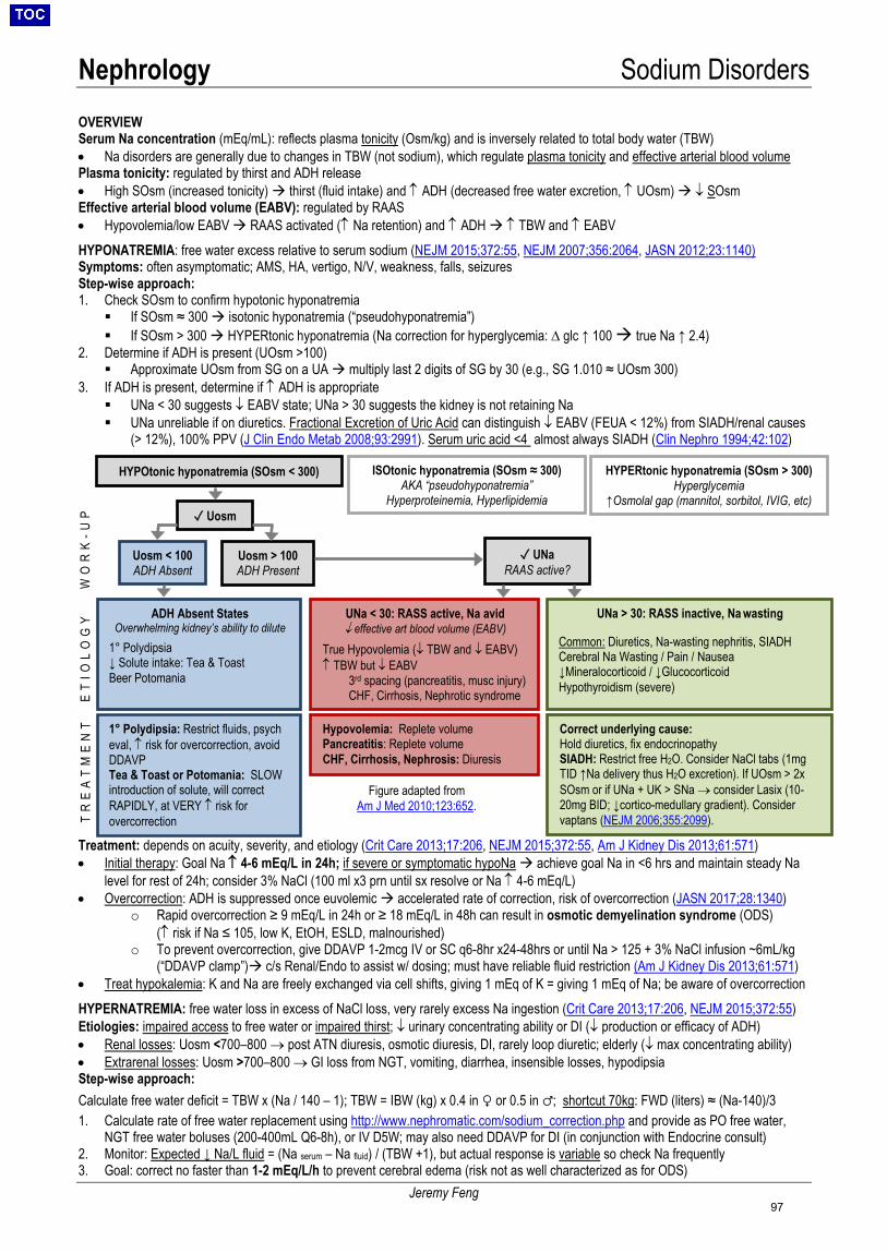
Nephrology Sodium Disorders
Jeremy Feng
OVERVIEW Serum Na concentration (mEq/mL): reflects plasma tonicity (Osm/kg) and is inversely related to total body water (TBW) • Na disorders are generally due to changes in TBW (not sodium), which regulate plasma tonicity and effective arterial blood volumePlasma tonicity: regulated by thirst and ADH release• High SOsm (increased tonicity) thirst (fluid intake) and ↑ ADH (decreased free water excretion, ↑ UOsm) ↓ SOsmEffective arterial blood volume (EABV): regulated by RAAS• Hypovolemia/low EABV RAAS activated (↑ Na retention) and ↑ ADH ↑ TBW and ↑ EABVHYPONATREMIA: free water excess relative to serum sodium (NEJM 2015;372:55, NEJM 2007;356:2064, JASN 2012;23:1140) Symptoms: often asymptomatic; AMS, HA, vertigo, N/V, weakness, falls, seizures Step-wise approach: 1. Check SOsm to confirm hypotonic hyponatremia If SOsm ≈ 300 isotonic hyponatremia (“pseudohyponatremia”) If SOsm > 300 HYPERtonic hyponatremia (Na correction for hyperglycemia: ∆ glc ↑ 100 true Na ↑ 2.4)
2. Determine if ADH is present (UOsm >100) Approximate UOsm from SG on a UA multiply last 2 digits of SG by 30 (e.g., SG 1.010 ≈ UOsm 300)
3. If ADH is present, determine if ↑ ADH is appropriate UNa < 30 suggests ↓ EABV state; UNa > 30 suggests the kidney is not retaining Na UNa unreliable if on diuretics. Fractional Excretion of Uric Acid can distinguish ↓ EABV (FEUA < 12%) from SIADH/renal causes
(> 12%), 100% PPV (J Clin Endo Metab 2008;93:2991). Serum uric acid <4 almost always SIADH (Clin Nephro 1994;42:102)
Treatment: depends on acuity, severity, and etiology (Crit Care 2013;17:206, NEJM 2015;372:55, Am J Kidney Dis 2013;61:571)• Initial therapy: Goal Na ↑ 4-6 mEq/L in 24h; if severe or symptomatic hypoNa achieve goal Na in <6 hrs and maintain steady Na
level for rest of 24h; consider 3% NaCl (100 ml x3 prn until sx resolve or Na ↑ 4-6 mEq/L)• Overcorrection: ADH is suppressed once euvolemic accelerated rate of correction, risk of overcorrection (JASN 2017;28:1340)
o Rapid overcorrection ≥ 9 mEq/L in 24h or ≥ 18 mEq/L in 48h can result in osmotic demyelination syndrome (ODS)(↑ risk if Na ≤ 105, low K, EtOH, ESLD, malnourished)
o To prevent overcorrection, give DDAVP 1-2mcg IV or SC q6-8hr x24-48hrs or until Na > 125 + 3% NaCl infusion ~6mL/kg(“DDAVP clamp”) c/s Renal/Endo to assist w/ dosing; must have reliable fluid restriction (Am J Kidney Dis 2013;61:571)
• Treat hypokalemia: K and Na are freely exchanged via cell shifts, giving 1 mEq of K = giving 1 mEq of Na; be aware of overcorrectionHYPERNATREMIA: free water loss in excess of NaCl loss, very rarely excess Na ingestion (Crit Care 2013;17:206, NEJM 2015;372:55)Etiologies: impaired access to free water or impaired thirst; ↓ urinary concentrating ability or DI (↓ production or efficacy of ADH) • Renal losses: Uosm <700–800 → post ATN diuresis, osmotic diuresis, DI, rarely loop diuretic; elderly (↓ max concentrating ability)• Extrarenal losses: Uosm >700–800 → GI loss from NGT, vomiting, diarrhea, insensible losses, hypodipsiaStep-wise approach:Calculate free water deficit = TBW x (Na / 140 – 1); TBW = IBW (kg) x 0.4 in ♀ or 0.5 in ♂; shortcut 70kg: FWD (liters) ≈ (Na-140)/3 1. Calculate rate of free water replacement using http://www.nephromatic.com/sodium_correction.php and provide as PO free water,
NGT free water boluses (200-400mL Q6-8h), or IV D5W; may also need DDAVP for DI (in conjunction with Endocrine consult)2. Monitor: Expected ↓ Na/L fluid = (Na serum – Na fluid) / (TBW +1), but actual response is variable so check Na frequently3. Goal: correct no faster than 1-2 mEq/L/h to prevent cerebral edema (risk not as well characterized as for ODS)
HYPOtonic hyponatremia (SOsm < 300) ISOtonic hyponatremia (SOsm ≈ 300) AKA “pseudohyponatremia”
Hyperproteinemia, Hyperlipidemia
HYPERtonic hyponatremia (SOsm > 300) Hyperglycemia
↑Osmolal gap (mannitol, sorbitol, IVIG, etc)
ET
IOL
OG
Y
✓ Uosm
Uosm < 100 ADH Absent
Uosm > 100 ADH Present
ADH Absent States Overwhelming kidney’s ability to dilute
1° Polydipsia ↓ Solute intake: Tea & Toast Beer Potomania
UNa < 30: RASS active, Na avid ↓ effective art blood volume (EABV)
True Hypovolemia (↓ TBW and ↓ EABV) ↑ TBW but ↓ EABV
3rd spacing (pancreatitis, musc injury) CHF, Cirrhosis, Nephrotic syndrome
UNa > 30: RASS inactive, Na wasting
Common: Diuretics, Na-wasting nephritis, SIADH Cerebral Na Wasting / Pain / Nausea ↓Mineralocorticoid / ↓Glucocorticoid Hypothyroidism (severe)
✓ UNaRAAS active?
WO
RK
-UP
1° Polydipsia: Restrict fluids, psych eval, ↑ risk for overcorrection, avoid DDAVP Tea & Toast or Potomania: SLOW introduction of solute, will correct RAPIDLY, at VERY ↑ risk for overcorrection
Hypovolemia: Replete volume Pancreatitis: Replete volume CHF, Cirrhosis, Nephrosis: Diuresis
Correct underlying cause: Hold diuretics, fix endocrinopathy SIADH: Restrict free H2O. Consider NaCl tabs (1mg TID ↑Na delivery thus H2O excretion). If UOsm > 2x SOsm or if UNa + UK > SNa → consider Lasix (10-20mg BID; ↓cortico-medullary gradient). Consider vaptans (NEJM 2006;355:2099). T
RE
AT
ME
NT
Figure adapted from Am J Med 2010;123:652.
97
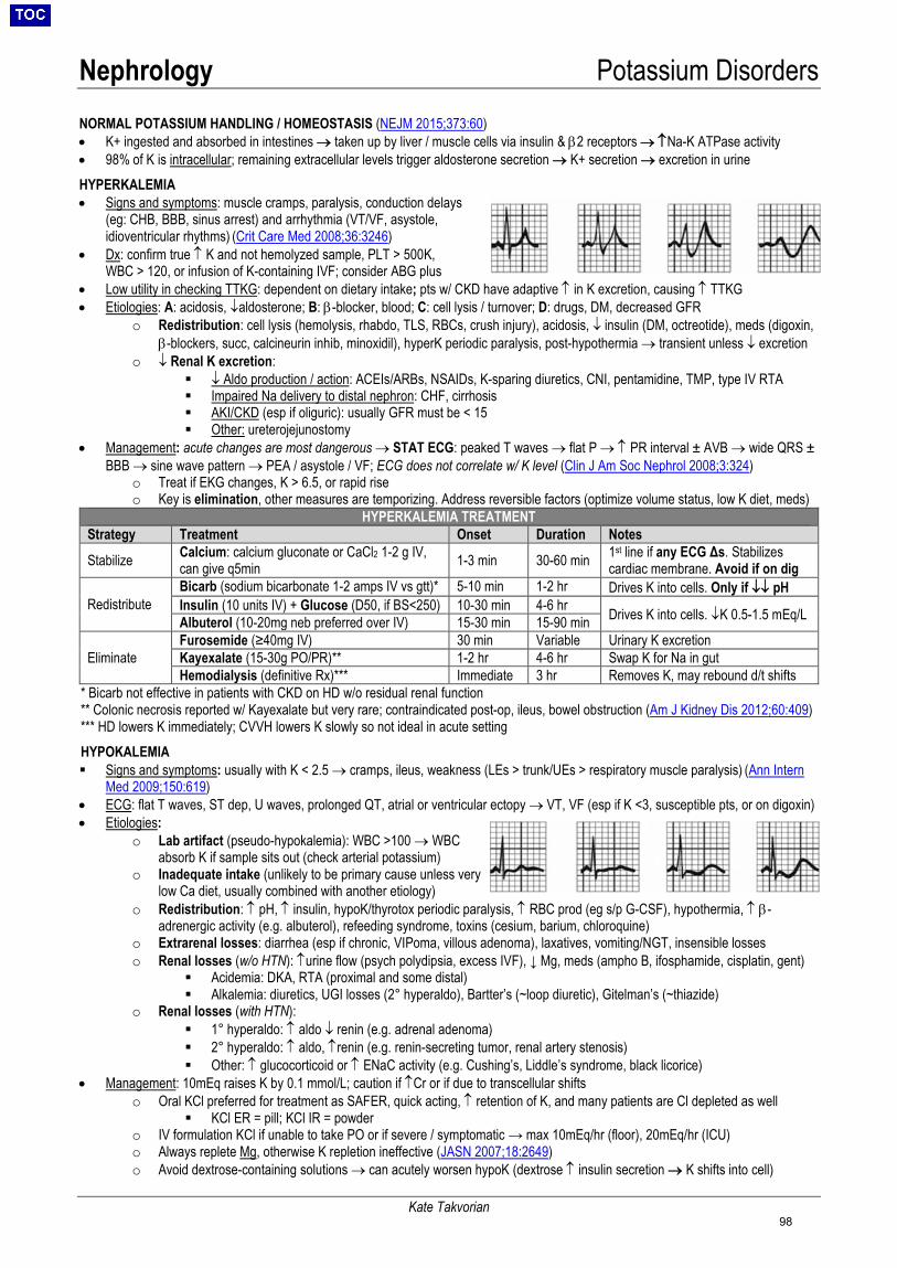
Nephrology Potassium Disorders
Kate Takvorian
NORMAL POTASSIUM HANDLING / HOMEOSTASIS (NEJM 2015;373:60) • K+ ingested and absorbed in intestines → taken up by liver / muscle cells via insulin & β2 receptors → ↑Na-K ATPase activity• 98% of K is intracellular; remaining extracellular levels trigger aldosterone secretion → K+ secretion → excretion in urineHYPERKALEMIA• Signs and symptoms: muscle cramps, paralysis, conduction delays
(eg: CHB, BBB, sinus arrest) and arrhythmia (VT/VF, asystole,idioventricular rhythms) (Crit Care Med 2008;36:3246)
• Dx: confirm true ↑ K and not hemolyzed sample, PLT > 500K,WBC > 120, or infusion of K-containing IVF; consider ABG plus
• Low utility in checking TTKG: dependent on dietary intake; pts w/ CKD have adaptive ↑ in K excretion, causing ↑ TTKG• Etiologies: A: acidosis, ↓aldosterone; B: β-blocker, blood; C: cell lysis / turnover; D: drugs, DM, decreased GFR
o Redistribution: cell lysis (hemolysis, rhabdo, TLS, RBCs, crush injury), acidosis, ↓ insulin (DM, octreotide), meds (digoxin,β-blockers, succ, calcineurin inhib, minoxidil), hyperK periodic paralysis, post-hypothermia → transient unless ↓ excretion
o ↓ Renal K excretion: ↓ Aldo production / action: ACEIs/ARBs, NSAIDs, K-sparing diuretics, CNI, pentamidine, TMP, type IV RTA Impaired Na delivery to distal nephron: CHF, cirrhosis AKI/CKD (esp if oliguric): usually GFR must be < 15 Other: ureterojejunostomy
• Management: acute changes are most dangerous → STAT ECG: peaked T waves → flat P → ↑ PR interval ± AVB → wide QRS ±BBB → sine wave pattern → PEA / asystole / VF; ECG does not correlate w/ K level (Clin J Am Soc Nephrol 2008;3:324)
o Treat if EKG changes, K > 6.5, or rapid riseo Key is elimination, other measures are temporizing. Address reversible factors (optimize volume status, low K diet, meds)
HYPERKALEMIA TREATMENT Strategy Treatment Onset Duration Notes
Stabilize Calcium: calcium gluconate or CaCl2 1-2 g IV, can give q5min 1-3 min 30-60 min 1st line if any ECG Δs. Stabilizes
cardiac membrane. Avoid if on dig
Redistribute Bicarb (sodium bicarbonate 1-2 amps IV vs gtt)* 5-10 min 1-2 hr Drives K into cells. Only if ↓↓ pH Insulin (10 units IV) + Glucose (D50, if BS<250) 10-30 min 4-6 hr Drives K into cells. ↓K 0.5-1.5 mEq/L Albuterol (10-20mg neb preferred over IV) 15-30 min 15-90 min
Eliminate Furosemide (≥40mg IV) 30 min Variable Urinary K excretion Kayexalate (15-30g PO/PR)** 1-2 hr 4-6 hr Swap K for Na in gut Hemodialysis (definitive Rx)*** Immediate 3 hr Removes K, may rebound d/t shifts
* Bicarb not effective in patients with CKD on HD w/o residual renal function** Colonic necrosis reported w/ Kayexalate but very rare; contraindicated post-op, ileus, bowel obstruction (Am J Kidney Dis 2012;60:409)*** HD lowers K immediately; CVVH lowers K slowly so not ideal in acute settingHYPOKALEMIA Signs and symptoms: usually with K < 2.5 → cramps, ileus, weakness (LEs > trunk/UEs > respiratory muscle paralysis) (Ann Intern
Med 2009;150:619) • ECG: flat T waves, ST dep, U waves, prolonged QT, atrial or ventricular ectopy → VT, VF (esp if K <3, susceptible pts, or on digoxin)• Etiologies:
o Lab artifact (pseudo-hypokalemia): WBC >100 → WBCabsorb K if sample sits out (check arterial potassium)
o Inadequate intake (unlikely to be primary cause unless verylow Ca diet, usually combined with another etiology)
o Redistribution: ↑ pH, ↑ insulin, hypoK/thyrotox periodic paralysis, ↑ RBC prod (eg s/p G-CSF), hypothermia, ↑ β-adrenergic activity (e.g. albuterol), refeeding syndrome, toxins (cesium, barium, chloroquine)
o Extrarenal losses: diarrhea (esp if chronic, VIPoma, villous adenoma), laxatives, vomiting/NGT, insensible losseso Renal losses (w/o HTN): ↑urine flow (psych polydipsia, excess IVF), ↓ Mg, meds (ampho B, ifosphamide, cisplatin, gent)
Acidemia: DKA, RTA (proximal and some distal) Alkalemia: diuretics, UGI losses (2° hyperaldo), Bartter’s (~loop diuretic), Gitelman’s (~thiazide)
o Renal losses (with HTN): 1° hyperaldo: ↑ aldo ↓ renin (e.g. adrenal adenoma) 2° hyperaldo: ↑ aldo, ↑renin (e.g. renin-secreting tumor, renal artery stenosis) Other: ↑ glucocorticoid or ↑ ENaC activity (e.g. Cushing’s, Liddle’s syndrome, black licorice)
• Management: 10mEq raises K by 0.1 mmol/L; caution if ↑Cr or if due to transcellular shiftso Oral KCl preferred for treatment as SAFER, quick acting, ↑ retention of K, and many patients are Cl depleted as well
KCl ER = pill; KCl IR = powdero IV formulation KCl if unable to take PO or if severe / symptomatic → max 10mEq/hr (floor), 20mEq/hr (ICU)o Always replete Mg, otherwise K repletion ineffective (JASN 2007;18:2649)o Avoid dextrose-containing solutions → can acutely worsen hypoK (dextrose ↑ insulin secretion → K shifts into cell)
98
Nephrology Magnesium & Phosphorus Disorders
Kate Takvorian
HYPOMAGNESEMIA • Signs/symptoms: other electrolyte disturbances (↓ K, ↓ Ca), weakness, anorexia, confusion, hyperreflexia, tetany,
↑ PR, ↑ QRS, ↑QTc, peaked / inverted T waves, U waves, VT / torsades, accentuation of digitalis toxicity• Etiologies:
o ↓ GI absorption: ↓ intake (EtOH, malnutrition), ↑ loss (diarrhea, pancreatitis, malabsorption,small bowel resection, PPIs)
o ↑ renal losses: thiazides, loops, amphoB, aminoglycosides, foscarnet, cyclosporine A, cisplatin, pentamidineo Can distinguish GI vs renal with 24hr urine Mg or FeMg (>10mg or >2% suggest renal wasting)
• Treatment: oral (very slow) vs. IV repletion (IV typically given inpatient)o MgSO4 1-2 gm IV over 15 min, max 1-2gm/h, up to 8gm in 24h
Give ½ dose if CrCl <30o Mg oxide 800-1600mg PO in divided doses (240mg Mg per 400 mg tab); limited by diarrheao If hypoMg due to thiazide or loop diuretic, add K-sparing diuretic to decrease Mg excretion
HYPERMAGNESEMIA (rarely pathologic) • Signs/symptoms (typically only if Mg >4): neuromuscular (hyporeflexia [first sign], areflexia, lethargy, weakness/paralysis,
resp failure), CV (hypotension, bradycardia, conduction defects [↑PR, ↑QRS, ↑QTc, CHB, cardiac arrest]), hypocalcemia(hyper Mg can suppress PTH)
• Etiologies: Mg intake > renal clearance (only method of excretion)o Medication overdose (Epsom salts, laxatives, Maalox, Mg enemas) → avoid these agents in ESRDo Increased Mg absorption with gastritis / PUD / colitiso Mild hyperMg may be seen in DKA, hypercatabolic states (TLS), lithium, adrenal insufficiency
• Treatment (symptomatic only): Ca gluconate 1 gm IV over 10 min vs gtt to counteract resp depression/hypotension.IVF, loop diuretics to enhance renal excretion. If oliguric/anuric ESRD, requires HD for removal.
HYPOPHOSPHATEMIA • Signs/symptoms (typically only if phos < 1.0mg/dL, esp if acute): ↓ intracellular ATP → AMS / encephalopathy, seizures,
CHF, hemolysis, respiratory depression, proximal myopathy, rhabdomyolysis, dysphagia / ileus, mineral ∆ (bone pain,hypercalciuria, rickets / osteomalacia) (JASN 2007;18:1999).
• Etiologies:o Redistribution (into cells): ↑insulin (DKA, HHNK, refeeding), acute respiratory alkalosis (↑pH→↑glycolysis),
hungry bone syndrome (deposition of Ca and phos in bone immediately following parathyroidectomy)o ↓ GI absorption: poor PO, chronic diarrhea, antacid use (aluminum, Mg), ↓ vit D (steatorrhea, chronic
diarrhea), overuse of phos binderso ↑ renal excretion: ↑ PTH (primary or secondary), Fanconi syndrome (multiple myeloma, meds), ↑ FGF-23
(genetic/paraneoplastic), meds (acetazolamide, tenofovir, metolazone, IV iron) (QJM 2010;103:449), osmoticdiuresis (glucosuria), proximally acting diuretics (acetazolamide, metolazone), CVVH (esp at high flow)
o Can determine if GI/redistribution vs renal with 24hr urine Phos or FePhos (>100mg or >5% → renal wasting)• Treatment:
o Severe (<1 mg/dL) or symptomatic: Na or K phos 0.08-0.50 mmol/kg IV over 6-8h (can give 15, 30, or 45mmoldoses at MGH); change to PO once >1.5mg/dL Give ½ dose in CKD/ESRD Aggressive IV tx can cause Ca precipitation, hypotension (often due to hypocalcemia), AKI, arrhythmia
o Asymptomatic (<2 mg/dL): Na or K phos 1mmol/kg/d PO in 3-4 divided doses (total 40-80mmol) NeutraPhos: 1 packet = 250mg Phos (8mmol), 7.1mEq K, & 6.9mEq Na; preferred if also need K or if
want lower Na K-Phos Neutral: 1 tablet = 250mg Phos (8mmol), 1.1mEq K, & 13 mEq Na; preferred if do not need K If poorly tolerated (causes diarrhea), can give scheduled skim milk (8oz = 8mmol Phos)
HYPERPHOSPHATEMIA • Signs/symptoms: acute hyperphosphatemia signs/symptoms result from effects of hypocalcemia (muscle cramps, tetany,
tingling, perioral numbness), acute phosphate nephropathy (bowel prep); rarely sx from chronic hyperphosphatemia• Etiologies:
o Acute phos load (TLS, rhabdo, exogenous/phosphate-containing laxatives); acute extracellular shift (DKA,lactic acidosis, severe hyperglycemia); acute or chronic kidney disease
o Increased tubular reabsorption (vit D tox, hypoPTH)o Pseudohyperphos (hyperglobulinemia, hyperlipidemia, hyperbili, hemolysis)
• Treatment: acute – normal saline (though can worsen hypoCa), dialysis; chronic – see Chronic Kidney Disease
99
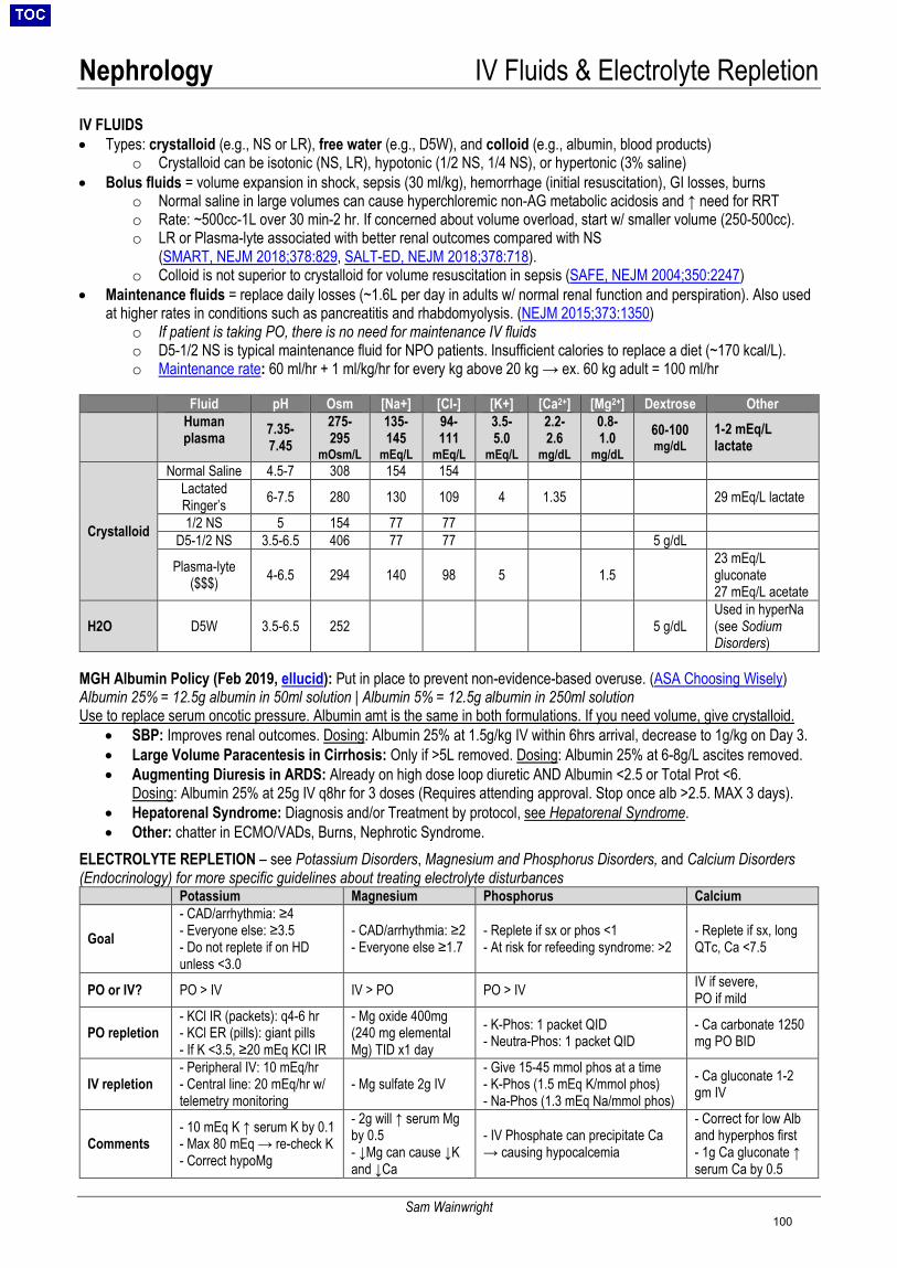
Nephrology IV Fluids & Electrolyte Repletion
Sam Wainwright
IV FLUIDS • Types: crystalloid (e.g., NS or LR), free water (e.g., D5W), and colloid (e.g., albumin, blood products)
o Crystalloid can be isotonic (NS, LR), hypotonic (1/2 NS, 1/4 NS), or hypertonic (3% saline)• Bolus fluids = volume expansion in shock, sepsis (30 ml/kg), hemorrhage (initial resuscitation), GI losses, burns
o Normal saline in large volumes can cause hyperchloremic non-AG metabolic acidosis and ↑ need for RRTo Rate: ~500cc-1L over 30 min-2 hr. If concerned about volume overload, start w/ smaller volume (250-500cc).o LR or Plasma-lyte associated with better renal outcomes compared with NS
(SMART, NEJM 2018;378:829, SALT-ED, NEJM 2018;378:718).o Colloid is not superior to crystalloid for volume resuscitation in sepsis (SAFE, NEJM 2004;350:2247)
• Maintenance fluids = replace daily losses (~1.6L per day in adults w/ normal renal function and perspiration). Also usedat higher rates in conditions such as pancreatitis and rhabdomyolysis. (NEJM 2015;373:1350)
o If patient is taking PO, there is no need for maintenance IV fluidso D5-1/2 NS is typical maintenance fluid for NPO patients. Insufficient calories to replace a diet (~170 kcal/L).o Maintenance rate: 60 ml/hr + 1 ml/kg/hr for every kg above 20 kg → ex. 60 kg adult = 100 ml/hr
Fluid pH Osm [Na+] [Cl-] [K+] [Ca2+] [Mg2+] Dextrose Other Human plasma 7.35-
7.45 275-295
mOsm/L
135-145
mEq/L
94-111
mEq/L
3.5-5.0
mEq/L
2.2-2.6
mg/dL
0.8-1.0
mg/dL 60-100mg/dL
1-2 mEq/Llactate
Crystalloid
Normal Saline 4.5-7 308 154 154 Lactated Ringer’s 6-7.5 280 130 109 4 1.35 29 mEq/L lactate
1/2 NS 5 154 77 77 D5-1/2 NS 3.5-6.5 406 77 77 5 g/dL
Plasma-lyte ($$$) 4-6.5 294 140 98 5 1.5
23 mEq/L gluconate 27 mEq/L acetate
H2O D5W 3.5-6.5 252 5 g/dL Used in hyperNa (see Sodium Disorders)
MGH Albumin Policy (Feb 2019, ellucid): Put in place to prevent non-evidence-based overuse. (ASA Choosing Wisely) Albumin 25% = 12.5g albumin in 50ml solution | Albumin 5% = 12.5g albumin in 250ml solution Use to replace serum oncotic pressure. Albumin amt is the same in both formulations. If you need volume, give crystalloid.
• SBP: Improves renal outcomes. Dosing: Albumin 25% at 1.5g/kg IV within 6hrs arrival, decrease to 1g/kg on Day 3.• Large Volume Paracentesis in Cirrhosis: Only if >5L removed. Dosing: Albumin 25% at 6-8g/L ascites removed.• Augmenting Diuresis in ARDS: Already on high dose loop diuretic AND Albumin <2.5 or Total Prot <6.
Dosing: Albumin 25% at 25g IV q8hr for 3 doses (Requires attending approval. Stop once alb >2.5. MAX 3 days).• Hepatorenal Syndrome: Diagnosis and/or Treatment by protocol, see Hepatorenal Syndrome.• Other: chatter in ECMO/VADs, Burns, Nephrotic Syndrome.
ELECTROLYTE REPLETION – see Potassium Disorders, Magnesium and Phosphorus Disorders, and Calcium Disorders (Endocrinology) for more specific guidelines about treating electrolyte disturbances
Potassium Magnesium Phosphorus Calcium
Goal - CAD/arrhythmia: ≥4- Everyone else: ≥3.5- Do not replete if on HDunless <3.0
- CAD/arrhythmia: ≥2- Everyone else ≥1.7
- Replete if sx or phos <1- At risk for refeeding syndrome: >2
- Replete if sx, longQTc, Ca <7.5
PO or IV? PO > IV IV > PO PO > IV IV if severe, PO if mild
PO repletion - KCl IR (packets): q4-6 hr- KCl ER (pills): giant pills- If K <3.5, ≥20 mEq KCl IR
- Mg oxide 400mg(240 mg elementalMg) TID x1 day
- K-Phos: 1 packet QID- Neutra-Phos: 1 packet QID
- Ca carbonate 1250mg PO BID
IV repletion - Peripheral IV: 10 mEq/hr- Central line: 20 mEq/hr w/telemetry monitoring
- Mg sulfate 2g IV- Give 15-45 mmol phos at a time- K-Phos (1.5 mEq K/mmol phos)- Na-Phos (1.3 mEq Na/mmol phos)
- Ca gluconate 1-2gm IV
Comments - 10 mEq K ↑ serum K by 0.1- Max 80 mEq → re-check K- Correct hypoMg
- 2g will ↑ serum Mgby 0.5- ↓Mg can cause ↓Kand ↓Ca
- IV Phosphate can precipitate Ca→ causing hypocalcemia
- Correct for low Alband hyperphos first- 1g Ca gluconate ↑serum Ca by 0.5
100
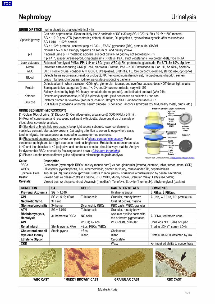
Nephrology Urinalysis
Elizabeth Kurtz
URINE DIPSTICK – urine should be analyzed within 2-4 hr
Specific gravity Can help approximate UOsm: multiply last 2 decimals of SG x 30 (eg SG 1.020 20 x 30 ~ 600 mosms) SG < 1.010: post-ATN (concentrating defect), diuretics, DI, polydipsia, hypovolemic hypoNa after resuscitation SG 1.010 – 1.025: normal SG > 1.025: prerenal, contrast (esp >1.030), ↓EABV, glycosuria (DM), proteinuria, SIADH
pH Normal 4.5 – 8, but strongly depends on serum pH and dietary intake If normal urine pH + metabolic acidosis, suspect distal RTA (kidney not secreting NH4+) If pH ≥ 7, suspect urease-producing organisms (Proteus, PsA), strict vegetarians (low protein diet), type I RTA
Leuk esterase Released from lysed PMNs; FP: ↓pH or ↓SG (lyses WBCs); FN: proteinuria, glucosuria. For UTI, Sn 80%, Sp low Nitrite Indicates nitrate-reducing GNR (E. coli, Klebsiella, Proteus, PsA – NOT Enterococcus). For UTI, Sn 60%, Sp>90% WBC UTI; if sterile pyuria, consider AIN, GC/CT, Ureaplasma, urethritis, TB, foreign body, exercise, steroid use, cyclophos
Blood Detects heme (glomerular, renal, or urologic); FP: hemoglobinuria (hemolysis), myoglobinuria (rhabdo), semen, drugs (rifampin, chloroquine, iodine), peroxidase-producing bacteria
Protein Detects albumin when excretion >300mg/d: glomerular, tubular, and overflow causes; does NOT detect light chains Semiquantitative categories (trace, 1+, 2+, and 3+) are not reliable, vary with SG Falsely elevated by high SG, heavy hematuria (heme protein), and iodinated contrast (w/in 24h)
Ketones Detects only acetoacetate, NOT β-hydroxybutyrate; yield decreases as collected urine sits
Glucose Reflects glomerular overflow (serum glucose >180mg/dl or SGLT-inhibitor/mutation) OR PCT failure (glucosuria w/ normal serum glucose consider Fanconi’s syndrome 2/2 MM, heavy metal, drugs, etc.)
URINE SEDIMENT (MICROSCOPY) (1) Obtain 10cc of urine (2) Dipstick (3) Centrifuge using a balance @ 3000 RPM x 3-5 min.(4) Pour off supernatant and resuspend sediment with pipette; place one drop of sample onslide, place coverslip, analyze.(5) Standard or bright field microscopy: keep light source subdued, lower condenser tomaximize contrast, start at low power (10x) paying attention to coverslip edge where caststend to migrate, increase power as needed to examine formed elements.(6) Phase contrast microscopy: review components of phase contrast microscopy. Raisecondenser up high and turn light source to maximal brightness. Rotate the condenser annulusto 40 and the objective to 40 (objective and condenser annulus should always match). Analyzefor dysmorphic RBCs or casts by focusing up and down. (Click here for tutorial).(7) Please use the urine sediment guide adjacent to microscope to guide analysis.Cells: Description RBCs Glomerular (dysmorphic RBCs “mickey mouse ears”) vs non-glomerular (trauma, exercise, infxn, tumor, stone, SCD) WBCs UTI/cystitis, pyelonephritis, AIN, atheroembolic, glomerular injury, renal/bladder TB, nephrolithiasis Epithelial Cells Tubular (ATN), transitional (proximal urethra to renal pelvis), squamous (contamination by genital secretions) Casts: Viewed best w/ phase contrast: Hyaline, RBC, WBC, Muddy brown, Granular, Waxy, Fatty (see below) Crystals: Viewed best w/ phase contrast: Acyclovir (“needles”), Tenofovir, Struvite (↑ urine pH), ethylene glycol (oxalate)
CONDITION UA CELLS CASTS / CRYSTALS COMMENTS Pre-renal Azotemia SG > 1.010 Hyaline, granular ↓ FENa, ↓ FEUrea CIN SG >1.010; +Prot Tubular cells Granular, muddy brown ↓ UNa, ↓ FENa, FP: proteinuria Nephrotic Synd. 3+ Prot Oval fat bodies, hyaline Glomerulonephritis 3+ heme Dysmorphic RBCs RBC casts, WBC, granular ATN SG ~ 1.010 Tubular cells Granular, muddy brown Rhabdomyolysis, Hemolysis 3+ heme w/o RBCs NO cells Acellular hyaline casts with
red or brown pigmentation ↓ FENa, red/brown urine AIN WBCs; +/- eos WBC casts, granular Urine eos NOT Sens or Spec Renal Infarct Sterile pyuria; +Pro +Eos, RBCs, WBCs ↑ urine LDH (↑ serum LDH) Cholesterol emboli Sterile pyuria +Eos Cholesterol Myeloma kidney Bland Bland Proteinuria NOT detected by UA Ethylene Glycol Ca oxalate CKD Waxy +/- impaired ability to concentrate
WBC CAST “MUDDY BROWN” CAST GRANULAR CAST RBC CAST
Adapted from Olympus website, “Introduction to Phase Contrast”
101
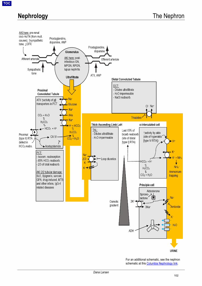
Nephrology The Nephron
Dana Larsen
For an additional schematic, see the nephron schematic at this Columbia Nephrology link.
102
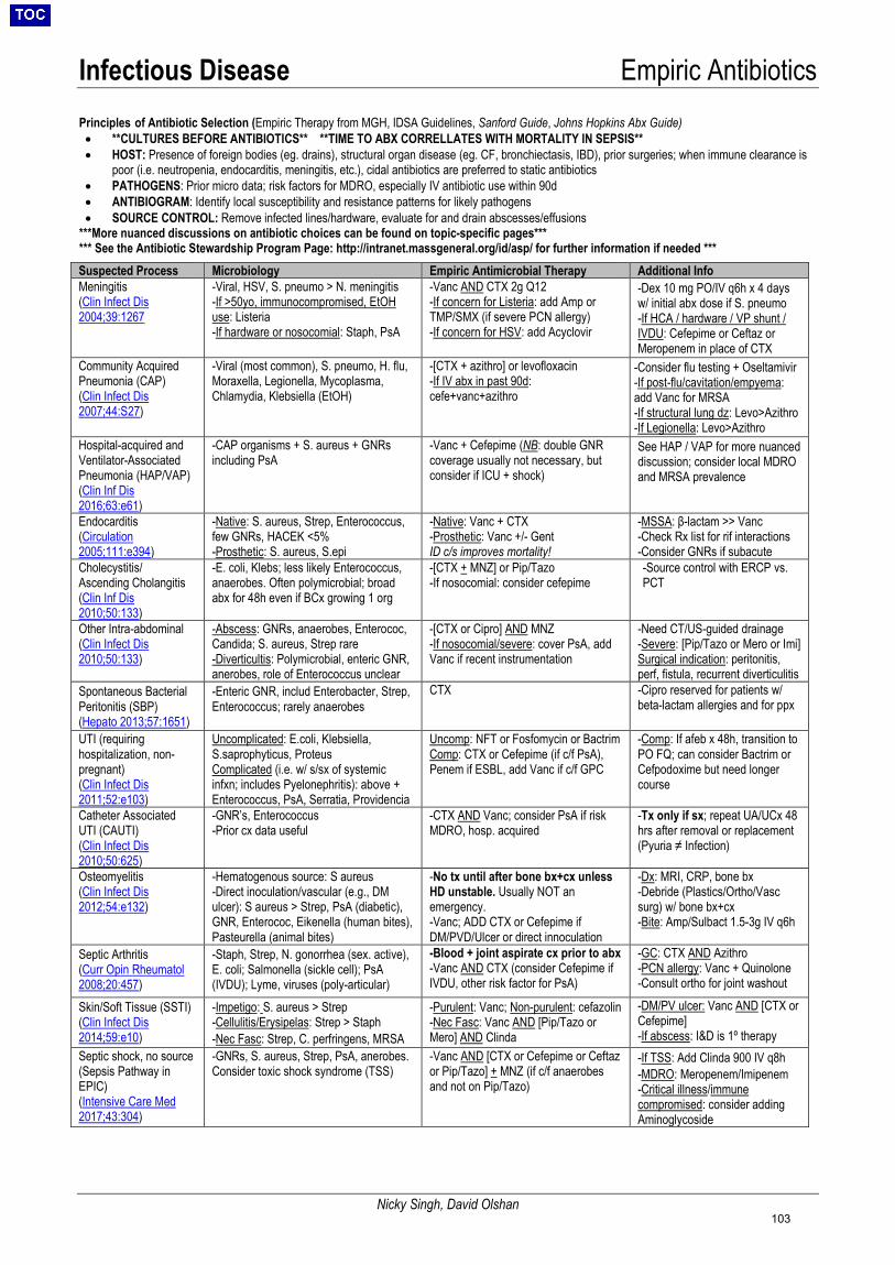
Infectious Disease Empiric Antibiotics
Nicky Singh, David Olshan
Principles of Antibiotic Selection (Empiric Therapy from MGH, IDSA Guidelines, Sanford Guide, Johns Hopkins Abx Guide) • **CULTURES BEFORE ANTIBIOTICS** **TIME TO ABX CORRELLATES WITH MORTALITY IN SEPSIS** • HOST: Presence of foreign bodies (eg. drains), structural organ disease (eg. CF, bronchiectasis, IBD), prior surgeries; when immune clearance is
poor (i.e. neutropenia, endocarditis, meningitis, etc.), cidal antibiotics are preferred to static antibiotics• PATHOGENS: Prior micro data; risk factors for MDRO, especially IV antibiotic use within 90d• ANTIBIOGRAM: Identify local susceptibility and resistance patterns for likely pathogens• SOURCE CONTROL: Remove infected lines/hardware, evaluate for and drain abscesses/effusions
***More nuanced discussions on antibiotic choices can be found on topic-specific pages****** See the Antibiotic Stewardship Program Page: http://intranet.massgeneral.org/id/asp/ for further information if needed *** Suspected Process Microbiology Empiric Antimicrobial Therapy Additional Info Meningitis (Clin Infect Dis 2004;39:1267
-Viral, HSV, S. pneumo > N. meningitis -If >50yo, immunocompromised, EtOHuse: Listeria-If hardware or nosocomial: Staph, PsA
-Vanc AND CTX 2g Q12-If concern for Listeria: add Amp orTMP/SMX (if severe PCN allergy)-If concern for HSV: add Acyclovir
-Dex 10 mg PO/IV q6h x 4 daysw/ initial abx dose if S. pneumo-If HCA / hardware / VP shunt /IVDU: Cefepime or Ceftaz or Meropenem in place of CTX
Community Acquired Pneumonia (CAP) (Clin Infect Dis 2007;44:S27)
-Viral (most common), S. pneumo, H. flu,Moraxella, Legionella, Mycoplasma,Chlamydia, Klebsiella (EtOH)
-[CTX + azithro] or levofloxacin -If IV abx in past 90d:cefe+vanc+azithro
-Consider flu testing + Oseltamivir-If post-flu/cavitation/empyema:add Vanc for MRSA-If structural lung dz: Levo>Azithro-If Legionella: Levo>Azithro
Hospital-acquired and Ventilator-Associated Pneumonia (HAP/VAP) (Clin Inf Dis 2016;63:e61)
-CAP organisms + S. aureus + GNRsincluding PsA
-Vanc + Cefepime (NB: double GNR coverage usually not necessary, butconsider if ICU + shock)
See HAP / VAP for more nuanced discussion; consider local MDRO and MRSA prevalence
Endocarditis (Circulation 2005;111:e394)
-Native: S. aureus, Strep, Enterococcus,few GNRs, HACEK <5%-Prosthetic: S. aureus, S.epi
-Native: Vanc + CTX-Prosthetic: Vanc +/- GentID c/s improves mortality!
-MSSA: β-lactam >> Vanc-Check Rx list for rif interactions-Consider GNRs if subacute
Cholecystitis/ Ascending Cholangitis (Clin Inf Dis 2010;50:133)
-E. coli, Klebs; less likely Enterococcus,anaerobes. Often polymicrobial; broadabx for 48h even if BCx growing 1 org
-[CTX + MNZ] or Pip/Tazo -If nosocomial: consider cefepime
-Source control with ERCP vs.PCT
Other Intra-abdominal (Clin Infect Dis 2010;50:133)
-Abscess: GNRs, anaerobes, Enterococ,Candida; S. aureus, Strep rare-Diverticultis: Polymicrobial, enteric GNR,anerobes, role of Enterococcus unclear
-[CTX or Cipro] AND MNZ -If nosocomial/severe: cover PsA, addVanc if recent instrumentation
-Need CT/US-guided drainage-Severe: [Pip/Tazo or Mero or Imi]Surgical indication: peritonitis,perf, fistula, recurrent diverticulitis
Spontaneous Bacterial Peritonitis (SBP) (Hepato 2013;57:1651)
-Enteric GNR, includ Enterobacter, Strep,Enterococcus; rarely anaerobes
CTX -Cipro reserved for patients w/beta-lactam allergies and for ppx
UTI (requiring hospitalization, non-pregnant)(Clin Infect Dis 2011;52:e103)
Uncomplicated: E.coli, Klebsiella, S.saprophyticus, ProteusComplicated (i.e. w/ s/sx of systemicinfxn; includes Pyelonephritis): above +Enterococcus, PsA, Serratia, Providencia
Uncomp: NFT or Fosfomycin or Bactrim Comp: CTX or Cefepime (if c/f PsA), Penem if ESBL, add Vanc if c/f GPC
-Comp: If afeb x 48h, transition toPO FQ; can consider Bactrim or Cefpodoxime but need longer course
Catheter Associated UTI (CAUTI)(Clin Infect Dis 2010;50:625)
-GNR’s, Enterococcus-Prior cx data useful
-CTX AND Vanc; consider PsA if riskMDRO, hosp. acquired
-Tx only if sx; repeat UA/UCx 48hrs after removal or replacement(Pyuria ≠ Infection)
Osteomyelitis (Clin Infect Dis 2012;54:e132)
-Hematogenous source: S aureus-Direct inoculation/vascular (e.g., DMulcer): S aureus > Strep, PsA (diabetic),GNR, Enterococ, Eikenella (human bites),Pasteurella (animal bites)
-No tx until after bone bx+cx unlessHD unstable. Usually NOT anemergency.-Vanc; ADD CTX or Cefepime ifDM/PVD/Ulcer or direct innoculation
-Dx: MRI, CRP, bone bx-Debride (Plastics/Ortho/Vascsurg) w/ bone bx+cx-Bite: Amp/Sulbact 1.5-3g IV q6h
Septic Arthritis (Curr Opin Rheumatol 2008;20:457)
-Staph, Strep, N. gonorrhea (sex. active),E. coli; Salmonella (sickle cell); PsA(IVDU); Lyme, viruses (poly-articular)
-Blood + joint aspirate cx prior to abx-Vanc AND CTX (consider Cefepime ifIVDU, other risk factor for PsA)
-GC: CTX AND Azithro-PCN allergy: Vanc + Quinolone-Consult ortho for joint washout
Skin/Soft Tissue (SSTI)
(Clin Infect Dis 2014;59:e10)
-Impetigo: S. aureus > Strep-Cellulitis/Erysipelas: Strep > Staph-Nec Fasc: Strep, C. perfringens, MRSA
-Purulent: Vanc; Non-purulent: cefazolin-Nec Fasc: Vanc AND [Pip/Tazo or Mero] AND Clinda
-DM/PV ulcer: Vanc AND [CTX or Cefepime]-If abscess: I&D is 1º therapy
Septic shock, no source (Sepsis Pathway in EPIC) (Intensive Care Med 2017;43:304)
-GNRs, S. aureus, Strep, PsA, anerobes.Consider toxic shock syndrome (TSS)
-Vanc AND [CTX or Cefepime or Ceftazor Pip/Tazo] + MNZ (if c/f anaerobesand not on Pip/Tazo)
-If TSS: Add Clinda 900 IV q8h-MDRO: Meropenem/Imipenem-Critical illness/immunecompromised: consider addingAminoglycoside
103
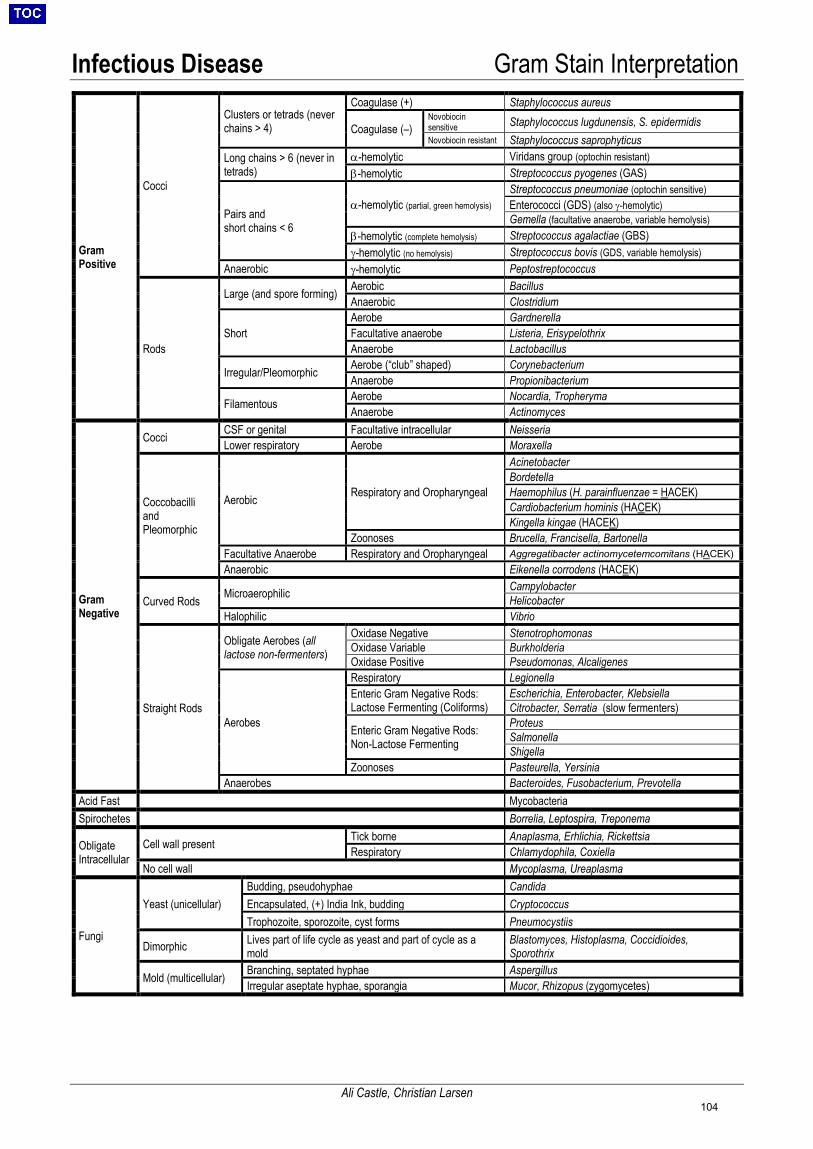
Infectious Disease Gram Stain Interpretation
Ali Castle, Christian Larsen
Gram Positive
Cocci
Clusters or tetrads (never chains > 4)
Coagulase (+) Staphylococcus aureus
Coagulase (–) Novobiocin sensitive Staphylococcus lugdunensis, S. epidermidis Novobiocin resistant Staphylococcus saprophyticus
Long chains > 6 (never in tetrads)
α-hemolytic Viridans group (optochin resistant) β-hemolytic Streptococcus pyogenes (GAS)
Pairs and short chains < 6
α-hemolytic (partial, green hemolysis) Streptococcus pneumoniae (optochin sensitive) Enterococci (GDS) (also γ-hemolytic) Gemella (facultative anaerobe, variable hemolysis)
β-hemolytic (complete hemolysis) Streptococcus agalactiae (GBS) γ-hemolytic (no hemolysis) Streptococcus bovis (GDS, variable hemolysis)
Anaerobic γ-hemolytic Peptostreptococcus
Rods
Large (and spore forming) Aerobic Bacillus Anaerobic Clostridium
Short Aerobe Gardnerella Facultative anaerobe Listeria, Erisypelothrix Anaerobe Lactobacillus
Irregular/Pleomorphic Aerobe (“club” shaped) Corynebacterium Anaerobe Propionibacterium
Filamentous Aerobe Nocardia, Tropheryma Anaerobe Actinomyces
Gram Negative
Cocci CSF or genital Facultative intracellular Neisseria Lower respiratory Aerobe Moraxella
Coccobacilli and Pleomorphic
Aerobic Respiratory and Oropharyngeal
Acinetobacter Bordetella Haemophilus (H. parainfluenzae = HACEK) Cardiobacterium hominis (HACEK) Kingella kingae (HACEK)
Zoonoses Brucella, Francisella, Bartonella Facultative Anaerobe Respiratory and Oropharyngeal Aggregatibacter actinomycetemcomitans (HACEK) Anaerobic Eikenella corrodens (HACEK)
Curved Rods Microaerophilic Campylobacter Helicobacter
Halophilic Vibrio
Straight Rods
Obligate Aerobes (all lactose non-fermenters)
Oxidase Negative Stenotrophomonas Oxidase Variable Burkholderia Oxidase Positive Pseudomonas, Alcaligenes
Aerobes
Respiratory Legionella Enteric Gram Negative Rods: Lactose Fermenting (Coliforms)
Escherichia, Enterobacter, Klebsiella Citrobacter, Serratia (slow fermenters)
Enteric Gram Negative Rods: Non-Lactose Fermenting
Proteus Salmonella Shigella
Zoonoses Pasteurella, Yersinia Anaerobes Bacteroides, Fusobacterium, Prevotella
Acid Fast Mycobacteria Spirochetes Borrelia, Leptospira, Treponema
Obligate Intracellular
Cell wall present Tick borne Anaplasma, Erhlichia, Rickettsia Respiratory Chlamydophila, Coxiella
No cell wall Mycoplasma, Ureaplasma
Fungi
Yeast (unicellular) Budding, pseudohyphae Candida Encapsulated, (+) India Ink, budding Cryptococcus Trophozoite, sporozoite, cyst forms Pneumocystiis
Dimorphic Lives part of life cycle as yeast and part of cycle as a mold
Blastomyces, Histoplasma, Coccidioides, Sporothrix
Mold (multicellular) Branching, septated hyphae Aspergillus Irregular aseptate hyphae, sporangia Mucor, Rhizopus (zygomycetes)
104
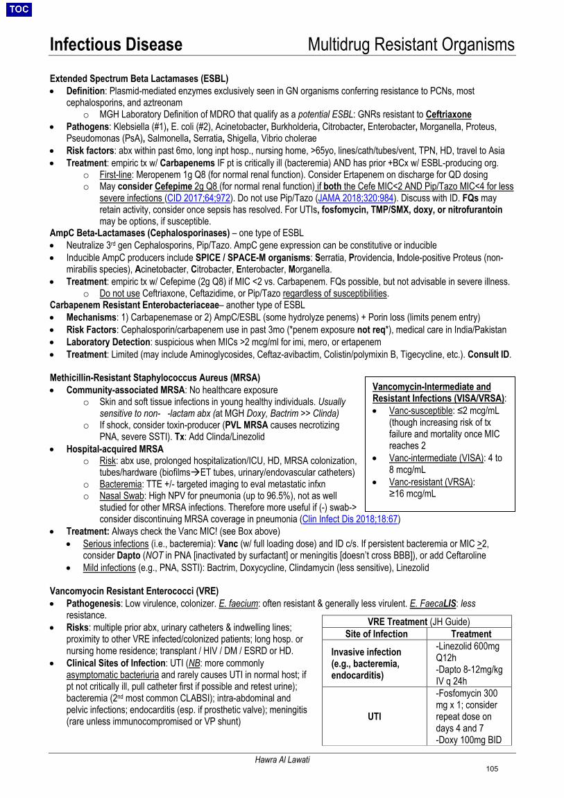
Infectious Disease Multidrug Resistant Organisms
Hawra Al Lawati
Extended Spectrum Beta Lactamases (ESBL) • Definition: Plasmid-mediated enzymes exclusively seen in GN organisms conferring resistance to PCNs, most
cephalosporins, and aztreonamo MGH Laboratory Definition of MDRO that qualify as a potential ESBL: GNRs resistant to Ceftriaxone
• Pathogens: Klebsiella (#1), E. coli (#2), Acinetobacter, Burkholderia, Citrobacter, Enterobacter, Morganella, Proteus,Pseudomonas (PsA), Salmonella, Serratia, Shigella, Vibrio cholerae
• Risk factors: abx within past 6mo, long inpt hosp., nursing home, >65yo, lines/cath/tubes/vent, TPN, HD, travel to Asia• Treatment: empiric tx w/ Carbapenems IF pt is critically ill (bacteremia) AND has prior +BCx w/ ESBL-producing org.
o First-line: Meropenem 1g Q8 (for normal renal function). Consider Ertapenem on discharge for QD dosingo May consider Cefepime 2g Q8 (for normal renal function) if both the Cefe MIC<2 AND Pip/Tazo MIC<4 for less
severe infections (CID 2017;64;972). Do not use Pip/Tazo (JAMA 2018;320:984). Discuss with ID. FQs mayretain activity, consider once sepsis has resolved. For UTIs, fosfomycin, TMP/SMX, doxy, or nitrofurantoinmay be options, if susceptible.
AmpC Beta-Lactamases (Cephalosporinases) – one type of ESBL • Neutralize 3rd gen Cephalosporins, Pip/Tazo. AmpC gene expression can be constitutive or inducible• Inducible AmpC producers include SPICE / SPACE-M organisms: Serratia, Providencia, Indole-positive Proteus (non-
mirabilis species), Acinetobacter, Citrobacter, Enterobacter, Morganella.• Treatment: empiric tx w/ Cefepime (2g Q8) if MIC <2 vs. Carbapenem. FQs possible, but not advisable in severe illness.
o Do not use Ceftriaxone, Ceftazidime, or Pip/Tazo regardless of susceptibilities.Carbapenem Resistant Enterobacteriaceae– another type of ESBL • Mechanisms: 1) Carbapenemase or 2) AmpC/ESBL (some hydrolyze penems) + Porin loss (limits penem entry)• Risk Factors: Cephalosporin/carbapenem use in past 3mo (*penem exposure not req*), medical care in India/Pakistan• Laboratory Detection: suspicious when MICs >2 mcg/ml for imi, mero, or ertapenem• Treatment: Limited (may include Aminoglycosides, Ceftaz-avibactim, Colistin/polymixin B, Tigecycline, etc.). Consult ID.
Methicillin-Resistant Staphylococcus Aureus (MRSA) • Community-associated MRSA: No healthcare exposure
o Skin and soft tissue infections in young healthy individuals. Usuallysensitive to non-β -lactam abx (at MGH Doxy, Bactrim >> Clinda)
o If shock, consider toxin-producer (PVL MRSA causes necrotizingPNA, severe SSTI). Tx: Add Clinda/Linezolid
• Hospital-acquired MRSAo Risk: abx use, prolonged hospitalization/ICU, HD, MRSA colonization,
tubes/hardware (biofilmsET tubes, urinary/endovascular catheters)o Bacteremia: TTE +/- targeted imaging to eval metastatic infxno Nasal Swab: High NPV for pneumonia (up to 96.5%), not as well
studied for other MRSA infections. Therefore more useful if (-) swab-> consider discontinuing MRSA coverage in pneumonia (Clin Infect Dis 2018;18:67)
• Treatment: Always check the Vanc MIC! (see Box above)• Serious infections (i.e., bacteremia): Vanc (w/ full loading dose) and ID c/s. If persistent bacteremia or MIC >2,
consider Dapto (NOT in PNA [inactivated by surfactant] or meningitis [doesn’t cross BBB]), or add Ceftaroline• Mild infections (e.g., PNA, SSTI): Bactrim, Doxycycline, Clindamycin (less sensitive), Linezolid
Vancomyocin Resistant Enterococci (VRE) • Pathogenesis: Low virulence, colonizer. E. faecium: often resistant & generally less virulent. E. FaecaLIS: less
resistance. • Risks: multiple prior abx, urinary catheters & indwelling lines;
proximity to other VRE infected/colonized patients; long hosp. or nursing home residence; transplant / HIV / DM / ESRD or HD.
• Clinical Sites of Infection: UTI (NB: more commonlyasymptomatic bacteriuria and rarely causes UTI in normal host; ifpt not critically ill, pull catheter first if possible and retest urine);bacteremia (2nd most common CLABSI); intra-abdominal andpelvic infections; endocarditis (esp. if prosthetic valve); meningitis(rare unless immunocompromised or VP shunt)
VRE Treatment (JH Guide)Site of Infection Treatment
Invasive infection (e.g., bacteremia, endocarditis)
-Linezolid 600mgQ12h-Dapto 8-12mg/kgIV q 24h
UTI
-Fosfomycin 300mg x 1; considerrepeat dose ondays 4 and 7-Doxy 100mg BID
Vancomycin-Intermediate and Resistant Infections (VISA/VRSA): • Vanc-susceptible: ≤2 mcg/mL
(though increasing risk of txfailure and mortality once MICreaches 2
• Vanc-intermediate (VISA): 4 to8 mcg/mL
• Vanc-resistant (VRSA):≥16 mcg/mL
105
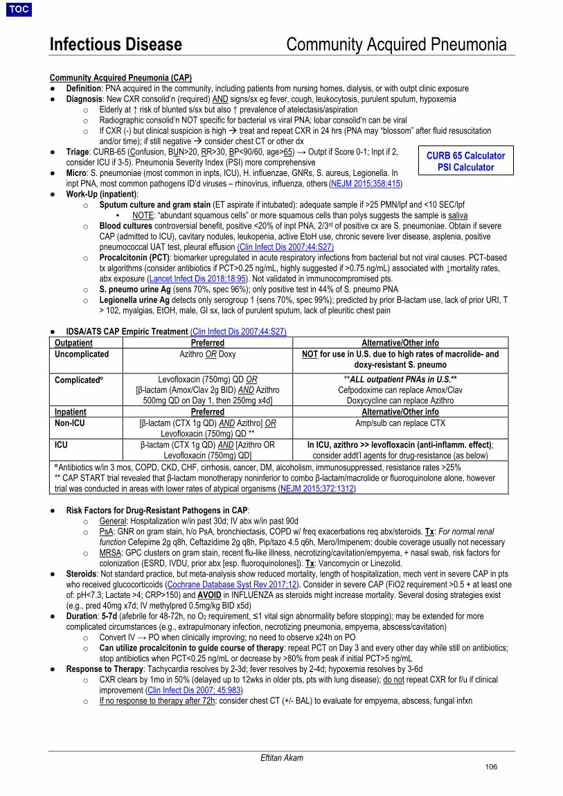
Infectious Disease Community Acquired Pneumonia Community Acquired Pneumonia (CAP) ● Definition: PNA acquired in the community, including patients from nursing homes, dialysis, or with outpt clinic exposure● Diagnosis: New CXR consolid’n (required) AND signs/sx eg fever, cough, leukocytosis, purulent sputum, hypoxemia
o Elderly at ↑ risk of blunted s/sx but also ↑ prevalence of atelectasis/aspirationo Radiographic consolid’n NOT specific for bacterial vs viral PNA; lobar consolid’n can be viralo If CXR (-) but clinical suspicion is high treat and repeat CXR in 24 hrs (PNA may “blossom” after fluid resuscitation
and/or time); if still negative consider chest CT or other dx● Triage: CURB-65 (Confusion, BUN>20, RR>30, BP<90/60, age>65) → Outpt if Score 0-1; Inpt if 2,
consider ICU if 3-5). Pneumonia Severity Index (PSI) more comprehensive● Micro: S. pneumoniae (most common in inpts, ICU), H. influenzae, GNRs, S. aureus, Legionella. In
inpt PNA, most common pathogens ID’d viruses – rhinovirus, influenza, others (NEJM 2015;358:415)● Work-Up (inpatient):
o Sputum culture and gram stain (ET aspirate if intubated): adequate sample if >25 PMN/lpf and <10 SEC/lpf▪ NOTE: “abundant squamous cells” or more squamous cells than polys suggests the sample is saliva
o Blood cultures controversial benefit, positive <20% of inpt PNA, 2/3rd of positive cx are S. pneumoniae. Obtain if severeCAP (admitted to ICU), cavitary nodules, leukopenia, active EtoH use, chronic severe liver disease, asplenia, positivepneumococcal UAT test, pleural effusion (Clin Infect Dis 2007;44:S27)
o Procalcitonin (PCT): biomarker upregulated in acute respiratory infections from bacterial but not viral causes. PCT-basedtx algorithms (consider antibiotics if PCT>0.25 ng/mL, highly suggested if >0.75 ng/mL) associated with ↓mortality rates,abx exposure (Lancet Infect Dis 2018;18:95). Not validated in immunocompromised pts.
o S. pneumo urine Ag (sens 70%, spec 96%); only positive test in 44% of S. pneumo PNAo Legionella urine Ag detects only serogroup 1 (sens 70%, spec 99%); predicted by prior B-lactam use, lack of prior URI, T
> 102, myalgias, EtOH, male, GI sx, lack of purulent sputum, lack of pleuritic chest pain
● IDSA/ATS CAP Empiric Treatment (Clin Infect Dis 2007;44:S27)Outpatient Preferred Alternative/Other info Uncomplicated Azithro OR Doxy NOT for use in U.S. due to high rates of macrolide- and
doxy-resistant S. pneumo Complicated° Levofloxacin (750mg) QD OR
[β-lactam (Amox/Clav 2g BID) AND Azithro 500mg QD on Day 1, then 250mg x4d]
**ALL outpatient PNAs in U.S.** Cefpodoxime can replace Amox/Clav
Doxycycline can replace Azithro Inpatient Preferred Alternative/Other info Non-ICU [β-lactam (CTX 1g QD) AND Azithro] OR
Levofloxacin (750mg) QD ** Amp/sulb can replace CTX
ICU β-lactam (CTX 1g QD) AND [Azithro OR Levofloxacin (750mg) QD]
In ICU, azithro >> levofloxacin (anti-inflamm. effect); consider addt’l agents for drug-resistance (as below)
°Antibiotics w/in 3 mos, COPD, CKD, CHF, cirrhosis, cancer, DM, alcoholism, immunosuppressed, resistance rates >25% ** CAP START trial revealed that β-lactam monotherapy noninferior to combo β-lactam/macrolide or fluoroquinolone alone, however trial was conducted in areas with lower rates of atypical organisms (NEJM 2015;372:1312)
● Risk Factors for Drug-Resistant Pathogens in CAP:o General: Hospitalization w/in past 30d; IV abx w/in past 90do PsA: GNR on gram stain, h/o PsA, bronchiectasis, COPD w/ freq exacerbations req abx/steroids. Tx: For normal renal
function Cefepime 2g q8h, Ceftazidime 2g q8h, Pip/tazo 4.5 q6h, Mero/Imipenem; double coverage usually not necessaryo MRSA: GPC clusters on gram stain, recent flu-like illness, necrotizing/cavitation/empyema, + nasal swab, risk factors for
colonization (ESRD, IVDU, prior abx [esp. fluoroquinolones]). Tx: Vancomycin or Linezolid.● Steroids: Not standard practice, but meta-analysis show reduced mortality, length of hospitalization, mech vent in severe CAP in pts
who received glucocorticoids (Cochrane Database Syst Rev 2017;12). Consider in severe CAP (FiO2 requirement >0.5 + at least oneof: pH<7.3; Lactate >4; CRP>150) and AVOID in INFLUENZA as steroids might increase mortality. Several dosing strategies exist(e.g., pred 40mg x7d; IV methylpred 0.5mg/kg BID x5d)
● Duration: 5-7d (afebrile for 48-72h, no O2 requirement, ≤1 vital sign abnormality before stopping); may be extended for morecomplicated circumstances (e.g., extrapulmonary infection, necrotizing pneumonia, empyema, abscess/cavitation)
o Convert IV → PO when clinically improving; no need to observe x24h on POo Can utilize procalcitonin to guide course of therapy: repeat PCT on Day 3 and every other day while still on antibiotics;
stop antibiotics when PCT<0.25 ng/mL or decrease by >80% from peak if initial PCT>5 ng/mL● Response to Therapy: Tachycardia resolves by 2-3d; fever resolves by 2-4d; hypoxemia resolves by 3-6d
o CXR clears by 1mo in 50% (delayed up to 12wks in older pts, pts with lung disease); do not repeat CXR for f/u if clinicalimprovement (Clin Infect Dis 2007; 45:983)
o If no response to therapy after 72h: consider chest CT (+/- BAL) to evaluate for empyema, abscess, fungal infxn
CURB 65 Calculator PSI Calculator
Eftitan Akam 106

Infectious Disease HAP/VAP & Aspiration Pneumonia
Hawra Al Lawati
Hospital-Acquired and Ventilator-Associated Pneumonia • Definitions:
o Hospital-Acquired (HAP): Pneumonia that develops >48 hrs after admissiono Ventilator-Associated (VAP): HAP that develops >48 hrs after endotracheal intubation
• Common Microbiology: Enteric GNRs (Klebsiella, E. coli), MRSA/MSSA, PsA, Acinetobacter• MDRO Risk Factors:
o For MDR pathogens: IV abx use within 90 days (most important risk factor); high local prevalence (>10%) of MDRGNRs and MRSA; structural lung disease (CF, bronchiectasis)
o Additional risk factors for MDR VAP: septic shock/ICU, ARDS, >5 days hospitalization or Renal ReplacementTherapy preceding onset
• Workup: CXR, sputum/blood cx, MRSA swab (see MDRO section regarding interpretation); consider induced sputum,bronch with BAL
• Antibiotic Choices and Empiric Treatment:o HAP/VAP w/o MDRO risk: 1 antipseudomonal agent (preference for B-lactam)o HAP/VAP w/ MDRO risk: 1 antipseudomonal agent (preference for B-lactam) AND 1 anti-MRSA agent
o Consider empiric double PsA coverage (e.g., 2 antipseudomonal agents (1 B-lactam and 1 Non-B-lactam))IF: hx of MDR PsA, septic shock w/ suspicion for PsA, CF/bronchiectasis, febrile neutropenia + PsA bacteremia
o Typical empiric regimen at MGH: Vancomycin + Cefepimeo MRSA: Vancomycin IV (trough 15-20) OR Linezolid 600mg IV q12hrso Antipseudomonal agents (B-lactams): normal renal func Cefepime OR Ceftazidime 2g IV q8h OR Pip/Tazo 4.5g IV
q6h OR Meropenem 1g IV q8h (aztreonam 2g IV q8h only if severe PCN allergy https://id.partners.org/allergy/)o Antipseudomonal agents (Non-beta-lactams): Tobramycin 5-7mg/kg IV x 1 then dose by level OR Levofloxacin
750mg (PsA susceptibility at MGH is 73%) IV QD OR polymyxin B (Call ID)• Tailoring Therapy:
o Improvement after 48h or pathogen identification→ Narrow abx/convert to PO/discontinue MRSA + PsA coverage ifpossible. In VAP, if negative tracheal aspirate, consider d/c antibiotics after 72 hours (NPV 94% for VAP)
o No improvement after 48h→ Broaden to cover MDROs (if not currently covering), consider other sites ofinfection/abscess
o Duration: 7d for both HAP/VAP. Can also utilize serial procalcitonin levels (1-3 day turnaround at MGH)→discontinue abx when <0.25ng/mL Eur Respir J 2009; 34:1364
Aspiration Pneumonia: • Definition: Pneumonia caused by the excessive entry of secretions, particulate matter, or fluid into airways (NB: ALL
pneumonias are secondary to micro-aspiration events; the term ‘aspiration pneumonia’ refers to macro-aspiration events)• Predisposing Factors: ↓ consciousness (seizure/overdose), esophageal dysmotility, post-bronchial obstruction, gum dzMicrobiology: Classically caused by polymicrobial infections including oral anaerobes (Peptostreptococcus, Fusobacterium,Bacteroides), but most common organisms are GNRs and standard CAP/HAP organisms. Am J Respir CC Med2003;167:1650• Characteristics: Indolent, putrid sputum, pulmonary necrosis w/ cavitation/abscess/empyema• Workup: CXR, sputum culture (anaerobic respiratory culture not performed at MGH due to low utility)• Empiric Treatment: CAP treatment ± anaerobic coverage (see below)• Anaerobic Coverage: Per IDSA guidelines, anaerobic coverage only recommended in pts w/ loss of consciousness
secondary to alcohol/drug overdose or seizure AND concomitant gingival disease or esophageal dysmotility. ClinInfect Dis 2007;44:S22o First line: ampicillin-sulbactam (or amox/clavulanate if not severely ill); alternative: [CTX + metro] OR clindamycin
• Duration: 7d (unless complicated by cavitation/abscess/empyema)
Aspiration Pneumonitis: • Definition: Aspiration of chemical substances into the airways without bacterial infection• Clinical Manifestations: Abrupt onset (2hr), low-grade fever, ↑ WBC, hypoxemia, CXR consolidation (RML/RLL upright,
RUL supine) may be indistinguishable from pneumonia in the acute setting!• Treatment: If concern for aspiration pneumonia (i.e., bacterial infection), may cover with abx for 48hrs d/c if no
consolidation develops on CXR OR if signs/sx/consolidation resolve rapidly (less likely to be PNA)
107

Infectious Disease Viral Respiratory / Head & Neck Infections
Will Simmons, Christian Larsen
Viral Respiratory Infections ● Epidemiology:
o URI: rhinovirus (30-50%), coronavirus (10-15%), influenza (5-15%), parainfluenza (5%), RSV (5%)o LRTI (bronchitis, bronchiolitis, PNA): influenza, RSV, parainfluenza, adenovirus (Lancet 2011;377:1264)
● Presentation:o Risk factors: immunosuppression (T cell defects: HIV, transplant), extremes of ageo Symptoms: fever, dry cough, myalgias, dyspnea, sore throat, rhinorrhea, malaise, confusion, anorexia, wheezingo Labs: leukocytosis or leukopenia (esp. lymphopenia), high CK (influenza)o Complications: viral PNA (continued worsening after onset); secondary bacterial PNA (initial improvement
followed by worsening after ~7days micro: S. pneumo [1st], S. aureus [2nd]), ARDS● Diagnosis:
o Rapid influenza 50-70% Se, >90% Spe; influenza A/B and RSV PCR >95% Se & Spe (J Clin Micro2013;51:2421)
▪ Se/Spe depends on high quality nasopharynx swab. Can test multiple days if clinical suspicion high; ifLRTI consider BAL vs ET aspiration
o Viral resp panel (adenovirus, parainfluenza, metapneumovirus) nasopharyngeal swab > induced sputum● Treatment (Influenza)
o Indications: Hospitalized, severe disease, or risk for complications (>65, SNF, pregnant, immunosuppressed,DM, CHF/CAD, CKD, COPD, SCD, asthma, BMI>40, neurological disease)
o Rx: Oseltamivir 75mg BID x5d (dose reduce for CKD; no data to support double dose if severe/ICU (BMJ2013;346:3039)); initiate within 48h of sx (note: >48h OK if severe disease or hospitalized pt)
o Ppx: Oseltamivir 75mg daily recommended for all residents of institutional settings during outbreaks (14d)o High dose influenza vaccine in adults >65, solid organ transplant recipients (J Infect Dis 2018;217:1718)
Head and Neck InfectionsEpidemiology: (Principles of Crit Care. Chow AW. 4th edition. McGraw-Hill, NY 2015)
o Organisms: Streptococci (mainly Viridans), Oral anaerobes (Fusobacterium spp., Bacteroides sp).o Typically from odontogenic or tonsillar infection with spread to adjacent tissue planes.o Diagnostics: Panorex, CT Neck. IR or ENT for deep cx.o Rx: B-lactam+ anaerobic agent or B-lactamase inhb. early involvement of ENT, airway monitoring.
● Ludwig’s Angina:o Definition: Infection involving the
submandibular space.o Risk factors: Periodontal infections,
especially involving 2nd and 3rd molars (70-85% of cases)
o Symptoms: Fevers, mouth pain,submandibular swelling/erythema, tongueswelling, stiff neck, drooling.
o Complications: Mandibular osteo, abscesses.● Lateral Pharyngeal Space Infections:
o Carotid Sheath (IJ, ICA, CN9)o Lemierre’s Syndrome: septic
thrombophlebitis of IJ o Usually with + blood cx’s; pt’s at risk for
septic embolio Add enoxaparin for clot management.
● Deep Neck Space Infectionso Definition: Infection, originating from
oropharynx involving retropharyngeal,“danger,” and prevertebral spaces
o Symptoms: fevers, neck pain, chestpain
o Complications: Mediastinitis (via dangerspace), vertebral osteo, paravertebralabscess
108
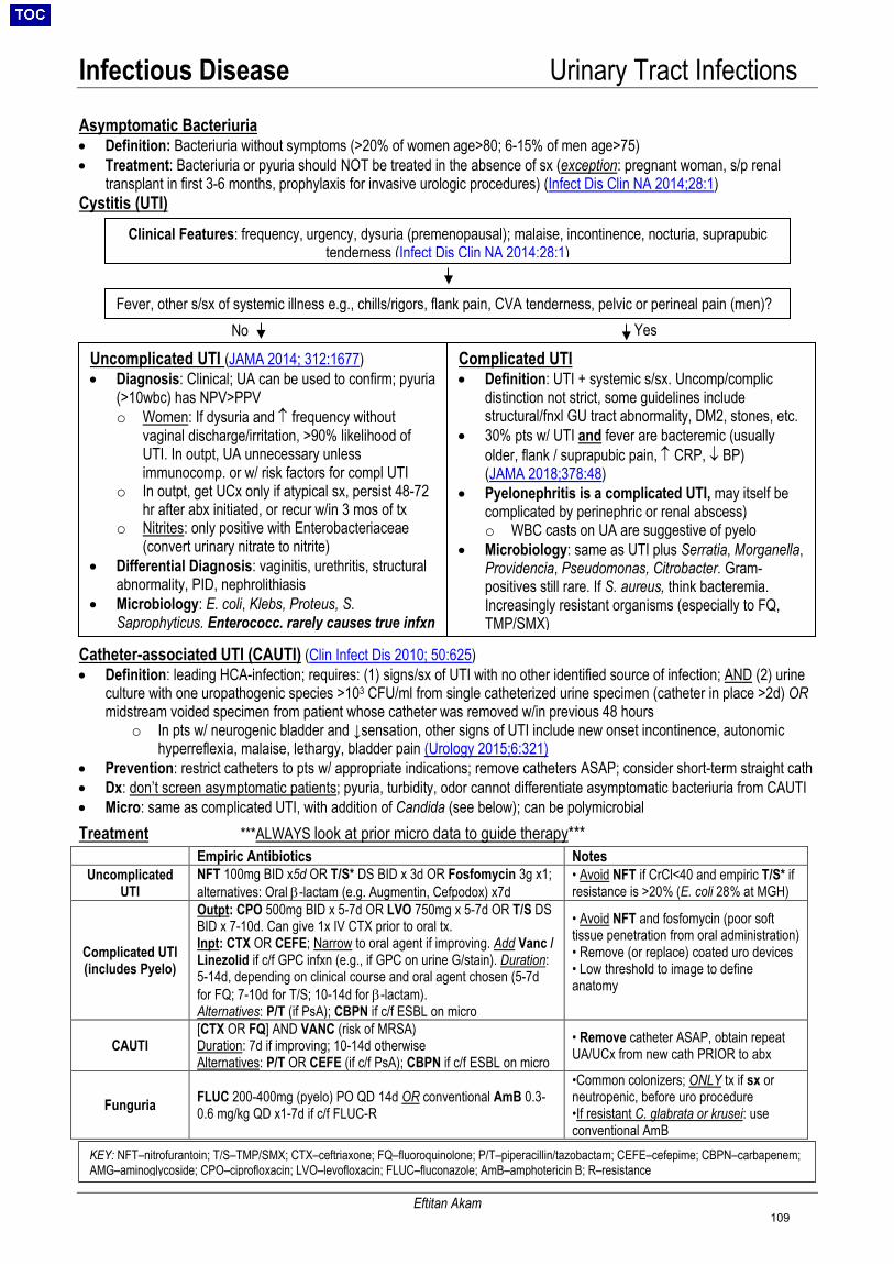
Infectious Disease Urinary Tract Infections
Eftitan Akam
Clinical Features: frequency, urgency, dysuria (premenopausal); malaise, incontinence, nocturia, suprapubic tenderness (Infect Dis Clin NA 2014;28:1)
Asymptomatic Bacteriuria • Definition: Bacteriuria without symptoms (>20% of women age>80; 6-15% of men age>75)• Treatment: Bacteriuria or pyuria should NOT be treated in the absence of sx (exception: pregnant woman, s/p renal
transplant in first 3-6 months, prophylaxis for invasive urologic procedures) (Infect Dis Clin NA 2014;28:1)Cystitis (UTI)
Catheter-associated UTI (CAUTI) (Clin Infect Dis 2010; 50:625) • Definition: leading HCA-infection; requires: (1) signs/sx of UTI with no other identified source of infection; AND (2) urine
culture with one uropathogenic species >103 CFU/ml from single catheterized urine specimen (catheter in place >2d) ORmidstream voided specimen from patient whose catheter was removed w/in previous 48 hours
o In pts w/ neurogenic bladder and ↓sensation, other signs of UTI include new onset incontinence, autonomichyperreflexia, malaise, lethargy, bladder pain (Urology 2015;6:321)
• Prevention: restrict catheters to pts w/ appropriate indications; remove catheters ASAP; consider short-term straight cath• Dx: don’t screen asymptomatic patients; pyuria, turbidity, odor cannot differentiate asymptomatic bacteriuria from CAUTI• Micro: same as complicated UTI, with addition of Candida (see below); can be polymicrobialTreatment ***ALWAYS look at prior micro data to guide therapy***
Empiric Antibiotics Notes Uncomplicated
UTI NFT 100mg BID x5d OR T/S* DS BID x 3d OR Fosfomycin 3g x1; alternatives: Oral β-lactam (e.g. Augmentin, Cefpodox) x7d
• Avoid NFT if CrCl<40 and empiric T/S* ifresistance is >20% (E. coli 28% at MGH)
Complicated UTI (includes Pyelo)
Outpt: CPO 500mg BID x 5-7d OR LVO 750mg x 5-7d OR T/S DS BID x 7-10d. Can give 1x IV CTX prior to oral tx. Inpt: CTX OR CEFE; Narrow to oral agent if improving. Add Vanc / Linezolid if c/f GPC infxn (e.g., if GPC on urine G/stain). Duration: 5-14d, depending on clinical course and oral agent chosen (5-7dfor FQ; 7-10d for T/S; 10-14d for β-lactam).Alternatives: P/T (if PsA); CBPN if c/f ESBL on micro
• Avoid NFT and fosfomycin (poor softtissue penetration from oral administration)• Remove (or replace) coated uro devices• Low threshold to image to defineanatomy
CAUTI [CTX OR FQ] AND VANC (risk of MRSA) Duration: 7d if improving; 10-14d otherwise Alternatives: P/T OR CEFE (if c/f PsA); CBPN if c/f ESBL on micro
• Remove catheter ASAP, obtain repeatUA/UCx from new cath PRIOR to abx
Funguria FLUC 200-400mg (pyelo) PO QD 14d OR conventional AmB 0.3-0.6 mg/kg QD x1-7d if c/f FLUC-R
•Common colonizers; ONLY tx if sx orneutropenic, before uro procedure•If resistant C. glabrata or krusei: useconventional AmB
KEY: NFT–nitrofurantoin; T/S–TMP/SMX; CTX–ceftriaxone; FQ–fluoroquinolone; P/T–piperacillin/tazobactam; CEFE–cefepime; CBPN–carbapenem; AMG–aminoglycoside; CPO–ciprofloxacin; LVO–levofloxacin; FLUC–fluconazole; AmB–amphotericin B; R–resistance
Fever, other s/sx of systemic illness e.g., chills/rigors, flank pain, CVA tenderness, pelvic or perineal pain (men)?
Uncomplicated UTI (JAMA 2014; 312:1677) • Diagnosis: Clinical; UA can be used to confirm; pyuria
(>10wbc) has NPV>PPVo Women: If dysuria and ↑ frequency without
vaginal discharge/irritation, >90% likelihood ofUTI. In outpt, UA unnecessary unlessimmunocomp. or w/ risk factors for compl UTI
o In outpt, get UCx only if atypical sx, persist 48-72hr after abx initiated, or recur w/in 3 mos of tx
o Nitrites: only positive with Enterobacteriaceae(convert urinary nitrate to nitrite)
• Differential Diagnosis: vaginitis, urethritis, structuralabnormality, PID, nephrolithiasis
• Microbiology: E. coli, Klebs, Proteus, S.Saprophyticus. Enterococc. rarely causes true infxn
Complicated UTI • Definition: UTI + systemic s/sx. Uncomp/complic
distinction not strict, some guidelines includestructural/fnxl GU tract abnormality, DM2, stones, etc.
• 30% pts w/ UTI and fever are bacteremic (usuallyolder, flank / suprapubic pain, ↑ CRP, ↓ BP)(JAMA 2018;378:48)
• Pyelonephritis is a complicated UTI, may itself becomplicated by perinephric or renal abscess)o WBC casts on UA are suggestive of pyelo
• Microbiology: same as UTI plus Serratia, Morganella,Providencia, Pseudomonas, Citrobacter. Gram-positives still rare. If S. aureus, think bacteremia.Increasingly resistant organisms (especially to FQ,TMP/SMX)
Yes No
109
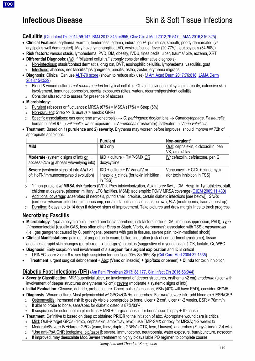
Infectious Disease Skin & Soft Tissue Infections
Jimmy Lam and Theodora Karagounis
Cellulitis (Clin Infect Dis 2014;59:147, BMJ 2012;345:e4955, Clev Clin J Med 2012;79:547, JAMA 2016;316:325)• Clinical Features: erythema, warmth, tenderness, edema, induration +/- purulence; smooth, poorly demarcated (vs.
erysipelas-well demarcated). May have lymphangitis, LAD, vesicles/bullae, fever (20-77%), leukocytosis (34-50%)• Risk factors: venous stasis, lymphedema, PVD, DM, obesity, IVDU, tinea pedis, ulcer, trauma/ bite, eczema, XRT• Differential Diagnosis: (NB: if “bilateral cellulitis,” strongly consider alternative diagnosis)o Non-infectious: stasis/contact dermatitis, drug rxn, DVT, eosinophilic cellulitis, lymphedema, vasculitis, gouto Infectious: abscess, nec fasciitis/gas gangrene, bursitis, osteo, zoster, erythema migrans• Diagnosis: Clinical. Can use ALT-70 score (shown to reduce abx use) (J Am Acad Derm 2017;76:618; JAMA Derm
2018;154:529)o Blood & wound cultures not recommended for typical cellulitis. Obtain if: evidence of systemic toxicity, extensive skin
involvement, immunosuppression, special exposures (bites, water), recurrent/persistent cellulitis.o Consider ultrasound to assess for presence of abscess• Microbiology:o Purulent (abscess or fluctuance): MRSA (67%) > MSSA (17%) > Strep (5%)o Non-purulent: Strep >> S. aureus > aerobic GNRso Specific associations: gas gangrene (myonecrosis) → C. perfringens; dog/cat bite → Capnocytophaga, Pasteurella;
human bite/IVDU → Eikenella; water exposure → Aeromonas (freshwater); saltwater → Vibrio vulnificus• Treatment: Based on 1) purulence and 2) severity. Erythema may worsen before improves; should improve w/ 72h of
appropriate antibiotics.Purulent Non-purulent*
Mild I&D only Oral: cephalexin, dicloxacillin, pen VK, amox/clav
Moderate (systemic signs of infx or abcess>2cm or abcess w/overlying infx)
I&D + culture + TMP-SMX OR doxycycline
IV: cefazolin, ceftriaxone, pen G
Severe (systemic signs of infx AND >1 of: HoTN/immunocomp/rapid evolution)
I&D + culture + IV Vanc/IV or linezolid + clinda (for toxin inhibition in TSS)
Vancomycin + CTX + clindamycin (for toxin inhibition in TSS)
o *If non-purulent w/ MRSA risk factors (IVDU, Prev infx/colonization, Abx in prev 8wks, DM, Hosp. in 1yr, athletes, staff,children at daycare, prisoner, military, LTC facilities, MSM): add empiric PO/IV MRSA coverage (CJEM 2009;11:430)
o Additional coverage: anaerobes (if necrosis, putrid smell, crepitus, certain diabetic infections [see below]); GNRs(cirrhosis w/severe infection, immunocomp, certain diabetic infections [as below]); PsA (neutropenic, trauma, post-op)
o Duration: 5 days; up to 14 days if delayed signs of improvement. Take pictures and draw margin lines to track progress.
Necrotizing Fasciitis • Microbiology: Type I (polymicrobial [mixed aerobes/anaerobes]; risk factors include DM, immunosuppression, PVD); Type
II (monomicrobial [usually GAS, less often other Strep or Staph, Vibrio, Aeromonas]; associated with TSS); myonecrosis(i.e., gas gangrene; caused by C. perfringens, presents with gas in tissues, severe pain, toxin-mediated shock)
• Clinical Manifestations: pain out of proportion to exam, bullae, induration (risk of compartment syndrome), tissueanesthesia, rapid skin changes (purple-red → blue-grey), crepitus (suggestive of myonecrosis); ↑ CK, lactate, Cr, WBC
• Diagnosis: Early suspicion and involvement of a surgeon for surgical exploration and ID is criticalo LRINEC score > or = 6 raises high suspicion for nec fasc; 90% Se 95% Sp (Crit Care Med 2004;32:1535)• Treatment: urgent surgical debridement + Abx: (Vanc or linezolid) + (pip/tazo or penem) + Clinda for toxin inhibition
Diabetic Foot Infections (DFI) (Am Fam Physician 2013; 88:177, Clin Infect Dis 2016;63:944)• Severity Classification: Mild (superficial ulcer, no involvement of deeper structures, erythema <2 cm); moderate (ulcer with
involvement of deeper structures or erythema >2 cm); severe (moderate + systemic signs of infx)• Initial Evaluation: Cleanse, debride, probe, culture. Check pulses/sensation, ABIs (40% will have PAD), consider XR/MRI• Diagnosis: Wound culture. Most polymicrobial w/ GPCs>GNRs, anaerobes. For mod-severe infx: add blood cx + ESR/CRPo Osteomyelitis: Increased risk if: grossly visible bone/probe to bone, ulcer > 2 cm2, ulcer >1-2 weeks, ESR > 70mm/ho If able to probe to bone, sens/spec for diabetic osteo is 87%/83%o If suspicious for osteo, obtain plain films ± MRI ± surgical consult for bone/tissue biopsy ± ID consult• Treatment: Definitive tx based on deep cx obtained PRIOR to the initiation of abx. Appropriate wound care is critical.o Mild: Oraltarget GPCs (diclox, cephalexin, amox/clav, levo); use TMP-SMX or doxy for MRSA; 1-2 weeks txo Moderate/Severe IVtarget GPCs (vanc, linez, dapto), GNRs* (CTX, levo, Unasyn), anaerobes (Flagyl/clinda); 2-4 wkso *Use anti-PsA GNR (cefepime, pip/tazo) if: severe, immunocomp, neutropenia, water exposure, burn/puncture, nosocomo If improved, may deescalate Mod/Severe treatment to highly bioavailable PO regimen to complete course
110
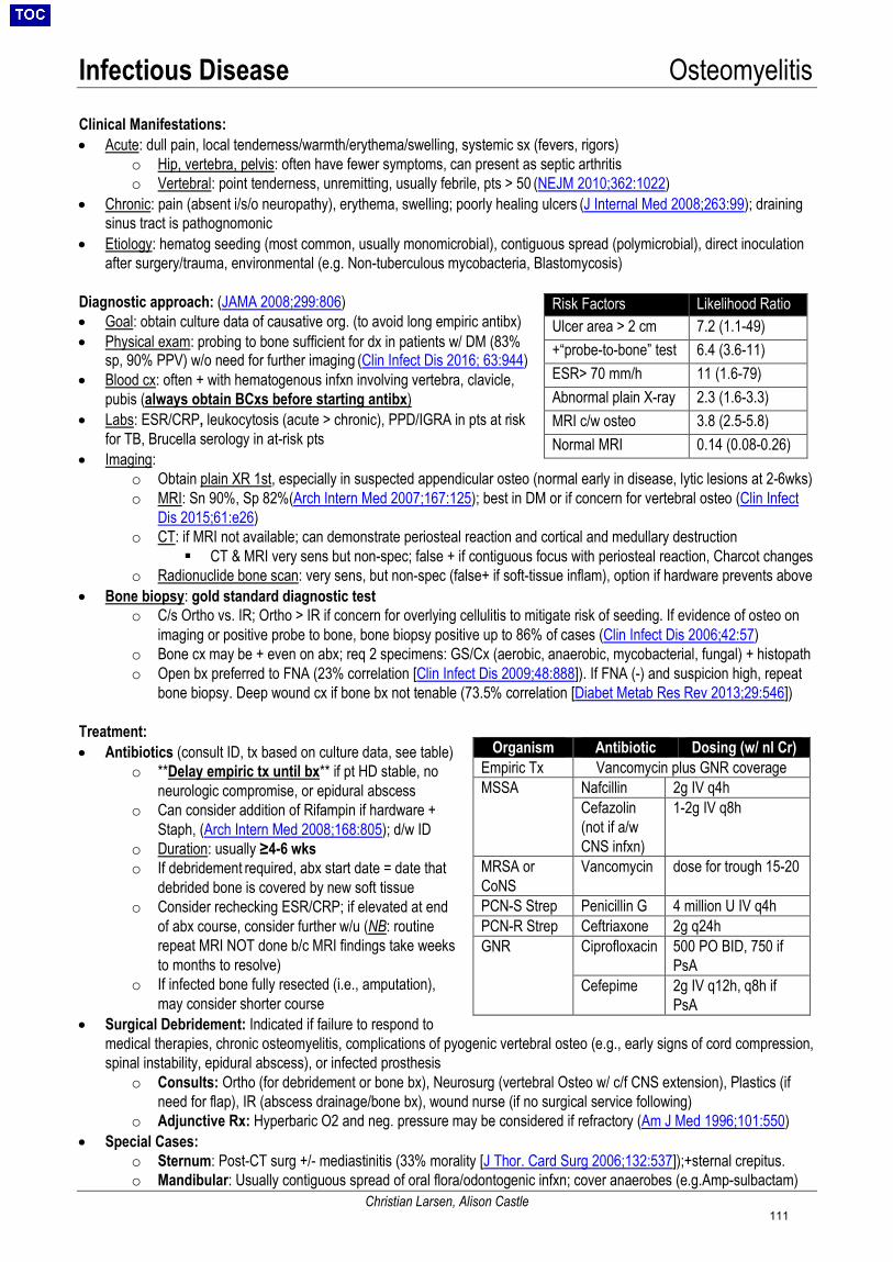
Infectious Disease Osteomyelitis
Christian Larsen, Alison Castle
Clinical Manifestations: • Acute: dull pain, local tenderness/warmth/erythema/swelling, systemic sx (fevers, rigors)
o Hip, vertebra, pelvis: often have fewer symptoms, can present as septic arthritiso Vertebral: point tenderness, unremitting, usually febrile, pts > 50 (NEJM 2010;362:1022)
• Chronic: pain (absent i/s/o neuropathy), erythema, swelling; poorly healing ulcers (J Internal Med 2008;263:99); drainingsinus tract is pathognomonic
• Etiology: hematog seeding (most common, usually monomicrobial), contiguous spread (polymicrobial), direct inoculationafter surgery/trauma, environmental (e.g. Non-tuberculous mycobacteria, Blastomycosis)
Diagnostic approach: (JAMA 2008;299:806) • Goal: obtain culture data of causative org. (to avoid long empiric antibx)• Physical exam: probing to bone sufficient for dx in patients w/ DM (83%
sp, 90% PPV) w/o need for further imaging (Clin Infect Dis 2016; 63:944)• Blood cx: often + with hematogenous infxn involving vertebra, clavicle,
pubis (always obtain BCxs before starting antibx)• Labs: ESR/CRP, leukocytosis (acute > chronic), PPD/IGRA in pts at risk
for TB, Brucella serology in at-risk pts• Imaging:
o Obtain plain XR 1st, especially in suspected appendicular osteo (normal early in disease, lytic lesions at 2-6wks)o MRI: Sn 90%, Sp 82%(Arch Intern Med 2007;167:125); best in DM or if concern for vertebral osteo (Clin Infect
Dis 2015;61:e26)o CT: if MRI not available; can demonstrate periosteal reaction and cortical and medullary destruction
CT & MRI very sens but non-spec; false + if contiguous focus with periosteal reaction, Charcot changeso Radionuclide bone scan: very sens, but non-spec (false+ if soft-tissue inflam), option if hardware prevents above
• Bone biopsy: gold standard diagnostic testo C/s Ortho vs. IR; Ortho > IR if concern for overlying cellulitis to mitigate risk of seeding. If evidence of osteo on
imaging or positive probe to bone, bone biopsy positive up to 86% of cases (Clin Infect Dis 2006;42:57)o Bone cx may be + even on abx; req 2 specimens: GS/Cx (aerobic, anaerobic, mycobacterial, fungal) + histopatho Open bx preferred to FNA (23% correlation [Clin Infect Dis 2009;48:888]). If FNA (-) and suspicion high, repeat
bone biopsy. Deep wound cx if bone bx not tenable (73.5% correlation [Diabet Metab Res Rev 2013;29:546])
Treatment: • Antibiotics (consult ID, tx based on culture data, see table)
o **Delay empiric tx until bx** if pt HD stable, noneurologic compromise, or epidural abscess
o Can consider addition of Rifampin if hardware +Staph, (Arch Intern Med 2008;168:805); d/w ID
o Duration: usually ≥4-6 wkso If debridement required, abx start date = date that
debrided bone is covered by new soft tissueo Consider rechecking ESR/CRP; if elevated at end
of abx course, consider further w/u (NB: routinerepeat MRI NOT done b/c MRI findings take weeksto months to resolve)
o If infected bone fully resected (i.e., amputation),may consider shorter course
• Surgical Debridement: Indicated if failure to respond tomedical therapies, chronic osteomyelitis, complications of pyogenic vertebral osteo (e.g., early signs of cord compression,spinal instability, epidural abscess), or infected prosthesis
o Consults: Ortho (for debridement or bone bx), Neurosurg (vertebral Osteo w/ c/f CNS extension), Plastics (ifneed for flap), IR (abscess drainage/bone bx), wound nurse (if no surgical service following)
o Adjunctive Rx: Hyperbaric O2 and neg. pressure may be considered if refractory (Am J Med 1996;101:550)• Special Cases:
o Sternum: Post-CT surg +/- mediastinitis (33% morality [J Thor. Card Surg 2006;132:537]);+sternal crepitus.o Mandibular: Usually contiguous spread of oral flora/odontogenic infxn; cover anaerobes (e.g.Amp-sulbactam)
Risk Factors Likelihood Ratio Ulcer area > 2 cm 7.2 (1.1-49) +“probe-to-bone” test 6.4 (3.6-11) ESR> 70 mm/h 11 (1.6-79) Abnormal plain X-ray 2.3 (1.6-3.3) MRI c/w osteo 3.8 (2.5-5.8) Normal MRI 0.14 (0.08-0.26)
Organism Antibiotic Dosing (w/ nl Cr) Empiric Tx Vancomycin plus GNR coverage MSSA Nafcillin 2g IV q4h
Cefazolin (not if a/w CNS infxn)
1-2g IV q8h
MRSA or CoNS
Vancomycin dose for trough 15-20
PCN-S Strep Penicillin G 4 million U IV q4h PCN-R Strep Ceftriaxone 2g q24h GNR Ciprofloxacin 500 PO BID, 750 if
PsA Cefepime 2g IV q12h, q8h if
PsA
111
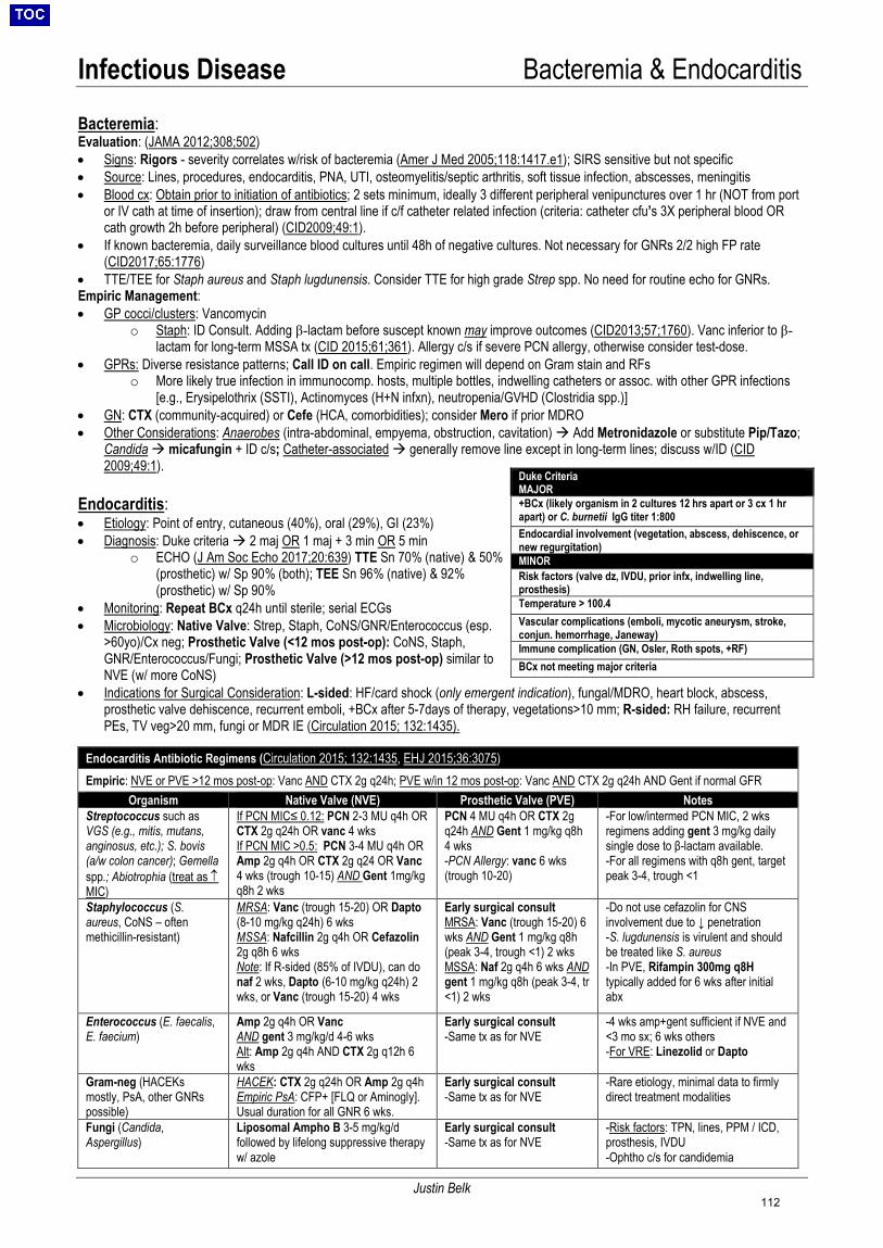
Infectious Disease Bacteremia & Endocarditis
Justin Belk
Bacteremia: Evaluation: (JAMA 2012;308;502) • Signs: Rigors - severity correlates w/risk of bacteremia (Amer J Med 2005;118:1417.e1); SIRS sensitive but not specific• Source: Lines, procedures, endocarditis, PNA, UTI, osteomyelitis/septic arthritis, soft tissue infection, abscesses, meningitis• Blood cx: Obtain prior to initiation of antibiotics; 2 sets minimum, ideally 3 different peripheral venipunctures over 1 hr (NOT from port
or IV cath at time of insertion); draw from central line if c/f catheter related infection (criteria: catheter cfu’s 3X peripheral blood ORcath growth 2h before peripheral) (CID2009;49:1).
• If known bacteremia, daily surveillance blood cultures until 48h of negative cultures. Not necessary for GNRs 2/2 high FP rate(CID2017;65:1776)
• TTE/TEE for Staph aureus and Staph lugdunensis. Consider TTE for high grade Strep spp. No need for routine echo for GNRs.Empiric Management:• GP cocci/clusters: Vancomycin
o Staph: ID Consult. Adding β-lactam before suscept known may improve outcomes (CID2013;57;1760). Vanc inferior to β-lactam for long-term MSSA tx (CID 2015;61;361). Allergy c/s if severe PCN allergy, otherwise consider test-dose.
• GPRs: Diverse resistance patterns; Call ID on call. Empiric regimen will depend on Gram stain and RFso More likely true infection in immunocomp. hosts, multiple bottles, indwelling catheters or assoc. with other GPR infections
[e.g., Erysipelothrix (SSTI), Actinomyces (H+N infxn), neutropenia/GVHD (Clostridia spp.)]• GN: CTX (community-acquired) or Cefe (HCA, comorbidities); consider Mero if prior MDRO• Other Considerations: Anaerobes (intra-abdominal, empyema, obstruction, cavitation) Add Metronidazole or substitute Pip/Tazo;
Candida micafungin + ID c/s; Catheter-associated generally remove line except in long-term lines; discuss w/ID (CID2009;49:1).
Endocarditis: • Etiology: Point of entry, cutaneous (40%), oral (29%), GI (23%)• Diagnosis: Duke criteria 2 maj OR 1 maj + 3 min OR 5 min
o ECHO (J Am Soc Echo 2017;20:639) TTE Sn 70% (native) & 50%(prosthetic) w/ Sp 90% (both); TEE Sn 96% (native) & 92%(prosthetic) w/ Sp 90%
• Monitoring: Repeat BCx q24h until sterile; serial ECGs• Microbiology: Native Valve: Strep, Staph, CoNS/GNR/Enterococcus (esp.
>60yo)/Cx neg; Prosthetic Valve (<12 mos post-op): CoNS, Staph,GNR/Enterococcus/Fungi; Prosthetic Valve (>12 mos post-op) similar toNVE (w/ more CoNS)
• Indications for Surgical Consideration: L-sided: HF/card shock (only emergent indication), fungal/MDRO, heart block, abscess,prosthetic valve dehiscence, recurrent emboli, +BCx after 5-7days of therapy, vegetations>10 mm; R-sided: RH failure, recurrentPEs, TV veg>20 mm, fungi or MDR IE (Circulation 2015; 132:1435).
Duke Criteria MAJOR +BCx (likely organism in 2 cultures 12 hrs apart or 3 cx 1 hr apart) or C. burnetii IgG titer 1:800Endocardial involvement (vegetation, abscess, dehiscence, or new regurgitation) MINOR Risk factors (valve dz, IVDU, prior infx, indwelling line, prosthesis) Temperature > 100.4 Vascular complications (emboli, mycotic aneurysm, stroke, conjun. hemorrhage, Janeway) Immune complication (GN, Osler, Roth spots, +RF) BCx not meeting major criteria
Endocarditis Antibiotic Regimens (Circulation 2015; 132:1435, EHJ 2015;36:3075)Empiric: NVE or PVE >12 mos post-op: Vanc AND CTX 2g q24h; PVE w/in 12 mos post-op: Vanc AND CTX 2g q24h AND Gent if normal GFR
Organism Native Valve (NVE) Prosthetic Valve (PVE) Notes Streptococcus such as VGS (e.g., mitis, mutans, anginosus, etc.); S. bovis (a/w colon cancer); Gemella spp.; Abiotrophia (treat as ↑ MIC)
If PCN MIC≤ 0.12: PCN 2-3 MU q4h OR CTX 2g q24h OR vanc 4 wks If PCN MIC >0.5: PCN 3-4 MU q4h OR Amp 2g q4h OR CTX 2g q24 OR Vanc 4 wks (trough 10-15) AND Gent 1mg/kg q8h 2 wks
PCN 4 MU q4h OR CTX 2g q24h AND Gent 1 mg/kg q8h 4 wks -PCN Allergy: vanc 6 wks(trough 10-20)
-For low/intermed PCN MIC, 2 wksregimens adding gent 3 mg/kg dailysingle dose to β-lactam available.-For all regimens with q8h gent, targetpeak 3-4, trough <1
Staphylococcus (S. aureus, CoNS – often methicillin-resistant)
MRSA: Vanc (trough 15-20) OR Dapto (8-10 mg/kg q24h) 6 wks MSSA: Nafcillin 2g q4h OR Cefazolin 2g q8h 6 wks Note: If R-sided (85% of IVDU), can do naf 2 wks, Dapto (6-10 mg/kg q24h) 2 wks, or Vanc (trough 15-20) 4 wks
Early surgical consult MRSA: Vanc (trough 15-20) 6 wks AND Gent 1 mg/kg q8h (peak 3-4, trough <1) 2 wks MSSA: Naf 2g q4h 6 wks AND gent 1 mg/kg q8h (peak 3-4, tr <1) 2 wks
-Do not use cefazolin for CNSinvolvement due to ↓ penetration-S. lugdunensis is virulent and shouldbe treated like S. aureus-In PVE, Rifampin 300mg q8Htypically added for 6 wks after initialabx
Enterococcus (E. faecalis, E. faecium)
Amp 2g q4h OR Vanc AND gent 3 mg/kg/d 4-6 wks Alt: Amp 2g q4h AND CTX 2g q12h 6 wks
Early surgical consult -Same tx as for NVE
-4 wks amp+gent sufficient if NVE and<3 mo sx; 6 wks others-For VRE: Linezolid or Dapto
Gram-neg (HACEKs mostly, PsA, other GNRs possible)
HACEK: CTX 2g q24h OR Amp 2g q4h Empiric PsA: CFP+ [FLQ or Aminogly]. Usual duration for all GNR 6 wks.
Early surgical consult -Same tx as for NVE
-Rare etiology, minimal data to firmlydirect treatment modalities
Fungi (Candida, Aspergillus)
Liposomal Ampho B 3-5 mg/kg/d followed by lifelong suppressive therapy w/ azole
Early surgical consult -Same tx as for NVE
-Risk factors: TPN, lines, PPM / ICD,prosthesis, IVDU-Ophtho c/s for candidemia
112
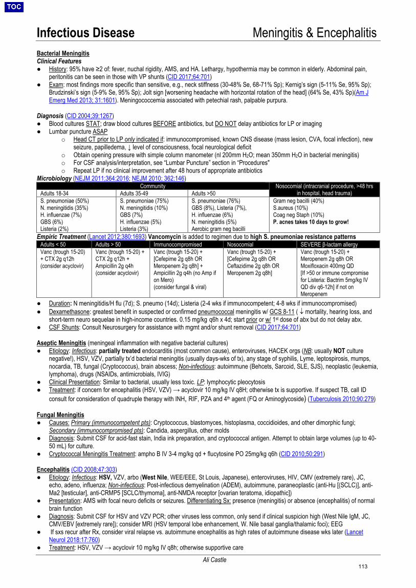
Infectious Disease Meningitis & Encephalitis
Ali Castle
Bacterial Meningitis Clinical Features● History: 95% have ≥2 of: fever, nuchal rigidity, AMS, and HA. Lethargy, hypothermia may be common in elderly. Abdominal pain,
peritonitis can be seen in those with VP shunts (CID 2017;64:701)● Exam: most findings more specific than sensitive, e.g., neck stiffness (30-48% Se, 68-71% Sp); Kernig’s sign (5-11% Se, 95% Sp);
Brudzinski’s sign (5-9% Se, 95% Sp); Jolt sign [worsening headache with horizontal rotation of the head] (64% Se, 43% Sp)(Am JEmerg Med 2013; 31:1601). Meningococcemia associated with petechial rash, palpable purpura.
Diagnosis (CID 2004;39:1267)● Blood cultures STAT; draw blood cultures BEFORE antibiotics, but DO NOT delay antibiotics for LP or imaging● Lumbar puncture ASAP
o Head CT prior to LP only indicated if: immunocompromised, known CNS disease (mass lesion, CVA, focal infection), newseizure, papilledema, ↓ level of consciousness, focal neurological deficit
o Obtain opening pressure with simple column manometer (nl 200mm H2O; mean 350mm H2O in bacterial meningitis)o For CSF analysis/interpretation, see “Lumbar Puncture” section in “Procedures"o Repeat LP if no clinical improvement after 48 hours of appropriate antibiotics
Microbiology (NEJM 2011;364:2016; NEJM 2010; 362:146)Community Nosocomial (intracranial procedure, >48 hrs
in hospital, head trauma) Adults 18-34 Adults 35-49 Adults >50 S. pneumoniae (50%)N. meningitidis (35%)H. influenzae (7%)GBS (6%)Listeria (2%)
S. pneumoniae (75%)N. meningitidis (10%)GBS (7%)H. influenzae (5%)Listeria (3%)
S. pneumoniae (76%)GBS (8%), Listeria (7%),H. influenzae (6%)N. meningitidis (5%)Aerobic gram neg bacilli
Gram neg bacilli (40%) S.aureus (10%)Coag neg Staph (10%)P. acnes takes 10 days to grow!
Empiric Treatment (Lancet 2012;380:1693) Vancomycin is added to regimen due to high S. pneumoniae resistance patterns Adults < 50 Adults > 50 Immunocompromised Nosocomial SEVERE β-lactam allergy Vanc (trough 15-20) + CTX 2g q12h(consider acyclovir)
Vanc (trough 15-20) + CTX 2g q12h + Ampicillin 2g q4h (consider acyclovir)
Vanc (trough 15-20) + [Cefepime 2g q8h OR Meropenem 2g q8h] + Ampicillin 2g q4h (no Amp if on Mero) (consider fungal & viral)
Vanc (trough 15-20) + [Cefepime 2g q8h OR Ceftazidime 2g q8h OR Meropenem 2g q8h]
Vanc (trough 15-20) + Meropenem 2g q8h OR Moxifloxacin 400mg QD [If >50 or immune compromise for Listeria: Bactrim 5mg/kg IV QD div q6-12h] if not on Meropenem
● Duration: N meningitidis/H flu (7d); S. pneumo (14d); Listeria (2-4 wks if immunocompetent; 4-8 wks if immunocompromised)● Dexamethasone: greatest benefit in suspected or confirmed pneumococcal meningitis w/ GCS 8-11 ( ↓ mortality, hearing loss, and
short-term neuro sequelae in high-income countries. 0.15 mg/kg q6h x 4d; start prior or w/ 1st dose of abx but do not delay abx.● CSF Shunts: Consult Neurosurgery for assistance with mgmt and/or shunt removal (CID 2017;64:701)
Aseptic Meningitis (meningeal inflammation with negative bacterial cultures) ● Etiology: Infectious: partially treated endocarditis (most common cause), enteroviruses, HACEK orgs (NB: usually NOT culture
negative!), HSV, VZV, partially tx’d bacterial meningitis (usually days-wks of tx), any stage of syphilis, Lyme, leptospirosis, mumps,nocardia, TB, fungal (Cryptococcus), brain abscess; Non-infectious: autoimmune (Behcets, Sarcoid, SLE, SJS), neoplastic (leukemia,lymphoma), drugs (NSAIDs, antimicrobials, IVIG)
● Clinical Presentation: Similar to bacterial, usually less toxic. LP: lymphocytic pleocytosis● Treatment: if concern for encephalitis (HSV, VZV) → acyclovir 10 mg/kg IV q8H; otherwise tx is supportive. If suspect TB, call ID
consult for consideration of quadruple therapy with INH, RIF, PZA and 4th agent (FQ or Aminoglycoside) (Tuberculosis 2010;90:279)
Fungal Meningitis ● Causes: Primary (immunocompetent pts): Cryptococcus, blastomyces, histoplasma, coccidioides, and other dimorphic fungi;
Secondary (immunocompromised pts): Candida, aspergillus, other molds● Diagnosis: Submit CSF for acid-fast stain, India ink preparation, and cryptococcal antigen. Attempt to obtain large volumes (up to 40-
50 mL) for culture.● Cryptococcal Meningitis Treatment: ampho B IV 3-4 mg/kg qd + flucytosine PO 25mg/kg q6h (CID 2010;50:291)
Encephalitis (CID 2008;47:303) ● Etiology: Infectious: HSV, VZV, arbo (West Nile, WEE/EEE, St Louis, Japanese), enteroviruses, HIV, CMV (extremely rare), JC,
echo, adeno, influenza; Non-infectious: Post-infectious demyelination (ADEM), autoimmune, paraneoplastic (anti-Hu [(SCLC)], anti-Ma2 [testicular], anti-CRMP5 [SCLC/thymoma], anti-NMDA receptor [ovarian teratoma, idiopathic])
● Presentation: AMS with focal neuro deficits or seizures. Differentiating Sx: presence (meningitis) or absence (encephalitis) of normalbrain function
● Diagnosis: Submit CSF for HSV and VZV PCR; other viruses less common, only send if clinical suspicion high (West Nile IgM, JC,CMV/EBV [extremely rare]); consider MRI (HSV temporal lobe enhancement, W. Nile basal ganglia/thalamic foci); EEG
● If sxs recur after Rx, consider viral relapse vs. autoimmune encephalitis as high rates of autoimmune disease wks later (LancetNeurol 2018:17:760)
● Treatment: HSV, VZV → acyclovir 10 mg/kg IV q8h; otherwise supportive care
113
Infectious Disease C. Difficile Infection
Hawra Al Lawati
General Information Risk factors: Antibiotics within last 3 months. All antibiotics have been associated with CDI, including 3rd/4th gen cephs, fluoroquinolones, carbapenems and clindamycin. Receipt of abx by pt previously in same bed and ward abx prescribing patterns also weakly associated.↑age, CKD, IBD, chemo/immunocompro, ± PPI/H2RA. Pathogenesis: fecal-oral, colonized host; most often infection requires both acquisition of C. diff plus loss of gut microbial abundance/diversity (i.e., due to abx). Symptoms are toxin-mediated: toxin A (enterotoxic) & toxin B (cytotoxic). Community-acquired CDI: 33% of new cases; p/w diarrhea w/o traditional RFs (abx, ↑age). Sources: contaminated food, H2O, pet, asymptomatic colonization in family members, babies, outpt medicine visits, ↓ mortality. Infect Drug Resist 2014;7:63
Clinical Manifestations • Features: loose/watery diarrhea (+/- mucous/occult blood); fever, abd pain, isolated ↑↑ WBC; ileus in severe infection.
Clin Infect Dis 2002;34:1585• Complications: fulminant colitis (65% mortal), ileus (<1% pts), toxic megacolon, perf, protein-losing enteropathy, AKI
Diagnosis-Glutamate dehydrogenase antigen produced by all c.diff strains (toxigenic &non-toxigenic)-Toxin A/B EIA: more specific to toxin producing strains (poor sensitivity)-NAAT/PCR toxin gene: can be + even in the absence of active infection (strain may have toxin gene but not necessarilyproduce the toxin) Open forum ID 2014MGH protocol: GDH EIA &toxin A/B assay first. If both positive = positive test. If both negative = negative test. If discordantresults but high clinical suscpicion for active C. difficile, call micro lab to request PCR testing. (NB: discordant results can be2/2 asymptomatic (non-toxin producing) colonization)• DO NOT retest within 1-2 weeks without significant clinical change. J Clin Micro 2008;46:3795• DO NOT test for “cure”. Studies have shown that toxin A+B EIA may remain positive for a long as 30 days in patients who
have resolution of symptoms. Am J Gastroenterol2013; 108:478• Consider early CT A/P if suspected C. diff w/ ileus or if concern for complications. Flex sig used in rare cases.Treatment (MGH ID Recs, updated IDSA guidelines) Initial Episode:
Category Criteria Treatment Non-severe WBC < 15 AND Cr<1.5 -Vanc 125 mg PO q6h or Fidaxomicin* 200 BID. D/c
cholestyramine if using (binds vanc)-Metronidazole no longer first-line (due to ↑↑ resistance)-Stop antiperistaltics and all non-essential antibiotics
Severe WBC>15 OR Cr>1.5 As above Fulminant Hypotension or shock, ileus,
megacolon -Vanc 500 mg PO q6h and metronidazole 500 mg IV q8h- If ileus: consider adding Vanc PR 500mg in 100cc retentionenema Q6H)-Obtain imaging, ID and surgical cs
Duration: 10d for non-fulminant infections; If concurrent abx use, treat CDI through abx course plus 7-14d after abx completion; PO vanc preferred for continuation once colitis resolves
*Fidaxomicin: bactericidal, less CDI recurrence than Vanc, some data for increased rates of cure but $$$$. Clin Infect Dis 2011;53:440, Lancet Infect Dis 2012;12:281 Recurrence: Occurs in 25% of pts after 1st episode w/in 30d • 1st recurrence: Tapered and pulsed PO Vanc regimen for 6-8 wks OR fidaxomicin 200mg BID x10d if Vanc used for
initial episode• 2nd recurrence (40-60% pts after 1st recur): Tapered and pulsed vanc regimen for 6-8 wks OR 125mg PO Vanc x10d
followed by Rifaximin 400mg TID x20d• 3rd recurrence: Evaluate for fecal microbiota transplant (FMT); improves outcomes compared to 14d PO Vanc. Can
also trial regimens used to tx 2nd recurrence (as above). NEJM 2013;368:407 BMC Med 2016;14:134Other Considerations: • PPX: PO vanc 125 mg BID lowers risk of recurrence from 27% to 4%; consider for pts needing abx with hx of
fulminant/recurrent CDI Clin Infect Dis 2016; 63:651• Probiotics: Administration around first dose of abx reduces risk of CDI by >50% with no significant adverse effects
(based on systemic review, not yet in guidelines) Gastroent 2017;152:1889• Bezlotoxumab: Promising new option; decreased recurrence compared to placebo when added to standard of care• FMT: obtain ID consult for initiation of FMT if refractory C. diff despite treatment (>2 recurrences)
114

Infectious Disease Invasive Fungal Infections
Dora Karagounis
Diagnostic Testing Summary: Risk Factors: heme malignancy, HSCT >> solid organ transplant >> patients on biologic therapies Fungal Markers: • 1,3-β-D Glucan (BDG) (Clin Infect Dis 2011;52:750) Cell wall polysaccharide, detects Candida, Aspergillus, Pneumocystis, Fusarium,
Trichosporon, Histo, Coccidio; CANNOT detect Mucor, Rhizopus, Blasto, Crypto; Sens 77%, Spec 86% w/ cut-off 80; false+ w/ IVIG, albumin, HD• Galactomannan (GM) (Cochrane Database Syst Rev 2015:CD007394): Aspergillus cell wall component, detect Aspergillus; Sens 65-80% for
serum test (BAL 90-95%), Spec 88%. False + w/ some TPN formulations. Can be used serially to monitor tx in pts.• Histo urine/serum Ag: Sens 90% urine Ag if disseminated (serum Ag Sens 80%); Spec limited by cross-reactivity• Crypto Ag: serum Ag Sens & Spec > 90% if disseminated, less so for pulm dz only• Blastomycoses: urine > serum Ag, high Sens, but modest Spec due to cross-reactivity Culture: Candida grows in blood/urine cx but decreased Sens if deep tissue infxn; if high c/f Coccidio, alert lab (biohazard)Antibody Detection: Clinically most useful if testing for Coccidio
Treatment Summary: x indicates activity, shading indicates 1st line Tx C. glabrata & krusei C. albicans Crypto Endemic Aspergillus Mucor
Fluconazole ** x x Itraconazole x x x Voriconazole x x x x x Posaconazole x x x x x x Isavuconazole* x x x x Micafungin x x x*** Ampho B x x x x x x
*Only approved for Invasive Aspergillosis/Mucor. **91% of MGH glabrata fluc-S. ***vori + mica not superior to vori alone
Pathogen-Specific Information: Invasive Opportunistic Fungi: Strongly recommend ID consult for most of these infections • Candida/yeast (risk factors: neutropenia, immunocompromised, TPN, IVDU, CVC, prior abdominal surgery)
o Spectrum of illness: sepsis (25% mortality), macronodular skin lesions (10%), endophthalmitis, endocarditis, osteoo Diagnostics: blood cultures (Candidemia never contaminant in blood), obtain optho exam, TTE, & ID consult o Treatment: Micafungin 1st line/empiric, transition to fluc if albicans, high dose fluc/vori if glabrata/krusei (or AmphoB for resistant strains);
Duration: 2 weeks after 1st neg cx and no dissemination in candidemia, longer for deep-seated infxno Source: Non-neutropenic: lines most likely source (remove early); neutropenic: GI most likely. o Prophylaxis: fluconazole, posaconazole or micafungin (for SOT, SCT, neutropenic)
• Cryptococcus/yeast (RFs: immunocompromised, liver dz, HIV, but can occur in immunocompetent) (Clin Infect Dis 2010;50:291)o Spectrum of illness: meningitis, pulmonary, cutaneous nodules, liver abscesseso Diagnostics: Serum/CSF CrAg, LP/CSF OP >20, ↓glucose, ↑TP, lymphs, +India ink o Treatment: amphoB + flucytosine (x2 wks), followed by fluconazole (>8 wks), serial LPs if OP>25 or symptoms of ↑ICPo Prophylaxis: typically not recommended
• Pneumocystis/yeast (risk factors: HIV with CD4 <200, steroids equiv to pred 20 mg x4 wks)o Spectrum of illness: Pulm symptom onset over days/weeks, PTX, hypoxia out of proportion to CXR (BL diffuse GGO)o Diagnostics: LDH >500 (sens not spec), BAL > induced sputum for silver stain, 1,3-BDG (Eur J Clin Microbiol Infect Dis 2014;33:1173) o Treatment: TMP/SMX with steroids (if A-a > 35, PaO2 < 70); Alternatives: atovaquone or pentamidine; Duration: 21 dayso Prophylaxis: TMP/SMX (1 SS qD or 1 DS MWF), atovaquone or dapsone (see UpToDate for ppx criteria)
• Aspergillus/mold (risk factors: immunocompromised esp neutropenia/steroids/transplant, COPD with prolonged ICU stay)o Spectrum of illness: invasive pulm w/ hemoptysis, PTX, aspergilloma, sinusitis, CNS, endophthalmitiso Diagnostics: CT with halo sign, BAL/sputum culture, 1,3-BDG (not spec), GM (spec, can trend in tx, BAL > sputum)o Treatment: vori (requires monitoring of drug levels and drug-drug int) or isavuconazole; Duration: Pulm: 6-12 weeks minimumo Prophylaxis: consider posaconazole in grade III-IV GVHD (NEJM 2007;356:348), vori in lung transplant w/ h/o aspergillus.
• Mucor/mold (risk factors: DKA, iron overload, heme malig, prolonged neutropenia, immunocomp.) (Semin Respir Crit Care Med 2015;36:692.o Spectrum of illness: rhino/orbital/cerebral invasion, pulmonary, GI, renal, black eschars over ulcers, rapidly progressive o Diagnostics: culture, wet prep (non-septating hyphae with wide-angle branches), CT with reverse halo signo Treatment: DEBRIDEMENT, AmphoB, consider posaconazole or isavuconazole (for salvage therapy or if renal disease)
Endemic Fungi: • Histoplasmosis (Endemic areas: Houston, OH/MS river valleys, Central America, Asia, Africa) (Clin Infect Dis 2007;45:807)
o Spectrum of illness: PNA, meningitis, mediastinal disease, disseminated disease.o Diagnostics: Ag from urine/serum/BAL, Cx, note chest imaging may appear similar to sarcoido Treatment: Itraconazole (mild-mod) or ampho B (severe), followed by itraconazole; Duration: 6-12 weekso Prophylaxis: for both Histo and Blasto (below), consider itraconazole ppx for HIV+ with CD4 <150 in hyperendemic areas
• Blastomycosis: (Endemic areas: OH/MS river valleys) o Spectrum of illness: fever, PNA, ARDS in severe, ulcerated skin lesions, prostatitis, CNSo Diagnostics: wet prep (broad-based, budding yeast), culture, urine > serum Ag, never colonizero Treatment: Itraconazole (mild-mod) or ampho B (severe), followed by itraconazole; Duration: 6-12 mos
• Coccidiomycosis: (Endemic areas: SW and S US)o Spectrum of illness: fever, cough, rash, HA, eosinophilia, meningitis, osteo.o Diagnostics: serologies, cx, spherules on bx/aspirateo Treatment: fluconazole or itraconazole, consider amphoB if severe; Duration: 3-12 moso Prophylaxis: fluconazole for 1° ppx ONLY for transplant recipients in endemic areas, not in HIV; use fluconazole for 2o ppx
115
Infectious Disease Tuberculosis
Rebecca Abelman
Epidemiology: 1 in 4 infected worldwide; US incidence 2.8/100,000 persons; 5.6% HIV coinfection; 4% MDR (CDC MMWR TB US 2017)
Risk Factors: Acquisition: travel hx to/from high-prevalence area, homelessness or incarceration, PWID, health care work, racial/ethnic minority; Reactivation: risk is 5% in first 2 years and 5%-10% over lifetime, but higher if pt has >1 of the following: HIV+, immunosupp, CKD (esp. HD), DM, cancer, transplant, TNFα inhibitors, silicosis, malabsorption malnutrition, tobacco, EtOH (NEJM 2011;364:1441)
Screening for Latent TB: Decision to test based on likelihood of exposure + likelihood of progression to active disease. IGRA preferred (Quant-GOLD test of choice at MGH); TST acceptable (NB: only 60% spec in pts who received BCG vaccine). Both IGRA and TST are 80-90% sens and >95% spec in immunocompetent hosts, ↓ sens in immunocompromised pts. Neither test rules in/out active TB and they can be discordant ~30% of the time. If + test but no risk factors, repeat either IGRA or TST prior to treatment. (Clin Infect Dis 2017;64:111)
Clinical Manifestations: • Primary TB: fever, chest pain, cough, arthralgias. CXR often
normal or (+) hilar LAD• Reactivation TB: fever, cough, hemoptysis, night sweats, weight loss; CXR often involves posterior/apical upper lobe or superior
aspect of lower lobe, or cavitation [seen in 1/3 of pts, a/w ↑ org. burden ↑ infectious, AFB+]; more common than primary TB!
Diagnostics for Active Pulmonary and Extrapulmonary TB (J Clin Microbiol 2007:45:4064; Lancet 2007;369:9578)Site of Infxn Diagnostic Tests
Sputum Send expectorated or induced sputum for AFB smear and culture x3 ≥8hrs apart (get NAAT/PCR on one of the specimens); smear may be (-) if burden is low: ~20% smear negative (HIV-), ~60% smear negative (HIV+)
Bronchi Send brushings, washings, BAL, sputum for AFB smear, NAAT/PCR and culture; +/- transbronchial biopsy. Obtain post-bronch induced sputum to increase yield.
Ascites or pleural fluid
Adenosine Deaminase (ADA): if >39 units/L → high sens/spec; Free IFN-gamma (if elevated, high sens/spec); AFB smear, NAAT/PCR, and culture (poor sensitivity, helpful if positive)
CSF At least 3 large vol (10-15cc) serial LPs if possible (Increases dx yield). Cell counts w/ ↓ glucose, ↑ protein, lymphocyte predominance; elev ADA useful adjunct. Send smear, Cx, and NAAT
Wound/Tissue AFB-positive staining and caseating granulomas; if cytopenic, consider bone marrow biopsy Urine Classically sterile pyuria; send first AM void (large vol -50cc) for culture x 3 days to improve yield Blood Must send mycobacterial cultures for AFB (isolators)
Patient Isolation: clinical decision based on likelihood of active TB• When: cough, dyspnea, or hemoptysis + >1 risk factor (HIV+, foreign born, substance use disorder, homeless, recent incarceration,
prior TB or exposure); first obtain CXR; if CXR normal (and HIV- or CD4>200), TB less likely. If CXR abnormal/equivocal (or HIV+and CD4<200), maintain isolation and obtain 3 sputum samples for AFB smear and mycobacterial culture as above. Consider ID c/s.
• Discontinue: If alt dx OR AFB smear neg x 3 with very low suspicion OR on TB tx x 2wks + AFB smear neg x3 + clinical improvement
Approach to Treatment: (Clin Infect Dis 2016;63:e147; NEJM 2015;373:2149)• Prior to Starting Treatment:
o General: check baseline LFTs/Cr, visual acuity/color discrimination, screen for HIV, Hep A/B/C, DM, EtOH use, pregnancyo Before treating latent TB: need to rule out active TB (obtain relevant history, CXR)o Before treating active TB: obtain ID consult, send TB for drug sensitivity testing
• Treatment Regimens:o Latent TB: INH 5mg/kg daily (max 300mg) + B6 for 6-9mo, OR RIF 10mg/kg daily (max 600mg) for 4mo, OR INH + Rifapentine
weekly for 3mo (requires DOT)o Active TB: INH + RIF + pyrazinamide (PZA) + ethambutol (EMB) daily for 2 mo, followed by INH + RIF for 4 moo Quinolones: 1st line tx with MDR-TB, so avoid in bacterial PNA if suspicious of active TB (will ↓ diagnostic yield and ↑ risk of
resistance) • Drug-resistant TB: suspect if previously treated, treatment failure, from prevalent area (India, China, Russia, S. Africa), or known
exposure. Treatment regimen depends on drug susceptibility profile; usually for 12-24 month tx course. 80% mortality• HIV co-infection: if CD4<50, start ART within 2 weeks after starting TB therapy. If CD4>50 with severe clinical disease, start in 2
weeks, and if CD4>50 and absence of severe clinical disease, may start within 8 weeks to reduce risk of IRIS. In TB meningitis,consider deferring ART initiation to 8 weeks.
• Extrapulmonary TB: highly variable presentations, variable multi-drug regimen, duration depends on site of infection & response. Addglucocorticoids for TB meningitis for 25% RR reduction in mortality (Cochrane Sys Rev 2016;4:1-64) may also consider in TBpericarditis
• Medication Side Effects: hepatotoxicity (INH, RIF, PZA), optic neuritis (EMB), peripheral neuropathy (INH add pyridoxine [B6] withinitiation of treatment), orange bodily fluids (RIF), numerous drug-drug interactions (especially RIF)
TST size* at 48-72 hrs Patient population
>5 mm HIV, prior TB hx, CXR c/w prior TB,silicosis, immunosuppression
>10 mm Diabetes, CKD, IVDU>15 mm No risk factors NB: size reflects skin induration, NOT erythema
116
Infectious Disease HIV/AIDS & Opportunistic Infections
Rebecca Abelman, Andrew Hoekzema
Definition and Clinical Manifestations: • Acute HIV: mono-like syndrome rash, LAD, fever, oral ulcers, pharyngitis, myalgias, diarrhea; presents 3-6 wks after infection• AIDS: HIV+ with: CD4 count<200 or CD4 T-cell <14% of total lymphs or AIDS defining illness
HIV Screening and Diagnostics: • SCREEN ALL 13-64Y ONCE, every pregnancy; risk-based: at least annually in IVDU, CSW, MSM >1 partner since last test, partners of all
high-risk pts, another STI; In MA: opt out verbal consent (“We’ll be conducting a number of tests, including for HIV”)• 4th gen combined HIV 1/2 Ab/p24 Ag assay: mean detection limit @ 18d (5d sooner than 3rd gen) (STD 2017;44:739)• HIV RNA PCR/viral load (VL): mean 12d, high sens/spec but slow, expensive; used for: (1) concern for acute HIV (Ab/Ag testing are
negative early in disease course); (2) confirmation of HIV diagnosis; (3) viral load
PrEP (Pre-Exp Ppx): sero-discordant couples w/ HIV+ partner on ARVs < 6mos, CSW, high-risk IVDU/MSM/ heterosexuals, recent STD. • Regimen: TDF/FTC (Truvada) QD reduces risk (40-75%, >95% w/excellent adherence), d/c when risk is no longer present. HIV testing
and STI testing q3mos while on therapy; test for HBV prior to initiation• If the partner of the patient is HIV positive but has an undetectable VL, risk of HIV transmission is near 0! (JAMA 2016;316:171)nPEP (Non-Occupational Post-Exp Ppx): persons presenting at ≤72hrs after non-occupational high-risk exposure from HIV+ source; case-by-case decision if HIV status of source unknown; test w/ HIV Ab/Ag at baseline & test for STIs, HBV, HCV.• Regimen: TDF/FTC (Truvada) + [raltegravir or dolutegravir] x28d; if ≥1 course nPEP in last year, consider PrEP
Basic Evaluation for Newly Diagnosed HIV/AIDS Patients • CD4 count, VL, genotype/resistance testing, CBC w/diff, BMP, U/A, LFTs, lipase, A1c/FLP, Hep A/B/C, hCG, cervical and/or anal pap,
RPR, GC/CT, PPD or IGRA; CMV, VZV, toxo, mycobacterial BCx if CD4 < 100, dilated eye exam if CD4<50• Initiate ARV early through referral (p36222) at all CD4 levels to decrease mortality (NEJM 2015;373:795) In many cases, ART can be
initiated on site, even prior to genotype return, even in high-risk patients (AIDS 2018;32:17) Make sure ID is involved in this decision.Treatment for ARV-Naïve Patients many options, choose based on indiv pt factors, drug-drug interactions, resistance testing • 1st line: 2 NRTI “backbone” (typically TAF/FTC or TDF/FTC [Truvada]) + 1 from diff. class, typically integrase inhibitorHospital Management of HIV/AIDS Patients:• If patient is on ART: determine regimen & adherence; typically continue ARVs (interruptions can ↑ disease progression)
o If must hold ARVs because of significant non-adherence or recent severe adverse reaction, hold all ARVs and consult IDo Beware of drug-drug interactions, particularly with boosted PIs (e.g. PPIs, check http://arv.ucsf.edu/insite?page=ar-00-02)
• If patient not yet on ART: prioritize OI tx, ppx, consult ID for help on early inpt vs outpt initiation of ART• IRIS: worsening sx of underlying infx (TB, MAC, CMV, others) 1-3 mos post-ART initiation, high risk if low CD4 count
o Nevertheless, early ARV init safe after OI dx, except in crypto meningitis (PLoS ONE 2009;4:e5575)Opportunistic Infections
1. Opportunistic Infections Prophylaxis Summary Recommendations for HIV in the US (JAMA 2018;320:379)CD4 Opportunistic Infection Prophylaxis Criteria for D/C
Any CD4 count
Influenza, HAV, HBV, HPV, VZV, S.pneumo, TB
Vax: Flu; HAV, HBV, HPV, PCV 13, PPSV23 after 8 wks; no live vax w/ CD4<200; latent TB: INH/B6 x 9mo
None
< 200 Pneumocystis jirovecii (or hx of thrush)
TMP-SMX DS QD (preferred) or 1 SS QD or dapsone 100mg QD or atovaquone 1500mg QD
CD4 >200 x 3mo
<150 Histo (only if endemic; not in MA) Itraconazole 200 mg PO QD CD4 > 150 x 6 mo < 100 Toxoplasma TMP-SMX DS QD or dapsone 50mg QD +
pyrimethamine 50mg qWk + leucovorin 25 qWk CD4 > 200 x 3mo
< 50 Mycobact. avium complex (MAC) Ppx no longer reccommended if ARVs started CD4>100 x3mo Treatment of OIs in Adults with HIV/AIDS – Also see “Invasive Fungal Infections”
Pathogen Diagnosis 1st Line Treatment MAC Cx (blood/sputum/bronch/marrow/tissue), AFB stain Azithro 600mg qday or clarithro 500mg BID
+ ethambutol 15mg/kg QDPneumocystis jirovecii Typically induced sputum (sens 50-90%) or BAL wash
(sens >90%) for dx; Cx not reliable TMP-SMX (15-20 mg/kg/day of TMP IV) x 21d, ± steroids if PaO2 < 70 or A-a >35
Toxoplasma gondii CT/MRI: ring-enhancing, most pts have IgG+ but not IgM+, brain Bx if Rx fails (r/o CNS lymphoma)
Pyrimeth 200mg x1; then by weight + sulfadiazine + leucovorin x6wks
Herpes Simplex Virus (HSV) Oral/genital: DFA, PCR, viral cx CNS: LP + CSF PCR
Acycl. 400 PO q8h or valacycl. 1g PO q12h x5-10d; CNS: acycl. 10mg/kg IV q8h x3wk
Cytomegalovirus (CMV) Retinitis: exam; Colitis/esophagitis: bx; PNA: bronch; Neuro: LP with PCR, brain Bx, Blood: PCR
In general: ganciclovir or foscarnet IV, switch to PO w/improvement
PML MRI: non-enhancing lesions, LP with JCV PCR Only disease-modifying tx is ARVs Cryptococcus (rare in US pts)
Serum and CSF CrAg, serum and/or CSF culture, ↑ CSF opening pressure
Ambisome + flucytosine x 2 wkthen high-dose fluc x 8 wkthen low-dose x 1yr
Mucocutaneous candidiasis (esophageal/oral)
Clincal dx. White plaque removed w/tongue depressor, +KOH; EGD + Bx
Oral: fluc 100mg PO x7-14d vs nystatin S&S; Eso: fluc 100-400mg PO/IV x14-21d)
117
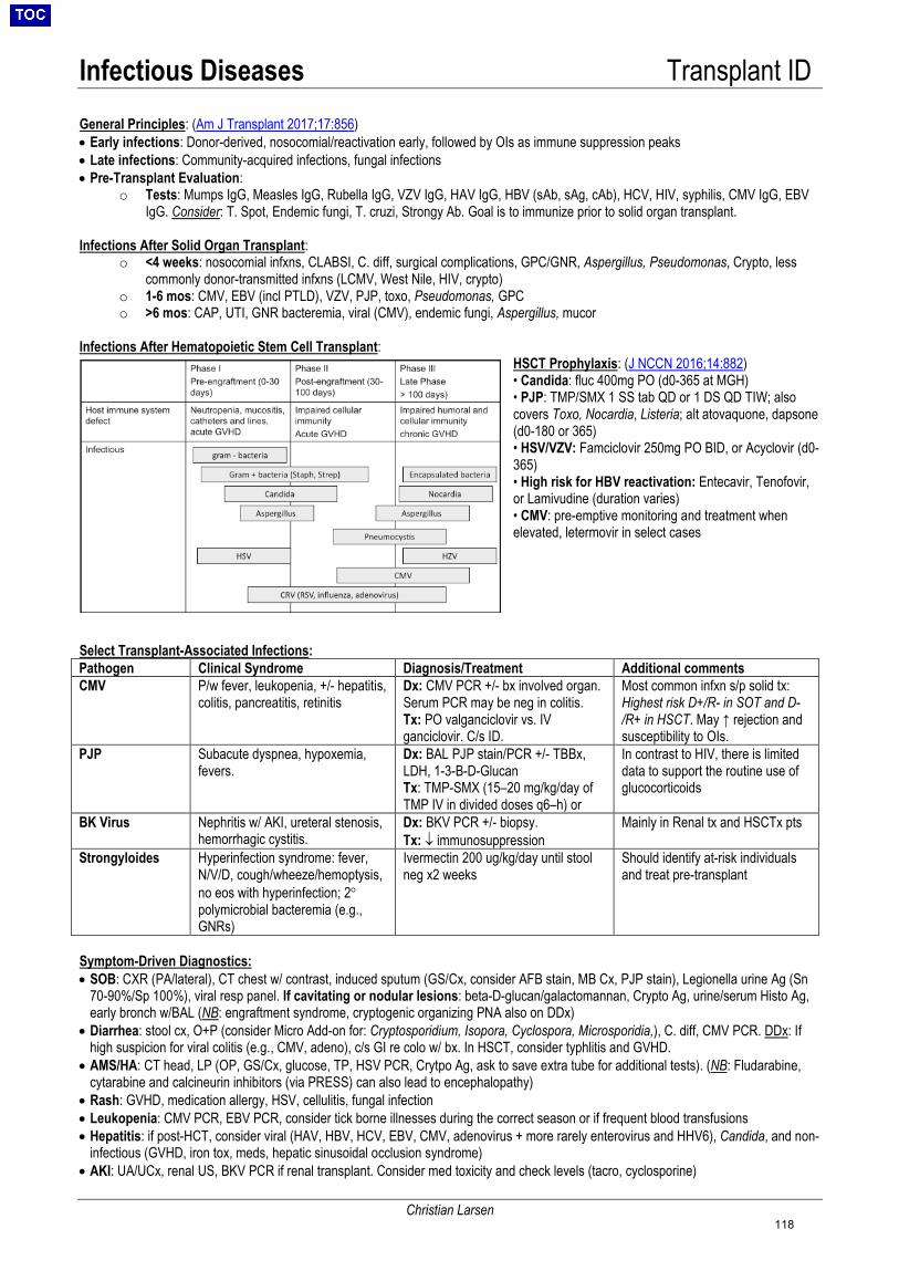
Infectious Diseases Transplant ID
Christian Larsen
General Principles: (Am J Transplant 2017;17:856) • Early infections: Donor-derived, nosocomial/reactivation early, followed by OIs as immune suppression peaks• Late infections: Community-acquired infections, fungal infections• Pre-Transplant Evaluation:
o Tests: Mumps IgG, Measles IgG, Rubella IgG, VZV IgG, HAV IgG, HBV (sAb, sAg, cAb), HCV, HIV, syphilis, CMV IgG, EBVIgG. Consider: T. Spot, Endemic fungi, T. cruzi, Strongy Ab. Goal is to immunize prior to solid organ transplant.
Infections After Solid Organ Transplant: o <4 weeks: nosocomial infxns, CLABSI, C. diff, surgical complications, GPC/GNR, Aspergillus, Pseudomonas, Crypto, less
commonly donor-transmitted infxns (LCMV, West Nile, HIV, crypto)o 1-6 mos: CMV, EBV (incl PTLD), VZV, PJP, toxo, Pseudomonas, GPCo >6 mos: CAP, UTI, GNR bacteremia, viral (CMV), endemic fungi, Aspergillus, mucor
Infections After Hematopoietic Stem Cell Transplant: HSCT Prophylaxis: (J NCCN 2016;14:882) • Candida: fluc 400mg PO (d0-365 at MGH)• PJP: TMP/SMX 1 SS tab QD or 1 DS QD TIW; alsocovers Toxo, Nocardia, Listeria; alt atovaquone, dapsone(d0-180 or 365)• HSV/VZV: Famciclovir 250mg PO BID, or Acyclovir (d0-365)• High risk for HBV reactivation: Entecavir, Tenofovir,or Lamivudine (duration varies)• CMV: pre-emptive monitoring and treatment whenelevated, letermovir in select cases
Select Transplant-Associated Infections: Pathogen Clinical Syndrome Diagnosis/Treatment Additional comments CMV P/w fever, leukopenia, +/- hepatitis,
colitis, pancreatitis, retinitis Dx: CMV PCR +/- bx involved organ. Serum PCR may be neg in colitis. Tx: PO valganciclovir vs. IV ganciclovir. C/s ID.
Most common infxn s/p solid tx: Highest risk D+/R- in SOT and D-/R+ in HSCT. May ↑ rejection and susceptibility to OIs.
PJP Subacute dyspnea, hypoxemia, fevers.
Dx: BAL PJP stain/PCR +/- TBBx, LDH, 1-3-B-D-Glucan Tx: TMP-SMX (15–20 mg/kg/day of TMP IV in divided doses q6–h) or
In contrast to HIV, there is limited data to support the routine use of glucocorticoids
BK Virus Nephritis w/ AKI, ureteral stenosis, hemorrhagic cystitis.
Dx: BKV PCR +/- biopsy. Tx: ↓ immunosuppression
Mainly in Renal tx and HSCTx pts
Strongyloides Hyperinfection syndrome: fever, N/V/D, cough/wheeze/hemoptysis, no eos with hyperinfection; 2° polymicrobial bacteremia (e.g., GNRs)
Ivermectin 200 ug/kg/day until stool neg x2 weeks
Should identify at-risk individuals and treat pre-transplant
Symptom-Driven Diagnostics: • SOB: CXR (PA/lateral), CT chest w/ contrast, induced sputum (GS/Cx, consider AFB stain, MB Cx, PJP stain), Legionella urine Ag (Sn
70-90%/Sp 100%), viral resp panel. If cavitating or nodular lesions: beta-D-glucan/galactomannan, Crypto Ag, urine/serum Histo Ag,early bronch w/BAL (NB: engraftment syndrome, cryptogenic organizing PNA also on DDx)
• Diarrhea: stool cx, O+P (consider Micro Add-on for: Cryptosporidium, Isopora, Cyclospora, Microsporidia,), C. diff, CMV PCR. DDx: Ifhigh suspicion for viral colitis (e.g., CMV, adeno), c/s GI re colo w/ bx. In HSCT, consider typhlitis and GVHD.
• AMS/HA: CT head, LP (OP, GS/Cx, glucose, TP, HSV PCR, Crytpo Ag, ask to save extra tube for additional tests). (NB: Fludarabine,cytarabine and calcineurin inhibitors (via PRESS) can also lead to encephalopathy)
• Rash: GVHD, medication allergy, HSV, cellulitis, fungal infection• Leukopenia: CMV PCR, EBV PCR, consider tick borne illnesses during the correct season or if frequent blood transfusions• Hepatitis: if post-HCT, consider viral (HAV, HBV, HCV, EBV, CMV, adenovirus + more rarely enterovirus and HHV6), Candida, and non-
infectious (GVHD, iron tox, meds, hepatic sinusoidal occlusion syndrome)• AKI: UA/UCx, renal US, BKV PCR if renal transplant. Consider med toxicity and check levels (tacro, cyclosporine)
118
Infectious Diseases STIs & Travel Medicine
Ijeoma Eleazu, Brady Page, Hawra Al Lawati
STIs
Travel Medicine Pre-travel evaluation - Patient : Medical conditions (immunosuppressed ?), allergies (particularly to vaccines), pregnant/planning to get pregnant, immunizationhx, prior travel history (experience with malaria prophylaxis/prior travel related illlnesses), med list- Trip: Place, duration, season, purpose of a trip (medical / visiting family), itinerary (urban vs. rural areas, cruise ship, exposure toanimals, cave exploration, water exposure)- USE MGH Developed tool (Heading Home Healthy) to enter your patient’s details for specific recommendationsImmunizations1. Ensure routine vaccinations are uptodate2. Use CDC site to get country-specific recommedation on vaccination (clinician view & select country).Travel Specific Vaccines: yellow fever, hep A, meningococcal, typhoid, Japanese encephalitis, Pre-exposure rabies, cholera.Malaria ProphylaxisSearch by country in CDC tool to know what and when to prescribe (based on resistance patterns).Start ~ 1 week before travel and up to 4 weeks after.Daily meds: Atovaquone-proguanil (Malarone), Doxycycline, Primaquine. Weekly meds: Mefloquine, Chloroquine.
Traveler’s diarrhea Bacteria most common pathogens: ETEC, C. Jejuni, Shigella & Salmonella spp. Tx : Loperamide/Bismuth. Antibiotics (flouroquinolones/azithromycin) if moderate/ severe cases (interfere with activity or dysentery). CDC Yellow Book Infections in a Returning Traveler - Assess if life threatening illness or if transmissable via respiratory, contact, etc (isolate pts)- Broad ddx which depends on exposure risk, host/patient vulnerability and incubation periods.- Common culprits : Malaria, TB, STIs, Tick-Borne disease, Mosquito-Borne illnesses, Enteric fever (NEJM 2017;376:548)
LESIONS Symptoms Diagnosis Treatment
Syphilis (T pallidum)
1°: painless, firm, round ulcer 2°: fever, condyloma lata of skin/mucus membranes, LAD, myalgia, uveitis, meningitis 3°: aortitis/aneurysm, disseminated gummas, CN palsies, tabes dorsalis (impaired gait, sensation, reflexes)
VDRL, RPR (nontreponemal): titers are used to track infection. Detects cardiolipin Ab; Nonspec TPPA, TP-EIA (treponemal); detects Ab. specific to T. pallidum, but positive for life once syphilis acquired
1°/2°: PCN G benzathine 2.4 million units IM x1 3°: PCN G benzathine 2.4 million units IM qweek x3 Neuro: IV/IM PCN G x10-14d (CID 2011;53:S110)
Genital herpes (HSV2>HSV1)
Prodrome → painful vesicles → ulcers 1° infection may include systemic sxs
Confirm clinical dx with PCR or viral culture
Chronic suppressive (if 6 outbreaks/yr) vs episodic therapy (acyclovir, valacyclovir, famcyclovir)
Lymphograuloma venereum (C trachomatis)
1°: transient, painless anogenital lesion 2°: 2-6w later, painful inguinal LAD, back pain, systemic sxs 3°: ‘Genitoanorectal syndrome’ from pelvic and abd LAD with inflamm diarrhea, fistulas, strictures, abscesses, necrosis
Positive IgG/complement fixation + clinical diagnosis; NAAT inpipeline
Doxy 100mg bid x21d + aspiration of buboes
DISCHARGES Symptoms Diagnosis Treatment
Gonorrhea (N gonorrhoeae),
Chlamydia (C trachomatis)
♀: mucopurulent cervicitis, urethritis, PID(abd pain, adnexal/cervical tenderness, +/-fever), frequently asymptomatic♂: painful urethritis with purulentdischarge, epididymitisAll: pharyngitis
♀: vaginal swab NAAT > urineNAAT♂: dirty catch urine NAATAll: consider pharyngeal/analswab based on hx (pts can self-swab)
Ceftriaxone 250mg IM + azithromycin 1g po Note: CTX resistant Gonorrhea in Asia (MIC ≥0.125 µg/m). Consider obtaining Cx to test for resistance (Emerg Infect Dis 2018; 24:381)
Doxy not alternative to azithro but used in PID to tx C trachomatis
Trichomoniasis (T vaginalis)
Purulent malodorous vaginal discharge, pruritus, dysuria, frequency, dyspareunia Usually asymptomatic in ♂
Wet mount → vaginal swab NAAT
Metronidazole 2g po or tinidazole 2g po (better tolerated than MTZ) + GC/CT tx; treat partner
119
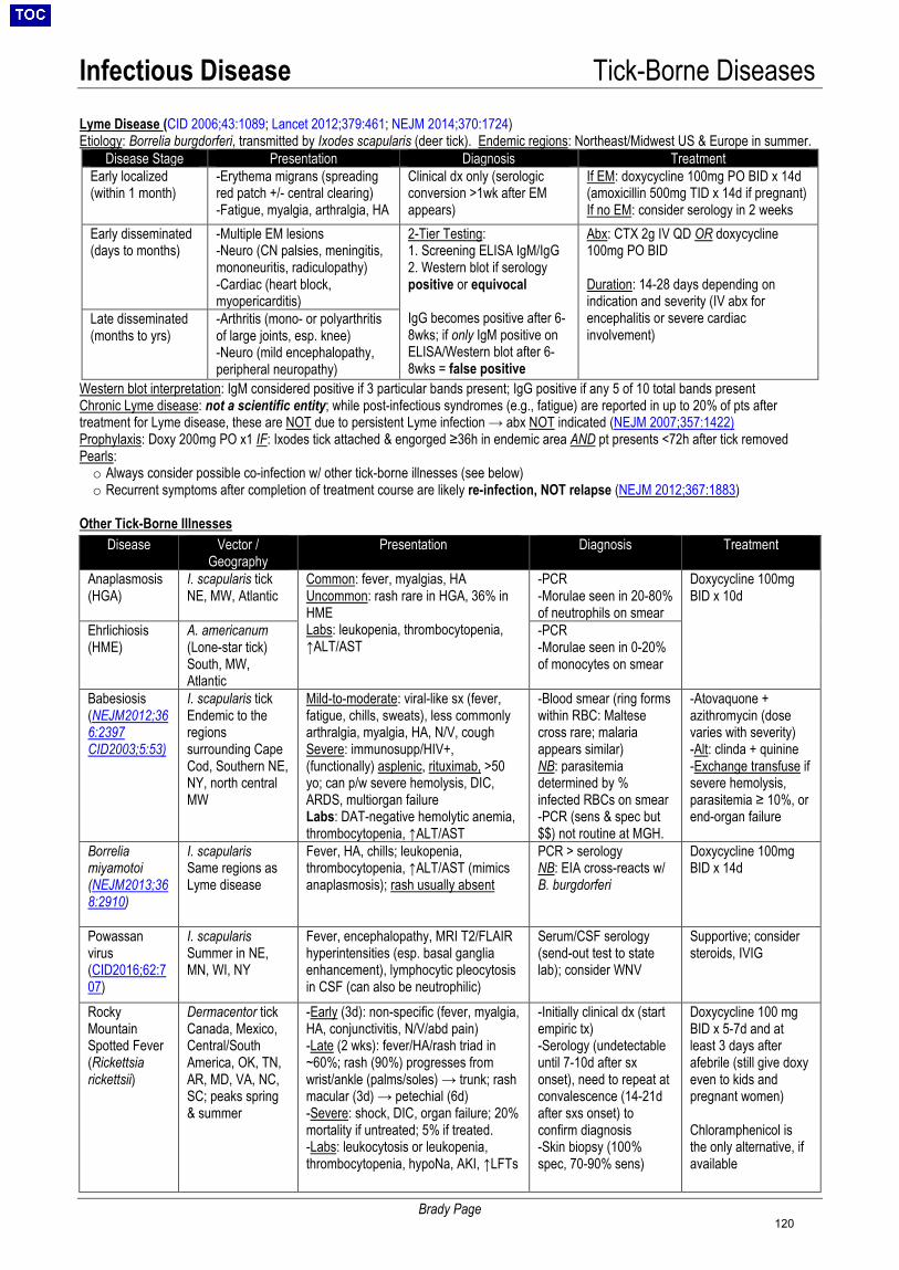
Infectious Disease Tick-Borne Diseases
Brady Page
Lyme Disease (CID 2006;43:1089; Lancet 2012;379:461; NEJM 2014;370:1724) Etiology: Borrelia burgdorferi, transmitted by Ixodes scapularis (deer tick). Endemic regions: Northeast/Midwest US & Europe in summer.
Disease Stage Presentation Diagnosis Treatment Early localized (within 1 month)
-Erythema migrans (spreadingred patch +/- central clearing)-Fatigue, myalgia, arthralgia, HA
Clinical dx only (serologic conversion >1wk after EM appears)
If EM: doxycycline 100mg PO BID x 14d (amoxicillin 500mg TID x 14d if pregnant) If no EM: consider serology in 2 weeks
Early disseminated (days to months)
-Multiple EM lesions-Neuro (CN palsies, meningitis,mononeuritis, radiculopathy)-Cardiac (heart block,myopericarditis)
2-Tier Testing:1. Screening ELISA IgM/IgG2. Western blot if serologypositive or equivocal
IgG becomes positive after 6-8wks; if only IgM positive on ELISA/Western blot after 6-8wks = false positive
Abx: CTX 2g IV QD OR doxycycline 100mg PO BID
Duration: 14-28 days depending on indication and severity (IV abx for encephalitis or severe cardiac involvement)
Late disseminated (months to yrs)
-Arthritis (mono- or polyarthritisof large joints, esp. knee)-Neuro (mild encephalopathy,peripheral neuropathy)
Western blot interpretation: IgM considered positive if 3 particular bands present; IgG positive if any 5 of 10 total bands present Chronic Lyme disease: not a scientific entity; while post-infectious syndromes (e.g., fatigue) are reported in up to 20% of pts after treatment for Lyme disease, these are NOT due to persistent Lyme infection → abx NOT indicated (NEJM 2007;357:1422) Prophylaxis: Doxy 200mg PO x1 IF: Ixodes tick attached & engorged ≥36h in endemic area AND pt presents <72h after tick removed Pearls: o Always consider possible co-infection w/ other tick-borne illnesses (see below)o Recurrent symptoms after completion of treatment course are likely re-infection, NOT relapse (NEJM 2012;367:1883)
Other Tick-Borne Illnesses
Disease Vector / Geography
Presentation Diagnosis Treatment
Anaplasmosis (HGA)
I. scapularis tickNE, MW, Atlantic
Common: fever, myalgias, HA Uncommon: rash rare in HGA, 36% in HME Labs: leukopenia, thrombocytopenia, ↑ALT/AST
-PCR-Morulae seen in 20-80%of neutrophils on smear
Doxycycline 100mg BID x 10d
Ehrlichiosis (HME)
A. americanum(Lone-star tick)South, MW,Atlantic
-PCR-Morulae seen in 0-20%of monocytes on smear
Babesiosis (NEJM2012;366:2397 CID2003;5:53)
I. scapularis tickEndemic to theregionssurrounding CapeCod, Southern NE,NY, north centralMW
Mild-to-moderate: viral-like sx (fever, fatigue, chills, sweats), less commonly arthralgia, myalgia, HA, N/V, cough Severe: immunosupp/HIV+, (functionally) asplenic, rituximab, >50 yo; can p/w severe hemolysis, DIC, ARDS, multiorgan failure Labs: DAT-negative hemolytic anemia, thrombocytopenia, ↑ALT/AST
-Blood smear (ring formswithin RBC: Maltesecross rare; malariaappears similar)NB: parasitemiadetermined by %infected RBCs on smear-PCR (sens & spec but$$) not routine at MGH.
-Atovaquone +azithromycin (dosevaries with severity)-Alt: clinda + quinine-Exchange transfuse ifsevere hemolysis,parasitemia ≥ 10%, orend-organ failure
Borrelia miyamotoi (NEJM2013;368:2910)
I. scapularisSame regions asLyme disease
Fever, HA, chills; leukopenia, thrombocytopenia, ↑ALT/AST (mimics anaplasmosis); rash usually absent
PCR > serology NB: EIA cross-reacts w/ B. burgdorferi
Doxycycline 100mg BID x 14d
Powassan virus (CID2016;62:707)
I. scapularisSummer in NE,MN, WI, NY
Fever, encephalopathy, MRI T2/FLAIR hyperintensities (esp. basal ganglia enhancement), lymphocytic pleocytosis in CSF (can also be neutrophilic)
Serum/CSF serology (send-out test to state lab); consider WNV
Supportive; consider steroids, IVIG
Rocky Mountain Spotted Fever (Rickettsia rickettsii)
Dermacentor tick Canada, Mexico, Central/South America, OK, TN, AR, MD, VA, NC, SC; peaks spring & summer
-Early (3d): non-specific (fever, myalgia,HA, conjunctivitis, N/V/abd pain)-Late (2 wks): fever/HA/rash triad in~60%; rash (90%) progresses fromwrist/ankle (palms/soles) → trunk; rashmacular (3d) → petechial (6d)-Severe: shock, DIC, organ failure; 20%mortality if untreated; 5% if treated.-Labs: leukocytosis or leukopenia,thrombocytopenia, hypoNa, AKI, ↑LFTs
-Initially clinical dx (startempiric tx)-Serology (undetectableuntil 7-10d after sxonset), need to repeat atconvalescence (14-21dafter sxs onset) toconfirm diagnosis-Skin biopsy (100%spec, 70-90% sens)
Doxycycline 100 mg BID x 5-7d and at least 3 days after afebrile (still give doxy even to kids and pregnant women)
Chloramphenicol is the only alternative, if available
120
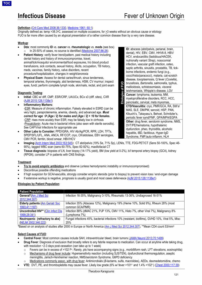
Infectious Disease Fever of Unknown Origin
Theodora Karagounis
Definition (Crit Care Med 2008;36:1330, Medicine 1961; 60:1) Originally defined as: temp >38.3oC, assessed on multiple occasions, for >3 weeks without an obvious cause or etiology FUO is far more often caused by an atypical presentation of a rather common disease than by a very rare disease.
Workup: • Ddx: most commonly ID vs. cancer vs. rheumatologic vs. meds (see box)
o In 25-50% of cases, no source is identified (Medicine 2007;86:26)• Patient History: verify fever trend/pattern, past medical history including
dental history and history of immunocompromise, travel,animal/tick/mosquito/ environmental/food exposures, h/o blood producttransfusions, sick contacts, sexual history, illicits, occupation, TB history,meds, vaccines, family history, valve disorders, recentprocedure/hospitalization, changes in weight/anorexia
• Physical Exam: Assess for dental caries/thrush, sinus tenderness,temporal arteries, thyromegaly, abd tenderness, HSM, CV murmur; inspecteyes, fundi; perform complete lymph node, skin/nails, rectal, and joint exam
Diagnostic Testing: • Initial: CBC w/ diff, CMP, ESR/CRP, UA/UCx, BCx x3 (diff. sites), CXR
(AJM 2015:128;1138e1)• Inflammatory Markers:
-ESR: Measure of chronic inflammation. Falsely elevated in ESRD (can bevery high), paraproteinemia, anemia, obesity, and advanced age. Mustcorrect for age (Age / 2) for males and (Age / 2) + 10 for females.-CRP: rises more acutely than ESR; may be falsely low in cirrhosis-Procalcitonin: Acute rise in bacterial infxns (also seen with sterile serositis).See CAP/Viral Infections for appropriate use
• Other Labs to Consider: PPD/IGRA, HIV Ab/Ag/PCR, RPR, LDH, TFTs,SPEP/SFL/UFL, ANA, ANCA, RF/CCP, cryo, CK/aldolase, EBV serologies,CMV PCR, ferritin, blood smear, HBV/HCV
• Imaging (Arch Intern Med 2003;163:545): CT abd/pelvis (19% Se, 71% Sp), LENIs, TTE, FDG-PET/CT (Sens 50-100%, Spec 46-90%), tagged WBC scan (sens 60-75%, Spec 82-92%), maxillofacial CT
• Tissue diagnosis: biopsies of LN, liver biopsy (14-17% yield), BM (low yield at 0-2%), b/l temporal artery biopsy (GCA), kidney(RPGN), consider LP in patients with CNS findings.
Treatment: • Try to avoid empiric antibiotics and observe (unless hemodynamic instability or immunocompromised)• Discontinue possible offending medications• If high suspicion for GCA/vasculitis, strongly consider empiric steroids (prior to biopsy) to prevent vision loss / end-organ damage• If extensive workup is negative, prognosis is usually good and most cases defervesce (AJM 2015:128;1138e1)Etiologies by Patient Population:
Patient Population General*(Am J Med Sci 2012;344:307)
Infection 16-35%, Malignancy 3-10%, Rheumatic 13-36%, Undiagnosed 16-51%
Elderly patients (Am Geriatr Soc 1993;41:1187)
Infection 35% (Abscess 12%), Malignancy 19% (Heme 10%, Solid 9%), Rheum 28% (most common GCA/PMR)
Uncontrolled HIV** (Clin Infect Dis 1999;28:341)
Infection 88% (dMAC 21%, PJP 13%, CMV 11%, Histo 7%, other Viral 7%), Malignancy 8% (Lymphoma 7%)
Neutropenic (refractory to abx)
(NEJM 2002;346:222) Fungal infections 45%, bacterial infections 10% (resistant, biofilms), GVHD 10%, Viral 5%, Misc 25%
*Based on on analysis of studies after 2000 in Europe or North America (Am J Med Sci 2012;344:307), **Mean CD4 count 53/mm3 Select Causes of FUO: • Central Fever: Most common causes include SAH, intraventricular bleed, brain tumors (JAMA Neurol 2013;70:1499)• Drug Fever: Diagnosis of exclusion that broadly refers to any febrile response to medication. Can occur at anytime while taking drug,
with resolution ~2-3 days post-cessation (can take up to 1 week)o Fevers can be in excess of >102 Fo. Rarely, pts have accompanying signs (e.g., morbilliform rash, LFT elevations, eosinophilia)o Mechanisms of drug fever include: Hypersensitivity reaction (including SJS/TEN), dysfunctional thermoregulation, aseptic
meningtitis, Jarisch-Herxheimer reaction, NMS/serotonin Syndrome, G6PD deficiencyo Medications commonly assoc. with drug fever: Antimicrobials (B-lactams, sulfa, macrolides), AEDs, dexmedetomidine, chemo
• VTE: DVT, PE, and thromboplebitis may cause fever. Likely low grade (6% w/ fever >101° and 1.4% >102°) (Chest 2000;117:39)
Etio
logi
es o
f FUO
ID: abscess (abd/pelvis, perianal, brain, dental), HIV, EBV, CMV, HHV6-8, HBV/ HCV, endocarditis (fastidious/HACEK, nutrionally variant Strep), nosocomial infection, vascular graft infection, osteo, septic arthritis, sinusitis, prostatitis, TB, tick-borne infections, endemic fungi (e.g., cocci/histo/paracocci), malaria, cat-scratch disease, toxoplasmosis, Q fever (Coxiella), brucellosis, Bartonella, salmonella, typhus, mellioidosis, schistosomiasis, visceral leishmaniasis, Whipple’s disease, LGV Cancer: lymphoma, leukemia, MM, myeloproliferative disorders, RCC, HCC, pancreatic, cervical, mets myxomas, CTD/Vasculitis: cryo, PMR/GCA, RA, Still’s/ MAS, SLE, DM/PM, sarcoid, HSP, PAN, Kikuchi’s, Takayasu’s, Behcet, Schnitzler’s, periodic fever synd/FMF, GPA/MPA/EGPA Other: drug fever, serotonin syndrome, NMS, DVT/PE/hematoma, hypothalamic dysfunction, pheo, thyroiditis, alcoholic hepatitis, IBD, factitious, Hyper-IgD Syndrome, FMF/auto-inflamm, HLH
121
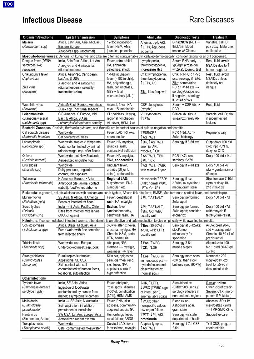
Infectious Disease Rare Diseases
Brady Page
Organism/Syndrome Epi & Transmission Symptoms Abnl Labs Diagnostic Tests Treatment Malaria (Plasmodium spp)
Africa, Latin Am, Asia, MidEast, Eastern Europe
12-35d incubation;fever, HSM, AMS,jaundice, petechiae
Anemia, ↓plt, AKI, ↑LFTs, ↓glucose, acidemia
BinaxNOW (RDT) + thick/thin blood smear w/ Giemsa
Variable, call ID; ppx doxy, Malarone, mefloquine Anopheles spp. (nocturnal)
Mosquito-borne viruses: Dengue, chikungunya, and zika are often indistinguishable clinically/epidemiologically; consider testing for all 3 if concerned. Dengue fever (DENV serotypes 1-4; Flavivirus)
India, Asia/Pac, Africa, Lat Am Fever, retro-orbital HA, arthralgia, petechiae, shock
Lymphopenia, thrombocytopenia, increasing Hct
Serum RNA early → IgG/IgM (cross-rxn w/ Zika); tourniq. test
Rest, fluid; avoid NSAIDs due to ↑ hemorrhagic sx
A aegypti and A albopictus (diurnal feeders)
Chikungunya fever (Alphavirus)
Zika virus (Flavivirus)
Africa, Asia/Pac, Caribbean, Lat Am, S USA
1-14d incubation;fever (>102 in chik),HA, polyarthrlagia,rash, conjunctivitis,GBS + fetalmicrocephaly (zika)
Chik: lymphopenia, thrombocytopenia, ↑LFTs, AKI
Zika: labs freq. wnl
Chik: RT-PCR if <7d sxs; serology if ≥7d. Zika: serum/urine PCR if <14d sxs → serology/plaque red. if negative; serology if ≥14d of sxs
Rest, fluid; avoid NSAIDs unless definitely not dengue
A aegypti and A albopictus (diurnal feeders); sexually-transmitted (zika)
West Nile virus (Flavivirus)
Africa/MEast, Europe, Americas Asympt; fever, HA, myal, 1% meningitis
CSF pleocytosis (lymphs)
Serum + CSF Abs > PCR
Rest, fluid Culex spp. (nocturnal feeders)
Leishmaniasis, cutaneous/visceral (Leishmania spp)
C/S America, S Europe, Mid East, E Africa, S Asia
CL: painless ulcer(s), regional lymphaden. VL: fever, HSM, ↓wt
VL: cytopenias, ↑LFTs
Clinical dx, tissue smear/cx; rarely Ab
Varable, call ID; abx if superinfected lesions Lutzomyia/Phlebotomus sandfly
Bacterial Zoonoses: Coxiella, Bartonella quintana, and Brucella are important causes of culture-negative endocarditis. Cat scratch disease (Bartonella henslae)
Worldwide Fever, LAD 1-3 wks, neuro, ocular
↑ESR/CRP, ↑AST/ALT
PCR 1-3d; Ab 1-2wks; histology
Regimens vary Cat bite/scratch, fleas
Leptospirosis (Leptospira spp.)
Worldwide; tropics > temperate Fever, HA, myalgia, jaundice, rash, conjunctival suffusion
↑Bili, ↑AST/ALT, anemia, AKI, hypoNa, ↑CK
Serology if 3-5d sxs Outpt doxy 100 bid x7d; inpt PCN G, doxy, or CTX
Water contaminated by animal urine/sewage, esp. after floods
Q fever (Coxiella burnetii)
Worldwide (not New Zealand) Fever, HA, myalgia, PNA, endocarditis
↑AST/ALT, ↑Bili, ↓Plt, ↑CK
PCR if <7d sxs, serology if ≥7d
Doxy 100 bid x14d Aerosolized ungulate fluid
Brucellosis (Brucella spp)
Worldwide Undulant fever, arthritis (SI joint, spine), endocarditis
↑AST/ALT, ↓WBC with relative ↑lymp
Serology if 7-1d sxs Doxy 100 bid x6 wks + gentamicin or rifampin
Dairy products, ungulate contact, lab exposure
Tularemia (Francisella tularensis)
N America, Europe > Asia Regional LAD; 6 syndromes: PNA, glandular, etc.
Nonspecific;↑ESR/CRP; normal WBC, LFTs, Cr; ↓Plt
Serology if sxs ≥2wks; cx cysteine+ media; gram stain
Streptomycin 7-10d; cipro or doxy 10-21d if mild dz
Arthropod bite, animal contact (rabbit), food/water, airborne
Rickettsia: In general, rickettsial diseases with eschars are scrub typhus, African tick-bite fever, RMSF, Mediterranean spotted fever, and rickettsialpox. Murine typhus (Rickettsia typhi)
SE Asia, N Africa, N America Fever, centrifugal rash, HA, myalgia
↓Plt, ↑AST/ALT Serology performed 2wks apart
Doxy 100 bid x7d Feces of infected rat fleas
Scrub typhus (Orientia tsutsugamushi)
India → E Asia; Pacific, Chile Eschar, fever, lymphadenopathy, centrifugal rash, HA
↓Plt, ↑AST/ALT, ↑Bili, AKI, WBC usually wnl
Serology performed 2wks apart; consider eschar bx
Doxy 100 bid x7d; azithromycin if tetracycline-resist.
Bites from infected mite larvae (AKA chiggers)
Helminths: If concerned about intestinal worms, albendazole is an effective and safe medication to give empirically while awaiting lab results. Schistosomiasis (Schistosoma spp)
Africa, Brazil, MidEast, Asia 3-8wks after ix: fever,urticaria, myalgia, HAChronic: HSM, portalHTN, hematuria
↑Eos (30-60%) in acute, ↓Plt, LFTs usually wnl
Serology at 6-12wks; stool/urine microscopy for speciation
Acute: pred 20-40 x5d + praziquantel Chronic: 40-60 x1 of praziquantel
Fresh water with free cercariae from infected snails
Trichinellosis (Trichinella spp)
Worldwide, esp. Europe Abd pain, N/V, diarrhea → myalgia, weakness, +/- fever
↑Eos, ↑WBC, ↑CK, ↑LDH
Serology 2-8d; muscle biopsy
Albendazole 400 bid + pred 30-60 qd x8-14d
Undercooked meat, esp. pork
Strongyloidiasis (Strongyloides stercoralis)
Rural tropics/subtropics; Appalachia, SE USA
Skin rxn, epigastric pain, diarrhea, resp. sxs; fever, N/V, sepsis or shock if hyperinfection
↑Eos, ↑WBC; in immunosupp pts → hyperinfection and disseminated dz (normal eos.)
Serology more sens (83+%) than stool but less spec (95+%)
Ivermectin 200 mcg/kg/day x2d; treat for x5-7d if disseminated dz
Skin contact with soil contaminated w/ human feces, fecal-oral, autoinfection
Other Infections Typhoid fever (Salmonella enterica serotype Typhi)
India, SE Asia, Africa Fever, abd pain, ‘rose spots’, diarrhea (>50%), constipation (30%), HSM, AMS
↓HR, ↑LFTs, ↓WBC (↑WBC sign of intest. perf.), anemia, abnl coags
Stool/blood cx (BMBx 90% sens.); serology effective in non-endemic regions
S Asia: azithro Other: ciprofloxacin Severe: CTX (mero-penem if Pakistan)
Ingestion of food/water contaminated by human fecal matter; asymptomatic carriers
Melioidosis (Burkholderia pseudomallei)
India → SE Asia; N Australia Fever, PNA, skin abscess, community-acquired sepsis, GU
↑WBC; other nonspecific values c/w organ failure
Blood cx on Ashdown’s agar, gram stain
Abscess I&D + IV mero/ceftaz x2wks → TMP-SMX x3mo
Soil; aspiration, inhalation, percutaneous inoculation
Hantavirus (Sin nombre, Andes)
SW USA, Lat Am, Europe, Asia Hemorrhagic fever, renal failure, ARDS
↑PTT, ↓Plt, AKI, proteinuria
Serology via state department of health
Supportive care Aerosolized rodent excreta
Toxoplasmosis (Toxoplasma gondii)
Worldwide Cervical LAD, fever for wks/mos, myalgia
Atypical lymphs, ↑AST/ALT
Serology 1-7d; CSF 2-5d
Tx if CNS, preg, or chorioretinitis Cats; contaminated meat/water
122
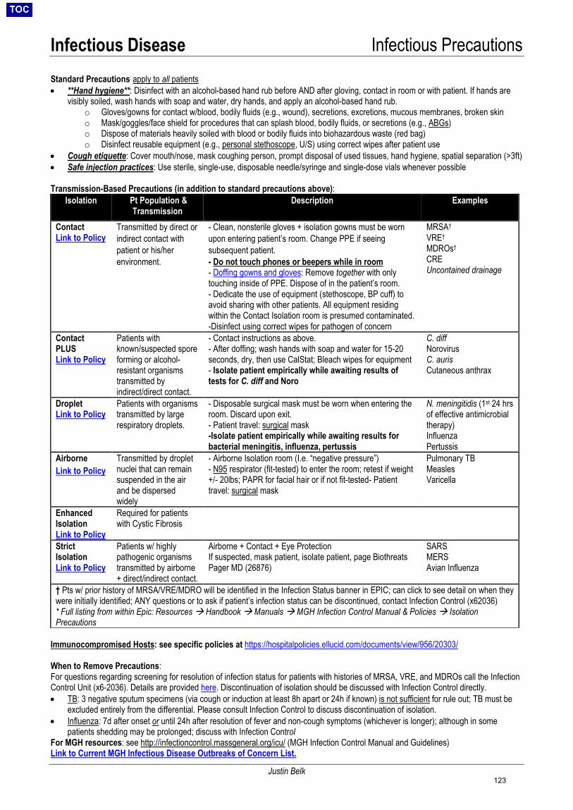
Infectious Disease Infectious Precautions
Justin Belk
Standard Precautions: apply to all patients • **Hand hygiene**: Disinfect with an alcohol-based hand rub before AND after gloving, contact in room or with patient. If hands are
visibly soiled, wash hands with soap and water, dry hands, and apply an alcohol-based hand rub.o Gloves/gowns for contact w/blood, bodily fluids (e.g., wound), secretions, excretions, mucous membranes, broken skino Mask/goggles/face shield for procedures that can splash blood, bodily fluids, or secretions (e.g., ABGs)o Dispose of materials heavily soiled with blood or bodily fluids into biohazardous waste (red bag)o Disinfect reusable equipment (e.g., personal stethoscope, U/S) using correct wipes after patient use
• Cough etiquette: Cover mouth/nose, mask coughing person, prompt disposal of used tissues, hand hygiene, spatial separation (>3ft)• Safe injection practices: Use sterile, single-use, disposable needle/syringe and single-dose vials whenever possible
Transmission-Based Precautions (in addition to standard precautions above): Isolation Pt Population &
Transmission Description Examples
Contact Link to Policy
Transmitted by direct or indirect contact with patient or his/her environment.
- Clean, nonsterile gloves + isolation gowns must be wornupon entering patient’s room. Change PPE if seeingsubsequent patient.- Do not touch phones or beepers while in room- Doffing gowns and gloves: Remove together with onlytouching inside of PPE. Dispose of in the patient’s room.- Dedicate the use of equipment (stethoscope, BP cuff) toavoid sharing with other patients. All equipment residingwithin the Contact Isolation room is presumed contaminated.-Disinfect using correct wipes for pathogen of concern
MRSA† VRE† MDROs† CRE Uncontained drainage
Contact PLUS Link to Policy
Patients with known/suspected spore forming or alcohol-resistant organisms transmitted by indirect/direct contact.
- Contact instructions as above.- After doffing; wash hands with soap and water for 15-20seconds, dry, then use CalStat; Bleach wipes for equipment- Isolate patient empirically while awaiting results oftests for C. diff and Noro
C. diffNorovirusC. aurisCutaneous anthrax
Droplet Link to Policy
Patients with organisms transmitted by large respiratory droplets.
- Disposable surgical mask must be worn when entering theroom. Discard upon exit.- Patient travel: surgical mask-Isolate patient empirically while awaiting results forbacterial meningitis, influenza, pertussis
N. meningitidis (1st 24 hrsof effective antimicrobialtherapy)InfluenzaPertussis
Airborne Link to Policy
Transmitted by droplet nuclei that can remain suspended in the air and be dispersed widely
- Airborne Isolation room (I.e. “negative pressure”)- N95 respirator (fit-tested) to enter the room; retest if weight+/- 20lbs; PAPR for facial hair or if not fit-tested- Patienttravel: surgical mask
Pulmonary TB Measles Varicella
Enhanced Isolation Link to Policy
Required for patients with Cystic Fibrosis
Strict Isolation Link to Policy
Patients w/ highly pathogenic organisms transmitted by airborne + direct/indirect contact.
Airborne + Contact + Eye Protection If suspected, mask patient, isolate patient, page Biothreats Pager MD (26876)
SARS MERS Avian Influenza
† Pts w/ prior history of MRSA/VRE/MDRO will be identified in the Infection Status banner in EPIC; can click to see detail on when they were initially identified; ANY questions or to ask if patient’s infection status can be discontinued, contact Infection Control (x62036) * Full listing from within Epic: Resources Handbook Manuals MGH Infection Control Manual & Policies IsolationPrecautions
Immunocompromised Hosts: see specific policies at https://hospitalpolicies.ellucid.com/documents/view/956/20303/
When to Remove Precautions: For questions regarding screening for resolution of infection status for patients with histories of MRSA, VRE, and MDROs call the Infection Control Unit (x6-2036). Details are provided here. Discontinuation of isolation should be discussed with Infection Control directly. • TB: 3 negative sputum specimens (via cough or induction at least 8h apart or 24h if known) is not sufficient for rule out; TB must be
excluded entirely from the differential. Please consult Infection Control to discuss discontinuation of isolation.• Influenza: 7d after onset or until 24h after resolution of fever and non-cough symptoms (whichever is longer); although in some
patients shedding may be prolonged; discuss with Infection ControlFor MGH resources: see http://infectioncontrol.massgeneral.org/icu/ (MGH Infection Control Manual and Guidelines) Link to Current MGH Infectious Disease Outbreaks of Concern List.
123
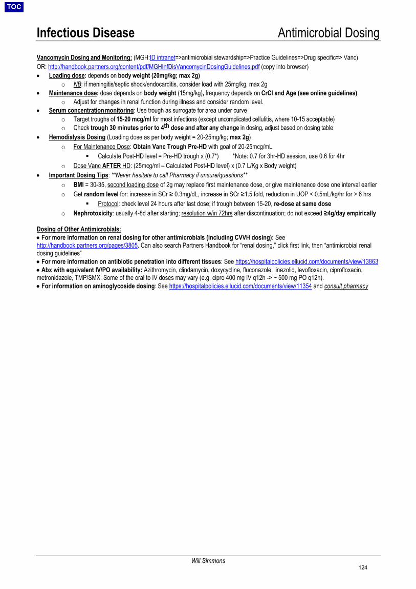
Infectious Disease Antimicrobial Dosing
Will Simmons
Vancomycin Dosing and Monitoring: (MGH:ID intranet=>antimicrobial stewardship=>Practice Guidelines=>Drug specific=> Vanc) OR: http://handbook.partners.org/content/pdf/MGHInfDisVancomycinDosingGuidelines.pdf (copy into browser) • Loading dose: depends on body weight (20mg/kg; max 2g)
o NB: if meningitis/septic shock/endocarditis, consider load with 25mg/kg, max 2g• Maintenance dose: dose depends on body weight (15mg/kg), frequency depends on CrCl and Age (see online guidelines)
o Adjust for changes in renal function during illness and consider random level.• Serum concentration monitoring: Use trough as surrogate for area under curve
o Target troughs of 15-20 mcg/ml for most infections (except uncomplicated cellulitis, where 10-15 acceptable)o Check trough 30 minutes prior to 4th dose and after any change in dosing, adjust based on dosing table
• Hemodialysis Dosing (Loading dose as per body weight = 20-25mg/kg; max 2g)o For Maintenance Dose: Obtain Vanc Trough Pre-HD with goal of 20-25mcg/mL
Calculate Post-HD level = Pre-HD trough x (0.7*) *Note: 0.7 for 3hr-HD session, use 0.6 for 4hro Dose Vanc AFTER HD: (25mcg/ml – Calculated Post-HD level) x (0.7 L/Kg x Body weight)
• Important Dosing Tips: **Never hesitate to call Pharmacy if unsure/questions**o BMI = 30-35, second loading dose of 2g may replace first maintenance dose, or give maintenance dose one interval earliero Get random level for: increase in SCr ≥ 0.3mg/dL, increase in SCr ≥1.5 fold, reduction in UOP < 0.5mL/kg/hr for > 6 hrs
Protocol: check level 24 hours after last dose; if trough between 15-20, re-dose at same doseo Nephrotoxicity: usually 4-8d after starting; resolution w/in 72hrs after discontinuation; do not exceed ≥4g/day empirically
Dosing of Other Antimicrobials: • For more information on renal dosing for other antimicrobials (including CVVH dosing): Seehttp://handbook.partners.org/pages/3805. Can also search Partners Handbook for “renal dosing,” click first link, then “antimicrobial renaldosing guidelines” • For more information on antibiotic penetration into different tissues: See https://hospitalpolicies.ellucid.com/documents/view/13863• Abx with equivalent IV/PO availability: Azithromycin, clindamycin, doxycycline, fluconazole, linezolid, levofloxacin, ciprofloxacin,metronidazole, TMP/SMX. Some of the oral to IV doses may vary (e.g. cipro 400 mg IV q12h -> ~ 500 mg PO q12h).• For information on aminoglycoside dosing: See https://hospitalpolicies.ellucid.com/documents/view/11354 and consult pharmacy
124

Hematology Anemia & Pancytopenia
Eftitan Akam, Charlotte Lee
GENERAL APPROACH TO ANEMIA (Williams Hematology 2018) • Presentation: Hypoxic sx (fatigue, dizziness, DOE, pallor, angina, claudication, retinal hemorrhage), nonspecific sx (cramps, abd
pain, n/v), compensatory mechanisms (hyperventilation, tachycardia, palpitations, orthostasis, ↑pulsation, flow murmur, bruit)• Associations: Jaundice (hemolysis), glossitis (folate / B12 def), motor / sensory deficits (B12 def), PICA / koilonychias / angular
cheilitis (Fe def), splenomegaly (cirrhosis, infxn), constipation / bone pain (myeloma), melena / +FOBT (GIB, CRC), Mediterr / Asian /Black (thal/SS), unusual thromboses (PNH), petechiae / ecchymoses (coagulopathy, pancytopenia)
• Initial labs (draw/add on labs prior to transfusion!):o CBC (other cell lines, MCV, RDW), retic count, special slide, T&So Determine hypo- (retic index [RI] <2%) vs hyper-proliferative (RI >2%)o Very low RI (<0.1%) indicative of aplastic anemia or red cell aplasia
• Additional labs depend on retic index:o RI <2% -“Anemia labs”: Fe/TIBC/ferritin, folate / B12 (in last 6 mo.), Cr, LFTs, TSHo RI >2% - “Hemolysis labs”: LDH, bilirubin, haptoglobin, DAT, UA, Coags
CLASSIFICATION OF ANEMIA (NEJM 2014;371:1324, Lancet 2018;391:155, Williams Hematology 2018) UNDERPRODUCTION (RI <2%)
Microcytic (MCV < 80 µm3) Normocytic (MCV 80-99 µm3) Macrocytic (MCV ≥ 100 µm3) Iron deficiency anemia (IDA) • ↓ Fe, ↑ TIBC, ↓ ferritin (<30 high Sp.),
Fe/TIBC <16%, ↑ RDWAnemia of inflammation • ↓ Fe, ↓/nl TIBC, ↑ ferritin,
Fe/TIBC ↓/nl (10-20%)Thalassemias
• Fe studies nl, MCV ↓ (often <70),MCV/RBC <13 (high Sp.), Hbelectrophoresis
Sideroblastic anemia • ↑ ferritin, Fe/TIBC nl, basophilic
stippling (Pb), ringed sideroblasts (BM)
Inflammation & variant: • Early anemia of inflammation• Early IDA• Mixed IDA & ↓ folate/B12 (dimorphic: ↑
RDW)Organ-specific: • Renal (CKD/ESRD): ↓Epo (should
↑10x per 10% Hct drop)• Endocrine (↓thyroid, pituitary, adrenal,
parathyroid, testosterone): ↓Epo• Marrow (red cell aplasia, AA, MDS,
myelofibrosis, myelophthisis, PNH,MM): SPEP, serum FLC, BMBx
Megaloblastic: smear shows hyper-seg PMNs and macro-ovalocytes • Folate: ↑ homocysteine, nl MMA• B12: ↑ homocysteine, ↑ MMA
(↑ anti-IF Ab, ↑ gastrin if PA;falsely normal B12 possible)
• Early myeloproliferative d/oNon-megaloblastic:• Cirrhosis, EtOH• Reticulocytosis: lysis or bleed• Hypothyroidism• MDS (refractory anemia) & MMMeds: HAART, 5FU, AraC, Hydrea
DESTRUCTION / LOSS (RI >2%) Extrinsic (transfused RBC has shortened life span) Intrinsic (transfused RBC has normal life span)
MAHA (-DAT, +schisto): see Thrombocytopenia: TMAs for DDx • Smear (≥2 schisto/HPF), PLT ~25K, LDH ↑↑, indir bili ↑, hapto ↓Immune (+DAT, +spherocytosis): Ab- and/or complement-mediated• Warm autoimmune (CLL, HIV, lymphoma, SLE): +DAT anti-IgG/-C3• Cold autoimmune (EBV, CMV, Mycoplasma): +DAT anti-C3• Alloimmune: acute v. delayed hemolytic transfusion rxn• Drug: PCN, cephalo, quinine, ampho B, NSAIDs, procainamideNon-immune (-DAT, +/- RBC inclusion):• Infection: babesia, malaria, bartonella, C. perfringens, H. flu (type B)• Toxin: lead, copper, insect / spider / snake bites, hypotonic infusion
Hereditary: • Hb disease (SS, HbC, thal): Hb electrophoresis• Enzyme deficiency (G6PD, PK): levels often nml in
attack; check 4wk later & repeat in 3mo if neg.• Membrane defect: spherocytosis, elliptocytosisAcquired (new onset):• PNH (paroxysmal nocturnal hemoglobinuria): flow
cytometry +/- FLAER for GPI anchor, smear nml, UA(hgb/hemosiderin), thrombosis (intra-abd/cerebral)
Acute blood loss: GI blood loss, hematoma
APPROACH TO PERIPHERAL BLOOD SMEAR (NEJM 2005;353:498)• Low power (200x): Scan slide for WBC distribution. Identify the “thick” edge and the “feathered” or thin edge.• Med power (400x): Examine feathered edge for rouleaux, parasites, abnormal WBC, platelet aggregation / microsatellites.• Oil Immersion (1000x): Assess the size, shape, and morphology of major cell lineages:
o RBC: Examine where RBCs are close but not touching, compare to lymphocyte nucleus size for scaleo WBC: Concentrate on edges and thin end of film, normal WBC include PMN, eos, basos, lymphocytes, monocytes
RBCs Hypochromia / microcytes (↓Fe, thal); spherocytes (AIHA, HS); schistocytes (valve, MAHA); target cells (thal, Hb dz, liver dz, asplenia); tear drops / nRBCs (myelofibrosis, myelophthisis, thal); bite cells / Heinz bodies (G6PD); basophilic stippling (thal, Pb, sideroblastic); acanthocytes / echinocytes (liver dz/renal dz); Howell-Jolly bodies (sickle dz, asplenia)
WBCs HypOsegmented PMNs (MDS); hypERsegmented PMNs (folate/B12); toxic granulation (sepsis); myeloid vs. lymphoid precursors (left shift, myelocytes, blasts, Auer rods), basophils (MPD)
Platelets Clumping (pseudothrombocytopenia); large platelets (↑ production, ITP, or congenital disease)
IRON DEFICIENCY ANEMIA (NEJM 2015;372:1832; Blood 2019;133:30)• Etiology: ↑loss due to chronic bleeding (PUD/UGIB [↑BUN], colon CA/LGIB, menses, intravascular hemolysis), ↑demand (Epo,
pregnancy, blood donation), ↓intake (malnutrition) or ↓absorption (IBD/post-gastrectomy/celiac). If unexplained or refractory to POiron, eval. for celiac, AI gastritis, H. pylori, which accounted for 5%, 27%, 19% of unexplained IDA (Haematologica 2005;90:585).
Reticulocyte index (RI) calculator: https://www.mdcalc.com/absolute-
reticulocyte-count-reticulocyte-index
125
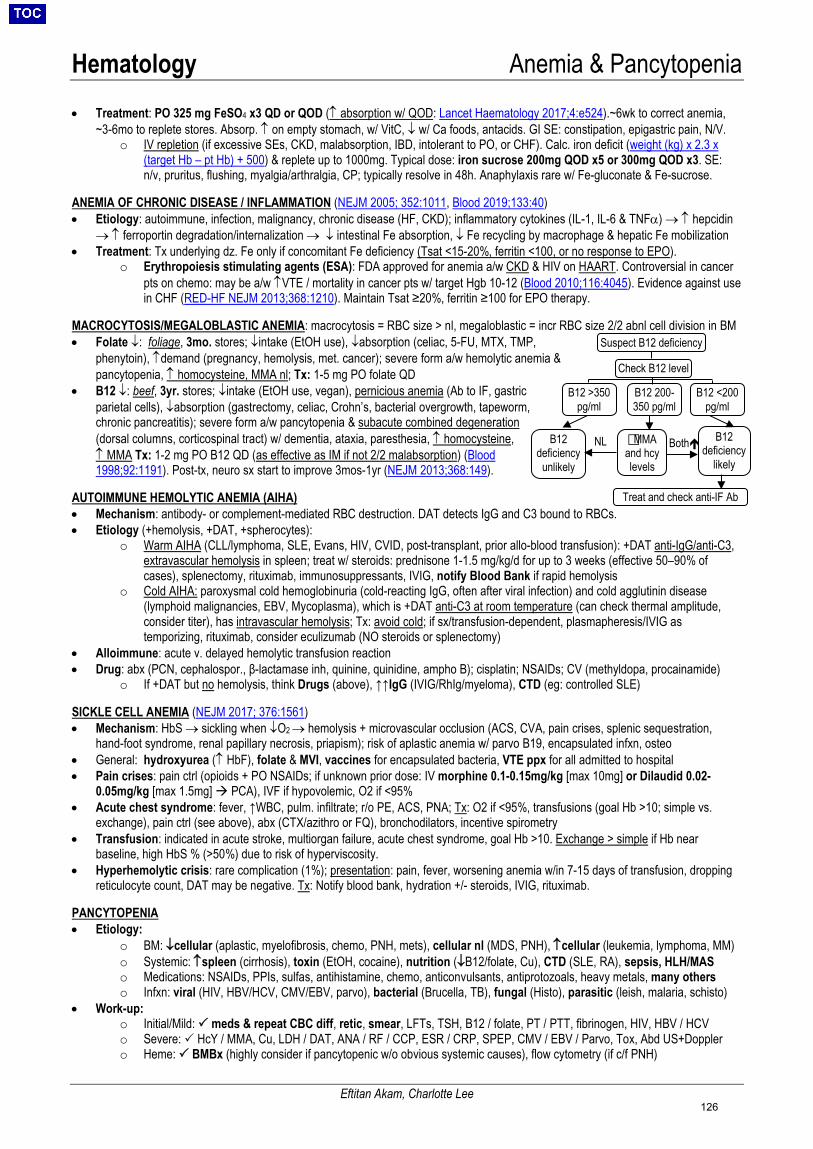
Hematology Anemia & Pancytopenia
Eftitan Akam, Charlotte Lee
• Treatment: PO 325 mg FeSO4 x3 QD or QOD (↑ absorption w/ QOD: Lancet Haematology 2017;4:e524).~6wk to correct anemia,~3-6mo to replete stores. Absorp. ↑ on empty stomach, w/ VitC, ↓ w/ Ca foods, antacids. GI SE: constipation, epigastric pain, N/V.
o IV repletion (if excessive SEs, CKD, malabsorption, IBD, intolerant to PO, or CHF). Calc. iron deficit (weight (kg) x 2.3 x(target Hb – pt Hb) + 500) & replete up to 1000mg. Typical dose: iron sucrose 200mg QOD x5 or 300mg QOD x3. SE:n/v, pruritus, flushing, myalgia/arthralgia, CP; typically resolve in 48h. Anaphylaxis rare w/ Fe-gluconate & Fe-sucrose.
ANEMIA OF CHRONIC DISEASE / INFLAMMATION (NEJM 2005; 352:1011, Blood 2019;133:40) • Etiology: autoimmune, infection, malignancy, chronic disease (HF, CKD); inflammatory cytokines (IL-1, IL-6 & TNFα) → ↑ hepcidin
→↑ ferroportin degradation/internalization → ↓ intestinal Fe absorption, ↓ Fe recycling by macrophage & hepatic Fe mobilization• Treatment: Tx underlying dz. Fe only if concomitant Fe deficiency (Tsat <15-20%, ferritin <100, or no response to EPO).
o Erythropoiesis stimulating agents (ESA): FDA approved for anemia a/w CKD & HIV on HAART. Controversial in cancerpts on chemo: may be a/w ↑VTE / mortality in cancer pts w/ target Hgb 10-12 (Blood 2010;116:4045). Evidence against usein CHF (RED-HF NEJM 2013;368:1210). Maintain Tsat ≥20%, ferritin ≥100 for EPO therapy.
MACROCYTOSIS/MEGALOBLASTIC ANEMIA: macrocytosis = RBC size > nl, megaloblastic = incr RBC size 2/2 abnl cell division in BM • Folate ↓: foliage, 3mo. stores; ↓intake (EtOH use), ↓absorption (celiac, 5-FU, MTX, TMP,
phenytoin), ↑demand (pregnancy, hemolysis, met. cancer); severe form a/w hemolytic anemia & pancytopenia, ↑ homocysteine, MMA nl; Tx: 1-5 mg PO folate QD
• B12 ↓: beef, 3yr. stores; ↓intake (EtOH use, vegan), pernicious anemia (Ab to IF, gastric parietal cells), ↓absorption (gastrectomy, celiac, Crohn’s, bacterial overgrowth, tapeworm, chronic pancreatitis); severe form a/w pancytopenia & subacute combined degeneration (dorsal columns, corticospinal tract) w/ dementia, ataxia, paresthesia, ↑ homocysteine, ↑ MMA Tx: 1-2 mg PO B12 QD (as effective as IM if not 2/2 malabsorption) (Blood 1998;92:1191). Post-tx, neuro sx start to improve 3mos-1yr (NEJM 2013;368:149).
AUTOIMMUNE HEMOLYTIC ANEMIA (AIHA) • Mechanism: antibody- or complement-mediated RBC destruction. DAT detects IgG and C3 bound to RBCs.• Etiology (+hemolysis, +DAT, +spherocytes):
o Warm AIHA (CLL/lymphoma, SLE, Evans, HIV, CVID, post-transplant, prior allo-blood transfusion): +DAT anti-IgG/anti-C3,extravascular hemolysis in spleen; treat w/ steroids: prednisone 1-1.5 mg/kg/d for up to 3 weeks (effective 50–90% ofcases), splenectomy, rituximab, immunosuppressants, IVIG, notify Blood Bank if rapid hemolysis
o Cold AIHA: paroxysmal cold hemoglobinuria (cold-reacting IgG, often after viral infection) and cold agglutinin disease(lymphoid malignancies, EBV, Mycoplasma), which is +DAT anti-C3 at room temperature (can check thermal amplitude,consider titer), has intravascular hemolysis; Tx: avoid cold; if sx/transfusion-dependent, plasmapheresis/IVIG astemporizing, rituximab, consider eculizumab (NO steroids or splenectomy)
• Alloimmune: acute v. delayed hemolytic transfusion reaction• Drug: abx (PCN, cephalospor., β-lactamase inh, quinine, quinidine, ampho B); cisplatin; NSAIDs; CV (methyldopa, procainamide)
o If +DAT but no hemolysis, think Drugs (above), ↑↑IgG (IVIG/RhIg/myeloma), CTD (eg: controlled SLE)
SICKLE CELL ANEMIA (NEJM 2017; 376:1561) • Mechanism: HbS → sickling when ↓O2 → hemolysis + microvascular occlusion (ACS, CVA, pain crises, splenic sequestration,
hand-foot syndrome, renal papillary necrosis, priapism); risk of aplastic anemia w/ parvo B19, encapsulated infxn, osteo• General: hydroxyurea (↑ HbF), folate & MVI, vaccines for encapsulated bacteria, VTE ppx for all admitted to hospital• Pain crises: pain ctrl (opioids + PO NSAIDs; if unknown prior dose: IV morphine 0.1-0.15mg/kg [max 10mg] or Dilaudid 0.02-
0.05mg/kg [max 1.5mg] PCA), IVF if hypovolemic, O2 if <95%• Acute chest syndrome: fever, ↑WBC, pulm. infiltrate; r/o PE, ACS, PNA; Tx: O2 if <95%, transfusions (goal Hb >10; simple vs.
exchange), pain ctrl (see above), abx (CTX/azithro or FQ), bronchodilators, incentive spirometry• Transfusion: indicated in acute stroke, multiorgan failure, acute chest syndrome, goal Hb >10. Exchange > simple if Hb near
baseline, high HbS % (>50%) due to risk of hyperviscosity.• Hyperhemolytic crisis: rare complication (1%); presentation: pain, fever, worsening anemia w/in 7-15 days of transfusion, dropping
reticulocyte count, DAT may be negative. Tx: Notify blood bank, hydration +/- steroids, IVIG, rituximab.
PANCYTOPENIA • Etiology:
o BM: ↓cellular (aplastic, myelofibrosis, chemo, PNH, mets), cellular nl (MDS, PNH), ↑cellular (leukemia, lymphoma, MM)o Systemic: ↑spleen (cirrhosis), toxin (EtOH, cocaine), nutrition (↓B12/folate, Cu), CTD (SLE, RA), sepsis, HLH/MASo Medications: NSAIDs, PPIs, sulfas, antihistamine, chemo, anticonvulsants, antiprotozoals, heavy metals, many otherso Infxn: viral (HIV, HBV/HCV, CMV/EBV, parvo), bacterial (Brucella, TB), fungal (Histo), parasitic (leish, malaria, schisto)
• Work-up:o Initial/Mild: meds & repeat CBC diff, retic, smear, LFTs, TSH, B12 / folate, PT / PTT, fibrinogen, HIV, HBV / HCVo Severe: HcY / MMA, Cu, LDH / DAT, ANA / RF / CCP, ESR / CRP, SPEP, CMV / EBV / Parvo, Tox, Abd US+Dopplero Heme: BMBx (highly consider if pancytopenic w/o obvious systemic causes), flow cytometry (if c/f PNH)
Suspect B12 deficiency
Check B12 level
B12 >350 pg/ml
B12 200-350 pg/ml
B12 <200 pg/ml
B12 deficiency
unlikely
Treat and check anti-IF Ab
Both NL MMA and hcy levels
B12 deficiency
likely
126
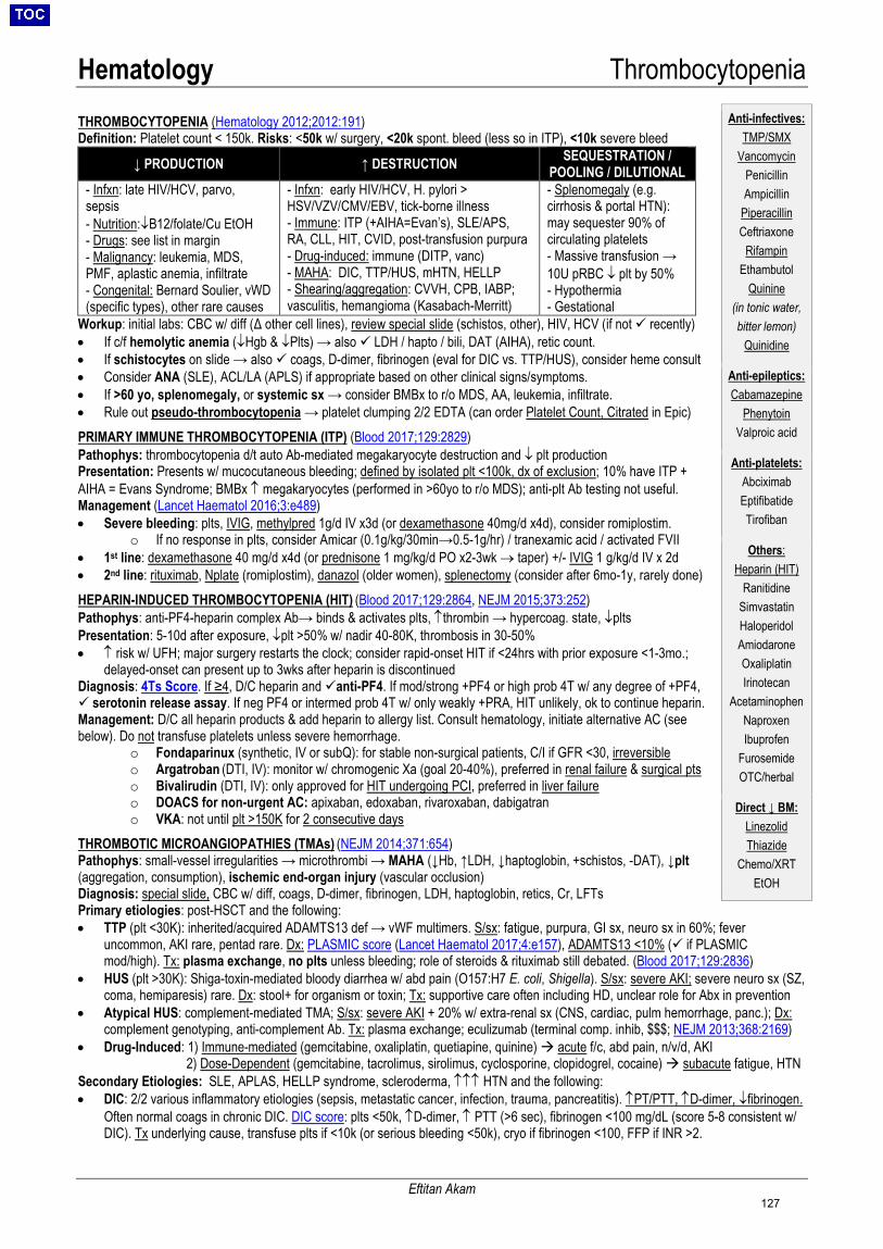
Hematology Thrombocytopenia
Eftitan Akam
THROMBOCYTOPENIA (Hematology 2012;2012:191) Definition: Platelet count < 150k. Risks: <50k w/ surgery, <20k spont. bleed (less so in ITP), <10k severe bleed
↓ PRODUCTION ↑ DESTRUCTION SEQUESTRATION / POOLING / DILUTIONAL
- Infxn: late HIV/HCV, parvo,sepsis- Nutrition:↓B12/folate/Cu EtOH- Drugs: see list in margin- Malignancy: leukemia, MDS,PMF, aplastic anemia, infiltrate- Congenital: Bernard Soulier, vWD(specific types), other rare causes
- Infxn: early HIV/HCV, H. pylori >HSV/VZV/CMV/EBV, tick-borne illness- Immune: ITP (+AIHA=Evan’s), SLE/APS,RA, CLL, HIT, CVID, post-transfusion purpura- Drug-induced: immune (DITP, vanc)- MAHA: DIC, TTP/HUS, mHTN, HELLP- Shearing/aggregation: CVVH, CPB, IABP;vasculitis, hemangioma (Kasabach-Merritt)
- Splenomegaly (e.g.cirrhosis & portal HTN):may sequester 90% ofcirculating platelets- Massive transfusion →10U pRBC ↓ plt by 50%- Hypothermia- Gestational
Workup: initial labs: CBC w/ diff (Δ other cell lines), review special slide (schistos, other), HIV, HCV (if not recently) • If c/f hemolytic anemia (↓Hgb & ↓Plts) → also LDH / hapto / bili, DAT (AIHA), retic count.• If schistocytes on slide → also coags, D-dimer, fibrinogen (eval for DIC vs. TTP/HUS), consider heme consult• Consider ANA (SLE), ACL/LA (APLS) if appropriate based on other clinical signs/symptoms.• If >60 yo, splenomegaly, or systemic sx → consider BMBx to r/o MDS, AA, leukemia, infiltrate.• Rule out pseudo-thrombocytopenia → platelet clumping 2/2 EDTA (can order Platelet Count, Citrated in Epic)PRIMARY IMMUNE THROMBOCYTOPENIA (ITP) (Blood 2017;129:2829) Pathophys: thrombocytopenia d/t auto Ab-mediated megakaryocyte destruction and ↓ plt production Presentation: Presents w/ mucocutaneous bleeding; defined by isolated plt <100k, dx of exclusion; 10% have ITP + AIHA = Evans Syndrome; BMBx ↑ megakaryocytes (performed in >60yo to r/o MDS); anti-plt Ab testing not useful. Management (Lancet Haematol 2016;3:e489)• Severe bleeding: plts, IVIG, methylpred 1g/d IV x3d (or dexamethasone 40mg/d x4d), consider romiplostim.
o If no response in plts, consider Amicar (0.1g/kg/30min→0.5-1g/hr) / tranexamic acid / activated FVII• 1st line: dexamethasone 40 mg/d x4d (or prednisone 1 mg/kg/d PO x2-3wk → taper) +/- IVIG 1 g/kg/d IV x 2d• 2nd line: rituximab, Nplate (romiplostim), danazol (older women), splenectomy (consider after 6mo-1y, rarely done)HEPARIN-INDUCED THROMBOCYTOPENIA (HIT) (Blood 2017;129:2864, NEJM 2015;373:252)Pathophys: anti-PF4-heparin complex Ab→ binds & activates plts, ↑thrombin → hypercoag. state, ↓plts Presentation: 5-10d after exposure, ↓plt >50% w/ nadir 40-80K, thrombosis in 30-50% • ↑ risk w/ UFH; major surgery restarts the clock; consider rapid-onset HIT if <24hrs with prior exposure <1-3mo.;
delayed-onset can present up to 3wks after heparin is discontinuedDiagnosis: 4Ts Score. If ≥4, D/C heparin and anti-PF4. If mod/strong +PF4 or high prob 4T w/ any degree of +PF4, serotonin release assay. If neg PF4 or intermed prob 4T w/ only weakly +PRA, HIT unlikely, ok to continue heparin.Management: D/C all heparin products & add heparin to allergy list. Consult hematology, initiate alternative AC (seebelow). Do not transfuse platelets unless severe hemorrhage.
o Fondaparinux (synthetic, IV or subQ): for stable non-surgical patients, C/I if GFR <30, irreversibleo Argatroban (DTI, IV): monitor w/ chromogenic Xa (goal 20-40%), preferred in renal failure & surgical ptso Bivalirudin (DTI, IV): only approved for HIT undergoing PCI, preferred in liver failureo DOACS for non-urgent AC: apixaban, edoxaban, rivaroxaban, dabigatrano VKA: not until plt >150K for 2 consecutive days
THROMBOTIC MICROANGIOPATHIES (TMAs) (NEJM 2014;371:654) Pathophys: small-vessel irregularities → microthrombi → MAHA (↓Hb, ↑LDH, ↓haptoglobin, +schistos, -DAT), ↓plt (aggregation, consumption), ischemic end-organ injury (vascular occlusion) Diagnosis: special slide, CBC w/ diff, coags, D-dimer, fibrinogen, LDH, haptoglobin, retics, Cr, LFTs Primary etiologies: post-HSCT and the following: • TTP (plt <30K): inherited/acquired ADAMTS13 def → vWF multimers. S/sx: fatigue, purpura, GI sx, neuro sx in 60%; fever
uncommon, AKI rare, pentad rare. Dx: PLASMIC score (Lancet Haematol 2017;4:e157), ADAMTS13 <10% ( if PLASMICmod/high). Tx: plasma exchange, no plts unless bleeding; role of steroids & rituximab still debated. (Blood 2017;129:2836)
• HUS (plt >30K): Shiga-toxin-mediated bloody diarrhea w/ abd pain (O157:H7 E. coli, Shigella). S/sx: severe AKI; severe neuro sx (SZ,coma, hemiparesis) rare. Dx: stool+ for organism or toxin; Tx: supportive care often including HD, unclear role for Abx in prevention
• Atypical HUS: complement-mediated TMA; S/sx: severe AKI + 20% w/ extra-renal sx (CNS, cardiac, pulm hemorrhage, panc.); Dx:complement genotyping, anti-complement Ab. Tx: plasma exchange; eculizumab (terminal comp. inhib, $$$; NEJM 2013;368:2169)
• Drug-Induced: 1) Immune-mediated (gemcitabine, oxaliplatin, quetiapine, quinine) acute f/c, abd pain, n/v/d, AKI2) Dose-Dependent (gemcitabine, tacrolimus, sirolimus, cyclosporine, clopidogrel, cocaine) subacute fatigue, HTN
Secondary Etiologies: SLE, APLAS, HELLP syndrome, scleroderma, ↑↑↑ HTN and the following: • DIC: 2/2 various inflammatory etiologies (sepsis, metastatic cancer, infection, trauma, pancreatitis). ↑PT/PTT, ↑D-dimer, ↓fibrinogen.
Often normal coags in chronic DIC. DIC score: plts <50k, ↑D-dimer, ↑ PTT (>6 sec), fibrinogen <100 mg/dL (score 5-8 consistent w/DIC). Tx underlying cause, transfuse plts if <10k (or serious bleeding <50k), cryo if fibrinogen <100, FFP if INR >2.
Anti-infectives: TMP/SMX
Vancomycin Penicillin Ampicillin
Piperacillin Ceftriaxone
Rifampin Ethambutol
Quinine (in tonic water, bitter lemon)
Quinidine
Anti-epileptics: Cabamazepine
Phenytoin Valproic acid
Anti-platelets: Abciximab Eptifibatide Tirofiban
Others: Heparin (HIT)
Ranitidine Simvastatin Haloperidol Amiodarone Oxaliplatin Irinotecan
Acetaminophen Naproxen Ibuprofen
Furosemide OTC/herbal
Direct ↓ BM: Linezolid Thiazide
Chemo/XRT EtOH
127
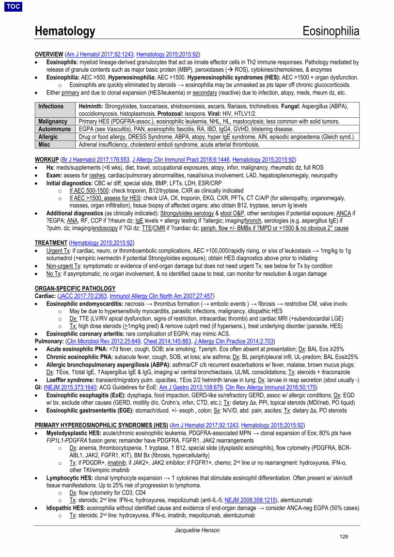
Hematology Eosinophilia
Jacqueline Henson
OVERVIEW (Am J Hematol 2017;92:1243, Hematology 2015;2015:92) • Eosinophils: myeloid lineage-derived granulocytes that act as innate effector cells in Th2 immune responses. Pathology mediated by
release of granule contents such as major basic protein (MBP), peroxidases ( ROS), cytokines/chemokines, & enzymes• Eosinophilia: AEC >500. Hypereosinophilia: AEC >1500. Hypereosinophilic syndromes (HES): AEC >1500 + organ dysfunction.
o Eosinophils are quickly eliminated by steroids → eosinophilia may be unmasked as pts taper off chronic glucocorticoids.• Either primary and due to clonal expansion (HES/leukemia) or secondary (reactive) due to infection, atopy, meds, rheum dz, etc.
Infections Helminth: Strongyloides, toxocariasis, shistosomiasis, ascaris, filariasis, trichinellosis. Fungal: Aspergillus (ABPA), coccidiomycosis, histoplasmosis. Protozoal: isospora. Viral: HIV, HTLV1/2.
Malignancy Primary HES (PDGFRA-assoc.), eosinophilic leukemia, NHL, HL, mastocytosis; less common with solid tumors. Autoimmune EGPA (see Vasculitis), PAN, eosinophilic fasciitis, RA, IBD, IgG4, GVHD, blistering disease. Allergic Drug or food allergy, DRESS Syndrome, ABPA, atopy, hyper IgE syndrome, AIN, episodic angioedema (Gleich synd.). Misc Adrenal insufficiency, cholesterol emboli syndrome, acute arterial thrombosis.
WORKUP (Br J Haematol 2017;176:553, J Allergy Clin Immunol Pract 2018;6:1446, Hematology 2015;2015:92)• Hx: meds/supplements (<6 wks), diet, travel, occupational exposures, atopy, infxn, malignancy, rheumatic dz, full ROS• Exam: assess for rashes, cardiac/pulmonary abnormalities, nasal/sinus involvement, LAD, hepatosplenomegaly, neuropathy• Initial diagnostics: CBC w/ diff, special slide, BMP, LFTs, LDH, ESR/CRP
o If AEC 500-1500: check troponin, B12/tryptase, CXR as clinically indicatedo If AEC >1500, assess for HES: check U/A, CK, troponin, EKG, CXR, PFTs, CT C/A/P (for adenopathy, organomegaly,
masses, organ infiltration), tissue biopsy of affected organs; also obtain B12, tryptase, serum Ig levels• Additional diagnostics (as clinically indicated): Strongyloides serology & stool O&P, other serologies if potential exposure; ANCA if
?EGPA; ANA, RF, CCP if ?rheum dz; IgE levels + allergy testing if ?allergic; imaging/bronch, serologies (e.g. aspergillus IgE) if?pulm. dz; imaging/endoscopy if ?GI dz; TTE/CMR if ?cardiac dz; periph. flow +/- BMBx if ?MPD or >1500 & no obvious 2° cause
TREATMENT (Hematology 2015;2015:92) • Urgent Tx: if cardiac, neuro, or thromboembolic complications, AEC >100,000/rapidly rising, or s/sx of leukostasis → 1mg/kg to 1g
solumedrol (+empiric ivermectin if potential Strongyloides exposure); obtain HES diagnostics above prior to initiating• Non-urgent Tx: symptomatic or evidence of end-organ damage but does not need urgent Tx; see below for Tx by condition• No Tx: if asymptomatic, no organ involvement, & no identified cause to treat, can monitor for resolution & organ damage
ORGAN-SPECIFIC PATHOLOGY Cardiac: (JACC 2017;70:2363, Immunol Allergy Clin North Am 2007;27:457) • Eosinophilic endomyocarditis: necrosis → thrombus formation (→ embolic events ) → fibrosis → restrictive CM, valve involv.
o May be due to hypersensitivity myocarditis, parasitic infections, malignancy, idiopathic HESo Dx: TTE (LV/RV apical dysfunction, signs of restriction, intracardiac thrombi) and cardiac MRI (+subendocardial LGE)o Tx: high dose steroids (>1mg/kg pred) & remove culprit med (if hypersens.), treat underlying disorder (parasite, HES)
• Eosinophilic coronary arteritis: rare complication of EGPA; may mimic ACS.Pulmonary: (Clin Microbiol Rev 2012;25:649, Chest 2014;145:883, J Allergy Clin Practice 2014;2:703)• Acute eosinophilic PNA: <7d fever, cough, SOB; a/w smoking; periph. Eos often absent at presentation; Dx: BAL Eos ≥25%• Chronic eosinophilic PNA: subacute fever, cough, SOB, wt loss; a/w asthma; Dx: BL periph/pleural infil, UL-predom; BAL Eos≥25% • Allergic bronchopulmonary aspergillosis (ABPA): asthma/CF c/b recurrent exacerbations w/ fever, malaise, brown mucus plugs;
Dx: Eos, total IgE, Aspergillus IgE & IgG, imaging w/ central bronchiectasis, UL/ML consolidations; Tx: steroids + itraconazole• Loeffler syndrome: transient/migratory pulm. opacities, Eos 2/2 helminth larvae in lung; Dx: larvae in resp secretion (stool usually -)GI: (NEJM 2015;373:1640; ACG Guidelines for EoE: Am J Gastro 2013;108:679, Clin Rev Allergy Immunol 2016;50:175)• Eosinophilic esophagitis (EoE): dysphagia, food impaction, GERD-like sx/refractory GERD, assoc w/ allergic conditions; Dx: EGD
w/ bx, exclude other causes (GERD, motility d/o, Crohn’s, infxn, CTD, etc.); Tx: dietary Δs, PPI, topical steroids (MDI/neb, PO liquid)• Eosinophilic gastroenteritis (EGE): stomach/duod. +/- esoph., colon; Sx: N/V/D, abd. pain, ascites; Tx: dietary Δs, PO steroids
PRIMARY HYPEREOSINOPHILIC SYNDROMES (HES) (Am J Hematol 2017;92:1243, Hematology 2015;2015:92) • Myelodysplastic HES: acute/chronic eosinophilic leukemia, PDGFRA-associated MPN → clonal expansion of Eos; 80% pts have
FIP1L1-PDGFRA fusion gene; remainder have PDGFRA, FGFR1, JAK2 rearrangementso Dx: anemia, thrombocytopenia, tryptase, B12, special slide (dysplastic eosinophils), flow cytometry (PDGFRA, BCR-
ABL1, JAK2, FGFR1, KIT), BM Bx (fibrosis, hypercellularity)o Tx: if PDGDR+, imatinib; if JAK2+, JAK2 inhibitor; if FGFR1+, chemo; 2nd line or no rearrangment: hydroxyurea, IFN-α,
other TKI/empiric imatinib• Lymphocytic HES: clonal lymphocyte expansion → cytokines that stimulate eosinophil differentiation. Often present w/ skin/soft
tissue manifestations. Up to 25% risk of progression to lymphoma.o Dx: flow cytometry for CD3, CD4o Tx: steroids; 2nd line: IFN-α, hydroxyurea, mepolizumab (anti-IL-5; NEJM 2008;358:1215), alemtuzumab
• Idiopathic HES: eosinophilia without identified cause and evidence of end-organ damage → consider ANCA-neg EGPA (50% cases)o Tx: steroids; 2nd line: hydroxyurea, IFN-α, imatinib, mepolizumab, alemtuzumab
128
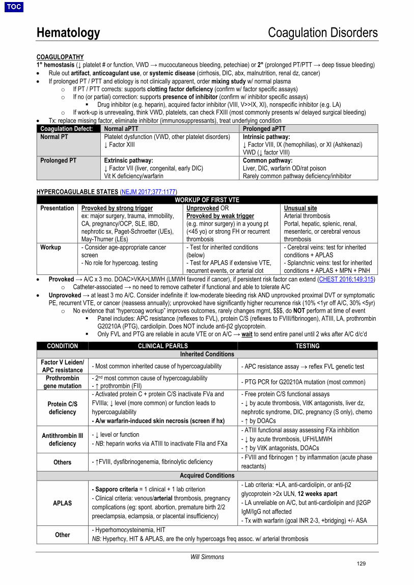
Hematology Coagulation Disorders
Will Simmons
COAGULOPATHY 1° hemostasis (↓ platelet # or function, VWD → mucocutaneous bleeding, petechiae) or 2° (prolonged PT/PTT → deep tissue bleeding) • Rule out artifact, anticoagulant use, or systemic disease (cirrhosis, DIC, abx, malnutrition, renal dz, cancer)• If prolonged PT / PTT and etiology is not clinically apparent, order mixing study w/ normal plasma
o If PT / PTT corrects: supports clotting factor deficiency (confirm w/ factor specific assays)o If no (or partial) correction: supports presence of inhibitor (confirm w/ inhibitor specific assays)
Drug inhibitor (e.g. heparin), acquired factor inhibitor (VIII, V>>IX, XI), nonspecific inhibitor (e.g. LA)o If work-up is unrevealing, think VWD, platelets, can check FXIII (most commonly presents w/ delayed surgical bleeding)
• Tx: replace missing factor, eliminate inhibitor (immunosuppressants), treat underlying conditionCoagulation Defect: Normal aPTT Prolonged aPTT Normal PT Platelet dysfunction (VWD, other platelet disorders)
↓ Factor XIII Intrinsic pathway: ↓ Factor VIII, IX (hemophilias), or XI (Ashkenazi) VWD (↓ factor VIII)
Prolonged PT Extrinsic pathway: ↓ Factor VII (liver, congenital, early DIC) Vit K deficiency/warfarin
Common pathway: Liver, DIC, warfarin OD/rat poison Rarely common pathway deficiency/inhibitor
HYPERCOAGULABLE STATES (NEJM 2017;377:1177)WORKUP OF FIRST VTE
Presentation Provoked by strong trigger ex: major surgery, trauma, immobility, CA, pregnancy/OCP, SLE, IBD, nephrotic sx, Paget-Schroetter (UEs), May-Thurner (LEs)
Unprovoked OR Provoked by weak trigger (e.g. minor surgery) in a young pt (<45 yo) or strong FH or recurrent thrombosis
Unusual site Arterial thrombosis Portal, hepatic, splenic, renal, mesenteric, or cerebral venous thrombosis
Workup - Consider age-appropriate cancerscreen- No role for hypercoag. testing
- Test for inherited conditions(below)- Test for APLAS if extensive VTE,recurrent events, or arterial clot
- Cerebral veins: test for inheritedconditions + APLAS- Splanchnic veins: test for inheritedconditions + APLAS + MPN + PNH
• Provoked → A/C x 3 mo. DOAC>VKA>LMWH (LMWH favored if cancer), if persistent risk factor can extend (CHEST 2016;149:315)o Catheter-associated → no need to remove catheter if functional and able to tolerate A/C
• Unprovoked → at least 3 mo A/C. Consider indefinite if: low-moderate bleeding risk AND unprovoked proximal DVT or symptomaticPE, recurrent VTE, or cancer (reassess annually); unprovoked have significantly higher recurrence risk (10% <1yr off A/C, 30% <5yr)
o No evidence that “hypercoag workup” improves outcomes, rarely changes mgmt, $$$, do NOT perform at time of event Panel includes: APC resistance (reflexes to FVL), protein C/S (reflexes to FVIII/fibrinogen), ATIII, LA, prothrombin
G20210A (PTG), cardiolipin. Does NOT include anti-β2 glycoprotein. Only FVL and PTG are reliable in acute VTE or on A/C → wait to send entire panel until 2 wks after A/C d/c’d
CONDITION CLINICAL PEARLS TESTING Inherited Conditions
Factor V Leiden/ APC resistance - Most common inherited cause of hypercoagulability - APC resistance assay → reflex FVL genetic test
Prothrombin gene mutation
- 2nd most common cause of hypercoagulability- ↑ prothrombin (FII) - PTG PCR for G20210A mutation (most common)
Protein C/S deficiency
- Activated protein C + protein C/S inactivate FVa andFVIIIa; ↓ level (more common) or function leads tohypercoagulability- A/w warfarin-induced skin necrosis (screen if hx)
- Free protein C/S functional assays- ↓ by acute thrombosis, VitK antagonists, liver dz,nephrotic syndrome, DIC, pregnancy (S only), chemo- ↑ by DOACs
Antithrombin III deficiency
- ↓ level or function- NB: heparin works via ATIII to inactivate FIIa and FXa
- ATIII functional assay assessing FXa inhibition- ↓ by acute thrombosis, UFH/LMWH- ↑ by VitK antagonists, DOACs
Others - ↑FVIII, dysfibrinogenemia, fibrinolytic deficiency - FVIII and fibrinogen ↑ by inflammation (acute phasereactants)
Acquired Conditions
APLAS
- Sapporo criteria = 1 clinical + 1 lab criterion- Clinical criteria: venous/arterial thrombosis, pregnancycomplications (eg: spont. abortion, premature birth 2/2preeclampsia, eclampsia, or placental insufficiency)
- Lab criteria: +LA, anti-cardiolipin, or anti-β2glycoprotein >2x ULN, 12 weeks apart- LA unreliable on A/C, but anti-cardiolipin and β2GPIgM/IgG not affected- Tx with warfarin (goal INR 2-3, +bridging) +/- ASA
Other - Hyperhomocysteinemia, HITNB: Hyperhcy, HIT & APLAS, are the only hypercoags freq assoc. w/ arterial thrombosis
129
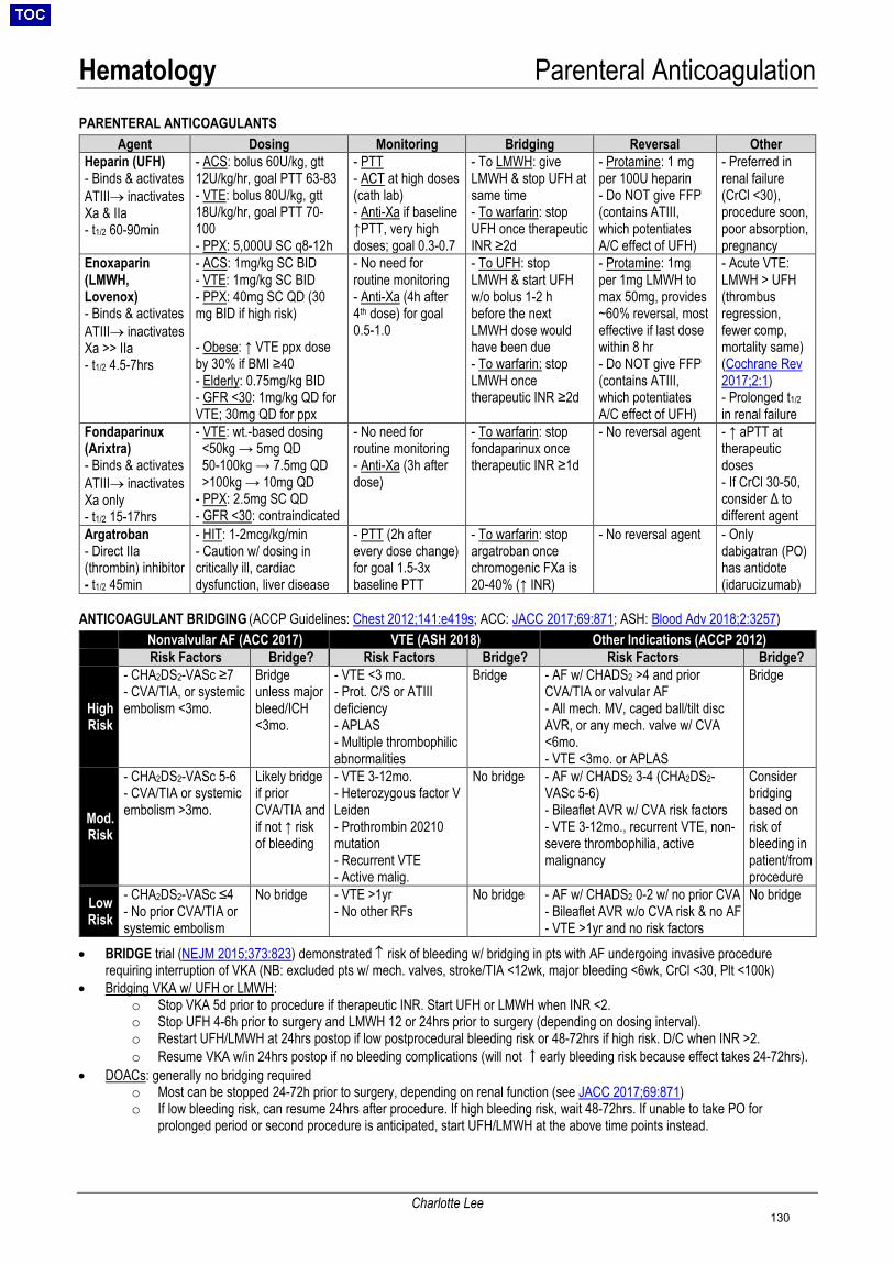
Hematology Parenteral Anticoagulation
Charlotte Lee
PARENTERAL ANTICOAGULANTS
Agent Dosing Monitoring Bridging Reversal Other Heparin (UFH) - Binds & activatesATIII→ inactivatesXa & IIa- t1/2 60-90min
- ACS: bolus 60U/kg, gtt12U/kg/hr, goal PTT 63-83- VTE: bolus 80U/kg, gtt18U/kg/hr, goal PTT 70-100- PPX: 5,000U SC q8-12h
- PTT- ACT at high doses(cath lab)- Anti-Xa if baseline↑PTT, very highdoses; goal 0.3-0.7
- To LMWH: giveLMWH & stop UFH atsame time- To warfarin: stopUFH once therapeuticINR ≥2d
- Protamine: 1 mgper 100U heparin- Do NOT give FFP(contains ATIII,which potentiatesA/C effect of UFH)
- Preferred inrenal failure(CrCl <30),procedure soon,poor absorption,pregnancy
Enoxaparin (LMWH, Lovenox) - Binds & activatesATIII→ inactivatesXa >> IIa- t1/2 4.5-7hrs
- ACS: 1mg/kg SC BID- VTE: 1mg/kg SC BID- PPX: 40mg SC QD (30mg BID if high risk)
- Obese: ↑ VTE ppx doseby 30% if BMI ≥40- Elderly: 0.75mg/kg BID- GFR <30: 1mg/kg QD forVTE; 30mg QD for ppx
- No need forroutine monitoring- Anti-Xa (4h after4th dose) for goal0.5-1.0
- To UFH: stopLMWH & start UFHw/o bolus 1-2 hbefore the nextLMWH dose wouldhave been due- To warfarin: stopLMWH oncetherapeutic INR ≥2d
- Protamine: 1mgper 1mg LMWH tomax 50mg, provides~60% reversal, mosteffective if last dosewithin 8 hr- Do NOT give FFP(contains ATIII,which potentiatesA/C effect of UFH)
- Acute VTE:LMWH > UFH(thrombusregression,fewer comp,mortality same)(Cochrane Rev2017;2:1)- Prolonged t1/2 in renal failure
Fondaparinux (Arixtra) - Binds & activatesATIII→ inactivatesXa only- t1/2 15-17hrs
- VTE: wt.-based dosing<50kg → 5mg QD50-100kg → 7.5mg QD>100kg → 10mg QD
- PPX: 2.5mg SC QD- GFR <30: contraindicated
- No need forroutine monitoring- Anti-Xa (3h afterdose)
- To warfarin: stopfondaparinux oncetherapeutic INR ≥1d
- No reversal agent - ↑ aPTT attherapeuticdoses- If CrCl 30-50,consider Δ todifferent agent
Argatroban - Direct IIa(thrombin) inhibitor- t1/2 45min
- HIT: 1-2mcg/kg/min- Caution w/ dosing incritically ill, cardiacdysfunction, liver disease
- PTT (2h afterevery dose change)for goal 1.5-3xbaseline PTT
- To warfarin: stopargatroban oncechromogenic FXa is20-40% (↑ INR)
- No reversal agent - Onlydabigatran (PO)has antidote(idarucizumab)
ANTICOAGULANT BRIDGING (ACCP Guidelines: Chest 2012;141:e419s; ACC: JACC 2017;69:871; ASH: Blood Adv 2018;2:3257)
Nonvalvular AF (ACC 2017) VTE (ASH 2018) Other Indications (ACCP 2012) Risk Factors Bridge? Risk Factors Bridge? Risk Factors Bridge?
High Risk
- CHA2DS2-VASc ≥7- CVA/TIA, or systemicembolism <3mo.
Bridge unless major bleed/ICH <3mo.
- VTE <3 mo.- Prot. C/S or ATIIIdeficiency- APLAS- Multiple thrombophilicabnormalities
Bridge - AF w/ CHADS2 >4 and priorCVA/TIA or valvular AF- All mech. MV, caged ball/tilt discAVR, or any mech. valve w/ CVA<6mo.- VTE <3mo. or APLAS
Bridge
Mod. Risk
- CHA2DS2-VASc 5-6- CVA/TIA or systemicembolism >3mo.
Likely bridge if prior CVA/TIA and if not ↑ risk of bleeding
- VTE 3-12mo.- Heterozygous factor VLeiden- Prothrombin 20210mutation- Recurrent VTE- Active malig.
No bridge - AF w/ CHADS2 3-4 (CHA2DS2-VASc 5-6)- Bileaflet AVR w/ CVA risk factors- VTE 3-12mo., recurrent VTE, non-severe thrombophilia, activemalignancy
Consider bridging based on risk of bleeding in patient/from procedure
Low Risk
- CHA2DS2-VASc ≤4- No prior CVA/TIA orsystemic embolism
No bridge - VTE >1yr- No other RFs
No bridge - AF w/ CHADS2 0-2 w/ no prior CVA- Bileaflet AVR w/o CVA risk & no AF- VTE >1yr and no risk factors
No bridge
• BRIDGE trial (NEJM 2015;373:823) demonstrated ↑ risk of bleeding w/ bridging in pts with AF undergoing invasive procedurerequiring interruption of VKA (NB: excluded pts w/ mech. valves, stroke/TIA <12wk, major bleeding <6wk, CrCl <30, Plt <100k)
• Bridging VKA w/ UFH or LMWH:o Stop VKA 5d prior to procedure if therapeutic INR. Start UFH or LMWH when INR <2.o Stop UFH 4-6h prior to surgery and LMWH 12 or 24hrs prior to surgery (depending on dosing interval).o Restart UFH/LMWH at 24hrs postop if low postprocedural bleeding risk or 48-72hrs if high risk. D/C when INR >2.o Resume VKA w/in 24hrs postop if no bleeding complications (will not ↑early bleeding risk because effect takes 24-72hrs).
• DOACs: generally no bridging requiredo Most can be stopped 24-72h prior to surgery, depending on renal function (see JACC 2017;69:871)o If low bleeding risk, can resume 24hrs after procedure. If high bleeding risk, wait 48-72hrs. If unable to take PO for
prolonged period or second procedure is anticipated, start UFH/LMWH at the above time points instead.
130

Hematology Oral Anticoagulation
Charlotte Lee, Shawn Li
ORAL ANTICOAGULANTS (ASH Guidelines: Blood Adv 2018;2:3257; ACCP Guidelines: Chest 2012;141:e152S, Chest 2012;141:e44S) Agent Dosing/Monitoring Bridging/Reversal Warfarin (Coumadin) - Vitamin Kantagonist: inhibitsvitamin K-dependent gamma-carboxylation of FII, VII, IX, X, ProteinC, S- t1/2 40h (variable)
Dose: - Initiation: 5mg QD x2d; if frail, HF, kidney/liver dz:consider 2.5mg; If BMI >40: consider 7.5mg- Adjust by INR, which lags 48h behind dose Δ
Monitoring: (UW Dosing Nomogram) - INR <2: ↑ up to 10-20%/wk- INR 2–3: no change- INR 3–4: ↓ 10%/wk- INR >4: hold until INR 2-3, restart ↓5-15%/wk- If overlap w/ direct thrombin inhibitor, checkchromogenic FXa: goal 20-40%
Bridging: - To parenteral A/C: start IV w/o bolus when INR <2- From parenteral A/C: see Parenteral AnticoagulationReversal: (IV vitamin K faster than PO at 6h, same at 24h)- Active bleeding• INR >1.7 → IV vitamin K 10mg + FFP 10cc/kg Q4H
(3-5U in 70kg)• Kcentra (4-factor PCC) if life-threatening (Circ
2013;128:1234; Transfusion 2016;56:799)- No active bleeding• INR >10 → PO vitamin K 2.5-5 mg OR IV 1-2.5 mg• INR <10 → hold warfarin, no need for reversal
Dabigatran (Pradaxa) - Direct thrombin(IIa) inhibitor- t1/2 12-17 h- 80% renalclearance- P-gp substrate
Dose: - Non-valvular AF: 150mg PO BID if GFR >30, 75mg PO BID if GFR 15-30 (RE-LY NEJM2009;361:1139)- VTE: 150mg PO BID after 5d UFH/ LMWH (RE-COVER NEJM 2009;361:2342)- PPX: 220mg PO QD (RE-NOVATE II ThrombHaemost 2011;106:721)
Bridging: - To parenteral A/C: start 12h after last dose- From parental A/C: start <2h before next dose or gtt D/C- To warfarin: start 3d before dabigatran D/C if GFR >50;2d if GFR 31-50, 1d if GFR 15-30; parenteral bridge PRN- From warfarin: hold warfarin, start when INR <2Reversal: (can be dialyzed, lipophilic)- Idarucizumab (REVERSE NEJM 2017;377:431)
Rivaroxaban (Xarelto) - Direct Xa inhibitor- t1/2 5-13 h- 66% renal clear.- interacts w/ CYP-3A4 & P-gp inhib.*Once daily dosing
Dose: - NV AF: 20mg PO QD if GFR >30, 15mg if GFR 15-30 (ROCKET-AF NEJM 2011;365:883)- VTE: 15mg PO BID x21d, then 20 mg QD(EINSTEIN-DVT NEJM 2010;363:2499; EINSTEIN-PE NEJM 2012;366:1287)- PPX: 10mg PO QD (MAGELLAN NEJM2013;368:513)
Bridging: (J Thromb Thrombolysis 2016;41:206) - To parenteral A/C: start when next DOAC dose due- From LMWH: start DOAC within 0-2h of next dose- From UFH: start DOAC immediately after stoppingheparin gtt (for edoxaban, start 4h after stopping UFH)- From warfarin:• Start rivaroxaban when INR <3• Start apixaban when INR <2• Start edoxaban when INR ≤2.5- To warfarin:• Rivaroxaban/apixaban: coadminister until INR ≥2• Edoxaban: cut edoxaban dose by ½ and begin
warfarin, D/C edoxaban once INR ≥2
Reversal: (cannot dialyze, protein-bound) - Andexanet alfa (recombinant FXa): give if life-threateningbleed (ANNEXA-R NEJM 2015;373:2413; ANNEXA-4NEJM 2019)- PER977/Ciraparantag (under FDA review): reverses oralXa, IIa inhibitors, heparin, enoxaparin, fondaparinux(NEJM 2014;371:2141)
Apixaban (Eliquis) - Direct Xa inhibitor- t1/2 8-15h- 25% renal clear.*Can use in renalimpairment if GFR>15
Dose: - NV AF: 5mg PO BID if GFR >30, 2.5 mg BID if 2/3:GFR 15-30, Wt <60kg, age >80 (ARISTOTLE NEJM2011;365:981)- VTE: 10mg BID x7d, then 5mg BID x6mo, then2.5mg BID if need (AMPLIFY NEJM 2013;369:799)- PPX: 2.5 mg BID (NEJM 2009;361:594)
Edoxaban (Savaysa, Lixiana) - Direct Xa inhibitor- t1/2 6-11 h- 50% renalclearance*Once daily dosing
Dose: - NV AF: 60mg PO QD, 30mg QD if CrCl 30-50 or wt≤60kg (ENGAGE-AF NEJM 2013;369:2093)- VTE: 60mg QD after 5d UFH/ LMWH, 30mg QD ifCrCl 30-50 or wt ≤60kg or taking P-gp inhibitors(NEJM 2013;369:1406)- PPX: not FDA-approved (15-30mg PO QD)
CHOOSING ANTICOAGULATION AGENT (ACCP Guidelines for VTE: Chest 2016;149:315; ASH Guidelines for VTE: Blood Adv 2018;2:3257, ACC/AHA/HRS Guidelines for AF: JACC 2019; JACC 2014;64:e1, ACCP Guidelines for AF: Chest 2018;154:1121)• VTE: DOACs (dabigatran, rivaroxaban, apixaban, edoxaban) > VKA > LMWH• VTE & active malignancy: LMWH or edoxaban (NEJM 2018;378:615) > other DOACs, VKA; apixaban may be > LMWH (ADAM-VTE,
ASH 2018); DOAC ppx ↓VTE risk in int/high risk ambulatory pts w/ CA (AVERT NEJM 2019;380:711; CASSINI NEJM 2019;380:720)• VTE & obesity (BMI ≥40, weight ≥120 kg): VKA, LMWH, or rivaroxaban > other DOACs• Recurrent VTE on non-LMWH A/C: switch to LMWH; Recurrent VTE on LMWH: increase LMWH dose• Mechanical valve: VKA; VKA > dabigatran (RE-ALIGN NEJM 2013;369:1206)• Non-valvular AF: DOAC > VKA; Valvular AF: VKA• AF + PCI: dual therapy (P2Y12i + OAC) vs. triple therapy (ASA + P2Y12i + OAC): triple therapy ↑bleeding, ?↓ ischemic events
o Dual therapy options: (1) P2Y12i (clopidogrel or ticag) + VKA (WOEST Lancet 2013;381:1107); (2) P2Y12i (clopidogrel) + lowdose rivaroxaban 15mg QD (PIONEER AF NEJM 2016;375:2423: ↓bleeding, similar CV death/MI/CVA; (3) P2Y12i (clopidogrel)+ dabigatran 150mg BID (RE-DUAL PCI NEJM 2017;377:1513: ↓bleeding, similar MI/CVA/death/unplanned revascularization
o If triple therapy chosen, consider transition to dual therapy at 4-6 weeks• APLS: VKA; VKA > rivaroxaban in high-risk APLS (TRAPS Blood 2018;132:1365)• Stable ischemic CAD: very lose dose rivaroxaban (2.5mg BID) + ASA → ↓MACE compared to ASA alone; ↑ major bleeding but no
difference in ICH or fatal bleeding (COMPASS NEJM 2017;377:1319)
131
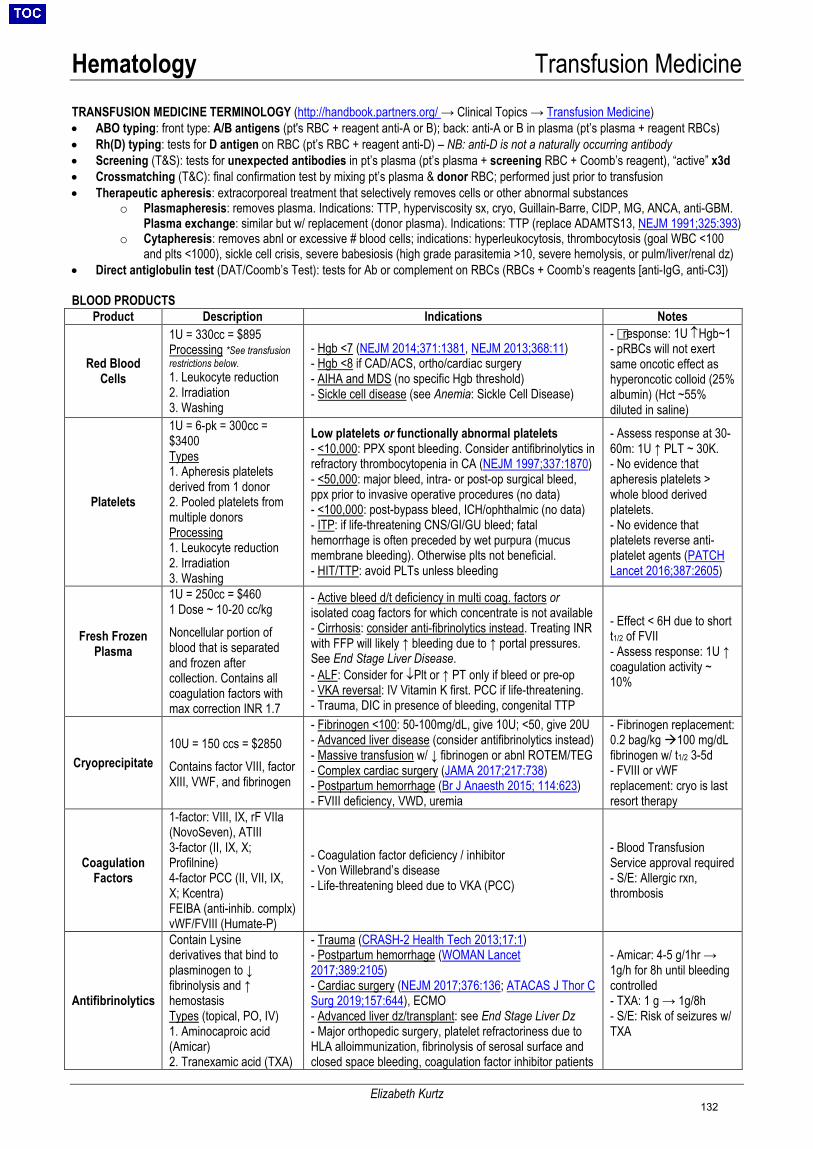
Hematology Transfusion Medicine
Elizabeth Kurtz
TRANSFUSION MEDICINE TERMINOLOGY (http://handbook.partners.org/ → Clinical Topics → Transfusion Medicine) • ABO typing: front type: A/B antigens (pt's RBC + reagent anti-A or B); back: anti-A or B in plasma (pt’s plasma + reagent RBCs)• Rh(D) typing: tests for D antigen on RBC (pt’s RBC + reagent anti-D) – NB: anti-D is not a naturally occurring antibody• Screening (T&S): tests for unexpected antibodies in pt’s plasma (pt’s plasma + screening RBC + Coomb’s reagent), “active” x3d• Crossmatching (T&C): final confirmation test by mixing pt’s plasma & donor RBC; performed just prior to transfusion• Therapeutic apheresis: extracorporeal treatment that selectively removes cells or other abnormal substances
o Plasmapheresis: removes plasma. Indications: TTP, hyperviscosity sx, cryo, Guillain-Barre, CIDP, MG, ANCA, anti-GBM.Plasma exchange: similar but w/ replacement (donor plasma). Indications: TTP (replace ADAMTS13, NEJM 1991;325:393)
o Cytapheresis: removes abnl or excessive # blood cells; indications: hyperleukocytosis, thrombocytosis (goal WBC <100and plts <1000), sickle cell crisis, severe babesiosis (high grade parasitemia >10, severe hemolysis, or pulm/liver/renal dz)
• Direct antiglobulin test (DAT/Coomb’s Test): tests for Ab or complement on RBCs (RBCs + Coomb’s reagents [anti-IgG, anti-C3])
BLOOD PRODUCTS Product Description Indications Notes
Red Blood Cells
1U = 330cc = $895 Processing *See transfusion restrictions below.1. Leukocyte reduction2. Irradiation3. Washing
- Hgb <7 (NEJM 2014;371:1381, NEJM 2013;368:11)- Hgb <8 if CAD/ACS, ortho/cardiac surgery- AIHA and MDS (no specific Hgb threshold)- Sickle cell disease (see Anemia: Sickle Cell Disease)
- response: 1U ↑Hgb~1- pRBCs will not exertsame oncotic effect ashyperoncotic colloid (25%albumin) (Hct ~55%diluted in saline)
Platelets
1U = 6-pk = 300cc = $3400 Types 1. Apheresis plateletsderived from 1 donor2. Pooled platelets frommultiple donorsProcessing1. Leukocyte reduction2. Irradiation3. Washing
Low platelets or functionally abnormal platelets - <10,000: PPX spont bleeding. Consider antifibrinolytics inrefractory thrombocytopenia in CA (NEJM 1997;337:1870)- <50,000: major bleed, intra- or post-op surgical bleed,ppx prior to invasive operative procedures (no data)- <100,000: post-bypass bleed, ICH/ophthalmic (no data)- ITP: if life-threatening CNS/GI/GU bleed; fatalhemorrhage is often preceded by wet purpura (mucusmembrane bleeding). Otherwise plts not beneficial.- HIT/TTP: avoid PLTs unless bleeding
- Assess response at 30-60m: 1U ↑ PLT ~ 30K.- No evidence thatapheresis platelets >whole blood derivedplatelets.- No evidence thatplatelets reverse anti-platelet agents (PATCHLancet 2016;387:2605)
Fresh Frozen Plasma
1U = 250cc = $460 1 Dose ~ 10-20 cc/kg Noncellular portion of blood that is separated and frozen after collection. Contains all coagulation factors with max correction INR 1.7
- Active bleed d/t deficiency in multi coag. factors orisolated coag factors for which concentrate is not available- Cirrhosis: consider anti-fibrinolytics instead. Treating INRwith FFP will likely ↑ bleeding due to ↑ portal pressures.See End Stage Liver Disease.- ALF: Consider for ↓Plt or ↑ PT only if bleed or pre-op- VKA reversal: IV Vitamin K first. PCC if life-threatening.- Trauma, DIC in presence of bleeding, congenital TTP
- Effect < 6H due to shortt1/2 of FVII- Assess response: 1U ↑coagulation activity ~10%
Cryoprecipitate 10U = 150 ccs = $2850 Contains factor VIII, factor XIII, VWF, and fibrinogen
- Fibrinogen <100: 50-100mg/dL, give 10U; <50, give 20U- Advanced liver disease (consider antifibrinolytics instead)- Massive transfusion w/ ↓ fibrinogen or abnl ROTEM/TEG- Complex cardiac surgery (JAMA 2017;217:738)- Postpartum hemorrhage (Br J Anaesth 2015; 114:623)- FVIII deficiency, VWD, uremia
- Fibrinogen replacement:0.2 bag/kg 100 mg/dLfibrinogen w/ t1/2 3-5d- FVIII or vWFreplacement: cryo is lastresort therapy
Coagulation Factors
1-factor: VIII, IX, rF VIIa(NovoSeven), ATIII3-factor (II, IX, X;Profilnine) 4-factor PCC (II, VII, IX,X; Kcentra)FEIBA (anti-inhib. complx)vWF/FVIII (Humate-P)
- Coagulation factor deficiency / inhibitor- Von Willebrand’s disease- Life-threatening bleed due to VKA (PCC)
- Blood TransfusionService approval required- S/E: Allergic rxn,thrombosis
Antifibrinolytics
Contain Lysine derivatives that bind to plasminogen to ↓ fibrinolysis and ↑ hemostasis Types (topical, PO, IV) 1. Aminocaproic acid(Amicar)2. Tranexamic acid (TXA)
- Trauma (CRASH-2 Health Tech 2013;17:1)- Postpartum hemorrhage (WOMAN Lancet2017;389:2105)- Cardiac surgery (NEJM 2017;376:136; ATACAS J Thor CSurg 2019;157:644), ECMO - Advanced liver dz/transplant: see End Stage Liver Dz- Major orthopedic surgery, platelet refractoriness due toHLA alloimmunization, fibrinolysis of serosal surface andclosed space bleeding, coagulation factor inhibitor patients
- Amicar: 4-5 g/1hr →1g/h for 8h until bleedingcontrolled- TXA: 1 g → 1g/8h- S/E: Risk of seizures w/TXA
132
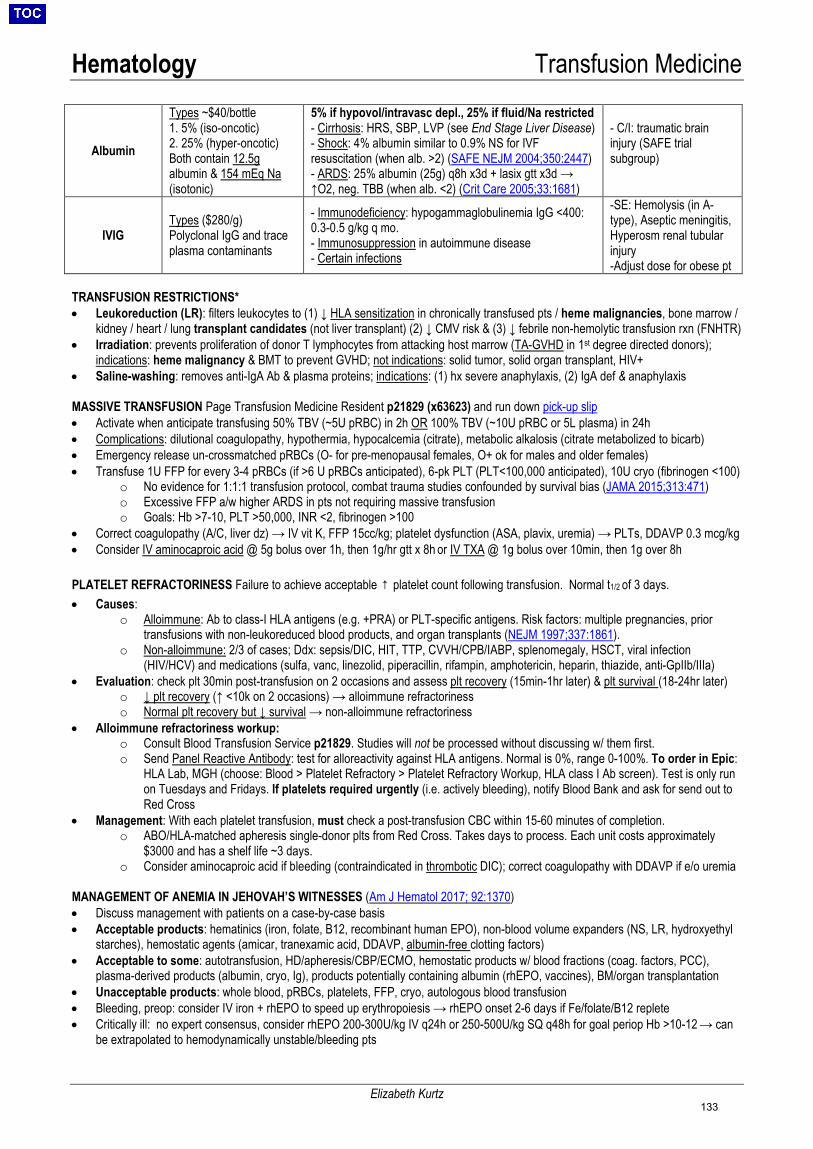
Hematology Transfusion Medicine
Elizabeth Kurtz
Albumin
Types ~$40/bottle 1. 5% (iso-oncotic) 2. 25% (hyper-oncotic) Both contain 12.5g albumin & 154 mEq Na (isotonic)
5% if hypovol/intravasc depl., 25% if fluid/Na restricted - Cirrhosis: HRS, SBP, LVP (see End Stage Liver Disease) - Shock: 4% albumin similar to 0.9% NS for IVF resuscitation (when alb. >2) (SAFE NEJM 2004;350:2447) - ARDS: 25% albumin (25g) q8h x3d + lasix gtt x3d → ↑O2, neg. TBB (when alb. <2) (Crit Care 2005;33:1681)
- C/I: traumatic brain injury (SAFE trial subgroup)
IVIG Types ($280/g) Polyclonal IgG and trace plasma contaminants
- Immunodeficiency: hypogammaglobulinemia IgG <400: 0.3-0.5 g/kg q mo. - Immunosuppression in autoimmune disease - Certain infections
-SE: Hemolysis (in A-type), Aseptic meningitis, Hyperosm renal tubular injury -Adjust dose for obese pt
TRANSFUSION RESTRICTIONS* • Leukoreduction (LR): filters leukocytes to (1) ↓ HLA sensitization in chronically transfused pts / heme malignancies, bone marrow /
kidney / heart / lung transplant candidates (not liver transplant) (2) ↓ CMV risk & (3) ↓ febrile non-hemolytic transfusion rxn (FNHTR) • Irradiation: prevents proliferation of donor T lymphocytes from attacking host marrow (TA-GVHD in 1st degree directed donors);
indications: heme malignancy & BMT to prevent GVHD; not indications: solid tumor, solid organ transplant, HIV+ • Saline-washing: removes anti-IgA Ab & plasma proteins; indications: (1) hx severe anaphylaxis, (2) IgA def & anaphylaxis MASSIVE TRANSFUSION Page Transfusion Medicine Resident p21829 (x63623) and run down pick-up slip • Activate when anticipate transfusing 50% TBV (~5U pRBC) in 2h OR 100% TBV (~10U pRBC or 5L plasma) in 24h • Complications: dilutional coagulopathy, hypothermia, hypocalcemia (citrate), metabolic alkalosis (citrate metabolized to bicarb) • Emergency release un-crossmatched pRBCs (O- for pre-menopausal females, O+ ok for males and older females) • Transfuse 1U FFP for every 3-4 pRBCs (if >6 U pRBCs anticipated), 6-pk PLT (PLT<100,000 anticipated), 10U cryo (fibrinogen <100)
o No evidence for 1:1:1 transfusion protocol, combat trauma studies confounded by survival bias (JAMA 2015;313:471) o Excessive FFP a/w higher ARDS in pts not requiring massive transfusion o Goals: Hb >7-10, PLT >50,000, INR <2, fibrinogen >100
• Correct coagulopathy (A/C, liver dz) → IV vit K, FFP 15cc/kg; platelet dysfunction (ASA, plavix, uremia) → PLTs, DDAVP 0.3 mcg/kg • Consider IV aminocaproic acid @ 5g bolus over 1h, then 1g/hr gtt x 8h or IV TXA @ 1g bolus over 10min, then 1g over 8h PLATELET REFRACTORINESS Failure to achieve acceptable ↑ platelet count following transfusion. Normal t1/2 of 3 days. • Causes:
o Alloimmune: Ab to class-I HLA antigens (e.g. +PRA) or PLT-specific antigens. Risk factors: multiple pregnancies, prior transfusions with non-leukoreduced blood products, and organ transplants (NEJM 1997;337:1861).
o Non-alloimmune: 2/3 of cases; Ddx: sepsis/DIC, HIT, TTP, CVVH/CPB/IABP, splenomegaly, HSCT, viral infection (HIV/HCV) and medications (sulfa, vanc, linezolid, piperacillin, rifampin, amphotericin, heparin, thiazide, anti-GpIIb/IIIa)
• Evaluation: check plt 30min post-transfusion on 2 occasions and assess plt recovery (15min-1hr later) & plt survival (18-24hr later) o ↓ plt recovery (↑ <10k on 2 occasions) → alloimmune refractoriness o Normal plt recovery but ↓ survival → non-alloimmune refractoriness
• Alloimmune refractoriness workup: o Consult Blood Transfusion Service p21829. Studies will not be processed without discussing w/ them first. o Send Panel Reactive Antibody: test for alloreactivity against HLA antigens. Normal is 0%, range 0-100%. To order in Epic:
HLA Lab, MGH (choose: Blood > Platelet Refractory > Platelet Refractory Workup, HLA class I Ab screen). Test is only run on Tuesdays and Fridays. If platelets required urgently (i.e. actively bleeding), notify Blood Bank and ask for send out to Red Cross
• Management: With each platelet transfusion, must check a post-transfusion CBC within 15-60 minutes of completion. o ABO/HLA-matched apheresis single-donor plts from Red Cross. Takes days to process. Each unit costs approximately
$3000 and has a shelf life ~3 days. o Consider aminocaproic acid if bleeding (contraindicated in thrombotic DIC); correct coagulopathy with DDAVP if e/o uremia
MANAGEMENT OF ANEMIA IN JEHOVAH’S WITNESSES (Am J Hematol 2017; 92:1370)
• Discuss management with patients on a case-by-case basis • Acceptable products: hematinics (iron, folate, B12, recombinant human EPO), non-blood volume expanders (NS, LR, hydroxyethyl
starches), hemostatic agents (amicar, tranexamic acid, DDAVP, albumin-free clotting factors) • Acceptable to some: autotransfusion, HD/apheresis/CBP/ECMO, hemostatic products w/ blood fractions (coag. factors, PCC),
plasma-derived products (albumin, cryo, Ig), products potentially containing albumin (rhEPO, vaccines), BM/organ transplantation • Unacceptable products: whole blood, pRBCs, platelets, FFP, cryo, autologous blood transfusion • Bleeding, preop: consider IV iron + rhEPO to speed up erythropoiesis → rhEPO onset 2-6 days if Fe/folate/B12 replete • Critically ill: no expert consensus, consider rhEPO 200-300U/kg IV q24h or 250-500U/kg SQ q48h for goal periop Hb >10-12 → can
be extrapolated to hemodynamically unstable/bleeding pts
133
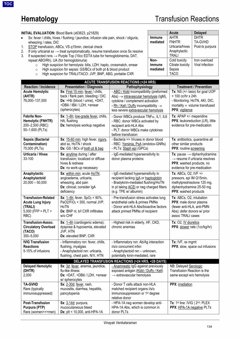
Hematology Transfusion Reactions
Vinayak Venkataraman
INITIAL EVALUATION: Blood Bank (x63623, p21829) • Sx: fever / chills, hives / flushing / jaundice, infusion site pain, shock / oliguria,
wheezing / rales, DIC 1. STOP transfusion, ABCs, VS q15min, clerical check 2. If only urticarial sx → treat symptomatically, resume transfusion once Sx resolve 3. If suspected rxns → Purple Top (10cc EDTA tube for hemoglobinemia, DAT,
repeat ABO/Rh), UA (for hemoglobinuria) o High suspicion for hemolysis: bilis, LDH, hapto, crossmatch, smear o High suspicion for sepsis: GS/BCx of both pt & blood product o High suspicion for TRALI/TACO: JVP, BNP, ABG, portable CXR
ACUTE TRANSFUSION REACTIONS (<24 HRS)
Reaction / Incidence Presentation / Diagnosis Pathophysiology Treatment / Prevention Acute Hemolytic (AHTR) 76,000–137,000
Sx: First 15 min; fever / chills, back / flank pain, bleeding / DIC Dx: +Hb (blood / urine), +DAT, +DBili / IBili / LDH, +smear (spherocytes)
- ABO / Kidd incompatibility (preformed Abs) → intravascular hemolysis (IgM), cytokine / complement activation - Rh / Kell / Duffy incompatibility → less severe extravascular hemolysis
Tx: NS (+/- lasix) for goal UOP > 100 cc/hr x 24h - Monitoring: HoTN, AKI, DIC, mortality ∝ volume transfused PPX: vigilance
Febrile Non-Hemolytic (FNHTR) 200–2,500 (RBC) 50–1,600 (PLTs)
Sx: 1–6h; low-grade fever, chills, HA, flushing Dx: hemolysis workup negative
- Donor WBCs produce TNFα, IL1, IL6 - RBC: donor WBCs activated by recipient anti-HLA Abs - PLT: donor WBCs make cytokines before transfusion
Tx: APAP +/- meperidine PPX: leukoreduction (LR), little evidence for pre-medication
Sepsis (Bacterial Contamination) 75,000 (PLTs)
Sx: 15-60 min; high fever, rigors, abd sx, HoTN / shock Dx: GS / BCx of both pt & bag
- Bacteria >> Viruses in donor blood - RBC: Yersinia, PsA (endotox-GNRs) - PLTs: Staph epi (GPCs)
Tx: antibiotics, quarantine all other similar products PPX: routine screening
Urticaria / Hives 33-100
Sx: anytime during / after transfusion; localized or diffuse hives & redness Dx: no work-up necessary
- IgE-mediated hypersensitivity to donor plasma proteins
Tx: pause → diphenhydramine → resume if urticaria resolves PPX: washed products, no evidence for pre-medication
Anaphylactic Anaphylactoid 20,000 – 50,000
Sx: within min; acute HoTN, angioedema, urticaria, wheezing, abd pain Dx: clinical; consider IgA deficiency
- IgE-mediated hypersensitivity in recipient lacking IgA or haptoglobin - Bradykinin-mediated flushing/HoTN in pt taking ACEi or neg charged filters (e.g. TPE w/ albumin)
Tx: ABCs, O2, IVF +/- pressors, epi IM Q15min, methylprednisolone 125 mg, diphenhydramine 25-50 mg PPX: washed products
Transfusion-Related Acute Lung Injury (TRALI) 5,000 (FFP > PLT > RBC)
Sx: 1–6h; fever, SpO2 < 90%, PaO2/FiO2 < 300, normal JVP, HoTN Dx: BNP nl, b/l CXR infiltrates w/o CHF
- Pre-transfusion stress activates lung endothelial cells & primes PMNs - Donor anti-HLA Abs/bioactive factors attack primed PMNs of recipient
Tx: ABCs, O2, intubation PPX: male donor plasma (fewer anti-HLA, anti-PMN Abs); defer donors w/ prior assoc TRALI cases
Transfusion-Assoc. Circulatory Overload (TACO) 350–5,000
Sx: 1–6h (cardiogenic edema); dyspnea & hypoxemia, elevated JVP, HTN Dx: elevated BNP, CXR
- Highest risk in elderly, HF, CKD, chronic anemias
Tx: O2, IV diuretics PPX: slower rate (1cc/kg/hr)
IVIG Transfusion Reactions 5-15% of infusions
- Inflammatory rxn: fever, chills, flushing, myalgias - Anaphylactoid rxn: urticaria, flushing, chest pain, N/V, HTN
- Inflammatory rxn: Ab/Ag interaction i/s/o concurrent infxn - Anaphylactoid rxn – unknown, potentially kinin-mediated, rare
Tx: IVF, sx mgmt PPX: slow, space out infusions
DELAYED TRANSFUSION REACTIONS (>24 HRS, <28 DAYS) Delayed Hemolytic (DHTR) 2,000
Sx: 3d; fever, anemia, jaundice, flu-like illness Dx: +DAT, +DBili / LDH, +smear w/ spherocytes
- Anamnestic IgG against previously exposed antigen (Kidd / Duffy / Kell) → extravascular hemolysis
NB: Delayed Serologic Transfusion Reaction is the same except w/o hemolysis
TA-GVHD Rare (typically immunosuppressed)
Sx: 2-30d; fever, rash, mucositis, diarrhea, hepatitis, pancytopenia
- Donor T cells attack non-HLA matched recipient organs i/s/o immunosuppression or 1st degree relative donor
PPX: irradiation
Post-Transfusion Purpura (PTP) Rare (women>>>men)
Sx: 3-14d; purpura, mucocutaneous bleed Dx: plt < 10,000, anti-HPA-1A
- HPA-1A neg women develop anti-HPA-1A Abs, which is common in donor PLTs
Tx: 1st line: IVIG | 2nd: PLEX PPX: HPA-1A negative PLTs
Acute Delayed Immune mediated
AHTR FNHTR Urticaria/hives Anaphylactic TRALI
DHTR TA-GVHD Post-tx purpura
Non-immune mediated
Cold toxicity Citrate toxicity Sepsis TACO
Iron overload Viral infection
134
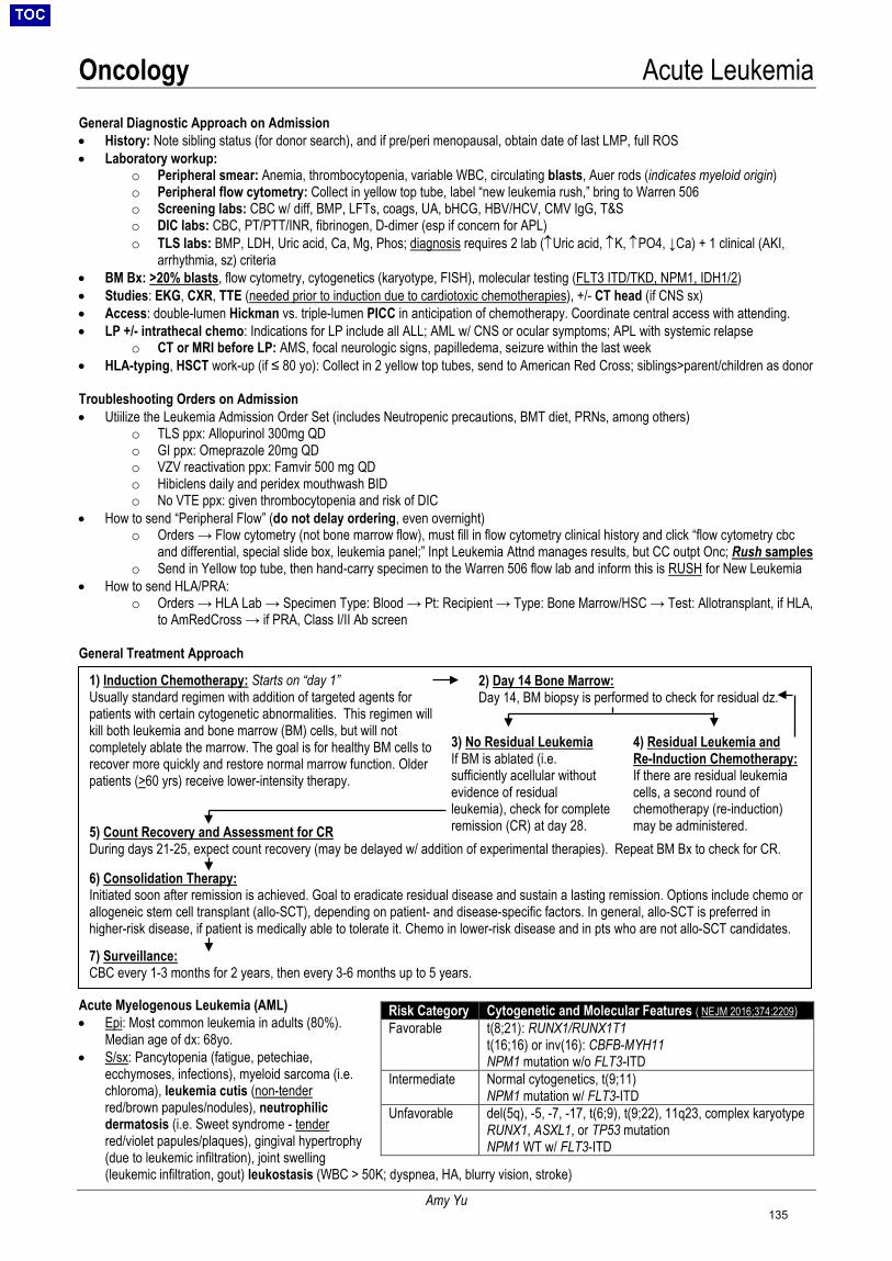
Oncology Acute Leukemia
Amy Yu
General Diagnostic Approach on Admission • History: Note sibling status (for donor search), and if pre/peri menopausal, obtain date of last LMP, full ROS • Laboratory workup:
o Peripheral smear: Anemia, thrombocytopenia, variable WBC, circulating blasts, Auer rods (indicates myeloid origin) o Peripheral flow cytometry: Collect in yellow top tube, label “new leukemia rush,” bring to Warren 506 o Screening labs: CBC w/ diff, BMP, LFTs, coags, UA, bHCG, HBV/HCV, CMV IgG, T&S o DIC labs: CBC, PT/PTT/INR, fibrinogen, D-dimer (esp if concern for APL) o TLS labs: BMP, LDH, Uric acid, Ca, Mg, Phos; diagnosis requires 2 lab (↑Uric acid, ↑K, ↑PO4, ↓Ca) + 1 clinical (AKI,
arrhythmia, sz) criteria • BM Bx: >20% blasts, flow cytometry, cytogenetics (karyotype, FISH), molecular testing (FLT3 ITD/TKD, NPM1, IDH1/2) • Studies: EKG, CXR, TTE (needed prior to induction due to cardiotoxic chemotherapies), +/- CT head (if CNS sx) • Access: double-lumen Hickman vs. triple-lumen PICC in anticipation of chemotherapy. Coordinate central access with attending. • LP +/- intrathecal chemo: Indications for LP include all ALL; AML w/ CNS or ocular symptoms; APL with systemic relapse
o CT or MRI before LP: AMS, focal neurologic signs, papilledema, seizure within the last week • HLA-typing, HSCT work-up (if ≤ 80 yo): Collect in 2 yellow top tubes, send to American Red Cross; siblings>parent/children as donor Troubleshooting Orders on Admission • Utiilize the Leukemia Admission Order Set (includes Neutropenic precautions, BMT diet, PRNs, among others)
o TLS ppx: Allopurinol 300mg QD o GI ppx: Omeprazole 20mg QD o VZV reactivation ppx: Famvir 500 mg QD o Hibiclens daily and peridex mouthwash BID o No VTE ppx: given thrombocytopenia and risk of DIC
• How to send “Peripheral Flow” (do not delay ordering, even overnight) o Orders → Flow cytometry (not bone marrow flow), must fill in flow cytometry clinical history and click “flow cytometry cbc
and differential, special slide box, leukemia panel;” Inpt Leukemia Attnd manages results, but CC outpt Onc; Rush samples o Send in Yellow top tube, then hand-carry specimen to the Warren 506 flow lab and inform this is RUSH for New Leukemia
• How to send HLA/PRA: o Orders → HLA Lab → Specimen Type: Blood → Pt: Recipient → Type: Bone Marrow/HSC → Test: Allotransplant, if HLA,
to AmRedCross → if PRA, Class I/II Ab screen General Treatment Approach Acute Myelogenous Leukemia (AML) • Epi: Most common leukemia in adults (80%).
Median age of dx: 68yo. • S/sx: Pancytopenia (fatigue, petechiae,
ecchymoses, infections), myeloid sarcoma (i.e. chloroma), leukemia cutis (non-tender red/brown papules/nodules), neutrophilic dermatosis (i.e. Sweet syndrome - tender red/violet papules/plaques), gingival hypertrophy (due to leukemic infiltration), joint swelling (leukemic infiltration, gout) leukostasis (WBC > 50K; dyspnea, HA, blurry vision, stroke)
Risk Category Cytogenetic and Molecular Features ( NEJM 2016;374:2209) Favorable t(8;21): RUNX1/RUNX1T1
t(16;16) or inv(16): CBFB-MYH11 NPM1 mutation w/o FLT3-ITD
Intermediate Normal cytogenetics, t(9;11) NPM1 mutation w/ FLT3-ITD
Unfavorable del(5q), -5, -7, -17, t(6;9), t(9;22), 11q23, complex karyotype RUNX1, ASXL1, or TP53 mutation NPM1 WT w/ FLT3-ITD
1) Induction Chemotherapy: Starts on “day 1” Usually standard regimen with addition of targeted agents for patients with certain cytogenetic abnormalities. This regimen will kill both leukemia and bone marrow (BM) cells, but will not completely ablate the marrow. The goal is for healthy BM cells to recover more quickly and restore normal marrow function. Older patients (>60 yrs) receive lower-intensity therapy.
2) Day 14 Bone Marrow: Day 14, BM biopsy is performed to check for residual dz.
6) Consolidation Therapy: Initiated soon after remission is achieved. Goal to eradicate residual disease and sustain a lasting remission. Options include chemo or allogeneic stem cell transplant (allo-SCT), depending on patient- and disease-specific factors. In general, allo-SCT is preferred in higher-risk disease, if patient is medically able to tolerate it. Chemo in lower-risk disease and in pts who are not allo-SCT candidates.
5) Count Recovery and Assessment for CR During days 21-25, expect count recovery (may be delayed w/ addition of experimental therapies). Repeat BM Bx to check for CR.
7) Surveillance: CBC every 1-3 months for 2 years, then every 3-6 months up to 5 years.
4) Residual Leukemia and Re-Induction Chemotherapy: If there are residual leukemia cells, a second round of chemotherapy (re-induction) may be administered.
3) No Residual Leukemia If BM is ablated (i.e. sufficiently acellular without evidence of residual leukemia), check for complete remission (CR) at day 28.
135
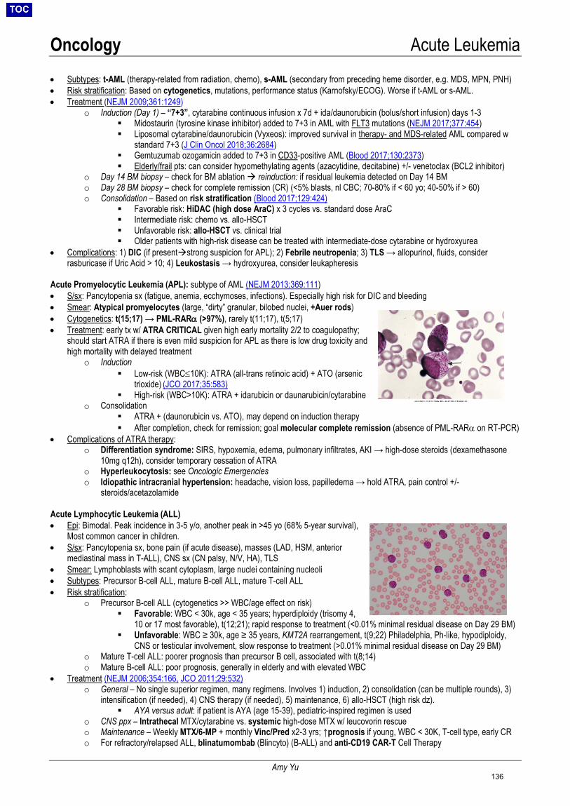
Oncology Acute Leukemia
Amy Yu
• Subtypes: t-AML (therapy-related from radiation, chemo), s-AML (secondary from preceding heme disorder, e.g. MDS, MPN, PNH) • Risk stratification: Based on cytogenetics, mutations, performance status (Karnofsky/ECOG). Worse if t-AML or s-AML. • Treatment (NEJM 2009;361:1249)
o Induction (Day 1) – “7+3”, cytarabine continuous infusion x 7d + ida/daunorubicin (bolus/short infusion) days 1-3 Midostaurin (tyrosine kinase inhibitor) added to 7+3 in AML with FLT3 mutations (NEJM 2017;377:454) Liposomal cytarabine/daunorubicin (Vyxeos): improved survival in therapy- and MDS-related AML compared w
standard 7+3 (J Clin Oncol 2018;36:2684) Gemtuzumab ozogamicin added to 7+3 in CD33-positive AML (Blood 2017;130:2373) Elderly/frail pts: can consider hypomethylating agents (azacytidine, decitabine) +/- venetoclax (BCL2 inhibitor)
o Day 14 BM biopsy – check for BM ablation reinduction: if residual leukemia detected on Day 14 BM o Day 28 BM biopsy – check for complete remission (CR) (<5% blasts, nl CBC; 70-80% if < 60 yo; 40-50% if > 60) o Consolidation – Based on risk stratification (Blood 2017;129:424)
Favorable risk: HiDAC (high dose AraC) x 3 cycles vs. standard dose AraC Intermediate risk: chemo vs. allo-HSCT Unfavorable risk: allo-HSCT vs. clinical trial Older patients with high-risk disease can be treated with intermediate-dose cytarabine or hydroxyurea
• Complications: 1) DIC (if presentstrong suspicion for APL); 2) Febrile neutropenia; 3) TLS → allopurinol, fluids, consider rasburicase if Uric Acid > 10; 4) Leukostasis → hydroxyurea, consider leukapheresis
Acute Promyelocytic Leukemia (APL): subtype of AML (NEJM 2013;369:111) • S/sx: Pancytopenia sx (fatigue, anemia, ecchymoses, infections). Especially high risk for DIC and bleeding • Smear: Atypical promyelocytes (large, “dirty” granular, bilobed nuclei, +Auer rods) • Cytogenetics: t(15;17) → PML-RARα (>97%), rarely t(11;17), t(5;17) • Treatment: early tx w/ ATRA CRITICAL given high early mortality 2/2 to coagulopathy;
should start ATRA if there is even mild suspicion for APL as there is low drug toxicity and high mortality with delayed treatment
o Induction Low-risk (WBC≤10K): ATRA (all-trans retinoic acid) + ATO (arsenic
trioxide) (JCO 2017;35:583) High-risk (WBC>10K): ATRA + idarubicin or daunarubicin/cytarabine
o Consolidation ATRA + (daunorubicin vs. ATO), may depend on induction therapy After completion, check for remission; goal molecular complete remission (absence of PML-RARα on RT-PCR)
• Complications of ATRA therapy: o Differentiation syndrome: SIRS, hypoxemia, edema, pulmonary infiltrates, AKI → high-dose steroids (dexamethasone
10mg q12h), consider temporary cessation of ATRA o Hyperleukocytosis: see Oncologic Emergencies o Idiopathic intracranial hypertension: headache, vision loss, papilledema → hold ATRA, pain control +/-
steroids/acetazolamide Acute Lymphocytic Leukemia (ALL) • Epi: Bimodal. Peak incidence in 3-5 y/o, another peak in >45 yo (68% 5-year survival),
Most common cancer in children. • S/sx: Pancytopenia sx, bone pain (if acute disease), masses (LAD, HSM, anterior
mediastinal mass in T-ALL), CNS sx (CN palsy, N/V, HA), TLS • Smear: Lymphoblasts with scant cytoplasm, large nuclei containing nucleoli • Subtypes: Precursor B-cell ALL, mature B-cell ALL, mature T-cell ALL • Risk stratification:
o Precursor B-cell ALL (cytogenetics >> WBC/age effect on risk) Favorable: WBC < 30k, age < 35 years; hyperdiploidy (trisomy 4,
10 or 17 most favorable), t(12;21); rapid response to treatment (<0.01% minimal residual disease on Day 29 BM) Unfavorable: WBC ≥ 30k, age ≥ 35 years, KMT2A rearrangement, t(9;22) Philadelphia, Ph-like, hypodiploidy,
CNS or testicular involvement, slow response to treatment (>0.01% minimal residual disease on Day 29 BM) o Mature T-cell ALL: poorer prognosis than precursor B cell, associated with t(8;14) o Mature B-cell ALL: poor prognosis, generally in elderly and with elevated WBC
• Treatment (NEJM 2006;354:166, JCO 2011;29:532) o General – No single superior regimen, many regimens. Involves 1) induction, 2) consolidation (can be multiple rounds), 3)
intensification (if needed), 4) CNS therapy (if needed), 5) maintenance, 6) allo-HSCT (high risk dz). AYA versus adult: if patient is AYA (age 15-39), pediatric-inspired regimen is used
o CNS ppx – Intrathecal MTX/cytarabine vs. systemic high-dose MTX w/ leucovorin rescue o Maintenance – Weekly MTX/6-MP + monthly Vinc/Pred x2-3 yrs; ↑prognosis if young, WBC < 30K, T-cell type, early CR o For refractory/relapsed ALL, blinatumombab (Blincyto) (B-ALL) and anti-CD19 CAR-T Cell Therapy
136
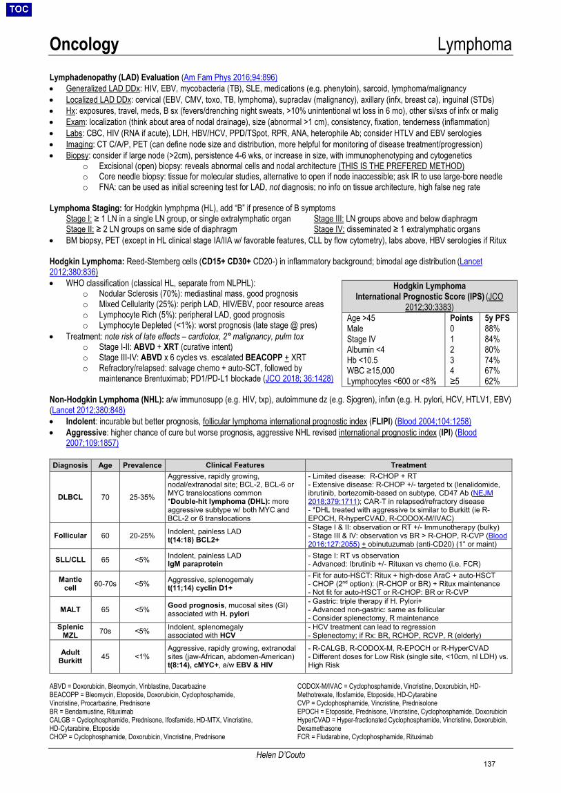
Oncology Lymphoma
Helen D’Couto
Lymphadenopathy (LAD) Evaluation (Am Fam Phys 2016;94:896)
• Generalized LAD DDx: HIV, EBV, mycobacteria (TB), SLE, medications (e.g. phenytoin), sarcoid, lymphoma/malignancy • Localized LAD DDx: cervical (EBV, CMV, toxo, TB, lymphoma), supraclav (malignancy), axillary (infx, breast ca), inguinal (STDs) • Hx: exposures, travel, meds, B sx (fevers/drenching night sweats, >10% unintentional wt loss in 6 mo), other si/sxs of infx or malig • Exam: localization (think about area of nodal drainage), size (abnormal >1 cm), consistency, fixation, tenderness (inflammation) • Labs: CBC, HIV (RNA if acute), LDH, HBV/HCV, PPD/TSpot, RPR, ANA, heterophile Ab; consider HTLV and EBV serologies • Imaging: CT C/A/P, PET (can define node size and distribution, more helpful for monitoring of disease treatment/progression) • Biopsy: consider if large node (>2cm), persistence 4-6 wks, or increase in size, with immunophenotyping and cytogenetics
o Excisional (open) biopsy: reveals abnormal cells and nodal architecture (THIS IS THE PREFERED METHOD) o Core needle biopsy: tissue for molecular studies, alternative to open if node inaccessible; ask IR to use large-bore needle o FNA: can be used as initial screening test for LAD, not diagnosis; no info on tissue architecture, high false neg rate
Lymphoma Staging: for Hodgkin lymphpma (HL), add “B” if presence of B symptoms
Stage I: ≥ 1 LN in a single LN group, or single extralymphatic organ Stage III: LN groups above and below diaphragm Stage II: ≥ 2 LN groups on same side of diaphragm Stage IV: disseminated ≥ 1 extralymphatic organs
• BM biopsy, PET (except in HL clinical stage IA/IIA w/ favorable features, CLL by flow cytometry), labs above, HBV serologies if Ritux Hodgkin Lymphoma: Reed-Sternberg cells (CD15+ CD30+ CD20-) in inflammatory background; bimodal age distribution (Lancet 2012;380:836) • WHO classification (classical HL, separate from NLPHL):
o Nodular Sclerosis (70%): mediastinal mass, good prognosis o Mixed Cellularity (25%): periph LAD, HIV/EBV, poor resource areas o Lymphocyte Rich (5%): peripheral LAD, good prognosis o Lymphocyte Depleted (<1%): worst prognosis (late stage @ pres)
• Treatment: note risk of late effects – cardiotox, 2° malignancy, pulm tox o Stage I-II: ABVD + XRT (curative intent) o Stage III-IV: ABVD x 6 cycles vs. escalated BEACOPP + XRT o Refractory/relapsed: salvage chemo + auto-SCT, followed by
maintenance Brentuximab; PD1/PD-L1 blockade (JCO 2018; 36:1428)
Non-Hodgkin Lymphoma (NHL): a/w immunosupp (e.g. HIV, txp), autoimmune dz (e.g. Sjogren), infxn (e.g. H. pylori, HCV, HTLV1, EBV) (Lancet 2012;380:848)
• Indolent: incurable but better prognosis, follicular lymphoma international prognostic index (FLIPI) (Blood 2004;104:1258) • Aggressive: higher chance of cure but worse prognosis, aggressive NHL revised international prognostic index (IPI) (Blood
2007;109:1857)
ABVD = Doxorubicin, Bleomycin, Vinblastine, Dacarbazine BEACOPP = Bleomycin, Etoposide, Doxorubicin, Cyclophosphamide, Vincristine, Procarbazine, Prednisone BR = Bendamustine, Rituximab CALGB = Cyclophosphamide, Prednisone, Ifosfamide, HD-MTX, Vincristine, HD-Cytarabine, Etoposide CHOP = Cyclophosphamide, Doxorubicin, Vincristine, Prednisone
CODOX-M/IVAC = Cyclophosphamide, Vincristine, Doxorubicin, HD-Methotrexate, Ifosfamide, Etoposide, HD-Cytarabine CVP = Cyclophosphamide, Vincristine, Prednisolone EPOCH = Etoposide, Prednisone, Vincristine, Cyclophosphamide, Doxorubicin HyperCVAD = Hyper-fractionated Cyclophosphamide, Vincristine, Doxorubicin, Dexamethasone FCR = Fludarabine, Cyclophosphamide, Rituximab
Hodgkin Lymphoma International Prognostic Score (IPS) (JCO
2012;30:3383)
Age >45 Male Stage IV Albumin <4 Hb <10.5 WBC ≥15,000 Lymphocytes <600 or <8%
Points 0 1 2 3 4 ≥5
5y PFS 88% 84% 80% 74% 67% 62%
Diagnosis Age Prevalence Clinical Features Treatment
DLBCL 70 25-35%
Aggressive, rapidly growing, nodal/extranodal site; BCL-2, BCL-6 or MYC translocations common *Double-hit lymphoma (DHL): more aggressive subtype w/ both MYC and BCL-2 or 6 translocations
- Limited disease: R-CHOP + RT - Extensive disease: R-CHOP +/- targeted tx (lenalidomide, ibrutinib, bortezomib-based on subtype, CD47 Ab (NEJM 2018;379:1711); CAR-T in relapsed/refractory disease - *DHL treated with aggressive tx similar to Burkitt (ie R-EPOCH, R-hyperCVAD, R-CODOX-M/IVAC)
Follicular 60 20-25% Indolent, painless LAD t(14:18) BCL2+
- Stage I & II: observation or RT +/- Immunotherapy (bulky) - Stage III & IV: observation vs BR > R-CHOP, R-CVP (Blood 2016;127:2055) + obinutuzumab (anti-CD20) (1° or maint)
SLL/CLL 65 <5% Indolent, painless LAD IgM paraprotein
- Stage I: RT vs observation - Advanced: Ibrutinib +/- Rituxan vs chemo (i.e. FCR)
Mantle cell 60-70s <5% Aggressive, splenogemaly
t(11;14) cyclin D1+ - Fit for auto-HSCT: Ritux + high-dose AraC + auto-HSCT - CHOP (2nd option): (R-CHOP or BR) + Ritux maintenance - Not fit for auto-HSCT or R-CHOP: BR or R-CVP
MALT 65 <5% Good prognosis, mucosal sites (GI) associated with H. pylori
- Gastric: triple therapy if H. Pylori+ - Advanced non-gastric: same as follicular - Consider splenectomy, R maintenance
Splenic MZL 70s <5% Indolent, splenomegaly
associated with HCV - HCV treatment can lead to regression - Splenectomy; if Rx: BR, RCHOP, RCVP, R (elderly)
Adult Burkitt 45 <1%
Aggressive, rapidly growing, extranodal sites (jaw-African, abdomen-American) t(8:14), cMYC+, a/w EBV & HIV
- R-CALGB, R-CODOX-M, R-EPOCH or R-HyperCVAD - Different doses for Low Risk (single site, <10cm, nl LDH) vs. High Risk
137

Oncology Plasma Cell Disorders
David Qualls
EVALUATION OF PLASMA CELL DISORDERS (Am Fam Phys 2005;71:105, Leukemia 2009;23:215) • Serum protein electrophoresis (SPEP): detects & quantifies monoclonal M-proteins
o M-protein (paraprotein, monoclonal protein): monoclonal Ig secreted by abnormally expanded clone of B-cells and plasma cells, seen in monoclonal B-cell proliferative disorders, cryoglobulinemia, and autoimmune disease (e.g. RA, SS)
• Immunofixation (IF): confirms polyclonal or monoclonal nature of serum proteins, and determines the type of M-protein, based on its heavy (IgG, IgM, IgA) and light (κ/λ) chain composition
• Serum free light chain (sFLC): detects polyclonal & monoclonal free light chains (κ/λ) (200-500x more sensitive than IF) o Normal: κ & λ polyclonal light chains in a κ/λ ratio of 0.26-1.65 confirms normal B-cell differentiation o Abnormal κ/λ ratio: monoclonal plasma (MM, primary amyloidosis) and B-cell (lymphomas) aberrations o Polyclonal decrease: immunosuppression and immunodeficiency states o ↑ sFLC & relatively unchanged κ/λ ratio: chronic infection, inflammatory disorders, polyclonal autoimmune disease o NB: Renal failure results in increased sFLC levels with κ/λ ratio up to 3 due to ↓clearance of FLC→ determine urine Bence
Jones protein (monoclonal light chains) by UPEP+IF κ/λ ratio 1.7-3.0, to determine if FLC are monoclonal • UPEP (24h): similar to SPEP but more sensitive in detecting FLC (BJP); if M protein detected, it is confirmed by IF • Specific Indications:
o SPEP & Ig levels: send if suspect primary hypogammaglobinemia (primary B-cell immunodeficiencies, e.g. CVID) or secondary immunodeficiencies (e.g. lymphoma, myeloma, immunosuppressive therapy, post-irradiation)
o SPEP+IF & sFLC: send if suspect clonal B-cell abnormalities (e.g. myeloma, Waldenstrom, amyloidosis, paraproteinemia) SPEP+IF alone is insensitive for primary amyloidosis, nonsecretory/oligosecretory myeloma, light-chain myeloma
o UPEP+IF: Not needed to screen for plasma cell disorders, as SPEP/IF/SFLC combination is sufficiently sensitive. Generally used after diagnosis is established to detect nephrotoxic FLC concentrations and monitor response to therapy.
TYPES OF PLASMA CELL DISORDERS (Lancet Oncol 2014;15:e358) • MGUS: 3% population > age 50, 7.5% > age 85; abnl sFLC ratio predicts prog to MM (~1% progress/yr); SPEP in 6 mo, then yearly. • MM (IgG or IgA): M-protein > 3 g/dL and/or 10-60% BM clonal cells + CRAB; or > 60% BM plasma cells, sFLC ratio > 100, or ≥1
focal lesion (plasmacytoma) on imaging w/o CRAB. [CRAB: Ca (>11 mg/dL), Renal dz (Cr >2 g/dL), Anemia (Hb <10 g/dL), Bone lesions (≥1 focal lesion on survey, CT, or PET)].
o Smoldering MM: absence of CRAB sx; treat if high risk (BM ≥ 10% with any M-protein, IgA, hypogammaglobulinemia, t(4;14), del(17p), MRI with bone marrow changes); 10% progress to full MM per year.
• Waldenstrom’s (IgM): Very rare; lymphoplasmacytic lymphoma in the bone marrow with IgM monoclonal gammopathy in the blood; anemia, mucocutaneous bleed, HSM, hyperviscosity (IgM = pentamer, thus more clumpedcheck viscosity), cryoglobulinemia.
• AL amyloidosis: 1) amyloid-related systemic syndrome, 2) (+) amyloid by Congo red, 3) amyloid is light-chain related, 4) monoclonal plasma cell d/o. Sx: cardiomyopathy, purpura, nephrotic syndrome, peripheral neuropathy, orthostasis, hepatomegaly, macroglossia.
• POEMS syndrome: (Polyneuropathy, Organomegaly, Endocrinopathy, M-protein, Skin changes), a/w ↑VEGF, sclerotic bone lesions, and Castleman’s disease.
MGUS Smoldering MM Multiple Myeloma Waldenstrom’s AL amyloidosis Clonal BM plasma cells (%) < 10 10-60 ≥ 10 (or plasmacytoma) > 10 < 10 M protein in serum (g/dL) < 3 ≥ 3 [IgG or IgA] Present > 3 (IgM) < 3 Clinical signs Absent Absent Present (CRAB) Present Present
MULTIPLE MYELOMA (MM) WORKUP AND MANAGEMENT (Nat Rev Dis Primers 2017;3:17046; NCCN 2019 MM guidelines) • Lab findings/workup: ↓AG, ↑globulin, ↑ESR, peripheral smear (rouleaux RBCs), ↑LDH, ↑β2M, SPEP/IF/sFLC, whole body low-
dose CT +/-PET (more sensitive than skeletal survey), BM biopsy (IHC, flow, cytogenetics, FISH) • Prognosis: depends on pt age, performance status, comorbidities), R-ISS staging (incorporates cytogenetics, LDH, β2M, albumin) • Treatment Agents: most common induction regimens combine a proteasome inhibitor, immunomodulator and steroids:
o Proteasome inhibitors: bortezomib (velcade - V), carfilzomib (Cz), ixazomib (Ix) o Immunomodulatory agents (IMiDs): lenalidomide (revlimid - R), pomalidomide (Pom), thalidomide (T), o Steroids, chemo: dexamethasone (D), prednisone (P), melphalan (M), cyclophosphamide (C), doxorubicin (dox) o Monoclonal abs: daratumumab (anti-CD38, Dara; NEJM 2018;378:518); elotuzumab (anti-SLAMF7, Elo)
• Induction & consolidation: NOT curative o Induction: Triplet therapy with RVD most common, other combos also seen; CyBorD used if renal failure at presentation. o If candidate for autologous SCT consolidation w/ auto-SCT; consider early SCT if > standard risk
Well-established PFS benefit with auto-SCT; improved OS also seen in most RCTs (NEJM 2017;376:1311) Early (SCT after recovery) vs. delayed SCT (at time of early relapse): better PFS, but no clear OS benefit
o Maintenance therapy (e.g. single agent R or V) following SCT or if not SCT candidate o Relapsed/refractory myeloma: combinations of above agents or repeat auto-SCT; CAR-T also under investigation
• Complications and adjunctive therapies o Bone disease: all patients should receive ppx bisphosphonate (pamidronate preferred over zoledronic acid) or denosumab;
palliative XRT/vertebroplasty/kyphoplasty for path fracture/cord compression (consult Neuro-IR) o Anemia: consider erythropoietin; Renal failure: cast nephropathy, Type II RTA, nephrotic syndrome, hyperCa, urate
nephropathy, type I cryoglobulinemia; Hyperviscosity: usually when IgM > 4 g/dL, IgG > 5 g/dL, IgA > 7 g/dL o ID PPX: consider PCP/HSV/fungal PPX if high-dose dexamethasone, VZV PPX if bortezomib, IVIG if recurrent infection o VTE PPX: consider if receiving immunomodulator (lenalidomide, pomalidomide)-based therapy (higher risk of thrombosis)
138

Oncology MDS & MPN
David Qualls
MYELODYSPLASTIC SYNDROME (MDS): Clonal stem cell mutation → ineffective/dysmorphic hematopoiesis → risk of AML • Presentation: Age > 50, cytopenia sxs (fatigue, bleed, infxns), most are asymptomatic with unexplained cytopenias (~90% anemia) • Risk factors: Male, exposure (benzene, tobacco), tx-related (alkylating agents, XRT), genetic (Down, Li-Fraumeni, Diamond-Blackfan) • Diagnosis:
o Smear: hypogranulated PMNs, pseudo-Pelger-Huet (hypolobated PMNs), ovalomacrocytosis, blasts (<20%) o BM Bx: usually hypercellular w/ single- or multi- lineage dysplasia, +/- blasts <20%, +/- ring sideroblasts, +/- fibrosis o Exclude other reasons for cytopenias: ANA, HIV/HCV, EBV/CMV/Parvo, EtOH, ↓B12/folate/copper, ↑Zinc, TSH, Fe/TIBC/ferritin,
DAT, SPEP/SFLC, CD55/59 flow (PNH), erythropoietin, review meds (e.g. MTX, Mycophenolate Mofetil, Cyclophosphamide) • Prognosis: Based on IPSS-R.; median survival ranges from 0.7 yr in “very high” risk, to 8.8 yrs in “very low” risk
o IPSS-R is based on blast %, cytogenetics, cytopenias. Correlates w/ survival & progression to AML • Treatment: Based on IPSS-R, performance status & age; see NCCN 2019 MDS Guidelines
o Low risk: Observation and supportive care. Anemia: Epo (if serum epo < 500), pRBC (watch for Fe overload). Neutropenia: abx prophy +/- G-CSF (if infxn). Thrombocytopenia: if suspect ITP, TPO agonist +/- steroids
o Intermediate/High risk: hypomethylating agent (decitabine, azacitidine) to prolong time to transplant or if poor SCT candidate If good PS: allogeneic HSCT (only curative tx, though with high up-front toxicity)
o Special variants: del (5q) = lenalidomide; hypoplastic MDS with PNH+ cells, HLA-DR15 or age <60 = ATG + Cyclosporine
MYELOPROLIFERATIVE NEOPLASMS (MPN): Clonal expansion of one or more myeloid lineages. Most common: CML, polycythemia vera (PV), essential thrombocythemia (ET), and primary myelofibrosis (PMF). Sequelae vary depending on lineage; PV and ET can progress to secondary MF; all can transform to AML. Goals of tx are to improve symptoms, prevent thrombosis, prevent transformation to AML; only potentially curative therapy for any MPN is allogeneic HCT. NCCN 2019 MPN Guidelines, NCCN 2019 CML Guideline
PV (↑Hgb ↑WBC ↑Plt)
ET (↑Plt)
Primary Myelofibrosis (MF) (↓Hgb ↓WBC ↓Plt)
CML (↓Hgb ↑WBC ↑Plt)
Sx
Hyperviscosity (HA, dizziness, Δ vision, abdominal pain, ruddy complexion), Thrombosis (VTE, stroke, Budd-Chiari), Aquagenic pruritus, Erythromelalgia
Similar to PV, Bleeding (2/2 acquired vWF disorder, consider if plt > 1 million)
Fatigue, night sweats, weight loss, abd pain, satiety, hepato-splenomegaly, anemia, thrombotic/ hemorrhagic events
Often asymptomatic; fatigue, night sweats, bleeding, abd pain, weight loss, splenomegaly (most common physical exam finding)
Dx Major WHO criteria: Hgb >16.5 (Men), Hgb >16 (Women) BM Bx showing trilineage proliferation Mutations: JAK2 V617F or JAK2 exon 12 mutation Minor WHO criteria: Low epo
Major WHO criteria: PLT >450k BM Bx shows enlarged megakaryocytes with hyperlobulated nuclei Mutations: JAK2 50%, CALR 30%, MPL 5% Minor WHO criteria: Other clonal markers
Major WHO criteria: BM Bx w/ “dry” tap showing reticulin or collagen fibrosis Mutations: JAK2 50%, CALR 40%, MPL 5% Minor WHO criteria: Leukoerythroblastic smear (left-shift, nucleated and teardrop RBCs), ↑LDH, anemia, splenomegaly
Mutation: BCR-ABL (by FISH +/- PCR) CBC with ↑granulocytes of all maturities (myelo, metamyelo, bands), basophilia, eosinophilia Can be chronic, accelerated, or blast phase. In blast phase, can convert to AML(80%)/ALL(20%)
Tx
All: Phlebotomy (goal HCT < 45), ASA 81 *if no bleeding, allopurinol, antihistamine; If age >60, ↑risk thrombosis: Hydroxyurea (but risk AML transformation) > interferon-α; 2nd line: Ruxolitinib (NEJM 2015;372:426)
All: ASA 81 (unless vWF disorder) If age>60 or ↑risk thrombosis: hydroxyurea > interferon-α > anagrelide (NEJM 2005;353:33)
Allo-HSCT (only cure), transfusion, hydroxyurea, ruxolitinib (JAK2 inhibitor, primary benefit is symptom reduction) (NEJM 2012;366:787)
BCR-ABL inhibitors: Imatinib, Nilotinib, Dasatinib. Allo-HSCT if resistant or in accelerated/blast phase.
Ddx ↑epo: hypoxia-induced (heart/lung dz, carboxy-Hb, smoking) vs. epo-producing tumor. ↓epo: activating epo receptor mutation (rare)
Infection, inflammation, iron deficiency, splenectomy, neoplasm
Other MPNs (especially ET); MDS; hairy cell leukemia; other marrow-infiltrating malignancies
Leukemoid rxn (↑LAP), drugs (steroids, GCSF, ATRA), infection (C. diff, mono), severe hemorrhage, splenectomy, DKA, organ necrosis.
Other MDS/MPN Types: • Chronic myelomonocytic leukemia (CMML): MDS/MPN overlap syndrome w/ monocytosis >1000 & splenomegaly • Systemic mastocytosis: rare, mast cells and precursors (CD34+); Dx: skin bx (cutaneous), BMBx (systemic), ↑tryptase, KIT D816V
mutation; Sx: flushing, pruritus, anaphylaxis, eosinophilia; Tx: no cure, treat sx; hydroxyurea, interferon-α; c-kit inhibitor Masitinib • Hypereosinophilic syndrome: peripheral eosinophilia (>1500) w/o other etiology; Dx: abs eos >1500 measured 2x, 1+ month apart
+/- tissue bx; Sx: skin lesions, neuropathy, clots, HSM; Tx: steroids, imatinib if FIP1L1–PDGFRA fusion gene, mepolizumab, HSCT Hemophagocytic lymphohistiocytosis (HLH): “cytokine storm” syndrome, 1° or 2° (infectious, inflammatory, neoplastic – esp lymphoma); Dx: pathologic mutation or 5+: fever, cytopenia, splenomegaly, ↑TG, ↑ferritin, ↓NK cells, ↑CD25, hemophagocytosis in BM/Spleen/LNs; Sx: fever, HSM, rash, sepsis; Tx: HLH-94 protocol (dexamethasone/etoposide then Cyclosporine A +/- IT MTX if CNS involvement), survival ~2 mo w/o therapy
139
Oncology Stem Cell Transplantation
David Qualls
TERMINOLOGY: • One-liners include: underlying malignancy; day since transplant (transplant day = day 0, day before = day -1, day after = day +1);
conditioning/cytoreduction regimen (conventional/myeloablative vs reduced-intensity/non-myeloablative); autologous vs allogeneic transplant; donor type (matched related/unrelated, haploidentical, umbilical cord) and source (bone marrow, peripheral stem cells, cord blood); GVHD prophylaxis regimen
• Example one-liner: “35M w/AML (FLT3-mutated) who is now day +4 from his myeloablative (flu/mel) matched related donor (MRD) peripheral blood stem-cell transplant (PBSCT) with tacrolimus/methotrexate GVHD prophylaxis (day 0 = 1/1/19).”
AUTOLOGOUS TRANSPLANT ALLOGENEIC TRANSPLANT Definition Transplant of self (patient)-mobilized and
harvested stem cells Transplant of non-self (donor) stem cells
Goals Reconstitute hematopoesis after high-potency chemo to kill all cells in BM (tumor /normal); intent is mostly curative except for myeloma (goal deep remission)
Reconstitute hematopoiesis after high dose chemo and graft-versus-tumor (GVT) effect to kill high-risk disease or treat profound marrow failure (e.g. aplastic anemia); always curative intent
Indications 1st relapsed lymphomas (40-50% 5YS) & all myelomas (35% 5YS); also for consolidation tx in amyloidosis, relapsed Waldenström, germ cell tumors
High-risk AML (40-60% 5YS) or AML >age 60 (ara-C consolidation contraindicated), ALL (40-50% 5YS), MDS (45% 5YS), TKI-resistant CML. Indolent relapsed lymphomas, autoimmune dz (SCID, aplastic anemia)
Source of cells Usually peripheral blood stem cells (PBSC) – less invasive, more rapid engraftment than BM harvest
Traditionally BM, now more commonly PBSC. Umbilical cord blood also used (delays engraftment)
Global timeline Mobilization (G-CSF; chemo) → harvest cells from self → high-dose chemo to kill disease → transplant → recovery of counts in 7-10 days (engraftment)→ monitor for infectious & transplant-related complications
Donor HLA matching → mobilize and harvest donor cells → ablate patient immune system/marrow → transplant → engraftment in 14-30 days (depending on transplant type) → monitor for infectious, transplant, and graft-versus-host (GVHD) complications
Graft-versus-host disease (GVHD)
No Yes, skin, liver, GI most commonly affected. Acute (w/in 6 months, peri-transplant mortality) and/or chronic (> 3 months, morbidity/mortality months-to-years later)
Graft-versus-tumor (GVT) effect
No Yes (therapeutic mechanism – goal for donor T cells to engraft and attack host tumor cells→ GVT effect)
Immunosupression No Yes (sometimes for 1-2 years) Time to engraftment 7-10 days 2-4 wks, depends on donor (time: cord blood > BM > PB)
TIMELINE (NEJM 2006;354:1813)
• Cell mobilization/collection (or harvest of bone marrow): few weeks prior to transplant admission; chemo + G-CSF to mobilize • Conditioning: day -8 to -3; varies based on conditioning regimen and donor type (Blood 2014;124:344)
o Goal: Eradicate/debulk tumor & create space for donor cell engraftment o Agents: Chemo (ex. alkylating agents - busulfan, cyclophosphamide, melphalan) ± total body irradiation ± mAb
• Transplantation: day 0, infusion of stem cells • Engraftment: day +7 to +30, defined as persistent ANC > 500 & Plt > 100k after nadir (3-6 days after completion of conditioning)
o Autologous: PBSC (7d) vs Allogeneic: PBSC (14d), BM (18d), dUCB (28d) o G-CSF (neupogen/filgrastim) accelerates neutrophil engraftment by a few days: 10 mcg/kg/d (Day +1 to ANC > 500) o Transfusions (irradiated & leukoreduced), Hct>25, Plt>10K (>50K if bleeding), attending-dependent
Check post-tx CBC in 15-60 min
ALLOGENEIC STEM CELL TRANSPLANT SPECIAL CONSIDERATIONS: • Stem cell donor source: donor cells are matched to pt by HLA typing to minimize GVHD; matching at alleles A, B, C, DR, DQ
o Matched-related donor (MRD): preferred, compatible siblings, matched at 10/10 HLA alleles o Matched-unrelated donor (MUD): common, NMDP database, matched at 8-9/10 HLA alleles o Haploidentical: any parent/sibling/child is a match, match 5/10 HLA alleles, ↑GVHD; post-SCT cyclophosphamide (PTCy)
removes alloreactive donor T-cells, reduces GVHD (Blood Rev 2015;29:63) o Stem cell collection: MRD, MUD, haploidentical stem cells can be collected via peripheral blood or direct from bone
marrow; PBSC has more GVHD but less graft failure, BM has less GVHD but higher graft failure o Umbilical Cord Blood (UCB): immature SC from fetus – allows for more HLA disparity and quick to obtain, but delayed
engraftment and ↑txp-mortality compared to MUD (similar DFS/OS, ↓severe GVHD) (Blood 2013;122:491) • Pre-transplant preparative (conditioning) regimen:
o Myeloablative conditioning: complete tumor eradication & ablation of host BM/immune cells prior to transplant ↑ toxicity, ↑ immunosuppr, ↑ txp-mortality, ↓ relapse; consider in young healthy patients, with MRD or no CR
140
Oncology Stem Cell Transplantation
David Qualls
o Reduced intensity conditioning (RIC): tumor debulking, create space in host BM/ immunosuppress enough to allow graft to be accepted; rely on Graft vs Tumor effect for cure (host and donor hematopoiesis coexists in mixed chimeric state and ↓GVHD; %donor cells in chimeric state predictive of relapse)
↓ toxicity, ↓ txp-mortality, ↑ relapse, consider in elderly w/ co-morbidities • GVHD PPX: day -3 to indefinite (tapered after months to years), goal is to prevent graft rejection & acute/chronic GVHD
o Immunosuppression regimens: combined Tacrolimus/Methotrexate or Tacrolimus/Sirolimus most common; in haploidentical transplants, post-transplant cyclophosphamide (PTCy) and Tacro/Cellcept is used.
Tacrolimus (FK506): calcineurin inhibitor, goal trough: 5-10 ug/L • Toxicity: AKI, ↑K, ↓Mg, ↑LFTs, N/V, TMA, tremor, ↑risk of DM
Sirolimus (Rapamycin): mTOR inhibitor; goal trough 3-12 ug/L • Toxicity: AKI, Sinusoidal obstruction syndrome (SOS), leukopenia, TMA, HLD, TCP
Methotrexate (MTX): anti-metabolite (inhibit thymidine), given at D1,3,6,11 w/ cyclosporine or tacrolimus • Toxicity: mucositis, myelosuppression, hepatotoxicity, AKI
Mycophenolate (MMF/Cellcept): anti-metabolite (inhibits purine synthesis) • Toxicity: myelosuppression, N/V/D
Post-transplant cyclophosphamide (PTCy): days +3 and +4; kills early alloreactive T-cells o T-cell depletion regimens: (ATG, decreased T-cell dose) no longer favored; decreased GVHD but no effect on OS
INFECTIOUS COMPLICATIONS: due to chemo-related pancytopenia & immunosuppression (ASBMT/IDSA Recommendations) • Infectious PPX: items with asterixis have well-established benefit and are employed at all institutions
o Bacterial: Cipro 500 BID or Levofloxacin 500 QD (Day -1 to ANC > 500) o Viral (HSV/VZV)*: Acyclovir 400 TID/800 BID or Famciclovir 500 BID (Day -1 to +365 [auto]; 2 yrs min & until off IS [allo]) o Fungal*: Fluconazole 400 QD or Vori 200 BID or Posaconazole 200 TID (Day -1 to ANC>500 [auto], until 3-6 mo [allo]) o PCP/Toxo*: Bactrim DS QD (start after engraftment as outpatient for 6 months [auto], >1 year or off IS [allo]) o CMV*: no ppx, if CMV+ pre-emptive treatment with IV ganciclovir or PO valganciclovir (Day -1 to +100)
Letermovir is a novel anti-CMV drug approved for use in high-risk allo-HCT patients • Day 0-30 (Pre-engraftment – neutropenic)
o Bacterial: GPCs & GNRs (F&N); neutropenic enterocolitis (typhlitis): pip/tazo vs. -penem vs. cefepime/flagyl + surgery c/s) o Viral: resp/enteral (adeno, flu, RSV, parainfluenza), HSV o Fungal: aspergillus, candida
• Day 30-90 (Early post-engraftment – poor cellular immunity) o Bacterial: GPCs & GNRs o Viral: resp/enteral (adeno, flu, RSV, parainfluenza), EBV (post-transplant lymphoproliferative disorder/PTLD), CMV, HHV6
(screen CMV/EBV if allo, adeno if T-cell depleted, HHV6 if UCB) o Fungal: aspergillus, candida, PCP o Parasitic: Toxo
• Day 90+ (Late post-engraftment – chronic GVHD, poor cellular & humoral immunity) o Bacterial: encapsulated bacteria (SHiN) o Viral: resp/enteral (adeno, flu, RSV, parainfluenza), EBV (PTLD), VZV, BK (hemorrhagic cystitis), JC (PML) o Fungal: aspergillus (nodular), PCP (interstitial) o Parasitic: Toxo (can mimic PCP PNA)
NON-INFECTIOUS COMPLICATIONS: due to immune-mediated organ damage, toxic effects of chemo, or immunosuppression • Non-infectious PPX:
o Tumor Lysis Syndrome: Allopurinol 300 QD (Admit to Day -1, but much lower risk in SCT than with induction chemo) o Hepatic veno-occlusive disease: Ursodiol 300 TID (Admit to Day +30)
• Day 0-30 (common to have mucositis, nausea/vomiting, alopecia, rash, diarrhea) o Nausea/Vomiting: optimal management varies based on timing relative to chemo initiation
Immediate (day 0-1): 5-HT3 blockade (Zofran, Aloxi), Neurokinin-1 antagonists (Emend), decadron Delayed (day 2-5 post chemo): dopamine (D2) blockade (Compazine, Reglan, Haldol) Late (5+ days post chemo): Ativan, steroids, Marinol (more helpful in younger pts, marijuana users)
o Mucositis: most HSCT patients get some degree of mucositis; duration and severity are worse in allogeneic HSCT. Treatment is focused on pain and caloric intake.
Pain: topical/IV opiates; low threshold for PCA Nutrition: TPN initiated if PO intake impaired by mucositis, and expected to continue for ≥ 1 week Palifermin (recombinant keratinocyte growth factor): can reduce duration, severity of mucositis
o Liver – Sinusoidal obstruction syndrome/veno-occlusive disease: Cause: direct cytotoxic injury to hepatic venules leading to hypercoaguable state and microthrombi Sx: RUQ pain, jaundice, ascites/edema; ↑ALT/AST/TBili, ↑INR/Cr (if acute liver failure or HRS)
141
Oncology Stem Cell Transplantation
David Qualls
Dx: Doppler U/S c/w reversal of portal vein flow, liver bx; dx criteria: Tbili >2mg/dL, hepatomegaly/RUQ pain, sudden weight gain (fluid) >2-5% baseline body weight
PPx: ursodiol 300 TID Rx defibrotide
o Pulm – Idiopathic interstitial pneumonitis/diffuse alveolar hemorrhage: Cause: direct cytotoxic injury to alveoli Sx: fever, hypoxemia, diffuse lung infiltrates (ARDS) Dx: bronchoscopy w/ serial lavage (r/o infection, blood) Rx: high-dose steroid
o Heme – Graft failure: Primary: persistent neutropenia without engraftment Secondary: delayed pancytopenia after initial engraftment (immune or infectious)
o Engraftment syndrome: sudden PMN recovery causing cytokine storm and vascular leak Sx: fever, rash, weight gain, bone pain; if severe – pulmonary edema, ↑ LFTs, AKI, seizures DDx: infection, medication, acute GVHD (dx of exclusion) Rx: high-dose IV steroids (*discuss with attending prior to initiation of steroid!)
• Day 30+ o Acute GVHD: ~40% in MRD, ~60% in MUD (cellular immune response, TH1 cell-mediated) (NEJM 2017;377:2167)
Risk factors: ↑ HLA mismatch, ↑ age, female donor/male recipient, TBI-myeloablation, PBSC > BM Cause: donor T-cell recognizes and attacks recipient native cells (usually day 0 to +100, but can be later) S/Sx: skin (rash, graded by biopsy findings, % body surface, desquamation), liver (cholestatic injury, graded by
bilirubin), GI (diarrhea, graded by volume of diarrhea/day) DDx: skin (viral, drug, engraftment), liver (viral, drug, SOS, TPN), GI (C. diff, CMV, adeno, GNR, drug) Rx: grade I (topical), II-IV (IV methylpred 1-2 mg/kg x 5d, if severe or steroid-refractory – MMF, etanercept,
ruxolitinib (Jakafi), antithymocyte globulin (ATG); many other agents proposed. Consider trial enrollment. o Chronic GVHD: 30-70% of patients s/p allo-HSCT (humoral immune response, TH2 cell-mediated) (NEJM 2017;377:2565)
Cause: both donor T-cell & B-cell mediated attacks on recipient after day +100 Risk factors: prior acute GVHD, HLA mismatch, ↑ age, PBSC > BM Sx: resembles scleroderma (sicca, dysphagia, arthritis, skin tightening, malar rash), lung (obliterative
bronchiolitis), liver (cholestasis), cytopenias/immunodeficiency; any organ system can be affected Rx: steroid +/- broad immunosuppression, photopheresis (ECP) for skin; novel agents including ruxolitinib
(Jakafi), ibrutinib, rituximab have been shown to be effective in steroid-resistant disease o PTLD (post-transplant lymphoproliferative disease): ~1% in allo-SCT; median day +70-90 (NEJM 2018;378:549)
Cause: IS leads to EBV reactivation (dormant in B cells) & clonal B cell proliferation (usually donor-derived) Risk factors: T-cell depleted donor graft, treatment with ATG, HLA-mismatch, cord blood transplant Dx: plasma EBV DNA monitoring can raise suspicion for PTLD (thousands of copies/microL compared with
hundreds); biopsy with immunophenotyping for true dx Rx: reduce IS, anti-viral, RTX-based chemo (if systemic) vs surgery/RT (if localized)
QUICK REFERENCE
(Day -8 conditioning to Day +30 engraftment) S/Sx monitor: DDx fever: DDx abdominal
pain/ascites: DDx dyspnea/hypoxia:
• Chemo toxicity: mucositis, N/V/D, s/sx infection
• GVHD sx: rash, jaundice, diarrhea (24h volume)
• SOS sx: RUQ pain, jaundice, ascites, edema
• Engraftment sx: fever, dyspnea, edema
• Infection (bacterial, viral, fungal, parasitic)
• Drug rxn • Engraftment (day 7-9
for auto, day 14-21 for allo)
• Tumor (initial lysis & cytokine release)
• Immobility (Atelectasis, aspiration, DVT/PE)
• GVHD
• Infection: typhlitis (abx, urgent surgical consult)
• Veno-occlusive disease: RUQ pain, jaundice, ascites, edema
• GVHD • Obstruction/ileus/
constipation
• Existing disease: CHF, COPD, asthma
• Infection: PNA (bacterial vs. fungal), aspiration
• Volume (often on maintenance IVF with chemo)
• Drug: chemo-induced lung injury or cardiotoxicity
• Engraftment (pulmonary edema from capillary leak)
• Pneumonitis • Alveolar hemorrhage • PE, TRALI, GVHD
142
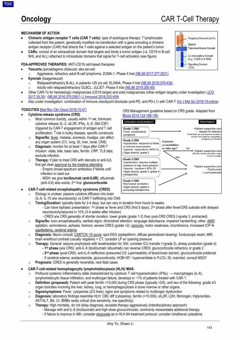
Oncology CAR T-Cell Therapy
Amy Yu, Shawn Li
MECHANISM OF ACTION • Chimeric antigen receptor T cells (CAR T cells): type of autologous therapy; T lymphocytes
collected from the patient, genetically modified via transfection with a gene encoding a chimeric antigen receptor (CAR) that directs the T-cells against a selected antigen on the patient’s tumor
• CARs: consist of an extracellular domain that targets and binds a tumor antigen (i.e. CD19 in B-cell NHL and ALL) attached to intracellular domains that signal for T-cell activation (see figure)
FDA-APPROVED THERAPIES: ANTI-CD19 cell-based therapies • Yescarta (axicabtagene ciloleucel; aka axi-cel)
o Aggressive, refractory adult B-cell lymphoma: ZUMA-1, Phase II trial (NEJM 2017;377:2531) • Kymriah (tisagenleucel)
o Relapsed/refractory B-ALL in patients <25 yrs old: ELIANA, Phase II trial (NEJM 2018;378:439) o Adults with relapsed/refractory DLBCL: JULIET- Phase II trial (NEJM 2019;280:45)
• Other CAR-Ts for hematologic malignancies (CD19 target) and solid malignancies (other antigen targets) under investigation (JCO 2017;35:30) (NEJM 2016;375:2561) (J Immunol 2018;200:459)
• Also under investigation: combination of immune checkpoint blockade (anti-PD, anti-PD-L1) with CAR-T (Int J Mol Sci 2018;19:online) TOXICITIES (Nat Rev Clin Oncol 2018;15:47) • Cytokine-release syndrome (CRS)
o Most common toxicity, usually within 1st wk; fulminant cytokine release (IL‐2, sIL2R, IFNγ, IL‐6, GM‐CSF) triggered by CAR-T engagement of antigen and T cell proliferation. ↑risk in bulky disease, specific constructs
o Signs/Sx: fever, malaise, anorexia, myalgia, can affect any organ system (CV, lung, GI, liver, renal, CNS)
o Diagnosis: monitor for at least 7 days after CAR-T infusion: vitals, tele, basic labs, ferritin, CRP, TLS labs; exclude infection
o Therapy: if plan to treat CRS with steroids or anti-IL6, first get clear approval by the treating attending
- Empiric broad-spectrum antibiotics if febrile until infection is ruled out - MGH: we give tocilizumab (anti-IL6R); siltuximab (anti‐IL6) also exists; 2nd line: glucocorticoids
• CAR-T-cell-related encephalopathy syndrome (CRES) o Etiology is unclear; passive cytokine diffusion into brain
(IL-6, IL-15 a/w neurotoxicity) vs CAR-T trafficking into CNS o Timing/Duration: typically lasts for 2-4 days, but can vary in duration from hours to weeks.
- Can have biphasic presentation: 1st phase w/ fever and CRS (first 5 days); 2nd phase after fever/CRS subside with delayed neurotoxicity/seizures in 10% (3-4 weeks after infusion) - CRES a/w CRS generally of shorter duration, lower grade (grade 1-2) than post-CRS CRES (>grade 3, protracted)
o Signs/Sx: toxic encephalopathy; earliest signs: diminished attention, language disturbance, impaired handwriting; other: AMS/ agitation, somnolence, aphasia, tremors; severe CRES (grade >2): seizures, motor weakness, incontinence, increased ICP papilledema, cerebral edema
o Diagnosis: Neuro consult; CARTOX‐10 score; spot EEG (epileptiform, diffuse generalized slowing); funduscopic exam; MRI brain w/without contrast (usually negative) > CT; consider LP w/ opening pressure
o Therapy: General: seizure prophylaxis with levetiracetam for 30d; consider ICU transfer (>grade 2), airway protection (grade 4) - 1st phase (a/w CRS): anti-IL-6 (tocilizumab/ siltuximab) can reverse CRES; glucocorticoids refractory or grade 2 - 2nd phase (post-CRS): anti-IL-6 ineffective (presumed 2/2 ↓permeability of blood-brain barrier); glucocorticoids preferred - If cerebral edema: acetazolamide, glucocorticoids; HOB>30°; hyperventilate to PaCO2 30; mannitol; consult NSGY
o Prognosis: CRES is generally reversible, rare fatal cases
• CAR-T-cell-related hemophagocytic lymphohistiocytosis (HLH)/ MAS: o Profound systemic inflammatory state characterized by cytotoxic T cell hyperactivation (IFNγ) → macrophages (IL-6),
lymphohistiocytic tissue infiltration, and multiorgan failure; develops in ~1% of patients treated with CAR‐T. o Definition (proposed): Patient with peak ferritin >10,000 during CRS phase (typically <D5), and two of the following: grade ≥3
organ toxicities involving the liver, kidney, lung; or hemophagocytosis in bone marrow or other organs. o Signs/symptoms: Fever, cytopenias (2/3 lines); signs and symptoms related to multiorgan dysfunction o Diagnosis: laboratory findings resemble HLH; CBC diff (cytopenia), ferritin (>10,000), sIL2R, LDH, fibrinogen, triglycerides,
AST/ALT, Bili, Cr; BMBx rarely critical (low sensitivity, low specificity). o Therapy: High mortality, do not delay diagnosis, escalate therapy aggressively (interdisciplinary approach)
- Manage with anti-IL-6 (tocilizumab) and high-dose glucocorticoids; commonly necessitates additional therapy - If failure to improve in 48h, consider etoposide as in HLH-94 treatment protocol; consider intrathecal cytarabine
CRS Management guideline based on CRS grade. Adapted from Blood 2014;124:188-195.
143

Oncology Solid Organ Malignancies
Lauren Banks
Organ Risk factors/ screening Staging/diagnostics Treatment Prostate Adenocarcinoma
(95%) Transitional cell
carcinoma, carcinosarcoma, basal cell carcinoma, lymphomas, or stromal sarcoma (~5%)
Androgen deprivation
therapy (ADT): orchiectomy, LHRH agonist (goserelin, histrelin, leuprolide, triptorelin) Androgen disruption:
abiraterone + prednisone, or enzalutamide
Risk factors: Age, AA race, genetic
factors (BRCA1/2 and family history), smoking
Screening (JAMA 2014;311:1143): Discuss PSA with men
>50 (DRE not recommended)
Staging: Path grade w/
Gleason Score (GS) NCCN risk based on
age, PSA, GS, stage TNM staging is
combined with PSA and GS for AJCC stages I- IV
Once biopsy & diagnosis: TRUS, MRI,
biomarkers, evaluate for metastases (CT, radio-nucleotide bone scan)
Low risk (T1c/T1-T2a + Gleason score ≤6 + PSA ≤10) (JCO 2015;33:3379, NEJM 2016;375:1415) Active surveillance (PSA, DRE +/- repeat bx),
external beam radiation therapy (EBRT) +/- brachytherapy (BT), or radical prostatectomy (RP) +/- EBRT & ADT
Intermediate risk (T2b-T2c or PSA 10-20 or GS 7) EBRT & ADT or EBRT & BT +/- ADT, RP +/- pelvic
lymph node dissection (PLND) +/- EBRT +/- ADT High risk (T3a/T3b-T4b or GS 8-10 or PSA >20) EBRT & ADT, ERBT & brachy & ADT, or RP with
extended PLND +/- EBRT +/- ADT Metastatic/recurrent (Lancet 2016;387:1163, NEJM 2015;373:737, NEJM 2010;363:411) Castration sensitive: ADT (LHRH agonist +/-
antiandrogen, LHRH antagonist or orchiectomy) + abiraterone w/ prednisone vs. docetaxel Castration resistant: ADT (testo<50ng/dL) +
doxcetaxel (chemo naïve) or cabazitaxel (dox exposed), mitoxantrone + prednisone, androgen disruption; sipuleucel-T, and bone-targeted radium-223 or denosumab/zoledronic acid (bone mets, no visceral mets) (NCCN Guidelines Version 1.2019)
Breast Infiltrating ductal
(76%) Invasive lobular
(8%) Ductal/lobular (7%) Mucinous (colloid)
(2.4% Tubular (1.5%) Medullary (1.2%) Papillary (1%)
TCH(P): Docetaxel,
carboplatin, trastuzumab +/-pertuzumab AC-TH(P):
doxorubicin/ cyclophosphamide followed by weekly paclitaxel + trastuzumab +/- pertuzumab Aromatase inhibitor
(AI): anastrozole, letrozole, exemestane
Risk factors: Age, genetics (BRCA1/2),
FHx, obesity after menopause, menopause >55y chest RT, 1st birth >30y, nulliparity, HRT, menarche <13y, ETOH, benign breast disease, smoking
Screening: q1-2y mammo after 50
(or 5-10y before earliest FHx, whichever is earlier), discuss before starting age 40.
Staging: TNM system used
for AJCC anatomic stage I-IV. AJCC 8th ed: added
Clinical Prognostic Stage Grp (TNM, grade, ER, PR, Her2; Recurrence score: OncotypeDx gene panel for those with Her2-, hormone rec+, node-, <5cm)
Diagnostics: Diagnostic mammo
1st (Bi-RADS score), targeted US, FNA or core bx (avoid excisional), breast MRI if young or to assess extent – good sensitivity, limited specificity 72%
Low Risk (I-IIB): Breast conserving surgery + RT or mastectomy;
chemo if high risk based on tumor biology Intermediate/High-Risk (Stage IIB-IIIC): Surgery & RT w/ neoadjuvant +/- adjuvant chemo &
HR/HER2 Tx (Neo)Adjuvant therapy: ER/PR+ (NEJM 2015;372:436): tamoxifen +/-
ovarian suppression (OS) or AI & OS (no OS if postmenopausal) x 2-5 yrs followed by 5 yrs of endo rx; chemo: ACT or TC HER2+ (Onco Targets Ther 2019;12:379): TCH(P)
or ACTH(P) + ER/PR rx if HR+; +/- HER2 inhibitor lapatinib TNBC (NEJM 2017:372;2147): AC-T +/-
capecitabine Metastatic/recurrent (Stage IV): ER+: tx above or fulvestrant (ER antagonist) +/-
CDK 4/6 or mTOR inhibitor (+OS if premenopausal) HER2+ (NEJM 2017;377:122): docetaxel/paclitaxel
+ trastuzumab + pertuzumab (1st line); T-DM1 (trastuzumab antibody drug conjugate) +/- lapatinib BRCA mutation (NEJM 2017;377:523, NEJM
2018;379:753): olaparib or talazoparib, platinum-based chemo
TNBC: AC-T +/- capecitabine (NCCN Guidelines Version 1.2019)
Pancreas Exocrine/ adenoca
(94%) Endocrine (6%) FOLFIRINOX:
leucovorin, 5-FU, irinotecan, oxaliplatin ChemoRT:
capecitabine or CI 5-FU + RT
Risk factors: Smoking, ETOH, obesity,
DM, chronic pancreatitis, age, male, FH pancreatic CA, HNPCC, BRCA1/2
Staging: Staged I-IV using
TNM system Diagnostics: CT C/A/P pancreas
protocol, EUS, MRCP, ERCP if indicated, CA19-9 Determine
resectability based on vascular involvement
Resectable: surgery + adjuvant chemoRT Borderline resectable: neoadjuvant chemo &
surgery +/- adjuvant chemoRT Locally adv: chemoRT or stereotactic body
radiation therapy (SBRT) Metastatic (NEJM 2011;364:1817): FOLFIRIMOX
or gemcitabine +/- nab-paclitaxel +/- palliative RT BRCA: FOLFIRINOX or gemcitabine/cisplatin, +/-
chemoRT MSI-H/dMMR (NEJM 2015;372:2509): consider
pembrolizumab (NCCN Guidelines Version 1.2019)
144
Oncology Solid Organ Malignancies
Lauren Banks
Colon and rectum Adenoca (98%) Neuroendocrine Lymphoma FOLFOX: oxaliplatin,
leucovorin, 5-FU CAPEOX:
capecitabine, oxaliplatin FOLFIRI: irinotecan,
leucovorin, 5-FU FOLFOXIRI:
irinotecan, oxaliplatin, leucovorin, fluorouracil EGFR inhibitor:
cetuximab, panitumumab
Risk factors (JAMA Oncol 2018;4:e173695): Obesity, physical
inactivity, smoking, red/processed meat, ETOH, adenomatous polyps, IBD, hereditary syndrome (FAP, HNPCC), age, AA race Risk of increased death
w/ R-sided compared w/ L-sided due to BRAF/KRAS mutation
Protective factors: ASA intake for pts 50-60
years Screening: Colo, flex sig, CT colo,
FIT, FOBT for ≥50yo
Staging: Staged I-IV using
TNM system Diagnostics: Colonoscopy, CT
C/A/P, CEA Pelvic MRI or
endorectal US for rectal CA Genetic testing (RAS,
BRAF, MSI status)
Colon: Stage I: surgery + observation Stage II: surgery + neo- vs. adjuvant chemo (5-
FU/leucovorin or capecitabine, add oxali if high-risk features) Stage III: surgery + adjuvant FOLFOX/CAPEOX Stage IV: resection of limited liver or lung mets +
FOLFOX/CAPEOX/FOLFIRI/FOLFOXIRI +/- bevacizumab KRAS/NRAS wt and left-sided tumors:
FOLFOX/FOLFIRI + EGFR inhibitor MSI-H/dMMR (NEJM 2015;372:2509):
immunotherapy (pembrolizumab, nivolumab or nivolumab + ipilimumab)
Rectal: Stage I: low anterior resection (LAR) or
abdominoperineal resection (APR) +/- neo- vs. adjuvant chemoRT Stage II-III: resection with neoadjuvant chemoRT
(NCCN Guidelines Version 1.2019 Colon Cancer) Lung NSCLC (84%) –
adenoca, large cell > SCC SCLC (13%) ChemoRT:
cisplatin/etoposide, cisplatin/vinblastine, carboplatin/ pemetrexed, cisplatin/pemetrexed, paclitaxel/carboplatin with RT EGFR inhibitors:
osimertinib, erlotinib, afatinib, gefitinib, dacomitinib *T790M∆ assoc w/ TKI resistance ALK/ROS1 inhibitors:
crizotinib, ceritinib, alectinib, brigatinib BRAF/MEK inhibitors:
dabrafenib/trametinib TRK inhibitor:
larotrectinib
Risk factors (Nat Rev Ca 2007;10:778): Smoking, asbestos,
occupational exposures, lung fibrosis, age, male, AA race, lower SE status 25% lung cancer
worldwide not due to smoking (50% of women with NSCLC are never smokers, 60-80% in Asian populations are women) more likely single mutation (ALK, EGFR, ROS1)
Screening (NEJM 2011;365:395): Annual low dose CT
chest for pts 55-74 yo with ≥30 pack-yr hx and smoking within last 15 yrs
Staging: NSCLC staged I-IV
using TNM system SCLC staged as
limited vs. extensive using TNM system
Diagnostics: CT chest/upper abd,
PET/CT, brain MRI Molecular testing for
NSCLC (EGFR, ALK, ROS1, PD-L1)
NSCLC: Stage IA: surgery vs. definitive RT Stage IB-IIIA: surgery if able +/- adjuvant
chemoRT Stage IIIB: definitive chemoRT + adjuvant
durvalumab (NEJM 2018;379:2342) Stage IV: targeted, immunotx, and systemic tx Targeted tx: EGFR inhibitor (EGFR sensitizing
mutations); ALK/ROS1 inhibitors (ALK or ROS1 rearrangement), BRAF/MEK inhibitors (BRAF V600E), TRK inhibitor (NTRK gene fusion) Immunotherapy (if no driver mutation)
(KEYNOTE-010, NEJM 2018;378:2078): pembrolizumab (1st line for ≥50% tumor PD-L1 expression); pembro + chemo (1st line option regardless of tumor PD-L1 expression level); alternative agents: nivolumab, atezolizumab Initial systemic tx: platinum agent + docetaxel,
pemetrexed, gemcitabine, or ramucirumab SCLC: Limited: surgery + chemo +/- mediastinal RT Extensive (NEJM 2018:379:2220): chemo &
atezolizumab +/- RT for lobar obstruction, SVC synd, bone/brain mets vs pal/supp care Chemo: platinum agents, etoposide, irinotecan
(NCCN Guidelines Version 3.2019) Melanoma Superficial
spreading (75%) Nodular (15-30%) Lentigo maligna
(~5%) Acral lentiginous
(<5%) Ocular (5%) BRAF/MEK targeted
tx: dabrafenib & trametinib, vemurafenib & cobimetinib, encorafenib & binimetinib (approved 6/2018)
Risk factors: Sun exposure (UVB >
UVA), atypical nevi, high nevi count, family or personal hx, immuno-suppression
Staging: TNM system Factors include:
Breslow thickness, ulceration, mitotic rate, lymphatic involvement, and distant metastases Serum LDH is an
important prognostic factor used in active surveillance and treatment
Surgical excision: Appropriate margins based on tumor thickness +/-
sentinel LN bx vs. complete regional LN dissection Adjuvant treatment or for metastatic disease: Immunotherapy (NEJM 2015;373:23, NEJM
2015;372:2521): anti-PD-1 monotherapy (pembrolizumab or nivolumab); combination nivolumab/ipilimumab Targeted tx (NEJM 2014;371:1867): BRAF/MEK
inhibitors (BRAF V600 activating mutations), KIT inhibitor imatinib (KIT-mutant tumors) Radiation: considered with symptomatic localized
disease (e.g. brain mets) Talimogene laherparepvec (T-VEC) (J Clin
Oncol 2015;33:2780): intralesional injection of HSV tumor cell lysis & GM-CSF expression (NCCN Guidelines Version 2.2019)
145
Oncology Chemotherapy & Toxicities
Lauren Banks
Common Chemotherapy Toxicities: • Severe N/V: any AC combinations (doxo/epi/ida/daunorubicin+ifos/cyclophosphamide), carmustine, dacarbazine, cisplatin,
mechlorethamine, streptozocin; HiDAC (AraC), aldesleukin/IFNα, amifostine > 300, ATO, azacitidine, bendamustine, busulfan, clofarabine, dactinomycin, irinotecan, melphalan, methotrexate > 250, temozolomide; refer to NCCN Guidelines1 for management
• Severe BM↓: busulfan, carmustine, cyclophosphamide, dacarbazine, ifosfamide, 5-FU, methotrexate, doxorubicin (daunorobicin), taxotere, taxol, carboplatin, melphalan, fludarabine
DRUG INDICATIONS TOXICITIES
Anti-Metabolites 5-Fluorouracil Breast, colorectal, pancreatic, gastric,
esophageal, H&N Coronary vasospasm, acute cerebellar ataxia, hand-foot syndrome, stomatitis, hemorrhage, GI ulcers/bleeding, hiccups, diarrhea, BM↓*
Capecitabine Breast, colorectal Monitor INR (if on Coumadin), hand-foot syndrome, SJS-TEN, N/V/D, cytopenias (worse with stage IV breast CA), liver toxicity
Cytarabine (HiDAC)
AML, ALL, CLL, meningeal leukemia Acute cerebellar ataxia, PRES, BM↓, chemical conjunctivitis (Rx dexamethasone eye drops), ↑LFTs, cutaneous tox, hand-foot syndrome
Gemcitabine Breast, ovarian, NSCLC, pancreas, bladder
Capillary leak syndrome, PRES, TMA/HUS, ARDS, ↑LFTs, N/V, hematuria
Mercaptopurine ALL (w MTX), CML Biliary cholestasis & hepatocellular necrosis, BM↓, (Consider TPMT SNP testing if severe BM↓), N/V/D
Fludarabine CLL, NHL, AML BM↓, IS, autoimmune hemolytic anemia, neurotoxicity, fatal pulm toxicity (when used w/ pentostatin for CLL)
Hydroxyurea CML, cervical, sickle cell, H&N BM↓, cutaneous tox, N/V/D, ↑LFTs, ↑Cr, ↑BUN Anti-Folates
Methotrexate ALL, breast, H&N, CTCL, SCLC, NSCLC, NHL, osteosarcoma
BM↓, aplastic anemia, AKI, ↑LFTs, hepatic fibrosis/cirrhosis, cutaneous tox, IS (PCP), pneumonitis/PF, teratogenic, ulcerative stomatitis/diarrhea, IF
Pemetrexed Mesothelioma, NSCLC BM↓, desquamating rash, pneumonitis, renal tox Alkylating Agents (all cause BM↓, infertility & increased risk of MDS/AML)
Busulfan HSCT conditioning, CML BM↓, sinusoidal obstruction/VOD, tamponade, ILD, seizures, renal tox Ifosfamide Testicular, breast, lung, HL, NHL, bone Hemorrhagic cystitis, encephalopathy, renal/pulmonary/cardiac tox Melphalan MM, ovarian BM↓, hypersensitivity, amnesia, pulmonary fibrosis, mucositis, rash, IF Carmustine CNS tumors, HL, NHL, MM BM↓, pulm tox (dose-related), ↑LFTs, renal tox, ocular sx Cyclophospha-mide
Leukemia, MM, breast, NHL, ovarian, retinoblastoma
Hemorrhagic cystitis, renal/cardio tox, pulm fibrosis, IF, sinusoidal obstruction/VOD, IS, hypoNa
Dacarbazine HL, melanoma Hepatic necrosis, teratogenic, hepatic vein thrombosis, ↑↑N/V, BM↓ Cisplatin Bladder, ovarian, testicular, H&N N/V, renal tox, ototoxicity, neurotoxicity, BM↓, ↑uric acid Carboplatin Ovarian, lung, H&N, CNS tumors N/V, ototoxicity, neurotoxicity, BM↓ (less nephrotoxic than cisplatin) Oxaliplatin Colorectal, pancreatic N/V, renal/pulm/liver tox, neurotoxicity, BM↓, rhabdo, ↑QT, PRES
Antibiotics Bleomycin HL/NHL, testicular, ovarian, H/N Late pulm fibrosis, dermatographia, hyperpigmentation, Raynaud’s Mitomycin Gastric, esoph, anal, bladder, pancreas BM↓, renal/cardiac tox, HUS, interstitial pneumonitis/ARDS, bladder fibrosis
Hormonal Therapy Tamoxifen Raloxifene
ER+ Breast Menopausal sx (hot flashes, vaginal atrophy/pruritus /bleeding), VTE (DVT/PE), endometrial cancer (tamoxifen only)
Anastrozole Letrozole Exemestane
ER+ Breast (post-menopausal) Sexual dysfunction, bone/joint pain, osteoporosis, premature menopause
Fulvestrant ER+ Breast (post-menopausal) Sexual dysfunction, bone/joint pain, osteoporosis, injection site reactions Megestrol Breast, endometrial Teratogenic, ↑weight, hypogonad, VTE, hot flashes, adrenal suppression Leuprolide Goserelin
Prostate, breast (goserelin) Hypogonadism, edema, depression, bone pain, HA, osteoporosis, transient worsening of prostate CA sx 2/2 brief testosterone surge (prevent w/ AR inhibitors), seizure, ↑CVD risk
Flutamide Nilutamide
Prostate Hot flashes, gynecomastia, ↓libido, N/V/D, muscle atrophy/pain, liver tox (> w/ F), interstitial pneumonitis, visual changes (N), osteoporosis
Bicaludamide Prostate Hypogonadism, sexual disfunction, depression, fatigue, liver tox, ILD Topoisomerase Inhibitors
Anthracyclines (Dauno/Epi/ Doxorubicin)
Breast, ALL, AML, MM, lung, bladder Cardiotoxicity (DCM, myopericarditis); BM↓, IS, 2nd malignancies, local tissue necrosis in setting of extravasation, liver/renal tox, typhlitis, “chemo brain”
Mitoxantrone Breast, ALL, AML, breast Cardiotoxicity (DCM, myopericarditis); BM↓, IS, N/V Irino/Topotecan Irinotecan: colorectal, SCLC
Topotecan: cervical, ovarian, SCLC BM↓, diarrhea, late ILD, thrombosis, typhlitis
Etoposide SCLC, testicular, KS, glioblastoma BM↓, acute infusion rxn (HoTN), metallic food taste, SJS/TEN
146
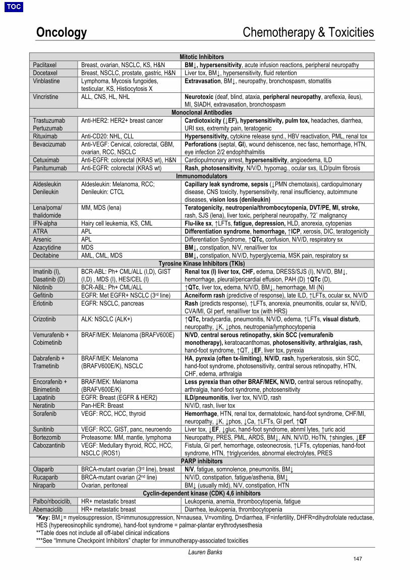
Oncology Chemotherapy & Toxicities
Lauren Banks
Mitotic Inhibitors Paclitaxel Breast, ovarian, NSCLC, KS, H&N BM↓, hypersensitivity, acute infusion reactions, peripheral neuropathy Docetaxel Breast, NSCLC, prostate, gastric, H&N Liver tox, BM↓, hypersensitivity, fluid retention Vinblastine Lymphoma, Mycosis fungoides,
testicular, KS, Histiocytosis X Extravasation, BM↓, neuropathy, bronchospasm, stomatitis
Vincristine ALL, CNS, HL, NHL Neurotoxic (deaf, blind, ataxia, peripheral neuropathy, areflexia, ileus), MI, SIADH, extravasation, bronchospasm
Monoclonal Antibodies Trastuzumab Pertuzumab
Anti-HER2: HER2+ breast cancer Cardiotoxicity (↓EF), hypersensitivity, pulm tox, headaches, diarrhea, URI sxs, extremity pain, teratogenic
Rituximab Anti-CD20: NHL, CLL Hypersensitivity, cytokine release synd., HBV reactivation, PML, renal tox Bevacizumab Anti-VEGF: Cervical, colorectal, GBM,
ovarian, RCC, NSCLC Perforations (septal, GI), wound dehiscence, nec fasc, hemorrhage, HTN, eye infection 2/2 endophthalmitis
Cetuximab Anti-EGFR: colorectal (KRAS wt), H&N Cardiopulmonary arrest, hypersensitivity, angioedema, ILD Panitumumab Anti-EGFR: colorectal (KRAS wt) Rash, photosensitivity, N/V/D, hypomag., ocular sxs, ILD/pulm fibrosis
Immunomodulators Aldesleukin Denileukin
Aldesleukin: Melanoma, RCC; Denileukin: CTCL
Capillary leak syndrome, sepsis (↓PMN chemotaxis), cardiopulmonary disease, CNS toxicity, hypersensitivity, renal insufficiency, autoimmune diseases, vision loss (denileukin)
Lena/poma/ thalidomide
MM, MDS (lena) Teratogenicity, neutropenia/thrombocytopenia, DVT/PE, MI, stroke, rash, SJS (lena), liver toxic, peripheral neuropathy, ?2˚ malignancy
IFN-alpha Hairy cell leukemia, KS, CML Flu-like sx, ↑LFTs, fatigue, depression, HLD, anorexia, cytopenias ATRA APL Differentiation syndrome, hemorrhage, ↑ICP, xerosis, DIC, teratogenicity Arsenic APL Differentiation Syndrome, ↑QTc, confusion, N/V/D, respiratory sx Azacytidine MDS BM↓, constipation, N/V, renal/liver tox Decitabine AML, CML, MDS BM↓, constipation, N/V/D, hyperglycemia, MSK pain, respiratory sx
Tyrosine Kinase Inhibitors (TKIs) Imatinib (I), Dasatinib (D)
BCR-ABL: Ph+ CML/ALL (I,D), GIST (I,D) , MDS (I), HES/CEL (I)
Renal tox (I) liver tox, CHF, edema, DRESS/SJS (I), N/V/D, BM↓, hemorrhage, pleural/pericardial effusion, PAH (D) ↑QTc (D),
Nilotinib BCR-ABL: Ph+ CML/ALL ↑QTc, liver tox, edema, N/V/D, BM↓, hemorrhage, MI (N) Gefitinib EGFR: Met EGFR+ NSCLC (3rd line) Acneiform rash (predictive of response), late ILD, ↑LFTs, ocular sx, N/V/D Erlotinib EGFR: NSCLC, pancreas Rash (predicts response), ↑LFTs, anorexia, pneumonitis, ocular sx, N/V/D,
CVA/MI, GI perf, renal/liver tox (with HRS) Crizotinib ALK: NSCLC (ALK+) ↑QTc, bradycardia, pneumonitis, N/V/D, edema, ↑LFTs, visual disturb,
neuropathy, ↓K, ↓phos, neutropenia/lymphocytopenia Vemurafenib + Cobimetinib
BRAF/MEK: Melanoma (BRAFV600E) N/VD, central serous retinopathy, skin SCC (vemurafenib monotherapy), keratoacanthomas, photosensitivity, arthralgias, rash, hand-foot syndrome, ↑QT, ↓EF, liver tox, pyrexia
Dabrafenib + Trametinib
BRAF/MEK: Melanoma (BRAFV600E/K), NSCLC
HA, pyrexia (often tx-limiting), N/V/D, rash, hyperkeratosis, skin SCC, hand-foot syndrome, photosensitivity, central serous retinopathy, HTN, CHF, edema, arthralgia
Encorafenib + Binimetinib
BRAF/MEK: Melanoma (BRAFV600E/K)
Less pyrexia than other BRAF/MEK, N/V/D, central serous retinopathy, arthralgia, hand-foot syndrome, photosensitivity
Lapatinib EGFR: Breast (EGFR & HER2) ILD/pneumonitis, liver tox, N/V/D, rash Neratinib Pan-HER: Breast N/V/D, rash, liver tox Sorafenib VEGF: RCC, HCC, thyroid Hemorrhage, HTN, renal tox, dermatotoxic, hand-foot syndrome, CHF/MI,
neuropathy, ↓K, ↓phos, ↓Ca, ↑LFTs, GI perf, ↑QT Sunitinib VEGF: RCC, GIST, panc, neuroendo Liver tox, ↓EF, ↓gluc, hand-foot syndrome, abnml lytes, ↑uric acid Bortezomib Proteasome: MM, mantle, lymphoma Neuropathy, PRES, PML, ARDS, BM↓, AIN, N/V/D, HoTN, ↑shingles, ↓EF Cabozantinib VEGF: Medullary thyroid, RCC, HCC,
NSCLC (ROS1) Fistula, GI perf, hemorrhage, osteonecrosis, ↑LFTs, cytopenias, hand-foot syndrome, HTN, ↑triglycerides, abnormal electrolytes, PRES
PARP inhibitors Olaparib BRCA-mutant ovarian (3rd line), breast N/V, fatigue, somnolence, pneumonitis, BM↓ Rucaparib BRCA-mutant ovarian (2nd line) N/V/D, constipation, fatigue/asthenia, BM↓ Niraparib Ovarian, peritoneal BM↓ (usually mild), N/V, constipation, HTN
Cyclin-dependent kinase (CDK) 4,6 inhibitors Palbo/ribociclib, HR+ metastatic breast Leukopenia, anemia, thrombocytopenia, fatigue Abemaciclib HR+ metastatic breast Diarrhea, leukopenia, thrombocytopenia
*Key: BM↓= myelosuppression, IS=immunosuppression, N=nausea, V=vomiting, D=diarrhea, IF=infertility, DHFR=dihydrofolate reductase, HES (hypereosinophilic syndrome), hand-foot syndrome = palmar-plantar erythrodysesthesia **Table does not include all off-label clinical indications ***See “Immune Checkpoint Inhibitors” chapter for immunotherapy-associated toxicities
147
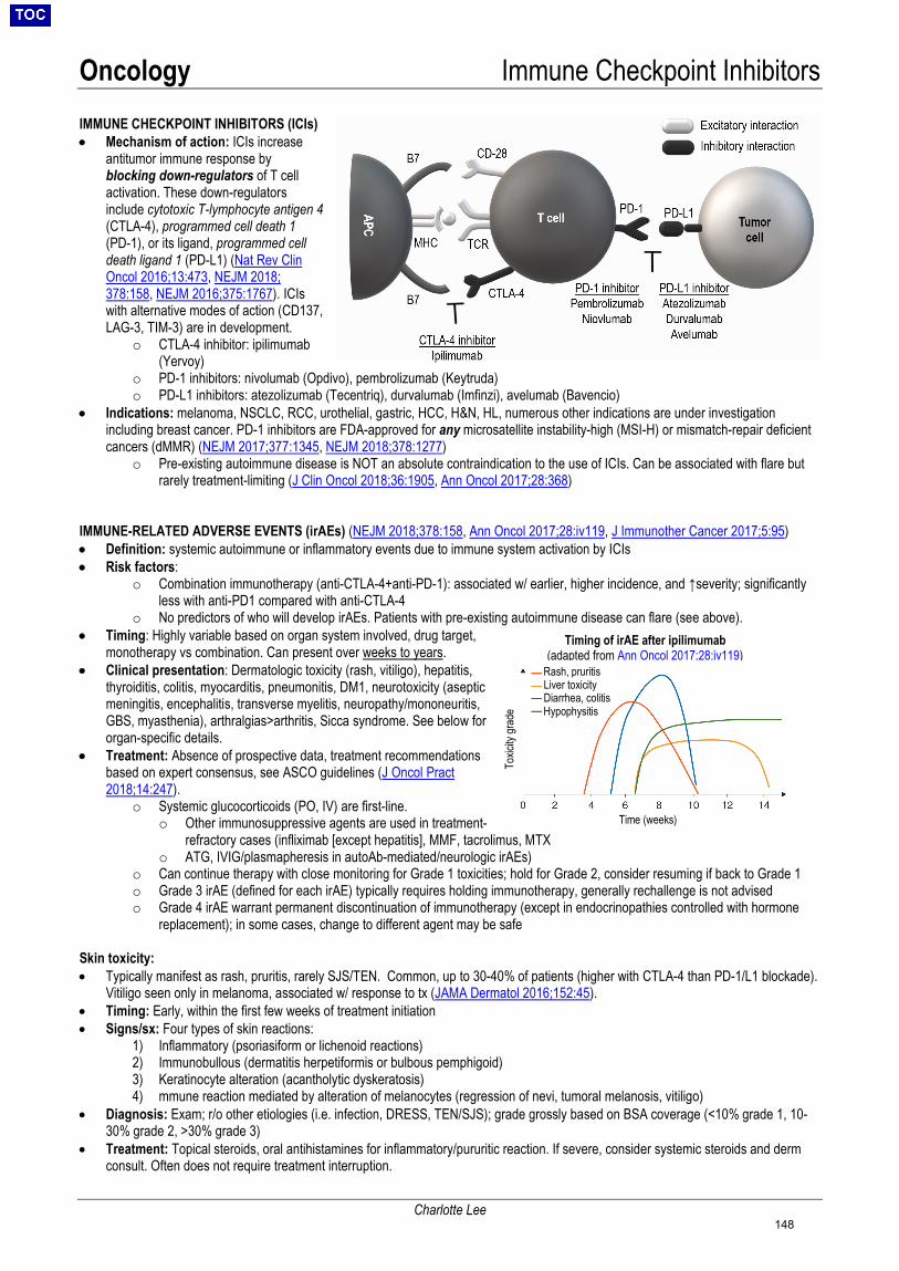
Oncology Immune Checkpoint Inhibitors
Charlotte Lee
IMMUNE CHECKPOINT INHIBITORS (ICIs) • Mechanism of action: ICIs increase
antitumor immune response by blocking down-regulators of T cell activation. These down-regulators include cytotoxic T-lymphocyte antigen 4 (CTLA-4), programmed cell death 1 (PD-1), or its ligand, programmed cell death ligand 1 (PD-L1) (Nat Rev Clin Oncol 2016;13:473, NEJM 2018; 378:158, NEJM 2016;375:1767). ICIs with alternative modes of action (CD137, LAG-3, TIM-3) are in development.
o CTLA-4 inhibitor: ipilimumab (Yervoy)
o PD-1 inhibitors: nivolumab (Opdivo), pembrolizumab (Keytruda) o PD-L1 inhibitors: atezolizumab (Tecentriq), durvalumab (Imfinzi), avelumab (Bavencio)
• Indications: melanoma, NSCLC, RCC, urothelial, gastric, HCC, H&N, HL, numerous other indications are under investigation including breast cancer. PD-1 inhibitors are FDA-approved for any microsatellite instability-high (MSI-H) or mismatch-repair deficient cancers (dMMR) (NEJM 2017;377:1345, NEJM 2018;378:1277)
o Pre-existing autoimmune disease is NOT an absolute contraindication to the use of ICIs. Can be associated with flare but rarely treatment-limiting (J Clin Oncol 2018;36:1905, Ann Oncol 2017;28:368)
IMMUNE-RELATED ADVERSE EVENTS (irAEs) (NEJM 2018;378:158, Ann Oncol 2017;28:iv119, J Immunother Cancer 2017;5:95) • Definition: systemic autoimmune or inflammatory events due to immune system activation by ICIs • Risk factors:
o Combination immunotherapy (anti-CTLA-4+anti-PD-1): associated w/ earlier, higher incidence, and ↑severity; significantly less with anti-PD1 compared with anti-CTLA-4
o No predictors of who will develop irAEs. Patients with pre-existing autoimmune disease can flare (see above). • Timing: Highly variable based on organ system involved, drug target,
monotherapy vs combination. Can present over weeks to years. • Clinical presentation: Dermatologic toxicity (rash, vitiligo), hepatitis,
thyroiditis, colitis, myocarditis, pneumonitis, DM1, neurotoxicity (aseptic meningitis, encephalitis, transverse myelitis, neuropathy/mononeuritis, GBS, myasthenia), arthralgias>arthritis, Sicca syndrome. See below for organ-specific details.
• Treatment: Absence of prospective data, treatment recommendations based on expert consensus, see ASCO guidelines (J Oncol Pract 2018;14:247).
o Systemic glucocorticoids (PO, IV) are first-line. o Other immunosuppressive agents are used in treatment-
refractory cases (infliximab [except hepatitis], MMF, tacrolimus, MTX o ATG, IVIG/plasmapheresis in autoAb-mediated/neurologic irAEs)
o Can continue therapy with close monitoring for Grade 1 toxicities; hold for Grade 2, consider resuming if back to Grade 1 o Grade 3 irAE (defined for each irAE) typically requires holding immunotherapy, generally rechallenge is not advised o Grade 4 irAE warrant permanent discontinuation of immunotherapy (except in endocrinopathies controlled with hormone
replacement); in some cases, change to different agent may be safe Skin toxicity: • Typically manifest as rash, pruritis, rarely SJS/TEN. Common, up to 30-40% of patients (higher with CTLA-4 than PD-1/L1 blockade).
Vitiligo seen only in melanoma, associated w/ response to tx (JAMA Dermatol 2016;152:45). • Timing: Early, within the first few weeks of treatment initiation • Signs/sx: Four types of skin reactions:
1) Inflammatory (psoriasiform or lichenoid reactions) 2) Immunobullous (dermatitis herpetiformis or bulbous pemphigoid) 3) Keratinocyte alteration (acantholytic dyskeratosis) 4) mmune reaction mediated by alteration of melanocytes (regression of nevi, tumoral melanosis, vitiligo)
• Diagnosis: Exam; r/o other etiologies (i.e. infection, DRESS, TEN/SJS); grade grossly based on BSA coverage (<10% grade 1, 10-30% grade 2, >30% grade 3)
• Treatment: Topical steroids, oral antihistamines for inflammatory/pururitic reaction. If severe, consider systemic steroids and derm consult. Often does not require treatment interruption.
Timing of irAE after ipilimumab (adapted from Ann Oncol 2017;28:iv119)
Rash, pruritis Liver toxicity Diarrhea, colitis Hypophysitis
Time (weeks)
Toxic
ity gr
ade
148
Oncology Immune Checkpoint Inhibitors
Charlotte Lee
Hypophysitis (JAMA Oncol 2018;4:173) • Primarily seen with anti-CTLA-4, estimated prevalence of 3.2%. Rarely with anti-PD-1/PD-L1 agents (0.5%).
o Mechanistically distinct from other irAEs; thought to be mediated by direct binding of ipilimumab to CTLA-4 expressed on normal cells of the anterior pituitary (Sci Transl Med 2014;6:230).
• Timing: Median onset is 8 weeks • Signs/sx: Headaches (can be severe) most common; fatigue, N/V, dizziness, weight loss, hot flashes, cold intolerance, hyponatremia
(anterior hypopituitarism); not associated with central diabetes insipidus (posterior pituitary spared) • Diagnosis: MRI brain/pituitary shows transient (generally resolved by 2 months), diffuse pituitary enlargement; test hormonal axes:
8AM serum cortisol + ACTH and/or cort stim; TSH w/ fT4/T4/T3; PRL; LH/FSH, serum testosterone/SHBG (in men); IGF-1. • Treatment: Symptoms resolve with appropriate hormone substitution; hormone deficiencies tend to persist
o Hypocortisolism: physiologic glucocorticoid replacement (prednisone 3-5mg daily equivalent; increase x2-3 with infection/illness), high-dose glucocorticoids do not improve outcomes (may be associated with reduced survival); counsel about adrenal crisis; obtain medical bracelet (Cancer 2018;124:3706, Oncologist 2016;21:804)
o Hypothyroidism (can also occur independent of hypophysitis): thyroid hormone replacement with levothyroxine (hyperthyroidism can also occur, but rarer; consult endocrine)
o Hypogonadism: consider testosterone replacement if persists o GH deficiency: GH theoretically contraindicated due to active malignancy, although no supportive evidence (Nat Clin Pract
Endocrinol Metab 2006;2:532) Colitis • More commonly seen with anti-CTLA-4: Grade 3/4 colitis is higher with ipilimumab (<10%) than with anti-PD-1 agents (1-2%). • Timing: 6-8 weeks (median) after initiation of therapy. • Signs/Sx: Diarrhea, abdominal pain • Diagnosis: CBC; BMP; CRP; ANCA; consider lactoferrin/calprotectin; flex-sig and/or colonoscopy; rule out alternative etiologies:
Clostridium difficile, bacterial or viral pathogens (stool Cx, O&P, CMV PCR, cryptosporidia); CTAP can show mild diffuse bowel thickening or segmental colitis associated with diverticulosis; GI consult for EGD/colo (can affect upper/lower) for grade 2 sxs (4-6 BM/d >baseline), pathology shows active acute colitis.
• Treatment: Symptomatically with antidiarrheal agents after exclusion of infection; Grade 1/2 (<3/4-6 BM >baseline): antidiarrheal; budesonide 9 mg PO or prednisone PO if fails to improve (G1>14d; G2>3d); Grade 3/4 (>6 BM over baseline): systemic glucocorticoids (prednisone 1-2 mg/kg or methylprednisolone 1-2 mg/kg IV) with taper; infliximab in refractory cases
Pneumonitis • More common w/ anti-PD-1, but serious toxicity rare. Combination therapy confers significantly higher risk. (Chest 2017;152:271, J
Clin Oncol 2017;35:709); risk also increased in combination with targeted therapy for lung ca. • Timing: Highly variable onset, later than other irAEs. • Signs/sx: Dyspnea (53%), cough (35%), increased sputum production; life-threatening presentations include acute interstitial
pneumonia/ARDS; 1/3rd of patients asymptomatic (J Clin Oncol 2017;35:709, Clin Cancer Res 2016;22:6051) • Diagnosis: Low threshold to obtain CT/HRCT, CXR often not helpful; workup Ddx (viral and bacterial PNA, COP, COPD
exacerbation, heart failure, lymphangitic carcinomatosis/disease progression, PE): VBG, influenza/RSV PCR, resp viral panel, BCx, SpCx and smear, sputum for PCP if at risk, consider BAL; NT-proBNP, troponin, TTE, CTPE/LENI. Imaging non-specific: GGOs, NSIP-like, COP-like, HP-like.
• Treatment: Oxygen, glucocorticoids (prednisone 1-2 mg/kg/d or methylprednisolone 1mg/kg) prolonged taper, consider empiric abx, diuresis
Myocarditis (J Am Coll Cardiol 2018;71:1755) • Rare, but serious adverse event of ICI associated with high mortality (46% death in severe myocarditis); risk much higher with
combination therapy • Timing: Generally within 3 months (Oncologist 2018;23:874, Lancet 2018;391:933) • Sisngs/sx: Sx of heart failure (SOB, LE edema), chest pain, palpitations, arrhythmia • Diagnosis: EKG/ tele; troponin, CK-MB, NT-proBNP, ESR/CRP; TTE; consider myocardial bx; CPK/aldolase; EMG/muscle bx • Treatment: pulse-dose glucocorticoids (1g IV x3d, then PO pred 1mg/kg); second line consider ATG/IVIG (if unstable), or
infliximab/MMF/tacro (if stable) (Oncologist 2018;23:879); BB, ACEi/ARB (EF low) Hepatitis (Oncologist 2018;23:991) • Timing: Median onset 6-14 weeks after treatment initiation • Signs/sx: Usually asymptomatic • Diagnosis: LFTs, r/o other etiologies such as viral hepatitis, ingestion/EtOH, drugs. Rarely consider liver bx if severe • Treatment: If LFTs ≥ 3-5 ULN, hold therapy and monitor labs closely. If persists >1-2 weeks, treat with methylprednisolone
(1mg/kg/day); 2nd line MMF (Cellcept), 3rd line ATG (Cancer Treat Rev 2016;44:51)
149
Oncology Oncologic Emergencies
Nicky Singh
Tumor Lysis Syndrome (NEJM 2011;364:1844, JCO 2008;26:2767) • Pathophys: Tumor lysis (iso of cytotoxic tx initiation, rarely spontaneous in NHL and acute leukemia) causes release of intracellular
components (nucleic acids catabolized to uric acid, K+, PO4+); Clinical effects (can be deadly): renal failure (↑uric acid precipitates in renal tubules); seizure/Ca-phos crystal deposition (↑phos → ↓Ca); arrhythmias (↑K); Timing: 1-2d after tx (can occur w/in hrs)
• Risk factors: o Malignancy risk: High: ALL/AML (WBC ≥100k), CLL (on venetoclax & ↑uric acid), stage 3/4 Burkitt’s/lymphoblastic NHL,
bulky DLBCL. Intermediate: ALL (WBC <100k), AML (WBC 25-100k or <25k & ↑LDH), Burkitt’s, DLBCL, CLL (chemo-specific & ↑WBC), rare chemo-sens bulky solid tumor. Low: HL, indolent NHL, CML, CLL (on alkylating tx & WBC <50k), MM
o High risk substrate: WBC > 50k, LDH > 2x ULN, bulky tumor (>10 cm), hypovolemia, uric acid > 7.5, renal failure • Labs/Workup: BMP (electrolytes, Cr), ionized Ca, Mg, phos (calculate Ca-phos product), uric acid, CBC+Diff; ECG • Diagnosis (Cairo-Bishop criteria):
o Laboratory diagnosis: 2+ criteria within 3d before or 7d after cytoxic therapy: uric acid ≥ 8 mg/dL, K ≥ 6 mEq/L, phos ≥ 4.5mg/dL, or Ca ≤ 7mg/dL. Criteria also satisfied if 25% change from baseline.
o Clinical diagnosis: lab TLS + 1+ criteria: Cr 1.5x ULN, arrhythmia, seizure, death (not attributable to chemo agent) • Prophylaxis and treatment: While treating, labs should be checked Q2-Q4H, patient should be on telemetry given electrolyte abn
o Hydration: Maintain UOP 80-100 cc/hr to for optimal excretion of uric acid and phos; can use diuretics prn Use bicarb only with marked acidosis, as ↑urine pH will ↓uric acid crystals but ↑Ca-phos crystals
o Electrolyte abnormalities: ↑K (hyperK tx), ↑phos (binders), ↓Ca (avoid until phos wnl or sx of ↓Ca) o Allopurinol: 100mg/m2 PO q8h or 200-400mg/m2 IV, administer 24-48 hr before chemo, cont until hyperuricemia resolved
Renally dose, note reduced clearance of other meds (ie: cyclophosphamide, MTX, 6-MP, azathioprine, ampicillin) o Rasburicase (discuss with attending): 0.2 mg/kg IV, administer if high risk or baseline uric acid ≥ 8 mg/dL
Risk of anaphylaxis, hemolysis. Contraindicated in G6PD deficiency (risk of methemoglobinemia) o Renal replacement therapy: indicated if Ca-phos product ≥70 mg2/dL2
Hyperviscosity Syndrome/Leukostasis (Blood 2012;119:2205) • Etiology: 1) hyperproteinemia from monoclonal gammopathies mostly commonly Waldenström’s macroglobulinemia (IgM),
uncommonly myeloma; 2) hyperleukocytosis/leukostasis seen in AML with blasts > 50k (uncommon in ALL/CLL unless very high counts); 3) other diseases such as rheumatoid disease, polycythemia, sickle cell, spherocytosis
• Signs/symptoms: Most common: pulmonary (SOB) and CNS (blurry vision 2/2 retinal venous engorgement, headache, dizziness, ataxia, confusion, coma), fever If concerned, page hematology fellow on call and clinical pathology resident for EMERGENCY VISCOSITY STUDY, p21828, (notify attending ASAP as pheresis will involve attending-level decision)
• Diagnosis: ↑ Ostwald tube serum viscosity, light chains, SPEP, WBC (often > 100k, but can be lower in blast crisis) o Lab artifacts from hyperleukocytosis: spurious ↑K (use ABG K), falsely low arterial pO2 (use oximeter)
• Treatment: always start with plasma volume expansion with IV NS o Hyperproteinemia: plasmapheresis (aiming for resolution of symptoms); reduces viscosity by 20-30% per session o Leukostasis: leukopheresis; cytoreduce (hydroxyurea); induction chemo; avoid RBC & plt transfusion (↑viscosity)
Metastatic Epidural Spinal Cord Compression (Seminars in Neurology 2010;30:245, Lancet Neurology 2008;7:459) • Primary CA: lung > prostate, breast > non-Hodgkin's lymphoma, renal cell, multiple myeloma, lymphoma • Location: TS (60%) > LS (25%) > CS (15%); multiple sites in 20-35%; ESCC score for spinal level (JNCCN 2016;14:70) • Symptoms: back pain (usually 1st sx; radicular, localized, worse at night/recumbent/valsalva) → weakness, gait instability → sensory
deficits (saddle anesthesia in cauda equina lesions), bowel/bladder dysfxn • Exam: pain precedes other sx by ~7 wks, weakness/ataxia, paresthesia, ↑ reflexes, ⊕ Babinski, ↓anal sphincter tone • Diagnosis: STAT vs. urgent full spine MRI with cord compression/metastasis protocol, alternative is CT myelography • Treatment: Call Spine Surgery & Radiation Oncology ASAP → more effective than chemo (except for heme, germ cell malignancies)
o Severe deficits: dexamethasone 96mg x1, then 24mg IV q6hr x3d, then taper x10d o Minimal deficits: dexamethasone 10mg IV x1, then 4mg IV q6hr
Brain Metastases with Increased Intracranial Pressure (Ann Palliat Med 2015;4:225, JCO 2015;33:3475) • Intracranial tumors present in ~10-30% of patients with metastatic disease; call Neurosurgery & Radiation Oncology • Primary CA: lung (48%), breast (18%), melanoma, RCC, osteosarcoma, head and neck, thyroid, colorectal • Symptoms: headaches (40-50%; “tension”, worse w/ Valsalva, N/V), focal neuro deficits (20-40%, hemiparesis most common),
cognitive dysfunction (30-35%), new onset seizures (10-20%), stroke (5-10%) • Diagnosis: contrast MRI ↑ sensitivity > non-enhanced MRI, CT I+ • Treatment: control vasogenic edema (dexamethasone 10mg IV x1, then 8mg BID), consider AED (usually not recommended for 1˚
ppx); avoid AC if c/f active hemorrhage; definitive treatment will ↓ local recurrence: stereotactic radiosurgery > whole-brain XRT (↑ neurocognitive impairment; hippocampal sparing helpful) > surgery
Superior Vena Cava Sydrome (SVC syndrome) (NEJM 2007;356:1862) • Etiology: External compression of SVC from a mediastinal mass (commonly lung CA or NHL) causing ↑upper body venous pressure • Symptoms: cerebral edema (HA, confusion, herniation), narrowing of larynx/pharynx (dyspnea, stridor, cough, dysphagia,
hoarsness), head/neck swelling (visually striking, often not clinically significant), hemodynamic instability (↓ venous return) • Diagnosis: venography, CT chest w/contrast, obtain/ensure tissue diagnosis to guide tx (extremely important!) • Treatment: secure airway, RT/chemo, intravascular stent (emergent/refractory), steroids (stridor/resp distress only, clear with onc)
150
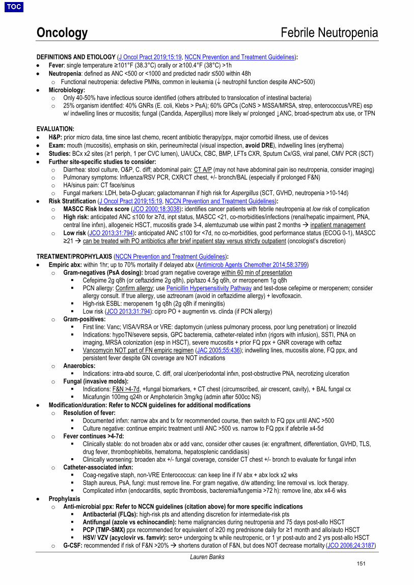
Oncology Febrile Neutropenia
Lauren Banks
DEFINITIONS AND ETIOLOGY (J Oncol Pract 2019;15:19, NCCN Prevention and Treatment Guidelines): • Fever: single temperature ≥101°F (38.3°C) orally or ≥100.4°F (38°C) >1h• Neutropenia: defined as ANC <500 or <1000 and predicted nadir ≤500 within 48h
o Functional neutropenia: defective PMNs, common in leukemia (↓ neutrophil function despite ANC>500)• Microbiology:
o Only 40-50% have infectious source identified (others attributed to translocation of intestinal bacteria)o 25% organism identified: 40% GNRs (E. coli, Klebs > PsA); 60% GPCs (CoNS > MSSA/MRSA, strep, enterococcus/VRE) esp
w/ indwelling lines or mucositis; fungal (Candida, Aspergillus) more likely w/ prolonged ↓ANC, broad-spectrum abx use, or TPN
EVALUATION: • H&P: prior micro data, time since last chemo, recent antibiotic therapy/ppx, major comorbid illness, use of devices• Exam: mouth (mucositis), emphasis on skin, perineum/rectal (visual inspection, avoid DRE), indwelling lines (erythema)• Studies: BCx x2 sites (≥1 periph, 1 per CVC lumen), UA/UCx, CBC, BMP, LFTs CXR, Sputum Cx/GS, viral panel, CMV PCR (SCT)• Further site-specific studies to consider:
o Diarrhea: stool culture, O&P, C. diff; abdominal pain: CT A/P (may not have abdominal pain iso neutropenia, consider imaging)o Pulmonary symptoms: Influenza/RSV PCR, CXR/CT chest, +/- bronch/BAL (especially if prolonged F&N)o HA/sinus pain: CT face/sinuso Fungal markers: LDH, beta-D-glucan; galactomannan if high risk for Aspergillus (SCT, GVHD, neutropenia >10-14d)
• Risk Stratification (J Oncol Pract 2019;15:19, NCCN Prevention and Treatment Guidelines):o MASCC Risk Index score (JCO 2000;18:3038): identifies cancer patients with febrile neutropenia at low risk of complicationo High risk: anticipated ANC ≤100 for ≥7d, inpt status, MASCC <21, co-morbidities/infections (renal/hepatic impairment, PNA,
central line infxn), allogeneic HSCT, mucositis grade 3-4, alemtuzumab use within past 2 months inpatient managemento Low risk (JCO 2013;31:794): anticipated ANC ≤100 for <7d, no co-morbidities, good performance status (ECOG 0-1), MASCC
≥21 can be treated with PO antibiotics after brief inpatient stay versus strictly outpatient (oncologist’s discretion)
TREATMENT/PROPHYLAXIS (NCCN Prevention and Treatment Guidelines): • Empiric abx: within 1hr; up to 70% mortality if delayed abx (Antimicrob Agents Chemother 2014;58:3799)
o Gram-negatives (PsA dosing): broad gram negative coverage within 60 min of presentation Cefepime 2g q8h (or ceftazidime 2g q8h), pip/tazo 4.5g q6h, or meropenem 1g q8h PCN allergy: Confirm allergy; use Penicillin Hypersensitivity Pathway and test-dose cefepime or meropenem; consider
allergy consult. If true allergy, use aztreonam (avoid in ceftazidime allergy) + levofloxacin. High-risk ESBL: meropenem 1g q8h (2g q8h if meningitis) Low risk (JCO 2013;31:794): cipro PO + augmentin vs. clinda (if PCN allergy)
o Gram-positives: First line: Vanc; VISA/VRSA or VRE: daptomycin (unless pulmonary process, poor lung penetration) or linezolid Indications: hypoTN/severe sepsis, GPC bacteremia, catheter-related infxn (rigors with infusion), SSTI, PNA on
imaging, MRSA colonization (esp in HSCT), severe mucositis + prior FQ ppx + GNR coverage with ceftaz Vancomycin NOT part of FN empiric regimen (JAC 2005;55:436); indwelling lines, mucositis alone, FQ ppx, and
persistent fever despite GN coverage are NOT indications o Anaerobics:
Indications: intra-abd source, C. diff, oral ulcer/periodontal infxn, post-obstructive PNA, necrotizing ulcerationo Fungal (invasive molds):
Indications: F&N >4-7d, +fungal biomarkers, + CT chest (circumscribed, air crescent, cavity), + BAL fungal cx Micafungin 100mg q24h or Amphotericin 3mg/kg (admin after 500cc NS)
• Modification/duration: Refer to NCCN guidelines for additional modificationso Resolution of fever:
Documented infxn: narrow abx and tx for recommended course, then switch to FQ ppx until ANC >500 Culture negative: continue empiric treatment until ANC >500 vs. narrow to FQ ppx if afebrile x4-5d
o Fever continues >4-7d: Clinically stable: do not broaden abx or add vanc, consider other causes (ie: engraftment, differentiation, GVHD, TLS,
drug fever, thrombophlebitis, hematoma, hepatosplenic candidiasis) Clinically worsening: broaden abx +/- fungal coverage, consider CT chest +/- bronch to evaluate for fungal infxn
o Catheter-associated infxn: Coag-negative staph, non-VRE Enterococcus: can keep line if IV abx + abx lock x2 wks Staph aureus, PsA, fungi: must remove line. For gram negative, d/w attending; line removal vs. lock therapy. Complicated infxn (endocarditis, septic thrombosis, bacteremia/fungemia >72 h): remove line, abx x4-6 wks
• Prophylaxiso Anti-microbial ppx: Refer to NCCN guidelines (citation above) for more specific indications
Antibacterial (FLQs): high-risk pts and attending discretion for intermediate-risk pts Antifungal (azole vs echinocandin): heme malignancies during neutropenia and 75 days post-allo HSCT PCP (TMP-SMX) ppx recommended for equivalent of ≥20 mg prednisone daily for ≥1 month and allo/auto HSCT HSV/ VZV (acyclovir vs. famvir): sero+ undergoing tx while neutropenic, or 1 yr post-auto and 2 yrs post-allo HSCT
o G-CSF: recommended if risk of F&N >20% shortens duration of F&N, but does NOT decrease mortality (JCO 2006;24:3187)
151
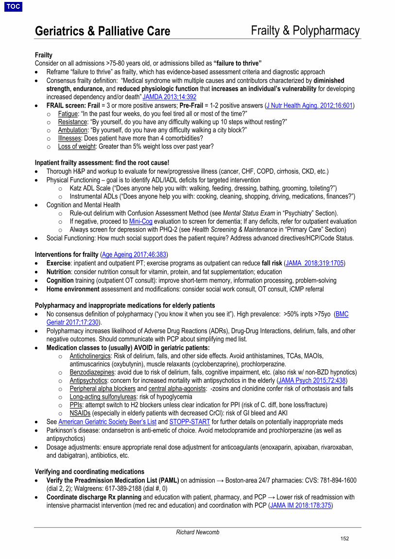
Geriatrics & Palliative Care Frailty & Polypharmacy
Richard Newcomb
Frailty Consider on all admissions >75-80 years old, or admissions billed as “failure to thrive” • Reframe “failure to thrive” as frailty, which has evidence-based assessment criteria and diagnostic approach• Consensus frailty definition: “Medical syndrome with multiple causes and contributors characterized by diminished
strength, endurance, and reduced physiologic function that increases an individual’s vulnerability for developingincreased dependency and/or death” JAMDA 2013;14:392
• FRAIL screen: Frail = 3 or more positive answers; Pre-Frail = 1-2 positive answers (J Nutr Health Aging. 2012;16:601)o Fatigue: “In the past four weeks, do you feel tired all or most of the time?”o Resistance: “By yourself, do you have any difficulty walking up 10 steps without resting?”o Ambulation: “By yourself, do you have any difficulty walking a city block?”o Illnesses: Does patient have more than 4 comorbidities?o Loss of weight: Greater than 5% weight loss over past year?
Inpatient frailty assessment: find the root cause! • Thorough H&P and workup to evaluate for new/progressive illness (cancer, CHF, COPD, cirrhosis, CKD, etc.)• Physical Functioning – goal is to identify ADL/IADL deficits for targeted intervention
o Katz ADL Scale (“Does anyone help you with: walking, feeding, dressing, bathing, grooming, toileting?”)o Instrumental ADLs (“Does anyone help you with: cooking, cleaning, shopping, driving, medications, finances?”)
• Cognition and Mental Healtho Rule-out delirium with Confusion Assessment Method (see Mental Status Exam in “Psychiatry” Section).o If negative, proceed to Mini-Cog evaluation to screen for dementia; If any deficits, refer for outpatient evaluationo Always screen for depression with PHQ-2 (see Health Screening & Maintenance in “Primary Care” Section)
• Social Functioning: How much social support does the patient require? Address advanced directives/HCP/Code Status.
Interventions for frailty (Age Ageing 2017;46:383) • Exercise: inpatient and outpatient PT; exercise programs as outpatient can reduce fall risk (JAMA 2018;319:1705)• Nutrition: consider nutrition consult for vitamin, protein, and fat supplementation; education• Cognition training (outpatient OT consult): improve short-term memory, information processing, problem-solving• Home environment assessment and modifications: consider social work consult, OT consult, iCMP referral
Polypharmacy and inappropriate medications for elderly patients • No consensus definition of polypharmacy (“you know it when you see it”). High prevalence: >50% inpts >75yo (BMC
Geriatr 2017;17:230).• Polypharmacy increases likelihood of Adverse Drug Reactions (ADRs), Drug-Drug Interactions, delirium, falls, and other
negative outcomes. Should communicate with PCP about simplifying med list.• Medication classes to (usually) AVOID in geriatric patients:
o Anticholinergics: Risk of delirium, falls, and other side effects. Avoid antihistamines, TCAs, MAOIs,antimuscarinics (oxybutynin), muscle relaxants (cyclobenzaprine), prochlorperazine.
o Benzodiazepines: avoid due to risk of delirium, falls, cognitive impairment, etc. (also risk w/ non-BZD hypnotics)o Antipsychotics: concern for increased mortality with antipsychotics in the elderly (JAMA Psych 2015;72:438)o Peripheral alpha blockers and central alpha-agonists: -zosins and clonidine confer risk of orthostasis and fallso Long-acting sulfonylureas: risk of hypoglycemiao PPIs: attempt switch to H2 blockers unless clear indication for PPI (risk of C. diff, bone loss/fracture)o NSAIDs (especially in elderly patients with decreased CrCl): risk of GI bleed and AKI
• See American Geriatric Society Beer’s List and STOPP-START for further details on potentially inappropriate meds• Parkinson’s disease: ondansetron is anti-emetic of choice. Avoid metoclopramide and prochlorperazine (as well as
antipsychotics)• Dosage adjustments: ensure appropriate renal dose adjustment for anticoagulants (enoxaparin, apixaban, rivaroxaban,
and dabigatran), antibiotics, etc.
Verifying and coordinating medications • Verify the Preadmission Medication List (PAML) on admission → Boston-area 24/7 pharmacies: CVS: 781-894-1600
(dial 2, 2); Walgreens: 617-389-2188 (dial #, 0)• Coordinate discharge Rx planning and education with patient, pharmacy, and PCP → Lower risk of readmission with
intensive pharmacist intervention (med rec and education) and coordination with PCP (JAMA IM 2018:178:375)
152
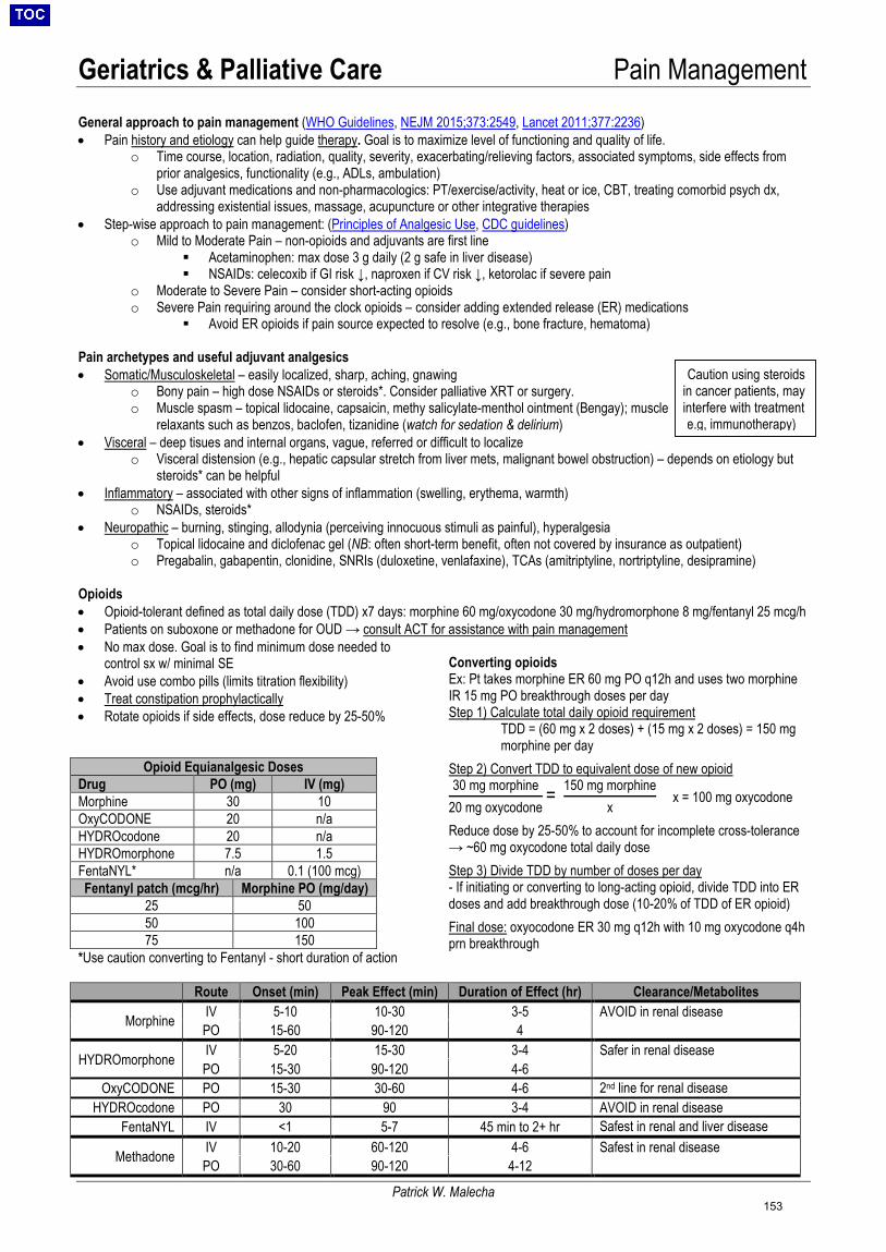
Geriatrics & Palliative Care Pain Management
Patrick W. Malecha
General approach to pain management (WHO Guidelines, NEJM 2015;373:2549, Lancet 2011;377:2236) • Pain history and etiology can help guide therapy. Goal is to maximize level of functioning and quality of life.
o Time course, location, radiation, quality, severity, exacerbating/relieving factors, associated symptoms, side effects fromprior analgesics, functionality (e.g., ADLs, ambulation)
o Use adjuvant medications and non-pharmacologics: PT/exercise/activity, heat or ice, CBT, treating comorbid psych dx,addressing existential issues, massage, acupuncture or other integrative therapies
• Step-wise approach to pain management: (Principles of Analgesic Use, CDC guidelines)o Mild to Moderate Pain – non-opioids and adjuvants are first line
Acetaminophen: max dose 3 g daily (2 g safe in liver disease) NSAIDs: celecoxib if GI risk ↓, naproxen if CV risk ↓, ketorolac if severe pain
o Moderate to Severe Pain – consider short-acting opioidso Severe Pain requiring around the clock opioids – consider adding extended release (ER) medications
Avoid ER opioids if pain source expected to resolve (e.g., bone fracture, hematoma)
Pain archetypes and useful adjuvant analgesics • Somatic/Musculoskeletal – easily localized, sharp, aching, gnawing
o Bony pain – high dose NSAIDs or steroids*. Consider palliative XRT or surgery.o Muscle spasm – topical lidocaine, capsaicin, methy salicylate-menthol ointment (Bengay); muscle
relaxants such as benzos, baclofen, tizanidine (watch for sedation & delirium)• Visceral – deep tisues and internal organs, vague, referred or difficult to localize
o Visceral distension (e.g., hepatic capsular stretch from liver mets, malignant bowel obstruction) – depends on etiology butsteroids* can be helpful
• Inflammatory – associated with other signs of inflammation (swelling, erythema, warmth)o NSAIDs, steroids*
• Neuropathic – burning, stinging, allodynia (perceiving innocuous stimuli as painful), hyperalgesiao Topical lidocaine and diclofenac gel (NB: often short-term benefit, often not covered by insurance as outpatient)o Pregabalin, gabapentin, clonidine, SNRIs (duloxetine, venlafaxine), TCAs (amitriptyline, nortriptyline, desipramine)
Opioids • Opioid-tolerant defined as total daily dose (TDD) x7 days: morphine 60 mg/oxycodone 30 mg/hydromorphone 8 mg/fentanyl 25 mcg/h• Patients on suboxone or methadone for OUD → consult ACT for assistance with pain management• No max dose. Goal is to find minimum dose needed to
control sx w/ minimal SE• Avoid use combo pills (limits titration flexibility)• Treat constipation prophylactically• Rotate opioids if side effects, dose reduce by 25-50%
Opioid Equianalgesic Doses Drug PO (mg) IV (mg) Morphine 30 10 OxyCODONE 20 n/a HYDROcodone 20 n/a HYDROmorphone 7.5 1.5 FentaNYL* n/a 0.1 (100 mcg) Fentanyl patch (mcg/hr) Morphine PO (mg/day)
25 50 50 100 75 150
*Use caution converting to Fentanyl - short duration of action
Converting opioids Ex: Pt takes morphine ER 60 mg PO q12h and uses two morphine IR 15 mg PO breakthrough doses per day Step 1) Calculate total daily opioid requirement
TDD = (60 mg x 2 doses) + (15 mg x 2 doses) = 150 mg morphine per day
Step 2) Convert TDD to equivalent dose of new opioid 30 mg morphine
20 mg oxycodone= 150 mg morphine
x x = 100 mg oxycodone
Reduce dose by 25-50% to account for incomplete cross-tolerance → ~60 mg oxycodone total daily doseStep 3) Divide TDD by number of doses per day- If initiating or converting to long-acting opioid, divide TDD into ERdoses and add breakthrough dose (10-20% of TDD of ER opioid) Final dose: oxyocodone ER 30 mg q12h with 10 mg oxycodone q4h prn breakthrough
Route Onset (min) Peak Effect (min) Duration of Effect (hr) Clearance/Metabolites
Morphine IV 5-10 10-30 3-5 AVOID in renal disease PO 15-60 90-120 4
HYDROmorphone IV 5-20 15-30 3-4 Safer in renal disease PO 15-30 90-120 4-6
OxyCODONE PO 15-30 30-60 4-6 2nd line for renal disease HYDROcodone PO 30 90 3-4 AVOID in renal disease
FentaNYL IV <1 5-7 45 min to 2+ hr Safest in renal and liver disease
Methadone IV 10-20 60-120 4-6 Safest in renal disease PO 30-60 90-120 4-12
*Caution using steroidsin cancer patients, mayinterfere with treatment(e.g, immunotherapy)
153
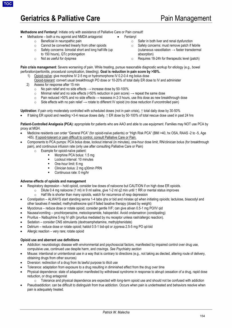
Geriatrics & Palliative Care Pain Management
Patrick W. Malecha
Methadone and Fentanyl: Initiate only with assistance of Palliative Care or Pain consult! • Methadone – both a mu agonist and NMDA antagonist
o Beneficial in neuropathic paino Cannot be converted linearly from other opioidso Safety concerns: bimodal short and long half-life (up
to 150 hours), QTc prolongationo Not as useful for dyspnea
• Fentanylo Safer in both liver and renal dysfunctiono Safety concerns: must remove patch if febrile
(cutaneous vasodilation → faster transdermalabsorption)
o Requires 18-24h for therapeutic level (patch)
Pain crisis management: Severe worsening of pain. While treating, pursue reasonable diagnostic workup for etiology (e.g., bowel perforation/peritonitis, procedural complication, bleeding). Goal is reduction in pain score by >50%.
1) Opioid-naïve: give morphine IV 2-5 mg or hydromorphone IV 0.2-0.4 mg bolus doseOpioid-tolerant: convert usual breakthrough PO dose or 10-20% of total daily ER dose to IV and administer
2) Assess for response after 15 mino No pain relief and no side effects → increase dose by 50-100%o Minimal relief and no side effects (<50% reduction in pain score) → repeat the same doseo Pain reduced >50% and no side effects→ reassess in 2-3 hours, use this dose as new breakthrough doseo Side effects with no pain relief → rotate to different IV opioid (no dose reduction if uncontrolled pain)
Uptitration: if pain only moderately controlled with scheduled doses (not in pain crisis), ↑ total daily dose by 30-50% • If taking ER opioid and needing >3-4 rescue doses daily, ↑ ER dose by 50-100% of total rescue dose used in past 24 hrs
Patient-Controlled Analgesia (PCA): appropriate for patients who are AAO and able to use equipment. Families may NOT use PCA by proxy at MGH. • Medicine residents can order “General PCA” (for opioid-naïve patients) or “High Risk PCA” (BMI >40, hx OSA, RAAS -2 to -5, Age
>65). If opioid-tolerant or pain difficult to control, consult Palliative Care or Pain.• Components to PCA pumps: PCA bolus dose, lockout interval (in minutes), one-hour dose limit, RN/clinician bolus (for breakthrough
pain), and continuous infusion rate (only use after consulting Palliative Care or Pain)o Example for opioid-naïve patient:
Morphine PCA bolus: 1.5 mg Lockout interval: 10 minutes One-hour limit: 6 mg Clinician bolus: 2 mg q30min PRN Continuous rate: 0 mg/hr
Adverse effects of opioids and management • Respiratory depression – hold opioid, consider low doses of naloxone but CAUTION if on high dose ER opioids.
o Dilute 0.4 mg naloxone (1 ml) in 9 ml saline, give 1-2 ml q2 min until ↑ RR or mental status improveso Half life is shorter than many opioids, watch for recurrence of resp depression
• Constipation – ALWAYS start standing senna 1-4 tabs qhs or bid and miralax qd when initiating opioids; lactulose, bisacodyl andother laxatives if needed; methylnaltrexone qod if failed laxative therapy (dosed by weight)
• Myoclonus – reduce dose or rotate opioid, consider gentle IVF; can give ativan 0.5-1 mg PO/IV qid• Nausea/vomiting -- prochlorperazine, metoclopramide, haloperidol. Avoid ondansetron (constipating)• Pruritus – Nalbuphine 5 mg IV q6h (pruritus mediated by mu receptor unless rash/allergic reaction).• Sedation – consider CNS stimulants (dextroamphetamine, methylphenidate)• Delirium – reduce dose or rotate opioid; haldol 0.5-1 bid-qid or zyprexa 2.5-5 mg PO qd-bid• Allergic reaction – very rare; rotate opioid
Opioid use and aberrant use definitions • Addiction: neurobiologic disease with environmental and psychosocial factors, manifested by impaired control over drug use,
compulsive use, continued use despite harm, and cravings. See Psychiatry section• Misuse: intentional or unintentional use in a way that is contrary to directions (e.g., not taking as diected, altering route of delivery,
obtaining drugs from other sources)• Diversion: redirection of a drug from its lawful purpose to illicit use• Tolerance: adaptation from exposure to a drug resulting in diminished effect from the drug over time• Physical dependence: state of adapation manifested by withdrawal syndrome in response to abrupt cessation of a drug, rapid dose
reduction, or drug antagonisto Tolerance and physical dependence are expected with long-term opioid use and should not be confused with addiction
• Pseudoaddiction: can be difficult to distinguish from true addiction. Occurs when pain is undertreated and behaviors resolve whenpain is adequately treated.
154

Non-pain Symptom Management
Meg Allison
Searchable Resources: Palliative care network of Wisconsin (www.mypcnow.org/fast-facts), wwww.capc.org, Pink and Green Books
Palliation in serious illness and end of life can be challenging and often is helped by a Palliative Care consultation. • “Comort measures only” is NOT a one-size-fits-all set of orders (e.g., indwelling Foley may be more tolerable than frequent urinary
incontinence, diuretics may still be indicated for relief of dyspnea or edema, etc.)• For persistent/recurring sx meds should be made standing, with additional PRNs for breakthrough
Anxiety: often exacerbated by medications (steroids, appetite stimulants, etc.), undertreated pain, and dyspnea • Treat underlying causes, use non-pharmacologic strategies (integrative therapies c/s, SW & spiritual care for coping/support)• For acute anxiolysis: olanzapine 2.5-5mg q6hr prn, lower doses if elderly. Avoid BZDs due to delirium risk.• For longer-term management, consider usual meds (SSRIs, SNRIs, TCAs)
Depressed mood: Can be difficult to distinguish between MDD, demoralization, and adjustment disorder. See Psychiatry section • Treat uncontrolled symptoms, especially pain. Screen for delirium.• CBT/psychotherapy may be better for existential demoralization than in MDD (Am J Hosp Palliat Care. 2016;33(1):93)
o Some amount of depressed mood is expected, doesn’t necessarily need medication but benefits from psychosocial support• If nearing end-of-life, CNS stimulants (methylphenidate, dextroamphetamine) > SSRIs due to faster onset of action
Delirium: common and often multifactorial • Prevention: remove unnecessary lines/catheters/restraints; lights on, shades & pt up during day; limit nighttime interruptions and
lights/TV; frequent reorientation, use signage; minimize staff/room changes; manage other symptoms• No FDA-approved delirium med, but if hallucinations or agitation interfering w/ staff or pt safety → Haldol 0.5-1mg IV q4hr PRN.
Consider atypical antipsychotics. See Neuro section on delirium for further details.
Nausea/vomiting, Diarrhea, Constipation: See relevant GI sections
Xerostomia: side effect of chemo/XRT, head/neck surgery, or medications • Oral hygiene, oral hydration, saliva substitutes such as Biotene
Anorexia/Cachexia: common in AIDS, heart failure, COPD, advanced cancer. Often highly concerning for family > patient. • Rule out reversible causes (other sx causing poor PO intake). In general, allow PO for comfort if near end-of-life.• Meds to consider: dexamethasone, megestrol (VTE risk), dronabinol, mirtazapine• NB: during dying process, artificial nutrition and hydration risk may outweigh benefit
Fatigue: often related to disease progression, medications, other treatments, deconditioning, malnutrition, sleep disturbances, sx’s • Treat uncontrolled sx. For cancer-related fatigue, exercise and psychological interventions >> medications (JAMA Oncol. 2017;3:
961). Consider steroid trial (J Clin Oncol. 2013;31:3076). Psychostimulants (modafinil, methylphenidate) have limited evidence.
Dyspnea: exacerbated by deconditioning, cachexia, worsens at EOL, exacerbates anxiety. Does not always correlate w/hypoxemia. • Treat underlying causes depending on etiology; consider bedside fan• For refractory dyspnea, opioids are gold standard (often at lower doses than required for pain). BZDs less supported by evidence; can
be used for associated anxiety and must weigh risk of delirium.
Secretions: pooled secretions “death rattle”. Disturbing to observers, less bothersome to pt • Stop feeds/fluids, don’t deep suction (uncomfortable to pt), continue oral care• Glycopyrrolate 0.2-0.4mg IV q4 PRN. Less deliriogenic than other anti-cholinergics• Other alternatives: scopolamine patch, atropine, hyoscyamine (may cause delirium)
Insomnia (inpatient management) • Avoid BZDs and non-BZD hypnotics (e.g., zolpidem, zaleplon, eszopiclone) for inpatient management due to delirium risk. Avoid H1
blockers (diphenhydramine, hydroxyzine) due to risk of delirium, next-day sedation, anticholinergic side effects.• Use non-FDA approved treatments on a short-term basis: melatonin (3-5mg Q6PM), trazodone (12.5-50mg QHS, QTc prolonging),
mirtazapine (7.5mg QHS)• Use with caution: quetiapine (12.5-25mg QHS, Qtc prolonging) – there is concern for increased mortality with antipsychotics in the
elderly (JAMA Psych 2015;72:438). Reserve for patients with additional indication (e.g., patients who require pharmacologic tx foragitated delirium).
Catastrophic hemoptysis or hemorrhage: often preceded by “sentinel” small bleed. Be sure to prep pt/family for possibility. • Dark linens/basins present in the room (↓ contrast w/ blood). PPE for caregivers, suctioning, warm blankets (hemorrhage chills)• Consider pre-drawn crisis meds, goal is rapid anxiolysis and sedation (BZDs q5-10 min +/- opioids, though bleeding usually painless)
Geriatrics & Palliative Care
155
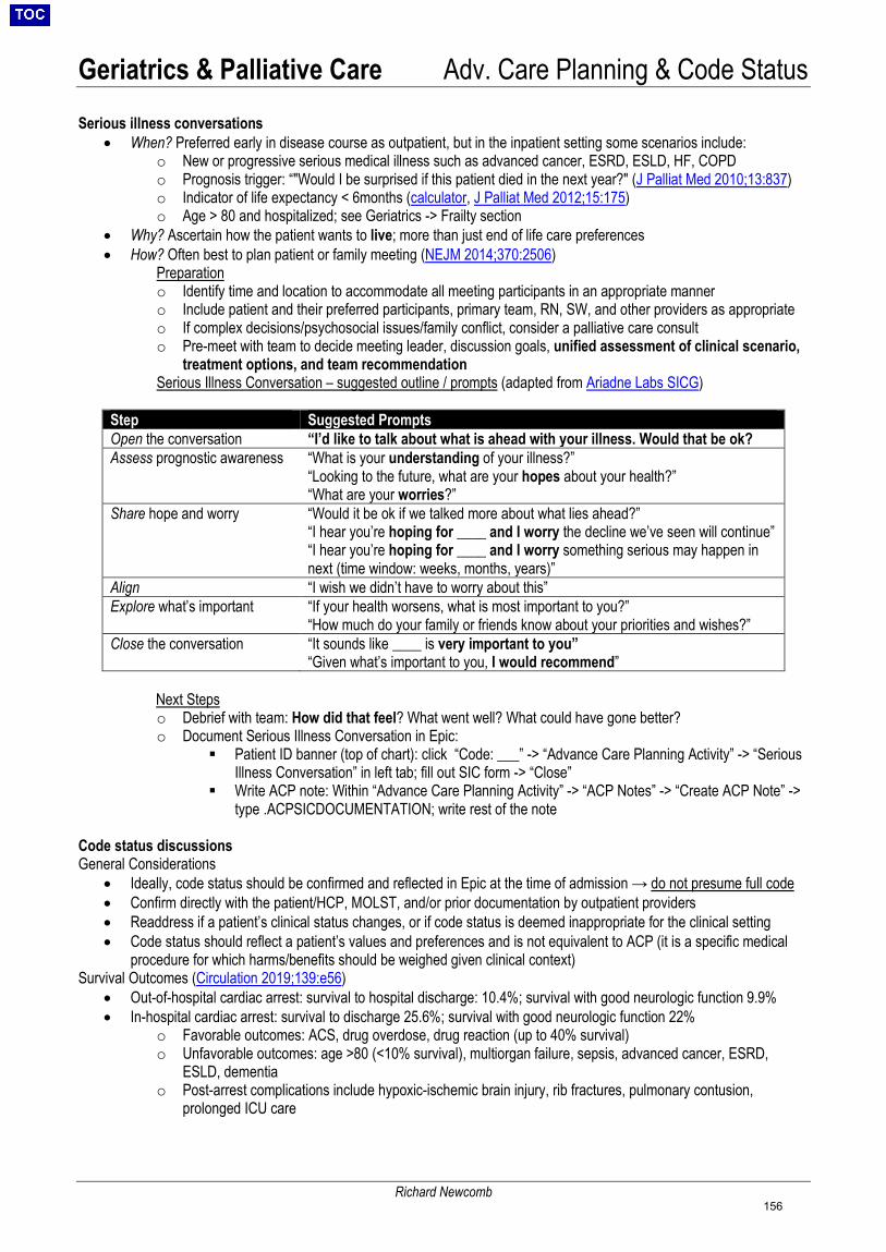
Geriatrics & Palliative Care Adv. Care Planning & Code Status
Richard Newcomb
Serious illness conversations • When? Preferred early in disease course as outpatient, but in the inpatient setting some scenarios include:
o New or progressive serious medical illness such as advanced cancer, ESRD, ESLD, HF, COPDo Prognosis trigger: “"Would I be surprised if this patient died in the next year?" (J Palliat Med 2010;13:837)o Indicator of life expectancy < 6months (calculator, J Palliat Med 2012;15:175)o Age > 80 and hospitalized; see Geriatrics -> Frailty section
• Why? Ascertain how the patient wants to live; more than just end of life care preferences• How? Often best to plan patient or family meeting (NEJM 2014;370:2506)
Preparation o Identify time and location to accommodate all meeting participants in an appropriate mannero Include patient and their preferred participants, primary team, RN, SW, and other providers as appropriateo If complex decisions/psychosocial issues/family conflict, consider a palliative care consulto Pre-meet with team to decide meeting leader, discussion goals, unified assessment of clinical scenario,
treatment options, and team recommendationSerious Illness Conversation – suggested outline / prompts (adapted from Ariadne Labs SICG)
Step Suggested Prompts Open the conversation “I’d like to talk about what is ahead with your illness. Would that be ok? Assess prognostic awareness “What is your understanding of your illness?”
“Looking to the future, what are your hopes about your health?” “What are your worries?”
Share hope and worry “Would it be ok if we talked more about what lies ahead?” “I hear you’re hoping for ____ and I worry the decline we’ve seen will continue” “I hear you’re hoping for ____ and I worry something serious may happen in next (time window: weeks, months, years)”
Align “I wish we didn’t have to worry about this” Explore what’s important “If your health worsens, what is most important to you?”
“How much do your family or friends know about your priorities and wishes?” Close the conversation “It sounds like ____ is very important to you”
“Given what’s important to you, I would recommend”
Next Steps o Debrief with team: How did that feel? What went well? What could have gone better?o Document Serious Illness Conversation in Epic:
Patient ID banner (top of chart): click “Code: ___” -> “Advance Care Planning Activity” -> “SeriousIllness Conversation” in left tab; fill out SIC form -> “Close”
Write ACP note: Within “Advance Care Planning Activity” -> “ACP Notes” -> “Create ACP Note” ->type .ACPSICDOCUMENTATION; write rest of the note
Code status discussions General Considerations
• Ideally, code status should be confirmed and reflected in Epic at the time of admission → do not presume full code• Confirm directly with the patient/HCP, MOLST, and/or prior documentation by outpatient providers• Readdress if a patient’s clinical status changes, or if code status is deemed inappropriate for the clinical setting• Code status should reflect a patient’s values and preferences and is not equivalent to ACP (it is a specific medical
procedure for which harms/benefits should be weighed given clinical context)Survival Outcomes (Circulation 2019;139:e56)
• Out-of-hospital cardiac arrest: survival to hospital discharge: 10.4%; survival with good neurologic function 9.9%• In-hospital cardiac arrest: survival to discharge 25.6%; survival with good neurologic function 22%
o Favorable outcomes: ACS, drug overdose, drug reaction (up to 40% survival)o Unfavorable outcomes: age >80 (<10% survival), multiorgan failure, sepsis, advanced cancer, ESRD,
ESLD, dementiao Post-arrest complications include hypoxic-ischemic brain injury, rib fractures, pulmonary contusion,
prolonged ICU care
156
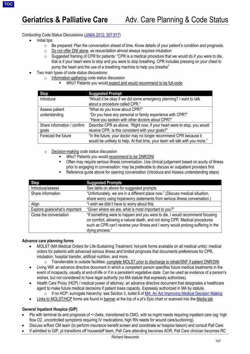
Geriatrics & Palliative Care Adv. Care Planning & Code Status
Richard Newcomb
Conducting Code Status Discussions (JAMA 2012: 307:917)
• Initial tips:o Be prepared: Plan the conversation ahead of time. Know details of your patient’s condition and prognosis.o Do not offer DNI alone, as resuscitation almost always requires intubationo Suggested framing of CPR for patients: “CPR is a medical procedure that we would do if you were to die,
that is if your heart were to stop and you were to stop breathing. CPR includes pressing on your chest topump the heart and the use of a breathing machine to help you breathe”
• Two main types of code status discussions:o Information-gathering code status discussion
Who? Patients you would expect and would recommend to be full-code
Step Suggested Prompt Introduce “Would it be okay if we did some emergency planning? I want to talk
about a procedure called CPR.” Assess patient understanding
“What do you know about CPR?” “Do you have any personal or family experience with CPR?” “Have you spoken with other doctors about CPR?”
Share information / confirm goals
Describe CPR as above. “Right now, if your heart were to stop, you would receive CPR. Is this consistent with your goals?”
Forecast the future “In the future, your doctor may no longer recommend CPR because it would be unlikely to help. At that time, your team will talk with you more.”
o Decision-making code status discussion Who? Patients you would recommend to be DNR/DNI Often may require serious illness conversation. Use clinical judgement based on acuity of illness
prior to engaging in conversation; may be preferable to discuss w/ outpatient providers first Reference guide above for opening conversation (Introduce and Assess understanding steps)
Step Suggested Prompts Introduce/assess See table on above for suggested prompts Share information “Unfortunately, we are in a different place now.” (Discuss medical situation,
share worry using hope/worry statements from serious illness conversation.) Align “I wish we didn’t have to worry about this. Explore goals/what’s important “Given where we are, what is most important to you?” Close the conversation “If something were to happen and you were to die, I would recommend focusing
on comfort, allowing a natural death, and not doing CPR. Medical procedures such as CPR can’t reverse your illness and I worry would prolong suffering in the dying process.”
Advance care planning forms • MOLST (MA Medical Orders for Life-Sustaining Treatment; hot-pink forms available on all medical units): medical
orders for patients with advanced serious illness and limited prognosis that documents preferences for CPR,intubation, hospital transfer, artificial nutrition, and more.
o Transferrable to outside facilities; complete MOLST prior to discharge to rehab/SNF if patient DNR/DNI• Living Will: an advance directive document in which a competent person specifies future medical treatments in the
event of incapacity, usually at end-of-life or if in a persistent vegetative state. Can be used as evidence of a person’swishes, but not considered to have legal authority (no MA statute that expressly authorizes).
• Health Care Proxy (HCP) / medical power of attorney: an advance directive document that designates a healthcareagent to make future medical decisions if patient loses capacity. Expressly authorized in MA by statute.
o If no HCP: surrogate hierarchy: see Section 3, bullet 6 of MA: An Act Improving Medical Decision Making• Links to MOLST/HCP forms are found in banner at the top of a pt’s Epic chart or scanned into the Media tab
General Inpatient Hospice (GIP) • Pts with terminal dx and prognosis of <~2wks, transitioned to CMO, with sx mgmt needs requiring inpatient care (eg: high
flow O2, uncontrolled symptoms requiring IV medications, high RN needs for wound care/suctioning)• Discuss w/floor CM team (to perform insurance benefit screen and coordinate w/ hospice liaison) and consult Pall Care• If admitted to GIP, pt transitions off housestaff team, Pall Care attending becomes AOR, Pall Care clinician becomes RC
157
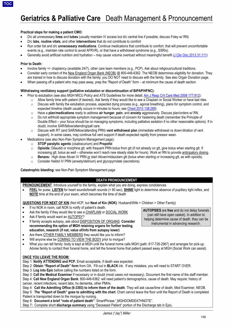
Geriatrics & Palliative Care Death Management & Pronouncement
James (“Jay”) Miller
Practical steps for making a patient CMO: • D/c all unnecessary lines and tubes (usually maintain IV access but d/c central line if possible; discuss Foley w/ RN)• D/c labs, routine vitals, and other interventions that do not contribute to comfort• Run order list and d/c unnecessary medications. Continue medications that contribute to comfort, that will prevent uncomfortable
events (e.g., maintain rate control to avoid AFRVR), or that have a withdrawal syndrome (e.g., SSRIs).• Generally avoid artificial nutrition and hydration – may cause volume overload without meaningful benefit (J Clin Onc 2013 31:111)
Prior to Death: • Involve family +/- chaplaincy (available 24/7), other care team members (e.g., PCP). Ask about religious/cultural traditions.• Consider early contact of the New England Organ Bank (NEOB) @ 800-446-6362. The NEOB determines eligibility for donation. They
are trained in how to discuss donation with the family; you DO NOT need to discuss with the family. See also Organ Donation page.• When passing off a patient who may pass away, prep the “Report of Death” form – at minimum the cause of death section
Withdrawing ventilatory support (palliative extubation or discontinuation of BiPAP/HFNC): • Prior to extubation (see also MGH MICU Policy and ATS Guidelines for more detail; Am J Resp Crit Care Med 2008 177:912):
o Allow family time with patient (if desired). Ask family if they would like to see a Chaplain or Social Worker or have last rites.o Discuss with family the extubation process, expected dying process (e.g., agonal breathing), plans for symptom control, and
expected timeline (death usually occurs in minutes to hours; see Chest 2010 138:289)o Have a plan/medications ready to address air hunger, pain, and anxiety aggressively. Discuss plan/orders w/ RN.o Do not withhold appropriate symptom management because of concern for hastening death (remember the Principle of
Double Effect – your focus should be on managing symptoms, including palliative sedation if no other reasonable options). If indoubt, involve SAR/fellow/attending/pall care.
o Discuss with RT (and SAR/fellow/attending PRN) vent withdrawal plan (immediate withdrawal vs down-titration of ventsupport). In some cases, may continue full vent support if death expected rapidly from pressor wean.
• Medications (see also Non-Pain Symptom Management page):o STOP paralytic agents (cisatracurium) and Propofolo Opioids: Dilaudid or morphine gtt, with frequent PRN bolus from gtt (if not already on gtt, give bolus when starting gtt. If
increasing gtt, bolus as well – otherwise won’t reach new steady state for hours). Work w/ RN to provide anticipatory dosing.o Benzos: High dose Ativan IV PRN or start Ativan/midazolam gtt (bolus when starting or increasing gtt, as with opioids)o Consider Haldol IV PRN (anxiety/delirium) and glycopyrrolate (secretions)
Catastrophic bleeding: see Non-Pain Symptom Management page
DEATH PRONOUNCEMENT PRONOUNCEMENT. Introduce yourself to the family, explain what you are doing, express condolences • FEEL for pulse, LISTEN for heart sounds/breath sounds (> 60 sec), SHINE light to determine absence of pupillary light reflex, and
NOTE time at the end of your exam, which becomes the time of death
AUTOPSIES are free and do not delay funerals (can still have open casket). In addition to
helping determine cause of death, they can be instrumental in advancing research.
QUESTIONS FOR NEXT OF KIN (Not HCP, but Next of Kin (NOK): Husband/Wife > Children > Other Family) • If no NOK in room, call NOK to notify of patient’s death.• Ask the family if they would like to see a CHAPLAIN or SOCIAL WORK• Ask if family would want an AUTOPSY?• If family accepts autopsy, ask obout DISPOSITION OF ORGANS. Consider
recommending the option of MGH retaining organs for further testing, education, research (if not, value of/info from autopsy lower)
• Are there OTHER FAMILY MEMBERS they would like you to inform?• Will anyone else be COMING TO VIEW THE BODY prior to morgue?• What you can tell family: body is kept at MGH until the funeral home calls MGH (path: 617-726-2967) and arranges for pick-up.
Advise family to contact their funeral home, and tell the funeral home that patient passed away at MGH (Social Work can assist).
ONCE YOU LEAVE THE ROOM: Step 1: Notify ATTENDING and PCP. Email acceptable, if death was expected. Step 2: Obtain “Report of Death” form from OA. Fill out in BLACK ink. If any mistakes, you will need to START OVER. Step 3: Log into Epic before calling the numbers listed on the form. Step 3: Call the Medical Examiner if necessary or in doubt (most cases not necessary). Document the first name of the staff member. Step 4: Call New England Organ Bank: 800-446-6362: will need patient’s demographics, cause of death. May require: history of cancer, recent infections, recent labs, hx dementia, other PMHx. Step 4: Call the Admitting Office (6-3393) to inform them of the death. They will ask cause/time of death, Med Examiner, NEOB. Step 5: The “Report of Death” goes to admitting with the chart. Chart cannot leave the floor until the Report of Death is completed. Patient is transported down to the morgue by nursing. Step 6: Document a brief “note of patient death”: SmartPhrase “.MGHDOMDEATHNOTE”. Step 7: Complete short discharge summary using “Deceased Patient” portion of the Discharge tab in Epic.
158
Geriatrics & Palliative Care Organ Donation
Patrick Malecha
Organ donation after brain or circulatory death • ~75% of transplanted organs are from deceased donors, including donation after brain death (DBD) and donation after circulatory
death (DCD). DCD represents ~8% of organs procured nationally, ~20% in the Boston area (NEJM 2007;357:209)• DBD = death based on neurologic criteria (“brain death,” or irreversible loss of all functions of the brain, including the brain stem)• DCD = death based on cardiopulmonary criteria (irreversible cessation of circulatory and respiratory function and mechanical
ventilatory support is no longer medically indicated, but criteria for brain death are NOT fulfilled)o Organs from DCD and DBD donors have similar long-term outcomes (NEJM 2002;347:248)
Eligibility for organ donation • Medical team determines that discontinuation of medical support is appropriate and discusses this with the HCP or legal next-of-kin• DO NOT broach the topic of potential organ donation with family; New England Organ Bank (NEOB) is specifically trained to do this.• If family wishes for withdrawal of support, the medical team notifies NEOB (800) 446-6362 who will coordinate the process for consent
and donation (NB: this process can take up to 24 hours)
Care of the patient prior to organ donation • Patients with potential for organ donation need to maintain organ viability in response to potentially severe autonomic and
inflammatory responses that occur after severe neurologic insult or brain death.• Interventions often require a delicate balance to preserve multiple organs: (Crit Care Med. 2015;43:1291, NEJM 2004;351:2730)
o Continuous temperature monitoring, telemetry, and lab monitoring for renal function, electrolytes, acid-base statuso Hemodynamics – goal MAP 60-110 (JAMA Surg. 2014;149:969)
Hypertensive autonomic storm after brain death. Esmolol to preserve cardiac function. (Am J Transplant.2005;5:684)
Fluids and vasopressors for hypotension/vasoplegia. Consider vasopressin before catecholamines (helps w/ DI) Dobutamine for reduced EF
o Maintenance of normothermia via external warming or coolingo Urine output monitoring – goal 0.5-1.0 cc/kg/hr. Monitor for DI with severe neurologic injury or brain death.o Proper ventilatory support and pulmonary toilet – lung-protective LTVV as in ARDSNet. Prevent pneumonia with head
elevation, etc. (JAMA. 2010;304:2620)o Maintenance of eunatremia, euvolemia, and acid-base statuso Consider glucocorticoids for adrenal insufficiency; thyroid hormone for EF <45% or hemodynamic instability (limited
evidence)o Empiric antibiotics if concern for infection
Death pronouncement in the operating room for DCD patients • Generally, withdrawal of medical support, including extubation, occurs in the OR after pt is prepped by surgical team• All members of the organ recovery teams must be outside of the room from the time of withdrawal of support to declaration of death;
otherwise this poses a conflict of interest. Family may be present in the OR if they wish.• Medical team (MD and RN) are present to coordinate end of life care from time of withdrawal of support to death, including PRN
palliative medications. NEOB staff may not participate in the administration of medications or declaration of death.• Death must occur and be declared within 2 hours of extubation, otherwise organs are deemed nonviable.
o “Dead-donor rule” (DDR) = recovery of organs cannot be the cause of death, and organs should be taken only from personswho are already dead (NEJM 2013;369:1287)
• MD declares death based on the irreversible cessation of circulatory and respiratory function (checks carotid artery for pulsations andauscultates for breath sounds using a sterile ultrasound cover over stethoscope)
o PEA arrest meets criteria for cessation of circulatory function so long as there is no pulsatile flow on arterial line. Death canbe declared even if cardiac electrical activity persists.
o After death is declared, a a 5-minute observation period begins to ensure no ROSC• Death paperwork should be signed by declaring MD in the OR (i.e., bring prepped death paperwork with you)
159

Rheumatology Approach to Rheumatologic Disease
Julie Fiore, Mazin Abdelghany
Overview: rheumatologic diseases may be roughly separated into 4 categories:
Arthritis Connective tissue disease Vasculitis Other RA, spondyloarthropathies, PMR, crystalline arthritis, OA
SLE, Sjögren’s, scleroderma, MCTD, UCTD, myositis (DM/PM)
small, medium, and large vessel vasculitides (e.g., GCA, PAN, GPA, EGPA)
Autoinflammatory diseases, bone/tendon/bursal disease, sarcoid, IgG4-related disease
RF, anti-CCP ANA, C3/C4, anti-Sm, anti-dsDNA, anti-Scl70, anti-histone, anti-U1RNP,
anti-Ro/La, antiphospholipid Abs
ANCA, cryoglobulins (limited role for ACE, IgG4)
NB: Always consider malignancy and infection as alternative diagnoses prior to initiation of immunosuppressants unless at risk of permanent organ damage (i.e., do not withhold glucocorticoids when suspecting GCA, mononeuritis multiplex, RPGN, etc.).
Rheumatologic ROS: Fevers, rashes/photosensitivity, alopecia, nail/nailfold abnormalities, sicca symptoms, conjunctivitis, uveitis, episcleritis, scleritis, Raynaud's, oral/genital ulcers, polychondritis, enthesitis, serositis sx, thomboses, neuropathy, pregnancy loss.
Basic Labs: CBC w/ diff, BMP, LFTs, UA, random urine protein:Cr ratio, ESR/CRP, CK, aldolase, TSH, hepatitis B/C serologies
Inflammatory Arthritis Sex Age Serology Comments Rheumatoid arthritis F > M 35-65 RF+/CCP+ (70%-80%) Symmetric chronic inflammatory polyarthritis
Psoriatic arthritis‡ M = F 30-55 RF+ (2%-10%) Asymmetric, large joints; 50% w/ RA distrib; majority w/ history of psoriasis, examine nails
Reactive arthritis (post-infx)‡ M > F 16-50 RF-, HLA-B27+ (50%-80%) a/w enthesitis, LBP, ocular sx, GU/GI sxEnteropathic arthritis‡ M = F All ages RF- a/w enthesitis, oral ulcers; 20% w/IBD Axial spondyloarthritis (e.g., ankylosing spondylitis)‡ M > F 15-45 HLA-B27+ (>50%) Asymmetric, large joints; LE>UE, enthesitis,
dactylitis; a/w “silent” GU infxn (e.g., GC/Chla) Gout M > F >25 RF- Intermittent inflammatory; usu. monoarticular Pseudogout F = M >60 RF- 5% w/ RA-like arthritis lasting for wks to months
Viral arthritis F > M All ages RF+ (<10%); consider Parvovirus B19 Ab
Acute symmetric polyarthritis w/ RA distribution; <10% develop chronic polyarthritis
‡Seronegative spondyloarthropathy (SpA)
Connective Tissue Disease Sex Age Serology/Tests Comments
Systemic lupus erythematosus (SLE) F > M 15-40
ANA+ (>95%), anti-dsDNA (70%), anti-Sm/ RNP (30%), anti-histone, C3/4
Sx: rash, oral ulcers, arthritis, serositis, renal dz, neuro d/o, heme abnormalities. ↑ CK suggests myositis
Sjögren’s syndrome (SS) F > M 40-60Anti-Ro(SSA)/La(SSB), [often ANA≥1:320 and RF+], lip bx
Sx: sicca sx, parotid gland inflam., dental caries; RA/SLE a/w 2° SS; if dx uncertain, perform lip bx; if salivary gland enlarged, consider IgG4
Systemic sclerosis (SSc), (a.k.a., scleroderma)
F:M 4:1 30-50
ANA+ (95%), anti-Scl-70*, ACA*, anti-RNA-pol 3* *any are >99% specific
Types: Local (linear/morphea) vs. systemic (dcSSc, lcSSc, SSc sine scleroderma) Sx: CREST; scleroderma renal crisis; ILD
Mixed connective tissue dz 80% F 20-30 ANA (speckled), anti-U1RNP (100%)
Sx: clin. ft. of SLE, SSc, myositis overlapping over many yrs.; pHTN
Dermatomyositis/polymyositis F:M 2:1 40-50
ANA+ (up to 80%), cytoplasmic (e.g., anti-Jo1)
Sx: Proximal muscle weakness, rashes, GERD/dysphagia; may cause antisynthetase syndrome (ILD, myopathy, arthritis)
Vasculitis Large-vessel vasculitis
Takayasu arteritis; giant cell arteritis (GCA) Medium-vessel vasculitis
Polyarteritis nodosa; Kawasaki disease (usually in children, can affect large vessels) Small-vessel vasculitis
ANCA-associated: (Granulomatosis with polyangiitis, eosinophilic granulomatosis with polyangiitis, microscopic polyangiitis) Immune complex-associated: (Cryoglobulinemic, IgA [Henoch-Schönlein purpura], hypocomplementemic urticarial (anti-C1q)
160
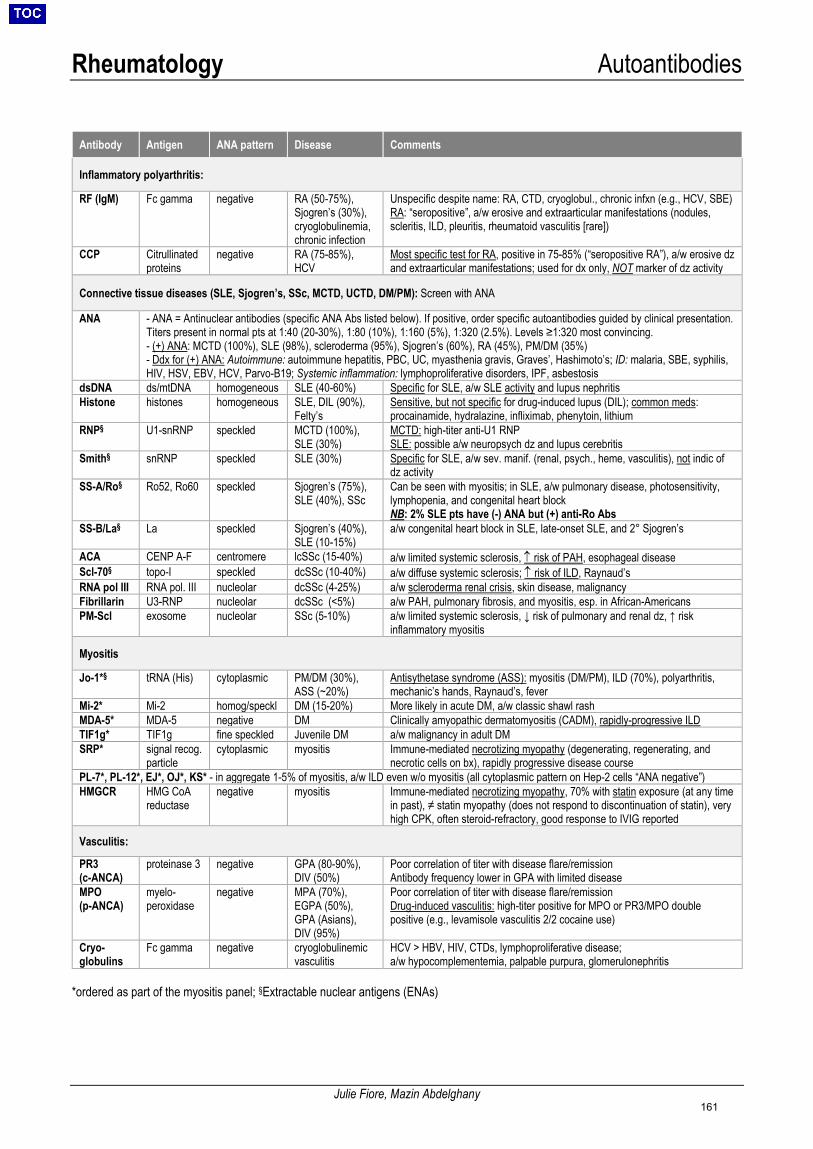
Rheumatology Autoantibodies
Julie Fiore, Mazin Abdelghany
*ordered as part of the myositis panel; §Extractable nuclear antigens (ENAs)
Antibody Antigen ANA pattern Disease Comments
Inflammatory polyarthritis:
RF (IgM) Fc gamma negative RA (50-75%), Sjogren’s (30%), cryoglobulinemia, chronic infection
Unspecific despite name: RA, CTD, cryoglobul., chronic infxn (e.g., HCV, SBE) RA: “seropositive”, a/w erosive and extraarticular manifestations (nodules, scleritis, ILD, pleuritis, rheumatoid vasculitis [rare])
CCP Citrullinated proteins
negative RA (75-85%), HCV
Most specific test for RA, positive in 75-85% (“seropositive RA”), a/w erosive dz and extraarticular manifestations; used for dx only, NOT marker of dz activity
Connective tissue diseases (SLE, Sjogren’s, SSc, MCTD, UCTD, DM/PM): Screen with ANA
ANA - ANA = Antinuclear antibodies (specific ANA Abs listed below). If positive, order specific autoantibodies guided by clinical presentation.Titers present in normal pts at 1:40 (20-30%), 1:80 (10%), 1:160 (5%), 1:320 (2.5%). Levels ≥1:320 most convincing.- (+) ANA: MCTD (100%), SLE (98%), scleroderma (95%), Sjogren’s (60%), RA (45%), PM/DM (35%)- Ddx for (+) ANA: Autoimmune: autoimmune hepatitis, PBC, UC, myasthenia gravis, Graves’, Hashimoto’s; ID: malaria, SBE, syphilis,HIV, HSV, EBV, HCV, Parvo-B19; Systemic inflammation: lymphoproliferative disorders, IPF, asbestosis
dsDNA ds/mtDNA homogeneous SLE (40-60%) Specific for SLE, a/w SLE activity and lupus nephritis Histone histones homogeneous SLE, DIL (90%),
Felty’s Sensitive, but not specific for drug-induced lupus (DIL); common meds: procainamide, hydralazine, infliximab, phenytoin, lithium
RNP§ U1-snRNP speckled MCTD (100%), SLE (30%)
MCTD: high-titer anti-U1 RNP SLE: possible a/w neuropsych dz and lupus cerebritis
Smith§ snRNP speckled SLE (30%) Specific for SLE, a/w sev. manif. (renal, psych., heme, vasculitis), not indic of dz activity
SS-A/Ro§ Ro52, Ro60 speckled Sjogren’s (75%), SLE (40%), SSc
Can be seen with myositis; in SLE, a/w pulmonary disease, photosensitivity, lymphopenia, and congenital heart block NB: 2% SLE pts have (-) ANA but (+) anti-Ro Abs
SS-B/La§ La speckled Sjogren’s (40%), SLE (10-15%)
a/w congenital heart block in SLE, late-onset SLE, and 2° Sjogren’s
ACA CENP A-F centromere lcSSc (15-40%) a/w limited systemic sclerosis, ↑ risk of PAH, esophageal disease Scl-70§ topo-I speckled dcSSc (10-40%) a/w diffuse systemic sclerosis; ↑ risk of ILD, Raynaud’s RNA pol III RNA pol. III nucleolar dcSSc (4-25%) a/w scleroderma renal crisis, skin disease, malignancy Fibrillarin U3-RNP nucleolar dcSSc (<5%) a/w PAH, pulmonary fibrosis, and myositis, esp. in African-Americans PM-Scl exosome nucleolar SSc (5-10%) a/w limited systemic sclerosis, ↓ risk of pulmonary and renal dz, ↑ risk
inflammatory myositis
Myositis
Jo-1*§ tRNA (His) cytoplasmic PM/DM (30%), ASS (~20%)
Antisythetase syndrome (ASS): myositis (DM/PM), ILD (70%), polyarthritis, mechanic’s hands, Raynaud’s, fever
Mi-2* Mi-2 homog/speckl DM (15-20%) More likely in acute DM, a/w classic shawl rash MDA-5* MDA-5 negative DM Clinically amyopathic dermatomyositis (CADM), rapidly-progressive ILD TIF1g* TIF1g fine speckled Juvenile DM a/w malignancy in adult DM SRP* signal recog.
particle cytoplasmic myositis Immune-mediated necrotizing myopathy (degenerating, regenerating, and
necrotic cells on bx), rapidly progressive disease course PL-7*, PL-12*, EJ*, OJ*, KS* - in aggregate 1-5% of myositis, a/w ILD even w/o myositis (all cytoplasmic pattern on Hep-2 cells “ANA negative”) HMGCR HMG CoA
reductase negative myositis Immune-mediated necrotizing myopathy, 70% with statin exposure (at any time
in past), ≠ statin myopathy (does not respond to discontinuation of statin), very high CPK, often steroid-refractory, good response to IVIG reported
Vasculitis:
PR3 (c-ANCA)
proteinase 3 negative GPA (80-90%), DIV (50%)
Poor correlation of titer with disease flare/remission Antibody frequency lower in GPA with limited disease
MPO (p-ANCA)
myelo-peroxidase
negative MPA (70%), EGPA (50%), GPA (Asians), DIV (95%)
Poor correlation of titer with disease flare/remission Drug-induced vasculitis: high-titer positive for MPO or PR3/MPO double positive (e.g., levamisole vasculitis 2/2 cocaine use)
Cryo-globulins
Fc gamma negative cryoglobulinemic vasculitis
HCV > HBV, HIV, CTDs, lymphoproliferative disease; a/w hypocomplementemia, palpable purpura, glomerulonephritis
161
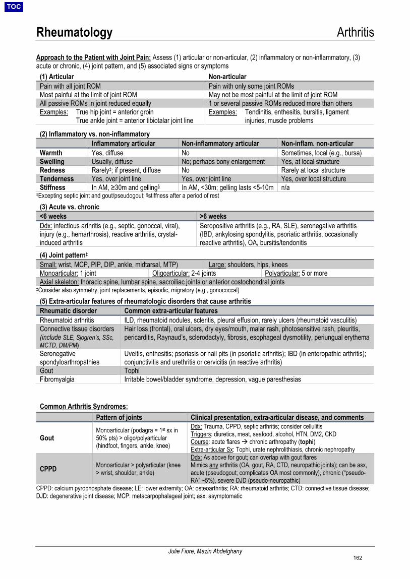
Rheumatology Arthritis
Julie Fiore, Mazin Abdelghany
Approach to the Patient with Joint Pain: Assess (1) articular or non-articular, (2) inflammatory or non-inflammatory, (3) acute or chronic, (4) joint pattern, and (5) associated signs or symptoms
(1) Articular Non-articular Pain with all joint ROM Pain with only some joint ROMs Most painful at the limit of joint ROM May not be most painful at the limit of joint ROM All passive ROMs in joint reduced equally 1 or several passive ROMs reduced more than others Examples: True hip joint = anterior groin
True ankle joint = anterior tibiotalar joint line Examples: Tendinitis, enthesitis, bursitis, ligament
injuries, muscle problems
‡Excepting septic joint and gout/pseudogout; §stiffness after a period of rest (3) Acute vs. chronic<6 weeks >6 weeksDdx: infectious arthritis (e.g., septic, gonoccal, viral), injury (e.g., hemarthrosis), reactive arthritis, crystal-induced arthritis
Seropositive arthritis (e.g., RA, SLE), seronegative arthritis (IBD, ankylosing spondylitis, psoriatic arthritis, occasionally reactive arthritis), OA, bursitis/tendonitis
(4) Joint pattern‡
Small: wrist, MCP, PIP, DIP, ankle, midtarsal, MTP) Large: shoulders, hips, knees Monoarticular: 1 joint Oligoarticular: 2-4 joints Polyarticular: 5 or more Axial skeleton: thoracic spine, lumbar spine, sacroiliac joints or anterior costochondral joints
‡Consider also symmetry, joint replacements, episodic, migratory (e.g., gonococcal) (5) Extra-articular features of rheumatologic disorders that cause arthritisRheumatic disorder Common extra-articular features Rheumatoid arthritis ILD, rheumatoid nodules, scleritis, pleural effusion, rarely ulcers (rheumatoid vasculitis) Connective tissue disorders (include SLE, Sjogren’s, SSc, MCTD, DM/PM)
Hair loss (frontal), oral ulcers, dry eyes/mouth, malar rash, photosensitive rash, pleuritis, pericarditis, Raynaud’s, sclerodactyly, fibrosis, esophageal dysmotility, periungual erythema
Seronegative spondyloarthropathies
Uveitis, enthesitis; psoriasis or nail pits (in psoriatic arthritis); IBD (in enteropathic arthritis); conjunctivitis and urethritis or cervicitis (in reactive arthritis)
Gout Tophi Fibromyalgia Irritable bowel/bladder syndrome, depression, vague paresthesias
Common Arthritis Syndromes:
Pattern of joints Clinical presentation, extra-articular disease, and comments
Gout Monoarticular (podagra = 1st sx in 50% pts) > oligo/polyarticular (hindfoot, fingers, ankle, knee)
Ddx: Trauma, CPPD, septic arthritis; consider cellulitis Triggers: diuretics, meat, seafood, alcohol, HTN, DM2, CKD Course: acute flares chronic arthropathy (tophi) Extra-articular Sx: Tophi, urate nephrolithiasis, chronic nephropathy
CPPD Monoarticular > polyarticular (knee > wrist, shoulder, ankle)
Ddx: As above for gout; can overlap with gout flares Mimics any arthritis (OA, gout, RA, CTD, neuropathic joints); can be asx, acute (pseudogout; complicates OA most commonly), chronic (“pseudo-RA” ~5%), severe DJD (pseudo-neuropathic)
CPPD: calcium pyrophosphate disease; LE: lower extremity; OA: osteoarthritis; RA: rheumatoid arthritis; CTD: connective tissue disease; DJD: degenerative joint disease; MCP: metacarpophalageal joint; asx: asymptomatic
(2) Inflammatory vs. non-inflammatoryInflammatory articular Non-inflammatory articular Non-inflam. non-articular
Warmth Yes, diffuse No Sometimes, local (e.g., bursa) Swelling Usually, diffuse No; perhaps bony enlargement Yes, at local structure Redness Rarely‡; if present, diffuse No Rarely at local structure Tenderness Yes, over joint line Yes, over joint line Yes, over local structure Stiffness In AM, ≥30m and gelling§ In AM, <30m; gelling lasts <5-10m n/a
162

Rheumatology Arthritis
Julie Fiore, Mazin Abdelghany
(Cont.) Pattern of joints Clinical presentation, extra-articular disease, and comments
RA Symmetric, pred. small (peripheral) polyarthritis (MCP, PIP, wrists, MTP)
Ddx: HCV/HBV, Lyme, crystal, SLE, psoriatic arthritis, sarcoid arthropathy. Typical Sx: palindromic rheumatism (episodic sx, migratory, 30-60% develop RA), monoarthritis (eventual polyarthritis in days-wks) Extra-articular: Anemia, rheumatoid nodules, ILD, pleural effusion, episcleritis/scleritis, splenomegaly (Felty’s), a/w Sjogren’s, rarely vasculitis
Ankylosing spondylitis* Spine and SI joints Gradual onset; low back pain, buttock pain, impaired spine mobility
Extra-articular: Synovitis, enthesitis, dactylitis, uveitis, psoriasis, IBD Psoriatic arthritis*
See 5 patterns in right column; axial (spine) involvement 42%
Ddx: RA, OA. 70% w/ psoriasis; 5 patterns: distal (DIP), asymm. oligo., symm. poly., arthritis mutilans, spondyloarthritis (sacroiliitis, spondylitis). Extra-articular: Synovitis, enthesitis, dactylitis, nail pits/onycholysis, uveitis
Enteropathic arthritis*
Axial (spondylitis/ sacroiliitis) and/or periph
Ddx: Behçet’s, celiac, pseudomembranous colitis, Whipple’s, parasitic. 6-46% of IBD pts. Extra-articular: Enthesitis, dactylitis, E nodosum
Reactive arthritis*
Asymm. oligo > mono > small polyarthritis; LE slightly > UE
1-4 wks post-infxn, a/w enteric: Salmonella, Shigella, Yersinia,Campylobacter, C. diff; GU: Chlamydia, E. coli, Ureaplasma, MycoplasmaExtra-articular: Conjunctivitis, GU sx, oral ulcers, keratoderma, E nodosum
Septic arthritis Monoarth. (50% knee) Usu. hematogenous; pts w/ RA ↑risk; Staph > strep > GNRs Osteoarthritis Knees/hips, 1st MTP,
PIP, DIP, C- and L-spine Limits ROM, bony swell, joint deforms/unstab., stages (1) pain limits high-impact activity → (2) pain constant, affects ADLs → (3) intense pain
Other arthritis: Viral polyarthritis (mimics RA), CTD (e.g., SLE, Sjogren’s, SSc), vasculitis, adult-onset Still’s, sarcoidosis, osteochondroma, osteoid osteoma, pigmented villonodular synovitis, amyloidosis, hemophilia, sickle cell disease
*All Seronegative spondyloarthropathies should be considered in ddx for each other
Diagnosis Treatment (non-pharm management for all-PT/OT, exercise)
Gout
Arthrocentesis: Negatively birefringent needle-shaped crystals, 10k<WBC<100k, diagnostic score (Arch Intern Med 2010;170:1120); culture as septic arthritis can co-exist. If any suspicion for septic arthritis, start empirical abx until Cx negative.
Acute: Rx depends on pt’s comorbidities. Colchicine (1.2mg x1, 0.6mg 1h later, then 0.6mg QD until 2-3d after resolu), PO GC (pred 40mg until resolu., then taper), NSAIDs (until 1-2d after resolu. [usu. 5-7d]), or intra-articular GC injection Chronic: Urate lowering if: ≥2 attacks/yr, CKD, urate nephrolithiasis, tophi (urate goal <6); Δdiet; d/c diuretics. Do not stop urate lowering therapy during acute attack.
CPPD Arthrocentesis: Small pos. birefringent rhomboid crystals, 10k<WBC<100k; XR: chondrocalcinosis, crowned dens
Acute: ≤2 joints → intra-articular GC injection 1st line; 2nd is same as gout (prefer colchicine w/in 24h of sx onset) Chronic: Consider HCQ, low-dose GC, MTX
RA Exclude other dz (esp. psoriatic, viral, polyarticular gout/CPPD, SLE). RF (70% sn, 85% sp), anti-CCP (75% sn, 95% sp), 30% ANA+; extremity XRs.
Acute/flares: GC or NSAIDs and initiate DMARD if not on Chronic: DMARD (MTX > HCQ, SSZ, leflunomide); if pt fails monotherapy, consider combination; if fails combo, transition to biologic (infliximab, abatacept, tocilizumab)
Ankylosing spondylitis*
Sacroiliitis (XR or MRI), LBP, ↑ESR/CRP, HLA-B27 (90% sn/sp)
NSAIDs 1st line, no GC, DMARDs not effective TNFα inh. 2nd (infliximab, etanercept, adalimumab)
Psoriatic arthritis*
Clinical dx, ↑ ESR/CRP (40%), HLA-B27, CASPAR criteria (91%sn;99%sp)
NSAIDs 1st line; if mod/sev, MTX > SSZ, leflunomide Sev. and erosive: TNFα inh. (inflix, adalimumab, golimumab)
Enteropathic arthritis*
Joint pain, LBP, always exclude septic arthritis, HLA-B27 (50-75%)
Usually improves w/ Rx of IBD. NSAIDs 1st line, d/w GI as can ↑IBD inflam. 2nd SSZ > MTX, azathioprine. 3rd TNFα inh.
Reactive arthritis*
Presence of preceding infection, arthrocentesis, stool culture, GC/Chla
Treat GU infxn; GI infxn may not need Rx. Acute: 1st NSAIDs, 2nd intra-articular GC, 3rd PO GC. Chronic: >6mo, SSZ>MTX
Septic arthritis Arthrocent: Fluid GS/Cx, joint WBC usually 50-150k
Antibiotics (3-4 wks) and joint drainage/wash out (ortho c/s)
Osteoarthritis Clinical, age>45, AM stiff <30m, slow progression, no warmth, musc wasting
PT, braces, PRN NSAIDs, consider duloxetine (60-120mg), intra-articular GC injection, severe sx → refer to ortho
*Seronegative spondyloarthropathies; GC: glucocorticoid; MTX: methotrexate; CTD: connective tissue disease; SLE: systemic lupuserythematosus; HCQ: hydroxychloroquine; SSZ: sulfasalazine; LBP: low back pain
163

Rheumatology Connective Tissue Diseases
Sneha Kannan, Avanthi Raghavan
Dz Clinical presentation Work-up Treatment Complications SL
E Discoid/malar rash (spares nasolabial fold), photosensitivity, serositis, nephritis, oral/nasal ulcers, psychosis, arthritis, cytopenias, constitutional sx (fever, weight loss, fatigue)
+ANA, +anti-dsDNA (up to70% pts, a/w SLE activity andlupus nephritis), +anti-Sm(30% pts, high specificity,remains + in remission), +anti-RNP (25%), +anti-SS-A/Ro(30%), +anti-SS-B/La, (20%).2012 SLICC criteria (ArthritisRheum 2012; 64: 2677)
Initial: hydroxychloroquine/ chloroquine for all ± steroids 2nd line: MMF, AZA, MTX, RTX, cyclophosphamide
- High riskVTE/ATE- CNS, Renal dz- 40% w/ APLAS- Osteonecrosis(both 2/2 SLEand steroids)
Sjog
ren’
s
Sicca sx (dry mouth/eyes), caries, parotid enlargement, vasculitis, interstitial nephritis, neuropathy, cytopenias
+ANA, +anti-SS-A (Ro, 70%pts), +anti-SS-B (La, 50-70%),+RFconsider salivary biopsy
Sicca only: sx mgmt Systemic: Hydroxychloroquine/ chloroquine, MTX, AZA, RTX, cyclophosphamide, glucocorticoids
5-10% lifetimerisk of NHL
Myos
itis (
polym
yosit
is, d
erm
atom
yosit
is,
inclu
sion
body
myo
sitis)
Proximal > distal muscle weakness: difficulty with stairs, standing from seated position, reaching above head
Extramuscular: constitutional sx, arthralgias, dysphagia, pulm. sx inc. cough and DOE (ILD), HTN, DM2
DM skin findings: heliotrope rash, poikiloderma (chest: V-sign; back: shawl sign; thigh: Holster sign), Gottron’s papules, panniculitis DM sine myositis: skin features w/o muscle weakness, a/w severe ILD (10-20% of DM)
Abs: +anti-Jo1 (a/w ILD, mechanic hands, arthritis), +anti-Mi2 (15-20%, a/w acuteonset, shawl sign, goodprognosis), +anti-MDA5
Labs: CK, aldolase (J Rheumatol 2003;30:2077)
Muscle biopsy DM: CD4 cells PM/IBM: CD8 cells
Initiation: prednisone 1mg/kg (up to 100mg/d) x4-6wk, then taper
Maintenance: AZA/MTX
Resistant/severe: pulse steroids, AZA, MTX, MMF, IVIG, RTX, cyclophosphamide (if ILD)
(DM) occult malignancy (9-32% incidence): commonly ovarian, breast, colon, lung, NHL, nasopharyngeal cancer
Additional: ILD in 10%; upper esophageal involvement; increased risk of MI
Syst
emic
Scler
osis
(aka
scler
oder
ma)
Localized: (affects skin only): linear (e.g., en coup de sabre), morphea
Systemic: may be limited cutaneous [lcSSc] (67% pts; skin thickening in hands/face only, commonly with CREST sx) or diffuse cutaneous [dcSSc] (33% pts; diffuse skin thickening, more significant multi-organ dz, less commonly with CREST sx)
CREST sx: Calcific nodules, Raynaud’s, Esophageal dysmotility, Sclerodactyly, Telangiectasias
Other systemic sx: renal crisis, ILD (>70%), PAH (10-40%)
NB: systemic sclerosis sine sclerodermapts with scleroderma but w/o skin findings
+anti-centromere (a/w lcSSc,only seen in 5% pts withdcSSc)+anti-Scl-70 (a/w dcSSc),+RNA-pol-III (a/w dcSSc andscleroderma renal crisis),+fibrillarin (a/w severe dcSSc,esp. in African-Americans(Arthritis Rheum 1996;39:1151)
Skin: MMF, MTX GI: PPIs, motility agents Lung: CCBs, endothelin-1 antag., PDE inhibitors, prostacyclin agonists MSK: Low dose pred, hydroxychloroquine, MTX Raynaud’s: CCBs
Increased risk of multiple cancers
Scleroderma renal crisis (up to 20%): AKI, abrupt HTN; a/w anti-RNA-pol III; treat with ACEi (captopril) + avoid steroids
MCTD
Overlap of SLE, systemic sclerosis, and polymyositis; Raynaud’s; non-erosive arthritis;
+anti-RNP by definition SLE features: glucocorticoids, RTX Scleroderma features: less responsive to steroids
Main cause of death is PAH
UCTD
/Ove
rlap
synd
rom
es Early Raynaud’s, ILD, inflammatory
arthritis not meeting classification criteria for RA, non-specific rash
Diagnosis of exclusion; does not meet criteria for diagnosis of specific disease
Managed according to symptoms
According to dominant clinical presentation
164
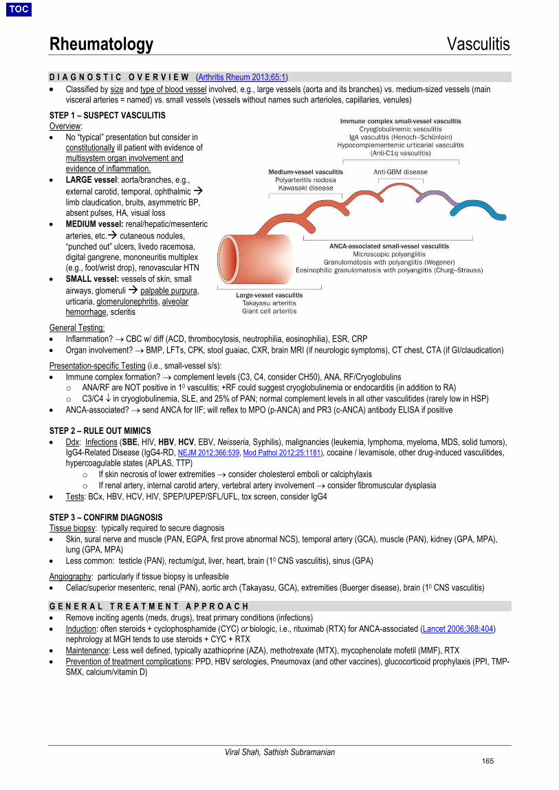
Rheumatology Vasculitis
Viral Shah, Sathish Subramanian
D I A G N O S T I C O V E R V I E W (Arthritis Rheum 2013;65:1) • Classified by size and type of blood vessel involved, e.g., large vessels (aorta and its branches) vs. medium-sized vessels (main
visceral arteries = named) vs. small vessels (vessels without names such arterioles, capillaries, venules) STEP 1 – SUSPECT VASCULITIS Overview: • No “typical” presentation but consider in
constitutionally ill patient with evidence ofmultisystem organ involvement andevidence of inflammation.
• LARGE vessel: aorta/branches, e.g.,external carotid, temporal, ophthalmic limb claudication, bruits, asymmetric BP,absent pulses, HA, visual loss
• MEDIUM vessel: renal/hepatic/mesentericarteries, etc. cutaneous nodules,“punched out” ulcers, livedo racemosa,digital gangrene, mononeuritis multiplex(e.g., foot/wrist drop), renovascular HTN
• SMALL vessel: vessels of skin, smallairways, glomeruli palpable purpura,urticaria, glomerulonephritis, alveolarhemorrhage, scleritis
General Testing: • Inflammation? → CBC w/ diff (ACD, thrombocytosis, neutrophilia, eosinophilia), ESR, CRP• Organ involvement? → BMP, LFTs, CPK, stool guaiac, CXR, brain MRI (if neurologic symptoms), CT chest, CTA (if GI/claudication)Presentation-specific Testing (i.e., small-vessel s/s):• Immune complex formation? → complement levels (C3, C4, consider CH50), ANA, RF/Cryoglobulins
o ANA/RF are NOT positive in 10 vasculitis; +RF could suggest cryoglobulinemia or endocarditis (in addition to RA)o C3/C4 ↓ in cryoglobulinemia, SLE, and 25% of PAN; normal complement levels in all other vasculitides (rarely low in HSP)
• ANCA-associated? → send ANCA for IIF; will reflex to MPO (p-ANCA) and PR3 (c-ANCA) antibody ELISA if positive
STEP 2 – RULE OUT MIMICS • Ddx: Infections (SBE, HIV, HBV, HCV, EBV, Neisseria, Syphilis), malignancies (leukemia, lymphoma, myeloma, MDS, solid tumors),
IgG4-Related Disease (IgG4-RD, NEJM 2012;366:539, Mod Pathol 2012;25:1181), cocaine / levamisole, other drug-induced vasculitides,hypercoagulable states (APLAS, TTP)
o If skin necrosis of lower extremities → consider cholesterol emboli or calciphylaxiso If renal artery, internal carotid artery, vertebral artery involvement → consider fibromuscular dysplasia
• Tests: BCx, HBV, HCV, HIV, SPEP/UPEP/SFL/UFL, tox screen, consider IgG4
STEP 3 – CONFIRM DIAGNOSIS Tissue biopsy: typically required to secure diagnosis • Skin, sural nerve and muscle (PAN, EGPA, first prove abnormal NCS), temporal artery (GCA), muscle (PAN), kidney (GPA, MPA),
lung (GPA, MPA)• Less common: testicle (PAN), rectum/gut, liver, heart, brain (10 CNS vasculitis), sinus (GPA)Angiography: particularly if tissue biopsy is unfeasible• Celiac/superior mesenteric, renal (PAN), aortic arch (Takayasu, GCA), extremities (Buerger disease), brain (10 CNS vasculitis)
G E N E R A L T R E A T M E N T A P P R O A C H• Remove inciting agents (meds, drugs), treat primary conditions (infections)• Induction: often steroids + cyclophosphamide (CYC) or biologic, i.e., rituximab (RTX) for ANCA-associated (Lancet 2006;368:404)
nephrology at MGH tends to use steroids + CYC + RTX• Maintenance: Less well defined, typically azathioprine (AZA), methotrexate (MTX), mycophenolate mofetil (MMF), RTX• Prevention of treatment complications: PPD, HBV serologies, Pneumovax (and other vaccines), glucocorticoid prophylaxis (PPI, TMP-
SMX, calcium/vitamin D)
165
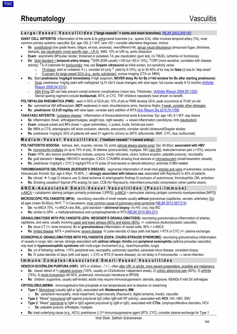
Rheumatology Vasculitis
Viral Shah, Sathish Subramanian
L a r g e - V e s s e l V a s c u l i t i d e s (“large vessels” = aorta and main branches), NEJM 2003;349:160GIANT CELL ARTERITIS: Inflammation of the aorta & its extracranial branches (i.e., spares ICA), often involves temporal artery (TA), most common primary systemic vasculitis; Epi: age >50, 2:1 M:F, rare <50 = consider alternative diagnoses, mimics. • Sx: constitutional (low grade fevers, fatigue, wt loss, anorexia), new/different HA, abrupt visual disturbance (amaurosis fugax, blindness,
diplopia), jaw claudication (most specific sign – LR 4), AMS, 10% w/ URI sx, aortic dissection• Exam: asymmetric BP/pulse; tender, thickened or pulseless TA; jaw claudication (gum test, r/o TMJD), ischemia on fundoscopy• Dx: Gold standard = temporal artery biopsy; ↑ESR (ESR usually >100 but <50 in 10%), ↑CRP (more sensitive, correlates with disease
activity); ↑IL-6 (rationale for tocilizumab); may use Doppler ultrasound as initial screen, but sensitivity varieso TA biopsy: start w/ unilateral; if (-), consider b/l (only ↑ yield by 5-10%); up to 30-45% of bx may be false (-) due to “skip areas”o If concern for large-vessel GCA (e.g., aorta, subclavian): pursue imaging (CTA vs. MRA)
• Rx: Start prednisone 1mg/kg/d immediately if high suspicion; NEVER delay Rx for Bx (>14d window for Bx after starting prednisone)- Dose: prednisone 1mg/kg (start with methylpred 1g IV x3d if visual changes) with slow taper; full course usually 9-12 months (Arthritis
Rheum 2006;54:3310)- ASA 81mg QD can help prevent cranial ischemic complications (vision loss, TIA/stroke). (Arthritis Rheum 2004;50:1332)- Steroid sparing regimens include tocilizumab, MTX, or CYC. TNF inhibitors repeatedly have shown no benefit.
POLYMYALGIA RHEUMATICA (PMR): seen in 50% of GCA pts; 10% of pts w/ PMR develop GCA, peak occurrence at 70-80 yrs old • Sx: symmetrical AM stiffness/pain (NOT weakness) in neck, shoulders/prox arms, hips/prox thighs; if weak, consider other etiologies• Rx: prednisone 12.5-25 mg/day with slow taper, consider early addition of MTX (Ann Rheum Dis 2015;74:1799)TAKAYASU ARTERITIS: “pulseless disease,” inflammation of thoracoabdominal aorta & branches: Epi: age <40, 8:1 M:F, esp Asians• Sx: inflammation (fever, arthralgias/myalgias, weight loss, night sweats) → vessel inflammation (carotidynia, limb claudication)• Exam: unequal pulses and BPs (lower > upper extremities), ↓ pulses, bruits, formal eye exam• Dx: MRA or CTA; arteriography will show occlusion, stenosis, aneurysms; consider carotid ultrasound/Doppler studies• Rx: prednisone 1mg/kg/d; 50% of patients will need 2nd agent for chronic sx (MTX, leflunomide, MMF, CYC, Aza, tocilizumab)M e d i u m - V e s s e l V a s c u l i t i d e s (“medium vessels” = named artery) POLYARTERITIS NODOSA: kidneys, skin, muscles, nerves, GI, joints (almost always spares lung); Epi: 40-60yo; associated with HBV • Sx: mononeuritis multiplex (in up to 70% of pts), GI distress (post-prandial), myalgias, AKI (rare GN), testicular/ovarian pain (>10%), seizures• Exam: HTN, skin lesions (erythematous nodules, purpura, livedo reticularis, ulcers, bullous eruption, palpable purpura), neuropathy• Dx: gold standard = biopsy; HBV/HCV serologies, C3/C4, CTA/MRA showing focal stenosis or microaneurysm (renal/mesenteric vessels)• Rx: prednisone 1mg/kg/d ± CYC 2 mg/kg/d PO or IV pulse (if mod-severe or steroid-refractory); antivirals if HBV-relatedTHROMBOANGIITIS OBLITERANS (BUERGER’S DISEASE): segmental inflammation of small-med arteries and veins of extremities; occlusive intravascular thrombi; Epi: age ≤ 45yo, 70-90% ♂, strongly associated with tobacco use; associated with Raynaud’s in 40% of patients • Dx: clinical 1) age 2) tobacco use 3) distal ischemia 4) arteriographic findings 5) exclusion of autoimmune, thrombophilia, DM, embolism• Rx: Smoking cessation! Iloprost (PG analog) for pain; CCB (for Raynaud’s); intermittent pneumatic compression (when painful ulcers)A N C A - A s s o c i a t e d S m a l l - V e s s e l V a s c u l i t i d e s ( P a u c i - I m m u n e )c-ANCA = cytoplasmic staining (antigen primarily proteinase 3 [PR3]); p-ANCA = perinuclear staining (antigen commonly myeloperoxidase [MPO])MICROSCOPIC POLYANGIITIS (MPA): necrotizing vasculitis of small vessels usually without granulomas (capillaries, venules, arterioles); Epi: all ages (mean 50-60yo), M>F, ↑ in Caucasians; most common cause of pulmonary-renal syndrome (NEJM 2012;367:214) • Dx: +p-ANCA 70%, +c-ANCA rare, BAL, gold standard = skin/renal biopsy; r/o HIV, cryo, hep B/C• Rx: similar to GPA → methylprednisolone and cyclophosphamide or RTX (NEJM 2010;363:221)GRANULOMATOSIS WITH POLYANGIITIS (GPA; WEGENER’S GRANULOMATOSIS): necrotizing granulomatous inflammation of arteries, capillaries, and veins, usually involving upper and lower airways (90%) and kidney (80%), +/- cutaneous (leukocytoclastic vasculitis) • Dx: sinus CT (+/- bone erosions), Bx w/ granulomatous inflammation of vessel walls, 90% + c-ANCA• Rx: limited disease: MTX + prednisone; severe disease: IV pulse steroids x3 days (with oral taper) + RTX or CYC (+/- plasma exchange)EOSINOPHILIC GRANULOMATOSIS WITH POLYANGIITIS (EGPA; CHURG-STRAUSS SYNDROME): necrotizing granulomatous inflammation of vessels in lungs, skin, nerves; strongly associated with asthma /allergic rhinitis and peripheral eosinophilia (asthma precedes vasculitis); may lead to hypereosinophilic syndrome with multi-organ involvement (e.g., heart/myocarditis, lungs) • Dx: >4 of following: asthma, >10% peripheral eos, neuropathy, pulmonary opacities, paranasal sinus disease, consistent biopsy• Rx: IV pulse steroids x3 days (with oral taper) ± CYC or RTX (if severe disease); do not delay rx if mononeuritis → nerve infarctionI m m u n e C o m p l e x - A s s o c i a t e d S m a l l - V e s s e l V a s c u l i t i d e sHENOCH-SCHÖNLEIN PURPURA (HSP): 90% in children; ♂>♀; often after URI; in adults, more severe presentation, possible a/w malignancy • Sx: classic tetrad of 1) palpable purpura (100%, usually on LEs/buttocks =dependent areas), 2) colicky abdominal pain (60%), 3) arthritis
(75%), 4) renal involvement (40-50%, proteinuria, microscopic hematuria to RPGN)• Rx: children: supportive, usually self-limited; adults may require immunosuppression: steroids, dapsone. NSAIDs if mild GI/ arthralgiasCRYOGLOBULINEMIA: immunoglobulins that precipitate at low temperatures and re-dissolve on rewarming• Type 1: Monoclonal (usually IgM or IgG), associated with Waldenstrom’s, MM
o Sx: peripheral neuropathy, renal impairment, hyperviscosity (Raynaud’s, digital ischemia, livedo), vasculitis• Type 2: “Mixed” monoclonal IgM against polyclonal IgG (often IgM with RF activity), associated with HCV, HIV, HBV, EBV• Type 3: “Mixed” polyclonal Ig (IgM or IgG) against polyclonal Ig (IgM or IgG), associated with CTDs, lymphoproliferative disorders, HCV
o Sx: palpable purpura, arthralgias, myalgias• Rx: treat underlying cause (e.g., HCV); prednisone ± 2nd immunosuppressive agent (RTX, CYC); consider plasma exchange for Type 1
166
Rheumatology Miscellaneous Rheumatologic Diseases
Yousef Badran
Behcet’s Disease: Autoinflammatory condition characterized by recurrent aphthae, vasculitis, and skin/GI/neuro/joint sx • Epi: W > M, 20-40 y/o, ↑ Turkey, Middle East, and Asian countries• Sx: Recurrent painful oral ulcers and ≥2 of the following: painful genital ulcers (specific), ocular disease (most commonly
uveitis or retinitis]), skin lesions (pustules, folliculitis, papules, erythema nodosum), negative pathergy test (skin pustuleformation to needle prick [NB: not sensitive in Caucasians])Other manifestations include: GI (similar to IBD), neurologic disease (both parenchymal and extra-parenchymal), vasculardisease (ATE/VTE, vasculitis [small/medium/large vessels], aneurysms [esp. pulmonary artery, OJRD 2011;6:15]) orarthritis (nonerosive, asymmetric). Less common: kidneys, heart, lung disease
• Dx: Clinical dx only, no specific laboratory tests exist; may have ↑ ESR/CRP• Rx (Ann Rheum Dis. 2018; 77:808)
o Mild (arthritis, ulcers): Colchicine 1-2 mg daily, low dose prednisone. Apremilast (PDE-4 inhibitor) for ulcers (NEJM2015;372:1510)
o Severe: Prednisone 1mg/kg/d; may add 2nd line agents: AZA, anti-TNF, IFNα, CYC, CP, MTX. If organ failure (esp.ophthalmic involvement): IV pulse steroids x3 days
Familial Mediterranean Fever (FMF): Autoinflammatory disorder due to mutations in MEFV gene; autosomal recessive inheritance; characterized by recurrent bouts of fever and serosal inflammation • Epi: Most common in Jews, Armenians, Turks, and Arabs. Onset <10 yrs old (65% pts), <20 yrs old (90% pts)• Sx: Recurrent acute attacks (1-3 days, resolve spontaneously) of fever associated w/ peritonitis (often mistaken for surgical
abdomen), unilateral pleuritis, arthritis (monoarticular, sterile joint), or skin lesions (erysipelas-like). Other manifestationsinclude: exertional myalgia, pericarditis, testicular pain, and aseptic meningitis.o Long-term complications: Secondary (AA) amyloidosis: renal disease (major cause of mortality), SBO, infertility
• Dx: During acute attack: ↑ WBC, ↑ ESR/CRP. Check UA for amyloidosis (proteinuria). Genetic testing for confirmation.Diagnostic criteria: Livenh et al. Requires 1 major or 2 minor criteria. (Arthritis Rheum 1997; 40:1879)
• Rx: Colchicine 1-3 mg/day (to prevent acute attacks and progression to amyloidosis). 5-10% colchicine resistant, add onIL-1 inhibitors.
Adult Onset Still’s Disease (AOSD): Systemic inflammatory disorder characterized by fevers, arthritis, and rash. Can present as single episode (wks-mos), multiple flares, or be persistently active. • Epi: W = M. Bimodal 15-25 yrs old and 36-46 yrs old• Sx: fever; arthralgias; evanescent, salmon-colored maculopapular rash that coincides w/ fever, usually on the trunk,
may be precipitated by trauma (Koebner phenomenon); pericarditis; pleural effusions; macrophage activating syndrome• Dx: Yamaguchi criteria requires >5 features, including >2 major criteria (J Rheumatol 1992;19:424):
o Major: Fever ≥39ºC for ≥1 week, arthralgias/arthritis ≥2 weeks, salmon-colored rash, ↑ WBC (≥10K + ≥80% PMN)o Minor: Sore throat, LAD, HSM, ↑AST/ALT, ↑ LDH, negative ANA/RFo Other labs (not part of criteria): ↑ ESR/CRP, ferritin >3000 ng/mL (if >10,000, consider MAS spectrum), ↑ plt, ↓ Hgb
• Rx:o Mild: NSAIDso Severe: Prednisone 0.5-1mg/kg/d (may not respond). If uncontrolled: MTX, anti-TNF, anti-IL6R, anti-IL1
Fibromyalgia: NOT a rheumatic disease. Chronic widespread musculoskeletal pain, often w/ fatigue, sleep disturbance, and multiple somatic symptoms. • Epi: W > M, generalized MSK pain in often ♀ 20-55 years of age. Often coexists with other inflammatory diseases like
SLE, other CTDs, RA. Often psychiatric comorbidities.• Sx: widespread MSK pain, fatigue, cognitive disturbance (decreased attention & ability to perform complex tasks),
psychiatric sx (depression), headache, parasthesias, IBS. Pan-positive ROS not uncommon.• Dx: clinical diagnosis, multiple tender points often used to assess, but specific number NOT needed for diagnosis.
Newer criteria involve widespread pain index (WPI) and symptom severity (SS) scale (Arthritis Care Res (Hoboken) 2010;62:600)o Labs: normal ESR, CRP, TSH, CBC, BMP
• Rx: Initial therapy: patient education, exercise program; Pharmacologic therapy: first-line includes amitriptyline,duloxetine, or milnacipran; also may consider cyclobenzaprine, gabapentin, and pregabalin (monotherapy > combotherapy). Avoid narcotics.
167

Rheumatology Rheumatologic Medications
Jules Elofson
DRUG/CLASS INDICATIONS COMMON TOXICITIES Azathioprine (AZA; Imuran)
NB: 6-MP is downstream metabolite of AZA
DM/PM, RA, SLE nephritis, vasculitis
GI, bruising, myelosuppression, lymphoproliferative d/o, hepatotoxicity; test for TPMT deficiency as low levels can ↑ toxicity (TPMT metabolizes 6-MP to inactive metabolites deficiency increases circulating 6-MP levels); do not give with xanthine oxidaseinhibitors (allopurinol, febuxostat)
Cyclophosphamide (CYC; Cytoxan) SLE (LN), vasculitis (most severe)
myelosuppression, hemorrhagic cystitis (MESNA for ppx), lymphoma, infertility (cumulative dose, leuprolide ppx)
Hydroxychloroquine (HCQ; Plaquenil) RA, SLE, Sjogren’s N/V, retinopathy (q1y retinal exam), dizziness, alopecia, myelosuppression; G6PD
Leflunomide (LFM; Arava) PsA, RA N/V, alopecia, rash, diarrhea, HTN, hepatotoxicity, URI, dizziness/HA, teratogen
Methotrexate (MTX; Rheumatrex, Trexall, Otrexup, Rasuvo)
RA (first line), PsA myelosuppression, hepatotoxicity (give with folate), pneumonitis, stomatitis, rash, teratogen
Mycophenolate Mofetil (MMF; CellCept)
AAV, DM/PM, PsA, Scleroderma, SLE
Cardiac (HTN, edema, CP, tachycardia), HA, insomnia, diarrhea, rash, pain, fever, stomatitis
Sulfasalazine (5-ASA; Azulfidine) AS, IBD, JRA, psoriasis, RA
sore throat, stomatitis, myelosuppression, N/V, rash, HA
Apremilast (Otezla); PDE4 inhibitor PsA, severe psoriasis N/D, URI, depression Tofacitinib (Xeljanz); JAK inhibitor RA, AS, psoriasis Infection, lymphoma, diarrhea BIOLOGIC, non-TNFŦ Ŧ Abatacept (Orencia); CTLA-4 PsA, RA URI, HA, nausea, HTN, dizziness, dyspesia Anakinra (Kineret); anti-IL-1R AOSD/MAS, gout,
Schnitzler syndrome myelosuppression (neutropenia), rash/injection reactions, HA, arthralgia, fever
Belimumab (Benlysta); anti-BAFF SLE Depression, HA, infusion reaction, PML, GI Canakinumab (Ilaris); anti-IL-1b CAPS, CAD (CANTOS
trial) Infection, HA, vertigo, GI, MSK pain, nasopharyngitis
Rituximab (Rituxan); anti-CD20 APLAS, GPA/MPA, IgG4-RD, Scl-ILD, (SLE)
URI, HTN, infusion reaction, TLS, PML, fever, rash/pruritus, LE edema
Tocilizumab (Actemra); anti-IL-6R GCA, RA URI, hepatotoxicity, HLD Secukinumab (Cosentyx); anti-IL17A AS, PsA, psoriasis URI Ustekinumab (Stelara); anti-IL-12/23 PsA, psoriasis URI, PRESS, seizures IVIG APLAS, DM/PM, IBM,
IMNM, Kawasaki’s transfusion reactions/anaphylaxis, aseptic meningitis, thromboembolism, HA
BIOLOGIC, TNF inhibitionŦ Ŧ Adalimumab (Humira); anti-TNF AS, IBD, PsA, psoriasis,
RA HA, nausea, rash, URI, CPK elevation, infection, drug-induced lupus
Infliximab (Remicade); anti-TNF AS, IBD, PsA, psoriasis, RA
HA, nausea, diarrhea, ALT elevation, infection, drug-induced lupus
Golimumab (Simponi); anti-TNF AS, IBD, PsA, RA injection reactions, URI, drug-induced lupus Certolizumab (Cimzia); anti-TNF AS (axial), IBD, RA Nausea, infection, URI, drug-induced lupus (rare) Etanercept (Enbrel); sol. TNF-R AS, PsA, psoriasis, RA HA, rash, nausea, diarrhea, infection, okay w/ HCV;
drug-induced lupus Ŧ Ŧ All can cause HBV /TB reactivation (check hepatitis serologies, PPD and/or IGRA prior to starting); if positive, start antiviral prophylaxis with entecavir (HBV reactivation) and prophylaxis with INH (latent tuberculosis) as per ID/rheum. (NB: TNF-alpha inhibitors are safe in HCV infectionmay even be beneficial, as TNF-alpha promotes liver fibrosis, Expert Opin Biol Ther 2012;12:193)
AAV (ANCA-associated vasculitis), AOSD (Adult-onset Still's disease), APLAS (Anti-phospholipid antibody syndrome), AS (Ankylosing spondylitis), DM (dermatomyositis), EGPA (eosinophilic granulomatosis with polyangiitis), GCA (giant cell arteritis), GPA (Granulomatosis with polyangiitis), IBD (inflammatory bowel disease), IMNM (Immune-mediated necrotizing myopathy), JRA (juvenile rheumatoid arthritis), MAS (Macrophage activating syndrome), MPA (Microscopic polyangiitis), PM (polymyositis), PsA (Psoriatic Arthritis), RA (rheumatoid arthritis), SLE (systemic lupus erythematosus), UC (Ulc. colitis)
168

Endocrinology Outpatient Type 2 Diabetes Management
Max Petersen
Pre-Diabetes (Diab Care 2019:42:S13) • Diagnosis: A1c 5.7-6.4%; fasting plasma glucose (FPG) 100-125; or OGTT w/ 2hr plasma glucose (PG) 140-199• Monitoring: A1c at least q1y; if A1c 6-6.4%, screen q6mo (25-50% 5-year risk of progression to diabetes if A1c 6-6.5%)• Treatment: Lifestyle interventions most effective; metformin also effective, esp. if BMI > 35 or age <45 (DPP, NEJM 2002;346:393)
Diabetes (Diab Care 2019:42:S13)• Diagnosis: A1c > 6.5%; FPG >126; 75g OGTT with 2hr PG >200; or random glucose >200 & symptoms. Unless diagnosis is made by
symptoms & random glucose >200, confirm with repeat or additional test. (NB: for T1DM, check TSH, celiac screen at diagnosis).• Treatment: goal A1c < 7%; liberalize to < 8-8.5% if life expectancy <10 years or high risk for hypoglycemia.
Screening: Beginning at age > 45 years OR if BMI > 25 (or > 23 in Asian-Americans) + risk factors (1st degree relative with DM, nonwhite, history of CVD, hypertension, HDL<35, triglycerides>250, PCOS, sedentary); screen q3y if normal (ADA Guidelines 2018)
Healthcare Maintenance for Diabetic Patients Every visit • Review blood sugar log goal AM fasting blood glucose 80-130
• Blood pressure goal SBP <140; ACEi/ARB first line• Weight, BMI weight center referral if BMI > 40 or > 35 with poor control; nutrition referral for all patients• Foot exam (inspect skin, joints, pulses, sensation) esp if known neuropathy or PVD; ABIs/vascular referral if PVD• Smoking cessation counseling (Advise, Assist, Arrange)
Q3-6mo • A1c Q6 months if controlled; Q3-6 months if A1c above targetAnnually • Lipids: moderate-intensity statin if 40-75 yrs old w/ ASCVD<7.5%; high-intensity statin if ASCVD>7.5%; ASA for 2°
prevention of CVD (limited evidence for 1° prevention)• Urine mAlb/Cr, BMP consider ACEi/ARB if hypertensive w/ either proteinuria or GFR<60; refer to renal if GFR<30• Monofilament exam if fail to feel at 4/10 specific sites, + for neuropathy (see PCOI for specific sites)• Retinopathy screen w/ dilated eye exam or retinal photography; can consider Q2-3yr if normal exam(s)• LFTs consider elastography and/or hepatology referral if elevated to evaluate for NASH
Vaccines • Influenza annually• Hepatitis B series if age <60 and not immune• PPSV23 x1 age <65; re-dose x1 >65 with at least 5 years between doses
Basal insulin management Criteria for initiation
• Consider if A1c ≥ 9%, random BG ≥ 300, fasting BG ≥ 250, or if symptomatic; also if < 65yo on two agents with A1c>8% (or ≥ 65yo and A1c > 8.5%) on two occasions at least three months apart; or when the A1c is quickly rising
• Able to perform self-monitoring with glucometer; consider referral to DM educatorInitial dose
• Starting dose: 0.1-0.2U/kg/day or 10U/day (if weight >80kg, may consider starting at 20U/day)• Choice of agent: choose long-acting (glargine, detemir QD) or intermediate-acting (NPH BID cheaper!)• Route: pen (easier to use, more expensive) vs. needle/syringe
Titration • Increase by 2-4U or 10-15% Q3 days until AM fasting BS is 80-130; savvy patients can self-titrate• If hypoglycemia occurs or FPG < 80 without clear reason, decrease dose by 10-20% or 4U, whichever is greater
Prandial insulin management Criteria • Consider if A1c still not at goal with basal insulin and fasting glucose within target range (80-130)Initial dose
• Strategy 1: Add 1 rapid-acting insulin before largest meal start w/ 4U or 0.1U/kg or 10% basal dose• Strategy 2: Change to mixed insulin (e.g. fixed 70/30, NPH + regular) BID (before breakfast and dinner). Divide
current basal dose into 2/3 AM, 1/3 PM or 1/2 AM, 1/2 PM. Counsel to avoid missing meals to avoid hypoglycemia.Titration • Increase dose by 1-2U or 10-15% q3d until target glucose reached (pre-prandial: 80-130; 1-2h post-prandial < 180)
• If A1c still not controlled: add rapid-acting insulin to another meal and titrate as above• If hypoglycemia occurs or FPG < 80 without clear reason, decrease dose by 10-20% or 4U, whichever is greater
Insulin supplies • Needles: Come as universal pen needles, or attached to syringes, made by many companies. 32G 4mm - less painful (higher gauge
thinner and shorter needle), but obese patients and high insulin doses often require deeper/wider needle.• Syringes: Boxes of 100. Pt on basal/bolus insulin needs 4 syringes/day (4 boxes/3 months). Pt on long-acting insulin only needs 1
box/3 months. Choose the smallest syringe that will hold the dose (smaller barrel clearer scale markings).Use this barrel size… With this dose range… 3/10 mL 30 units or less 1/2 mL 31-50 units1 mL 51-100 units
• Alcohol swabs (or patients can wash hands/skin with soap and water)• Glucometer & test strips: Many choices (insurance dependent), each with own strip brand. Most test strips come in boxes of 50-100.• ** All durable medical equipment including test strips and glucometers, require an ICD-10 code on the script itself **
169
Endocrinology Outpatient Type 2 Diabetes Management
Max Petersen
Non-Insulin Agents
Drug/Dose Range % ↓ A1c Contraindications Patients (pts) who
benefit/ Pros Side Effects/Considerations Cost*
Metformin: 1st line anti-diabetic medication; many effects, primary mechanism is decreasing hepatic glucose production Metformin (Glucophage)
500-1000mg BID1-2
GFR cutoffs: - <45ml/min don’t initiate- <30ml/min discontinue
- First line therapy- Weight loss- Improve lipids
- Diarrhea/GI (nausea, bloating)- B12 deficiency- Lactic acidosis in patients
with severe liver/renal disease;hold 48 hrs s/p IV contrast
$5 (IR) $8 (ER)
Metformin pearls: To increase adherence, warn patients about GI side effects and educate patients that side effects go away with time for most; can be minimized by taking WITH food. Start with 500mg daily and increase dose by 500mg each week to a dose of 1000mg BID; each dose increment helps – no diminishing returns. If GI side effects occur at a higher dose (side effects are dose-dependent), decrease and try at later date. Many patients tolerate the ER formulation better than IR. Insulin Secretagogues: stimulate release of insulin from pancreatic beta cells, thus only effective in pts who still have beta cell function Sulfonylureas: Glipizide 2.5-20mg QD Glimepiride 1-8mg QD
1-2- Severe renal or hepatic
impairment- NOTE: cross-reactivity
in pts with allergy tosulfa abx is low
- Affordable second oralagent
- Weight gain- Hypoglycemia (esp.
glyburide)- FDA Special Warning on ↑ CV
mortality
$5 - $10
Meglinitides: Repaglinide (Prandin)
0.25-4mg QAC 0.5- 0.7
- Mod to severe hepaticimpairment
- Concurrent gemfibroziltherapy
- Use like bolus insulin(short-acting)
- Pts with CKD- Pts at risk for nocturnal
hypoglycemia
- Weight gain- Avoid use with clopidogrel
(may lead to hypoglycemia)
$15 -$20
GLP-1 Receptor Agonists: stimulate glucose-dependent insulin release from beta cells, therefore lower risk of hypoglycemia Exenatide (Byetta)
5-10mg BIDLiraglutide (Victoza) 0.6-1.8 QD Dulaglutide (Trulicity) 0.75-1.5 Qwk
0.5-1.1
- FDA Black BoxWarning: risk ofthyroid C-cell tumors(e.g., medullary), soavoid if hx of thyroidcancer or if pt w/ MEN2
- ASCVD and kidneydisease benefits
- Can use in pts at highrisk for hypoglycemia
- Weight loss
- GI: n/v, diarrhea- Injection site reactions- Delayed gastric emptying- ↑ risk of pancreatitis
$600-$750
DPP-4 Inhibitors: inhibit degradation of DPP4, increasing glucose-dependent insulin secretion and decreasing glucagon secretion Sitagliptin (Januvia) 25mg-100mg QD Saxagliptin (Onglyza) 2.5-5mg QD Linagliptin (Tradjenta) 5mg QD
0.5-0.8
- No contraindications,but very weak
- Safe in pts withCKD/ESRD
- No hypoglycemia (safein elderly)
- Weight neutral
- Saxagliptin ↑hospitalizations for CHF
$375-$475
SGLT-2 Inhibitors: block renal glucose reabsorption, increasing glucosuria Canagliflozin (Invokana)
100-300mg QDEmpagliflozin (Jardiance)
5-10 mg QDDapagliflozin (Farxiga)
5-10 mg QD
0.8-0.9
GFR cutoffs: - <45ml/min don’t initiate- <30ml/min discontinue- History of bladder
cancer (dapagliflozin)
- Benefits for ASCVD,CHF and CKD
- Weight loss- Pts at risk for
hypoglycemia- ↓BP 3-5 mmHg
- FDA Black Box Warning: ↑risk of amputation (canagliflozin)
- UTI & genital fungalinfections
- Small risk of euglycemic DKA- Risk of dehydration/
hypotension- Risk of fracture (canagliflozin)- May ↑ LDL cholesterol
$475-$500
Thiazolidinediones: increase insulin sensitivity by acting on adipose, muscle, and liver to ↑glucose uptake, ↓ectopic lipid deposition Pioglitizone (Actos)
15-30mg QD 1-1.6
- Avoid use in pts w/history of bladdercancer
- Pts at risk forhypoglycemia
- Pts with CKD
- FDA Black Box Warning:increased risk of CHF
- Weight gain- Risk of fracture increased
$10
* Monthly costs in Boston area pharmacies (GoodRx)
PEARLS FOR CHOOSING A SECOND NON-INSULIN AGENT (Diab Care 2019;42:S90) • If prominent ASCVD: GLP-1RA or SGLT2i• If prominent heart failure or CKD (but eGFR > 45 mL/min): SGLT2i• If weight loss desired: GLP-1RA or SGLT2i• If cost is a major concern: SU or TZD
170
Endocrinology Inpatient Diabetes Management
Max Petersen
I N S U L I N N O M E N C L A T U R E
Basal insulin: fixed intermediate / long-acting for basic metabolic requirements Prandial insulin: fixed rapid / short-acting to cover meal carbohydrates Correctional insulin: sliding scale rapid / short-acting to correct hyperglycemia, BG > 150 mg/dL (graded scale of 1-2 units for each increment of 50 mg/dL) Pre-Mixes (avoid in hospital, but consider for transition to outpatient regimen): NPH/regular (Humulin 70/30, 50/50), protamine aspart/aspart (NovoLog 70/30); protamine lispro/lispro (Humalog 75/25, 50/50) Insulin pumps: consult diabetes/endocrine; pt may self-manage if alert and able IV insulin: Use in ICU if BG>180 x 2 and anticipated ICU LOS > 3 days; reference MICU Insulin Protocol in Partners Handbook. Ensure an active source of dextrose. Always overlap with SC insulin by 2-3h when stopping insulin gtt.
I N P A T I E N T M A N A G E M E N T (Diabetes Care 2019;42;S173)
Glycemic targets • Floor: fasting 100-140 mg/dL, random <180 mg/dL • ICU: 140-180 mg/dL (NOT stricter) (NICE-SUGAR, NEJM 2009;360:1283)
Check FSBG AC & QHS (at least for 24-48h) in (1) known diabetics, (2) non-diabetics with BG > 140 mg/dL, (3) those receiving therapies a/w hyperglycemia (corticosteroids, octreotide, tube feeds, TPN) * Note: FSBGs inaccurate in hypotension (esp. on pressors) and hypothermia due to altered blood flow to skin. Confirm w/ serum glucose.
Hypoglycemia • ↑ Risk: T1DM, malnutrition, emesis, ↓ body weight, ↓ PO intake, ↓ steroid dose, AKI (↓ insulin clearance), CKD (esp. dialysis) • Manifestations: < 70: shakiness, anxiety, diaphoresis, visual ∆, HA, AMS
< 55: seizure, coma Beware of hypoglycemia unawareness in T1DM and longstanding T2DM
• Tx: PO if able (15g gel, tabs, juice); IV (0.5-1 amp D50 = 12.5-25g), recheck in 15 min and chase with PO if due to insulin OD • Tip: If sulfonylurea OD → Tx: 50-75 mcg octreotide subQ • Review and adjust insulin regimen if hypoglycemia (BG<70) or BG<100 occurs while inpatient, even if asymptomatic
Admission Orders (NEJM 2006;355:1903) 1. Hold home oral antihyperglycemic agents (NEVER hold basal insulin for T1DM) 2. Continue home insulin regimen with dose reduction (~25-50% reduction) given expected change in diet while hospitalized.
Hypoglycemia is associated with increased mortality in elderly, so reasonable to be cautious. 3. If not on home insulin, and well controlled, reasonable to start with ISS and soon change to basal-bolus once TDD established 4. If not on home insulin, and not well controlled, start with basal-bolus!
TDD 0.5 U/kg/day (0.2-0.3 if age>75, lean, ESRD, ESLD, frail): 50% basal, 50% prandial + ISS 5. Check A1c in all patients with hyperglycemia if not done in last 3 months 6. If NPO: no need to ↓ basal if truly only covering basal needs, but this is unusual. Consider 50% dose reduction or 0.2 U/kg/day for
basal insulin. Be sure to change correctional ISS and FSBG from TID AC to q6h. 7. Correctional insulin sliding scale: use low-dose if insulin-sensitive/ESRD/ESLD/frail, otherwise moderate-dose for most T2DM
Adjusting Insulin Dosing: In general increase by no more than 20% of total daily insulin requirement every day
Special Situations: 1. Glucocorticoids: rule of thumb give NPH 0.1U/kg/d for every 10mg pred, up to 0.4U/kg/d; if dexamethasone, use glargine instead 2. Tube Feeds: If not on insulin already, start with RISS q6h. Convert to NPH BID based on needs. If on insulin, use ½ basal (NPH
BID) + ½ bolus (regular insulin q6h) + correctional. If TF stopped, give D5W at TF rate until next NPH dose, and ↓ NPH dose by 50% or more based on pre-TF insulin requirements. TPN: regular insulin can be added to TPN (discuss w/ nutrition), does not cover basal!
Disposition: If new home insulin → nutrition c/s + floor RN teaching and arrange outpatient f/u. Using discharge order set, send rx for glucometer, test strips, lancets, syringes/vials or pens/needles to MGH outpatient pharmacy and bring up to floor for RN teaching.
FORMULATIONS (§MGH formulary) Type (Onset) Name Peak Duration Rapid (10 min) lispro§ (Humalog), aspart (Novolog), glulisine (Apidra) 0.5-2.5 hr < 5 hr Short (30 min) regular§ (Humulin R, Novolin R) 2.5-5 hr 4-12 hr Intermediate (1-2 hr) NPH§ (Humulin N, Novolin N) BID 4-12 hr 12-18 hr Long (3-4 hr) glargine§ (Lantus) QD, detemir (Levemir) BID, degludec (Tresiba) QD none 24 hr
Fasting or AM BG high (w/ other BGs in range) → ↑ basal insulin dose* Fasting BG high + HS BG high (w/ other BGs in range) → ↑ pre-dinner prandial insulin dose Pre-lunch or dinner BG high (w/ other BGs in range) → ↑ prandial insulin dose of preceding meal BG rising steadily over course of day → ↑ prandial insulin dose at each meal
*Avoid titrating basal insulin more than q2-3d (d/t long half life, requires time to reach steady state) to avoid “stacking” and hypoglycemia
171

Endocrinology DKA & HHS
Max Petersen
D K A : D I A B E T I C K E T O A C I D O S I SPathophysiology: Think about each element of Diabetic Keto-Acidosis • Diabetes: ↓ insulin & ↑ opposing hormones (glucagon, catechols, cortisol) → hyperglycemia → osmotic diuresis• Ketones: ↓ insulin → ↑ lipolysis → ↑ free fatty acids → ↑ ketones [acetoacetate, β-hydroxybutyrate, acetone (fruity breath)]• Acidosis: ↑ β-hydroxybutyrate and acetoacetate, and contraction alkalosis with total body HCO3- deficit (NEJM 2015;372:546)Precipitants (the “I’s”): infection (30-40% of cases), initial presentation of DM (20-25% of cases), insulin non-adherence, inflammation (pancreatitis – but ↑ amylase / lipase in DKA even w/o this), ischemia/infarction (MI, CVA, gut), intoxication (EtOH, cocaine), iatrogenesis (e.g., SGLT2 inhibitors, steroids, thiazides, dobutamine/terbutaline, atypical anti-psychotics), infant (pregnancy) Presentation: dehydration, polydipsia/polyuria, N/V/abd pain, weakness, AMS, Kussmaul’s respirations, fruity breath (acetone) Dx: BG 250-800, pH <7.3, AG >10, urine/serum ketones. Consider euglycemic DKA in pt on SGLT2i, EtOH liver dz, pregnancy. • Check BMP, CBC + diff, UA, serum osm, serum β-hydroxybutyrate, ABG/VBG. Consider TnT, EKG, BCx, UCx, CXR, lipase/amylase.• Note: sodium correction represents what the sodium will be once glucose is corrected, NOT what the sodium currently is! When to
use absolute sodium value: when calculating anion gap. When to use corrected value: to assess for underlying hypotonic hypoNa .o Corrrection Formula: add 1.6 mEq/L for every 100 mg/dL of serum glucose > 100 mg/dL (e.g. if 300 mg/dL, add 3.2 mEq/L)
• Note: UA ketone does not test for β-hydroxybutyrate (BOHB), which is the predominant ketone in DKA (must measure from serum)Management: *Prioritize ABCs, volume status, identifying precipitant THEN electrolytes THEN glucose• Labs: BMP q2h until AG closes, then q4h until normal K+; VBG, β-hydroxybutyrate q2-4h; FSBG q1h while on insulin gtt• Step 1: Volume resuscitation (typically 5-8L deficit)
o Bolus NS (15-20cc/kg/hr) [unless CHF, ESLD, ESRD, hypoxemia] for initial resuscitationo Calculate corrected Na → if Na low, start NS ± K+ at 250-500cc/hr; if Na normal/high or hyperCl acidosis, start ½NS ± K+
at 250-500cc/hro Add D5 to IVF once BG<200 (DKA) or <300 (HHS)
• Step 2: Potassium repletionPotassium Action K<3.3 Give 20-40 mEq KCl IV per hour + hold insulin! 3.3<K<5.3 Add 20 mEq K to IVF K>5.3 Continue to monitor q2h
• Step 3: Insulin therapy (Diab Care 2009;32:1335)o Pearls:
The #1 goal of insulin therapy in DKA is to stop ketogenesis and close the AG; glucose correction is secondary Don’t start insulin until you have control of K+
Don’t stop the insulin gtt unless true hypoglycemia (<65 mg/dL) or hypokalemia (<3.3 mM) occurso Initial: Bolus 0.1 U/kg, then start 0.1 U/kg/hr IV gtt; OR no bolus and start 0.14 U/kg/hr IV gtt
Goal is to ↓ BG by 50-75 mg/dL each hour For mild DKA, subcutaneous insulin regimens may be used instead of IV (Cochr Dat Syst Rev 2016;1:CD011281)
o Titrating Insulin Drip: MICU insulin dtt protocol is for general glycemic management, NOT for DKA If BG does not ↓ by 50-75 mg/dL in the first hour, re-bolus (DKA) or double the gtt (HHS) No evidence for hourly titration of the insulin infusion rate in DKA while BG>200 Once BG <200 (DKA) or <300 (HHS), ↓ gtt to 0.02-0.05 U/kg/hr
• Goal is to maintain BG at 150-200 (DKA) or 250-300 (HHS)For BG < 150 ∆ Insulin gtt and glucose source BG 91-149 BG 66-90 BG ≤ 65
↓ gtt by 25% + ↑ D5 gtt 50 cc/hr ↓ gtt by 50% + ½ amp D50 + continue D5 gtt hold insulin + 1 amp D50 + continue D5 gtt
Other Electrolytes: • HCO3-: no proven benefit w/ pH > 6.9. If pH < 6.9, give 2 amps HCO3 dissolved in 400mL sterile water w/ 20mEq KCl over 2h• Phos: Total body deficit but serum phos may be ↑ / nml; will ↓ w/ insulin; only replete if < 1.0 to prevent cardiac dysfxnTransitioning to SQ Insulin: Start if BG < 200 and pt is able to eat and two of the following are met: AG<12, HCO3>15, pH>7.3. Start basal regimen w/ either: home glargine dose OR glargine at 0.25-0.4 U/kg/d OR glargine at (# units on IV gtt over past 6h x 4 x 0.7). Start bolus regimen w/ either: 0.25-0.4 U/kg/d divided (if T1DM or unknown) OR ISS only (if T2DM). Overlap IV/SQ insulin by 2-4h.
H H S : H Y P E R O S M O L A R H Y P E R G L Y C E M I C S T A T EPathophysiology: Hyperglycemia → osmotic diuresis → volume depletion; ketogenesis suppressed by low (but present) insulin levels
Precipitants: Same as DKA (NB: pts w/ T2DM and burnt-out pancreas can also present with DKA)
Presentation: AMS (25-50%), seizures, focal neuro, volume depletion, after days-weeks of evolution (versus hours-days in DKA)
Diagnosis: Glucose > 600 mg/dL (frequently >1000), osmolality > 320 mOsm/kg, pH > 7.3, absent or minimal ketones
Treatment: As above for DKA w/ modifications: more aggressive IVF (~8-10 L deficit); goal glucose 250-300 mg/dL (in DKA, 150-200); transition to SQ insulin when BG<300 and mental status improved and patient is able to eat
172
Endocrinology Adrenal Insufficiency
Caitlin Colling
E T I O L O G Y (Lancet 2014;383:2152, NEJM 2009;360:2328) Primary AI • Mechanism: ↓ adrenal hormone → ↑ ACTH. Lesion localizes to the adrenal gland.• Causes: Autoimmune (80-90% cases in developed countries; anti-21-hydroxylase Ab in 86%, autoimmune polyglandular
syndromes) >> infxn (TB, HIV, CMV, histo, meningococcus), bilateral adrenal hemorrhage (infxn, DIC, APLAS), malignancy(mets), genetic (CAH, adrenal leukodystrophy), meds (keto/fluconazole, etomidate, phenobarb, phenytoin, rifampin, opioids)
Secondary AI • Mechanism: ↓ ACTH → ↓ adrenal hormone. Lesion localizes to pituitary gland.• Causes: Chronic glucocorticoids, opioids, medroxyprogesterone and megestrol. Ask about topical, inhaled and intra-articular
steroids. Other major etiologic consideration is a pituitary lesion (see Pituitary Disorders)C L I N I C A L M A N I F E S T A T I O N SPrimary AND Secondary: • Signs/symptoms: weakness, fatigue, anorexia, N/V, abd pain, weight loss, orthostatic hypotension, vasodilatory shock• Labs abnormalities: hyponatremia, hypoglycemia, hypercalcemia, non-AG acidosis, anemia, eosinophilia, lymphocytosisPrimary only (low serum aldo): hyperK, salt craving, hyperpigmentation (mucous membranes, creases, pressure areas)Secondary only (normal serum aldo): ± hypopituitarism
D I A G N O S I S• Screening test: AM cortisol. Diagnostic test: Cosyntropin stimulation test (aka “cort stim”) (JCEM 2016;101:364)• 6-8AM cortisol: Definite AI if ≤ 3 µg/dL (some sources say <5 µg /dL suggestive); definitely not AI if ≥ 18 µg /dL• Cort stim protocol: Check serum cortisol and ACTH → give cosyntropin (ACTH) 250 μg IV/IM serum cortisol 30-60 min later
- Normal response: serum cortisol at 30-60 min is ≥ 18 µg/dL (note: this rules out all cases of 1° AI + chronic cases of 2° AI)o In acute 2° AI, adrenal glands have not had time to atrophy, so cort stim test will be normal!
- Can be performed at any time of day; initial cortisol check will be higher in the morning but stim will always be appropriate- If positive cort stim, consult endocrine.
• Falsely low serum cortisol: ↓ albumin (e.g., cirrhotics, nephrotic syndrome, malnutrition, critical illness; ↓ bound and totalcortisol, but free cortisol may be nl); PM testing (cortisol responses are greatest in morning)
• Falsely high serum cortisol: Pregnancy, PO estrogens (↑cortisol binding globulin, ↑bound/total cortisol, free cortisol may be ↓• Additional labs for primary AI: ↑ ACTH >2x ULN, ↓ aldo; in addition, check plasma renin/aldo, 17-OH-Prog, 21-OH Ab• Additional labs for secondary AI: ↓ ACTH, nl aldo
A D R E N A L C R I S I S• Acute-onset AI with distributive shock i/s/o major stressor (infxn, trauma, major surgery, critical illness)• No known AI + not taking chronic steroids: Do not delay empiric therapy for testing; defer testing until clinically stable• Known AI or taking chronic steroids: Start therapy (see below); dx can be presumed by hx; no role for cort stim test• Consult endocrine if concerned for adrenal crisis.
T R E A T M E N T (JCEM 2016;101:364)• Adrenal crisis Stress dose steroids (Hydrocortisone 100 mg IV or dexamethasone 4mg IV x1) + fluid resuscitation. Follow
with hydrocortisone 50mg IV q8hr or dex 4mg IV q24hr ± fludrocortisone 0.1mg QD when off saline infusion if 1° AI.- May taper once patient’s clinical status improves and underlying precipitant is adequately addressed- Dexamethasone not detected in cortisol assay; steroid of choice if considering early cort stim dx (Clin Chem 2004;50:2345)- ***Remember to treat adrenal insufficiency BEFORE treating hypothyroidism otherwise you may precipitate adrenal crisis
• Chronic AI Glucocorticoid: hydrocortisone 15-25 mg PO QD (2/3 AM, 1/3 early PM) or prednisone 3-5 mg PO QAM;Mineralocorticoid (only in 1° AI): fludrocortisone 0.05-0.1 mg PO QD- Chronic AI + illness Sick Dose: “3x3 rule” = 3x daily dose for three days for outpatient / floor patients w/ minor
illnesses (stress dose as above for severe illness)- Also supply patients with medical alert bracelet if new diagnosis
S T E R O I D P E A R L S• Taper: not necessary if steroid use < 3 wks (independent of dose) → low risk of HPA suppression• Side effects of supra-physiologic doses: ↑ weight, insomnia, skin thinning, AMS, hyperglycemia, edema, osteoporosis, gastritis• Prophylaxis: PJP: if taking prednisone >20mg for >4 weeks plus second reason for immunocompromise; PUD: if also taking
aspirin/NSAIDs; Osteoporosis: start calcium 1200mg/day + vitamin D 800IU/day if on glucocorticoids (any dose) > 3 months(consider bisphosphonates for pts at intermediate to high risk of fracture); DM2: monitor glucose/A1C, consider NPH dose(0.1U/kg/day up to 0.4U/kg/day) with glucocorticoid if BG/A1C high
Steroid Equivalent Anti-inflammatory Dose (mg)
Relative Anti-inflammatory Activity
Relative Na Retention Activity
Duration (hrs)
Hydrocortisone 20 1 2 8-12Predniso(lo)ne 5 4 0.8 12-36Methylprednisolone 4 5 0.5 12-36Dexamethasone 0.75 30 0 36-72Fludrocortisone n/a 10 125 12-36
173
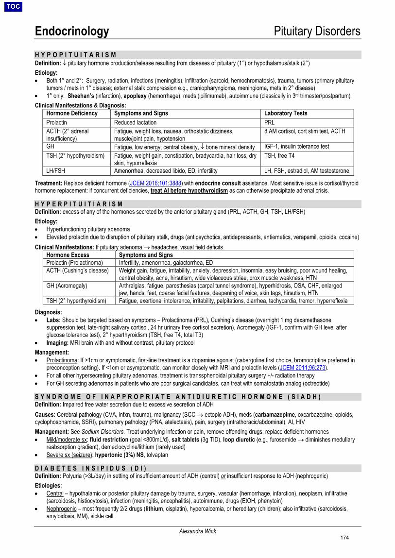
Endocrinology Pituitary Disorders
Alexandra Wick
H Y P O P I T U I T A R I S M Definition: ↓ pituitary hormone production/release resulting from diseases of pituitary (1°) or hypothalamus/stalk (2°)
Etiology: • Both 1° and 2°: Surgery, radiation, infections (meningitis), infiltration (sarcoid, hemochromatosis), trauma, tumors (primary pituitary
tumors / mets in 1° disease; external stalk compression e.g., craniopharyngioma, meningioma, mets in 2° disease) • 1° only: Sheehan’s (infarction), apoplexy (hemorrhage), meds (ipilimumab), autoimmune (classically in 3rd trimester/postpartum)
Clinical Manifestations & Diagnosis: Hormone Deficiency Symptoms and Signs Laboratory Tests Prolactin Reduced lactation PRL ACTH (2° adrenal insufficiency)
Fatigue, weight loss, nausea, orthostatic dizziness, muscle/joint pain, hypotension
8 AM cortisol, cort stim test, ACTH
GH Fatigue, low energy, central obesity, ↓ bone mineral density IGF-1, insulin tolerance test TSH (2° hypothyroidism) Fatigue, weight gain, constipation, bradycardia, hair loss, dry
skin, hyporreflexia TSH, free T4
LH/FSH Amenorrhea, decreased libido, ED, infertility LH, FSH, estradiol, AM testosterone
Treatment: Replace deficient hormone (JCEM 2016;101:3888) with endocrine consult assistance. Most sensitive issue is cortisol/thyroid hormone replacement: if concurrent deficiencies, treat AI before hypothyroidism as can otherwise precipitate adrenal crisis. H Y P E R P I T U I T I A R I S M Definition: excess of any of the hormones secreted by the anterior pituitary gland (PRL, ACTH, GH, TSH, LH/FSH)
Etiology: • Hyperfunctioning pituitary adenoma • Elevated prolactin due to disruption of pituitary stalk, drugs (antipsychotics, antidepressants, antiemetics, verapamil, opioids, cocaine)
Clinical Manifestations: If pituitary adenoma → headaches, visual field deficits Hormone Excess Symptoms and Signs Prolactin (Prolactinoma) Infertility, amenorrhea, galactorrhea, ED ACTH (Cushing’s disease) Weight gain, fatigue, irritability, anxiety, depression, insomnia, easy bruising, poor wound healing,
central obesity, acne, hirsutism, wide violaceous striae, prox muscle weakness, HTN GH (Acromegaly) Arthralgias, fatigue, paresthesias (carpal tunnel syndrome), hyperhidrosis, OSA, CHF, enlarged
jaw, hands, feet, coarse facial features, deepening of voice, skin tags, hirsutism, HTN TSH (2° hyperthyroidism) Fatigue, exertional intolerance, irritability, palpitations, diarrhea, tachycardia, tremor, hyperreflexia
Diagnosis: • Labs: Should be targeted based on symptoms – Prolactinoma (PRL), Cushing’s disease (overnight 1 mg dexamethasone
suppression test, late-night salivary cortisol, 24 hr urinary free cortisol excretion), Acromegaly (IGF-1, confirm with GH level after glucose tolerance test), 2° hyperthyroidism (TSH, free T4, total T3)
• Imaging: MRI brain with and without contrast, pituitary protocol
Management: • Prolactinoma: If >1cm or symptomatic, first-line treatment is a dopamine agonist (cabergoline first choice, bromocriptine preferred in
preconception setting). If <1cm or asymptomatic, can monitor closely with MRI and prolactin levels (JCEM 2011;96:273). • For all other hypersecreting pituitary adenomas, treatment is transsphenoidal pituitary surgery +/- radiation therapy • For GH secreting adenomas in patients who are poor surgical candidates, can treat with somatostatin analog (octreotide) S Y N D R O M E O F I N A P P R O P R I A T E A N T I D I U R E T I C H O R M O N E ( S I A D H ) Definition: Impaired free water secretion due to excessive secretion of ADH
Causes: Cerebral pathology (CVA, infxn, trauma), malignancy (SCC → ectopic ADH), meds (carbamazepime, oxcarbazepine, opioids, cyclophosphamide, SSRI), pulmonary pathology (PNA, atelectasis), pain, surgery (intrathoracic/abdominal), AI, HIV
Management: See Sodium Disorders. Treat underlying infection or pain, remove offending drugs, replace deficient hormones • Mild/moderate sx: fluid restriction (goal <800mL/d), salt tablets (3g TID), loop diuretic (e.g., furosemide → diminishes medullary
reabsorption gradient), demeclocycline/lithium (rarely used) • Severe sx (seizure): hypertonic (3%) NS, tolvaptan
D I A B E T E S I N S I P I D U S ( D I ) Definition: Polyuria (>3L/day) in setting of insufficient amount of ADH (central) or insufficient response to ADH (nephrogenic)
Etiologies: • Central – hypothalamic or posterior pituitary damage by trauma, surgery, vascular (hemorrhage, infarction), neoplasm, infiltrative
(sarcoidosis, histiocytosis), infection (meningitis, encephalitis), autoimmune, drugs (EtOH, phenytoin) • Nephrogenic – most frequently 2/2 drugs (lithium, cisplatin), hypercalcemia, or hereditary (children); also infiltrative (sarcoidosis,
amyloidosis, MM), sickle cell
174
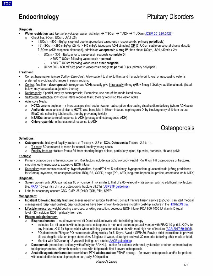
Endocrinology Pituitary Disorders
Hawra Al Lawati
Diagnosis: • Water restriction test: Normal physiology: water restriction ↑SOsm ↑ADH ↑UOsm (JCEM 2012;97:3426)
o Check Na, SOsm, UOsm, UVol q2hr If UOsm > 800 mEq/kg, stop test due to appropriate vasopressin response (dx: primary polydipsia) If (1) SOsm > 295 mEq/kg, (2) Na > 145 mEq/L (adequate ADH stimulus) OR (3) UOsm stable on several checks despite
↑ SOsm (ADH response plateaued), administer vasopressin 4 mcg IV, then check UOsm, UVol q30min x 2hr- UOsm < 300 mEq/kg prior to vasopressin suggests complete DI
- > 50% ↑ UOsm following vasopressin = central- < 50% ↑ UOsm following vasopressin = nephrogenic
- UOsm 300 - 800 mEq/kg prior to vasopressin suggests partial DI (vs. primary polydipsia)Treatment: • Correct hypernatremia (see Sodium Disorders). Allow patient to drink to thirst and if unable to drink, oral or nasogastric water is
preferred to avoid rapid changes in serum sodium.• Central: first line = desmopressin (exogenous ADH); usually give intranasally (5mcg qHS + 5mcg 1-3x/day); additional meds (listed
below) may be used as adjunctive therapy• Nephrogenic: if partial, may try desmopressin; if complete, use one of the meds listed below• Salt/protein restriction: low solute intake reduces thirst, thereby reducing free water intake• Adjunctive Meds:
o HCTZ: volume depletion → increases proximal sodium/water reabsorption, decreasing distal sodium delivery (where ADH acts)o Amiloride: mechanism similar to HCTZ; also beneficial in lithium-induced nephrogenic DI by blocking entry of lithium across
ENaC into collecting tubule cells, thereby preventing toxicityo NSAIDs: enhance renal response to ADH (prostaglandins antagonize ADH)o Chlorpropamide: enhances renal response to ADH
Osteoporosis Definitions: • Osteoporosis: history of fragility fracture or T-score ≤ -2.5 on DXA. Osteopenia: T-score -2.4 to -1.
o T-score: SD compared to mean for normal, healthy young adultso Fragility fracture: fracture from a fall from standing height or less, particularly spine, hip, wrist, humerus, rib, and pelvis
Etiology: • Primary osteoporosis is the most common. Risk factors include age ≥65, low body weight (<57.6 kg), FH osteoporosis or fractures,
smoking, early menopause, excessive EtOH intake• Secondary osteoporosis caused by: hyperthyroidism, hyperPTH, vit D deficiency, hypogonadism, glucocorticoids (≥5mg prednisone
for >3mos), myeloma, malabsorption (celiac, IBD), RA, COPD, drugs (PPI, AED, long-term heparin, leuprolide, aromatase inhib, MTX)
Diagnosis: • Screen women with DXA scan at age 65 or younger if risk similar to that of a 65-year-old white woman with no additional risk factors
(i.e. FRAX 10-year risk of major osteoporotic fracture ≥9.3%) (USPSTF guidelines)• Labs for secondary causes: CBC, CMP, 25(OH)D, TSH, PTH, SPEPManagement:• Inpatient following fragility fracture: assess need for surgical treatment, consult fracture liaison service (p25656), can start medical
management (bisphosphonates); bisphosphonates have been shown to decrease mortality post-hip fracture in the HORIZON trial.• Lifestyle measures: weight-bearing exercises, smoking cessation, decrease EtOH intake, RDA 800-1000 IU vitamin D (goal
level >30), calcium 1200 mg ideally from diet• Pharmacologic therapy:
o Bisphosphonates – must have normal vit D and calcium levels prior to initiating therapy Indicated for: all patients with osteoporosis, osteopenia in men and postmenopausal women with FRAX 10-yr risk >20% for
any fracture, >3% for hip; consider when initiating glucocorticoids in pts with med-high risk of fracture (ACR 2017;69:1095). PO alendronate 75mg or PO risendronate 35mg weekly for 5-10 yrs. Avoid if GFR<30. Provide strict instructions to prevent
pill esophagitis: take on empty stomach w/ full glass of water, sit upright and wait 30 min prior to taking other meds or food. Monitor with DXA scan q1-2 yrs until findings are stable (AACE guidelines)
o Denosumab (monoclonal antibody with affinity for RANKL) – option for patients with renal dysfunction or other contraindicationto bisphosphonates, q6month injection; treat with teriparatide first if severe osteoporosis
o Anabolic agents (teriparatide: recombinant PTH, abaloparatide: PTHrP analog) – for severe osteoporosis and/or for patientswith contraindications to bisphosphonates, daily SQ injection
175

Endocrinology Calcium Disorders
Caitlin Colling
H Y P E R C A L C E M I A ***MAKE SURE TO CORRECT CALCIUM FOR ALBUMIN: Corrected Ca = Serum Ca + 0.8 x (4-Alb)***
Definition: mild (corrected Ca < 12); moderate (corrected Ca 12-14); severe (corrected Ca >14)
Clinical Signs and Symptoms: MSK (“bones”)→ Osteitis fibrosa cystica (1° hyperPTH), bone pain, weakness; renal (“stones”) → Polydipsia, polyuria, nephrolithiasis, Type 1 RTA, AKI/CKD; GI (“groans”) → n/v, anorexia, constipation, ileus, pancreatitis; neuropsych (“overtones”) → fatigue, depression, anxiety, cognitive dysfunction to confusion, stupor, coma; CV → brady, short QTc, AV block, valve/vessel calcification
Diagnostic Approach:
Management: (BMJ 2015;305:h2723, NEJM 2005;352:373) • In general, asymptomatic mild-moderate hyperCa can be managed conservatively as outpatient; patients with symptomatic or severe
hyperCa (>14) should be admitted for treatment and endocrine consult. • Conservative measures: avoid contributory meds; oral hydration; oral PO4 repletion to 2.5-3.0 (IV could lead to hypoCa) • Volume resuscitation: patients are typically very dehydrated; bolus NS then gtt @ 200-300cc/hr with goal UOP 100-150cc/hr • Loop Diuretics: use ONLY if concurrent HF, CKD (and only once volume replete); otherwise, avoid, as they can worsen dehydration • Calcitonin: 4-8U/kg SC BID for 48 hours (will lower Ca by 1-2mg/dL). Tachyphylaxis usually occurs within 48-72h. • Bisphophonates: best studied in malignancy; zoledronate >> pamidronate (except in MM; more ATN). Takes 2-4d. Side effects:
hypoCa (check 25-OH-D & replete prior to admin), flu-like illness. Reduce dose if CKD. Avoid if CrCl < 30. • Denosumab: monoclonal Ab against RANKL blocks pre-osteoclast maturation; studied in bisphosphonate-refractory hyperCa • Other: Glucocorticoids (effective in calcitriol [1,25-OH-vit D]-mediated etiologies), HD (if refractory or life-threatening) • Special considerations for 1° hyperPTH: (JAMA Surg 2017;152:878, JCEM 2014;99:3607) Surgery is curative. Indicated if (a)
symptomatic OR (b) asymptomatic with Ca > 11.5, osteoporosis/vertebral fracture, CCl < 60, nephrolithiasis, or age < 50. If poor surgical candidate, consider cinacalcet, bisphosphonate, tamoxifen
H Y P O C A L C E M I A Clinical Signs/Symptoms: (BMJ 2008;336:1298) Neuromuscular (paresthesias, muscle cramps/spasms, tetany, Trousseau sign [carpal spasm w/ BP cuff inflation; 94% sens; 99% spec], Chvostek sign [circumoral muscle twitch w/ facial nerve tapping; poor sens; 85% spec]); seizures; ↑ QTc
Diagnostic Approach: Management: • Replete magnesium (hypoCa can be hard to correct without first correcting hypoMg causes PTH resistance and ↓ secretion) • IV Ca repletion: If severe (corrected Ca < 7.5, iCa < 1), symptomatic, or prolonged QT
- 1-2g IV Ca gluconate OR CaCl2 (in codes; via central line, risk of skin necrosis if extravasates) over 10-20 min - IV therapy ↑ serum levels for only 2-3h (chase w/ gtt or PO); telemetry recommended w/ IV repletion as arrhythmias may occur
• PO Ca repletion: If Ca > 7.5 or ASx; 1.5-2 g elemental Ca/day in divided doses; Ca citrate better absorbed vs CaCO3 esp if pt on PPI • Vitamin D repletion: 800-1000IU Vit D3 daily (if severely deficient, start with 50,000IU Vit D2 or D3 qweek x 6-8wks); in patients with
poor conversion of 25-OH-D (e.g., hypoparathyroidism, CKD), use calcitriol (starting dose: 0.25mcg PO QD)
PTH
a) Primary HPTH (adenoma > hyperplasia > carcinoma); associated w/ MEN b) FHH (24h urine calcium/Cr ratio <0.01); in 1° HPTH, UCCR >0.01 (unless pt w/ CKD/vit D deficiency) c) Tertiary HPTH d) Ectopic PTH production (rare)
↑ PTHrP Ectopic production (malignancy: lung SCC, breast, RCC)
↑ 1,25(OH)D Lymphoma, granulomatous disease (e.g., sarcoid)
↑ 25(OH)D Vitamin D toxicity
Normal: Multiple myeloma Hyperthyroidism Adrenal insuff. Pheo Vit A intox Drugs (thiazides, Li) Milk-alkali synd.
High Low
PTH
a) Hypoparathyroidism (surgical, autoimmune) b) Hypomagnesemia (causes PTH suppression/resistance)
↓ PO4 a) Vitamin D deficiency b) Pancreatitis c) Drugs (bisphos., phenytoin, cinacalcet)
↑ PO4 a) CKD (2° HPTH) b) Tumor lysis c) Rhabdo (early) d) Pseudohypoparathyroidism e) Critical illness f) Transfusions (citrate)
Low High
176
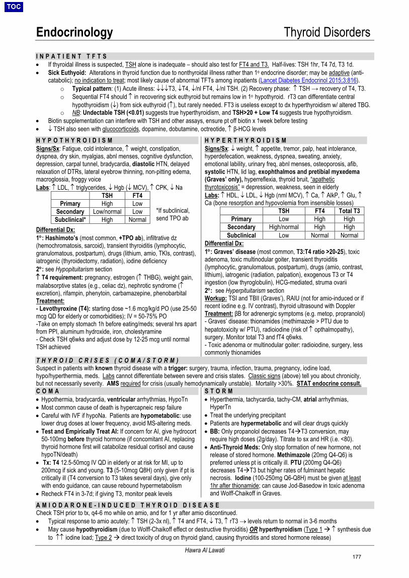
Endocrinology Thyroid Disorders
Hawra Al Lawati
I N P A T I E N T T F T S • If thyroidal illness is suspected, TSH alone is inadequate – should also test for FT4 and T3. Half-lives: TSH 1hr, T4 7d, T3 1d. • Sick Euthyoid: Alterations in thyroid function due to nonthyroidal illness rather than 1o endocrine disorder; may be adaptive (anti-
catabolic); no indication to treat; most likely cause of abnormal TFTs among inpatients (Lancet Diabetes Endocrinol 2015;3:816). o Typical pattern: (1) Acute illness: ↓↓↓T3, ↓T4, ↓/nl FT4, ↓/nl TSH. (2) Recovery phase: ↑ TSH → recovery of T4, T3. o Sequential FT4 should ↑ in recovering sick euthyroid but remains low in 1o hypothyroid. rT3 can differentiate central
hypothyroidism (↓) from sick euthyroid (↑), but rarely needed. FT3 is useless except to dx hyperthyroidism w/ altered TBG. o NB: Undectable TSH (<0.01) suggests true hyperthyroidism, and TSH>20 + Low T4 suggests true hypothyroidism.
• Biotin supplementation can interfere with TSH and other assays, ensure pt off biotin x 1week before testing • ↓ TSH also seen with glucocorticoids, dopamine, dobutamine, octreotide, ↑ β-HCG levels
H Y P O T H Y R O I D I S M Signs/Sx: Fatigue, cold intolerance, ↑ weight, constipation, dyspnea, dry skin, myalgias, abnl menses, cognitive dysfunction, depression, carpal tunnel, bradycardia, diastolic HTN, delayed relaxation of DTRs, lateral eyebrow thinning, non-pitting edema, macroglossia, froggy voice Labs: ↑ LDL, ↑ triglycerides, ↓ Hgb (↓ MCV), ↑ CPK, ↓ Na
*If subclinical, send TPO ab
Differential Dx: 1°: Hashimoto’s (most common, +TPO ab), infiltrative dz (hemochromatosis, sarcoid), transient thyroiditis (lymphocytic, granulomatous, postpartum), drugs (lithium, amio, TKIs, contrast), iatrogenic (thyroidectomy, radiation), iodine deficiency 2°: see Hypopituitarism section ↑ T4 requirement: pregnancy, estrogen (↑ THBG), weight gain, malabsorptive states (e.g., celiac dz), nephrotic syndrome (↑ excretion), rifampin, phenytoin, carbamazepine, phenobarbital Treatment: - Levothyroxine (T4): starting dose ~1.6 mcg/kg/d PO (use 25-50 mcg QD for elderly or comorbidities); IV = 50-75% PO -Take on empty stomach 1h before eating/meds; several hrs apart from PPI, aluminum hydroxide, iron, cholestyramine - Check TSH q6wks and adjust dose by 12-25 mcg until normal TSH achieved
TSH FT4 Primary High Low
Secondary Low/normal Low Subclinical* High Normal
H Y P E R T H Y R O I D I S M Signs/Sx: ↓ weight, ↑ appetite, tremor, palp, heat intolerance, hyperdefecation, weakness, dyspnea, sweating, anxiety, emotional lability, urinary freq, abnl menses, osteoporosis, afib, systolic HTN, lid lag, exophthalmos and pretibial myxedema (Graves’ only), hyperreflexia, thyroid bruit, “apathetic thyrotoxicosis” = depression, weakness, seen in elderly Labs: ↑ HDL, ↓ LDL, ↓ Hgb (nml MCV), ↑ Ca, ↑ AlkP, ↑ Glu, ↑ Ca (bone resorption and hypovolemia from insensible losses)
Differential Dx: 1°: Graves’ disease (most common, T3:T4 ratio >20-25), toxic adenoma, toxic multinodular goiter, transient thyroiditis (lymphocytic, granulomatous, postpartum), drugs (amio, contrast, lithium), iatrogenic (radiation, palpation), exogenous T3 or T4 ingestion (low thyroglobulin), HCG-mediated, struma ovarii 2°: see Hyperpituitarism section Workup: TSI and TBII (Graves’), RAIU (not for amio-induced or if recent iodine e.g. IV contrast), thyroid ultrasound with Doppler Treatment: βB for adrenergic symptoms (e.g. metop, propranolol) - Graves’ disease: thionamides (methimazole > PTU due to hepatotoxicity w/ PTU), radioiodine (risk of ↑ opthalmopathy), surgery. Monitor total T3 and fT4 q6wks. - Toxic adenoma or multinodular goiter: radioiodine, surgery, less commonly thionamides
TSH FT4 Total T3 Primary Low High High
Secondary High/normal High High Subclinical Low Normal Normal
T H Y R O I D C R I S E S ( C O M A / S T O R M ) Suspect in patients with known thyroid disease with a trigger: surgery, trauma, infection, trauma, pregnancy, iodine load, hypo/hyperthermia, meds. Labs cannot differentiate between severe and crisis states. Classic signs (above) tell you about chronicity, but not necessarily severity. AMS required for crisis (usually hemodynamically unstable). Mortality >30%. STAT endocrine consult. C O M A • Hypothermia, bradycardia, ventricular arrhythmias, HypoTn • Most common cause of death is hypercapneic resp failure • Careful with IVF if hypoNa. Patients are hypometabolic: use
lower drug doses at lower frequency, avoid MS-altering meds. • Test and Empirically Treat AI: If concern for AI, give hydrocort
50-100mg before thyroid hormone (if concomitant AI, replacing thyroid hormone first will catabolize residual cortisol and cause hypoTN/death)
• Tx: T4 12.5-50mcg IV QD in elderly or at risk for MI, up to 200mcg if sick and young. T3 (5-10mcg Q8H) only given if pt is critically ill (T4 conversion to T3 takes several days), give only with endo guidance, can cause rebound hypermetabolism
• Recheck FT4 in 3-7d; if giving T3, monitor peak levels
S T O R M • Hyperthermia, tachycardia, tachy-CM, atrial arrhythmias,
HyperTn • Treat the underlying precipitant • Patients are hypermetabolic and will clear drugs quickly • BB: Only propanolol decreases T4T3 conversion, may
require high doses (2g/day). Titrate to sx and HR (i.e. <80). • Anti-Thyroid Meds: Only stop formation of new hormone, not
release of stored hormone. Methimazole (20mg Q4-Q6) is preferred unless pt is critically ill. PTU (200mg Q4-Q6) decreases T4T3 but higher rates of fulminant hepatic necrosis. Iodine (100-250mg Q6-Q8H) must be given at least 1hr after thionamide; can cause Jod-Basedow in toxic adenoma and Wolff-Chaikoff in Graves.
A M I O D A R O N E - I N D U C E D T H Y R O I D D I S E A S E Check TSH prior to tx, q4-6 mo while on amio, and for 1 yr after amio discontinued. • Typical response to amio acutely: ↑ TSH (2-3x nl), ↑ T4 and FT4, ↓ T3, ↑ rT3 → levels return to normal in 3-6 months • May cause hypothyroidism (due to Wolff-Chaikoff effect or destructive thyroiditis) OR hyperthyroidism (Type 1 ↑ synthesis due
to ↑↑ iodine load; Type 2 direct toxicity of drug on thyroid gland, causing thyroiditis and stored hormone release)
177
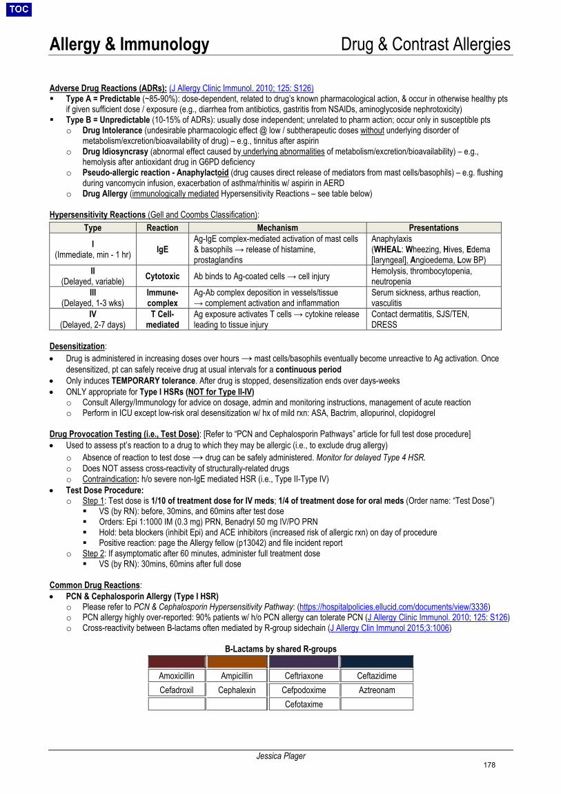
Allergy & Immunology Drug & Contrast Allergies
Jessica Plager
Adverse Drug Reactions (ADRs): (J Allergy Clinic Immunol. 2010; 125: S126) Type A = Predictable (~85-90%): dose-dependent, related to drug’s known pharmacological action, & occur in otherwise healthy pts
if given sufficient dose / exposure (e.g., diarrhea from antibiotics, gastritis from NSAIDs, aminoglycoside nephrotoxicity) Type B = Unpredictable (10-15% of ADRs): usually dose independent; unrelated to pharm action; occur only in susceptible pts
o Drug Intolerance (undesirable pharmacologic effect @ low / subtherapeutic doses without underlying disorder ofmetabolism/excretion/bioavailability of drug) – e.g., tinnitus after aspirin
o Drug Idiosyncrasy (abnormal effect caused by underlying abnormalities of metabolism/excretion/bioavailability) – e.g.,hemolysis after antioxidant drug in G6PD deficiency
o Pseudo-allergic reaction - Anaphylactoid (drug causes direct release of mediators from mast cells/basophils) – e.g. flushingduring vancomycin infusion, exacerbation of asthma/rhinitis w/ aspirin in AERD
o Drug Allergy (immunologically mediated Hypersensitivity Reactions – see table below)
Hypersensitivity Reactions (Gell and Coombs Classification):
Type Reaction Mechanism Presentations
I (Immediate, min - 1 hr) IgE
Ag-IgE complex-mediated activation of mast cells & basophils → release of histamine, prostaglandins
Anaphylaxis (WHEAL: Wheezing, Hives, Edema [laryngeal], Angioedema, Low BP)
II (Delayed, variable) Cytotoxic Ab binds to Ag-coated cells → cell injury Hemolysis, thrombocytopenia,
neutropenia III
(Delayed, 1-3 wks) Immune-complex
Ag-Ab complex deposition in vessels/tissue → complement activation and inflammation
Serum sickness, arthus reaction, vasculitis
IV (Delayed, 2-7 days)
T Cell-mediated
Ag exposure activates T cells → cytokine release leading to tissue injury
Contact dermatitis, SJS/TEN, DRESS
Desensitization: • Drug is administered in increasing doses over hours → mast cells/basophils eventually become unreactive to Ag activation. Once
desensitized, pt can safely receive drug at usual intervals for a continuous period• Only induces TEMPORARY tolerance. After drug is stopped, desensitization ends over days-weeks• ONLY appropriate for Type I HSRs (NOT for Type II-IV)
o Consult Allergy/Immunology for advice on dosage, admin and monitoring instructions, management of acute reactiono Perform in ICU except low-risk oral desensitization w/ hx of mild rxn: ASA, Bactrim, allopurinol, clopidogrel
Drug Provocation Testing (i.e., Test Dose): [Refer to “PCN and Cephalosporin Pathways” article for full test dose procedure] • Used to assess pt’s reaction to a drug to which they may be allergic (i.e., to exclude drug allergy)
o Absence of reaction to test dose → drug can be safely administered. Monitor for delayed Type 4 HSR.o Does NOT assess cross-reactivity of structurally-related drugso Contraindication: h/o severe non-IgE mediated HSR (i.e., Type II-Type IV)
• Test Dose Procedure:o Step 1: Test dose is 1/10 of treatment dose for IV meds; 1/4 of treatment dose for oral meds (Order name: “Test Dose”)
VS (by RN): before, 30mins, and 60mins after test dose Orders: Epi 1:1000 IM (0.3 mg) PRN, Benadryl 50 mg IV/PO PRN Hold: beta blockers (inhibit Epi) and ACE inhibitors (increased risk of allergic rxn) on day of procedure Positive reaction: page the Allergy fellow (p13042) and file incident report
o Step 2: If asymptomatic after 60 minutes, administer full treatment dose VS (by RN): 30mins, 60mins after full dose
Common Drug Reactions: • PCN & Cephalosporin Allergy (Type I HSR)
o Please refer to PCN & Cephalosporin Hypersensitivity Pathway: (https://hospitalpolicies.ellucid.com/documents/view/3336)o PCN allergy highly over-reported: 90% patients w/ h/o PCN allergy can tolerate PCN (J Allergy Clinic Immunol. 2010; 125: S126)o Cross-reactivity between B-lactams often mediated by R-group sidechain (J Allergy Clin Immunol 2015;3:1006)
B-Lactams by shared R-groups
Amoxicillin Ampicillin Ceftriaxone Ceftazidime Cefadroxil Cephalexin Cefpodoxime Aztreonam
Cefotaxime
178
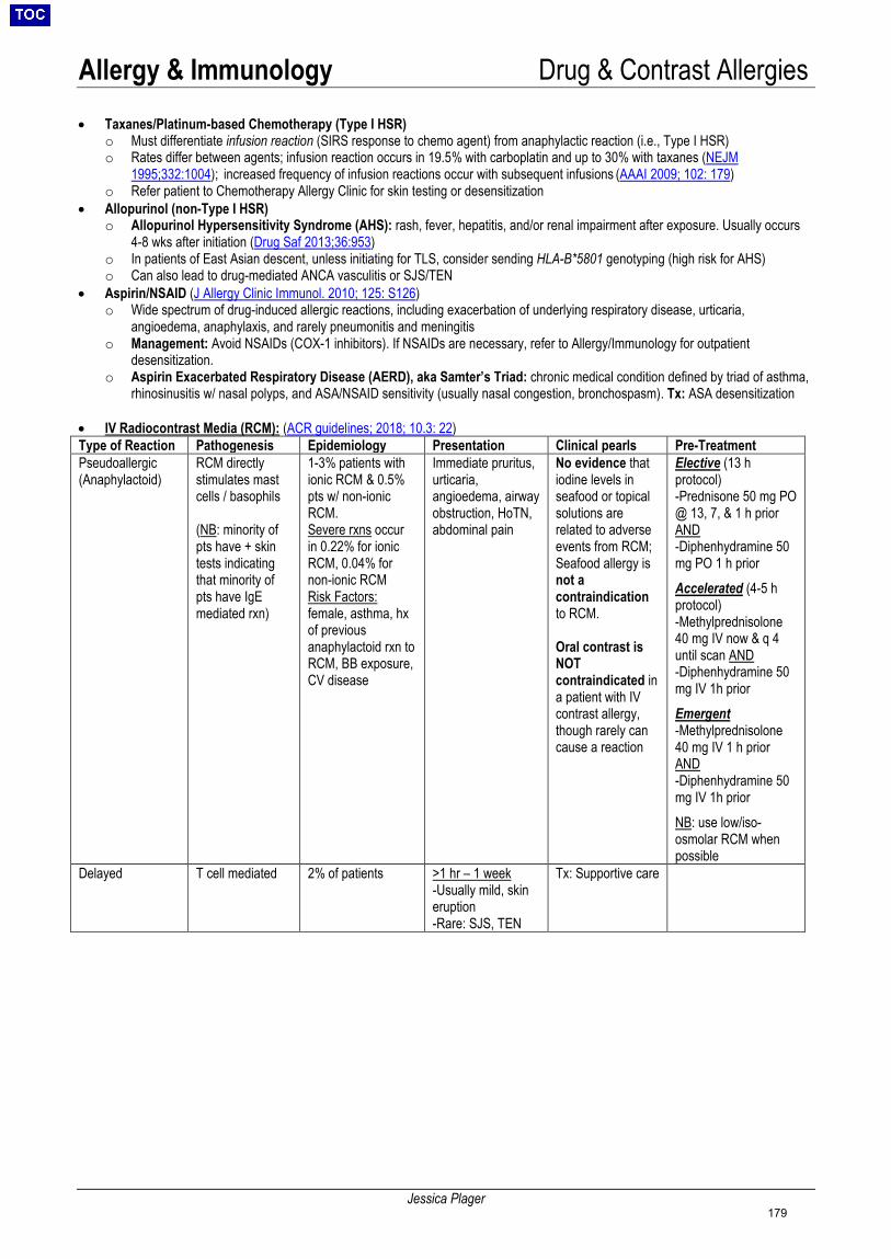
Allergy & Immunology Drug & Contrast Allergies
Jessica Plager
• Taxanes/Platinum-based Chemotherapy (Type I HSR)o Must differentiate infusion reaction (SIRS response to chemo agent) from anaphylactic reaction (i.e., Type I HSR)o Rates differ between agents; infusion reaction occurs in 19.5% with carboplatin and up to 30% with taxanes (NEJM
1995;332:1004); increased frequency of infusion reactions occur with subsequent infusions (AAAI 2009; 102: 179)o Refer patient to Chemotherapy Allergy Clinic for skin testing or desensitization
• Allopurinol (non-Type I HSR)o Allopurinol Hypersensitivity Syndrome (AHS): rash, fever, hepatitis, and/or renal impairment after exposure. Usually occurs
4-8 wks after initiation (Drug Saf 2013;36:953)o In patients of East Asian descent, unless initiating for TLS, consider sending HLA-B*5801 genotyping (high risk for AHS)o Can also lead to drug-mediated ANCA vasculitis or SJS/TEN
• Aspirin/NSAID (J Allergy Clinic Immunol. 2010; 125: S126)o Wide spectrum of drug-induced allergic reactions, including exacerbation of underlying respiratory disease, urticaria,
angioedema, anaphylaxis, and rarely pneumonitis and meningitiso Management: Avoid NSAIDs (COX-1 inhibitors). If NSAIDs are necessary, refer to Allergy/Immunology for outpatient
desensitization.o Aspirin Exacerbated Respiratory Disease (AERD), aka Samter’s Triad: chronic medical condition defined by triad of asthma,
rhinosinusitis w/ nasal polyps, and ASA/NSAID sensitivity (usually nasal congestion, bronchospasm). Tx: ASA desensitization
• IV Radiocontrast Media (RCM): (ACR guidelines; 2018; 10.3: 22)Type of Reaction Pathogenesis Epidemiology Presentation Clinical pearls Pre-Treatment Pseudoallergic (Anaphylactoid)
RCM directly stimulates mast cells / basophils
(NB: minority of pts have + skin tests indicating that minority of pts have IgE mediated rxn)
1-3% patients withionic RCM & 0.5%pts w/ non-ionicRCM.Severe rxns occurin 0.22% for ionicRCM, 0.04% fornon-ionic RCMRisk Factors:female, asthma, hx of previous anaphylactoid rxn to RCM, BB exposure, CV disease
Immediate pruritus, urticaria, angioedema, airway obstruction, HoTN, abdominal pain
No evidence that iodine levels in seafood or topical solutions are related to adverse events from RCM; Seafood allergy is not a contraindication to RCM.
Oral contrast is NOT contraindicated in a patient with IV contrast allergy, though rarely can cause a reaction
Elective (13 h protocol) -Prednisone 50 mg PO@ 13, 7, & 1 h priorAND-Diphenhydramine 50mg PO 1 h priorAccelerated (4-5 h protocol) -Methylprednisolone40 mg IV now & q 4until scan AND-Diphenhydramine 50mg IV 1h priorEmergent -Methylprednisolone40 mg IV 1 h priorAND-Diphenhydramine 50mg IV 1h priorNB: use low/iso-osmolar RCM when possible
Delayed T cell mediated 2% of patients >1 hr – 1 week-Usually mild, skineruption-Rare: SJS, TEN
Tx: Supportive care
179
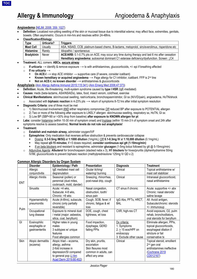
Allergy & Immunology Angioedema & Anaphylaxis
Jessica Plager
Angioedema (NEJM; 2008; 359: 1027) • Definition: Localized non-pitting swelling of the skin or mucosal tissue due to interstitial edema; may affect face, extremities, genitals,
bowels. Often asymmetric. Occurs in min-hrs and resolves within 24-48hrs.• Classification/Etiology:
Type Urticaria? Triggers Mast Cell Usually ASA, NSAID, CCB, platinum-based chemo, B-lactams, metoprolol, siro/everolimus, risperidone etc Histamine Rarely Idiopathic / spontaneous Bradykinin Never ACE/ARB: 0.1-0.7% pts on ACE; may occur any time during therapy and last 6 mo after cessation
Hereditary angioedema: autosomal dominant C1 esterase deficiency/dysfunction. Screen: ↓C4 • Treatment: ALL comers: ABCs, secure airway
o If urticaria → identify & remove exposure → tx with antihistamines, glucocorticoids, +/- epi if breathing affectedo If no urticaria →
On ACE-I → stop ACE inhibitor → supportive care (if severe, consider icatibant) Known hereditary or acquired angioedema → Page allergy for C1-inhibitor, icatibant. FFP is 2nd line Not on ACE-I; no known disorder → antihistamines & glucocorticoids
Anaphylaxis (Ann Allergy Asthma Immunol 2015;115:341) (Ann Emerg Med 2006;47:373) • Definition: Acute, life-threatening, multi-system syndrome caused by type I HSR (IgE-mediated)• Causes: meds (beta-lactams, ASA/NSAIDs), latex, food, insect venom, cold/heat, exercise• Clinical Manifestations: skin/mucosal swelling, rash/urticaria, bronchospasm/stridor, GI sx (N/V/D/pain), angioedema, HoTN/shock
o Associated with biphasic reaction in 4-23% pts → return of symptoms 8-72 hrs after initial symptom resolution• Diagnostic Criteria: one of three must be met
o 1) Skin/mucosal involvement AND either respiratory compromise OR reduced BP after exposure to POTENTIAL allergeno 2) Two or more of the following after exposure to LIKELY allergen: skin/mucosa swelling, respiratory sx, HoTN, GI sxo 3) Low BP (SBP<90 or >30% drop from baseline) after exposure to KNOWN allergen for pt
• Labs: consider histamine (within 10-30 min of symptom onset) and tryptase (within 15 min-3 h of symptom onset and 24h aftersymptoms resolve to assess baseline). Normal levels do not rule out anaphylaxis!
• Treatment:o Establish and maintain airway, administer oxygen/IVFo Epinephrine: Only medication that reverses airflow obstruction & prevents cardiovascular collapse
Dosing: 0.3-0.5mg IM/SQ at 1:1000 dilution (1mg/mL) OR 0.1-0.3mg IV at 1:10,000 dilution (0.1mg/mL) May repeat q5-15 minutes; if >3 doses required, consider continuous epi gtt (1-10mcg/min) If on beta blockers and resistant to epinephrine, administer glucagon (1-5mg bolus followed by gtt @ 5-15mcg/min)
o Adjunctive Agents: Albuterol for bronchospasm (stacked nebs x 3), H1 blockers for hives/pruritis (diphenhydramine 50mgIV/IM, glucocorticoids to prevent biphasic reaction (methylprednisolone 125mg IV QD x 2)
Common Allergic Disorders by Organ System Disorder Epidemiology / Path Presentation Diagnosis Treatment
ENT
Allergic conjunctivitis
IgE mediated mast cell degranulation
Ocular itching/ watering/ burning
Clinical Topical antihistamine w/ mast cell stabilizer
Allergic rhinitis Seasonal (pollen) or perennial (dust mites, cockroach, mold, dander)
Sneezing, rhinorrhea, post-nasal drip, cough
Clinical Intranasal glucocorticoid, nasal antihistamine
Sinusitis Acute: <4 wks, Subacute: 4-8 wks, Chronic: >8 wks
Nasal congestion, obstruction, tooth/ sinus pain
CT sinus if chronic Acute: supportive +/- abx Chronic: nasal steroids/ saline lavage
Pulm
Hypersensitivity pneumonitis
Acute (4-6hrs), subacute, chronic (only partially reversible)
Cough, SOB, fever; if chronic, fatigue & wt loss
IgG Abs, PFTs, HRCT, BAL
All: Avoid antigen, Subacute/chronic: steroids +/- immunosup
Occupational lung disease
Exposure to mineral dust / metal (major: asbestos, silica, coal, beryllium)
DOE, cough, chest tightness, wt loss
CXR, high-res CT Avoid exposure, O2, pulm rehab, bronchodilators, oral steroids for beryllium
GI Eosinophilic esophagitis or gastroenteritis
Higher rates in young males 3 subtypes w/ unique features Food allergies common
Food impaction, dysphagia, GERD failing PPIs
Dx criteria: 1. Symptoms2. > 15 eos/HPF onendoscopy3. Exclude other cause
Eliminate allergen, PPIs, topical glucocorticoids, esophageal dilation if stricture or fail conservative tx
Skin Atopic dermatitis (eczema)
Atopic triad – eczema, allergy, asthma 2-fold increase indepression/SI comparedto general pop (J AmAcad Derm 2019;80:402)
Dry skin, pruritis, excoriation Skin flexures most common in adults, can affect any area
Clinical Topical steroid, emollient 2nd gen oral antihistamines ineffective (Cochrane 2019 CD012167)
180
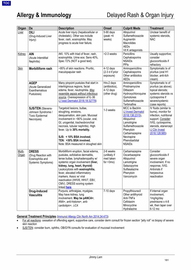
Allergy & Immunology Delayed Rash & Organ Injury
Jimmy Lam
Organ Dx Description Onset Culprit Meds Treatment Liver DILI
(Drug-Induced Liver Injury)
Acute liver injury (hepatocellular or cholestatic). Other sxs include fever, rash, eosinophilia. May progress to acute liver failure.
5-90 days(peak >4weeks)
Allopurinol Sulfamethoxazole Augmentin Macrolides AEDs Vit K antagonists
Unclear benefit of systemic steroids, NAC
Kidney AIN (Acute Interstitial Nephritis)
AKI. 10% with triad of fever, rash, eosinophilia. Urine eos: Sens 40%, Spec 72% (NOT a good test).
>2-3 weeks Penicillins Cephalosporins NSAIDs PPIs
Usually supportive; consider glucocorticoids if refractory
Skin Morbilliform rash ~95% of skin reactions. Pruritic, maculopapular rash
4-12 days(2-3 d if priorexposure)
Aminopenicillins Cephalosporins Other antibiotics AEDs
Symptomatic tx of pruritus (oral H1 blocker, anti-itch cream)
AGEP (Acute Generalized Exanthematous Pustulosis)
Many pinpoint pustules that start in intertriginous regions, facial edema, fever, neutrophilia. May resemble sepsis without infectious source. Overexpression of IL-36 (J Ivest Dermatol 2018;18:32779)
Hrs-2 days (antibiotics), 4-12 days(other drug)
Aminopenicillins Pristinamycine Diltiazem Hydroxychloroquine Quinolones Sulfamethoxazole Terbinafine
Symptomatic tx of pruritus (as above); topical steroids; systemic steroids vs cyclosporine if severe/systemic (case reports)
SJS/TEN (Stevens-Johnson Syndrome / Toxic Epidermal Necrolysis)
Targetoid lesions, bullae, sloughing, erosions, desquamation, skin pain. Mucosal involvement in ~90% (ocular, oral, GI, urogenital, tracheobronchial necrosis, tubular nephritis). High fever. Up to 30% mortality.
SJS: < 10% BSA involved. TEN: >30% BSA involved. Note: BSA measured in sloughed skin
1-3 weeks MCC is Bactrim (J Invest Dermatol 2018;138:2315), Allopurinol Lamotrigine Sulfamethoxazole Phenytoin Carbamazepine Nevirapine Phenobarbital NSAIDs
IV fluids (similar to burns), monitor for infection, nutritional support. Consider: IVIG, cyclosporine, steroids, etanercept (J Clin Invest 2018;128:985)
Multi-Organ
DRESS (Drug Reaction with Eosinophilia and Systemic Symptoms)
Morbilliform eruption, facial edema, pustules, exfoliative dermatitis, tense bullae, lymphadenopathy w/ systemic organ involvement (liver, kidney, lung, heart, thyroid) Leukocytosis with eosinophilia, fever, elevated inflammatory markers. Assoc w/ viral reactivation (HHV6, HHV7, EBV, CMV). DRESS scoring system linked here.
2-6 weeks(unlikely ifmed takenfor >3mo)
Carbamazepine Oxcarbazepine Allopurinol Lamotrigine Salazopyrine Sulfasalazine Phenytoin Vancomycin
Consider glucocorticoids if severe organ involvement. If no response, IVIG. Antivirals if herpesvirus reactivation
Drug-Induced Vasculitis
Purpura, arthralgias, myalgias. May have kidney, lung involvement. May be pANCA+, ANA+, anti-histone+, anti-cardiolipin+, ↓C4
7-10 days Propylthiouracil Other antithyroid Anti-TNFa Cefotaxim Minocycline Hydralazine
If internal organ involvement, 1mg/kg/day prednisone x 4-8 wk, then taper over 6-12 mo
General Treatment Principles (Immunol Allergy Clin North Am 2014;34:473)• For all reactions: cessation of offending agent, supportive care, consider derm consult for frozen section “jelly roll” vs biopsy of severe
skin reaction • SJS/TEN: consider burn, ophtho, OB/GYN consults for evaluation of mucosal involvement
181
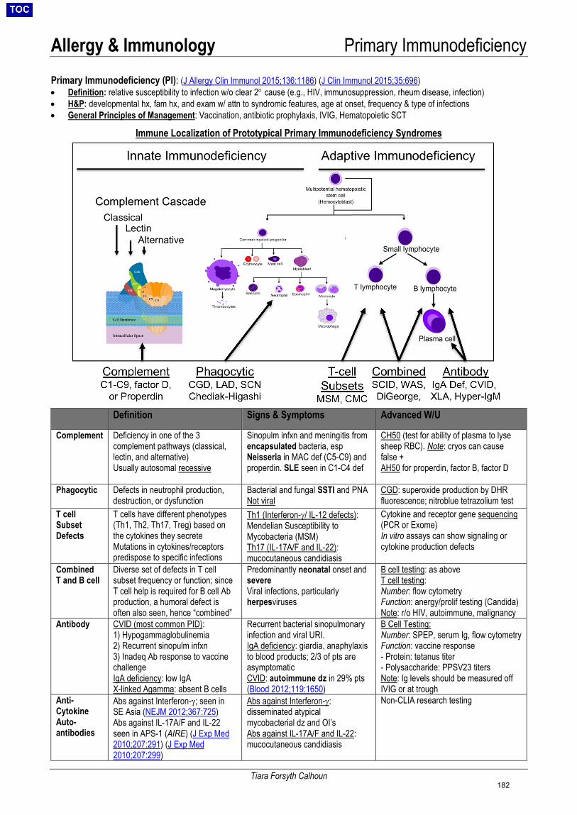
Allergy & Immunology Primary Immunodeficiency
Tiara Forsyth Calhoun
Primary Immunodeficiency (PI): (J Allergy Clin Immunol 2015;136:1186) (J Clin Immunol 2015;35:696) • Definition: relative susceptibility to infection w/o clear 2° cause (e.g., HIV, immunosuppression, rheum disease, infection)• H&P: developmental hx, fam hx, and exam w/ attn to syndromic features, age at onset, frequency & type of infections• General Principles of Management: Vaccination, antibiotic prophylaxis, IVIG, Hematopoietic SCT
Immune Localization of Prototypical Primary Immunodeficiency Syndromes
Definition Signs & Symptoms Advanced W/U
Complement Deficiency in one of the 3 complement pathways (classical, lectin, and alternative) Usually autosomal recessive
Sinopulm infxn and meningitis from encapsulated bacteria, esp Neisseria in MAC def (C5-C9) and properdin. SLE seen in C1-C4 def
CH50 (test for ability of plasma to lyse sheep RBC). Note: cryos can cause false + AH50 for properdin, factor B, factor D
Phagocytic Defects in neutrophil production, destruction, or dysfunction
Bacterial and fungal SSTI and PNA Not viral
CGD: superoxide production by DHR fluorescence; nitroblue tetrazolium test
T cell Subset Defects
T cells have different phenotypes (Th1, Th2, Th17, Treg) based on the cytokines they secrete Mutations in cytokines/receptors predispose to specific infections
Th1 (Interferon-γ/ IL-12 defects): Mendelian Susceptibility to Mycobacteria (MSM) Th17 (IL-17A/F and IL-22): mucocutaneous candidiasis
Cytokine and receptor gene sequencing (PCR or Exome) In vitro assays can show signaling or cytokine production defects
Combined T and B cell
Diverse set of defects in T cell subset frequency or function; since T cell help is required for B cell Ab production, a humoral defect is often also seen, hence “combined”
Predominantly neonatal onset and severe Viral infections, particularly herpesviruses
B cell testing: as above T cell testing: Number: flow cytometry Function: anergy/prolif testing (Candida) Note: r/o HIV, autoimmune, malignancy
Antibody CVID (most common PID): 1) Hypogammaglobulinemia2) Recurrent sinopulm infxn3) Inadeq Ab response to vaccinechallengeIgA deficiency: low IgAX-linked Agamma: absent B cells
Recurrent bacterial sinopulmonary infection and viral URI. IgA deficiency: giardia, anaphylaxis to blood products; 2/3 of pts are asymptomatic CVID: autoimmune dz in 29% pts (Blood 2012;119:1650)
B Cell Testing: Number: SPEP, serum Ig, flow cytometry Function: vaccine response - Protein: tetanus titer- Polysaccharide: PPSV23 titersNote: Ig levels should be measured offIVIG or at trough
Anti-Cytokine Auto-antibodies
Abs against Interferon-γ; seen in SE Asia (NEJM 2012;367:725) Abs against IL-17A/F and IL-22 seen in APS-1 (AIRE) (J Exp Med 2010;207:291) (J Exp Med 2010;207:299)
Abs against Interferon-γ: disseminated atypical mycobacterial dz and OI’s Abs against IL-17A/F and IL-22: mucocutaneous candidiasis
Non-CLIA research testing
182
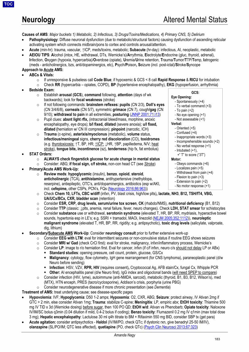
Neurology Altered Mental Status
Amanda Nagy
GCS: Eye Opening:
- Spontaneously (+4)- To verbal command (+3)- To pain (+2)- No eye opening (+1)- Not assessable (+1)
Verbal: - Oriented (+5)- Confused (+4)- Inappropriate words (+3)- Incomprehensible sounds (+2)- No verbal response (+1)- Intubated (+1),
+” T” to score (“3T”) Motor:
- Obeys commands (+6)- Localizes pain (+5)- Withdrawal from pain (+4)- Flexion to pain (+3)- Extension to pain (+2)- No motor response (+1)
Causes of AMS: Major buckets 1) Metabolic, 2) Infectious, 3) Drugs/Toxins/Medications, 4) Primary CNS, 5) Delirium • Pathophysiology: Diffuse neuronal dysfunction (due to metabolic/structural factors) causing dysfunction of ascending reticular
activating system which connects midbrain/pons to cortex and controls arousal/attention.• Acute (min-hr): trauma, vascular, ↑ICP, meds/toxins, metabolic; Subacute (hr-day): infectious, AI, neoplastic, metabolic• AEIOU TIPS: Alcohol (intox, HE, withdrawal, DTs, Wernicke’s)/Arrythmia, Electrolyte/Endocrine (gluc, thyroid, adrenal),
Infection, Oxygen (hypoxia, hypercarbia)/Overdose (opiate), Uremia/Urine retention, Trauma/Tumor/TTP/Temp, Iatrogenic(meds - anticholinergics, bzs, anti/dopaminergics, etc), Psych/Poison, Seizure (incl. post-ictal)/Stroke/Syncope
Approach to Acute AMS: • ABCs & Vitals:
o If unresponsive & pulseless call Code Blue; if hypoxemic & GCS < 8 call Rapid Response & RICU for intubationo Check RR (hypercarbia – opiates, COPD), BP (hypertensive encephalopathy), EKG (hypoperfusion, arrhythmia)
• Bedside Exam:o Establish arousal (GCS), command following, attention (days of wk
backwards); look for focal weakness (stroke)o If not following commands: brainstem reflexes: pupils (CN 2/3), Doll’s eyes
(CN 3/4/6/8), corneals (CN 5/7), symmetric grimace (CN 7), cough/gag (CN9/10); withdrawal to pain in all extremities, posturing (JNNP 2001;71:i13)
o Pupil clues: absnt light rflx, (intracranial bleed/mass, morphine, anoxicencephalopathy, eye drops); b/l fixed, dilated (severe anoxia); u/l fixed,dilated (herniation w/ CN III compression); pinpoint (narcotic, ICH)
o Trauma (c-spine), asterixis/myoclonus (metabolic), volume status,infectious/meningeal signs, cherry red discoloration (CO), toxidromes(e.g. thyrotoxicosis: ↑T, BP, HR; ↑ICP: ↓HR, ↑BP, papilledema, N/V; heatstroke), tongue bite, incontinence (sz), tenderness (hip fx, fat embolus)
• STAT Orders:o ALWAYS check fingerstick glucose for acute change in mental statuso Consider: ABG; if focal sign, c/f stroke, non-con head CT (see Stroke)
• Primary/Acute AMS Work-Up:o Review meds: hypoglycemic (insulin), benzo, opioid, steroid,
anticholinergic (TCA), antihistamine, antihypertensive (methyldopa,reserpine), antiepileptic, OTC’s, anti/dopaminergics, antibiotics (esp w/AKI,incl. cefepime, other CSPs, PCN’s, FQs (Neurology 2016;86:963))
o Check Chem 10, LFTs, CBC w/diff (infxn, PV, blast crisis, high/low plts), lactate, NH3, B12, TSH/fT4, VBG,UA/UCx/BCx, CXR, bladder scan (retention)
o Consider ESR, CRP, drug levels, serum/urine tox screen, CK (rhabdo/NMS), nutritional deficiency (B1, B12)o Consider TTP (classic: ↓plts, anemia, renal failure, fever, neuro changes). Check LDH, STAT smear for schistocyteso Consider substance use or withdrawal, serotonin syndrome (elevated T, HR, BP, RR, mydriasis, hyperactive bowel
sounds, hypertonia esp in LE’s; e.g. SSRI + tramadol, MAOi, linezolid (NEJM 2005;352:1112)), neurolepticmalignant syndrome (elevated T, HR, BP, RR, rigidity; e.g. antipsychotic), toxic drug levels (salicylate, valproate,dig, lithium)
• Secondary/Subacute AMS Work-Up: Consider neurology consult prior to further extensive work-upo Consider EEG with LTM: eval for intermittent seizures or non-convulsive status if routine EEG shows seizureso Consider MRI w/ Gad (check CrCl first): eval for stroke, malignancy, infxn/inflammatory process, Wernicke’so Consider LP: image to r/o herniation first. Eval for cancer, infxn (if c/f infxn, neuro c/s should not delay LP or ABx) Standard studies: opening pressure, cell count, protein, glucose, GS/Cx Malignancy: cytology, flow cytometry, IgH gene rearrangement (for CNS lymphoma), paraneoplastic panel (d/w
Neuro before sending) Infection: HSV, VZV, RPR, HIV (requires consent), Cryptococcal Ag, AFB stain/Cx, fungal Cx, Whipple PCR Other: AI encephalitis panel (d/w Neuro first), IgG index and oligoclonal bands (will need SPEP to compare)
o Consider infection (HIV, lyme), autoimmune (anti-NMDA, sarcoid), metabolic (thyroid, B1, B3, B12, Wilson’s), med(MTX), HTN enceph, PRES (tacro/cyclosporine), Addison’s crisis, porphyria (urine PBG)
o Consider neurodegenerative disease if more chronic presentation (see Dementia)Treatment of AMS: treat underlying cause; see disease-specific pages • Hypovolemia: IVF; Hypoglycemia: D50 1-2 amps; Hypoxemia: O2, CXR, ABG; Seizure: protect airway, IV Ativan 2mg if
GTC > 2 min, else consider Ativan 1mg; Trauma: stabilize C-spine; Meningitis: LP, empiric abx; EtOH toxicity: Thiamine 500mg IV TID x 3d (Wernicke dosing) before sugar, then 100 PO QD; EtOH w/d: Ativan vs Phenobarb; Opiate toxicity: NaloxoneIV/IM/SC bolus q3min (0.04 dilution if mild, 0.4-2 bolus if coding); Benzo toxicity: Flumazenil 0.2 mg IV q1min (max total dose3 mg); Hepatic encephalopathy: Lactulose 30 ml q4h titrate to BM + Rifaximin 550 mg BID, consider SBP tx (get para)
• Acute agitation: consider antipsychotics - Haldol (IV/IM/PO, check QTc; if dystonic rxn, give benadryl 25-50 IM/IV),olanzapine (SL/PO/IM; QTC less affected), quetiapine (PO, check QTc) (Psych Clin Neurosci 2013;67:323)
183
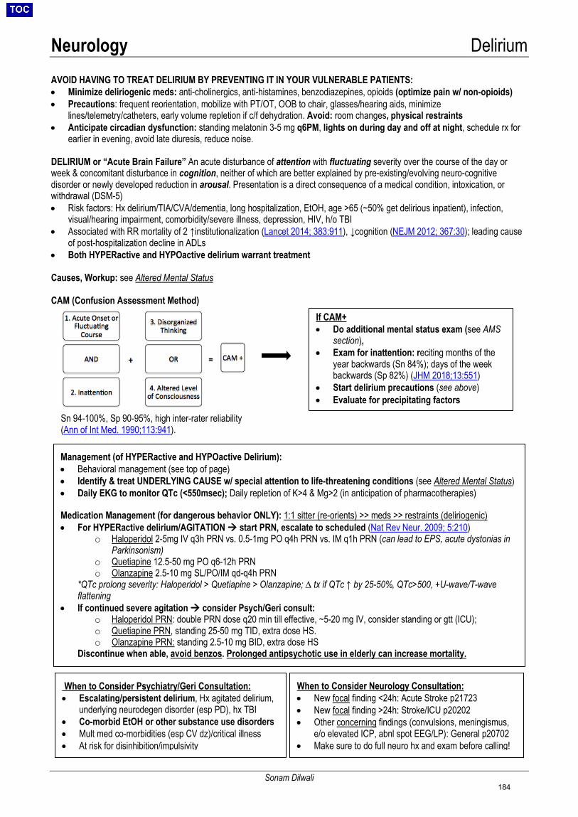
Neurology Delirium
Sonam Dilwali
AVOID HAVING TO TREAT DELIRIUM BY PREVENTING IT IN YOUR VULNERABLE PATIENTS: • Minimize deliriogenic meds: anti-cholinergics, anti-histamines, benzodiazepines, opioids (optimize pain w/ non-opioids)• Precautions: frequent reorientation, mobilize with PT/OT, OOB to chair, glasses/hearing aids, minimize
lines/telemetry/catheters, early volume repletion if c/f dehydration. Avoid: room changes, physical restraints• Anticipate circadian dysfunction: standing melatonin 3-5 mg q6PM, lights on during day and off at night, schedule rx for
earlier in evening, avoid late diuresis, reduce noise.
DELIRIUM or “Acute Brain Failure” An acute disturbance of attention with fluctuating severity over the course of the day or week & concomitant disturbance in cognition, neither of which are better explained by pre-existing/evolving neuro-cognitive disorder or newly developed reduction in arousal. Presentation is a direct consequence of a medical condition, intoxication, or withdrawal (DSM-5) • Risk factors: Hx delirium/TIA/CVA/dementia, long hospitalization, EtOH, age >65 (~50% get delirious inpatient), infection,
visual/hearing impairment, comorbidity/severe illness, depression, HIV, h/o TBI• Associated with RR mortality of 2 ↑institutionalization (Lancet 2014; 383:911), ↓cognition (NEJM 2012; 367:30); leading cause
of post-hospitalization decline in ADLs• Both HYPERactive and HYPOactive delirium warrant treatment
Causes, Workup: see Altered Mental Status
CAM (Confusion Assessment Method)
When to Consider Psychiatry/Geri Consultation: • Escalating/persistent delirium, Hx agitated delirium,
underlying neurodegen disorder (esp PD), hx TBI• Co-morbid EtOH or other substance use disorders• Mult med co-morbidities (esp CV dz)/critical illness• At risk for disinhibition/impulsivity
When to Consider Neurology Consultation: • New focal finding <24h: Acute Stroke p21723• New focal finding >24h: Stroke/ICU p20202• Other concerning findings (convulsions, meningismus,
e/o elevated ICP, abnl spot EEG/LP): General p20702• Make sure to do full neuro hx and exam before calling!
Management (of HYPERactive and HYPOactive Delirium): • Behavioral management (see top of page)• Identify & treat UNDERLYING CAUSE w/ special attention to life-threatening conditions (see Altered Mental Status)• Daily EKG to monitor QTc (<550msec); Daily repletion of K>4 & Mg>2 (in anticipation of pharmacotherapies)
Medication Management (for dangerous behavior ONLY): 1:1 sitter (re-orients) >> meds >> restraints (deliriogenic) • For HYPERactive delirium/AGITATION start PRN, escalate to scheduled (Nat Rev Neur. 2009; 5:210)
o Haloperidol 2-5mg IV q3h PRN vs. 0.5-1mg PO q4h PRN vs. IM q1h PRN (can lead to EPS, acute dystonias inParkinsonism)
o Quetiapine 12.5-50 mg PO q6-12h PRNo Olanzapine 2.5-10 mg SL/PO/IM qd-q4h PRN
*QTc prolong severity: Haloperidol > Quetiapine > Olanzapine; ∆ tx if QTc ↑ by 25-50%, QTc>500, +U-wave/T-waveflattening
• If continued severe agitation consider Psych/Geri consult:o Haloperidol PRN: double PRN dose q20 min till effective, ~5-20 mg IV, consider standing or gtt (ICU);o Quetiapine PRN, standing 25-50 mg TID, extra dose HS.o Olanzapine PRN: standing 2.5-10 mg BID, extra dose HS
Discontinue when able, avoid benzos. Prolonged antipsychotic use in elderly can increase mortality.
Sn 94-100%, Sp 90-95%, high inter-rater reliability (Ann of Int Med. 1990;113:941).
If CAM+ • Do additional mental status exam (see AMS
section),• Exam for inattention: reciting months of the
year backwards (Sn 84%); days of the weekbackwards (Sp 82%) (JHM 2018;13:551)
• Start delirium precautions (see above)• Evaluate for precipitating factors
184
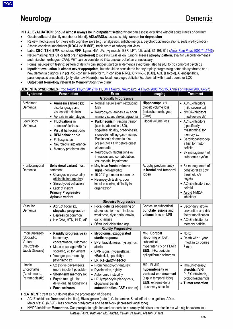
Neurology Dementia
Natalia Festa, Kathleen McFadden, Pavan Vaswani, Méabh O’Hare
INITIAL EVALUATION: Should almost always be in outpatient setting where can assess over time without acute illness or delirium • Obtain collateral (family member or friend), ADLs/IADLs, assess safety, screen for depression• Review medications for those with cognitive s/e’s (e.g., analgesics, anticholinergics, psychotropic medications, sedative-hypnotics)• Assess cognitive impairment (MOCA >> MMSE), track score at subsequent visits• Labs: CBC, TSH, BMP; consider: RPR, Lyme, HIV, UA, hvy metals, ESR, LFT, folic acid, B1, B6, B12 (Amer Fam Phys 2005;71:1745)• Neuroimaging: NCHCT or MRI brain (preferred) to r/o structural lesion (tumor), assess atrophy pattern, eval for vascular dementia
and microhemorrhages (CAA). PET can be considered if dx unclear but often unnecessary.• Formal neuropsych testing: pattern of deficits can suggest particular dementia syndrome; also helpful to r/o comorbid psych dz• Inpatient evaluation is almost never appropriate, but should be considered for any rapidly progressing dementia syndrome or a
new dementia diagnosis in pts <55 (consult Neuro for ?LP, consider RT-QuIC >14-3-3 [CJD], ACE [sarcoid], AI encephalitis,paraneoplastic encephalitis [only after d/w Neuro]), new focal neurologic deficits (?stroke), fall with head trauma or LOC
• Outpatient Neurology referral to Memory/Cognitive clinic
DEMENTIA SYNDROMES (Prog Neurol Psych 2012;16:11, BMJ Neurol, Neurosurg, & Psych 2005;75:v15, Annals of Neurol 2008;64:97)Syndrome Presentation Exam Imaging Treatment
Gradually Progressive Alzheimer Dementia
• Amnesia earliest sx;also language andvisuospatial deficits
• Apraxia in later stages
• Normal neuro exam (excludingMS)
• Neuropsych: amnesia w/ shortmemory span, alexia, agraphia
Hippocampal (+/- global) volume loss; ?microhemorrhages (CAA)
• AChE-inhibitors(mild-severe dz)
• NMDA-inhibitors(mod-severe dz)
Lewy Body Dementia
• Fluctuations inattention/alertness
• Visual hallucinations• REM behavior d/o• Falls/syncope• Neuroleptic intolerance• Memory problems late
• Parkinsonism: resting tremor(can be absent in LBD),cogwheel rigidity, bradykinesia,stooped/shuffling gait – namedParkinson’s dementia if sxpresent for >1 yr before onsetof dementia
• Neuropsych: fluctuations w/intrusions and confabulation,visuospatial impairment
Global volume loss • AChE-inhibitors(specificallyrivastigmine) formemory sx
• Carbidopa/levodopa trial for motordeficits
• Sx management ofautonomic dysfxn
Frontotemporal Dementia
Behavioral variant most common: • Changes in personality
(disinhibition, apathy)• Stereotyped behaviors• Lack of insightPrimary ProgressiveAphasia variant
• May have frontal releasesigns (non-specific)
• 15-20% get motor neuron dz• Neuropsych testing: poor
impulse control, difficulty inorganization
Atrophy predominantly in frontal and temporal lobes
• Sx management ofbehavioral sx (lowthreshold c/spsych)
• AChE-inhibitors nothelpful
• Avoid NMDA-inhibitors
Stepwise Progressive Vascular Dementia
• Abrupt focal sx,stepwise progression
• Depression common• Hx: CVA, HTN, HLD, AF
• Focal deficits (depending onstroke location), can include:weakness, dysarthria, ataxia,gait changes
• Often look older than age
Cortical or subcortical punctate lesions and volume-loss on MRI
• Secondary strokeprevention and riskfactor modification
• AChE-inhibitor formemory deficits
Rapidly Progressive Prion Diseases (Sporadic, Variant Creutzfeldt-Jacob Disease)
• Rapidly progressive sxin memory,concentration, judgment
• Mean onset age ~60 forsporadic, 28 for variant
• Younger pts: more sigpsychiatric sx
• Myoclonus, exaggeratedstartle response
• EPS: bradykinesia, nystagmus,ataxia
• UMN signs (hyperreflexia,+Babinksi, spasticity)
• LP: RT-QuIC>>14-3-3
MRI: Cortical ribboning on DWI, subcortical hyperintensity on FLAIR EEG: 1-Hz periodic epileptiform discharges
• No tx• Death w/in 1 year
(median dx course6 mo)
Limbic Encephalitis (Autoimmune, Paraneoplastic)
• Sx evolve days-weeks(more indolent possible)
• Short-term memory sx• Psych sx: agitation,
delusions, hallucinations• Focal seizures
• Prominent psych features• Dyskinesias, rigidity• Autonomic instability• LP: lymphocytic pleocytosis,
oligoclonal bands,autoantibodies (CSF + serum)
MRI: FLAIR hyperintensity or contrast enhancement (esp in temporal lobe) EEG: extreme delta brush very specific
• Immunotherapy:steroids, IVIG,PLEX, rituximab,cyclophosphamide
• Tumor resection
TREATMENT: treat sx but do not slow the progression of disease • AChE inhibitors: Donepezil (first line), Rivastigmine (patch), Galantamine. Small effect on cognition, ADLs.
Major s/e: GI (N/V/D); less common bradycardia and heart block (increased vagal tone)• NMDA inhibitors: Memantine. Can precipitate agitation and exacerbate neuropsychiatric sx (caution in pts with sig behavioral sx)
185

Neurology Headache & Vertigo
Simona Nedelcu, Pavan Vaswani
PRIMARY HEADACHES: ~90% of benign headaches are either tension (#1) or migraine (#2)
Tension HA: most common (~40% population), ♀>♂. Band-like, radiate forehead to occiput, mild-mod severity, 30m to 7d. Rarely seek eval. (Am Fam Physician 2002;66:797). • Abortives: NSAIDs, Tylenol. Can add caffeine/bultalbital, antiemetic
(metoclopramide, promethazine). Avoid med overuse: no more than 2 d/wk.• Preventatives: amitriptyline, nortriptyline, SSRI, tx OSA, smoking cessation
Migraine HA: sx >3/5 criteria POUND (Pounding, Photo/phonophobia, Onset 4-72hrs, Unilat, N/V, Disabling) (JAMA 2006;296:1274)• Migraine w/aura: 1 reversible sx: visual (scintillating scotoma, visual loss/field deficit), sensory (tingling, numbness), speech lang,
motor (wkness, hemiplegic migraine), basilar (dysarthria, vertigo, ataxia, diplopia), vestibular (vertigo), retinal (monocular field deficit).Can mimic stroke. Aura without migraine headache possible (acephalgic migraine)
• Menstrual migraine: before/during menstruation → tx w/ NSAIDs or sumatriptan(Neurology 2008;70:1555). Consider preventive tx perimenstrually w/ slowtriptan (frovatriptan) 2.5mg qD/BID (begin 2d premenstrally, for total 6d/month)
• Abortives: Avoid overuse HA (maximum 2d/wk). Tx early, escalate stepwise.o Mild/Mod pain: Tylenol, Mg (2g IV), NSAIDs, IVF (Headache 2012;52:467)o Mod/Sev pain: IVF, triptan, antiemetics (metoclopramide, prochlorperazine
10mg IV q8h), VPA 500mg IV, DHE, steroids (dexameth 10-25mg IV x1)(Headache 2012;52:114).
• Preventives: if >3 d/mo, long aura, or disability (Neurology 2012;78:1337)o BB/CCB: Propranolol 20mg BID, inc to 40-160mg/day. Metoprolol 25mg
BID, inc to 50-200mg/day. Verapamil 80mg TID, increase gradually.o Antidepressants: Amitryptiline/nortriptyline 10mg qHS, inc to 150mg.
Venlafaxine 37.5 mg qD, inc to 75-150 mg.o Anticonvulsants: Topiramate 25mg qD, inc gradually 100mg BID. VPA
500-1500 mg qD (avoid both of these in young ♀).o Supplements: magnesium 400mg QD, riboflavin 400mg QD, feverfewo Botox: requires referral to headache clinic
VERTIGO illusion of motion of self or world 2/2 vestib dysfxn; a/w N/V, postural/gait instability. Important to distinguish: central vs peripheral (Am Fam Physician 2017;95:154) • Hx/Exam: duration, episodic/persistent, triggers (position Δ), prior sx, assoc sx (5D’s for brainstem: dysarthria, diplopia, dysphagia,
dysphonia, dysmetria). Orthostatics. Dix-Hallpike. HINTS.• HINTS+ Exam: Everything must be c/w peripheral to be reassuring. In acute vertigo, Sn 97% Sp 85% for stroke (better than MRI!)
Head Impulse (pt looks at your nose, passively rotate head. No saccade = ambiguous. Catchup saccade = peripheral).Nystagmus (unidirectional e.g. always left-beating = peripheral; L-beating in L gaze, R-beating in R gaze, any vertical = central).Test of Skew (cover one eye then other, any vertical skew/correction = central) (Acad Em Med 2013;20:986)
Peripheral Sx: Usually severe nausea, mild imbalance, hearing loss/tinnitus. Ddx: benign positional paroxysmal vertigo (BPPV), infection (labyrinthitis, vesitibular neuritis, herpes zoster oticus), Meniere’s disease, vestibular migraine, otosclerosis, trauma (perilymphatic fistula) Imaging: if exam reassuring, none required
Central Sx: Usually mild nausea, severe imbalance, rare hearing sx. Ddx: cerebral infarction (vertebrobasilar ischemia), TIA, hemorrhage, toxic, cerebellopontine mass (vestib schwannoma, ependymoma, brainstem glioma, medulloblastoma, neurofibromatosis), multiple sclerosis, vestibular migraine Imaging: MRI Brain W/WO, coronal DWI, MRA H&N
• Treatment: metoclopramide, prochlorperazine, meclizine (2 wks max, vestib suppression), lorazepam, diazepam AND Vestibular PT
Acute dizziness
Add sx c/f medical etiology Med w/u
Episodic Ongoing
Position triggered
OrthostaticsDix Hallpike + Supine Roll Test
Hallpike (upbeat torsional) ORSupine Roll (horizontal)c/w BPPV
Other / None:Image to r/o
central lesion
Nystagmus?
Not Position triggered
Medical w/u, consider imaging
HINTS+ exam
No catchup saccade ORDirection changing nystagmus OR
Vertical skew ORNew unilateral hearing deficit
Image to r/o central lesion
+catchup saccade ANDUnidirectional nystagmus AND
No vertical skew ANDNo new unilateral hearing deficit
Likely peripheral
Indications for Imaging (MRI Brain W/ WO): >35 yo, abn neuro exam, acute, severe, positional, wrse w exertion, immunosuppressed, wakes at night Vessel imaging (MRA Head and Neck): vestibular, brainstem, retinal sx, motor sx
Triptans Nasal: Sumatriptan 5-20mg q2hrs (max 40mg/day), Zolmitriptan 5mg q2hrs (max 10mg/d) SC: Sumatriptiptan 4-6mg q1hr (max 12mg/d; 70-80% pts w/ sx reduction; 35% resolution) PO: Sumatriptan 25-100mg q2hr (max 200mg/day), Zolmitriptan 1.25-2.5 q2hrs (max 10mg/d) C/I in ischemic CVD/CAD 2/2 vasoconstriction, PVD, liver disease, basilar migraine, MAOIs within 2 wks No more than 4x/d, no more than 2d/wk Caution w/ SSRIs 2/2 risk of serotonin syndrome
DHE (Dihydroergotamine) IV: 0.5-1mg IV Q8h x5 days preferred to x1 dose (max 3mg/day) Nasal: 1 spray /nostril q15min (max 6 spray/day, 8/week); SC: 1mg SC q1hr (max 3mg/day) Same contraindications as Triptans, do not mix with triptans w/in 24hrs
Approach to Acute Dizziness
186
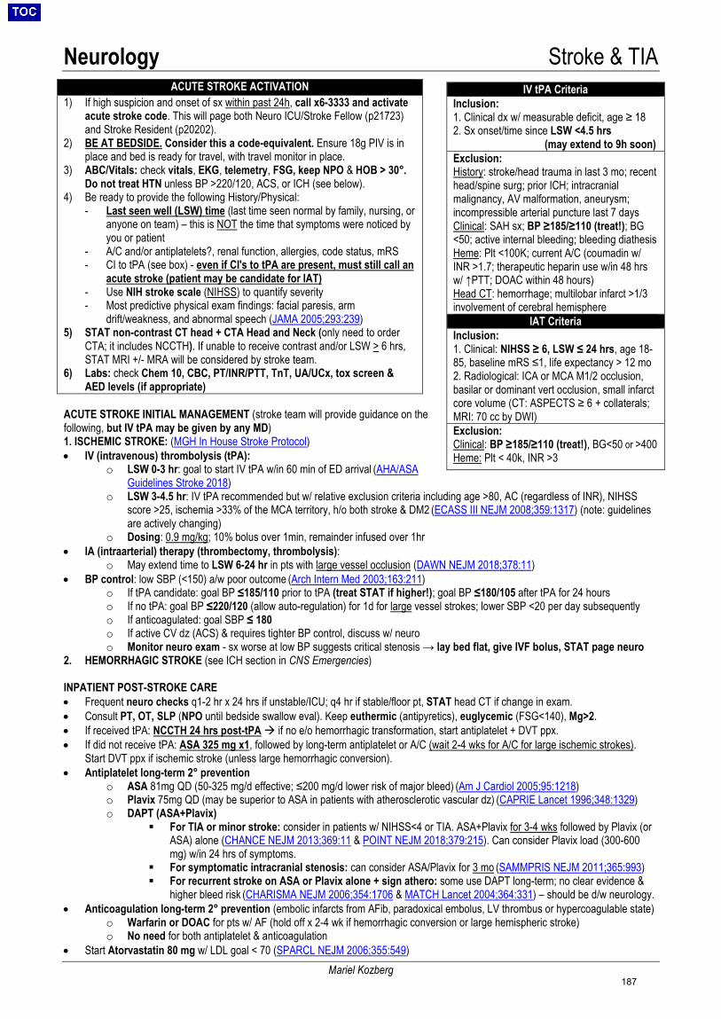
Neurology Stroke & TIA
Mariel Kozberg
ACUTE STROKE INITIAL MANAGEMENT (stroke team will provide guidance on the following, but IV tPA may be given by any MD) 1. ISCHEMIC STROKE: (MGH In House Stroke Protocol)• IV (intravenous) thrombolysis (tPA):
o LSW 0-3 hr: goal to start IV tPA w/in 60 min of ED arrival (AHA/ASAGuidelines Stroke 2018)
o LSW 3-4.5 hr: IV tPA recommended but w/ relative exclusion criteria including age >80, AC (regardless of INR), NIHSSscore >25, ischemia >33% of the MCA territory, h/o both stroke & DM2 (ECASS III NEJM 2008;359:1317) (note: guidelinesare actively changing)
o Dosing: 0.9 mg/kg; 10% bolus over 1min, remainder infused over 1hr• IA (intraarterial) therapy (thrombectomy, thrombolysis):
o May extend time to LSW 6-24 hr in pts with large vessel occlusion (DAWN NEJM 2018;378:11)• BP control: low SBP (<150) a/w poor outcome (Arch Intern Med 2003;163:211)
o If tPA candidate: goal BP ≤185/110 prior to tPA (treat STAT if higher!); goal BP ≤180/105 after tPA for 24 hours
o If no tPA: goal BP ≤220/120 (allow auto-regulation) for 1d for large vessel strokes; lower SBP <20 per day subsequentlyo If anticoagulated: goal SBP ≤ 180o If active CV dz (ACS) & requires tighter BP control, discuss w/ neuroo Monitor neuro exam - sx worse at low BP suggests critical stenosis → lay bed flat, give IVF bolus, STAT page neuro
2. HEMORRHAGIC STROKE (see ICH section in CNS Emergencies)
INPATIENT POST-STROKE CARE • Frequent neuro checks q1-2 hr x 24 hrs if unstable/ICU; q4 hr if stable/floor pt, STAT head CT if change in exam.• Consult PT, OT, SLP (NPO until bedside swallow eval). Keep euthermic (antipyretics), euglycemic (FSG<140), Mg>2.• If received tPA: NCCTH 24 hrs post-tPA if no e/o hemorrhagic transformation, start antiplatelet + DVT ppx.• If did not receive tPA: ASA 325 mg x1, followed by long-term antiplatelet or A/C (wait 2-4 wks for A/C for large ischemic strokes).
Start DVT ppx if ischemic stroke (unless large hemorrhagic conversion).• Antiplatelet long-term 2° prevention
o ASA 81mg QD (50-325 mg/d effective; ≤200 mg/d lower risk of major bleed) (Am J Cardiol 2005;95:1218)o Plavix 75mg QD (may be superior to ASA in patients with atherosclerotic vascular dz) (CAPRIE Lancet 1996;348:1329)o DAPT (ASA+Plavix)
For TIA or minor stroke: consider in patients w/ NIHSS<4 or TIA. ASA+Plavix for 3-4 wks followed by Plavix (orASA) alone (CHANCE NEJM 2013;369:11 & POINT NEJM 2018;379:215). Can consider Plavix load (300-600mg) w/in 24 hrs of symptoms.
For symptomatic intracranial stenosis: can consider ASA/Plavix for 3 mo (SAMMPRIS NEJM 2011;365:993) For recurrent stroke on ASA or Plavix alone + sign athero: some use DAPT long-term; no clear evidence &
higher bleed risk (CHARISMA NEJM 2006;354:1706 & MATCH Lancet 2004;364:331) – should be d/w neurology.• Anticoagulation long-term 2° prevention (embolic infarcts from AFib, paradoxical embolus, LV thrombus or hypercoagulable state)
o Warfarin or DOAC for pts w/ AF (hold off x 2-4 wk if hemorrhagic conversion or large hemispheric stroke)o No need for both antiplatelet & anticoagulation
• Start Atorvastatin 80 mg w/ LDL goal < 70 (SPARCL NEJM 2006;355:549)
ACUTE STROKE ACTIVATION 1) If high suspicion and onset of sx within past 24h, call x6-3333 and activate
acute stroke code. This will page both Neuro ICU/Stroke Fellow (p21723)and Stroke Resident (p20202).
2) BE AT BEDSIDE. Consider this a code-equivalent. Ensure 18g PIV is inplace and bed is ready for travel, with travel monitor in place.
3) ABC/Vitals: check vitals, EKG, telemetry, FSG, keep NPO & HOB > 30°.Do not treat HTN unless BP >220/120, ACS, or ICH (see below).
4) Be ready to provide the following History/Physical:- Last seen well (LSW) time (last time seen normal by family, nursing, or
anyone on team) – this is NOT the time that symptoms were noticed byyou or patient
- A/C and/or antiplatelets?, renal function, allergies, code status, mRS- CI to tPA (see box) - even if CI's to tPA are present, must still call an
acute stroke (patient may be candidate for IAT)- Use NIH stroke scale (NIHSS) to quantify severity- Most predictive physical exam findings: facial paresis, arm
drift/weakness, and abnormal speech (JAMA 2005;293:239)5) STAT non-contrast CT head + CTA Head and Neck (only need to order
CTA; it includes NCCTH). If unable to receive contrast and/or LSW > 6 hrs,STAT MRI +/- MRA will be considered by stroke team.
6) Labs: check Chem 10, CBC, PT/INR/PTT, TnT, UA/UCx, tox screen &AED levels (if appropriate)
IV tPA Criteria Inclusion: 1. Clinical dx w/ measurable deficit, age ≥ 182. Sx onset/time since LSW <4.5 hrs
(may extend to 9h soon) Exclusion: History: stroke/head trauma in last 3 mo; recent head/spine surg; prior ICH; intracranial malignancy, AV malformation, aneurysm; incompressible arterial puncture last 7 days Clinical: SAH sx; BP ≥185/≥110 (treat!); BG <50; active internal bleeding; bleeding diathesis Heme: Plt <100K; current A/C (coumadin w/ INR >1.7; therapeutic heparin use w/in 48 hrs w/ ↑PTT; DOAC within 48 hours) Head CT: hemorrhage; multilobar infarct >1/3 involvement of cerebral hemisphere
IAT Criteria Inclusion: 1. Clinical: NIHSS ≥ 6, LSW ≤ 24 hrs, age 18-85, baseline mRS ≤1, life expectancy > 12 mo2. Radiological: ICA or MCA M1/2 occlusion,basilar or dominant vert occlusion, small infarctcore volume (CT: ASPECTS ≥ 6 + collaterals;MRI: 70 cc by DWI)Exclusion: Clinical: BP ≥185/≥110 (treat!), BG<50 or >400 Heme: Plt < 40k, INR >3
187
Neurology Stroke & TIA
Mariel Kozberg
• Consider Fluoxetine 20 mg daily x 90d for motor recovery if ischemic stroke w/ hemiparesis/plegia (FLAME Lancet Neurol2011;10:123), but recent data does not support this use (FOCUS Lancet 2019;393:265).
• Work up cause/secondary prevention: (see additional details below in Special Considerations by Stroke Type)o Labs: lipids, A1c, TSH, ESR/CRP; if <60 y/o, send tox screen (cocaine), hypercoagulability w/u (if recommended by neuro)o Imaging: head and neck CTA or MRA (can do TOF if low GFR); carotid US as alternativeo Cardiac workup: EKG, TTE, inpatient telemetry followed by 30-day MCOT vs. LINQ if tele is negative for AFib
SPECIAL CONSIDERATIONS BY STROKE TYPE CARDIOEMBOLIC STROKE
SUSPECT WHEN: • ACA/MCA/PCA occlusion w/o sig vascular dz• Infarcts in multiple territories or cerebellar stroke• Known risk factors (LA/LV thrombus, AFib,
LVEF<25%, aortic disease, intracardiac shunt)• Hypercoagulability/hyperviscosity (solid organ or
heme malignancy, HbSS, cryo, clotting d/o)
DIAGNOSTIC WORKUP: • TTE (w/ bubble if pt < 60 y/o) - if PFO, r/o venous thrombus (LENIs/
MRV pelvis), can consider closure (RESPECT NEJM 2017;377:1022)• Inpatient telemetry followed by 30-day MCOT vs. LINQ at discharge
(unless known AFib)ACUTE MANAGEMENT CONCERNS: • Avoid immediate AC unless known intracardiac thrombus or
mechanical valve. Transition to long-term AC in 2-4 weeks.SYMPTOMATIC CAROTID STENOSIS
SUSPECT WHEN: • Carotid stenosis present on ipsilateral side• H/o amaurosis fugaxDIAGNOSTIC WORKUP:• CTA vs. MRA head & neck usually sufficient• Alternatives: carotid US (typically need carotid US
prior to consideration of CEA)
ACUTE MANAGEMENT CONCERNS: • If >50% symptomatic carotid stenosis consider carotid
revascularization (stent/angioplasty/endarterectomy) – ideally w/in 2weeks of sx (NASCET II NEJM 1998;339:1415)
• Consider anticoagulation if territory to be saved (d/w neurology)• Consider induced HTN if symptoms fluctuate with BP
INFECTIVE ENDOCARDITIS SUSPECT WHEN: • Unexplained fever w/ stroke in pt with valvular dzDIAGNOSTIC WORKUP:• Blood cultures, TTE followed by TEE if neg• CTA head to identify mycotic aneurysms (↑risk
bleeding)• If CTA negative, may need conventional angio
(CTA not as sensitive for mycotic aneurysms)
ACUTE MANAGEMENT CONCERNS: • Immediate antibiotics; caution with tPA• Early cardiac surgery if small non-hemorrhagic stroke; delayed cardiac
surgery (2-4 wk) if large or hemorrhagic stroke• Avoid anticoagulation or antiplatelet w/o a separate indication
CAROTID AND VERTEBRAL DISSECTIONS SUSPECT WHEN: • <60 y/o or posterior circulation stroke in pt w/o RFs• Neck pain, HA, or Horner’s syndrome• Trauma (vertebral fx), chiropractor, coughing spellsDIAGNOSTIC WORKUP:• CTA vs. MRA with T1 fat saturation• Consider comorbid conditions (Marfan’s, FMD)
ACUTE MANAGEMENT CONCERNS: • Goal of tx is to prevent stroke: highest risk in first few days• Immediate anticoagulation (if early) followed by antiplatelet. Prefer
antiplatelet if: sx onset >3d ago, dissection extends intradurally (no ACdue to risk of SAH), large infarct (risk of hemorrhage) (CADISS LancetNeurol 2015;14:361)
• High rate of recanalization Tx 3 months then re-image vesselCEREBRAL VENOUS SINUS THROMBOSIS
SUSPECT WHEN: • Positional HA, vomiting, papilledema, vision Δ• P/w SZ (common, may be difficult to control)DIAGNOSTIC WORKUP:• NCCTH: hyperdensity in torcula (dense delta sign)• CTV vs. MRV to assess intracranial venous system• Consider hypercoagulable workup
ACUTE MANAGEMENT CONCERNS: • Immediate anticoagulation even in presence of hemorrhage• AEDs if seizures (not indicated for ppx)• IV fluids, avoid dehydration, modify risk factors (smoking, OCPs)
TRANSIENT ISCHEMIC ATTACK (TIA) • Definition: Transient neurologic dysfunction caused by focal brain, spinal cord, or retinal ischemia w/o acute infarction or end-organ
injury as assessed clinically or by imaging• Causes: Atherothrombotic stenosis (ICA, vertebral, basilar, small vessel), embolic (arterial, aortic, cardiac, paradoxical), dissection
(ICA, vertebral) - identification will guide tx (antiplatelet therapy vs. search for underlying arrhythmia +/- anticoagulation)• Imaging: MRI (w/ DWI/ADC) w/in 24hr of sx onset and vessel imaging of head and neck for large vessel occlusive disease (e.g. MRA
(time of flight if low GFR) vs. CTA vs. carotid ultrasound• Cardiac w/u: TTE to excl thrombus & PFO (age <60) & tele/MCOT vs. LINQ monitoring to exclude AFib if suspected embolic TIA• ABCD2 score (Age, BP, Clinical features, Sx Duration, Diabetes) used to identify pts w/ high risk of ischemic stroke w/in 1 week of TIA• Management: Immediate intervention reduces the risk of recurrent stroke (1.5-3.5% risk within 48h), see 2° Prevention above
188
Neurology CNS Emergencies
Michael Young
Intracranial hemorrhage (ICH): epidural (EDH), subdural (SDH), subarachnoid (SAH), intraparenchymal hemorrhages (IPH) • Causes: trauma (all), HTN (IPH), ruptured aneurysm/AVM (SAH, IPH), cerebral amyloid (IPH), tumor (IPH; most common w/ met
breast Ca, lung Ca, melanoma, RCC, choroid, thyroid CA’s), cortical vein thrombosis (IPH), venous sinus thrombosis (IPH)• Presentation: typically acute focal neuro deficit, +/- progressive ↓consciousness, N/V. SAH: thunderclap HA, N/V, meningismus;
EDH/SDH: s/p trauma, lucid interval with EDH; IPH: focal neuro symptoms (may mimic ischemic stroke clinically), often with HA.• Tests: STAT imaging (NCHCT head for all; +CTA head if SAH/IPH), coags/PLTs; need f/u scan at 6h to assess progression• STAT Management:
− STAT Neurosurg (p21111) if SAH/SDH/EDH; otherwise, Neuro (acute stroke: p34282; non-acute stroke: p20202; in ED: p20000).− Elevate HOB to 30-45° to reduce ICP and prevent aspiration.− BP control: SBP < 140 (studied in SAH or ICH due to ruptured aneurysm/AVM), use IV labetalol or nicardipine drip (avoid
hydralazine if possible), place arterial line (INTERACT Lancet Neurol 2008;7:391, ATACH Crit Care Med 2010;38:637)− Correct coag: warfarin/INR>1.5: tx [vitamin K 10 mg IV x 1] AND [3-5U FFP or Kcentra] (call blood bank); ↓Plt (transfuse, goal
>50); uremia/antiplt use: consider DDAVP 0.3mcg/kg IV, heparin/LMWH (protamine), s/p tPA (check fibrinogen, give cryo, +/-amicar), rivaroxaban or apixaban: give Andexanet Alfa (Andexxa); dosing based on size and timing of last dose (call pharmacy)
− Venous sinus thrombosis (VST): anticoagulate w/ LMWH/heparin despite hemorrhage (Lancet 1991;338:597). Manage ↑ICPand seizures.
− Prognosis depends on age, GCS, pre-ICH cognitive impairment and ICH volume/location (FUNC Score) (Stroke 2008;39:2304)− Typically acceptable to restart DVT ppx in smaller hemorrhages if stable after 48hrs, but confirm with consultants
Elevated Intracranial Pressure (ICP) / Herniation • Etiologies: Mass (tumor, abscess, hemorrhage), cerebral edema (massive infarction, hyperammonemia, DKA), hydrocephalus
(tumor, intraventricular hemorrhage, leptomeningeal disease, meningitis), PRES. If ICP severely elevated then herniation(displacement and compression of brain tissue) ensues. LP in setting of significantly raised ICP may also result in herniation.
• Signs of hernation: fixed/dilated/asymmetric pupil (often 1 first) accompanied by nausea, somnolence/confusion, limited upgaze;flexor/extensor posturing; ipsilateral hemiparesis (uncal herniation); Cushing’s triad (bradycardia, HTN, & irregular breathing)
• Tests: STAT head CT (call x63050)• Management:
− STAT Neurosurg. (p21111) (for ICP monitor/EVD placement/decompressive hemicraniectomy)− Secure ABCs, elevate HOB to 30-45˚, keep head midline (to secure venous drainage), treat pain/agitation− Neuro-ICU level monitoring (p20202 can help coordinate)− Hyperventilate to PaCO2 ~ 30-35 mmHg (if suspect herniation, transiently reduceds ICP), only for short-term management− IV mannitol therapy 1g/kg q6h (use with caution in pts on HD) and/or 23% saline 15cc q6h. Check BMP, Sosm q6h. No mannitol
if osm gap >15, Na >160, or serum osm >340. No 23% saline if Na >160.− If related to edema from malignancy or bacterial infection, give 10mg IV dexamethasone x 1, then 4mg q6hrs− Complications during LP: if sx of herniation/opening pressures > 40 cm H2O, consider STAT head CT. Immediately replace
stylet into needle, only drain CSF in the manometer, STAT Neurosurgery (p21111).
Hypertensive Encephalopathy: PRES (“posterior reversible” encephalopathy syndrome) • Typically associated with severe HTN, but also relative HTN in setting of preeclampsia/eclampsia, cytotoxic/immunosuppressive
drugs (Cyclosporine, Tacrolimus, Cisplatin, Bevacizumab), acute/chronic renal failure, uremia, sepsis, vasculitides, TTP→ due toimpaired cerebral autoregulation and endothelial dysfunction, hypomagnesemia (NEJM 1996;334:494).
• Symptoms: HA, confusion, decreased consciousness, visual disturbances, seizures, can result in ICH and ↑ICP• Tests: Brain MRI +gad: FLAIR shows vasogenic edema w/in white matter in the posterior cerebral hemispheres; DWI/ADC normal
(but also can have strokes); additional regions can be involved including brainstem, cerebellum, basal ganglia, frontal lobes• Management: ICU if severe, strict BP control (reduce 25% daily, if severe use nicardipine or labetalol drip), treat seizures, Mg2+ (esp
in eclampsia), remove inciting factor• Prognosis: Often fully reversible; complications include progressive cerebral edema, ICH, stroke, death
Cord Compression: High level of suspicion in cancer patients with back pain, urinary sx or LE weakness • Etiologies: Subacute (tumor/mets, abscesses) vs acute (disc herniation, trauma, hemorrhage)• Symptoms: Back pain, motor weakness, hyperreflexia below lesion (*can be hypo in acute or w/cauda equina), +Babinski, loss of
sensation (typically c/w dermatome or level), bowel/bladder incontinence OR retention, loss of rectal tone, saddle anesthesia• Tests: STAT whole spine MRI w/contrast (cord compression protocol), call ED MRI (x63050) or inpt MRI (x64226)• STAT page NSG/Ortho spine +/- Rad Onc (x68652) for possible XRT if tumor-related• Dexamethasone (10mg IV x1 then 4mg IV q6h), esp in malignancy; solumedrol in acute cord injury 2/2 trauma is controversial
Other CNS Emergencies • Respiratory failure i/s/o neuromuscular disorders: See Weakness & Neuromuscular Disorders• NMS and serotonin syndrome: See Catatonia/NMS/Serotonin Syndrome• Status epilepticus: See Seizures/Epilepsy• Stroke: See Stroke.
189
Neurology Seizures
Amrit Misra & Sattar Khoshkhoo
Definitions (Epilepsia 2005;4:470, Continuum 2013;19:767, MGH Status Epilepticus Treatment Protocol): • Epilepsy: brain d/o characterized by enduring predisposition to generate seizures (unprovoked)• Status Epilepticus: at least 5 mins of continuous seizure or 2+ seizures w/ incomplete recovery of consciousness in between• Non-convulsive Status Epilepticus: non-convulsive electrographic seizure ≥10s or rhythmic EEG responsive to seizure treatment• Tonic: persistent flexion/extension; Clonic: limb jerking; Atonic: loss of postural tone; Myoclonic: sudden brief muscle contraction• Psychogenic Non-Epileptic Seizures (PNES): Important to distinguish from epileptic events. Features that are common in PNES
but rare in epilepsy: opisthotonus (arched back), undulating/asynchronous motor activity, eyes closed with resistance to lid opening,and side-to-side head movements (Epilepsy & Behav 2003;3:205)
• Classification (Epilepsia 2010;4:676):o Focal: Unilateral, occuring in one hemisphere +/- impaired consciousness (formerly simple partial, complex partial)o Generalized: Occuring in and rapidly engaging b/l distributed networks (formerly tonic-clonic, atonic, myoclonic, absence)
Etiology: Provoked vs Not? Primary epilepsy, vascular (stroke/ischemia/hemorrhage), withdrawal (EtOH/BZDs), mass lesions (tumor, abscess), trauma, metabolic (↓ glc, ↑CO2, ↓O2, ↓Ca), meds, infection (systemic and CNS), HTN or HoTN, high fever, eclampsia, PRES • Ddx: syncope, TIA, migraine, PNES (~30% also have epilepsy), myoclonus, dystonia• H&P: Previous sz history, prodrome (palpitation, sweating, N/V, aura), med list (many lower threshold), triggers (exertion,
pain/fatigue/emotional stress, cough/urination/defecation), tongue biting, incontinence, lateralizing signs, alcohol. GET COLLATERAL.• Labs: FSBG, Tox, AED levels, Lytes, CBC, LFTs/albumin, NH3, ABG, CK, INR, lactate, troponin, blood cx, b-hcg. Prl Sn ~50%• Monitoring: Tele (↑ risk for fatal cardiac arrhythmias during ictal/post-ictal period; ictal arrhythmias ↑ risk of sudden death)• Neuroimaging: Obtain in all w/ unprovoked 1st seizure (MRI more sens) (Neuro 2015;84:1705) or in any w/ focal neuro exam, h/o
trauma, malignancy, HIV or focal seizure (Neuro 2007;69:1772). Imaging changes management in ~10% (Neuro 2007;69:1996).• LP/BCx: If febrile, HIV/immunocompromised, or if no clear etiology• EEG: Within 24h-48h if not seizing, emergent EEG if seizing: DO NOT wait to manage. If emergent, contact EEG fellow (p16834)
Treatment of status epilepticus: ABCs:
VS, O2, EKG; Assess pt safety;
Place 2 PIVs (BZD + PHT incompatible)
First 5-10 min: Ativan 4 mg IV over 2 min →
repeat 4 mg IV q5min prn; If no IV, diazepam 20 mg PR or midaz 10 mg IM/nasal/buccal
Correct reversible causes:
FSBG (start IV thiamine + D50), lytes, fever, O2
Concurrently: Call neuro, RICU, continuous EEG;
Check AED levels and rebolus if needed → PHT, VPA, PHB 1 hr after load,
FOS-PHT 2 hrs after load (correct for alb); tx underlying cause
Persistent SZ (10-30min): Levetiracetam, VPA, fosphenytoin/phenytoin, phenobarb, +/-
lacosamide (need pre/post EKG to check PR)
Refractory SZ (30-60 min): Intubate, continuous EEG
Midaz (if HD unstable) +/- propofol gtt
Seizure PPX: No AED in 1st seizure unless abnormal EEG OR abnormal imaging (Epilepsia 2008;49:13). Early AED only reduces short term recurrence risk (<2yr) and is unlikely to change prognosis of sustained remission (3+ yrs) (Neuro 2015;84:1705).• ETOH seizure: Ppx not indicated when intoxication or withdrawal is the cause of seizure (Neuro 2006;67:s45)• Brain tumor: No ppx. If seizures occur, start AEDs: Keppra > Lacosamide (fewer chemo interactions)• TBI: Keppra 500-750 mg BID x 7 days (Neurosurg Focus 2008;25:E3)• ICH: AED only if clinical seizure or traumatic etiology, Keppra 500mg BID x 7days• PNES: Treatment with outpatient Cognitive Behavioral Therapy (CBT), psychiatry involvement. In acute setting it may be helpful to
educate patients about functional neurologic symptoms (http://www.neurosymptoms.org), and place social work consult.• In MA, No Driving for LOC event until 6 mos seizure free. Counsel pt and include in discharge summary.
AED Loading Dosing Goal Level Side Effects Levetiracetam (Keppra)
60mg/kg 1:1 PO:IV No goal, level to check adherence
Psychiatric sx (irritability, anxiety, depression, sedation, psychosis).
Valproic acid (Depakote)
20-40mg/kg 1:1 PO:IV 50-100 mcg/mL(>1h post load)
Teratogenic. Abnormal LFTs, weight gain, N/V, encephalopathy (↑ NH3), pancreatitis, thrombocytopenia. Good for mood disorders.
Phenytoin (Dilantin), Fosphenytoin
20 pheny equiv/kg
1:1 PO: IV
10-20 mcg/mL,correct for alb,(2h post load)
Teratogenic. Gingival hypertrophy, hair growth, rash, AMS, diplopia, ataxia, slurred speech, hypotension/arrythmia (if run faster than 50mg/min; Fosphenytoin is less cardiotoxic).
Lamotrigine (Lamictal) No Load 1:1 PO:IV 1-13 mg/L Rash, SJS, nausea, somnolence, dizziness, ataxia. Good in mood disorders.
Topiramate (Topamax) No Load Only PO N/A Weight loss, fatigue, teratogenic. Nephrolithiasis, cognitive, anxiety, anorexia, tremor
Lacosamide (Vimpat) 200-400mg 1:1 PO:IV N/A Headache, diplopia, dizziness, nausea, hypotension. Obtain EKG after load, and watch for PR prolongation.
Carbamazepine (Tegretol)
No Load Only PO 4-12 mcg/mL SIADH, N/V/D, rash, pruritis, fatigue, blurred vision, diplopia, lethargy.
190
Neurology Weakness & Neuromuscular Disorders
Chris Cashman, Méabh O’Hare
APPROACH TO WEAKNESS • Ask about functional issues (getting out of chair, tripping over curbs/stairs)• UMN signs: spasticity, increased tone, hyperreflexia, + Babinski | LMN signs: fasciculations, atrophy, decreased tone, hyporeflexia• Pattern: UMN (extensors in UEs, flexors in LEs), proximal (many myopathies), bulbar (dysphagia, dysarthria, diplopia)• Associated Sensory Sx: reduced sensation, tingling, burning, allodynia, hyperalgesia, decreased temperature sense, imbalance• Autonomic Sx: orthostasis, constipation, urinary retention, erectile dysfunction, changes in sweating, hair loss, post-prandial nausea• EMG/NCS: Can be helpful with localization, determining fiber type involved, determining if disease is axonal vs demyelinating (which
guides tx), and determining injury extent (which guides prognosis). Often higher yield at least 2-3 weeks into illness and as outpt.Localization Associated Sx/Signs Diagnostics Important/Common Causes
Brain Cortical signs (language/visual field/neglect), cerebellar sx, UMN signs
MRI Brain best initial test (+gado if c/f cancer, infxn, demyelinating dz)
Vascular (hemorrhage or ischemia), tumor, trauma, demyelinating
Spinal Cord Sensory level, bowel/bladder dysfxn, UMN signs.
MRI Spine (level based on sx, +gado if c/f cancer, infxn, demyelinating dz) CSF if c/f inflammatory or infxn
Transverse myelitis (MS, NMO, connective tissue dz), infxn (viral myelitis, HTLV), compression (tumor/disc/abscess), vascular, trauma, paraneoplastic, toxic, ↓B12/Cu
Anterior horn cell
LMN signs. If motor neuron dz: both UMN and LMN signs.
NCS/EMG +/- MRI brain and spine; LP
ALS, SMA, polio, acute flaccid myelitis (pediatric)
Radiculopathy Motor/sensory sx corresponding to nerve root. +Radiating pain.
MRI Spine (level based on sx) LP if polyradiculopathy NCS/EMG: helpful for localization (sensitivity imperfect clinical dx)
Nerve root compression (disc herniation, spondylosis) by far most common; polyradiculopathy: GBS, iatrogenic (post-op, chemo), ischemic, infxn (HIV, Lyme, CMV, EBV), DM (typically thoracic), sarcoid, malig.
Peripheral Neuropathy
Sensory symptoms; autonomic dysfxn if small fibers affected. Often symmetric and length dependent. GBS is ascending.
Labs: highest yield = A1c, B12 + MMA, SPEP + immunofixation Add’l labs (in select pts): Lyme, RPR, HIV, malnutrition (B1, B6, vit E, B3, Cu), vasculitis (ANCA, ANA, ESR, CRP, RF, C3/C4), celiac, ACE NCS/EMG: localization & pattern (NB: nL NCS doesn’t exclude small fiber dz)
Symmetric/length dependent: toxic/metabolic/nutritional (DM, chemo, ETOH, ↓B12, critical illness), paraprotein-related, hereditary (CMT); Polyradiculoneuropathy: GBS/CIDP, DM, Lyme; Mononeuropathy: compression/trauma; Mononeuropathy multiplex: vasculitis, amyloid, sarcoid, HNPP
NMJ Weakness is fatigable and improves with rest. A/w ptosis, diplopia. No sensory sx.
Ice pack test, tensilon (rarely) Labs: myasthenia panel (see below) NCS/EMG: repetitive stimulation, single fiber EMG CT chest I+ if above c/w myasthenia
Myasthenia Gravis, Lambert–Eaton, botulism, tick paralysis
Myopathy Proximal weakness most common. Pain uncommon.
Initial Labs: CK, aldolase, LDH, LFTs, TSH/fT4, PTH, ESR/CRP EMG: e/o muscle irritability, chronicity May need muscle biopsy
Critical illness, medication-related (steroids, statins, colchicine, cyclosporine, NRTI), inflammatory myopathies (Inclusion Body Myositis, Dermatomyositis, Polymyositis)
GUILLAIN-BARRÉ SYNDROME (Acute Inflammatory Demyelinating Polyradiculoneuropathy): Symmetric, ascending numbness & weakness. Absent reflexes (may be present acutely), facial weakness, autonomic instability, acute resp failure in 30% of patients. • Causes: Often recent infection (Campylobacter jejuni, HIV, CMV, EBV, Zika) or vaccination (rare)• Dx: LP w/ albuminocyt. dissoc. (high protein, norm WBCs). Anti-GQ1B in CMF variant. NCS/EMG: highest yield 2-3wks after sx onset• Tx: IVIg or plasmapheresis (equiv. outcomes); monitor respiratory function with NIF/VC TID to qD (done by RT)-more frequent if crisis• Elective intubation w/ 20-30-40 Rule: VC <20mL/kg, NIF weaker than -30cm H20, MEF <40 cm H20, OR ≥ 20% decline in ~24 hrsMYASTHENIA GRAVIS/LAMBERT EATON (MG/LEMS): Weakness of voluntary muscles, worse w/ exertion & repetitive movements andin the evening. Typically involves ocular (ptosis, diplopia), bulbar, respiratory, neck and proximal>distal limb muscles.• Cause: Auto-Abs against postsynaptic ACh-R in skeletal muscle (MG) or Voltage Gated Calcium Channels (LEMS)• Exam: Upgaze fatiguability – hold sustained upgaze for 1 min, look for development of ptosis. After observing ptosis, place ice on
eyes for 1 min, weakness will improve (Tensilon test rare, requires atropine at the bedside. Only improves MG not LEMS).• Dx: Ach-R Ab (80-90% seropositive, specific); if neg check anti-MUSK. EMG/NCS: Order w/repetitive stim; will show decremental
response (MG) or potentiation (LEMS). Chest CT I+: r/o thymoma (in 70-80% MG). Find underlying malignancy in LEMS.• Tx: Symptomatic (pyridostigmine); immunotherapy: rapid (IVIg, plasmapheresis), chronic (steroids+/-azathioprine/MMF); thymectomyMYASTHENIC CRISIS: MG exacerbation requiring intubation or delayed extubation post-surgery• Triggers: surgery, infection, IV contrast, pregnancy, certain drugs/meds (antibiotics including fluoroquinolones, aminoglycosides;
anticonvulsants; antipsychotics; BBs; CCBs; mag). AVOID succinylcholine during intubation.• Respiratory failure: bedside exam → follow number counting in single breath, assess cough/swallowing. Trend mechanics with RT:
NIFs/VC as above (see 20-30-40 Rule). Aggressive pulm toilet. HOLD pyridostigmine if bulbar sx and/or intubated (can ↑ secretions).
191
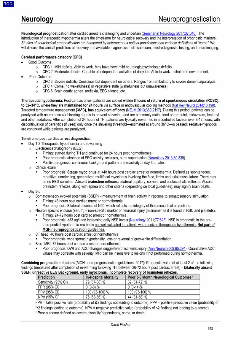
Neurology Neuroprognostication
David Fischer
Neurological prognostication after cardiac arrest is challenging and uncertain (Seminar in Neurology 2017;37:040). The introduction of therapeutic hypothermia alters the timeframe for neurological recovery and the interpretation of prognostic markers. Studies of neurological prognostication are hampered by heterogenous patient populations and variable definitions of “coma”. We will discuss the clinical predictors of recovery and available diagnostics – clinical exam, electrodiagnostic testing, and neuroimaging.
Cerebral performance category (CPC) • Good Outcome:
o CPC 1. Mild deficits. Able to work. May have have mild neurologic/psychologic deficits.o CPC 2. Moderate deficits. Capable of independent activities of daily life. Able to work in sheltered environment.
• Poor Outcome:o CPC 3. Severe deficits. Conscious but dependent on others. Ranges from ambulatory to severe dementia/paralysis.o CPC 4. Coma (no wakefulness) or vegetative state (wakefulness but unawareness).o CPC 5. Brain death: apnea, areflexia, EEG silence, etc.
Therapeutic hypothermia: Post-cardiac arrest patients are cooled within 6 hours of return of spontaneous circulation (ROSC), to 32–34°C, where they are maintained for 24 hours via surface or endovascular cooling methods (Nat Rev Neurol 2014;10:190). Targeted temperature management (36°C), has equivalent efficacy (NEJM 2013;369:2197). During this period, patients can be paralyzed with neuromuscular blocking agents to prevent shivering, and are commonly maintained on propofol, midazolam, fentanyl and other sedatives. After completion of 24 hours of TH, patients are typically rewarmed in a controlled fashion over 8-12 hours, with discontinuation of paralytics (if used) only once the shivering threshold—estimated at around 36°C—is passed; sedative-hypnotics are continued while patients are paralyzed.
Timeframe post cardiac arrest diagnostics: • Day 1-2 Therapeutic hypothermia and rewarming
o Electroencephalography (EEG) Timing: started during TH and continued for 24 hours post normothermia. Poor prognosis: absence of EEG activity, seizures, burst suppression (Neurology 2013;80:339). Positive prognosis: continuous background pattern and reactivity at day 3 or later.
o Clinical exam Poor prognosis: Status myoclonus at <48 hours post cardiac arrest or normothermia. Defined as spontaneous,
repetitive, unrelenting, generalized multifocal myoclonus involving the face, limbs and axial musculature. There maybe no EEG correlate. Absent brainstem reflexes: bilateral pupillary, corneal, and oculocephalic reflexes. Absentbrainstem reflexes, along with apnea and other criteria (depending on local guidelines), may signify brain death.
• Day 3-5o Somatosensory evoked potentials (SSEP) – measurement of brain activity in reponse to somatosensory stimulation Timing: 48 hours post cardiac arrest or normothermia. Poor prognosis: Bilateral absence of N20, which reflects the integrity of thalamocortical projections.
o Neuron specific enolase (serum) – non-specific marker of neuronal injury (misnomer as it is found in RBC and platelets). Timing: 24-72 hours post cardiac arrest or nomothermia. Poor prognosis: >33 ug/l and increasing daily NSE levels (Neurology 2011;77:623). NSE is prognostic in the pre-
therapeutic hypothermia era but is not well validated in patients who received therapeutic hypothermia. Not part ofMGH neuroprognostication guidelines.
o CT head, 48 hours post cardiac arrest or normothermia Poor prognosis: wide spread hypodensity, loss or reversal of grey-white differentiation.
o Brain MRI, 72 hours post cardiac arrest or normothermia Poor prognosis: DWI and ADC changes suggestive of ischemic injury (Ann Neurol 2009;65:394). Quantitative ADC
values may correlate with severity. MRI can be insensitive to lesions if not performed during normothermia.
Combining prognostic indicators (MGH neuroprognostication guidelines, 2017): Prognostic value of at least 2 of the following findings (measured after completion of re-warming following TH, between 36-72 hours post cardiac arrest) – bilaterally absent SSEP, unreactive EEG Background, early myoclonus, incomplete recovery of brainstem reflexes.
Prediction In-Hospital Mortality Poor 3-6 Month Neurological Outcomes* Sensitivity (95% CI) 79 (67-88) % 62 (51-72) % FPR (95% CI) 0 (0-8) % 0 (0-14)% PPV (95% CI) 100 (93-100) % 100 (93-100) % NPV (95% CI) 76 (63-86) % 44 (31-58) %
FPR = false positive rate (probability of ≥2 findings not leading to outcome). PPV = positive predictive value (probability of ≥2 findings leading to outcome). NPV = negative predictive value (probability of <2 findings not leading to outcome). * Poor outcome defined as severe disability/dependency, coma, or death.
192
Psychiatry Mental Status, Psychosis, & Agitation
Laura DiCola, Fiona Gispen
MENTAL STATUS: (document daily for pts w/ new AMS or worsening psychiatric sx) AFP 2009;80:809
PSYCHOSIS • Characteristics: delusions, hallucinations (auditory>visual), thought disorganization• Ddx: schizophrenia, schizoaffective, MDD w/ psychosis, bipolar w/ psychosis, malingering, substance-induced (cocaine,
amphetamines, MJ, bath salts, hallucinogens, EtOH), less frequently OCD/PTSD/borderline PD, intellectual disability,dementia, due to another medical condition (delirium, epilepsy, AIP, paraneoplastic limbic encephalitis)o New onset psychotic disorders in patients >50 is fairly rare. Medical cause of psychotic symptoms in this age group
(delirium, CNS pathology, dementia) is more likely unless known psych diagnosis.• Labs: CBC, BMP, UA, Utox+VPAIN, serum tox including EtOH, UA, med levels, delirium workup (see neuro page)• Refer to psych: outpatient = always, inpatient = if decompensated (can be associated with fear, agitation, aggression)Treatment Basics: Confirm home antipsychotics/mood stabilizers early in admission, continue only if patient reliably taking;otherwise, dose reduce. Ask if patient on long-acting injectable medication/date of last injection, ask which PRN medicationswork well for patient. Obtain Depakote, lithium, clozapine levels.• Antipsychotics:
o Best practice is generally to avoid multiple antipsychotics in 1 patient. If med list includes >1 antipsychotic, be sure toconfirm before ordering as it is unlikely they’re taking all in outpatient setting. Continue patient’s home Cogentin(benztropine) if prescribed to reduce EPS sx (particularly common in 1st gen high potency antipsychotics like Haldol).Clozapine is typically prescribed for treatment resistant schizophrenia/schizoaffective in the US but has the notableside effect of agranulocytosis. If patient on clozapine, consult psychiatry early to continue medication in house.
• Mood Stabilizers:o Include lithium, Depakote, lamotrigine, some antipsychotics. Confirm compliance with lamotrigine given risk of SJSo Consider lithium toxicity in patients who present with n/v/AKI/new NSAID/ACE/ARB/diuretic use: sx include nausea,
vomiting, diarrhea, tremor, ataxia, confusion/agitation seizures, nonconvulsive status, encephalopathy if severeAGITATION IN DELIRIUM: (see Delirium in neuro section) • Safety: #1 priority is patient and staff safety. LISTEN to nursing concerns.
o Very low threshold to page security, particularly if patient has a history of violenceo Can always page psychiatry (page APS resident after 6PM on weekdays/5PM on weekends)o Offer oral medications early. Consider lorazepam 1st line if strong suspicion for stimulant intoxication or catatoniao If patient requires restraints, ensure appropriate sedation as agitated patients are at risk of rhabdo/MSK injuryo 2nd generation antipsychotics carry a black-box warning for increased all-cause mortality in pts with dementia
(who commonly present with superimposed delirium) – goal is lowest effective dose for shortest time possible• Treat underlying cause:
o Carefully review pts’ medications and assess risk/benefit of continuing anticholinergics & benzodiazepines. If opiatesare required, consider preferentially using PO oxycodone or hydromorphone if IV needed
• Managemento Use behavioral strategies (including frequent re-orientation& light/physical activity (OOB/PT) cues) as first-lineo If medication is required for adults with QTc<550ms, can trial oral quetiapine (initial doses 12.5-25mg q6 hrs)o If requires IV, trial IV haloperidol (initial dose 2.5-5mg, 1-2mg in elderly/frail). May be effective and is less associated
with dystonia than IM or PO dosing. Prefer early psych consultation for pts requiring higher/more frequent doses.o Monitor QTc, replete mag ≥2.0 and K ≥4.0 while using antipsychotics.o AVOID antipsychotics in patients with Parkinsonian syndromes, catatonia, NMS
• IM medications: Use only as a last resort in case of emergencies. Consult psychiatry for pts requiring IMs.o IM haloperidol (5mg) should be co-administered with either IM diphenhydramine (25-50mg) or IM benztropine (0.5-
1mg) to reduce risk of dystonia although these medications may temporarily worsen delirium.o IM olanzapine or thorazine may be given alone but should be used cautiously in elderly pts given risk of orthostasiso IM olanzapine cannot be administered with IM benzos/barbiturates due to risk of cardiorespiratory depression
APPEARANCE/BEHAVIOR: grooming/hygiene, eye contact, attitude/cooperation, abnormal mvmt (fidgeting, tics, TD) SPEECH/LANGUAGE: mechanics (rate, volume, prosody, articulation, fluent or not if pt can place 5 words together = fluent); paucity of speech, mutism, echolalia (copying provider’s speech), verbigeration (repeating meaningless phrases) THOUGHT PROCESS: presence of disorganization (including derailing/tangentiality); also note vague use of references (common in psychosis); thought blocking (pt appears unable to produce responses to questions) MOOD/AFFECT: pt’s own description, observed affect, future views, self-attitude (worthlessness, grandiosity) THOUGHT CONTENT/PERCEPTIONS: SI/HI, delusions, hallucinations, overvalued ideas, obsessions, poverty of content COGNITION: level of consciousness, orientation, MOCA INSIGHT/JUDGMENT: give examples (insight: patient recognizes sx as pathological/accepts dx; judgment: pt takes
193
Psychiatry Consent, Capacity, & Legal
Laura DiCola
Three Elements of Valid Informed Consent Psychosomatics 1997;38:119; NEJM 2007; 357:1834 1. Relevant Clinical Information: At minimum: diagnosis, proposed intervention, its purpose, its risks/benefits, alternatives, and risks/benefits of
alternatives (including no intervention)2. Voluntary Decision: The decision must be voluntary and without coercion from hospital staff or family/friends3. Capacity: Confirm patient has the ability to make a decision about the specific question being addressed (see below)Exceptions to Informed Consent1. Emergency: Imminent risk of death or serious harm without medical intervention. All attempts should be made to find HCP/other surrogate
decision-maker. Always discuss with Attending of Record. Document emergent situation, lack of capacity, lack of available surrogate, needfor emergent intervention. Consider 2nd opinion/consulting MGH lawyer-on-call.
2. Lack of Capacity or Competency: Turn to the appropriate HCP, guardian or other surrogate decision-maker (see below)Capacity Assessment• Capacity: person’s ability to make an informed decision about a specific question. It can change over time.• Competence: legal designation made by judge. Determines a person’s ability to make decisions in multiple areas of life.• Any physician can make a determination of capacity. Psychiatry should be consulted only for capacity assessment in complex cases,
such as when neuropsychiatric illness may be impairing decision-making or when the pt, family, and medical team disagree on decision-making. Before consulting psychiatry, explain risks/benefits to patient and know patient’s expressed decision. If consult required, have risks& benefits of each intervention available to consultant.
• The strictness of the capacity test varies as the risk/benefit ratio of a decision changes: the more favorable the risk/benefit ratio, thelower the standard for capacity to consent and higher the standard to refuse, and vice versa.
Criteria for Determining Capacity (all must be met for patient to have capacity) NEJM 2007; 357:1834 NEJM 1988; 319:1635 Criterion Approach in Physician’s Assessment
Communicate a clear and stable choice Ask patient to indicate a choice. No expression is a presumption of incapacity. Frequent reversals of choice may indicate lack of capacity.
Understand relevant information Ask patient to describe his/her understanding of the information given by the physician (diagnosis, proposed intervention, purpose of intervention, risks/benefits, risks/benefits of alternatives including no intervention).
Appreciate the situation and its consequences
Ask patient to describe views of diagnosis, interventions, and likely outcomes. Is patient aware of her illness? Its seriousness? Consequences?
Be able to manipulate information provided in a rational fashion
Ask patient to compare treatment options, consequences, and reasons for choice. Does the patient weigh the risks and benefits logically?
Documenting Capacity Assessment: “Based upon my evaluation of the pt, he/she does/does not express a consistent preference regarding the proposed treatment, does/does not have a factual understanding of the current situation as evidenced by [example], does/does not appreciate the risks and benefits of treatment and non-treatment, and is able/unable to rationally manipulate information to make a decision as evidenced by [example]. Therefore, in my opinion, this pt has/lacks capacity to make this medical decision.” If capacity present: “We should respect the patient’s right to make this decision to [details].” If lacks capacity: “Surrogate decision-maker needed.”
Surrogate Decision-Makers• Encourage each pt to sign legal HCP form specifying surrogate. Surrogate should be activated when pt lacks capacity.• Surrogate’s job is to make the decision pt would have made for herself if she were able—not what the surrogate wants• HCP may be uncomfirmed (most common) or confirmed. Court-confirmed HCP is required when pt’s surrogate is activated & pt
actively objects to surrogate’s decision. If HCP confirmation required, contact Guardianship team.• Guardianship: Legal process by which the MA Probate Court grants a guardian the authority to make decisions on behalf of someone whom
a judge has ruled is not competent. Guardianship required when there is no HCP identified & pt is unable to designate a HCP. Note: apatient may not have capacity to make a certain medical decision and still be able to designate a HCP. For help: contact ‘Guardianshipteam’ of Lisa Lovett, LICSW, & Mary Lussier-Cushing, RN/PCo Rogers guardianship: Granted by court, allows guardian to authorize the use of specifically approved antipsychotics
• For emergent or life-threatening situations in which a patient lacks decisional capacity, emergency guardianship is not required toprovide lifesaving treatment & should not delay care. Consult MGH lawyer-on-call if any questions arise
Temporary Involuntary (Psychiatric) Hospitalization (Section 12 in MA - MGL ch.123 §12): Consult psych for all pts on 12a • Section 12a (the front of the “pink paper”): MD uses this form to apply for involuntary psych hospitalization of a pt who, based on MD’s exam
& opinion, requires hospitalization to avoid likelihood of serious harm by reason of mental illness• Authorizes pt’s transport to psych facility and, if necessary in the process, the use of restraint of the pt to maintain safety.• Issued when likelihood of serious harm to self &/or others is imminent (general rule of thumb is within <24-72 hrs) and:
o is the result of a “serious mental illness” (which must be supported in writing with specific evidence; “symptoms caused solely byalcohol or drug intake, organic brain damage or intellectual disability do not constitute a serious mental illness”)
o and meets ≥1 of the following 3 criteria: (1) Substantial risk of physical self-harm; (2) Substantial risk of physical harm to others; (3)Very substantial risk of physical self-impairment or injury as manifested by evidence that the person’s judgment is so affected (i.e., byserious mental illness) that he/she is unable to protect him/herself in the community.
• Section 12b (reverse side of the “pink paper”, “72 hr hold”): Completed by evaluating MD at receiving psychiatric facilityCivil Commitment for Substance Use Disorder Treatment (Section 35 in MA - MGL ch.123 §35)• Process by which the court may involuntarily commit someone to inpatient substance use disorder treatment when there is likelihood of
serious harm as a result of the disordered substance use; must be pursued via petition filed at courthouse
194
Psychiatry Catatonia, NMS, & Serotonin Syndrome
Fiona Gispen
CATATONIA: behavioral syndrome that occurs in the context of underlying psychiatric or general medical diagnosis, marked by inability to move normally despite full physical capacity; pathophysiology incompletely understood • Subtypes: retarded: immobility, mutism, withdrawal; excited: mania, hyperkinesis, stereotypy, disorientation; malignant:
accompanied by hyperthermia, autonomic instability, rigidity & delirium (Arch of Gen Psych 2009;66:1173)Etiology: (Schizophr Bull 2010;36:239, Behav Brain Sci 2002;25:555)
o Psychiatric: mood disorders > thought disorders (schizophrenia, autism) > dissociative disorderso Medical: seizures (including NCSE), PRES, CNS lesion, infection, TBI, PLE, delirium, anti-NMDAR encephalitis, SLEo Drug: dopamine-blockers, dopamine withdrawal, sedative/hypnotic withdrawal, hallucinogens, synthetic MJ, opiates
Diagnosis: DSM-V or Bush-Francis Catatonia Rating Scale (BFCRS), diagnose w/ ≥ 2 of 1st 14 (Psych Scand 1996;93:129) • Most common signs in order of decreasing frequency in recent study (World J Psych 2016;6:391) include:
o 80%+: immobility, mutism, withdrawal & refusal to eat, staringo 50%+: negativism (oppose/no response to instruction), posturing/catalepsy (spontaneous maint of posture), rigidityo 10%+: waxy flexibility (ability to mold limbs with initial resistance), stereotopy (repetitive, purposeless mvmts),
echophenomena (repetition of examiner’s words or mvmts), verbigerationo Other signs: automatic obedience, mitgehen, ambitendency (motorically stuck in indecisive movement), grasp reflex
• Exam: Observe for 30s outside pt room • Attempt to engage in conversation • Scratch head or gesture in exaggeratedmanner (echopraxia) • Examine for cogwheeling in arms, alternate force, attempt to reposture • Test for mitgehen •Extend hand & say, “Do not shake my hand” (ambitendency, pt will appear stuck) • Reach into pocket & say, “Stick outyour tongue. I want to put a pin in it” (automatic obedience) • Check for grasp reflex
• Ddx: catatonia: delirium, dementia, stroke, PD, stiff person & locked in syndromes, NCSE, akinetic & elective mutism,anti-NMDAR encephalitis; malignant: NMS, malignant hyperthermia, SS, DTs, CNS infection/vasculitis, antichol toxicity
Treatment: (Schizophr Bull 2010;36:239) o Hold D2 blockers (e.g., typical/atypical antipsychotics; prochlorperazine, promethazine, metoclopramide)o Ativan Challenge: 2mg IV x1 (1mg in frail elderly). If response, 2mg standing IV Ativan q6-8h, uptitrate as tolerated.
DO NOT HOLD FOR SEDATION (signs of catatonia can be mistaken for sedation write hold for resp depression)o If no response, then ECTo Adjunctive agents: Amantadine (100mg QD up to 600 QD), memantine (10-20mg QD), zolpidem, AEDs
NEUROLEPTIC MALIGNANT SYNDROME: (Am J Pysch 2007;164:870) • Overview: abrupt onset of 1) mental status ∆ 2) rigidity 3) fever & 4) autonomic dysfunction associated with DA blocking
agent or withdrawal of pro-dopamine agent (List of Meds Associated with NMS)• Risk Factors: initiation/increase of above agent (typically occurs within hours/days but can be idiosyncratic), hx of
NMS/catatonia, withdrawal from EtOH/sedatives, basal ganglia disorders, exhaustion, dehydration, agitation• Labs: ↑ WBC and CK = most common lab abnormalities (↑ CK only seen in 50% of cases). Low serum iron is 92-100 %
sensitive for NMS but not specific. May also see: mild elevations in LDH, alk phos, AST, ALT, electrolyte abnormalities• Ddx: serotonin syndrome, malignant hyperthermia, malignant catatonia (significant overlap), CNS infection, spinal cord
injury, seizure, heat stroke, acute dystonia, CNS vasculitis, thyrotoxicosis, drug intoxication/toxicity, withdrawal statesClinical Presentation Intervention
Early Mild rigidity, confusion, T<100.4F, HR <100
1. D/c offending agent & ?contributors (serotonergics, Li, anticholinergics)2. Aggressive fluids 3. Lorazepam 1-2mg IM/IV Q4-6H
Moderate (may require ECT)
Moderate rigidity, T100.4-104F, HR 100-120
4. ADD cooling measures +/- ICU5. ADD bromocriptine 2.5-5mg PO Q8H or amantadine 100mg PO Q8H
Severe (may require ECT)
Severe rigidity, +/- coma, T >104F, HR >120
6. ICU level of care (if intubation required, consider versed>propofol forsedation) 7. ADD dantrolene 1-2.5 mg/kg IV Q6H x 48hr
SEROTONIN SYNDROME (NEJM 2005;352:1112) • Overview: exposure to serotonergic agent leading to triad of 1) mental status ∆ 2) neuromuscular hyperreactivity (tremor,
hyperreflexia, clonus) & 3) autonomic instability (tachycardia, tachypnea, diaphoresis, mydriasis, hyperthermia, shivering,sialorrhea, urinary incontinence, diarrhea). Note: n/v/d common in SS prodrome but rarely seen in NMS
• Causative Agents: amphetamines, bupropion, buspirone, carbamazepine, carbidopa-levodopa, cocaine,cyclobenzaprine, diphenhydramine, fentanyl, levodopa, linezolid, lithium, LSD, MAOIs, MDMA, meperidine, methadone,methylene blue, metoclopramide, ondansetron, SNRIs, SSRIs, TCAs, tramadol, trazodone, triptans, tryptophan, VPA
• Diagnosis: Can use Hunter's criteria for diagnosis of serotonin toxicity if diagnostically unclear (QJM 2003;96:635)• Treatment: 1) Hold offending agent (generally will resolve w/in 24 hrs) 2) Use BZDs if agitation present (lorazepam 2 mg
IV, repeat PRN) 3) If unsuccessful, can use cyproheptadine 12 mg x1 then 2mg Q2h until clinical response seen. Verysevere cases with hyperthermia may require ICU level of care with intubation, sedation, and paralysis.
195
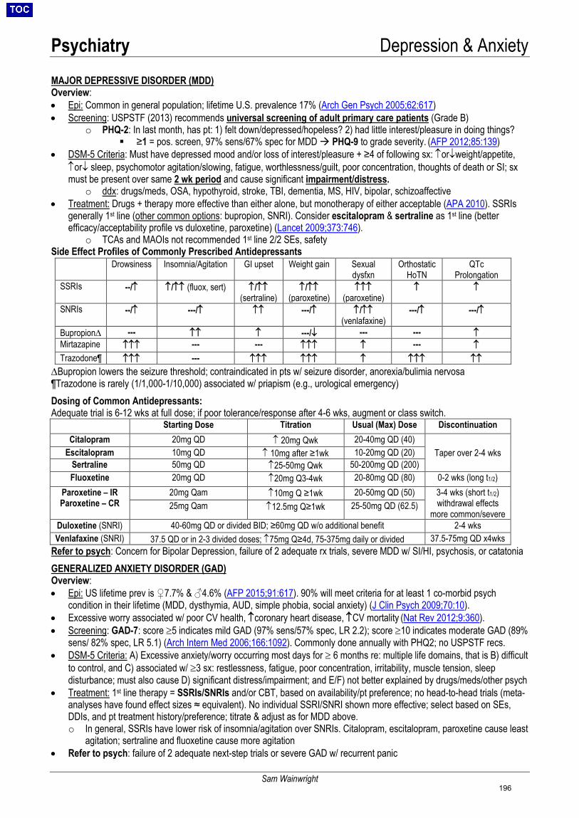
Psychiatry Depression & Anxiety
Sam Wainwright
MAJOR DEPRESSIVE DISORDER (MDD) Overview: • Epi: Common in general population; lifetime U.S. prevalence 17% (Arch Gen Psych 2005;62:617)• Screening: USPSTF (2013) recommends universal screening of adult primary care patients (Grade B)
o PHQ-2: In last month, has pt: 1) felt down/depressed/hopeless? 2) had little interest/pleasure in doing things? ≥1 = pos. screen, 97% sens/67% spec for MDD PHQ-9 to grade severity. (AFP 2012;85:139)
• DSM-5 Criteria: Must have depressed mood and/or loss of interest/pleasure + ≥4 of following sx: ↑or↓weight/appetite,↑or↓ sleep, psychomotor agitation/slowing, fatigue, worthlessness/guilt, poor concentration, thoughts of death or SI; sxmust be present over same 2 wk period and cause significant impairment/distress.
o ddx: drugs/meds, OSA, hypothyroid, stroke, TBI, dementia, MS, HIV, bipolar, schizoaffective• Treatment: Drugs + therapy more effective than either alone, but monotherapy of either acceptable (APA 2010). SSRIs
generally 1st line (other common options: bupropion, SNRI). Consider escitalopram & sertraline as 1st line (betterefficacy/acceptability profile vs duloxetine, paroxetine) (Lancet 2009;373:746).
o TCAs and MAOIs not recommended 1st line 2/2 SEs, safetySide Effect Profiles of Commonly Prescribed Antidepressants
∆Bupropion lowers the seizure threshold; contraindicated in pts w/ seizure disorder, anorexia/bulimia nervosa ¶Trazodone is rarely (1/1,000-1/10,000) associated w/ priapism (e.g., urological emergency) Dosing of Common Antidepressants: Adequate trial is 6-12 wks at full dose; if poor tolerance/response after 4-6 wks, augment or class switch.
Starting Dose Titration Usual (Max) Dose Discontinuation Citalopram 20mg QD ↑ 20mg Qwk 20-40mg QD (40)
Taper over 2-4 wks Escitalopram 10mg QD ↑ 10mg after ≥1wk 10-20mg QD (20)Sertraline 50mg QD ↑25-50mg Qwk 50-200mg QD (200)Fluoxetine 20mg QD ↑20mg Q3-4wk 20-80mg QD (80) 0-2 wks (long t1/2)
Paroxetine – IR Paroxetine – CR
20mg Qam ↑10mg Q ≥1wk 20-50mg QD (50) 3-4 wks (short t1/2)withdrawal effects
more common/severe 25mg Qam ↑12.5mg Q≥1wk 25-50mg QD (62.5)
Duloxetine (SNRI) 40-60mg QD or divided BID; ≥60mg QD w/o additional benefit 2-4 wksVenlafaxine (SNRI) 37.5 QD or in 2-3 divided doses; ↑75mg Q≥4d, 75-375mg daily or divided 37.5-75mg QD x4wks
Refer to psych: Concern for Bipolar Depression, failure of 2 adequate rx trials, severe MDD w/ SI/HI, psychosis, or catatonia GENERALIZED ANXIETY DISORDER (GAD) Overview: • Epi: US lifetime prev is ♀7.7% & ♂4.6% (AFP 2015;91:617). 90% will meet criteria for at least 1 co-morbid psych
condition in their lifetime (MDD, dysthymia, AUD, simple phobia, social anxiety) (J Clin Psych 2009;70:10).• Excessive worry associated w/ poor CV health, ↑coronary heart disease, ↑CV mortality (Nat Rev 2012;9:360).• Screening: GAD-7: score ≥5 indicates mild GAD (97% sens/57% spec, LR 2.2); score ≥10 indicates moderate GAD (89%
sens/ 82% spec, LR 5.1) (Arch Intern Med 2006;166:1092). Commonly done annually with PHQ2; no USPSTF recs.• DSM-5 Criteria: A) Excessive anxiety/worry occurring most days for ≥ 6 months re: multiple life domains, that is B) difficult
to control, and C) associated w/ ≥3 sx: restlessness, fatigue, poor concentration, irritability, muscle tension, sleepdisturbance; must also cause D) significant distress/impairment; and E/F) not better explained by drugs/meds/other psych
• Treatment: 1st line therapy = SSRIs/SNRIs and/or CBT, based on availability/pt preference; no head-to-head trials (meta-analyses have found effect sizes ≈ equivalent). No individual SSRI/SNRI shown more effective; select based on SEs,DDIs, and pt treatment history/preference; titrate & adjust as for MDD above.o In general, SSRIs have lower risk of insomnia/agitation over SNRIs. Citalopram, escitalopram, paroxetine cause least
agitation; sertraline and fluoxetine cause more agitation• Refer to psych: failure of 2 adequate next-step trials or severe GAD w/ recurrent panic
Drowsiness Insomnia/Agitation GI upset Weight gain Sexual dysfxn
Orthostatic HoTN
QTc Prolongation
SSRIs --/↑ ↑/↑↑ (fluox, sert) ↑/↑↑ (sertraline)
↑/↑↑ (paroxetine)
↑↑↑ (paroxetine)
↑ ↑
SNRIs --/↑ ---/↑ ↑↑ ---/↑ ↑/↑↑ (venlafaxine)
---/↑ ---/↑
Bupropion∆ --- ↑↑ ↑ ---/↓ --- --- ↑ Mirtazapine ↑↑↑ --- --- ↑↑↑ ↑ --- ↑ Trazodone¶ ↑↑↑ --- ↑↑↑ ↑↑↑ ↑ ↑↑↑ ↑↑
196
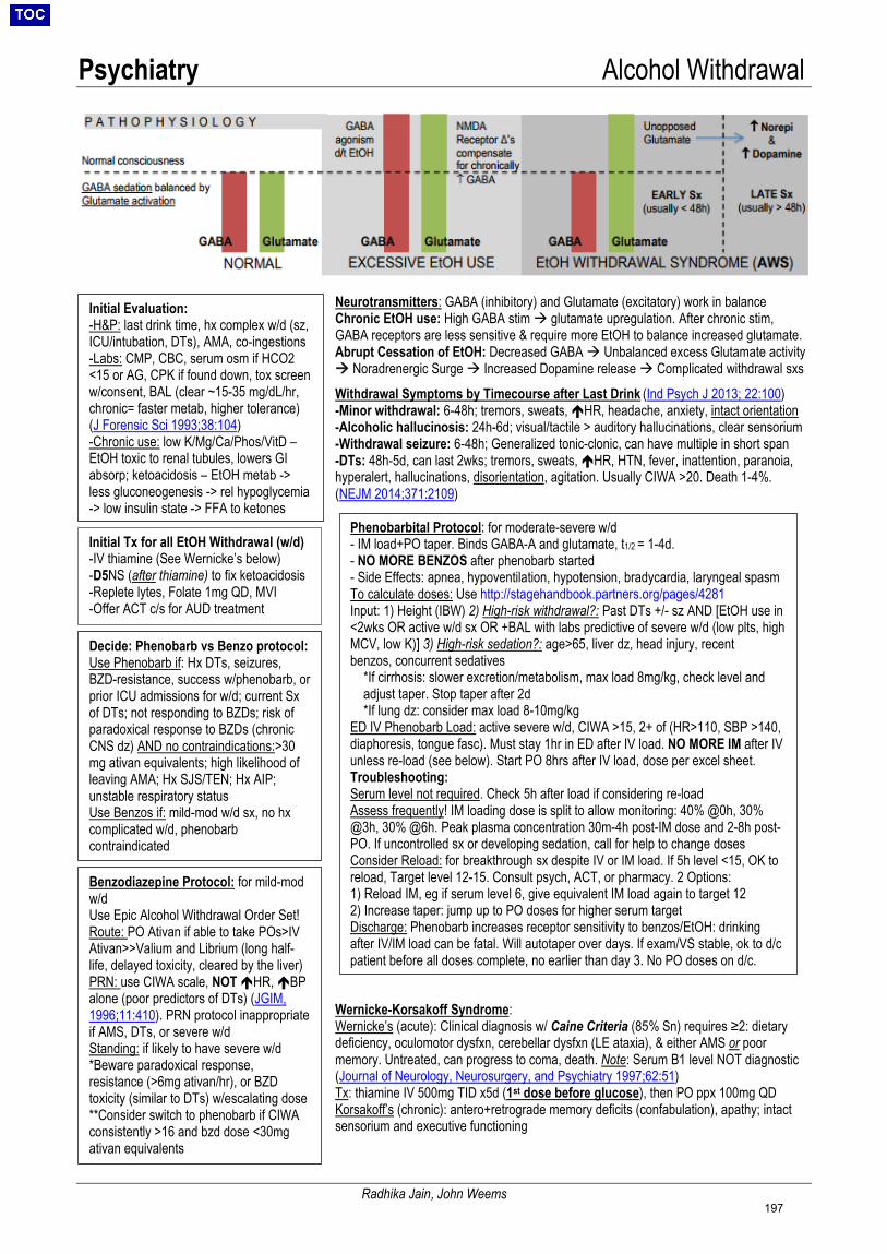
Psychiatry Alcohol Withdrawal
Radhika Jain, John Weems
Phenobarbital Protocol: for moderate-severe w/d - IM load+PO taper. Binds GABA-A and glutamate, t1/2 = 1-4d.- NO MORE BENZOS after phenobarb started- Side Effects: apnea, hypoventilation, hypotension, bradycardia, laryngeal spasmTo calculate doses: Use http://stagehandbook.partners.org/pages/4281Input: 1) Height (IBW) 2) High-risk withdrawal?: Past DTs +/- sz AND [EtOH use in<2wks OR active w/d sx OR +BAL with labs predictive of severe w/d (low plts, highMCV, low K)] 3) High-risk sedation?: age>65, liver dz, head injury, recentbenzos, concurrent sedatives
*If cirrhosis: slower excretion/metabolism, max load 8mg/kg, check level andadjust taper. Stop taper after 2d*If lung dz: consider max load 8-10mg/kg
ED IV Phenobarb Load: active severe w/d, CIWA >15, 2+ of (HR>110, SBP >140, diaphoresis, tongue fasc). Must stay 1hr in ED after IV load. NO MORE IM after IV unless re-load (see below). Start PO 8hrs after IV load, dose per excel sheet. Troubleshooting: Serum level not required. Check 5h after load if considering re-load Assess frequently! IM loading dose is split to allow monitoring: 40% @0h, 30% @3h, 30% @6h. Peak plasma concentration 30m-4h post-IM dose and 2-8h post- PO. If uncontrolled sx or developing sedation, call for help to change doses Consider Reload: for breakthrough sx despite IV or IM load. If 5h level <15, OK to reload, Target level 12-15. Consult psych, ACT, or pharmacy. 2 Options: 1) Reload IM, eg if serum level 6, give equivalent IM load again to target 122) Increase taper: jump up to PO doses for higher serum targetDischarge: Phenobarb increases receptor sensitivity to benzos/EtOH: drinkingafter IV/IM load can be fatal. Will autotaper over days. If exam/VS stable, ok to d/cpatient before all doses complete, no earlier than day 3. No PO doses on d/c.
Initial Evaluation: -H&P: last drink time, hx complex w/d (sz,ICU/intubation, DTs), AMA, co-ingestions-Labs: CMP, CBC, serum osm if HCO2<15 or AG, CPK if found down, tox screenw/consent, BAL (clear ~15-35 mg/dL/hr,chronic= faster metab, higher tolerance)(J Forensic Sci 1993;38:104)-Chronic use: low K/Mg/Ca/Phos/VitD –EtOH toxic to renal tubules, lowers GIabsorp; ketoacidosis – EtOH metab ->less gluconeogenesis -> rel hypoglycemia-> low insulin state -> FFA to ketones
Initial Tx for all EtOH Withdrawal (w/d) -IV thiamine (See Wernicke’s below)-D5NS (after thiamine) to fix ketoacidosis-Replete lytes, Folate 1mg QD, MVI-Offer ACT c/s for AUD treatment
Decide: Phenobarb vs Benzo protocol: Use Phenobarb if: Hx DTs, seizures, BZD-resistance, success w/phenobarb, or prior ICU admissions for w/d; current Sx of DTs; not responding to BZDs; risk of paradoxical response to BZDs (chronic CNS dz) AND no contraindications:>30 mg ativan equivalents; high likelihood of leaving AMA; Hx SJS/TEN; Hx AIP; unstable respiratory status Use Benzos if: mild-mod w/d sx, no hx complicated w/d, phenobarb contraindicated
Benzodiazepine Protocol: for mild-mod w/d Use Epic Alcohol Withdrawal Order Set! Route: PO Ativan if able to take POs>IV Ativan>>Valium and Librium (long half-life, delayed toxicity, cleared by the liver) PRN: use CIWA scale, NOT HR, BP alone (poor predictors of DTs) (JGIM, 1996;11:410). PRN protocol inappropriate if AMS, DTs, or severe w/d Standing: if likely to have severe w/d *Beware paradoxical response,resistance (>6mg ativan/hr), or BZDtoxicity (similar to DTs) w/escalating dose**Consider switch to phenobarb if CIWAconsistently >16 and bzd dose <30mgativan equivalents
Neurotransmitters: GABA (inhibitory) and Glutamate (excitatory) work in balance Chronic EtOH use: High GABA stim glutamate upregulation. After chronic stim, GABA receptors are less sensitive & require more EtOH to balance increased glutamate. Abrupt Cessation of EtOH: Decreased GABA Unbalanced excess Glutamate activity Noradrenergic Surge Increased Dopamine release Complicated withdrawal sxs
Withdrawal Symptoms by Timecourse after Last Drink (Ind Psych J 2013; 22:100)-Minor withdrawal: 6-48h; tremors, sweats, HR, headache, anxiety, intact orientation-Alcoholic hallucinosis: 24h-6d; visual/tactile > auditory hallucinations, clear sensorium-Withdrawal seizure: 6-48h; Generalized tonic-clonic, can have multiple in short span-DTs: 48h-5d, can last 2wks; tremors, sweats, HR, HTN, fever, inattention, paranoia,hyperalert, hallucinations, disorientation, agitation. Usually CIWA >20. Death 1-4%.(NEJM 2014;371:2109)
Wernicke-Korsakoff Syndrome: Wernicke’s (acute): Clinical diagnosis w/ Caine Criteria (85% Sn) requires ≥2: dietary deficiency, oculomotor dysfxn, cerebellar dysfxn (LE ataxia), & either AMS or poor memory. Untreated, can progress to coma, death. Note: Serum B1 level NOT diagnostic (Journal of Neurology, Neurosurgery, and Psychiatry 1997;62:51) Tx: thiamine IV 500mg TID x5d (1st dose before glucose), then PO ppx 100mg QD Korsakoff’s (chronic): antero+retrograde memory deficits (confabulation), apathy; intact sensorium and executive functioning
197
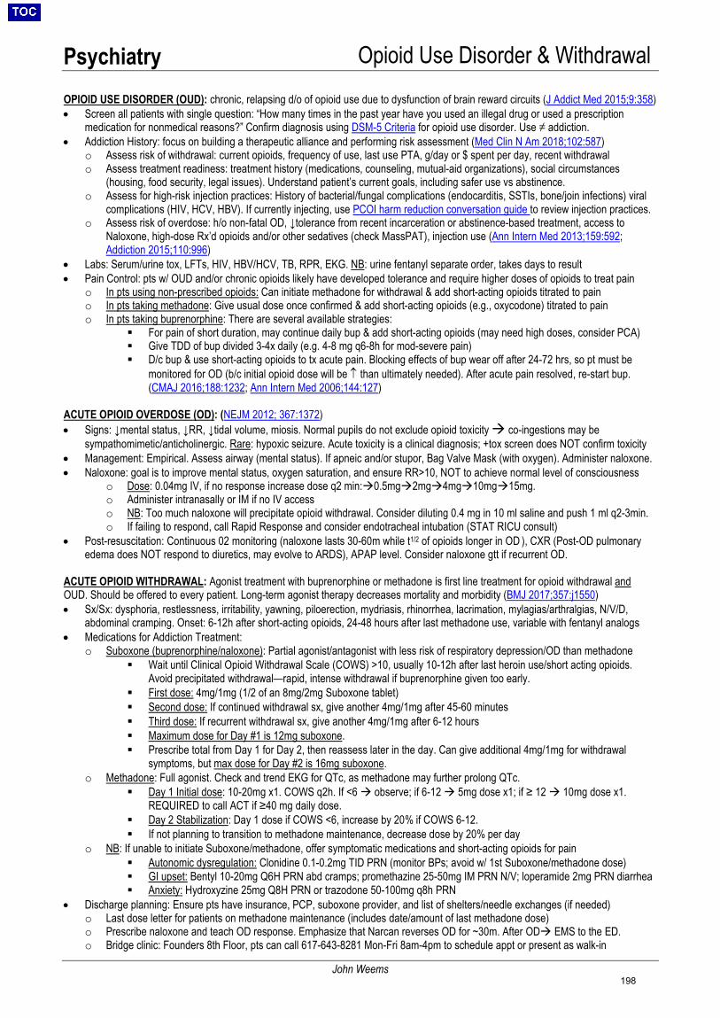
Psychiatry Opioid Use Disorder & Withdrawal
John Weems
OPIOID USE DISORDER (OUD): chronic, relapsing d/o of opioid use due to dysfunction of brain reward circuits (J Addict Med 2015;9:358) • Screen all patients with single question: “How many times in the past year have you used an illegal drug or used a prescription
medication for nonmedical reasons?” Confirm diagnosis using DSM-5 Criteria for opioid use disorder. Use ≠ addiction.• Addiction History: focus on building a therapeutic alliance and performing risk assessment (Med Clin N Am 2018;102:587)
o Assess risk of withdrawal: current opioids, frequency of use, last use PTA, g/day or $ spent per day, recent withdrawalo Assess treatment readiness: treatment history (medications, counseling, mutual-aid organizations), social circumstances
(housing, food security, legal issues). Understand patient’s current goals, including safer use vs abstinence.o Assess for high-risk injection practices: History of bacterial/fungal complications (endocarditis, SSTIs, bone/join infections) viral
complications (HIV, HCV, HBV). If currently injecting, use PCOI harm reduction conversation guide to review injection practices.o Assess risk of overdose: h/o non-fatal OD, ↓tolerance from recent incarceration or abstinence-based treatment, access to
Naloxone, high-dose Rx’d opioids and/or other sedatives (check MassPAT), injection use (Ann Intern Med 2013;159:592;Addiction 2015;110:996)
• Labs: Serum/urine tox, LFTs, HIV, HBV/HCV, TB, RPR, EKG. NB: urine fentanyl separate order, takes days to result• Pain Control: pts w/ OUD and/or chronic opioids likely have developed tolerance and require higher doses of opioids to treat pain
o In pts using non-prescribed opioids: Can initiate methadone for withdrawal & add short-acting opioids titrated to paino In pts taking methadone: Give usual dose once confirmed & add short-acting opioids (e.g., oxycodone) titrated to paino In pts taking buprenorphine: There are several available strategies:
For pain of short duration, may continue daily bup & add short-acting opioids (may need high doses, consider PCA) Give TDD of bup divided 3-4x daily (e.g. 4-8 mg q6-8h for mod-severe pain) D/c bup & use short-acting opioids to tx acute pain. Blocking effects of bup wear off after 24-72 hrs, so pt must be
monitored for OD (b/c initial opioid dose will be ↑ than ultimately needed). After acute pain resolved, re-start bup.(CMAJ 2016;188:1232; Ann Intern Med 2006;144:127)
ACUTE OPIOID OVERDOSE (OD): (NEJM 2012; 367:1372) • Signs: ↓mental status, ↓RR, ↓tidal volume, miosis. Normal pupils do not exclude opioid toxicity co-ingestions may be
sympathomimetic/anticholinergic. Rare: hypoxic seizure. Acute toxicity is a clinical diagnosis; +tox screen does NOT confirm toxicity• Management: Empirical. Assess airway (mental status). If apneic and/or stupor, Bag Valve Mask (with oxygen). Administer naloxone.• Naloxone: goal is to improve mental status, oxygen saturation, and ensure RR>10, NOT to achieve normal level of consciousness
o Dose: 0.04mg IV, if no response increase dose q2 min:0.5mg2mg4mg10mg15mg.o Administer intranasally or IM if no IV accesso NB: Too much naloxone will precipitate opioid withdrawal. Consider diluting 0.4 mg in 10 ml saline and push 1 ml q2-3min.o If failing to respond, call Rapid Response and consider endotracheal intubation (STAT RICU consult)
• Post-resuscitation: Continuous 02 monitoring (naloxone lasts 30-60m while t1/2 of opioids longer in OD ), CXR (Post-OD pulmonaryedema does NOT respond to diuretics, may evolve to ARDS), APAP level. Consider naloxone gtt if recurrent OD.
ACUTE OPIOID WITHDRAWAL: Agonist treatment with buprenorphine or methadone is first line treatment for opioid withdrawal and OUD. Should be offered to every patient. Long-term agonist therapy decreases mortality and morbidity (BMJ 2017;357:j1550) • Sx/Sx: dysphoria, restlessness, irritability, yawning, piloerection, mydriasis, rhinorrhea, lacrimation, mylagias/arthralgias, N/V/D,
abdominal cramping. Onset: 6-12h after short-acting opioids, 24-48 hours after last methadone use, variable with fentanyl analogs• Medications for Addiction Treatment:
o Suboxone (buprenorphine/naloxone): Partial agonist/antagonist with less risk of respiratory depression/OD than methadone Wait until Clinical Opioid Withdrawal Scale (COWS) >10, usually 10-12h after last heroin use/short acting opioids.
Avoid precipitated withdrawal—rapid, intense withdrawal if buprenorphine given too early. First dose: 4mg/1mg (1/2 of an 8mg/2mg Suboxone tablet) Second dose: If continued withdrawal sx, give another 4mg/1mg after 45-60 minutes Third dose: If recurrent withdrawal sx, give another 4mg/1mg after 6-12 hours Maximum dose for Day #1 is 12mg suboxone. Prescribe total from Day 1 for Day 2, then reassess later in the day. Can give additional 4mg/1mg for withdrawal
symptoms, but max dose for Day #2 is 16mg suboxone.o Methadone: Full agonist. Check and trend EKG for QTc, as methadone may further prolong QTc.
Day 1 Initial dose: 10-20mg x1. COWS q2h. If <6 observe; if 6-12 5mg dose x1; if ≥ 12 10mg dose x1.REQUIRED to call ACT if ≥40 mg daily dose.
Day 2 Stabilization: Day 1 dose if COWS <6, increase by 20% if COWS 6-12. If not planning to transition to methadone maintenance, decrease dose by 20% per day
o NB: If unable to initiate Suboxone/methadone, offer symptomatic medications and short-acting opioids for pain Autonomic dysregulation: Clonidine 0.1-0.2mg TID PRN (monitor BPs; avoid w/ 1st Suboxone/methadone dose) GI upset: Bentyl 10-20mg Q6H PRN abd cramps; promethazine 25-50mg IM PRN N/V; loperamide 2mg PRN diarrhea Anxiety: Hydroxyzine 25mg Q8H PRN or trazodone 50-100mg q8h PRN
• Discharge planning: Ensure pts have insurance, PCP, suboxone provider, and list of shelters/needle exchanges (if needed)o Last dose letter for patients on methadone maintenance (includes date/amount of last methadone dose)o Prescribe naloxone and teach OD response. Emphasize that Narcan reverses OD for ~30m. After OD EMS to the ED.o Bridge clinic: Founders 8th Floor, pts can call 617-643-8281 Mon-Fri 8am-4pm to schedule appt or present as walk-in
198

Psychiatry Substance Use Disorders
Julia Cromwell, Meghan Musselman
MGH Tox Screens: ▪ Basic Serum Toxicology Screen: Quantitative assays for ethanol, salicylates, acetaminophen; Qualitative assay for TCAs▪ Drug Screen, Prescription/OTC (“Full tox”): Send out to Mayo, will take >3 days to return (www.mayomedicallaboratories.com)
-Common OTCs: Caffeine, acetaminophen, salicylates, ibuprofen, naproxen, dextromethorphan, diphenhydramine, guaifenesin-Neuro/Psych Drugs: Barbiturates, AEDs (incl. carbamazepine, lamotrigine, levetiracetam, topiramate), propofol, TCAs, SSRIs,SNRIs, bupropion, phenothiazines (incl. chlorpromazine, thioridazine), clozapine, muscle relaxants (cyclobenzaprine,metaxalone), sleep meds (inc. zolpidem, zaleplon)-Others: Lidocaine, trazodone, theophylline, some pesticides-Limited use for illicit drugs: Benzos, some opiates (incl. codeine, meperidine, methadone, oxycodone, fentanyl), amphetamines-Drugs NOT on screen: Cocaine, lithium, digoxin, ethylene glycol, iron, lead (order separately and note some are send-outs)
▪ Urine Toxicology Screen: Amphetamines, barbiturates, benzodiazepines, THC, cocaine, opiates, phencyclidine▪ VPAIN (“Urine pain panel”): Buprenorphine, oxycodone, methadone, 6-monoacetyl morphine (heroin metabolite)
o NB: urine fentanyl is an add-on test—consider sending for suspected opioid OD (esp. with PEA arrest) given prevalence ofhigh prevalence of synthetic fentanyl analogues in the community
▪ Oral Fluid Drug Test: Differentiates specific TYPE of benzo (eg, lorazepam v diazepam), opiate (eg, codeine v heroin), amphetamine
Urine Test Characteristics: (Mayo Clin Proc 2008;83:66) Class Detection Time (days) False Positives False Negatives
Amphetamines 1-2d after use (2-4d for chronicexposure)
bupropion, labetalol, trazodone, ranitidine, pseudoephedrine, selegiline
Ritalin & atomoxetine won’t
test + Barbiturates 2-20d after use Fioricet (contains butalbital)
Benzodiazepines 1-5d (most), 2-30d after diazepam oxaprozin (NSAID) lorazepam, clonazepam <1mg/day
Cannabinoids 1-7d after use,up to 1mo heavy chronic use hemp products, Marinol synthetic
cannabinoids Opiates
(NOT methadone, fentanyl, meperidine)
1-3d after use, 6-8d after heavy usepoppy seeds (unlikely); At MGH, methadone,
naloxone, fluoroquinolones do NOT interfere
oxycodone, oxymorphone,
Suboxone Cocaine 2-4d after use, 1-3wks after heavy use NONE
Phencyclidine (PCP) 7-14d after use Lamictal, Effexor, Tramadol, dextromethorphan, doxylamine
Buprenorphine 5-10d high doses of opiates/methadone/tramadol; quinine, hydroxychloroquine, naltrexone
Methadone 1-5d quetiapine, diphenhydramine, doxylamine Oxycodone 2-4d substances that change urine color (e.g.,
Flagyl) will cause refusal
Cocaine Intoxication/Withdrawal: • Intoxication: Grandiosity, euphoria, hyperactivity, anorexia, anxiety, psychotic sx (formication, paranoia, AH/VH), fever, mydriasis.
Vasospasm can cause HTN emergency, stroke, MI and seizures. Tx: labetalol, phentolamine (AVOID unopposed alpha stimulation)• Withdrawal: Depression, fatigue, nightmares, cravings,↑ sleep/appetite. Tx: Acute: propranolol, quetiapine for severe sx; Chronic:
consider topiramate and/or baclofen for cravings/dependence (consult ACT) (Psychiatry 2005;2:44)Benzodiazepine Withdrawal: Manage withdrawal per EtOH protocol (see “Alcohol Withdrawal” section). Higher risk for delirium with BZD withdrawal. If possible, initiate taper with same BZD agent (eg due to extremely short half-life, alprazolam requires alprazolam taper).
Commonly used benzos Comparative dosages (approx) Half-life (hours) (approx) alprazolam (Xanax)* 0.5mg 6-27 (oral peak 1-2)chlordiazepoxide (Librium) 25mg 5-30 (oral peak 0.5-4)clonazepam (Klonopin)* 0.25mg 18-50 (oral peak 1-2)diazepam (Valium)* 5mg 20-50 (oral peak 0.5-1)lorazepam (Ativan)* 1mg 10-20 (oral peak 2-4)temazepam (Restoril) 10mg 3-19 (oral peak 1-2)triazolam (Halcion) 0.25mg 2-3 (oral peak 0.7-2)
*Most common illicit usage bc most commonly prescribedSpice/K2/Bath Salt Intoxication: Agitation/violence, hallucination/paranoia, anxiety, tachycardia, arrhythmia, myoclonus, diaphoresis. • Tx: low stimulation environment, IVF, consider IV BZDs to reduce agitation and prevent seizure (Curr Psychiatry Rep 2016;18:52)THC Intoxication: Euphoria followed by relaxation; tachycardia; hallucinations (especially w/ high potency THC, e.g., wax/dab)• Cannabinoid hyperemesis syndrome: Chronic user with recurrent N/V, abdominal pain; symptom relief w/ hot showers; mild
leukocytosis. Tx: IVF, antiemetic, and THC cessation; consider BZD, followed by antipsychotic and capsaicin (J Med Toxicol2017;13:71)
199

Primary Care Health Screening & Maintenance
Eftitan Akam
GENERAL SCREENING GUIDELINES (USPSTFa, ADAb, AACEc, ACC/AHAd, ACCPe, CDCf, ACSg) [Evidence Grade]
* CVD RFs: DM, HTN, personal Hx of any athero, FHx (<50 in male relatives, <60 in female relatives), BMI (30+), smoking^ DM RFs: prior abnl testing (A1c≥5.7), FHx, AA/Latinx/Asian/NA ancestry, Hx GDM, PCOS, CVD, HDL<35 or TG>250, physical inactivity*** Age 40 if ≥1st degree relative dx <65; Age 45 if AA or 1st degree relative dx <65; Age 50 for others expected to live >10yrs
ADDITIONAL SCREENING GUIDELINES FOR MEN (USPSTFa, ACSg) [Evidence grade] Age 18-40 40 45 50 55 60 65 70 75+ AAA If +tobacco hx [B]a Prostate CA FHx* Q2yg FHx, AA Q2yg Screen all men Q2y if life expectancy ≥ 10-15 yrsg
PSA 55-69yo if pt preference [C]a, recommend against if >70yo [D]a
Testicular CA Recommend against routine screening in all men[D]a * >1 first-degree relative with history of prostate cancer
ADDITIONAL SCREENING GUIDELINES FOR NON-PREGNANT WOMEN (USPSTFa) [Evidence grade] Age 18 19 20 21 25 30 35 40 45 50 55 60 65 70 75+ Breast CA
Consider BRCA counseling if +FHx. Screening tools available. [B]a
Individualized screening by
risk (Gail Model)
Q2y mammography age 50-74. Stop if <10yrs life expectancy.
[B]a
Cervical CA Q3y [A]a Q3y or Q5y + HPV co-testing [A]a Stop∑ STIs ≤24: GC/CT annually [B]a Screen based on risk assessment Contraception Discuss with everyone. Start folic acid at reproductive age if planning/capable of pregnancy [A]a Osteoporosis Consider earlier screening based on FRAX assessment [B]a DEXA [B]a ∑ Stop if 3 consecutive neg paps or 2 consecutive neg co-tests within 10 years w/ most recent test within 5 years. Continue x20 yrs s/p dx pre-cancerous lesion regardless of age. Do not resume age ≥65 for new sexual partner only.
Age 18 19 20 21 25 30 35 40 45 50 55 60 65 70 75+ Cardiovascular Screening / Preventative Health Recommendations
CVD Risk Assess RFs q4-6y [B]d (age, sex, total chol/HDL, SBP,DM, smoking)
Estimate risk w/ ASCVD calculator q4-6y [B]d If ASCVD risk ≥10% w/ ≥1 CVD RF*, consider statin [B]a
ASA for 1° ppx∆
1° ppx for CV events, colon CA if ≥10% 10-yr ASCVD risk, no ↑risk bleeding, & life exp ≥10 years [B]a,d,e; recent RCT w/ no sig benefit in
elderly (NEJM 2018;379:1509) Diabetes If HTN or BMI ≥25 (>23 Asian) w/ ≥1 DM RF^ [B]b Q3y. ↑interval if abnl result, annually in pre-DMb HTN Q3-5y; Q1y if borderline high BP, obese, AA [A]a Q1y [A]a HLD One-time
screening age <20
Men 20-45, women 20-55: screen Q5yr, ↑ if RF [C]cQ 3-5y in DM if wnl at dx
Men >45, women >55 Q1-2y if no RF*[A]c, Q1y in DM [B]c
Screen Q1y [A]c
Obesity Annual BMI refer for or offer intensive behavioral intervention if ≥30 (obese) [B]a Diet Intensive behavioral counseling if CVD RF* [B]a Exercise 150 min/wk mod exercise (30 min 5x/wk); 75 min/wk intense exercise (25 min 3x/wk)f
Universal Cancer Screening Colorectal CA Start 10 years prior to age of affected family member at dx*** Colo Q10y, flex sig q5y, FIT q1y [A]a Lung CA Q1y low-dose CT if 30 pys & quit w/in
last 15y [B]a Skin CA Insufficient evidence for routine skin exams by clinicians [I]a
Infectious Disease Screening HIV Screen at least once; repeat based on risk assessment [A]a HCV Screen x1 if born 1945-1965; additional screening based on risk assessment [B]a HBV Born in endemic region, getting HD or immunosuppression, HIV+, IVDU, MSM, close contact w/ HBV+ person [B]a Latent TB Screen if born or lived in high risk country (see CDC website) or high risk setting (homeless, jail) [B]a
Psych/SUD/Social Risk Factor Screening Depression Q1y [B]a; PHQ-2: in 2 wk how often (a)little interest/pleasure doing things & (b)down/depressed/hopeless EtOH Misuse Screen regularly with AUDIT-C [B]a Tobacco Every encounter [A]a. Advise to quit, Assist doing so (plan, quit date, QuitWorks, meds), Arrange f/u. Intimate Partner Violence
Screen regularly in women of reproductive age [B]a. HITS screen well-validated. Assess immediate safety & consider HAVEN referral.
No data but consider ongoing IPV, elder abuse screening
Fall Risk PT, Vit D [B]a
200
Primary Care Health Screening & Maintenance
Eftitan Akam
ADDITIONAL SCREENING GUIDELINES FOR SPECIAL POPULATIONS • MSM (men who have sex with men) and SMW (sexual minority women): see LGBTQ Health• Immigrants and refugees: see Immigrant & Refugee Health
Note that there is considerable discrepancy between societal guidelines created using the same evidence. Examples include: • Breast Cancer – USPSTF: biannual screening age 50-74 [B]; discussion of risks/benefits age 40-49 [C]; no
recommendation for women >75 given insufficient evidence [I]. ACS: annual mammography age 45-55; discusstransitioning to biennial screening at 55 until life expectancy <10 years; discuss initiation of annual screening starting atage 40. ACOG: offer screening mammography at 40; start screening at 50; discuss cessation at 75; screen Q1-2 years.
• Diabetes – USPSTF: screen overweight and obese adults aged 40-70; no specific interval guidance. We have selectedthe ADA guidelines because they account for additional risk factors and recommend a specific screening interval.
MANAGEMENT OF ABNORMAL PAP SMEAR (ASCCP Consensus Guidelines, 2012)
VACCINES - https://www.cdc.gov/vaccines/schedules/hcp/adult.html Vaccine Age # of Doses/ Special indications Influenza All, q1yr No live vaccine (intranasal): immunocompromised/pregnant TDaP / Td All, q10yrs (substitute Tdap for Td once) Extra dose Tdap for pregnant women PCV13/ PPSV23 All ≥65: PCV13, then PPSV23 12 mo later; if
PPSV23 already given <65, re-dose x1 at >5 yrs. Age 19-64: if CHF/CM, chronic lung dz, cirrhosis, DM, ETOH d/o, smoking; redose x1 at 65 (if >5y from 1st PPSV23 & 12mo from PCV13)
Special pops: CSF leak, cochlear implant, functional asplenia, immunocompromised (incl: immunodeficiency d/o, HIV, nephrotic syndrome, malignancy, tx; full list at CDC) Age >18: give PCV13 x1 PPSV23 8 wks later and re-dose x1 at 5 yrs if asplenia or immunocomp & again at age 65 (if >5yrs from 1st)
Varicella* Only if no evidence of immunity (presumed immune if U.S.-born pre-1980)
2 doses at least 1 mo apart
Zoster* 50+ (regardless of varicella infxn hx) 2 doses RZV 2-6 mo apart if >50yo (if received ZVL, at least 2 mo after dose). If >60, give RZV (preferred) or ZVL
HPV Women <27, Men <22 (or MSM, HIV <27) 3 doses at 0/1-2/6 months MMR* Only if no evidence of immunity 1-2 doses at least 28 days apart, 1 dose in women of
childbearing ageHep A Likely unvaccinated if born before 1991 2 doses at 0/6-12 mo for travel to endemic countries, MSM,
any drug use (not just IVDU), liver dz, clotting d/os, household contacts of those at risk
Hep B Likely unvaccinated if born before 1991 3 doses at 0/1/6 mo for mult partners, STI, MSM, + partner, IVDU, DM, ESRD, any liver dz, HIV, health worker/occup exposure, travel to endemic country, household contacts
Meningococcus (MenACWY/ MenB)
Usually vaccinated as teen. May need booster q5y if risk of infxn remains.
1-3 doses depending on type for living in dorms, asplenia,HIV, MSM, complement def, military, occup exposure, travel
Hib Usually vaccinated as child 1 dose if not immune for asplenia, SCD * Hold in pregnancy, malignancy, immunocompromised** Asplenia: see https://hospitalpolicies.ellucid.com/documents/view/12033
Abnormal Pap Cytology Results ASCUS LSIL HSIL AGC
Age 21-24 Preferred: Management as per LSIL (annual cytology). Alternative: HPV reflex. If HPV-, resume regular screening; if HPV+, manage as LSIL
Repeat cytology in 1 year: if neg, ASC-US, or LSIL, repeat in 12 mos; if neg repeat in 12 months; if neg x2 resume regular screening vs. if ≥ ASC-US on any repeat colpo
Colpo w/ endocervical curettage
For AGC-NOS, AGC-endocervical, or adenocarcinoma in situ (AIS) colpo w/ biopsy &endocervical sampling, w/additional endometrialsampling if ≥ 35yo or youngerw/ endometrial neoplasia RFs(unopposed estrogen,tamoxifen, early menarche,late menopause, PCOS, DM,obesity) or sux (AUBFor AGC-endometrial endometrial & endocervicalsampling
Age 25-29 Preferred: HPV reflex. If HPV+ colpo. If HPV- repeat co-testing in 3 years. Alternative: Repeat cytology in 1 year. If neg, resume routine screening. If ≥ ASC-US colpo
Colpo, even if have previous HPV- result
Option 1: Colpo w/ endocervical curettage Option 2: Immediate LEEP (not if pregnant or desiring pregnancy)
Age 30+ HPV reflex: If HPV- repeat co-testing in 1 year and if neg repeat co-testing in 3 years vs. if pos (cytology or HPV) colpo. If HPV reflex + or unknown colpo
201
Primary Care Women’s Health
Dinah Applewhite, Rashmi Jasrasaria
Vulvar/Vaginal Complaints (Obstet Gynecol 2008;5:1243)• Presentation: vaginal discharge, odor, pain, pruritus• Infectious vaginitis: more likely to present acutely
o Bacterial vaginosis (BV): malodorous discharge, most common and most pts asymptomatic, high prevalence in WSW Dx: 3/4 Amsel criteria (homogenous/thin/grey-white discharge smoothly coating vaginal walls, clue cells, fishy
smell on KOH, pH >4.5 (less reliable if post-menopause) → order “Genital culture female” in Epic, collected withrayon swab, gram stain assesses for clue cells (0-3 consistent with normal flora, 7-10 consistent with BV)
Tx: Flagyl 500mg BID x7d, clinda 300mg BID x 7d, or secnidazole 2gm x 1; can opt not to tx if not pregnanto Candida: curd-like discharge, pruritus
Dx: pH nl, order “Genital culture female” in Epic, collected with rayon swab, add on anti-fungal sens. if recurrent Tx: Monistat 7 (vaginal miconazole 2% 5g daily x 7d) OTC, fluconazole 150mg PO x1 requires Rx (cheaper)
o Trichomonas: purulent malodorous discharge, inflammation on exam (DDx includes gonorrhea and chlamydia) Dx: pH >5-6, trichomonads on microscopy, order “Trichomonas vaginalis antigen” collected with rayon swab Tx: Flagyl 2gm x1 for patient and sexual partners
• Dermatoses: more likely to present chronically, often require GYN referral and biopsyo Contact dermatitis: erythema, swelling, fissures, erosions → r/o Candida, remove offending agento Vulvar atrophy: 50% postmenopausal ♀ 2/2 decreased vaginal secretions, Δ vaginal flora → moisturizers, topical estradiolo Lichen simplex chronicus: intense pruritis + lichenified plaque i/s/o atopic dermatitis hx → antihistamines, steroidso Lichen sclerosis: onset 50-60s, cigarette paper skin + porcelain white papules i/s/o autoimmune dz, may lead to labia
minora fusion, clitoral hood phimosis, fissures, perianal dz, 5% incidence of malignancy → high potency steroids x4 wkso Lichen planus: “purple, papular, pruritic,” white lacy Wickham striae → high potency steroids, monitor for SCC
Abnormal Uterine Bleeding (AUB) (Obstet Gynecol Clin N Am 2017;44:179; Obstet Gynecol 2012;120:197) • Hx: quantity, pattern (breakthrough, intermenstrual, irregular), sexual/ OB hx, trauma, FH, cancer risks, meds (hormones, A/C)• Dx: pelvic exam; CBC, coags, hCG, TSH, PRL, UA, GCCT; +/- transvaginal pelvic US (TVPUS), hysteroscopy, endometrial bx (EMB)• DDx:
o Structural: polyps (↑ risk of endometrial CA if post-menopausal, on tamoxifen, >1.5cm → resection, Mirena), leiomyomas(fibroids → resection, hysterectomy, GnRH agonists), adenomyosis (boggy uterus → hormonal tx, hysterectomy)
o Malignancy/hyperplasia: must be excluded in any peri- or post-menopausal ♀ with AUB or younger pts with risk factors(unopposed estrogen, early menarche, late menopause, nulliparity, PCOS, obesity, DM, tamoxifen, FH) → dx with EMB +/-imaging → tx with progesterone for hyperplasia + EMB Q3-6mo OR hysterectomy
o Ovulatory dysfunction: endocrine d/o, PCOS, obesity → OCP, progestin-only (Mirena), endometrial ablation, hysterectomyo Coagulopathy: disorders of hemostasis or oral A/Co Iatrogenic: common with progestin-only contraceptives, especially initially
Polycystic Ovary Syndrome (PCOS): affects 5-10% of women of reproductive age (NEJM 2005;352:1223; J Clin Endo Met 2013;98:4565; Obstet Gynecol 2009;114:936)• Criteria: 2/3: oligo/ anovulation, clinical/ biochemical hyperandrogenism (i.e. hirsutism), polycystic ovaries on pelvic US• Workup: testosterone; exclude other dx (hCG, FSH, 17-OHP (pre-8AM), prolactin, TSH), screen for metabolic d/o, OSA, mood d/o• Tx: weight loss, OCP/ Mirena, spironolactone, metformin (if insulin resistant), fertility referral if/ when pt desires pregnancy
Infertility: Evaluate after 12 mo unprotected intercourse in <35 yo, 6 mo in >35 yo (Fertil Steril 2015;103:e44) • DDx: ovulatory dysfunction, fallopian tube abnormalities, uterine abnormalities, cervical factors, endometriosis• Hx: duration of infertility, prior OB/GYN hx (menstrual hx, pregnancies, PID, fibroids, cervical dysplasia, endometriosis, contraceptive
use, DTE exposure in utero), sexual hx (timing, frequency, lubrication, dyspareunia), meds, prior chemo/XRT, substance use, FH• Dx:
o Test ovulation: mid-luteal progesterone (day 21, 1 wk before expected menses, goal >3), home kit to check for LH surgeo Test ovarian reserve: FSH/estradiol (day 3, goal FSH <10, estradiol <80), clomiphene challenge testo Additional workup: chlamydia PCR, HSG (fallopian tubes), saline infusion sonohysterography (uterus)o Partner: semen analysis
• Tx: if testing abnormal, refer to reproductive endocrinology for aggressive induction of ovulation, IVF, or donor oocytes
Menopause: amenorrhea x12 mo w/o alt etiology (no need to check labs), avg onset age 51, suspect 1o ovarian insuff if <40 yo (Obstet Gynecol 2014;123:202) • Vasomotor sx (hot flashes): Systemic hormone tx (estrogen + progestin, estrogen monotx if hysterectomy): most effective therapy but
only recommended if <60 yo and for <5 yrs duration (J Clin Endo Met 2008;93:4567); start at 0.5 mg/day; side effects include breasttenderness, vaginal bleeding; ↓CRC, fracture risk; ↑breast CA, CVD, VTE; Ø inc risk of mortality after 5-7 yrs (JAMA2017;318(10):927); Alternatives: SSRIs (paroxetine), SNRIs (venlafaxine), gabapentin, clonidine
• Vaginal sx (dryness, burning, pain w/ intercourse)o Lubricants (KY): prior to intercourse; Moisturizers (Replens): longer-term reliefo Topical estrogen therapy: ring, tablet, cream; start at 0.3 mg/day; Ø inc risk of endometrial hyperplasiao SERM (ospemifene): for sx atrophy not relieved by nonpharm tx or not amenable to topical tx; adv effects incl ↑hot flashes
202
Primary Care Women’s Health
Dinah Applewhite, Rashmi Jasrasaria
Contraception: see Quick Start Algorithm (Contraceptive Technology 20th Ed CDC USMEC 2016) • 45% of pregnancies are unplanned → rule out pregnancy before initiating contraception → IUD, implant are first line• Hormonal methods take ~1 wk to work → use backup method for 7 days
Use 1y Failure Rate* Pros/Cons Contraindications
Estrogen-progestin Pill Daily 9% (0.3%) - Pros: ↓menses, PMS, cramps,
acne, endometrial/ovarian CA- Cons: N/V, breast tenderness,↓libido, spotting, require patient adherence
- VTE, thrombogenic mutation- Active breast or liver CA- Migraine w/ aura, >35 yo + any migraine- Uncontrolled HTN, DM w/ vasccomplications, CVD, valvular dz- >35 yo + >15 cig/day- ESLD
Ring (Nuva-Ring) 3 wks in, 1 wk out 9% (0.3%)
Patch (Xulane) Weekly x3 wks, 1 wk off; apply to arm, torso, or buttock
9% (0.3%)
Progestin-only IUD (hormone content Mirena > Kyleena > Skyla)
Mirena Q7Y, Kyleena Q5Y, Skyla Q3Y
0.2% - Pros: effective, long-acting- Cons: irregular bleeding, physicalcomplications (rare)
- Abnl uterine cavity, G/C at time ofinsertion, PID, endometrial/cervical/breast/liver Ca, APLAS, pregnancy, ESLD
Implant (Nexplanon)
Q3Y to upper inner arm 0.05% - Unexplained vaginal bleeding
- Breast/liver CA, APLAS, ESLD
Injection (Depot-Provera)
Q3mo IM/SQ to buttock 6% (0.2%)
- Pros: long-acting- Cons: irregular bleeding, weight gain (<5 lb), ↓BMD, prolonged return to fertility (1 yr)
- Unexplained vaginal bleeding- Breast/liver CA, APLAS, ESLD- Mult CV RF, uncontrolled HTN, DM w/vasc comp, iCMP, CVA, vasc dz
Pill (Micronor) Daily 9% (0.3%) - Pros: few contraindications- Cons: irregular bleeding, musttake at same time daily
- Bariatric surgery- Breast/liver CA, APLAS, ESLD
Hormone-free
Copper IUD (Paraguard) Q12Y 0.8%
- Pros: effective, long-acting, safe inESLD, emergency contraceptive- Cons: heavier bleeding, cramping
- Abnl uterine cavity, G/C at time ofinsertion, PID, endometrial/cervical CA,pregnancy
Male condom Every encounter 18% (2%) - Pros: STI prevention
- Cons: require patient adherence- Oil based lubricant w/ latex condom
Sterilization Permanent 0.15% - Pros: effective, long-acting- Cons: irreversible
- Surgical risk, patient unsure of decision
* Typical use – i.e. % women who will have unplanned pregnancy in 1 year on this method; (% Perfect use – not realistic for most)
Oral Contraceptives (OCPs): (CDC MMWR 2013;62(RR05):1) • Types: monophasic vs multiphasic/ triphasic; combined (estrogen + progestin) vs progestin-only• OCP selection:
o 2nd generation progestin-containing (levonorgestrel, norethindrone): ↓ VTE risko 3rd generation progestin-containing (norgestimate, desogestrel): ↓ androgenic SE, higher VTE risko Progestin-only (norethindrone 0.35 mg): if contraindication to estrogen, if ♀ lactating
• Switching OCPs due to SEs:o HA, nausea, breast tenderness → 2/2 estrogen excess → decrease estrogen dosing, change to QHSo Weight gain, acne, hirsutism, ↓ libido, mood Δ → 2/2 androgen or progestin excess → change to 3rd gen progestino Breakthrough bleeding → increase estrogen dosing if early cycle bleeding; increase progestin or change to triphasic if lateo Amenorrhea → rule out pregnancy, otherwise continue with same pill
Emergency Contraception: (Obstet Gynecol 2010;115:1100) • Sexual assault cases should be referred to the ED for an exam by a trained SANE RN• Plan B (levonorgestrel 1.5mg x1 or 0.75mg x2): OTC, use within 72 hrs, less reliable if BMI >30• Ella (ulipristal acetate 30 mg): requires rx, use within 120 hrs, more reliable in higher BMI• Paragard (copper IUD): requires office visit, ideally placed within 120 hrs (okay up to 160), most effective
Abortion: (Guttmacher Institute Fact Sheet; Am J Public Health 2017;107:1904; Obstet Gynecol 2014;123:676) • See PCOI for list of providers in MA. Ave cost ~$500, 50% pay out of pocket. 1/4 ♀ will have abortion by age 45 in USA.• Workup: confirm pregnancy/ LMP/ TVPUS, check CBC/ Rh, offer STI testing and immediate post-abortion contraception• Medical abortion (performed up to 10 wks gestation, 92% effective): mifepristone x1 → buccal misoprostal in 24-48 hr → pt passes
pregnancy at home over hrs, a/w cramping and bleeding, tx with NSAIDs → f/u bHCG or TVPUS usually in 14d• Surgical abortion (performed up to 24 wks gestation, 99% effective): same-day office procedure → no f/u unless complications• Counseling: 1-866-4-EXHALE
203
Primary Care Musculoskeletal Pain
Andrew Hoekzema, Sam Wainwright
Urinary incontinence: Very common (25% young women 75% of older women). Most women do not seek help. • Types: stress (leakage with coughing, laughing, etc.), urge (loss of urine preceded by feeling of urgency), mixed (most common,
stress + urge), overflow, and functional (impaired mobility/cognition/neurologic).• Dx: History: Review meds (anticholinergics, diuretics, etc.), bowel habits, caffeine/EtOH use, 72h voiding diary. Physical exam: check
for prolapse, fistula, diverticulum; cough stress test (can be supine, but standing w/ full bladder ↑sensitivity); urethral mobility (w/ptbearing down, displacement >30˚ or movement >2cm); rectal exam (fecal impaction, sphincter tone); neuro exam. Diagnostics: UA/cx,PVR (if suspect overflow, abnl >150cc), specialized urodynamic studies not indicated in initial eval of uncomplicated UI.
• Tx: All types: bladder training (timed voiding, use PCOI handout), lifestyle interventions (eg weight loss, ↓fluid/caffeine intake) andpelvic floor muscle exercises (eg Kegels, use PCOI handout, consider referral to pelvic floor PT). Stress/mixed: Pessaries (mixeddata, best for women who wish to avoid therapy/behavioral therapy, refer to urogyn for fitting), vaginal estrogen (in post-menopausalwomen w/ vaginal atrophy), and surgeries/procedures (eg midurethral sling, urethral bulking agents). Urgency: antimuscarinics(numerous side effects), beta-agonists (eg mirabegron, avoid w/uncontrolled HTN, ESRD, liver disease), and intravesicular botox
MUSCULOSKELETAL PAIN
KNEE PAIN • Pathophysiology: Detailed hx, incl: trauma, acute vs. chronic, constitutional sx, BMI, orthopedic hx. Elicit history of swelling,
stiffness, instability, popping or catching sensation, sensory/motor changes. Have pt point to area of pain with one finger.Location Etiologies Anterior Quadriceps or patellar injury, patellofemoral syndrome, Osgood-Schlatter, bursitis, RA, gout, pseudogout, septic joint Lateral Lateral meniscal tear, lateral collateral ligament injury, iliotibial band, lateral OA Medial Medial meniscal tear, medical collateral ligament injury, tibial plateau fracture, anserine bursitis, medial OA Popliteal Popliteal/Baker cyst, PCL injury, DVT • Knee Exam:Test Maneuver Positive in Lachman (similar to anterior drawer)
Pt supine with knee flexed, one hand on pt’s femur, just above knee. Other hand on pt’s tibia. Apply slight flexion and pull sharply towards your abdomen. If tibia feels unrestrained, positive test
ACL injury
Posterior drawer Pt supine with knee flexed, can stabilize foot by sitting on it. Place hands around tibia, apply pressure backward in place parallel to femur. If unrestrained motion, positive test.
PCL injury
McMurray One hand over medial joint line with knee fully flexed. Evert foot, apply valgus stress and gently flex/extend knee. If clicking around medial joint line, positive test.
Meniscal injury
• XR Imaging: If trauma <1wk old & c/f fracture, follow Ottawa Rules, Se 98%, Sp 49% (Ann Int Med 2004;140(2):121). [obtain if anyof the following: >55yo, isolated patellar tenderness, tenderness at head of fibula, cannot flex to 90°, or cannot bear weight for 4 steps(limp doesn’t count)]. If eval of chronic OA, get weightbearing views of both knees; add patellar view for patellar problems.
• Reserve MRI until 4 weeks conservative care unless suspect fracture, infection, or internal derangement (e.g. ACL, meniscal tear inyounger patients). Asymptomatic meniscal tears: 13% younger than 45 yo, 36% older than 45 yo (Clin Ortho Rel Res 1992;282:177)
• Treatment: Limited benefit of arthroscopy, especially in degenerative meniscal tears in age > 45 yo, patients with OA (BMJ2017;357:j1982). Start with NSAIDs, PT, weight loss first even with clicking, catching. Consider glucosamine+chondroitin or platelet-rich-plasma (AHRQ 2017;17-EHC011-EF)
SHOULDER PAIN • Pathophysiology: R/O neck etiology: neck pain, pain radiating to beyond elbow, numbness, tingling. Hx: Age, trauma, acute vs.
chronic, constitutional sxs, orthopedic hx. Get precise pain location, day/night, provoking activity, loss of ROM, weakness.Etiology Hx/PE Subacromial bursitis Referred pain to lateral upper arm. Impingement signs, painful arc 70°-120° abduction. Overuse; overhead
Rotator Cuff Age > 40 yo. Acute=trauma. Chronic=age, acromial spurring, overuse. Tendonopathy, partial or full thickness tears. Pain & weakness, worse w/ overhead reaching, loss of motion. Painful arc, impingement.
Glenohumeral Arthritis/ Adhesive Capsulitis
Aching, stiff; chronic loss of active and passive motion in all planes. OA: crepitus, age > 60 yo. Capsulitis: ↑risk with diabetes, thyroid disease, immobilization, often 40-60 yo.
Labral Tears & Instability
Young athletes. “Click, pop, catch.” Ant inferior → shot-blocking arm pulled back. Posterior → push-up. SLAP (Superior Labrum Anterior Posterior) baseball pitching, throwing, overhead weight lifting.
AC joint pain Young: traumatic sprain, fall with separation. Older: AC evolves into OA (can contribute to impingment) Pain, tenderness, possibly swelling over AC joint, positive cross arm test
• Shoulder Exam:Test Maneuver Positive in Drop-arm Ask patient to abduct arm at 90°. Test is positive if they cannot smoothly
adduct shoulder to waist-level. Rotator cuff tear
Neer Fully pronate forearm (thumb pointing backwards) then bring shoulder to full forward flexion. Test is positive if there is any pain.
Subacromial impingement, rotator cuff tear or tendonopathy Hawkins Forward flex shoulder to 90°. Then flex elbow to 90°. Then internally rotate the
shoulder. Test is positive if there is pain. External rotation Flex elbow to 90°. Patient externally rotates the shoulder while examiner Teres minor & infraspinatus
204
Primary Care Musculoskeletal Pain
Andrew Hoekzema, Sam Wainwright
provides resistance. Test is positive if there is pain. tear or tendonopathy Empty can Forward flex shoulder to 90°. Then internally rotate forearm (thumb points
downward). Patient resists examiner’s attempts to push arm downward. Test is positive if there is pain.
Supraspinatus tear or tendonopathy
• Imaging: X-ray if h/o trauma c/f fracture or dislocation, gross deformity, exam c/f RC tear or joint involvement (True AP ofglenohumeral joint, axillary lateral, & “Y view” of AC joint). MRI w/o contrast in pts with red flags, R/O acute massive RC tear, previousabnormal radiograph, persistent pain despite 2-3 mos of conservative therapy (e.g. activity avoidance, NSAIDs, PT and homeexercises). Partial thickness RC tear: treat like bursitis/tendonopathy/impingement.
LOW BACK PAIN: 84% lifetime acute back pain, 50% sciatica (Mayo Clin Proc 2015;90:1699) • Pathophysiology: Understudied, no widely accepted classification system, though some suggest doing so by distribution of pain
Definition Signs and Symptoms Etiologies Axial Originates from
muscles, discs, endplates, facet joints, SI joints.
85% of acute low back pain in primary care = nonspecific. Disc: young & ↑ w/ spine loading (i.e. sitting). Facet: > 40 yo, ↑ in extension and ↓ by sitting. SI pain: MVA/falls, rheum. Compression fx: older, trauma, osteopenia, steroids. Cancer: PMH, weight loss, night pain. Inflammatory back pain: AM stiffness, night pain. Infection: fever, night sweats, immunosuppression, IVDU.
Muscle/ligament injury, facet, DJD, vertebral compression fx, cancer, spondyloarthropathies, discitis/osteo/epidural abscess
Radicular Originates from disc herniation with nerve compression, spinal stenosis
90% disc herniation L4-S1. Sciatica is 95% sensitive, 88% specific for herniation. Leg > back pain. Dermatomal distribution of lancinating/burning pain. SLRT positive. L4-5: foot dorsiflex. L5-S1: foot plantarflex and ankle reflex.
Disk herniation, spinal/foraminal stenosis, can’t miss cauda equina (bladder/bowel involved)
• Imaging: Early MRI / x-rays if RED FLAGS: focal severe/progressive neuro deficits, cauda equina sx; trauma; suspect fracture,osteopenia risk (age >50 or <20, PMH, steroids); major risk factors/hx of cancer; fevers/wt loss/IVDU (Spine 1996;21:2885)o Otherwise, defer imaging until after initial 4-6 weeks treatment (Ann Intern Med 2007;147:478; Choosing Wisely guidelines)o NB: MRI/CT ASYMPTOMATIC FINDINGS (age 20 yo & 60 yo, respectively): herniated discs in 29%, 38%; disc bulge 30%, 69%;
and degenerative disc 37%, 88%. (Am J Neuroradiol 2015;36:811)o Always explore potential poor coping, fear/avoidance, and social/psychological stressors. Tx depression, anxiety, SUD!o See STarT Back Screening tool for further guidance
• Possibly effective and lower-risk therapies:o LBP resolves in 4-6 weeks in the majority of patients. Avoid bed rest! Activity as tolerated.o PT & exercise w/progressive home exercise (no demonstrated benefit in acute LBP, modest benefit for subacute/chronic)o Non-pharmacologic therapies: Acute LBP heat/cold, massage, manipulation, acupuncture. Chronic LBP yoga,
cognitive behavioral therapy, mindfulness, multidisciplinary rehabilitation. (Ann Intern Med 2017;166(7):514)o NSAIDs (ibuprofen 400-600 QID or naproxen 220 to 500 BID) are first line for limited duration if no contraindication.o Muscle relaxants: combo tx w/ NSAIDs may give add’l benefit acutely if NSAIDs alone ineffective (JAMA 2015;314(15):1572)o Duloxetine and Tramadol for chronic LBP (no benefit in acute) – second line after NSAIDs (Ann Intern Med 2017;166(7):480)o Radicular pain: if no improvement despite 6+wks of non-invasive tx, consider referral to Pain Med or PM&R for trial of epidural
steroids (limited evidence, benefits likely limited and short-term). Not recommended for acute or nonradicular pain.• Therapies with questionable evidence and/or higher risk of harm:
o Acetaminophen: if NSAIDs contraindicated - but little e/o effectiveness (Lancet 2014;384:1586)o Oral prednisone taper for acute sciatica: inconclusive evidence (Ann Intern Med 2017;166(7):480)o Gabapentin, pregabalin: option for sciatica though efficacy inconclusive (NEJM 2017;376:1111)
Opioids: Limited evidence of effectiveness, and higher risk of harm (JAMA 2018;319(9):872). Before prescribing, review potential benefits vs. risks. MA law: Must check PMP and limit 7 days for initial opioid prescription. Plan to d/c in 6-8 weeks if no benefit
LONG-TERM OPIOIDS FOR MSK PAIN: Screening, Prescribing and Monitoring (e.g. Pain Agreement, Tox Screens) Vanishingly limited evidence for chronic MSK pain. High risks of hyperalgesia, tolerance, dependence, addiction. Before prescribing longer-term opioids:
• Exhaust non-opioid options. Avoid benzodiazepines, hypnotics. Screen for sleep apnea, SUD, mental health. Stress that paincontrol is a mutual goal, complete pain relief is unlikely.
• Perform a risk assessment (Screener & Opioid Assessment for Patients with Pain (SOAPP)-Revised). Check MassPAT (also inEpic under PHS applications). Obtain prior records & speak to prior prescribers. Agree that single prescriber will provide scripts.
• Create a pain agreement with the patient: Discuss 6-8 week initial trial, safe use, secure storage and disposal of opioids.Educate that random UTox and oral drug swabs, random pill counts are for pt safety. Rx on 28-day cycle ending on weekday tofacilitate refills. Prescribe the patient naloxone to be used in case of overdose risks.
• Discontinue opioids if no significant benefit at 6-8 wks, significant side effects, risk > benefit, non-adherence.• Caution prescribing > 50 mg/day morphine equivalents (MME), avoid > 90 MME (obtain pain consult).
Follow-up for longer-term opioids: • See patients in office at least q1-3 months to review pain, function, side effects, compliance, and re-evaluate plan.• Early refill requests should trigger an appointment to assess reason, obtain tox screen, discuss proper use.
205
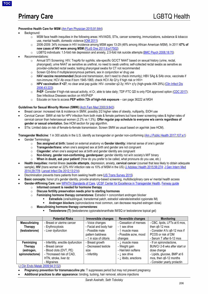
Primary Care LGBTQ Health
Sarah Axelrath, Seth Tobolsky
Preventive Health Care for MSM (Am Fam Physician 2015;91:844) • Background:
o MSM face health inequities in the following areas: HIV/AIDS, STIs, cancer screening, immunizations, substance & tobaccouse, mental health, domestic violence (IOM 2011)
o 2006-2009: 34% increase in HIV incidence among MSM ages 13-29 (48% among African American MSM). In 2011 67% ofnew cases of HIV were among MSM (PLoS One 2011;6:e17502)
o LGBTQ individuals: 1.5-fold risk depression and anxiety, 2.5-fold risk suicide attempts (BMC Psych 2008;18:70)• Recommendations:
o Annual STI Screening: HIV; TrepAb for syphilis; site-specific GC/CT NAAT based on sexual history (urine, rectal,pharyngeal); urine NAAT as sensitive as urethral, no need to swab urethra; self-collected rectal swabs as sensitive asprovider-collected rectal swabs; testing pharyngeal swabs for CT not recommended
o Screen Q3-6mo if multiple/anonymous partners, sex in conjunction w/ drug useo HAV vaccine recommended (fecal-oral transmission, don’t need to check immunity); HBV SAg & SAb once, vaccinate if
non-immune; HCV Ab once if born 1945-1965; check HCV Ab Q1y if high risk or HIV+o HPV vaccination if <27; no clear anal pap guide: HIV- consider q2-3y; HIV+ q1y (high-grade AIN 29%) (Clin Infect Dis
2006;43:223)o PrEP: Consider if high-risk sexual activity, nl Cr, able to take daily; TDF-FTC QD is only FDA approved option (CDC 2017);
See Infectious Diseases section on HIV/PrEPo Educate on how to access PEP within 72h of high-risk exposure – can page 36222 at MGH
Guidelines for Sexual Minority Women (SMW) (Arch Fam Med 2000;9:843) • Breast cancer: increased risk & incidence in SMW, possibly 2/2 higher rates of obesity, nulliparity, EtOH use• Cervical Cancer: SMW at risk for HPV infection from both male & female partners but have lower screening rates & higher rates of
cervical cancer than heterosexual women (2.2% vs 1.3%). Offer regular pap schedule to everyone w/a cervix regardless ofgender or sexual orientation. See HCM section for pap algorithm.
• STIs: Limited data on risk of female-to-female transmission. Screen SMW as usual based on age/risk (see HCM).
Transgender Medicine: 1 in 300 adults in the U.S. identify as transgender or gender non-conforming (Am J Public Health 2017;107:e1) • Gender Terminology
o Sex assigned at birth: based on external anatomy vs Gender identity: internal sense of one’s gendero Transgender/trans: when one’s assigned sex at birth and gender are not congruento Cisgender: when one’s assigned sex at birth and gender identity are congruento Non-binary, gender non-conforming, genderqueer: gender identity not w/in society’s M/F binaryo When in doubt, ask your patient! (How do you prefer to be called, what pronouns do you use, etc.)
• Health inequities: mental illness (suicide attempts, depression, anxiety, cervical cancer (counsel that less likely to obtain adequatesample), HIV (trans women w/ 22% HIV infection rate vs 10% of MSM in the US) (J Adolesc Health 2015;56:274; J Gen Intern Med2014;29:778; Lancet Infect Dis 2012;13:214)
• Discrimination prevents trans patients from seeking health care (US Trans Survey 2015)• Basic concepts: honor pt’s gender identity, provide anatomy-based screening, multidisciplinary care w/ mental health access• Gender-Affirming Care: see WPATH Standards of Care, UCSF Center for Excellence in Transgender Health, Fenway guide
o Informed consent is needed for hormone therapyo Discuss fertility preservation needs prior to starting hormoneso Feminizing hormone therapy cornerstones: Estradiol + concomitant androgen blocker
Estradiols (oral/sublingual, transdermal patch, estradiol valerate/estradiol cypionate IM) Androgen blockers (spironolactone most common, can decrease required estrogen dose)
o Masculinizing hormone therapy cornerstones: Testosterone (T) (testosterone cypionate/enanthate IM/SQ or testosterone topical gel)
Potential Risks Irreversible changes Reversible changes Monitoring
Masculinizing Therapy
(testosterone)
- Breast or uterine cancer- Erythrocytosis- Liver dysfunction
- Voice changes- Facial and body hair- Possible malepattern baldness- ↑ in size of clitoris
- Cessation of menses- ↑ sex drive- ↑ muscle mass- Possible acne, moodchanges
- CBC, lipids, LFTs at 6 mos,then q6-12 mos- Consider A1c q6-12 mos ifPCOS or risk of DM- Serum T after 6-12 mos
Feminizing Therapy
(estrogen, spironolactone)
- Infertility, erectile dysfunction- Breast cancer- Blood clots (DVT, PE)- ?Increased risk of CAD,HTN, stroke, liver dz- Migraines
- Breast growth- Decreased testiclesize- Infertility
- ↓ muscle mass- Weight gain- Hair/skin softens- ↓ sex drive- ↓ libido, erections
- If on spironolactone,BUN/Cr 2-8 wks after start ordose change- Lipids, glucose, BMP at 6mos, then q6-12 months- Consider yearly prolactin
(J Clin Endo Metab 2009;94:3132) • Pregnancy prevention for transmasculine pts: T suppresses period but may not prevent pregnancy• Additional practices to alter appearance: binding, tucking, hair removal, silicone injections
206
Primary Care Respiratory Complaints
Sam Wainwright
Chronic Cough (Am Fam Phys 2017;96:575): Subacute (3-8 weeks) vs. Chronic (>8 weeks) • Most common causes: upper airway cough syndrome (UACS), asthma, GERD; 18-62% pts have combo.
o Other causes: post-infxn (self-limiting but can last up to 3+ months, treat sxs), nonasthmatic eosinophilic bronchitis,chemical irritant (eg. cigarette smoke), psychogenic/habitual cough, bronchiectasis, cancer, TB, sarcoidosis.
o Normal CXR usually excludes bronchiectasis, persistent PNA, sarcoidosis, TB.• General approach: 1) Obtain good history (smoking status, URI hx, ACE-i use); consider CXR if no ACE-i or irritant exposure (except
smoking) and ↓suspicion for UACS/asthma/GERD; 2) Remove possible offending agent; 3) Start empiric tx for UACS/asthma/GERDsequentially until resolution → tx should be added to initial regimen; 4) Consider PFTs, esophageal pH monitoring, chest CT, sputumtests, cardiac studies if sxs persist despite treatment of usual causes.Etiology Characteristics Treatment
Upper Airway Cough Syndrome (UACS)
Formerly post-nasal drip syndrome. Most common cause of subacute and chronic cough. 2/2 nasal secretions stimulating cough receptors. PE of throat/nose may reveal cobblestoning. Common causes: allergic/non-allergic rhinitis, sinusitis.
Avoid environmental triggers of allergic rhinitis. Intranasal steroids, antihistamine nasal spray, oral antihistamine, oral decongestants, or saline nasal rinse can be used for symptom relief
Asthma Typically w/ episodic wheezing & dyspnea. Cough variant asthma p/w only cough. Pt may have h/o atopy. PE may reveal nasal polyps. Need spirometry w/ bronchodilator response & bronchoprovocation (e.g. metacholine challenge) for dx.
PRN bronchodilators +/- inhaled corticosteroids. Some pts may use only seasonally. See “Asthma” in pulmonology section for stepwise therapy.
GERD Epigastric burning sensation, sour taste in mouth, but sxs absent in >40% of patients.
Lifestyle modifications, moderate dose PPI (omeprazole 40 mg). Consider H pylori testing.
Respiratory tract infection
H/o recent viral illness. 2/2 postnasal drip/UACS or direct effect of virus on bronchial reactivity/cough receptors. Pts have been shown to experience transient bronchial hyperreactivity as well.
UACS tx as above. 2nd gen (cetirizine) or 3rd gen (fexofenadine) antihistamine. If bronchial hyperreactivity, tx w/ usual asthma care.
ACE Inhibitor Produces cough in 3-20% of pts. 2/2 ACEi mediated increase in bradykinin. Sxs can occur 1 wk – 6 mos after starting.
Withdraw ACEi (resolves within 1-4 weeks), change to ARB (not associated with cough).
Rhinosinusitis (Otolaryngol Head Neck Surg 2015;152:598): Acute (<1mo) vs. subacute (1-3mo) vs. chronic (>3mo, usually w/ anaerobes); recurrent (4 or more annual episodes) • Dx: rhinorrhea (viral - clear, bact - purulent) + nasal obstruction or facial pressure/pain/fullness. A/w anosmia, ear fullness, cough, H/A
Etiology Time Frame Treatment Bacterial: S. pneumo (41%), H. flu (35%), M. catarrhalis(4%), S. aureus (3%),anaerobes (7%), strep (7%)
>10 days, orworsening within 10days after initialimprovement (“doubleworsening”)
Watchful waiting* in pts w/ good follow-up vs. Augmentin 875mg BID** (Doxy 100mg BID in PCN-allergic) x 5-7d
• NSAIDs/Tylenol for pain• Saline irrigation/Netipot• Topical nasal steroids• Topical decongestant
(oxymetazoline) x 3d• Expectorants:(guaifenesin)Viral 7-10 days Symptom control, oral decongestant
Fungal: Mucor (invasive) in DM, immunocompromised
Acute(invasive) to more chronic (>3mo)
Surgical removal of fungal mucin or “fungal ball” (mycetoma). ENT emergency if invasive (destruction of sinus, erosion into orbit or brain)
* Watchful waiting: If stable after 7 days, can wait 10 more days w/sx management. If no improvement w/additional 10 days, start abx.** Higher dose Augmentin (2g BID or 90 mg/kg/d BID) in pts w/RFs for resistance (regional resistance pattern, Age 65+, hospitalized in last5d, abx use in last month, immunocompromised, DM/cardiac/renal/hepatic disease, severe infxn (fever >102F, suppurative complication)• Dx: ONLY if concern for complications or other etiology suspected: CT scan with contrast +/- MRI• Complications: Meningitis, periorbital/orbital cellulitis (pain, edema, proptosis, painful eye movement, diplopia),
subperiosteal/intracranial/epidural abscess, osteomyelitis of the sinus bones, septic cavernous sinus thrombosis.o Alarm symptoms: persistent fevers >102F; periorbital edema, inflammation, or erythema; CN palsies; abnormal
extraocular movements; proptosis; vision changes (diplopia, impaired vision); severe headache; AMS; meningeal signs.
Pharyngitis (IDSA Guidelines/JAMA 2012;308:1307): Most cases are viral (suspect if + conjunctivitis, coryza, cough, diarrhea, hoarseness, discrete ulcerative stomatitis, viral exanthema). Only 5-15% of adult sore throat visits are Group A Strep (GAS). • Exclude dangerous etiologies: epiglottitis, peritonsillar abscesses, infx in submandibular or retropharyngeal space, primary HIV• Identify & treat GAS to↓risk of suppurative complications (peritonsillar abscess, cervical lymphadenitis, mastoiditis), prevent
rheumatic fever (lower risk in adults), ↓transmission, & improve sx. ASO titers useful only in dx of non-suppurative sequelae of GAS.o Centor Criteria: 1 pt for each – tonsillar exudates, tender ant. cervical LAD, fever, ∅ cough.
0-2: No testing, treat sx. 3-4: Send Rapid Strep antigen detection test (Sens 70-90/Spec 90) + throat culture (if neg rapid but ↑clinical
suspicion)o Tx: PO Penicillin V 250mg QID vs 500mg BIDx10d; amoxicillin 500mg BIDx10d; IM Pencillin G benzathine 1.2 mill Ux1
PCN-allergic: Cephalexin 500mg BIDx10d Beta-lactam sensitivity: Clinda 300mg TIDx10d; Azithromycin 500 mg QDx1d, then 250 mg QDx4d
• Symptomatic Tx: OTC lozenges (e.g. Sucrets, Cepacol), throat sprays, NSAIDs/Tylenol for pain relief. No PO steroids.• Follow-up: If no improvement in sx in 5-7 days, evaluate for other infectious causes (e.g. mono, primary HIV, GC/chlamydia) or
suppurative complications such as tonsillopharyngeal cellulitis of abcess.
207
Primary Care Nodules
Sam Wainwright
Adrenal Nodules (Endocr Pract 2009;15:450, Eur J Endocr 2016;175:G1)
• Is it malignant? ↑Risk: diameter >4 cm, increased vascularity, irregular shape, high T2 signal on MRI, delayed contrastwashout. ↓Risk: if ≤10 HU on CT, homogenous and lipid-rich.
• Is it functionally active? Clinical exam & lab testing for all nodules (unless obvious myelolipoma) to r/o pheo & Cushing’s(see table). Also test for hyperaldo in hypertensive pts. Only test for production of excess sex hormones if clinicalstigmata. AVOID testing inpatients due to high false positive rates.
Diagnosis Suggestive Clinical Features Laboratory Tests Pheochromocytoma HTN, palpitations, headache, diaphoresis Serum metanephrines, 24H urine fractionated
metanephrine and catecholamines Cushing's syndrome Central obesity, prox muscle weakness, facial
plethora 1 mg dexamethasone suppression test is first line for screening in this population
Hyperaldosteronism HTN, hypokalemia Plasma aldo and PRA (d/c aldo antagonists before testing). May req. Adrenal Vein Sampling.
Adrenocortical cancer
Sxs related to excess glucocorticoid, mineralocorticoid, androgen/estrogen secretion
Serum DHEA-S and measure any other clinically indicated steroid based on clinical features
• Indications for adrenalectomy: > 4 cm on CT scan, malignant, or hormonally active; surgery after hormonal eval• Follow up: If indeterminate, repeat CT scan in 6-12 mos, then as clinically indicated. Consider cortisol & catechol testing
yearly x 4-5yrs (effectiveness of this practice unknown, EJE guidelines don’t recommend). Adrenalectomy if nodule growsby 20% or >1 cm.
Thyroid Nodules (Thyroid 2016;26:1, Endocr Pract 2016;22:622)
• Is it malignant? ↑Risk: pt h/o irradiation to head/neck, +family hx, or h/o thyroid cancer syndromes (i.e. MEN 2)• Workup: obtain ultrasound and check TSH (low TSH=less likely CA, high TSH=more likely CA)
o Low TSH - measure FT4 and FT3, obtain Thyroid radionuclide (123I) scan.▪ If “hot nodule,” consider Tx for hyperthyroidism if symptomatic. No biopsy necessary.▪ If “cold nodule,” refer for US-guided FNA
o If normal or high TSH, measure FT4 and TPO antibody, refer for thyroid U/S and FNA based on US findings.• FNA: All nodules >2cm or >1cm with moderately suspicious qualities. No FNA for purely cystic nodules.• Follow up: Based on U/S characteristics. If neg FNA but highly suspicious U/S findings, repeat US/FNA within 12 mo. If
low-moderate suspicious U/S findings, repeat U/S 12-24 mo, consider FNA if change. Can stop f/u after 2 neg FNAs.
Incidental Pulmonary Nodules (Radiology 2017;284:228, Chest 2013;143(Suppl 5), Thorax 2015;70:Suppl 2)
• NB: these guidelines are for incidental findings; recommendations for f/u of nodules found on LDCT for lung cancerscreening are different as that population is high risk (see Lung-RADS classification tables online)
• Is it malignant? Pt characteristics: ↑Risk w/ h/o smoking, emphysema, pulmonary fibrosis, extra-thoracic cancer,asbestos exposure, age. Nodule characteristics: Quality (subsolid/ground glass>solid), size, rate of growth, borders(irregular/spiculated border>smooth border), calcification (eccentric>popcorn/concentric/diffuse), location (upper>lowerlobe).
• Follow up: Tailored to patient and type of nodule. Subsolid (ground glass): if <6 mm, no routine f/u. If >6 mm, CT at 6-12months, then CT every 2 yrs until 5 yrs. Part solid: if <6mm, no routine f/u. If >6 mm, CT at 3-6 mos, then annual CT for 5yrs if unchanged and solid component <6 mm. Solid nodules: see below.
Nodule type <6mm 6-8 mm > 8 mmSingle solid nodule
Low risk No routine follow up CT at 6-12 months, then consider CT at 18-24 months
Consider CT at 3 months, PET/CT, or tissue sampling
High risk Optional CT at 12 months CT at 6-12 months, then CT at 18-24 months
Consider CT at 3 months, PET/CT, or tissue sampling
Multiple solid nodules Low risk No routine follow up CT at 3-6 months, then consider CT
at 18-24 months CT at 3-6 months, then consider CT at 18-24 months
High risk Optional CT at 12 months CT at 3-6 months, then at 18-24 months
CT at 3-6 months, then at 18-24 months
• Consider referral to the Pulmonary Nodule Clinic: refer in Epic or call x38728 for appointment
208
Primary Care Immigrant & Refugee Health
Helen D’Couto
Medical examination for newly arrived refugees and immigrants (CDC checklist)Introduction • Health system in US: describe role of PCP, how to obtain prescriptions/refills/labs/imaging/referrals, how to use ER• Know your Rights: Handouts re ICE (English) (Spanish); Red Cards (rights cards in different languages); compiled resourcesHistory & Physical: obtain prior medical records if possible• Mental health: PTSD, anxiety, depression; RHS-15 (screen at 2nd visit to minimize effect of re-traumatization)• HEENT: Vision, dental, hearing. See PCOI for low-cost referral options “Patient Education” → “Health Care Access”• Legal Status Screen: Do not document in medical record. “Do you have any questions about your immigration status?”
o If yes → Refer to Advice & Counsel session, e.g. LINC program MGH Chelsea or through local legal aid organizationso If possible asylum case → May refer directly to organization providing legal services, e.g. PAIR; MIRA
Vaccinations: • Age-appropriate vaccines as indicated (see Health Screening & Maintenance section)• If no vaccine documentation, check titers (including childhood vaccines such as MMR) or assume pt not vaccinated.Screening:• General screening: CBC w/ differential (eos, anemia), UA (hematuria),
glucose, gen chem, pregnancy test if appropriate; hepatitis, lead,micronutrient, chronic disease
• Tuberculosis: eval sxs, h/o sick contacts, PE; send T-spot (preferredwith prior exposure to BCG vaccine) or PPD
• STIs: Syphilis (TrepAb at MGH, VDRL/RPR elsewhere); HIV; GC/CT(urine NAAT) if ♀ < 25 & sexually active or ♀ > 25 + risk (h/o sexualassault, LE ⊕ on UA, sxs, new or >1 partner, partner w/ STI)
• Malaria (PCR most sensitive, blood films less sensitive if ∅sxs)o Test pts from Sub-Saharan Africa (SSA) who did not receive
pre-departure presumptive Rx w/ artesunate-combination(e.g. pregnant ♀ when Rx was C/I)
o Any pt from malaria-endemic country w/ sxs infection• Intestinal/Tissue Invasive Parasites: Test if no pre-departure Rx (albendazole, ivermectin) or incomplete: ≥2 stool O&P, Strongy
serologies. If SSA: schisto serologies.o Absolute eos: not specific or sensitive but if persistently elevated do work-up and consider empiric Strongy Rx.
Categories of immigration status • In MA, MassHealth eligibility is NOT contingent on immigration status. Refer all pts to PFS; PCOI page “Patient Education” → “Health
Care Access” → “Help Uninsured Patients Access Medical Care.” Link to videos for patients on the ACAType Basis Work Permit Green Card Details
LPR Varied Yes Yes Lawful Permanent Resident: Green card recipient; pathway to citizenship. Can apply for family members to get green card “family based” immigration.
U-Visa, T-VisaVAWA Crime Yes Yes
Eligible if victim of human trafficking (T) or victim of certain types of crime (U). Violence Against Women Act: Eligible if abused by spouse, child or parent who is LPR/citizen.
TPS Country Yes No Temporary Protected Status: Short list of countries, designated by Homeland Security, where conditions preclude safe return. Cannot be deported while country of origin on list.
Cancellation of Removal Discretionary Yes No Based on exception hardship to self or LPR/citizen spouse, parent,
child if deported; ineligible with certain criminal convictions.
DACA Age Yes No Deferred Action for Childhood Arrivals: Obama administration program deferring immigration cases of undocumented youth. Eligible for in-state tuition and work permits.
Refugee Fear Yes Yes Same legal standard as Asylum, based on persecution or well-founded fear, but granted prior to arrival in US. Maximum set annually by President (no limit to Asylum).
Asylum Fear 180-days s/pfiling After 1-yr
Well-founded fear of being persecuted based on race, religion, nationality, membership in social group or political opinion. Application due within 1yr date of entry. If granted, may also apply to spouse and children if in United States.
Withholding of Removal Fear No No Asylum/CAT/Withholding all part of same application. No 1yr rule;
may apply at any time. Ineligible with certain criminal convictions.
CAT Fear No No Convention Against Torture: similar to Withholding, but still eligible with criminal convictions.
Undocumented N/A No No Patients should seek legal counsel to ensure no options to apply for alternative statuses.
Parasitic Infections Peripheral eosinophilia Strongyloidiasis, filariasis,
schistosomiasis Hematuria, ♀ infertility, chronic pelvic pain
Schistosomiasis
Splenomegaly Malaria, schistosomiasis Chronic rash or itching Mycetoma, onchocerciasis,
other filarial diseases Esophageal dysmotility, HF, conduction dz
Chagas disease (Central/South America)
Seizures, CNS sxs Neurocysticercosis
209
Primary Care Outpatient Disease Management Index & Decision Aids Index of Outpatient Disease Management
Outpatient Cardiovascular Health Asthma COPD GERD & PUD Chronic Kidney Disease HIV (& PrEP) Travel Medicine Frailty Geriatric Syndromes Arthritis Outpatient Diabetes Thyroid Disorders Dementia Headache & Vertigo Depression & Anxiety ETOH Use Disorder Opioid Use Disorder Post-Acute Care
Calculators, Decision Aids, and Apps Calculators and Decision Aids for Providers: -ASCVD Risk Estimator Plus: http://tools.acc.org/ASCVD-Risk-Estimator-Plus/#!/calculate/estimate/-AUDIT Alcohol Use Questionnaire: http://auditscreen.org/using-audit-CAGE Questions for Alcohol Use: https://www.mdcalc.com/cage-questions-alcohol-use-CDC Adult Vaccine Schedule: https://www.cdc.gov/vaccines/schedules/hcp/imz/adult.html-Contraceptive Quick Start Algorithm: https://rhedi.org/wp-content/uploads/2017/08/QuickstartAlgorithm.pdf-FRAX Fracture Risk Assessment Tool: https://www.sheffield.ac.uk/FRAX/tool.aspx?country=9-PHQ-9: https://www.mdcalc.com/phq-9-patient-health-questionnaire-9-Pregnancy Due Dates/GA Calculator: https://www.mdcalc.com/pregnancy-due-dates-calculator
Patient-Provider Shared Decision Aids: -Mayo Shared Decision Making Resource Center (includes Cardiovascular Primary Prevention Choice, Depression MedicationChoice, Diabetes Medication Choice, Osteoporosis Decision Aid, PCI Choice, Smoking Cessation around the Time ofSurgery, Rheumatoid Arthritis Choice, and Statin Choice Decision Aid): https://shareddecisions.mayoclinic.org/
Primary Care Apps: -American Diabetes Association Standards of Care App: https://professional.diabetes.org/content-page/standards-care-app-1-Apps available in iTunes or Google store: AHRQ ePSS (repository of USPSTF guidelines and recommendations), ASCVDRisk Estimator, ACP Clinical Guidelines, CDC MEC Contraception Guide
Andrew Hoekzema210
40 53 54 68 92
117 119
152 152 162 169 177 185 186 196 197 198 249
Consultants Calling Consults
Meg Allison
TIPS FOR CALLING CONSULTS • To do BEFORE you call:
o Place order in Epic for consulto Existing outpatient specialist (e.g. cardiologist, oncologist) should be called first. You should also know name of the outpatient
provider before calling an inpatient consult.o Know your patient – you may be asked to provide additional information (current status, exam, workup). Briefly review the
admit note and briefly see/examine the patient if you have not done so previously. GI: melena/hematochezia, stool, current/prior Hct, coags, transfusions, past EGD/colo, vitals, IV access, NSAID/ASA use Cards: EKG/tele, prior stress/echo/cath (know anatomy), dry weight, biomarkers, current cardiac meds, outpt cardiologist Renal: Baseline Cr, CKD stage, on/off HD, dialysis access, electrolyte mgmt, current UOP, nephrotoxins, outpt nephrologist Onc: known cancers w/ stage/tx history, biopsy results (for new dx), current anticoagulants, special slide, outpt oncologist ID: current/past micro data, possible sources, current/prior abx (incl # of days), fever curve, hardware, travel, exposures
o Know your question – Bigelow JAR should specify consult question in task list. If not there, ASK. It is always OK to clarify.• To do DURING the page/call:
o Call as early in the day as possible (ideally before noon). The paging directory: ppd.partners.org or “Phone Directory” in Epico In your page to consulting team, include: pt name, MRN, location, call back #, brief consult question +/- level of urgencyo Avoid “curbside” questions. If there is a specific question about management, call a formal consult.o Tell the consultant a brief HPI, a clear explanation of the team’s thinking and a clear and specific question
• To do AFTER the call:o Invite the consultant to find you to relay their recommendations or tell them who will be covering for you
CALLING EMERGENT CONSULTS • Surgery: STAT to surgeon means life-threatening emergency (eg: hemorrhage, lost airway, perforated or ischemic bowel) → include
reason for consult in your page to help surgeon triage urgencyo Page “Senior Resident on call” under Emergency Surgery/Trauma (Churchill) Team (Red/Blue/White/Green)
• Psychiatry (eg: pt actively trying to leave AMA w/ unclear capacity; security concerns, major behavioral issues)o 8am-6pm: p33061 (Emergency Consult Resident). If weekend/Holiday: p17911 (weekend rounding psychiatrist)o 6pm-8am: Call APS (6-2994) or page APS resident at 27792
• Ophtho: Go through Partners Paging Directory. Backup/emergency number is 617-573-4063 (MEEI ED back desk).• Toxicology (ingestions/overdoses/exposures/interactions): Call Poison Control Massachusetts (617-355-6607 or 800-222-1222).
CALLING SURGICAL CONSULTS AT MGH • All surgical consults are considered urgent. For a non-urgent consult overnight, wait to page until AM.• In the ED: speak directly to (do not page) the surgery team that sits in Acute. Once patient on floor, page intern on the consulting
team. Do NOT page the ED Surgery resident who placed initial consult note. Do NOT page surgery attending.• New Ward consult → Page “Senior Resident on call” under Emergency Surgery/Trauma (Churchill) Team (Red/Blue/White/Green).
o Existing ward consults, page the intern for that Churchill service, not the team on call that day• New Private consult (patient had prior operation by MGH surgeon) → Page Senior for new consult on Baker surgery services
(Teams 1-6); team depends on which surgery attending is requested.o Team 1 (Surg Onc) → Paging Directory: “Surgical Oncology (Baker Team 1)”o Teams 2-6 (Berger, Colorectal, Pancreas, MIS, Endocrine) Paging Directory: “General/Gastrointestinal Surgery”
• Cardiac Surgery consult → Page “In-House Fellow” under “Cardiac Surgery” – you will usually be directed to the NP• Ortho consult → Page 20296 or “Orthopedics In-house” under “Orthopedics”• Transplant Surgery consult → Page “Intern” (6a-6p) or “House officer on call (6p-6a)” under “Transplant Surgery”• Vascular Surgery consult → Page “On-Call Resident” under “Vascular and Endovascular Surgery”
CALLING OTHER SUBSPECIALTY CONSULTS • ACT (Addiction Consult Team): Place consult in Epic (no need to call), for EtOH or other substance use disorders, suboxone, etc.• AMS (Anticoagulation Management Service): For established pts: p30104, or click AMS icon in Epic to determine existing AMS
RN; Inpatient for discharge – place Epic consult, if urgent or questions, page Discharge Pathway Service: p30103• Cardiology: login to Amion under “mghcardiology” to identify appropriate fellow• Chronic pain (cancer pain, pain in addiction): p17246; Acute pain service (epidurals and periop pain): p27246• Diabetes nurse educator: Service NP: p20737; MD: p14364• ENT: Call the MEEI ED at 617-573-3431 and they will page the consult resident• Optimum Care Committee (“OCC,” Ethics): Page ethics support pager: p32097 (Mon-Fri, 8am-4pm, except holidays)• Ophtho: Weekday 8am-5pm (routine): p23555 or look up through Partners Directory; Weekday 5pm-8am or Weekend/Holiday:
p23666 or call MEEI ED Back Desk 617-573-4063. Determine whether patient can travel to MEEI for an exam prior to calling consult.• Psychiatry: For non-emergent floor consult: start with order for psych consult in Epic
o Weekday, Weekend Night, Holiday Night: Call CL coordinators (6-2984). These consults will be seen within 24 hours.o Weekend or Holiday 8am-5pm: p17911 (weekend rounding psychiatrist)
• Transfusion reactions: Page blood bank resident: p21829, or fellow: p24346
211
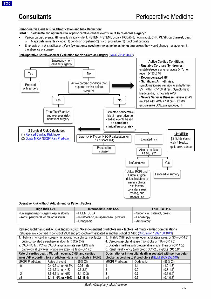
Consultants Perioperative Medicine
Mazin Abdelghany, Max Adelman
Peri-operative Cardiac Risk Stratification and Risk Reduction: GOAL: To estimate and optimize risk of peri-operative cardiac events, NOT to “clear for surgery” • Peri-op cardiac events: MI (usually clinically silent, NSTEMI > STEMI, usually POD#0-3, not intraop), CHF, VT/VF, card arrest, death
o Major determinants include: (1) condition of patient (2) risk of procedure (3) functional capacity• Emphasis on risk stratification. Very few patients need non-invasive/invasive testing unless they would change management in
the absence of surgery.Peri-Operative Cardiovascular Evaluation for Non-Cardiac Surgery (JACC 2014;64e77)
Operative Risk without Adjustment for Patient Factors High Risk >5% Intermediate Risk 1-5% Low Risk <1%
- Emergent major surgery, esp in elderly- Aortic, peripheral, or major vascular
- HEENT, CEA- Intrathoracic, intraperitoneal, prostate- Orthopedic
- Superficial, cataract, breast- Endoscopy- Ambulatory
Revised Goldman Cardiac Risk Index (RCRI): Six independent predictors (risk factors) of major cardiac complications Retrospectively derived in cohort of 2900 and prospectively validated in another cohort of 1400 (Circulation 1999;100:1043) 1. High-risk noncardiac surgery (as above; not a clinical risk factor
but incorporated elsewhere in algorithm) (OR 2.6)2. CAD (h/o MI, PCI or CABG, angina, nitrate use, EKG with
pathological Q waves, or positive exercise test) (OR 3.8)
3. HF (h/o CHF, pulmonary edema, bilateral rales, or S3) (OR 4.3)4. Cerebrovascular disease (h/o stroke or TIA) (OR 3.0)5. Diabetes mellitus with preoperative insulin therapy (OR 1.0!)6. Renal insufficiency (with preop SCr>2.0 mg/dL) (OR 0.9!)
Rate of cardiac death, MI, pulm edema, CHB, and cardiac arrest/VF according to # predictors (data from cohorts in RCRI)
Odds ratio for in-hospital death associated with peri-op beta-blocker according to # predictors (NEJM 2005;353:349)
#RCRI Predictors Rates of event (95% CI) #RCRI Predictors Odds ratio (95% CI) 0 1 2 ≥3
0.4-0.5% or ~0.5% 0.9-1.3% or ~1% 3.6-6.6% or ~5% 9.1-11.0% or ~10%
(0.05-1.5) (0.3-2.1) (2.1-10.3) (5.5-18.4)
1 2 3 ≥4
1.1 0.9 0.7 0.6
(1.0-1.3) (0.8-1.1) (0.6-0.9) (0.4-0.8)
Emergency non-cardiac surgery?
Yes
Proceed with surgery
No
Active cardiac condition that requires eval/tx before
surgery?
Yes
Treat/Test/Stabilizeand reassess risk-benefit of surgery
No
Estimated periperative risk of major adverse cardiac events based
on combined clinical/surgical risk
Low risk (<1% per NSQIP calculators or RCRI score 0-1)
Proceed to surgery
Elevated risk
Able to achieve ≥4 METs?*
No/unknown
Utilize RCRI and Gupta surgical
risk calculators to assess clinical
risk factors, consider stress
testing, and reduce risk
Yes
Proceed to surgery
*4+ METs:↑2 flights stairs; walk 4 blocks; golf, bowl, dance
2 Surgical Risk Calculators (1) Revised Cardiac Risk Index(2) Gupta MICA NSQIP Risk Prediction
Active Cardiac Conditions - Unstable Coronary Syndromes:unstable/severe angina, acute (< 7d) orrecent (< 30d) MI- Decompensated HF- Significant Arrhythmias:symptomatic/new ventricular arrhythmias,SVT with HR >100 at rest. Symptomaticbradycardia, high-grade AVB.- Severe Valvular Disease: severe sx AS(mGrad >40, AVA < 1.0 cm2), sx MS (progressive DOE, presyncope, HF)
212
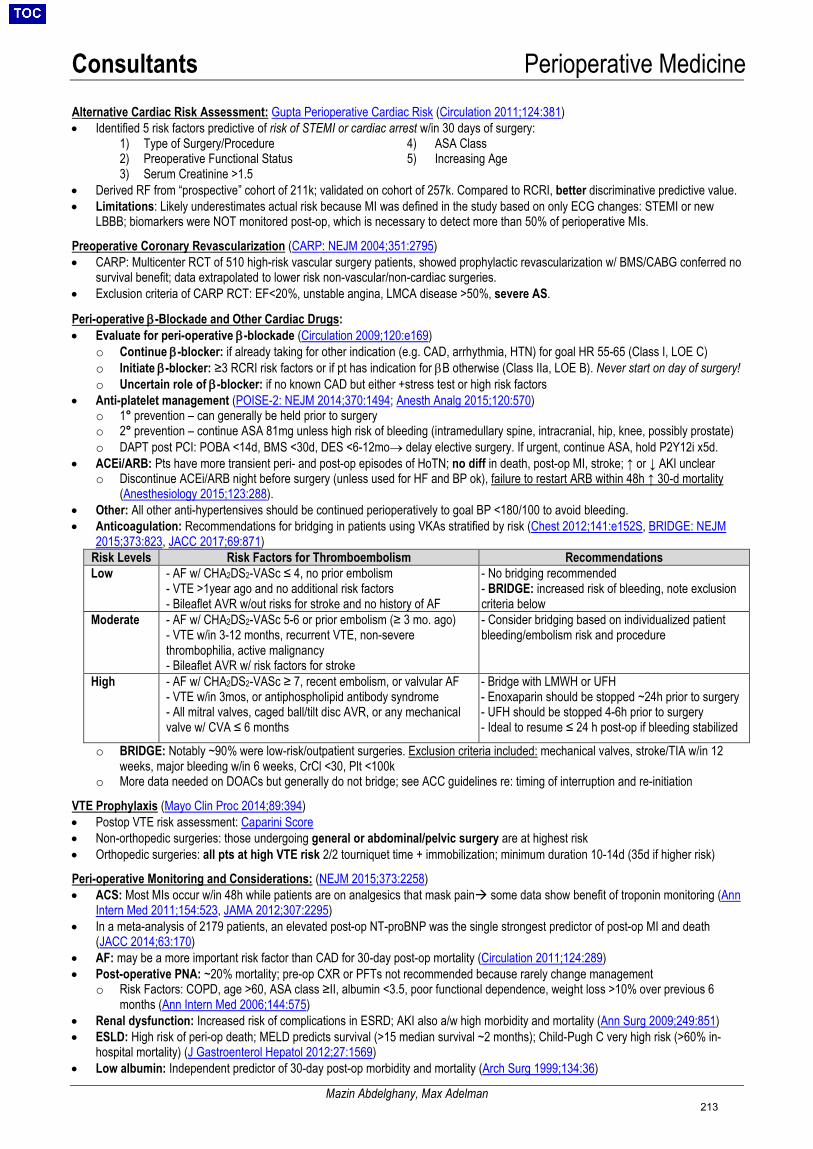
Consultants Perioperative Medicine
Mazin Abdelghany, Max Adelman
Alternative Cardiac Risk Assessment: Gupta Perioperative Cardiac Risk (Circulation 2011;124:381) • Identified 5 risk factors predictive of risk of STEMI or cardiac arrest w/in 30 days of surgery:
1) Type of Surgery/Procedure 4) ASA Class2) Preoperative Functional Status 5) Increasing Age3) Serum Creatinine >1.5
• Derived RF from “prospective” cohort of 211k; validated on cohort of 257k. Compared to RCRI, better discriminative predictive value.• Limitations: Likely underestimates actual risk because MI was defined in the study based on only ECG changes: STEMI or new
LBBB; biomarkers were NOT monitored post-op, which is necessary to detect more than 50% of perioperative MIs.
Preoperative Coronary Revascularization (CARP: NEJM 2004;351:2795) • CARP: Multicenter RCT of 510 high-risk vascular surgery patients, showed prophylactic revascularization w/ BMS/CABG conferred no
survival benefit; data extrapolated to lower risk non-vascular/non-cardiac surgeries.• Exclusion criteria of CARP RCT: EF<20%, unstable angina, LMCA disease >50%, severe AS.
Peri-operative β-Blockade and Other Cardiac Drugs:• Evaluate for peri-operative β-blockade (Circulation 2009;120:e169)
o Continue β-blocker: if already taking for other indication (e.g. CAD, arrhythmia, HTN) for goal HR 55-65 (Class I, LOE C)o Initiate β-blocker: ≥3 RCRI risk factors or if pt has indication for βB otherwise (Class IIa, LOE B). Never start on day of surgery!o Uncertain role of β-blocker: if no known CAD but either +stress test or high risk factors
• Anti-platelet management (POISE-2: NEJM 2014;370:1494; Anesth Analg 2015;120:570)o 1° prevention – can generally be held prior to surgeryo 2° prevention – continue ASA 81mg unless high risk of bleeding (intramedullary spine, intracranial, hip, knee, possibly prostate)o DAPT post PCI: POBA <14d, BMS <30d, DES <6-12mo→ delay elective surgery. If urgent, continue ASA, hold P2Y12i x5d.
• ACEi/ARB: Pts have more transient peri- and post-op episodes of HoTN; no diff in death, post-op MI, stroke; ↑ or ↓ AKI unclearo Discontinue ACEi/ARB night before surgery (unless used for HF and BP ok), failure to restart ARB within 48h ↑ 30-d mortality
(Anesthesiology 2015;123:288).• Other: All other anti-hypertensives should be continued perioperatively to goal BP <180/100 to avoid bleeding.• Anticoagulation: Recommendations for bridging in patients using VKAs stratified by risk (Chest 2012;141:e152S, BRIDGE: NEJM
2015;373:823, JACC 2017;69:871)Risk Levels Risk Factors for Thromboembolism Recommendations
Low - AF w/ CHA2DS2-VASc ≤ 4, no prior embolism- VTE >1year ago and no additional risk factors- Bileaflet AVR w/out risks for stroke and no history of AF
- No bridging recommended- BRIDGE: increased risk of bleeding, note exclusioncriteria below
Moderate - AF w/ CHA2DS2-VASc 5-6 or prior embolism (≥ 3 mo. ago)- VTE w/in 3-12 months, recurrent VTE, non-severethrombophilia, active malignancy- Bileaflet AVR w/ risk factors for stroke
- Consider bridging based on individualized patientbleeding/embolism risk and procedure
High - AF w/ CHA2DS2-VASc ≥ 7, recent embolism, or valvular AF- VTE w/in 3mos, or antiphospholipid antibody syndrome- All mitral valves, caged ball/tilt disc AVR, or any mechanicalvalve w/ CVA ≤ 6 months
- Bridge with LMWH or UFH- Enoxaparin should be stopped ~24h prior to surgery- UFH should be stopped 4-6h prior to surgery- Ideal to resume ≤ 24 h post-op if bleeding stabilized
o BRIDGE: Notably ~90% were low-risk/outpatient surgeries. Exclusion criteria included: mechanical valves, stroke/TIA w/in 12weeks, major bleeding w/in 6 weeks, CrCl <30, Plt <100k
o More data needed on DOACs but generally do not bridge; see ACC guidelines re: timing of interruption and re-initiation
VTE Prophylaxis (Mayo Clin Proc 2014;89:394)• Postop VTE risk assessment: Caparini Score• Non-orthopedic surgeries: those undergoing general or abdominal/pelvic surgery are at highest risk• Orthopedic surgeries: all pts at high VTE risk 2/2 tourniquet time + immobilization; minimum duration 10-14d (35d if higher risk)
Peri-operative Monitoring and Considerations: (NEJM 2015;373:2258)• ACS: Most MIs occur w/in 48h while patients are on analgesics that mask pain some data show benefit of troponin monitoring (Ann
Intern Med 2011;154:523, JAMA 2012;307:2295)• In a meta-analysis of 2179 patients, an elevated post-op NT-proBNP was the single strongest predictor of post-op MI and death
(JACC 2014;63:170)• AF: may be a more important risk factor than CAD for 30-day post-op mortality (Circulation 2011;124:289)• Post-operative PNA: ~20% mortality; pre-op CXR or PFTs not recommended because rarely change management
o Risk Factors: COPD, age >60, ASA class ≥II, albumin <3.5, poor functional dependence, weight loss >10% over previous 6months (Ann Intern Med 2006;144:575)
• Renal dysfunction: Increased risk of complications in ESRD; AKI also a/w high morbidity and mortality (Ann Surg 2009;249:851)• ESLD: High risk of peri-op death; MELD predicts survival (>15 median survival ~2 months); Child-Pugh C very high risk (>60% in-
hospital mortality) (J Gastroenterol Hepatol 2012;27:1569)• Low albumin: Independent predictor of 30-day post-op morbidity and mortality (Arch Surg 1999;134:36)
213
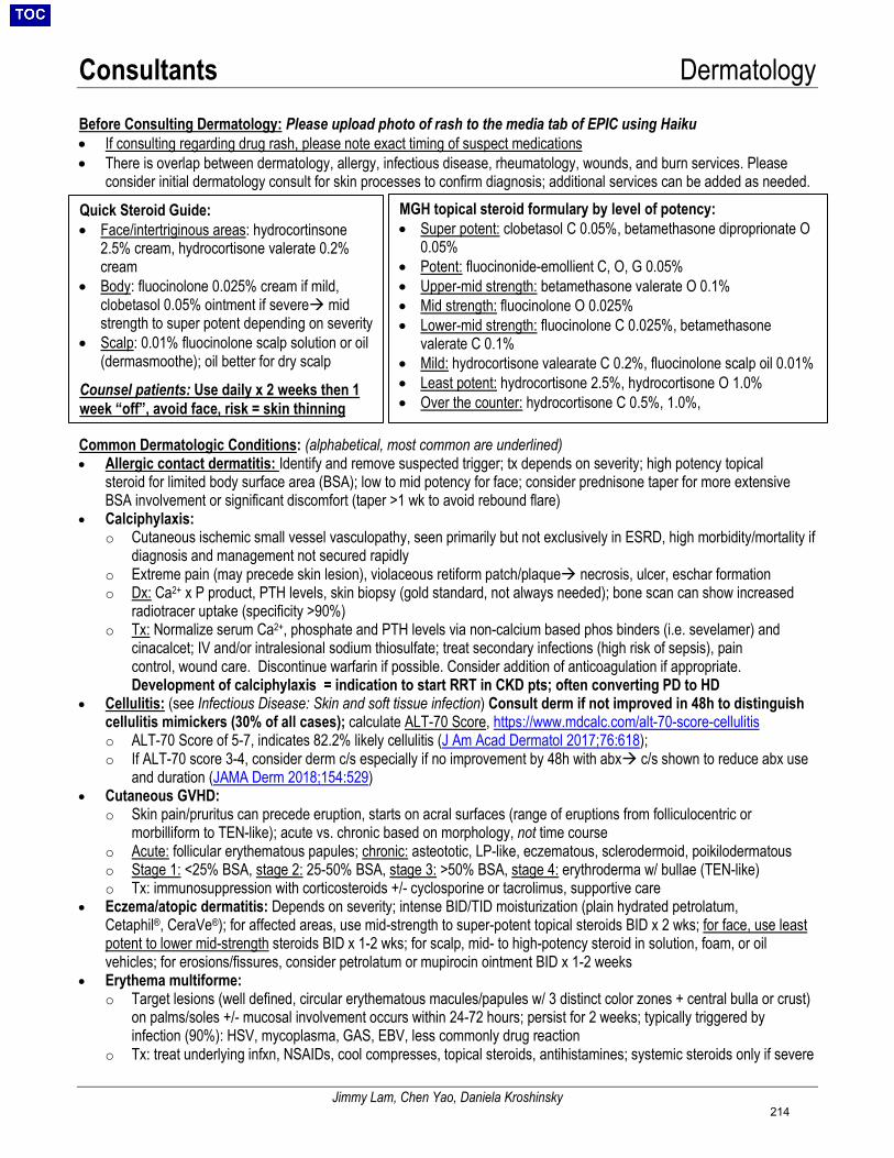
Consultants Dermatology
Jimmy Lam, Chen Yao, Daniela Kroshinsky
Before Consulting Dermatology: Please upload photo of rash to the media tab of EPIC using Haiku • If consulting regarding drug rash, please note exact timing of suspect medications• There is overlap between dermatology, allergy, infectious disease, rheumatology, wounds, and burn services. Please
consider initial dermatology consult for skin processes to confirm diagnosis; additional services can be added as needed.
Common Dermatologic Conditions: (alphabetical, most common are underlined) • Allergic contact dermatitis: Identify and remove suspected trigger; tx depends on severity; high potency topical
steroid for limited body surface area (BSA); low to mid potency for face; consider prednisone taper for more extensiveBSA involvement or significant discomfort (taper >1 wk to avoid rebound flare)
• Calciphylaxis:o Cutaneous ischemic small vessel vasculopathy, seen primarily but not exclusively in ESRD, high morbidity/mortality if
diagnosis and management not secured rapidlyo Extreme pain (may precede skin lesion), violaceous retiform patch/plaque necrosis, ulcer, eschar formationo Dx: Ca2+ x P product, PTH levels, skin biopsy (gold standard, not always needed); bone scan can show increased
radiotracer uptake (specificity >90%)o Tx: Normalize serum Ca2+, phosphate and PTH levels via non-calcium based phos binders (i.e. sevelamer) and
cinacalcet; IV and/or intralesional sodium thiosulfate; treat secondary infections (high risk of sepsis), paincontrol, wound care. Discontinue warfarin if possible. Consider addition of anticoagulation if appropriate.Development of calciphylaxis = indication to start RRT in CKD pts; often converting PD to HD
• Cellulitis: (see Infectious Disease: Skin and soft tissue infection) Consult derm if not improved in 48h to distinguishcellulitis mimickers (30% of all cases); calculate ALT-70 Score, https://www.mdcalc.com/alt-70-score-cellulitiso ALT-70 Score of 5-7, indicates 82.2% likely cellulitis (J Am Acad Dermatol 2017;76:618);o If ALT-70 score 3-4, consider derm c/s especially if no improvement by 48h with abx c/s shown to reduce abx use
and duration (JAMA Derm 2018;154:529)• Cutaneous GVHD:
o Skin pain/pruritus can precede eruption, starts on acral surfaces (range of eruptions from folliculocentric ormorbilliform to TEN-like); acute vs. chronic based on morphology, not time course
o Acute: follicular erythematous papules; chronic: asteototic, LP-like, eczematous, sclerodermoid, poikilodermatouso Stage 1: <25% BSA, stage 2: 25-50% BSA, stage 3: >50% BSA, stage 4: erythroderma w/ bullae (TEN-like)o Tx: immunosuppression with corticosteroids +/- cyclosporine or tacrolimus, supportive care
• Eczema/atopic dermatitis: Depends on severity; intense BID/TID moisturization (plain hydrated petrolatum,Cetaphil®, CeraVe®); for affected areas, use mid-strength to super-potent topical steroids BID x 2 wks; for face, use leastpotent to lower mid-strength steroids BID x 1-2 wks; for scalp, mid- to high-potency steroid in solution, foam, or oilvehicles; for erosions/fissures, consider petrolatum or mupirocin ointment BID x 1-2 weeks
• Erythema multiforme:o Target lesions (well defined, circular erythematous macules/papules w/ 3 distinct color zones + central bulla or crust)
on palms/soles +/- mucosal involvement occurs within 24-72 hours; persist for 2 weeks; typically triggered byinfection (90%): HSV, mycoplasma, GAS, EBV, less commonly drug reaction
o Tx: treat underlying infxn, NSAIDs, cool compresses, topical steroids, antihistamines; systemic steroids only if severe
MGH topical steroid formulary by level of potency: • Super potent: clobetasol C 0.05%, betamethasone diproprionate O
0.05% • Potent: fluocinonide-emollient C, O, G 0.05%• Upper-mid strength: betamethasone valerate O 0.1%• Mid strength: fluocinolone O 0.025%• Lower-mid strength: fluocinolone C 0.025%, betamethasone
valerate C 0.1% • Mild: hydrocortisone valearate C 0.2%, fluocinolone scalp oil 0.01%• Least potent: hydrocortisone 2.5%, hydrocortisone O 1.0%• Over the counter: hydrocortisone C 0.5%, 1.0%,
Quick Steroid Guide: • Face/intertriginous areas: hydrocortinsone
2.5% cream, hydrocortisone valerate 0.2%cream
• Body: fluocinolone 0.025% cream if mild,clobetasol 0.05% ointment if severe midstrength to super potent depending on severity
• Scalp: 0.01% fluocinolone scalp solution or oil(dermasmoothe); oil better for dry scalp
Counsel patients: Use daily x 2 weeks then 1 week “off”, avoid face, risk = skin thinning
214
Consultants Dermatology
Jimmy Lam, Chen Yao, Daniela Kroshinsky
• Erythroderma: Diffuse redness > 90% BSA.o Causes: psoriasis, atopic derm, mycosis fungoides, cutaneous T cell lymphoma, pityriasis rubra pilaris, drug, other.o Work-up: CBC w/ diff, BMP, detailed med rec, +/- HIV. Tx: liberal emollients or mid-potency topical steroids and
antihistamines; correct fluid/electrolyte imbalance; monitor for 2º infections, discontinue offending meds, c/s derm• Herpes simplex virus 1/2: (always confirm first diagnosis with DFA or PCR from vesicle base; culture can be done but
results can be very delayed; Tzanck smear effective but requires experienced practitioner)o Uncomplicated orolabial: primary is usually gingivostomatitisacyclovir 400mg TID x7-10d; if recurrent
valacyclovir 2000 mg PO q12h x 1d or famiciclovir 1500mg PO x1 at symptom onset; periocular skin involvementwarrants an ophthalmology consult to r/o herpetic keratitis
o Uncomplicated genital (immunocompetent): for 1º episode (<72 hr after onset), valacyclovir 1000 mg PO bid x 10d,or acyclovir 400 mg PO TID x10d, or famciclovir 250mg TID; recurrent episodes (<24 hr onset), use valacyclovir 500mg PO q12h x 3-5 d or acyclovir 400 mg PO TID x 5d
• Herpes zoster (shingles):o Uncomplicated, <72 hr (immunocompetent): valacyclovir 1000mg PO Q8H x7d or acyclovir 800 mg PO 5x/d x7-10do Disseminated: >20 vesicles outside two 1º dermatomes; acyclovir 10 mg/kg IV q8h; consider immunodeficiency w/uo Immunosuppressed: acyclovir 10 mg/kg IV q8h; IVFs if hypovolemic to decrease risk of acyclovir crystal
nephropathy; obtain DFA/viral culture; monitor carefully for dissemination (esp. encephalitis, pneumonia, hepatitis)o Herpes zoster ophthalmicus: Urgent Ophtho consult if concern for ocular involvemento Post-herpetic neuralgia: risk ↓ w/ early antiviral treatment (<72 hr); if higher risk (>50 yo w/ moderate-to-severe
acute pain) consider preventive tx w/ gabapentin 300 mg PO QD, titrate up to 3600 mg QD, divided TID as toleratedo Consider high lysine, low arginine diet and post-episode vaccination to prevent herpes recurrence
• Pressure Injury/Ulcers: must be documented in your H&Po NPUAP Staging: 1: non blanchable erythema of intact skin; 2: partial thickness skin loss with exposed dermis;
3: full thickness skin loss; 4: full thickness skin and tissue losso Wound CNS Consult for: stage 3/4 pressure injury, device related injuries, moisture associated skin damage, edema
drainage management, special bed surfaces (i.e. clinitron, bariatric) → IP Consult to Wound Nurseo Wound Consult (Plastics/Vascular collab) for: acute wound issues such as limb ischemia, wet gangrene, any wound
requiring OR debridement → IP Consult to Inpatient Wound Service; consider derm consult first to confirm etiology• Purpura fulminans: “DIC in skin” = true emergency; consult Hematology for possible factor replacement
o Microvascular skin occlusion w/ platelet-fibrin thrombi retiform purpura; Causes: infection (Strep, Staph, H. flu, N.meningitidis, Capnocytophaga, VZV, CMV, Babesia); catastrophic APS, CTD, malignancy, protein C/S deficiency
o Work-up: DIC labs, blood cultures, skin biopsy w/ GS and culture; Tx: broad-spectrum abx + supportive care.• Psoriasis: Depends on severity; short-term tx includes topical steroids, calcipotriene, intense moisturization +/- occlusion
w/ plastic wrap; long-term tx includes phototherapy, acitretin, MTX, biologics w/ outpt Derm f/u (JAAD 2011;65:137)• Seborrheic dermatitis: For face, least potent to lower mid-strength topical steroid BID x 1wk and/or ketoconazole 2%
cream BID x 4 wks, then 1-2x/wk for maintenance; alternatively pimecrolimus cream, tacrolimus 0.03 0r 0.1% ointment forscalp, ketoconazole 2% shampoo qHS
• Stasis dermatitis: LE compression (ACE wraps, stockings) with elevation; mid-strength to superpotent corticosteroidointment BID x 1-2 wks +/- occlusion with plastic wrap; mupirocin ointment BID x1-2 wks to erosions; intensivemoisturization (hydrated petrolatum) (Note: bilateral lower extremity cellulitis extremely rare – diagnosis of exclusion)
• Tinea pedis: Apply topical imidazole (econazole 1% cream QD or clotrimazole 1% cream BID x 2-4 wks) or allylamine(terbinafine 1% cream BID x 2 wks) to entire foot and webbed spaces between toes
• Urticaria (acute): Identify, remove trigger; histamine-mediated itch combination PO antihistamines (fexofenadine 180mg PO BID with diphenhydramine or hydroxyzine PO qHS; add ranitidine BID if not improved); dry skin itch cream-based emollients; neuropathic itch gabapentin; adjunct topical menthol (not on any open erosions, as it will sting)
• Vasculitis: idiopathic in up to 50%; small, medium, and large-vesselo Small-vessel vasculitis is also known as leukocytoclastic vasculits (LCV); most common
• Immune-complex: HSP, erythema elevatum diutinum, urticarial vasculitis, cryoglobulinemia, cutaneous smallvessel vasculitis (dx of exclusion); ANCA-mediated: GPA, MPA, EGPA; other: calciphylaxis (see above)
o Etiologies: 1) Drug-induced: abx, allopurinol, thz, hydantoins, PTU; 2) Connective-tissue disease; 3) Infections: HBV,HCV, Strep, URI, HIV, CMV, Mycobacteria, Neisseria spp.; 4) Malignancy
o Workup: UA w/ microscopy, CBC w/diff, BUN LFTs, HBV, HCV, cryos, complements, ANA, dsDNA, RF, ANCA
215

Consultants Dermatology
Jimmy Lam, Chen Yao, Daniela Kroshinsky
Drug Eruptions • Step 1: Make timeline to determine time course of drug initiation and development of rash• Step 2: Discontinue offending agent. Common drugs for each eruption listed, but any drug can be a culprit at any time
Time Course
Rash Other Signs/Sx Common Drugs
Treatment
Urticaria/ Anaphylaxis
Immediate (min-hr) – delayed (days)
Pruritic, well-circumscribed, erythematous papules/plaques with central pallor.
+/- angioedema, wheezing, GI sx, tachycardia, HoTN,
Any Antihistamines (benadryl + H2) + Steroids if severe+ IM epinephrine if s/sanaphylaxis- c/s allergyimmunology
Fixed Drug Eruption
Minutes-hours
Solitary sharply demarcated round red-brown patch or edematous plaque recurring in same location each time drug ingested. Can evolve to bullae. Oral/anogenital mucosa common sites.
Usually asymptomatic
Abx (sulfa, TMP, FQs, TCNs), NSAIDs, barbiturates
-Topical steroids ifsymptomatic
Acute Generalized Exanthematous Pustulosis (AGEP)
2-14 days Small non-follicular pustules on erythematous/edematous plaques, begin on face or intertriginous areas then become widespread. Usually within 24-48 hours of medication exposure. Burning, pruritus common.
Fever, marked neutrophilia +/- oral mucosal erosions, facial edema
Abx (PCN, macrolides) Can occur after only one exposure
-Anti-pyretic-Topical steroids
Exanthematous
4-14 days Pruritic, erythematous macules/papules start on trunk, spread centrifugally to symmetric extremities. May lead to erythroderma.
+/- low grade fever
Abx (PCN, sulfa), allopurinol, phenytoin, Requires repeat exposures
-Topical steroids,antihistamines(Note: may take 7-14dafter stopping drug toresolve)
SJS/TEN
4-21 days Prodromal fevers, malaise, myalgias/arthralgias. Pruritic atypical targetoid (amorphous, 2 color zones) macules bullae desquamation
<10% BSA = SJS 10-30% BSA = SJS/TEN overlap>30% BSA= TEN
Mucosal bullae, erosions & crusting, conjunctivitis + Nikolsky signComplications:2° infection, resp.compromise, GIB,visual impairment
Abx (sulfa), AEDs, NSAIDs, allopurinol, phenobarbital
-Cyclosporine(preferred at MGH)-Steroids possiblemortalitybenefit (JAMA Derm2017;153:514)butcontroversial -IVIG, anti-TNF-Burn level care if>30% BSA
DRESS
3-6 wks Morbilliform rash spreads downward symmetrically from face, can see SJS/TEN-like lesions and mucosal involvement.
Fever, arthralgias, eosinophilia, internal organ involvement (liver, kidney; rarely lung, heart), LAD.
Abx, AEDs, ARV (nevirapine, abacavir), carbamazepine
-Supportive care-IV Solumedrol(decreased risk ofbowel edema vs. PO),SLOW taper (3-6 wks)
216
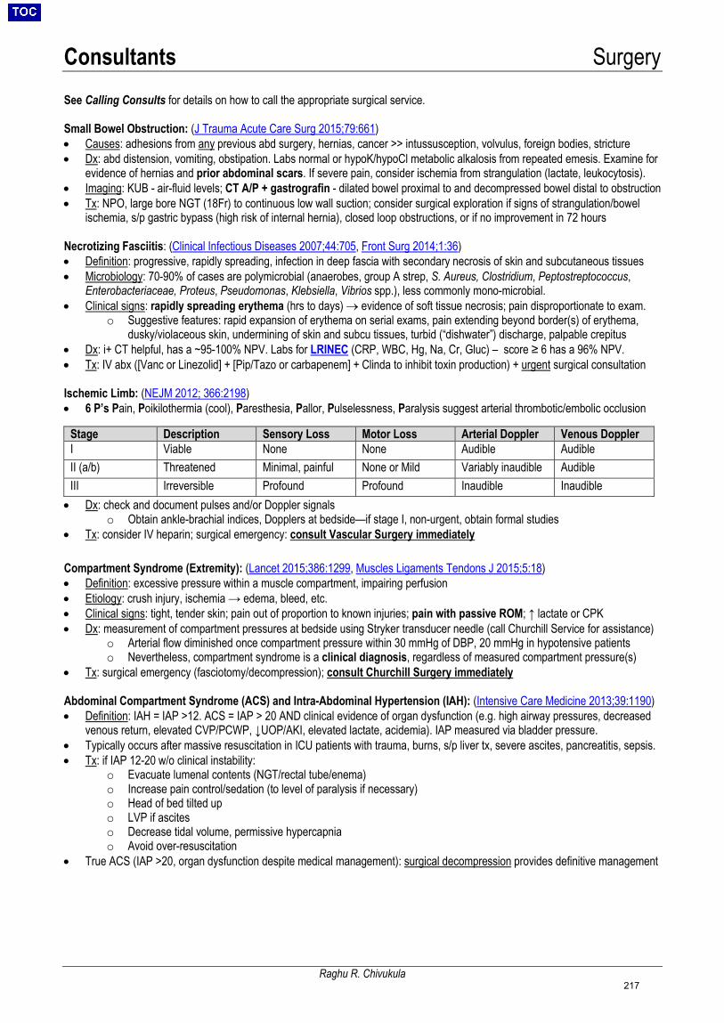
Consultants Surgery
Raghu R. Chivukula
See Calling Consults for details on how to call the appropriate surgical service.
Small Bowel Obstruction: (J Trauma Acute Care Surg 2015;79:661) • Causes: adhesions from any previous abd surgery, hernias, cancer >> intussusception, volvulus, foreign bodies, stricture• Dx: abd distension, vomiting, obstipation. Labs normal or hypoK/hypoCl metabolic alkalosis from repeated emesis. Examine for
evidence of hernias and prior abdominal scars. If severe pain, consider ischemia from strangulation (lactate, leukocytosis).• Imaging: KUB - air-fluid levels; CT A/P + gastrografin - dilated bowel proximal to and decompressed bowel distal to obstruction• Tx: NPO, large bore NGT (18Fr) to continuous low wall suction; consider surgical exploration if signs of strangulation/bowel
ischemia, s/p gastric bypass (high risk of internal hernia), closed loop obstructions, or if no improvement in 72 hours
Necrotizing Fasciitis: (Clinical Infectious Diseases 2007;44:705, Front Surg 2014;1:36) • Definition: progressive, rapidly spreading, infection in deep fascia with secondary necrosis of skin and subcutaneous tissues• Microbiology: 70-90% of cases are polymicrobial (anaerobes, group A strep, S. Aureus, Clostridium, Peptostreptococcus,
Enterobacteriaceae, Proteus, Pseudomonas, Klebsiella, Vibrios spp.), less commonly mono-microbial.• Clinical signs: rapidly spreading erythema (hrs to days) → evidence of soft tissue necrosis; pain disproportionate to exam.
o Suggestive features: rapid expansion of erythema on serial exams, pain extending beyond border(s) of erythema,dusky/violaceous skin, undermining of skin and subcu tissues, turbid (“dishwater”) discharge, palpable crepitus
• Dx: i+ CT helpful, has a ~95-100% NPV. Labs for LRINEC (CRP, WBC, Hg, Na, Cr, Gluc) – score ≥ 6 has a 96% NPV.• Tx: IV abx ([Vanc or Linezolid] + [Pip/Tazo or carbapenem] + Clinda to inhibit toxin production) + urgent surgical consultation
Ischemic Limb: (NEJM 2012; 366:2198) • 6 P’s Pain, Poikilothermia (cool), Paresthesia, Pallor, Pulselessness, Paralysis suggest arterial thrombotic/embolic occlusion
Stage Description Sensory Loss Motor Loss Arterial Doppler Venous Doppler I Viable None None Audible Audible II (a/b) Threatened Minimal, painful None or Mild Variably inaudible Audible III Irreversible Profound Profound Inaudible Inaudible • Dx: check and document pulses and/or Doppler signals
o Obtain ankle-brachial indices, Dopplers at bedside—if stage I, non-urgent, obtain formal studies• Tx: consider IV heparin; surgical emergency: consult Vascular Surgery immediately
Compartment Syndrome (Extremity): (Lancet 2015;386:1299, Muscles Ligaments Tendons J 2015;5:18) • Definition: excessive pressure within a muscle compartment, impairing perfusion• Etiology: crush injury, ischemia → edema, bleed, etc.• Clinical signs: tight, tender skin; pain out of proportion to known injuries; pain with passive ROM; ↑ lactate or CPK• Dx: measurement of compartment pressures at bedside using Stryker transducer needle (call Churchill Service for assistance)
o Arterial flow diminished once compartment pressure within 30 mmHg of DBP, 20 mmHg in hypotensive patientso Nevertheless, compartment syndrome is a clinical diagnosis, regardless of measured compartment pressure(s)
• Tx: surgical emergency (fasciotomy/decompression); consult Churchill Surgery immediately
Abdominal Compartment Syndrome (ACS) and Intra-Abdominal Hypertension (IAH): (Intensive Care Medicine 2013;39:1190) • Definition: IAH = IAP >12. ACS = IAP > 20 AND clinical evidence of organ dysfunction (e.g. high airway pressures, decreased
venous return, elevated CVP/PCWP, ↓UOP/AKI, elevated lactate, acidemia). IAP measured via bladder pressure.• Typically occurs after massive resuscitation in ICU patients with trauma, burns, s/p liver tx, severe ascites, pancreatitis, sepsis.• Tx: if IAP 12-20 w/o clinical instability:
o Evacuate lumenal contents (NGT/rectal tube/enema)o Increase pain control/sedation (to level of paralysis if necessary)o Head of bed tilted upo LVP if asciteso Decrease tidal volume, permissive hypercapniao Avoid over-resuscitation
• True ACS (IAP >20, organ dysfunction despite medical management): surgical decompression provides definitive management
217
Consultants Urology
Raghu R. Chivukula
Symptomatic Urolithiasis (kidney stones): • Evaluation/Management:
o Imaging: Non-contrast Stone Protocol CT (I–, O–): evaluates position, hydronephrosis, hints at composition; alternative isKUB and ultrasound (requires both), with non-diagnostic studies prompting CT
o UA/UCx: In all patients except those with urostomies. If positive cx, need decompression by stent (Urology) or percutaneousnephrostomy/PCN (GU IR)
o Rehydration: Normal Saline @ 150 mL/hr if tolerated (↑ ureteral peristalsis)o Alpha-Blockers: Tamsulosin 0.4mg PO QD (hold for SBP < 90) (ureteral relaxation)o Analgesia: Opioids preferred; NSAIDs/Ketorolac more effective but risk bleeding and AKIo Preoperative workup if requiring intervention: NPO / EKG / Coags / Type & Screen
• Consider Urology Consult: solitary or transplanted kidney, DM, immunosuppression, AKI, +UA/UCx, sepsis, inadequate pain control• Urosepsis Management: image ASAP, BCx/UCx, urgent Urology consult; IV abx to cover GNRs + enterococcus• Clinical Pearl: Patients with an acute abdomen lie still, pts with renal colic writhe in pain
Hematuria / Obstructed Catheter: microscopic hematuria defined as UA with >3RBC/hpf; consider glomerular source in proper context • DDx: UTI, INR>3, traumatic catheter placement, bladder CA (5th most common neoplasm), upper urinary tract CA, prostate CA• Workup: “The Three C’s”: 1) Hematuria protocol CT (3-phase: non-con, arterial phase, delays to assess ureters); 2) Urine cytology
once hematuria clears (blood interferes with test); 3) Outpatient cystoscopy• Tx: if obstructed (can’t void or catheter not draining / “clot retention”) or significant hematuria: Irrigate bladder via Whistle-Tip catheter
using a 60 cc catheter-tipped syringe – flush in and out with saline to remove clots until urine is clear; then place 3-Way Foley oncontinuous bladder irrigation (CBI, AKA Murphy drip)
o Start CBI after clot extraction; titrate to keep urine cranberry juice color or lighter
Urinary Retention • Urethral/bladder pathology:
o Etiology: BPH, UTI, constipation, neurogenic (MS, SC injury), DM, immobility, anticholinergics, opioids, benzos, pelvic surgo Treatments (improvement may take 3-12 months):
Aggressive bowel regimen, treat UTI, minimize narcotics and anticholinergics, encourage ambulation Alpha blockers (Finasteride does not help acute retention, takes 4-6 months to work) Clean intermittent catheterization (CIC) with bladder scans to ensure low residual volume vs chronic Foley/SPT
• Ureteral pathology: typically external compression on ureter by mass or LN → hydronephrosis. Often due to underlying malignancy,portends poor prognosis. Management depends on GOC, prognosis, GFR, need for nephrotoxic chemo. Options: PCN, ureteral stent.
Urinary Incontinence• Classifications: stress (leakage w/ coughing, etc.), urge (proceeded by urgency), mixed (most common), overflow (PVR >150),
functional (neurologic, impaired mobility/cognition)• Treatment:
o All types: lifestyle interventions, bladder training (timed voiding), Kegel pelvic floor exerciseso Stress: vaginal estrogen (post-menopausal women w/ vaginal atrophy), pessaries (mixed data), surgery (midurethral sling)o Urge: antimuscarinics (oxybutynin, tolerodine, beware of side effects), beta agonists (mirabegron, avoid w/ uncontrolled
HTN, ESRD, liver dz), intravesicular botox
Tubes and Drains • Foley Catheter: externally placed tube which travels through urethra and into bladder
o Placement: lay patient flat, sterile prep, hold penis upright, instill 10cc’s of 2% viscous lidocaine (order “UroJet”) intourethra, always insert Foley catheter to the hub. Inflate balloon only after return of urine with 10 cc’s of sterile water; gentlywithdraw catheter to bladder neck and feel the balloon settle. May need to flush catheter with 60cc’s to verify position: easyflush w/ drainage = in bladder
o Foley Size: Hx BPH → large Foley (18 Fr Coudé or larger). Keep curve up / nub on hub pointed toward umbilicus. Hxinstrumentation or urethral stricture → small Foley (14 Fr or smaller)
o Use Coudé catheter for elderly men/BPH or difficult placement—has a gentle upward curve to pass through the prostate(Coudé catheters can be ordered from Central Supply, ED or Ellison 6)
o Urethral trauma: leave catheter in for at least 5 days to allow for urethral healing• Suprapubic Tubes (SPT): externally placed tube which travels through the overlying skin and directly into the bladder
o Placed by GU IR. Once tract formed (after 1-2 changes by IR), change q6-12wks similar to Foleyo Staph aureus becomes a more common organism involved in infections
• Percutaneous nephrostomy tube (PCN): externally placed tube which travels through the overlying skin directly into the renal pelviso Placed by GU IR usually under local anesthesia. Cannot be coagulopathic, thrombocytopenic, or on ASA/Plavixo Urine collects in external bag. If low UOP into bags, passage of blood or concern for malposition - obtain CT A/P, call GU IR
• Ureteral stent: internally placed stent which maintains ureteral patency from level of renal pelvis to bladdero Placed by Urology in OR with general anesthesia, requires change every 3-6 months. May cause urinary urgency. Is NOT
changed in setting of infection• If stents/PCNs/Chronic Foley or SPT/ileal conduit or neobladder – UTIs should be treated only if symptomatic, NOT based on UA/UCx
218
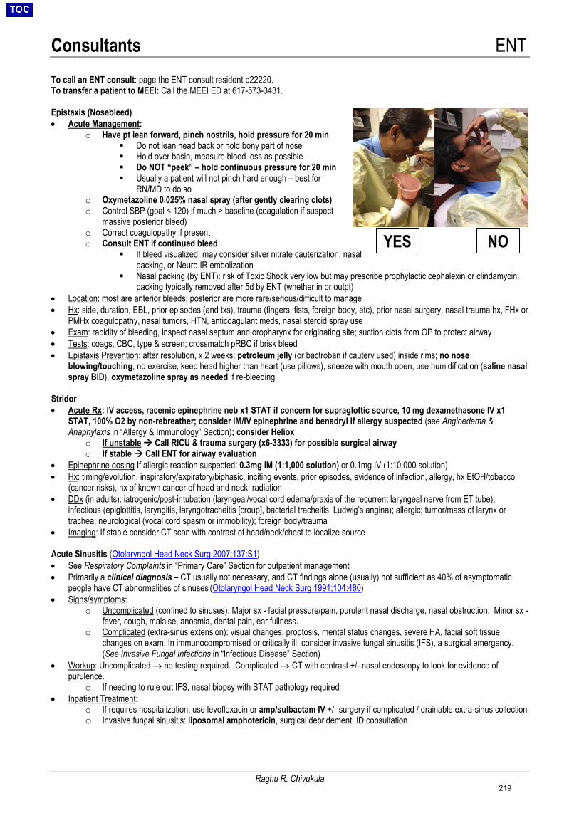
Consultants ENT
Raghu R. Chivukula
To call an ENT consult: page the ENT consult resident p22220. To transfer a patient to MEEI: Call the MEEI ED at 617-573-3431.
Epistaxis (Nosebleed)• Acute Management:
o Have pt lean forward, pinch nostrils, hold pressure for 20 min Do not lean head back or hold bony part of nose Hold over basin, measure blood loss as possible Do NOT “peek” – hold continuous pressure for 20 min Usually a patient will not pinch hard enough – best for
RN/MD to do soo Oxymetazoline 0.025% nasal spray (after gently clearing clots)o Control SBP (goal < 120) if much > baseline (coagulation if suspect
massive posterior bleed)o Correct coagulopathy if presento Consult ENT if continued bleed
If bleed visualized, may consider silver nitrate cauterization, nasalpacking, or Neuro IR embolization
Nasal packing (by ENT): risk of Toxic Shock very low but may prescribe prophylactic cephalexin or clindamycin;packing typically removed after 5d by ENT (whether in or outpt)
• Location: most are anterior bleeds; posterior are more rare/serious/difficult to manage• Hx: side, duration, EBL, prior episodes (and txs), trauma (fingers, fists, foreign body, etc), prior nasal surgery, nasal trauma hx, FHx or
PMHx coagulopathy, nasal tumors, HTN, anticoagulant meds, nasal steroid spray use• Exam: rapidity of bleeding, inspect nasal septum and oropharynx for originating site; suction clots from OP to protect airway• Tests: coags, CBC, type & screen; crossmatch pRBC if brisk bleed• Epistaxis Prevention: after resolution, x 2 weeks: petroleum jelly (or bactroban if cautery used) inside rims; no nose
blowing/touching, no exercise, keep head higher than heart (use pillows), sneeze with mouth open, use humidification (saline nasalspray BID), oxymetazoline spray as needed if re-bleeding
Stridor • Acute Rx: IV access, racemic epinephrine neb x1 STAT if concern for supraglottic source, 10 mg dexamethasone IV x1
STAT, 100% O2 by non-rebreather; consider IM/IV epinephrine and benadryl if allergy suspected (see Angioedema &Anaphylaxis in “Allergy & Immunology” Section); consider Heliox
o If unstable Call RICU & trauma surgery (x6-3333) for possible surgical airwayo If stable Call ENT for airway evaluation
• Epinephrine dosing If allergic reaction suspected: 0.3mg IM (1:1,000 solution) or 0.1mg IV (1:10,000 solution)• Hx: timing/evolution, inspiratory/expiratory/biphasic, inciting events, prior episodes, evidence of infection, allergy, hx EtOH/tobacco
(cancer risks), hx of known cancer of head and neck, radiation• DDx (in adults): iatrogenic/post-intubation (laryngeal/vocal cord edema/praxis of the recurrent laryngeal nerve from ET tube);
infectious (epiglottitis, laryngitis, laryngotracheitis [croup], bacterial tracheitis, Ludwig’s angina); allergic; tumor/mass of larynx ortrachea; neurological (vocal cord spasm or immobility); foreign body/trauma
• Imaging: If stable consider CT scan with contrast of head/neck/chest to localize source
Acute Sinusitis (Otolaryngol Head Neck Surg 2007;137:S1) • See Respiratory Complaints in “Primary Care” Section for outpatient management• Primarily a clinical diagnosis – CT usually not necessary, and CT findings alone (usually) not sufficient as 40% of asymptomatic
people have CT abnormalities of sinuses (Otolaryngol Head Neck Surg 1991;104:480)• Signs/symptoms:
o Uncomplicated (confined to sinuses): Major sx - facial pressure/pain, purulent nasal discharge, nasal obstruction. Minor sx -fever, cough, malaise, anosmia, dental pain, ear fullness.
o Complicated (extra-sinus extension): visual changes, proptosis, mental status changes, severe HA, facial soft tissuechanges on exam. In immunocompromised or critically ill, consider invasive fungal sinusitis (IFS), a surgical emergency.(See Invasive Fungal Infections in “Infectious Disease” Section)
• Workup: Uncomplicated → no testing required. Complicated → CT with contrast +/- nasal endoscopy to look for evidence ofpurulence.
o If needing to rule out IFS, nasal biopsy with STAT pathology required• Inpatient Treatment:
o If requires hospitalization, use levofloxacin or amp/sulbactam IV +/- surgery if complicated / drainable extra-sinus collectiono Invasive fungal sinusitis: liposomal amphotericin, surgical debridement, ID consultation
YES NO
219

Consultants Ophthalmology
Yasmin Islam
To call an Ophtho consult: Check vision using vision card and pupils prior to calling consult! General Inpatient Consult: page 21004; for Emergent /Overnight 617-573-4063 or MEEI Operator 617-523-7900.
High-Yield Pearls for the Wards: • Vision Loss: acute (requires urgent evaluation) vs. chronic (outpt referral) - assess patient with their glasses on!!• Glaucoma drops: prostaglandin analogs, beta-blockers, carbonic anhydrase inhibitors, or alpha 2 agonists; all lower IOP
o If brand-name drops unavailable, fractionate combo meds, ask pharm for substitution advice, or have pt bring in home meds• Ophthalmoscope: Available on most floors. Tropicamide (dilating drop) is available to order. Can make pt light-sensitive for 4 hours.• Dilating drops: 0.5% tropicamide (parasympathetic antagonist), 1-2 drops placed 15-20 minutes before exam• Finding the retina: dilate the eye and use the ophthalmoscope as in http://stanfordmedicine25.stanford.edu/the25/fundoscopic.html
Common Eye Pathology• The Red Eye: typically benign; refer to ophtho if no improvement or any ”ocular vital sign” changes (see above)
o Viral conjunctivitis: eyes “stuck shut” in AM, itchy, crusty discharge, ± URI symptoms, ± pre-auricular nodes, winter time Tx: supportive/isolation (typically adenovirus, highly infectious). Wash hands thoroughly if you suspect this!
o Allergic conjunctivitis: olopatadine 0.1% gtt bid x 5d. Clear Eyes/Visine not rec’d (rebound redness 2/2 alpha agonism)o Anterior uveitis: pain and true photophobia must be present ± eye injection. Refer to MEEI ED.o Contact lens keratitis: Have patients remove contact lens when admitted! Use glasses. P/w red/uncomfortable eye;
infection until proven otherwise. Refer to MEEI ED.• Blepharitis (inflamm of eyelids): p/w crusting/red eye/gritty feeling
o Tx: baby shampoo, warm compresses, abx ointments x 2 weeks, then daily lid hygiene. Tx Hordeolum ("stye") the same.• Dry Eye Syndrome (DES): p/w eye pain or “grit”/paradoxical tearing ±vague “blurriness.”
o Tx: artificial tears q1hrs prn first line tx, refer if no improvement• Corneal abrasion/exposure keratopathy: unilateral, redness, mild light sensitivity, common after sedation
o Dx: apply fluorescein (order in Epic) to the affected eye, illuminate with a blue light (e.g. ophthalmoscope, or smartphonescreen with Eye Handbook App), abrasion will light up green; keratopathy will look like “sandpaper” instead of smooth glass.
o Tx: abx ointment (Erythromycin 0.5%/bacitracin ophthalmic QID) + Lacrilube qhs. Consult if no improvement after 24 hrs.• Anisocoria (unequal pupils): old (20% population has at baseline) vs. new (can be trivial 2/2 anticholinergic vs. catastrophic from
herniation). NB: always ask for h/o ocular surgery as surgical pupil is a common benign cause.Miosis (Constricted Pupil) Mydriasis (Dilated Pupil)
↑ Cholinergic (eg: morphine, pilocarpine) ↑ Sympathetic (eg: atropine, CNIII paralysis) ↓ Sympathetic (eg: Horner’s) ↓ Cholinergic (eg: epinephrine, cocaine)
o If clinical suspicion for herniation (known bleed, CN3 palsy, obtundation, hemiparesis) → STAT head/neck CTAo Horner’s Syndrome: ptosis, miosis, ± anhidrosis. Wide ddx along pathway from posterior hypothalamus → C8-T2 →
superior cervical ganglion → up sympathetic chain along internal carotid and into orbit. Will require head and neckangiographic imaging to r/o potential carotid dissection.
• Retinal detachment: Presents with flashes/floaters/curtain coming over vision. Risk factors: myopia (near-sighted), trauma, diabeticretinopathy, prior eye surgery.
o Tx: Refer to MEEI ED. Will likely require vitreoretinal surgery.• Subconjunctival hemorrhage: blood between conjunctiva and sclera from ruptured vessel. No vision changes, not painful. Can be
2/2 associated blood dyscrasia, valsalva, trauma, spontaneous. Will resolve spontaneously. No need to consult ophtho.• Endophthalmitis: Infection within globe. Can be 2/2 trauma, surgery, or endogenous source (bacteremia/fungemia).
o Tx: Ophtho c/s, antibiotics/antifungals that will penetrate blood-brain barrier. May require vitrectomy (surgery).
Basic Eye Exam: “Ocular Vital Signs” - Visual Acuity (e.g. 20/200, CF, etc)- Pupils (4mm -> 2mm OD, No APD)- Visual Fields
- Extra Ocular Movements- Intraocular pressure- Color vision testing (Ishihara cards)
Common Abbreviations: APD Afferent pupillary defect AT Artificial tears cc/sc With/without refractive corr. CE Cataract extraction CF Count fingers (VA) CWS Cotton wool spot DES Dry eye syndrome EOM Extraocular movement HM Hand motion (VA) IOL Intraocular lens IOP Intraocular pressure LP Light perception (VA) MGD Meibomian gland dysfunction
NLP No light perception (VA) NPDR Non-prolif. diabetic retinopathy NS Nuclear sclerosis (i.e. cataract) OD/OS Right eye, left eye OU Both eyes PDR Prolif. diabetic retinopathy PF Pred Forte gtt (prednisolone) PFAT Preservative-free artificial tears PVD Posterior vitreous detachment RD Retinal detachment SLE Slit lamp exam SPK Superficial punctate keratitis/dry eye VA, VF Visual acuity, fields
Uvea = iris + ciliary body + choroid
220
Radiology Contact Information & Life Images
Craig Audin, Reece Goiffon
Main Number Technologists 617 – (643 / 724 / 726) – XXXX CT Blake 2 48518
CT ED 66760 Reading Rooms ED Radiology 63050 Dodd Reception 44212 GI Fluoro 44295 Teleradiology 44270 Mammography 63092 Cardiac CT 47132 MRI ED 49867 Cardiac MRI 66947 MRI Inpatient 85692 Chest CT 33899 Nuclear Medicine 68350 CXR Inpatient 42051 Pediatrics 61367 CXR Outpatient 62197 PET 64209 ED 41533 Scheduling 4XRAY ED Neuro 68188 US White 2 53074 GI CT 65162 GI Fluoro/KUB 32605 On Call Pagers GI MR 49919 Cardiac CT 22122 GI US White 2 60595 Cardiac MRI 33133 GI US Yawkey 6 31577 IR GI/GU 34071 IR (GI & VIR) 34723 IR Neuro Spine 33722 Mammography 40228 IR Neuro Vascular 21154 MSK 40516 IR Vascular 38553 Neuroradiology 41931 Mammography 20022 Nuclear Cardiology 43600 MSK/MSK IR 36321 Nuclear Medicine 61404 Neuro ED 39991 Pediatrics 42119 Neuro Inpatient 32535 PET 66737 Nuclear Medicine Resident On call Vascular 47115 Pediatrics On call
Consults – Weekdays 8am 12pm 5pm 7pm 8am
Cardiac CT Dodd XXXXXXXXXXXXXXXXXXXXXX Chest Dodd ED GI Dodd ED Neuro Neuro Consult (730am – 430pm) Neuro ED Vascular Dodd ED Other Reading Room ED
Consults – Weekends & Holidays 8am 12pm 5pm 7pm 8am
Cardiovascular Dodd ED Chest Dodd ED GI Dodd ED Neuro Neuro ED Other ED
• Life Imageso Upload images to lifeIMAGE and Epic: Partners Applications utilities MGH Upload Image to Radiology
(LifeImage) Access LifeImage find exam on CD/DVD upload imageso Send images to MGH PACS: upload to MGH request reado Retrieve images from The Cloud: ISDrequests.partners.org file an urgent ticketo Additional information:
Urgent reads: contact ISD (p34188, x30003) Multiple body parts: interpretations only given for selected body parts Multiple LifeImages of the same body part: upload all images request a read only on the most recent Exams will not be read if: requisition was for a different body part than the uploaded images; study >6
months old; a more recent LifeImage is available; US, fluoroscopy, or mammography
221

Radiology Radiology Basics
Craig Audin, Reece Goiffon
• X-ray:
5 Radiographic Densities Silhouette Sign: loss of the margin between two opposing structures of the same radiographic density • RUL – right paratracheal stripe• RML – right heart border• RLL – right hemidiaphragm
• LUL – aortic arch• Lingula – left heart border• LLL – left hemidiaphragm
• Computed Tomography (CT):o Hounsfield Units (HU): measurement of CT attenuationo Windowing and leveling: adjusting contrast and brightness to highlight structures
Window (contrast): range of Hounsfield units displayed across the grayscale• Wide window – best for large differences in attenuation• Narrow window – best for subtle differences in attenuation
Level (brightness): HU that corresponds to mid-gray• High level – best for structures with high attenuation• Low level – best for structures with low attenuation
o Phases of contrast:
Phase Time After Injection Structures Evaluated CTPE 15 s Pulmonary arteries Arterial (CTA) 30 s Aorta, systemic
arteries, renal cortices Late arterial 60 s Routine chest Portal Venous 70 s Routine abdomen Nephrographic 100 s Renal medulla Venous 120 s Peripheral veins Delayed (Urogram) 10-15 min Ureters, bladder
• Magnetic Resonance Imaging (MRI):
T1 & T2 Signal Evolution of Blood Protein Signal
o MRI safety: Device compatibility: www.mrisafety.com
Substance HU Air -1000Fat -100Water 0 Blood 50 Soft tissue 100 Bone 1000 Metal >2000
222

Radiology Contrast
Craig Audin, Reece Goiffon
• Indications:o IV: whenever possible, particularly for infection, tumors, and vessel imagingo PO positive (hyperdense): bowel obstruction, bowel wall pathology, differentiate bowel from other abd. structureso PO negative (hypodense): inflammatory bowel disease, GI bleed, mesenteric ischemiao Rectal: appendicitis, penetrating abdominal trauma
• Pregnancy and breast feeding: (ACR 2018)o Pregnancy:
Iodinated: no need to withhold contrast (no data to suggest potential harm to fetus) Gadolinium: unknown risk to fetus consider noncontrast or alternative study
o Breast feeding: mother’s informed decision to “pump and dump” for 12-24 h after scan• Renal function: (ACR 2018, MGPO 2017)
Contrast Induced Nephropathy Nephrogenic Systemic Fibrosis Risk factors Age >60 years, dialysis, kidney transplant, single
kidney, renal cancer, renal surgery, HTN on medication, DM, metformin
Dialysis, kidney transplant, single kidney, renal cancer, renal surgery, HTN on medication, DM
Screening (At risk pts only)
Outpatient: GFR within 30 d Inpatient: GFR within 24 h
Outpatient: • GFR 45-59: GFR within 6 wks• GFR <45: GFR within 2 dInpatient: GFR within 2 d
Prevention GFR ≥ 45: contrast per protocol GFR 30-44: pre-hydrate GFR < 30: non-contrast or alternative study • If necessary consult radiology
GFR ≥ 30: gadolinium per protocol GFR < 30: non-contrast or alternative study • If necessary consult radiology and
nephrology, obtain informed consentDialysis pts HD within 72 h after scan Prompt post-scan HD (PD inadequate) Repeat studies Decision is clinical and subjective
Insufficient evidence to hold contrast for 24 h No risk factors: proceed At risk pts: consult radiology
Metformin GFR ≥ 30: continue metformin GFR < 30 or AKI: hold for 48 h after scan
No need to hold metformin
• MGH prehydration protocol: (MGPO 2017)o PO (preferred): 1-2 L PO non-caffeinated beverage 12-24 h prior to scano IV (outpatient): NS 250 mL IV bolus @ 1 h prior to scano IV (inpatient): NS 100 mL/h IV 6-12 h before and 4-12 h after scan (ACR 2018)
• Contrast reactions: (ACR 2018)
Allergic Physiologic Indications for Premedication
Mild
Limited urticaria Itchy throat Nasal congestion URI symptoms
N/V, flushing/warmth HA/dizziness Mild HTN Transient vasovagal reaction
• Prior mild-moderate allergic reaction• None for prior physiologic reactions• None for shellfish allergies• No cross-reactivity between iodinated
contrast and gadolinium
Mode
rate Diffuse urticaria
Facial/laryngeal edema w/o dyspnea or hoarseness Bronchospasm w/o hypoxia
Protracted N/V HTN urgency Isolated CP Vasovagal reaction requiring tx
Seve
re Anaphylaxis
Facial/laryngeal edema w/ dyspnea or hoarseness Bronchospasm w/ hypoxemia
HTN emergency Arrhythmia Seizure Protracted vasovagal reaction
• Adult premedication protocol: (ACR 2018)o Elective (13 h protocol)
Prednisone 50 mg PO @ 13, 7, and 1 h prior, AND Diphenhydramine 50 mg PO @ 1 h prior
o Accelerated (4-5 h protocol) Methylprednisolone 40 mg IV now and q4h until scan, AND Diphenhydramine 50 mg IV @ 1 h prior
o Emergent (1 h protocol) – no evidence of efficacy, only if no alternatives Methylprednisolone 40 mg IV @ 1 h prior, AND Diphenhydramine 50 mg IV @ 1 h prior
Corticosteroid Dose Equivalents Prednisone 50 mg PO Hydrocortisone 200 mg Methylprednisolone 40 mg Dexamethasone 7.5 mg PO:IV 1:1
223
Radiology Protocols
Craig Audin, Reece Goiffon
• Ordering Studies:o All cross-sectional studies are protocoled by radiology – simply provide the necessary information:
Body part and modality Indication: clinical history relevant to the study Contrast: “per radiology discretion” unless specific reason otherwise Contraindications for contrast: kidney injury or prior allergic reaction (see Contrast)
o Questions: call the appropriate division or page the appropriate on-call radiologist (see Contact Information)• Level of Urgency:
o Routine: order of interpretation depends on acquisition timeo Urgent: takes priority over routine studieso STAT: means NOW, high acuity/life threatening emergencies
Patient must be ready for immediate transport Patient must be accompanied by a responding clinician capable of providing emergency care Responding clinician must be present for the entire exam Radiology will provide preliminary read: phone call for XR/US, at the scanner for cross-sectionals
• Overnight Reads:o Studies with full interpretations overnight: all ED studies, STAT studies, and acute CTPEso Verbal preliminary reads:
Typically done for ICU studies only• Inpatient studies are only reviewed overnight if there is an urgent clinical question (i.e., one
that would alter overnight management). Consider face-to-face consult in ED. After communication w/ the primary team, all verbalized prelim reads will be documented in the chart A full interpretation will be generated the following morning for all prelim reads
• Cardiovascular Protocols:o DVT imaging: US is initial test of choice (Cardiovascular Diagnosis and Therapy 2016;6:493)
CTV/MRV: primarily used for central venous thrombosis when initial US is equivocal or non-diagnostico Arterial imaging:
CTA: three phases (noncontrast, arterial, delays) stenosis, dissection, aneurysm Requisition: specify vessel of interest, field of view, and indication
o Coronary CTA: ECG-gated study of the heart only performed by CV CT on-call radiologist during normal hours Specify if body parts other than the heart should be imaged (thoracic aorta, CABG grafts, etc.)
o Other EKG-gated CTAs: Indications: any evaluation of the heart or ascending aorta EKG-gating is unnecessary for the descending thoracic aorta, abdominal aorta, and pulmonary arteries
o Noncontrast vascular studies: RP hematoma, pre-op aortic calcifications, coronary calcium score, follow-up aortic size
• ED Protocols:o Trauma: I+, single phase (arterial for chest, portal venous for abdomen/pelvis – images checked at the scanner
by radiology for possible delays) Blunt trauma: includes bone kernel reformats for improved visualization of bones Penetrating trauma: O+R+ for increased sensitivity of bowel injury
o Cervical spine: I-, need for CTA determined by radiology, bone kernel reformats in all 3 planes Images checked at the scanner by radiology only if IV contrast is required for another body part
o Appendicitis: I+ and O+/R+ (please specify PO or PR), kidneys through pelvis onlyo Neuro ED: call reading desk @ x68188
• GI/GU Protocols:o Stone protocol: I-O-, low dose
Order contrast-enhanced CT if there is concern for ANYTHING else (stones may still be visualized)o Routine abdomen/pelvis vs renal mass vs bladder cancer vs hematuria:
Routine abdomen/pelvis: I+O+, single phase (portal venous) workhorse protocol Renal mass: I+O+, two phases (noncontrast, nephrographic), abdomen only renal masses or cysts Bladder cancer: I+O+, two phases (portal venous, delayed) workup or monitoring of GU malignancy Hematuria: I+O-, “three” phases (noncontrast, nephrographic, urogram) hematuria, hydronephrosis
o CT urogram vs CT cystogram: Urogram: antegrade filling of ureters and bladder with IV contrast (delayed phase) Cystogram: retrograde filling of bladder with contrast via Foley catheter evaluation of bladder rupture
224
Radiology Protocols
Craig Audin, Reece Goiffon
o Arterially-enhancing tumors: MR CHIT: melanoma, RCC, choriocarcinoma, HCC, islet cell (neuroendocrine) tumors, thyroid
o Does my patient need to be NPO? IV contrast CT: 2 h Abdomen/pelvis CT: 8 h Non-contrast CT: no NPO
o Fluoroscopy protocols: Requisition: specify indication, h/o surgery or aspiration Barium swallow vs modified barium swallow vs UGI series vs SB follow-through:
• Barium swallow: esophagus, GE junction, proximal stomach dysphagia, GERD• Modified barium swallow: mouth, pharynx, upper esophagus dysphagia, aspiration• UGI series: barium swallow plus stomach, pylorus, and duodenal bulb bariatric surgery• SB follow-through: small bowel, terminal ileum, and proximal LB +/- UGI series beforehand
• Musculoskeletal Protocols:o Questions: page MSK IR on-call radiologist @ p36321
• Nuclear Medicine Protocols:o Overnight studies:
Tagged RBC study: BRBPR (NOT guaiac positive stools, melena, or massive bleeding)• Requirements: consult IR first for possible angiogram if study is positive
VQ scan: acute PE (NOT chronic PE), ONLY if results will alter management (i.e. AC tonight)• Requirements: CXR within 24 h, patient stable for duration of scan (~4 h)
HIDA scan: acute cholecystitis, ONLY if results will alter management (i.e. OR tonight)• Requirements: NPO 4-24 h prior to study, no opiates 12-24 h prior to study, bilirubin <10
o PET: Fasting: hold everything but meds and water
• Overnight is ideal, but AT LEAST 6 hours for non-DM patients• AT LEAST 4 hours for DM patients
o Continue long-acting insulin, hold short-acting insulin 4 h prior to scan Blood sugar thresholds
• FDG-PET brain < 175 mg/dL FDG-PET whole body < 250 mg/dL • Neuroradiology Protocols:
o Inpatients: page Neuro IP on-call radiologist @ p32535o Acute stroke:
Inpatients/ICU: page acute stroke consult fellow @ p21723 ED: activate ED2CT via the group pager
o Head CT: typically noncontrast Indications for contrast-enhanced head CT: infection and/or tumor AND contraindication for brain MRI
o Spine MRI: for more than 1 segment, please order total spine and specify indication Separate MRIs should not be ordered prior to neurology/NSGY consult
o Fluoroscopy-guided LPs: performed by neuroradiology fellows, NOT neuro IR Indications: difficult anatomy, and only after LP is attempted on floor
• Not to be used as an anesthesia service for unruly patients (typically performed withoutconscious sedation, although this can be arranged if required for patient safety)
• Thoracic Protocols:o All chest CTs are high resolution – traditional “high res chest CT” is now the diffuse lung disease CT (see below)o Routine chest vs CTPE vs CTA chest:
Routine chest: single phase (late arterial) workhorse protocol CTPE: single phase (pulmonary arterial) pulmonary arteries CTA chest: three phases (noncontrast, arterial, delays) systemic arteries
o Double rule out studies: Clinical concern for PE and aortic dissection Contrast can only be optimized for one (must pick CTPE or CTA)
o Diffuse lung disease (a.k.a. misnomer “high res CT”): Indications: ILD, lung transplant, air trapping Inspiratory and expiratory images, plus prone images to differentiate between atelectasis and fibrosis
o Nodule follow-up: (Radiology 2017;284:228) Indications: incidental nodule on prior CT, age >35 y, AND no history of malignancy or recent infection Fleischner Society 2017 Guidelines
225
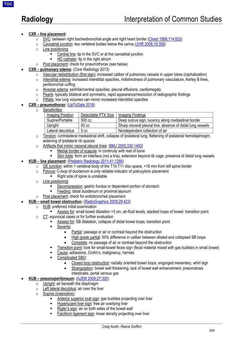
Radiology Interpretation of Common Studies
Craig Audin, Reece Goiffon
• CXR – line placement:o SVC: between right tracheobronchial angle and right heart border (Chest 1998;114:820)o Cavoatrial junction: two vertebral bodies below the carina (JVIR 2008;19:359)o Line positioning:
Central line: tip in the SVC or at the cavoatrial junction HD catheter: tip in the right atrium
o Post placement: check for pneumothorax (see below)• CXR – pulmonary edema: (Core Radiology 2013)
o Vascular redistribution (first sign): increased caliber of pulmonary vessels in upper lobes (cephalization)o Interstitial edema: increased interstitial opacities, indistinctness of pulmonary vasculature, Kerley B lines,
peribronchial cuffingo Alveolar edema: perihilar/central opacities, pleural effusions, cardiomegalyo Pearls: typically bilateral and symmetric, rapid appearance/resolution of radiographic findingso Pitfalls: low lung volumes can mimic increased interstitial opacities
• CXR – pneumothorax: (UpToDate 2018)o Sensitivities:
Imaging Position Detectable PTX Size Imaging Findings Supine/Portable 500 cc Deep sulcus sign, lucency along mediastinal border Upright 50 cc Sharp visceral pleural line, absence of distal lung vessels Lateral decubitus 5 cc Nondependent collection of air
o Tension: contralateral mediastinal shift, collapse of ipsilateral lung, flattening of ipsilateral hemidiaphragm,widening of ipsilateral rib spaces
o Artifacts that mimic visceral pleural lines: (BMJ 2005;330:1493) Medial border of scapula: in continuity with rest of bone Skin folds: form an interface (not a line), extension beyond rib cage, presence of distal lung vessels
• KUB – line placement: (Pediatric Radiology 2011;41:1266)o GE junction: within 1 vertebral body of the T10-T11 disc space, <16 mm from left spine bordero Pylorus: C-loop of duodenum is only reliable indicator of post-pyloric placement
Right side of spine is unreliableo Line positioning:
Decompression: gastric fundus or dependent portion of stomach Feeding: distal duodenum or proximal jejunum
o Post placement: check for endobronchial placement• KUB – small bowel obstruction: (RadioGraphics 2009;29:423)
o KUB: preferred initial examination Assess for: small bowel dilatation >3 cm, air-fluid levels, stacked loops of bowel, transition point
o CT: equivocal cases or for further evaluation Assess for: SB dilatation, collapse of distal bowel loops, transition point Severity:
• Partial: passage or air or contrast beyond the obstruction• High grade partial: 50% difference in caliber between dilated and collapsed SB loops• Complete: no passage of air or contrast beyond the obstruction
Transition point: look for small-bowel feces sign (fecal material mixed with gas bubbles in small bowel) Cause: adhesions, Crohn’s, malignancy, hernias Complicated SBO:
• Closed loop obstruction: radially oriented bowel loops, engorged mesentery, whirl sign• Strangulation: bowel wall thickening, lack of bowel wall enhancement, pneumatosis
intestinalis, portal venous gas• KUB – pneumoperitoneum: (AJEM 2009;27:320)
o Upright: air beneath the diaphragmo Left lateral decubitus: air over the livero Supine (insensitive):
Anterior superior oval sign: gas bubbles projecting over liver Hyperlucent liver sign: free air overlying liver Rigler’s sign: air on both sides of the bowel wall Falciform ligament sign: linear density projecting over liver
226

Radiology Interpretation of Common Studies
Craig Audin, Reece Goiffon
• US – cholecystitis: (AJR 2011;196:W367)o US is preferred initial examinationo Gallstones: echogenic foci with posterior shadowingo Common findings: gallbladder wall thickening >3 mm, gallbladder distension >40 mm, peri-cholecystic fluido Sonographic Murphy’s sign: 92% sensitivity (analgesics reduce sensitivity)o Gallstones and gallbladder wall thickening: 95% positive predictive value for acute cholecystitis
• US – deep venous thrombosis: (Cardiovascular Diagnosis and Therapy 2016;6:493)o Compression US: noncompressibility of vein, echogenic thrombus within vein, venous distensiono Venous duplex US: absence of color Doppler signal within vein, loss of flow phasicity, loss of response to
augmentation maneuverso CT venogram:
Alternative to US in critically ill patients who have undergone CTPE Pros: evaluation of pelvic veins and IVC, which are difficult to assess on US Cons: invasive, requires contrast, radiation, possible streak or mixing artifacts
• Cross sectional imaging:o Anatomy: http://www.radiologyassistant.nl/o CT and MRI basics: see Radiology Basicso Search pattern: see below
CT Head MRI Brain 1. Brain parenchyma
a. Mass lesion: brain windowsb. Intracranial hemorrhage: brain and subdural
windowsc. Infarction: stroke windows
2. Vessels3. CSF spaces: ventricles, sulci, cisterns4. Midline shift or herniation5. Soft tissues (great place to start for trauma head CTs)6. Bones/sinuses
1. Brain parenchymaa. Mass lesion: T1, T2, FLAIRb. Intracranial hemorrhage: SWI, T1, T2c. Infarction: DWI, ADC
2. Vessels: T2 for flow voids, T1 post-contrast, TOF ifnoncontrast MRA
3. CSF spaces: T24. Midline shift or herniation: coronals helpful5. Soft tissues6. Bones/sinuses
CT Chest MRCP 1. Lines and tubes (scout can be very helpful)2. Abdomen3. Soft tissues4. Bones5. Heart and mediastinum: thyroid, lymph nodes, heart
and pericardium, major vessels, esophagus6. Pleura: pleural effusion, pneumothorax7. Lungs: secondary pulmonary lobule is the key
a. Radiology Assistant Lung HRCT Basics
1. Choledocholithiasis: hypointense filling defectwithin CBD surrounded by hyperintense bile
CT Abdomen/Pelvis 1. Lung bases2. Liver/gallbladder: focal lesions, biliary ductal dilatation3. Spleen4. Pancreas: focal lesions, pancreatic ductal dilatation5. Adrenals6. Kidneys/ureters: hydronephrosis, stones, focal lesions7. Bladder/pelvic organs8. Peritoneum: free air or fluid9. Lymph nodes10. Vessels11. GI tract: bowel distension, bowel wall thickening12. Soft tissues13. Bones
227

Procedures Ultrasound Basics
Sean Mendez
Equipment: (NEJM 2011;364:749)Basic Terminology: • Frequency: 1Hz = 1cycle/sec; Medical U/S typically between 2-15MHz (derm U/S up to 100MHz).
- High frequency (> 5MHz): ↑ resolution, shallow tissue penetration. Ideal for vascular, skin, breast, thyroid.- Low frequency (2–5MHz): ↓ resolution, deeper tissue penetration. Ideal for abdominal, OB/GYN, cardiac.
• Gain: Signal amplification. Similar to brightness control.• Depth: Depth of field of view (FOV). Excessively large FOV ↓ spatial resolution; tight FOV limits view of nearby structures.• Attenuation: Reduced signal transduction through a medium = ↓ signal intensity behind it (bone/air have high attenuation)
Transducer (probe): Converts electricity into sound waves transmits sounds wave into tissue receives sound waves echoed back from tissue. Indicator (denoted by light or notch on probe) displays on left of the screen. Exception: echocardiography → indicator displays on right side. • SECTOR (cardiac) probe: Good for looking in small sonographic windows (i.e., between
intercostal spaces for cardiac or pulm imaging); low resolution, produces fan-like image.• LINEAR (vascular) probe: Good for shallow structures (i.e. vascular, soft tissue). Uses high
frequency with good resolution, produces rectangular image.• CURVED (abdominal) probe: Good for deeper structures (i.e. intra-abdominal). Uses lower
frequency; combines linear and sector probe image qualities.
Commonly Used Modes: • B-mode (brightness mode): Standard 2D gray-scale image.• D-mode (doppler): Detects flow to or away from transducer. Useful to find and
define vessels, flow across valveso Color → direction and velocity are color coded and superimposed on B-mode
image. “BART” (Blue is Away from probe, Red is Towards)o Power → detects very low flow but not direction, useful in vascular compromiseo Spectral → velocity presented graphically on a timeline
• M-mode (motion mode): Takes a slice of a B-mode image over time. Often used inTTE. Useful to assess lung sliding for pneumothorax.
General Imaging Concepts: Typical appearance of normal tissue: • Skin and pleura are smooth and brighter than surrounding tissue (echogenic or
hyperechoic)• Fat and muscle are dark, though varies depending on the tissue (hypoechoic)• Fluid (e.g. blood, effusion) appears black on ultrasound (anechoic), though thick
fluids (pus) can be brighter than typical fluid• Tendons and nerves are bright / hyperechoic when perpendicular to probe, but
dark / hypoechoic when angle is changed (anisotropy).• Bone has a bright hyperechoic rim (due to reflection) around a black / anechoic
image with a shadow beyond it
Artifacts: Elements seen on ultrasound image that do not exist in reality • Reflection: Proportional to the difference in acoustic impedence between two tissues
(↑ difference = ↑ reflection)Relative acoustic impedance: bone >> solid organ > fat >> lung >> air.
• Shadowing: ↓ signal beyond a strongly attenuatingOR reflecting structure (e.g. stones, bone)
• Enhancement: ↑ signal posterior to weakly attenuating(hypo or anechoic) structure (e.g., cysts)
• Mirror image: Structures in front of strong reflector(e.g. diaphragm) appear to lie behind it as well
• Reverberation: Evenly spaced lines at various depthsbeyond a strong reflector (e.g. A lines beyond pleura)
• Comet tail: Tiny, narrow reverberations beyond verystrong reflector (e.g. metal pellet) blending into a line
B mode
M mode
Rib Rib Pleura
Lung
Fascia
Muscle
Indicator
Enhancement Shadowing
228
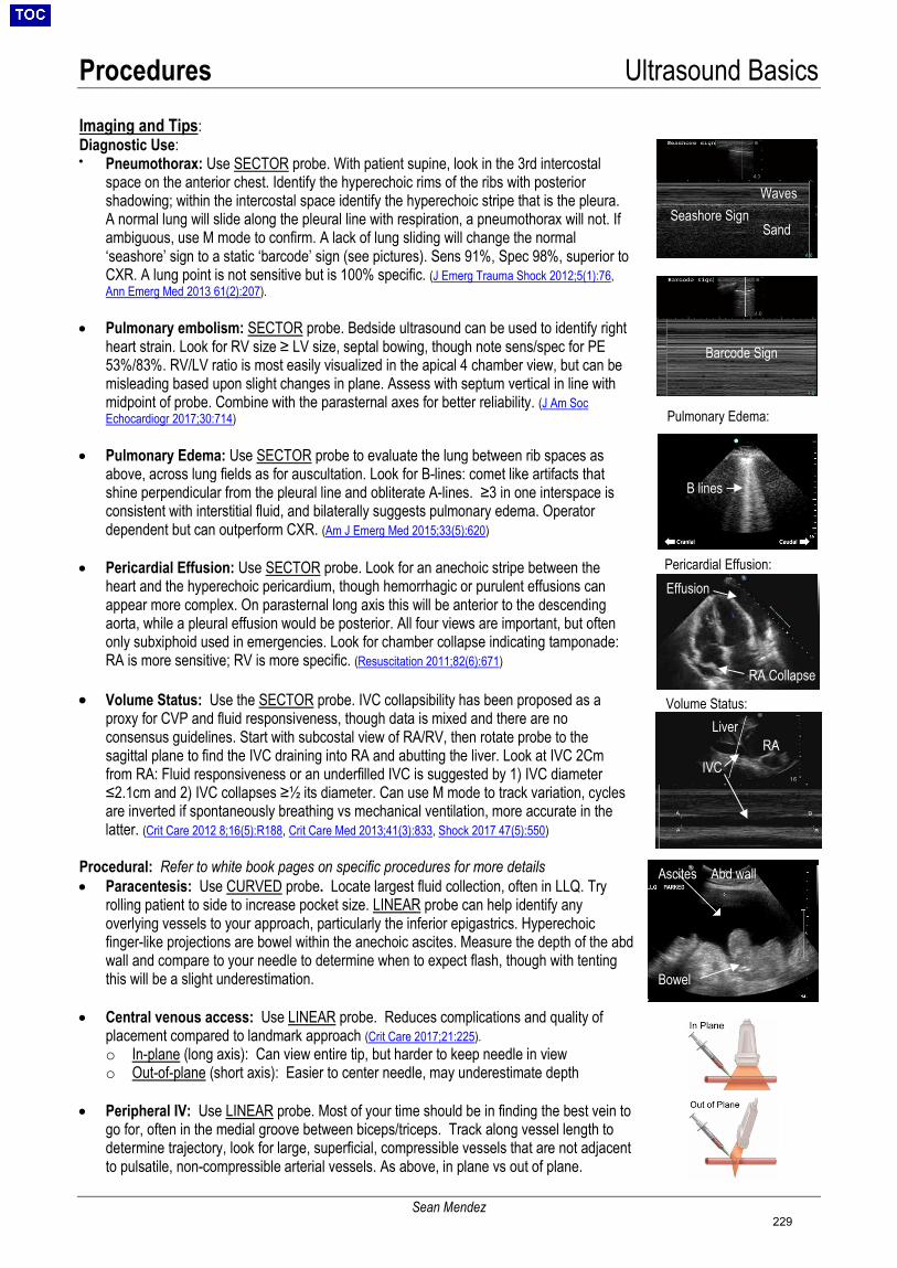
Procedures Ultrasound Basics
Sean Mendez
Imaging and Tips: Diagnostic Use: • Pneumothorax: Use SECTOR probe. With patient supine, look in the 3rd intercostal
space on the anterior chest. Identify the hyperechoic rims of the ribs with posteriorshadowing; within the intercostal space identify the hyperechoic stripe that is the pleura.A normal lung will slide along the pleural line with respiration, a pneumothorax will not. Ifambiguous, use M mode to confirm. A lack of lung sliding will change the normal‘seashore’ sign to a static ‘barcode’ sign (see pictures). Sens 91%, Spec 98%, superior toCXR. A lung point is not sensitive but is 100% specific. (J Emerg Trauma Shock 2012;5(1):76,Ann Emerg Med 2013 61(2):207).
• Pulmonary embolism: SECTOR probe. Bedside ultrasound can be used to identify right heart strain. Look for RV size ≥ LV size, septal bowing, though note sens/spec for PE 53%/83%. RV/LV ratio is most easily visualized in the apical 4 chamber view, but can be misleading based upon slight changes in plane. Assess with septum vertical in line with midpoint of probe. Combine with the parasternal axes for better reliability. (J Am Soc Echocardiogr 2017;30:714)
• Pulmonary Edema: Use SECTOR probe to evaluate the lung between rib spaces asabove, across lung fields as for auscultation. Look for B-lines: comet like artifacts thatshine perpendicular from the pleural line and obliterate A-lines. ≥3 in one interspace isconsistent with interstitial fluid, and bilaterally suggests pulmonary edema. Operatordependent but can outperform CXR. (Am J Emerg Med 2015;33(5):620)
• Pericardial Effusion: Use SECTOR probe. Look for an anechoic stripe between theheart and the hyperechoic pericardium, though hemorrhagic or purulent effusions canappear more complex. On parasternal long axis this will be anterior to the descendingaorta, while a pleural effusion would be posterior. All four views are important, but oftenonly subxiphoid used in emergencies. Look for chamber collapse indicating tamponade:RA is more sensitive; RV is more specific. (Resuscitation 2011;82(6):671)
• Volume Status: Use the SECTOR probe. IVC collapsibility has been proposed as aproxy for CVP and fluid responsiveness, though data is mixed and there are noconsensus guidelines. Start with subcostal view of RA/RV, then rotate probe to thesagittal plane to find the IVC draining into RA and abutting the liver. Look at IVC 2Cmfrom RA: Fluid responsiveness or an underfilled IVC is suggested by 1) IVC diameter≤2.1cm and 2) IVC collapses ≥½ its diameter. Can use M mode to track variation, cyclesare inverted if spontaneously breathing vs mechanical ventilation, more accurate in thelatter. (Crit Care 2012 8;16(5):R188, Crit Care Med 2013;41(3):833, Shock 2017 47(5):550)
Procedural: Refer to white book pages on specific procedures for more details • Paracentesis: Use CURVED probe. Locate largest fluid collection, often in LLQ. Try
rolling patient to side to increase pocket size. LINEAR probe can help identify anyoverlying vessels to your approach, particularly the inferior epigastrics. Hyperechoicfinger-like projections are bowel within the anechoic ascites. Measure the depth of the abdwall and compare to your needle to determine when to expect flash, though with tentingthis will be a slight underestimation.
• Central venous access: Use LINEAR probe. Reduces complications and quality ofplacement compared to landmark approach (Crit Care 2017;21:225).o In-plane (long axis): Can view entire tip, but harder to keep needle in viewo Out-of-plane (short axis): Easier to center needle, may underestimate depth
• Peripheral IV: Use LINEAR probe. Most of your time should be in finding the best vein togo for, often in the medial groove between biceps/triceps. Track along vessel length todetermine trajectory, look for large, superficial, compressible vessels that are not adjacentto pulsatile, non-compressible arterial vessels. As above, in plane vs out of plane.
B lines
Waves
Sand
Effusion
RA Collapse
Liver RA
IVC
Barcode Sign
Seashore Sign
Pulmonary Edema:
Pericardial Effusion:
Volume Status:
Abd wall
Bowel
Ascites
229
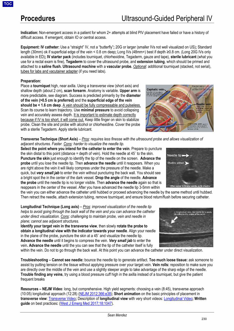
Procedures Ultrasound-Guided Peripheral IV
Sean Mendez
Indication: Non-emergent access in a patient for whom 2+ attempts at blind PIV placement have failed or have a history of difficult access. If emergent, obtain IO or central access.
Equipment: IV catheter: Use a “straight” IV, not a “butterfly”; 20G or larger (smaller IVs not well visualized on US); Standard length (30mm) ok if superficial edge of the vein < 0.8 cm deep; Long IVs (48mm+) best if depth ≥0.8 cm. (Long 20G IVs only available in ED); IV starter pack (includes tourniquet, chlorhexidine, Tegaderm, gauze and tape), sterile lubricant (what you use for a rectal exam is fine), Tegaderm to cover the ultrasound probe, and extension tubing, which should be primed and attached to a saline flush. Ultrasound machine with a vascular probe. Optional: additional tourniquet (stacked, not serial), tubes for labs and vacutainer adapter (if you need labs).
Preparation: Place a tourniquet high, near axilla. Using a transverse view (short axis) and shallow depth (about 2 cm), scan forearm. Anatomy is variable. Upper arm is more predictable, see diagram. Success is predicted primarily by the diameter of the vein (>0.5 cm is preferred) and the superficial edge of the vein should be < 1.6 cm deep. A vein should be fully compressable and pulseless. Scan its course to learn trajectory. Use minimal pressure to avoid collapsing vein and accurately assess depth. It is important to estimate depth correctly because if IV is too short, it will come out. Keep little finger on skin to stabilize probe. Clean the site and probe with alcohol or chlorhexidine. Cover the probe with a sterile Tegaderm. Apply sterile lubricant.
Transverse Technique (Short Axis) – Pros: requires less finesse with the ultrasound probe and allows visualization of adjacent structures. Faster. Cons: harder to visualize the needle tip. Select the point where you intend for the catheter to enter the vein. Prepare to puncture the skin distal to this point (distance = depth of vein). Hold the needle at 45˚ to the skin. Puncture the skin just enough to identify the tip of the needle on the screen. Advance the probe until you lose the needle tip. Then advance the needle until it reappears. When you are right above the vein it will likely compress under the pressure of the needle. Make a quick, but very small jab to enter the vein without puncturing the back wall. You should see a bright spot the in the center of the dark vessel. Drop the angle of the needle. Advance the probe until the needle tip is no longer visible. Then advance the needle again so that is reappears in the center of the vessel. After you have advanced the needle tip 3-5mm within the vein you can either advance the catheter until hubbed or proceed advancing the needle by the same method until hubbed. Then retract the needle, attach extension tubing, remove tourniquet, and ensure blood return/flush before securing catheter.
Longitudinal Technique (Long axis) – Pros: improved visualization of the needle tip helps to avoid going through the back wall of the vein and you can advance the catheter under direct visualization. Cons: challenging to maintain probe, vein and needle in plane; cannot see adjacent structures. Identify your target vein in the transverse view, then slowly rotate the probe to obtain a longitudinal view with the indicator towards your needle. Align your needle in the plane of the probe, puncture the skin at a 45˚ and visualize the needle tip. Advance the needle until it begins to compress the vein. Very small jab to enter the vein. Advance the needle until the you can see that the tip of the catheter itself is fully within the vein. Do not to go through the back wall. At this point you can advance the catheter under direct visualization.
Troubleshooting – Cannot see needle: bounce the needle tip to generate artifact. Too much loose tissue: ask someone to assist by putting tension on the tissue without applying pressure over your target vein. Vein rolls: reposition to make sure you are directly over the middle of the vein and use a slightly steeper angle to take advantage of the sharp edge of the needle. Trouble finding any veins, try using a blood pressure cuff high in the axilla instead of a tourniquet, but give the patient frequent breaks
Resources – NEJM Video: long, but comprehensive. High yield segments: choosing a vein (8:45), transverse approach (10:05) longitudinal approach (12:28) (NEJM 2012;366:e38); Short animation on the basic principles of placement in transverse view: Transverse Video; Description of longitudinal view with very short videos: Longitudinal Video; Written guide on best practices: (West J Emerg Med 2017;18:1047).
230
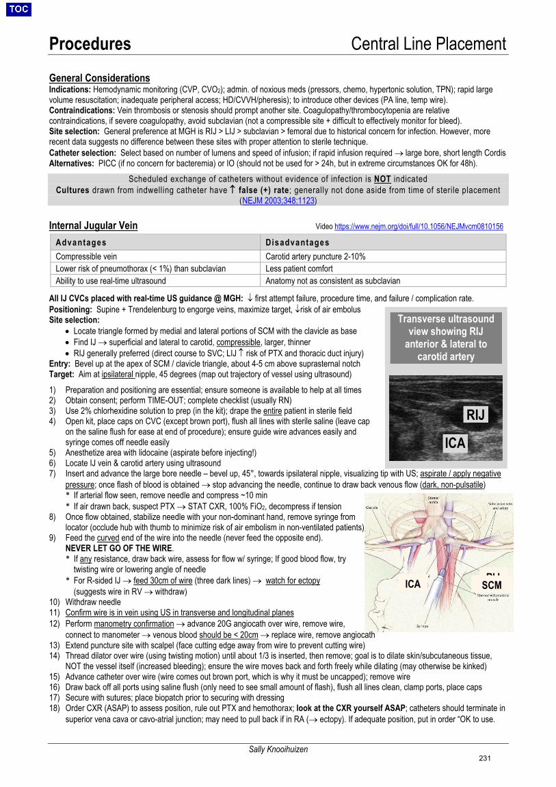
Procedures Central Line Placement
Sally Knooihuizen
General Considerations Indications: Hemodynamic monitoring (CVP, CVO2); admin. of noxious meds (pressors, chemo, hypertonic solution, TPN); rapid large volume resuscitation; inadequate peripheral access; HD/CVVH/pheresis); to introduce other devices (PA line, temp wire). Contraindications: Vein thrombosis or stenosis should prompt another site. Coagulopathy/thrombocytopenia are relative contraindications, if severe coagulopathy, avoid subclavian (not a compressible site + difficult to effectively monitor for bleed). Site selection: General preference at MGH is RIJ > LIJ > subclavian > femoral due to historical concern for infection. However, more recent data suggests no difference between these sites with proper attention to sterile technique. Catheter selection: Select based on number of lumens and speed of infusion; if rapid infusion required → large bore, short length Cordis Alternatives: PICC (if no concern for bacteremia) or IO (should not be used for > 24h, but in extreme circumstances OK for 48h).
Scheduled exchange of catheters without evidence of infection is NOT indicated Cultures drawn from indwelling catheter have ↑ false (+) rate; generally not done aside from time of sterile placement
(NEJM 2003;348:1123)
Internal Jugular Vein Video https://www.nejm.org/doi/full/10.1056/NEJMvcm0810156
Advantages Disadvantages Compressible vein Carotid artery puncture 2-10% Lower risk of pneumothorax (< 1%) than subclavian Less patient comfort Ability to use real-time ultrasound Anatomy not as consistent as subclavian
All IJ CVCs placed with real-time US guidance @ MGH: ↓ first attempt failure, procedure time, and failure / complication rate. Positioning: Supine + Trendelenburg to engorge veins, maximize target, ↓risk of air embolus Site selection:
• Locate triangle formed by medial and lateral portions of SCM with the clavicle as base• Find IJ → superficial and lateral to carotid, compressible, larger, thinner• RIJ generally preferred (direct course to SVC; LIJ ↑ risk of PTX and thoracic duct injury)
Entry: Bevel up at the apex of SCM / clavicle triangle, about 4-5 cm above suprasternal notch Target: Aim at ipsilateral nipple, 45 degrees (map out trajectory of vessel using ultrasound) 1) Preparation and positioning are essential; ensure someone is available to help at all times2) Obtain consent; perform TIME-OUT; complete checklist (usually RN)3) Use 2% chlorhexidine solution to prep (in the kit); drape the entire patient in sterile field4) Open kit, place caps on CVC (except brown port), flush all lines with sterile saline (leave cap
on the saline flush for ease at end of procedure); ensure guide wire advances easily andsyringe comes off needle easily
5) Anesthetize area with lidocaine (aspirate before injecting!)6) Locate IJ vein & carotid artery using ultrasound7) Insert and advance the large bore needle – bevel up, 45°, towards ipsilateral nipple, visualizing tip with US; aspirate / apply negative
pressure; once flash of blood is obtained → stop advancing the needle, continue to draw back venous flow (dark, non-pulsatile)* If arterial flow seen, remove needle and compress ~10 min* If air drawn back, suspect PTX → STAT CXR, 100% FiO2, decompress if tension
8) Once flow obtained, stabilize needle with your non-dominant hand, remove syringe fromlocator (occlude hub with thumb to minimize risk of air embolism in non-ventilated patients)
9) Feed the curved end of the wire into the needle (never feed the opposite end).NEVER LET GO OF THE WIRE.* If any resistance, draw back wire, assess for flow w/ syringe; If good blood flow, try
twisting wire or lowering angle of needle* For R-sided IJ → feed 30cm of wire (three dark lines) → watch for ectopy
(suggests wire in RV → withdraw)10) Withdraw needle11) Confirm wire is in vein using US in transverse and longitudinal planes12) Perform manometry confirmation → advance 20G angiocath over wire, remove wire,
connect to manometer → venous blood should be < 20cm → replace wire, remove angiocath13) Extend puncture site with scalpel (face cutting edge away from wire to prevent cutting wire)14) Thread dilator over wire (using twisting motion) until about 1/3 is inserted, then remove; goal is to dilate skin/subcutaneous tissue,
NOT the vessel itself (increased bleeding); ensure the wire moves back and forth freely while dilating (may otherwise be kinked)15) Advance catheter over wire (wire comes out brown port, which is why it must be uncapped); remove wire16) Draw back off all ports using saline flush (only need to see small amount of flash), flush all lines clean, clamp ports, place caps17) Secure with sutures; place biopatch prior to securing with dressing18) Order CXR (ASAP) to assess position, rule out PTX and hemothorax; look at the CXR yourself ASAP; catheters should terminate in
superior vena cava or cavo-atrial junction; may need to pull back if in RA (→ ectopy). If adequate position, put in order “OK to use.
Medial head of SCM
IJCA
Transverse ultrasound view showing RIJ
anterior & lateral to carotid artery
ICA
RIJ
RIJSCMICA
231
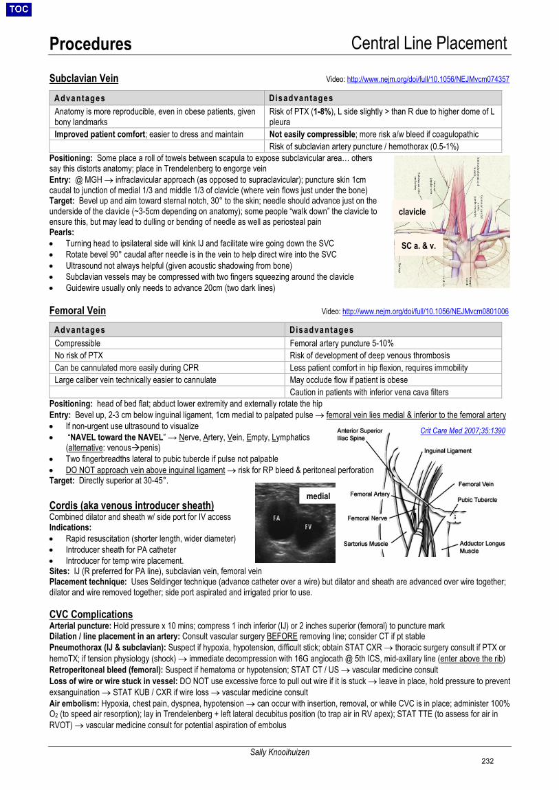
Procedures Central Line Placement
Sally Knooihuizen
Subclavian Vein Video: http://www.nejm.org/doi/full/10.1056/NEJMvcm074357
Positioning: Some place a roll of towels between scapula to expose subclavicular area… others say this distorts anatomy; place in Trendelenberg to engorge vein Entry: @ MGH → infraclavicular approach (as opposed to supraclavicular); puncture skin 1cm caudal to junction of medial 1/3 and middle 1/3 of clavicle (where vein flows just under the bone) Target: Bevel up and aim toward sternal notch, 30° to the skin; needle should advance just on the underside of the clavicle (~3-5cm depending on anatomy); some people “walk down” the clavicle to ensure this, but may lead to dulling or bending of needle as well as periosteal pain Pearls: • Turning head to ipsilateral side will kink IJ and facilitate wire going down the SVC• Rotate bevel 90° caudal after needle is in the vein to help direct wire into the SVC• Ultrasound not always helpful (given acoustic shadowing from bone)• Subclavian vessels may be compressed with two fingers squeezing around the clavicle• Guidewire usually only needs to advance 20cm (two dark lines)
Femoral Vein Video: http://www.nejm.org/doi/full/10.1056/NEJMvcm0801006
Positioning: head of bed flat; abduct lower extremity and externally rotate the hip Entry: Bevel up, 2-3 cm below inguinal ligament, 1cm medial to palpated pulse → femoral vein lies medial & inferior to the femoral artery • If non-urgent use ultrasound to visualize• “NAVEL toward the NAVEL” → Nerve, Artery, Vein, Empty, Lymphatics
(alternative: venouspenis)• Two fingerbreadths lateral to pubic tubercle if pulse not palpable• DO NOT approach vein above inguinal ligament → risk for RP bleed & peritoneal perforationTarget: Directly superior at 30-45°.
Cordis (aka venous introducer sheath) Combined dilator and sheath w/ side port for IV access Indications: • Rapid resuscitation (shorter length, wider diameter)• Introducer sheath for PA catheter• Introducer for temp wire placement.Sites: IJ (R preferred for PA line), subclavian vein, femoral veinPlacement technique: Uses Seldinger technique (advance catheter over a wire) but dilator and sheath are advanced over wire together;dilator and wire removed together; side port aspirated and irrigated prior to use.
CVC Complications Arterial puncture: Hold pressure x 10 mins; compress 1 inch inferior (IJ) or 2 inches superior (femoral) to puncture mark Dilation / line placement in an artery: Consult vascular surgery BEFORE removing line; consider CT if pt stable Pneumothorax (IJ & subclavian): Suspect if hypoxia, hypotension, difficult stick; obtain STAT CXR → thoracic surgery consult if PTX or hemoTX; if tension physiology (shock) → immediate decompression with 16G angiocath @ 5th ICS, mid-axillary line (enter above the rib) Retroperitoneal bleed (femoral): Suspect if hematoma or hypotension; STAT CT / US → vascular medicine consult Loss of wire or wire stuck in vessel: DO NOT use excessive force to pull out wire if it is stuck → leave in place, hold pressure to prevent exsanguination → STAT KUB / CXR if wire loss → vascular medicine consult Air embolism: Hypoxia, chest pain, dyspnea, hypotension → can occur with insertion, removal, or while CVC is in place; administer 100% O2 (to speed air resorption); lay in Trendelenberg + left lateral decubitus position (to trap air in RV apex); STAT TTE (to assess for air in RVOT) → vascular medicine consult for potential aspiration of embolus
Advantages Disadvantages Anatomy is more reproducible, even in obese patients, given bony landmarks
Risk of PTX (1-8%), L side slightly > than R due to higher dome of L pleura
Improved patient comfort; easier to dress and maintain Not easily compressible; more risk a/w bleed if coagulopathic Risk of subclavian artery puncture / hemothorax (0.5-1%)
Advantages Disadvantages Compressible Femoral artery puncture 5-10% No risk of PTX Risk of development of deep venous thrombosis Can be cannulated more easily during CPR Less patient comfort in hip flexion, requires immobility Large caliber vein technically easier to cannulate May occlude flow if patient is obese
Caution in patients with inferior vena cava filters
medial
clavicle
SC a. & v.
Crit Care Med 2007;35:1390
232
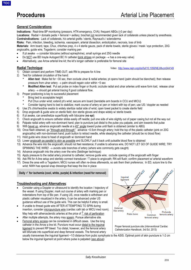
Procedures Arterial Line Placement
Sally Knooihuizen
General Considerations: Indications: Real-time BP monitoring (pressors, HTN emergency, CVA); frequent ABGs (>3 per day) Locations: Radial > dorsalis pedis > femoral > axillary; brachial not recommended given lack of collaterals unless placed by anesthesia. Contraindications: Lack of collaterals, h/o arterial grafts / stents, Raynaud’s / scleroderma Risks: Pain, infection, bleeding, ischemia, vasospasm, arterial dissection, embolization, necrosis, loss of limb Materials: Arm board, tape, Chux, chlorhex prep, 4 x 4 sterile gauze, pack of sterile towels, sterile gloves / mask / eye protection, 20G angiocaths, guide wire, Tegaderm; consider marking pen • If pt awake → consider lidocaine (without epinephrine), small syringe and 25G needle• Do NOT use BD Insyte Autogard BC IV catheter (pink stripes on package → has a one-way valve)• Alternatively, use Arrow arterial line kit; the kit’s longer catheter is preferable for femoral site
Radial Technique: Video: http://www.nejm.org/doi/full/10.1056/NEJMvcm044149 1) Obtain consent and perform TIME-OUT; ask RN to prepare for A line2) Test for collateral circulation of the hand:
* Allen test: Make fist for ~30 sec, then occlude ulnar & radial arteries; pt opens hand (palm should be blanched); then releasepressure from ulnar artery → palm should regain color within ~5 sec
* Modified Allen test: Put sat probe on index finger or thumb; occlude radial and ulnar arteries until wave form lost; release ulnarartery → should get arterial tracing if good collateral flow.
3) Proper positioning is key to successful placement* Bring bed to acceptable height* Put Chux under wrist; extend pt’s wrist; secure arm board (bendable arm boards in CCU and MICU)* Consider taping hand to bed to stabilize; mark course of artery w/ pen or indent with top of pen; use US / doppler as needed
4) Use 2% chlorhexidine swabs to widely sterilize radial side of wrist; open towel packet to create sterile field5) Drop angiocath & guide wire on sterile field; don sterile gloves and drape widely w/ sterile towels6) If pt awake, can anesthetize superficially with lidocaine (no epi)7) Check angiocath to ensure catheter slides easily off needle; pull one side of wire slightly out of paper casing but not all the way out8) Palpate radial artery with non-dominant hand; plan to puncture just distal to the pulse you palpate, and aim towards that pulse9) With bevel up, advance angiocath needle at a 45° angle toward pulse until flash is obtained (similar to ABG)10) Once flash obtained, go "through-and-through”: advance ~0.5cm through artery; hold the top of the plastic catheter (pink on 20G
angiocaths) with non-dominant hand; push button to retract needle, while steadying the catheter (should be no blood flow)11) Hold guide wire close to head of angiocath w/ dominant hand12) Lower angiocath as parallel to skin as possible and SLOWLY pull it back until pulsatile blood flow is obtained13) Advance the wire into the angiocath; should not feel resistance; if unable to advance wire, DO NOT LET GO OF GUIDE WIRE; TRY
SPINNING THE WIRE! → avoids side branches of artery (where wire commonly gets caught)14) Advance angiocath into the artery over the wire (Seldinger technique)15) Apply pressure to the radial artery proximal to catheter; remove guide wire; occlude opening of the angiocath with finger16) Ask RN for A-line setup and sterilely connect transducer / T-piece to angiocath; RN will flush; confirm placement w/ arterial waveform17) Dress the area with a Tegaderm; MICU nurses will often re-dress afterwards, so ask them their preference; In ED, suture line to the
wrist; NWH has special snap dressings that keep the line in place
Troubleshooting and Alternatives: • Consider using a Doppler or ultrasound to identify the location / trajectory of
the vessel. If using Doppler, mark out course of artery with marking pen orindentations from top of Bic pen. If using US, once needle is withdrawn andplastic catheter visualized in the artery, it can be advanced under USguidance without use of the guide wire. This can be helpful if artery is small.
• If unable to thread guide wire AFTER ATTEMPTING TO SPIN duringinsertion, consider micropuncture wire (cardiac cath lab or MICU med room).May help with atherosclerotic arteries at the price of ↑ risk of perforation
• After multiple attempts, the artery may spasm. Pursue alternative site.• Femoral artery access can be considered in difficult cases. Use the long
catheter in the Arrow a line kit. Puncture must occur distal to the inguinal ligament to prevent RP bleed. Too distal, however, and the femoral artery will bifurcate into superficial and deep femoral vessels. The femoral arteryusually transverses the inguinal ligament ~1/3 distance from pubic symphysis to the ASIS. Optimal point of skin puncture is 1-2 cmbelow the inguinal ligament at point where pulse is palpated (see above)
Proper femoral puncture site (Interventional CardiacCatheterization Handbook, 3rd Ed. 2013)
Daily for ischemia (cool, white, purple) & infection (need for removal)
233
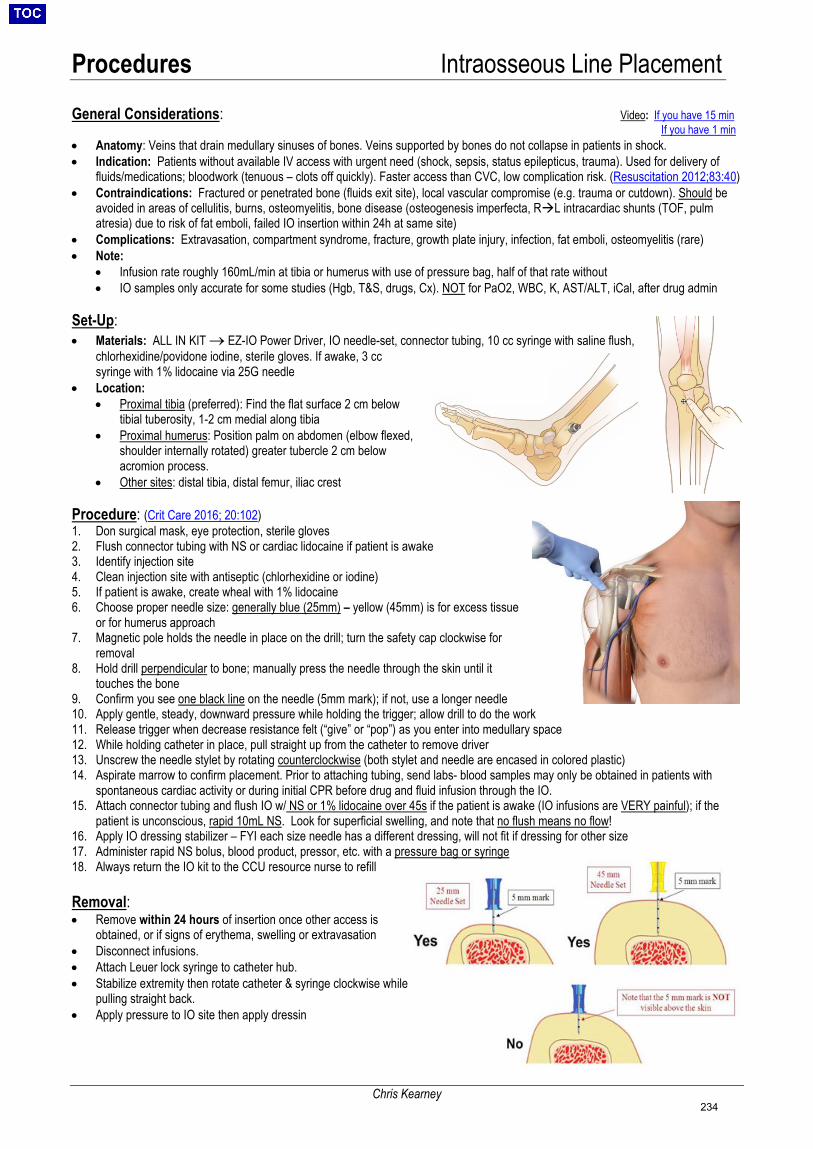
Procedures Intraosseous Line Placement
Chris Kearney
General Considerations: Video: If you have 15 min If you have 1 min
• Anatomy: Veins that drain medullary sinuses of bones. Veins supported by bones do not collapse in patients in shock.• Indication: Patients without available IV access with urgent need (shock, sepsis, status epilepticus, trauma). Used for delivery of
fluids/medications; bloodwork (tenuous – clots off quickly). Faster access than CVC, low complication risk. (Resuscitation 2012;83:40)• Contraindications: Fractured or penetrated bone (fluids exit site), local vascular compromise (e.g. trauma or cutdown). Should be
avoided in areas of cellulitis, burns, osteomyelitis, bone disease (osteogenesis imperfecta, RL intracardiac shunts (TOF, pulmatresia) due to risk of fat emboli, failed IO insertion within 24h at same site)
• Complications: Extravasation, compartment syndrome, fracture, growth plate injury, infection, fat emboli, osteomyelitis (rare)• Note:
• Infusion rate roughly 160mL/min at tibia or humerus with use of pressure bag, half of that rate without• IO samples only accurate for some studies (Hgb, T&S, drugs, Cx). NOT for PaO2, WBC, K, AST/ALT, iCal, after drug admin
Set-Up: • Materials: ALL IN KIT → EZ-IO Power Driver, IO needle-set, connector tubing, 10 cc syringe with saline flush,
chlorhexidine/povidone iodine, sterile gloves. If awake, 3 ccsyringe with 1% lidocaine via 25G needle
• Location:• Proximal tibia (preferred): Find the flat surface 2 cm below
tibial tuberosity, 1-2 cm medial along tibia• Proximal humerus: Position palm on abdomen (elbow flexed,
shoulder internally rotated) greater tubercle 2 cm belowacromion process.
• Other sites: distal tibia, distal femur, iliac crest
Procedure: (Crit Care 2016; 20:102) 1. Don surgical mask, eye protection, sterile gloves2. Flush connector tubing with NS or cardiac lidocaine if patient is awake3. Identify injection site4. Clean injection site with antiseptic (chlorhexidine or iodine)5. If patient is awake, create wheal with 1% lidocaine6. Choose proper needle size: generally blue (25mm) – yellow (45mm) is for excess tissue
or for humerus approach7. Magnetic pole holds the needle in place on the drill; turn the safety cap clockwise for
removal8. Hold drill perpendicular to bone; manually press the needle through the skin until it
touches the bone9. Confirm you see one black line on the needle (5mm mark); if not, use a longer needle10. Apply gentle, steady, downward pressure while holding the trigger; allow drill to do the work11. Release trigger when decrease resistance felt (“give” or “pop”) as you enter into medullary space12. While holding catheter in place, pull straight up from the catheter to remove driver13. Unscrew the needle stylet by rotating counterclockwise (both stylet and needle are encased in colored plastic)14. Aspirate marrow to confirm placement. Prior to attaching tubing, send labs- blood samples may only be obtained in patients with
spontaneous cardiac activity or during initial CPR before drug and fluid infusion through the IO.15. Attach connector tubing and flush IO w/ NS or 1% lidocaine over 45s if the patient is awake (IO infusions are VERY painful); if the
patient is unconscious, rapid 10mL NS. Look for superficial swelling, and note that no flush means no flow!16. Apply IO dressing stabilizer – FYI each size needle has a different dressing, will not fit if dressing for other size17. Administer rapid NS bolus, blood product, pressor, etc. with a pressure bag or syringe18. Always return the IO kit to the CCU resource nurse to refill
Removal: • Remove within 24 hours of insertion once other access is
obtained, or if signs of erythema, swelling or extravasation• Disconnect infusions.• Attach Leuer lock syringe to catheter hub.• Stabilize extremity then rotate catheter & syringe clockwise while
pulling straight back.• Apply pressure to IO site then apply dressin
234
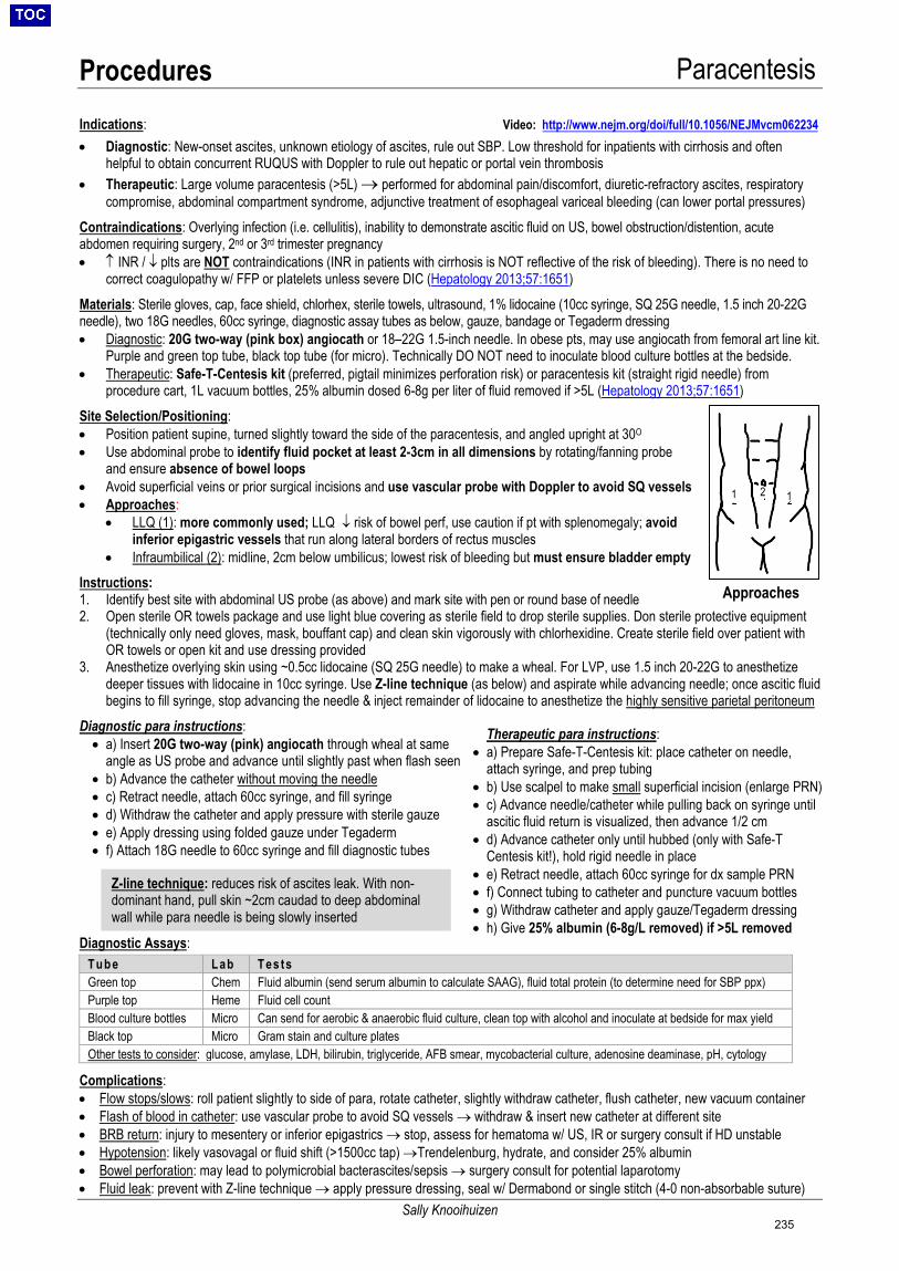
Procedures Paracentesis
Sally Knooihuizen
Therapeutic para instructions: • a) Prepare Safe-T-Centesis kit: place catheter on needle,
attach syringe, and prep tubing• b) Use scalpel to make small superficial incision (enlarge PRN)• c) Advance needle/catheter while pulling back on syringe until
ascitic fluid return is visualized, then advance 1/2 cm• d) Advance catheter only until hubbed (only with Safe-T
Centesis kit!), hold rigid needle in place• e) Retract needle, attach 60cc syringe for dx sample PRN• f) Connect tubing to catheter and puncture vacuum bottles• g) Withdraw catheter and apply gauze/Tegaderm dressing• h) Give 25% albumin (6-8g/L removed) if >5L removed
Z-line technique: reduces risk of ascites leak. With non-dominant hand, pull skin ~2cm caudad to deep abdominalwall while para needle is being slowly inserted
Indications: Video: http://www.nejm.org/doi/full/10.1056/NEJMvcm062234 • Diagnostic: New-onset ascites, unknown etiology of ascites, rule out SBP. Low threshold for inpatients with cirrhosis and often
helpful to obtain concurrent RUQUS with Doppler to rule out hepatic or portal vein thrombosis• Therapeutic: Large volume paracentesis (>5L) → performed for abdominal pain/discomfort, diuretic-refractory ascites, respiratory
compromise, abdominal compartment syndrome, adjunctive treatment of esophageal variceal bleeding (can lower portal pressures)Contraindications: Overlying infection (i.e. cellulitis), inability to demonstrate ascitic fluid on US, bowel obstruction/distention, acute abdomen requiring surgery, 2nd or 3rd trimester pregnancy • ↑ INR / ↓ plts are NOT contraindications (INR in patients with cirrhosis is NOT reflective of the risk of bleeding). There is no need to
correct coagulopathy w/ FFP or platelets unless severe DIC (Hepatology 2013;57:1651)Materials: Sterile gloves, cap, face shield, chlorhex, sterile towels, ultrasound, 1% lidocaine (10cc syringe, SQ 25G needle, 1.5 inch 20-22G needle), two 18G needles, 60cc syringe, diagnostic assay tubes as below, gauze, bandage or Tegaderm dressing • Diagnostic: 20G two-way (pink box) angiocath or 18–22G 1.5-inch needle. In obese pts, may use angiocath from femoral art line kit.
Purple and green top tube, black top tube (for micro). Technically DO NOT need to inoculate blood culture bottles at the bedside.• Therapeutic: Safe-T-Centesis kit (preferred, pigtail minimizes perforation risk) or paracentesis kit (straight rigid needle) from
procedure cart, 1L vacuum bottles, 25% albumin dosed 6-8g per liter of fluid removed if >5L (Hepatology 2013;57:1651)Site Selection/Positioning:• Position patient supine, turned slightly toward the side of the paracentesis, and angled upright at 30O
• Use abdominal probe to identify fluid pocket at least 2-3cm in all dimensions by rotating/fanning probeand ensure absence of bowel loops
• Avoid superficial veins or prior surgical incisions and use vascular probe with Doppler to avoid SQ vessels• Approaches:
• LLQ (1): more commonly used; LLQ ↓ risk of bowel perf, use caution if pt with splenomegaly; avoidinferior epigastric vessels that run along lateral borders of rectus muscles
• Infraumbilical (2): midline, 2cm below umbilicus; lowest risk of bleeding but must ensure bladder emptyInstructions: 1. Identify best site with abdominal US probe (as above) and mark site with pen or round base of needle2. Open sterile OR towels package and use light blue covering as sterile field to drop sterile supplies. Don sterile protective equipment
(technically only need gloves, mask, bouffant cap) and clean skin vigorously with chlorhexidine. Create sterile field over patient withOR towels or open kit and use dressing provided
3. Anesthetize overlying skin using ~0.5cc lidocaine (SQ 25G needle) to make a wheal. For LVP, use 1.5 inch 20-22G to anesthetizedeeper tissues with lidocaine in 10cc syringe. Use Z-line technique (as below) and aspirate while advancing needle; once ascitic fluidbegins to fill syringe, stop advancing the needle & inject remainder of lidocaine to anesthetize the highly sensitive parietal peritoneum
Diagnostic para instructions: • a) Insert 20G two-way (pink) angiocath through wheal at same
angle as US probe and advance until slightly past when flash seen • b) Advance the catheter without moving the needle • c) Retract needle, attach 60cc syringe, and fill syringe • d) Withdraw the catheter and apply pressure with sterile gauze• e) Apply dressing using folded gauze under Tegaderm• f) Attach 18G needle to 60cc syringe and fill diagnostic tubes
Diagnostic Assays:
Tube Lab Tes ts Green top Chem Fluid albumin (send serum albumin to calculate SAAG), fluid total protein (to determine need for SBP ppx) Purple top Heme Fluid cell count Blood culture bottles Micro Can send for aerobic & anaerobic fluid culture, clean top with alcohol and inoculate at bedside for max yield Black top Micro Gram stain and culture plates Other tests to consider: glucose, amylase, LDH, bilirubin, triglyceride, AFB smear, mycobacterial culture, adenosine deaminase, pH, cytology
Complications: • Flow stops/slows: roll patient slightly to side of para, rotate catheter, slightly withdraw catheter, flush catheter, new vacuum container• Flash of blood in catheter: use vascular probe to avoid SQ vessels → withdraw & insert new catheter at different site• BRB return: injury to mesentery or inferior epigastrics → stop, assess for hematoma w/ US, IR or surgery consult if HD unstable• Hypotension: likely vasovagal or fluid shift (>1500cc tap) →Trendelenburg, hydrate, and consider 25% albumin• Bowel perforation: may lead to polymicrobial bacterascites/sepsis → surgery consult for potential laparotomy• Fluid leak: prevent with Z-line technique → apply pressure dressing, seal w/ Dermabond or single stitch (4-0 non-absorbable suture)
Approaches
1 2 1
235
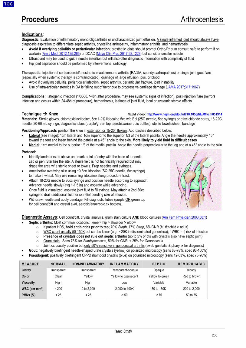
Procedures Arthrocentesis
Isaac Smith
Indications: Diagnostic: Evaluation of inflammatory mono/oligoarthritis or uncharacterized joint effusion. A single inflamed joint should always have diagnostic aspiration to differentiate septic arthritis, crystalline arthopathy, inflammatory arthritis, and hemarthrosis • Avoid if overlying cellulitis or periarticular infection; prosthetic joints should prompt Ortho/Rheum consult; safe to perform if on
warfarin (Am J Med. 2012;125:265) or DOAC (Mayo Clin Proc 2017;92:1223) but consider smaller needle• Ultrasound may be used to guide needle insertion but will also offer diagnostic information with complexity of fluid• Hip joint aspiration should be performed by interventional radiology
Therapeutic: Injection of corticosteroid/anesthetic in autoimmune arthritis (RA/JIA, spondyloarhropathies) or single-joint gout flare (especially when systemic therapy is contraindicated); drainage of large effusion, pus, or blood • Avoid if overlying cellulitis, periarticular infection, septic arthritis, periarticular fracture, joint instability• Use of intra-articular steroids in OA is falling out of favor due to progressive cartilage damage (JAMA 2017;317:1967)
Complications: Iatrogenic infection (1/3500, >48h after procedure, may see systemic signs of infection), post-injection flare (mirrors infection and occurs within 24-48h of procedure), hemarthrosis, leakage of joint fluid, local or systemic steroid effects
Technique Knee NEJM Video: http://www.nejm.org/doi/full/10.1056/NEJMvcm051914Materials: Sterile gloves, chlorhexidine/iodine, 5cc 1-2% lidocaine 5cc w/o Epi (25G needle, 5cc syringe) or ethyl chloride spray, 18-22G needle, 20-60 mL syringe, diagnostic tubes (purple/green top, aerobic/anaerobic bottles), sterile towels/sheet, bandage Positioning/Approach: position the knee in extension or 15-20° flexion. Approaches described below: • Lateral (see image): 1cm lateral and 1cm superior to the superior 1/3 of the lateral patella. Angle the needle approximately 45°
toward the feet and insert behind the patella at a 45° angle to the skin. More likely to yield fluid in difficult cases• Medial: 1cm medial to the superior 1/3 of the medial patella. Angle the needle perpendicular to the leg and at a 45° angle to the skinProtocol:• Identify landmarks as above and mark point of entry with the base of a needle
cap or pen. Sterilize the site. A sterile field is not technically required but maydrape the area w/ a sterile sheet or towels. Prep needles and syringes.
• Anesthetize overlying skin using ~0.5cc lidocaine (SQ 25G needle, 5cc syringe)to make a wheal. May use remaining lidocaine along procedure tract.
• Attach 18-20G needle to 30cc syringe and position needle according to approach.Advance needle slowly (avg 1-1.5 in) and aspirate while advancing.
• Once fluid is visualized, aspirate joint fluid to fill syringe. May attach a 2nd 30ccsyringe to drain additional fluid for sx relief pending size of effusion.
• Withdraw needle and apply bandage. Fill diagnostic tubes (purple OR green topfor cell count/diff and crystal eval, aerobic/anaerobic cx bottles).
Diagnostic Assays: Cell count/diff, crystal analysis, gram stain/culture AND blood cultures (Am Fam Physician;2003;68:1) • Septic arthritis: Most common locations: knee > hip > shoulder > elbow
o If patient HDS, hold antibiotics prior to tap; 70% Staph, 17% Strep, 8% GNR (H. flu child > adult)o WBC count usually 50-150K but can be lower (e.g., <20K in disseminated gonorrhea); ↑WBC = ↑ risk of infectiono Presence of crystals does not rule out septic arthritis (up to 5% of pts with crystals also have septic joint)o Gram stain: Sens 75% for Staphylococcus, 50% for GNR, < 25% for Gonococcuso Joint cx usually positive but only 50% sensitive in gonococcal arthritis (swab genitalia & pharynx for diagnosis)
• Gout: negatively birefingent needle-shaped urate crystals (yellow) on polarized microscopy (sens 63-78%, spec 93-100%)• Pseudogout: positively birefringent CPPD rhomboid crystals (blue) on polarized microscopy (sens 12-83%, spec 78-96%)
ME AS UR E NO RM AL NON-INFLAMMATORY INFL AM M ATOR Y SEPT I C HE MO RR H AGI C Clarity Transparent Transparent Transparent-opaque Opaque Bloody Color Clear Yellow Yellow to opalsecent Yellow to green Red to brown Viscosity High High Low Variable Variable WBC (per mm3) < 200 0 to 2,000 2,000 to 100K 50 to 150K 200 to 2,000 PMNs (%) < 25 < 25 ≥ 50 ≥ 75 50 to 75
236
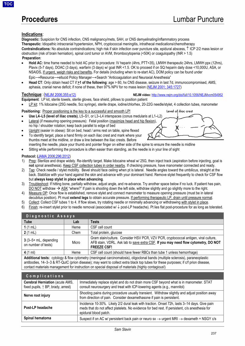
Procedures Lumbar Puncture
Sam Slavin
x
Indications:
Diagnostic: Suspicion for CNS infection, CNS malignancy/mets, SAH, or CNS demyelinating/inflammatory process Therapeutic: Idiopathic intracranial hypertension, NPH, cryptococcal meningitis, intrathecal medications/chemotherapy Contraindications: No absolute contraindications; high risk if skin infection over puncture site, epidural abscess, ↑ ICP 2/2 mass lesion or obstruction (risk of brain herniation), spinal cord tumor or AVM, thrombocytopenia (<50K) or coagulopathy (INR > 1.5) Preparation: • Hold AC: time frame needed to hold AC prior to procedure: IV heparin (4hrs, PTT<35), LMWH therapeutic 24hrs, LMWH ppx (12hrs),
Plavix (5-7 days), DOAC (3 days), warfarin (3 days) w/ goal INR <1.5. OK to proceed if on SQ heparin daily dose <10,000U, ASA, orNSAIDS. If urgent, weigh risks and benefits. For details (including when to re-start AC), DOM policy can be found underEpic→Resource→ellucid Policy Manager→Search “Anticoagulation and Neuraxial Anesthesia”
• Head CT: Only obtain head CT if >1 of the following: age > 60, hx CNS disease, seizure in last 7d, immunocompromised, AMS,aphasia, cranial nerve deficit; if none of these, then 97% NPV for no mass lesion (NEJM 2001; 345:1727)
Technique: (NEJM 2006;355:e12) NEJM video: http://www.nejm.org/doi/full/10.1056/NEJMvcm054952Equipment: LP kit, sterile towels, sterile gloves, face shield, pillows to position patient • LP kit: 1% lidocaine (25G needle, 5cc syringe), sterile drape, iodine/chlorhex, 20-22G needle/stylet, 4 collection tubes, manometerPositioning: Proper positioning is the key to a successful and smooth LP!• Use L4–L5 (level of iliac crests), L5–S1, or L3–L4 interspaces (conus medularis at L1–L2)• Lateral (if measuring opening pressure): Fetal position (maximize head and hip flexion),
no hip / shoulder rotation; keep back parallel to edge of bed• Upright (easier in obese): Sit on bed, head / arms rest on table, spine flexed• To identify target, place a hand firmly on each iliac crest and mark where your
thumbs meet at the midline, or draw a line between the iliac crests. Beforeinserting the needle, place your thumb and pointer finger on either side of the spine to ensure the needle is midline
• Sitting while performing the procedure is often easier than standing, as the needle is in your line of sightProtocol: (JAMA 2006;296:2012)1) Prep: Sterilize and drape widely. Re-identify target. Make lidocaine wheal w/ 25G, then inject track (aspiration before injecting, goal is
not spinal anesthesia). Keep CSF collection tubes in order nearby. If checking pressure, have manometer connected and ready.2) Tap: Check needle / stylet mobility. Bevel should face ceiling when pt is lateral. Needle angles toward the umbilicus, straight at the
back. Stabilize with your hand against the skin and advance with your dominant hand. Remove stylet frequently to check for CSF flowbut always keep stylet in place when advancing.
3) Troubleshoot: If hitting bone, partially withdraw, adjust angle, and re-advance. Try another space below if no luck. If patient has pain,DO NOT withdraw ASK “where?” If pain is shooting down the left side, withdraw slightly and go slightly more to the right.
4) Measure OP: Once flow is established, remove stylet and connect manometer to measure opening pressure (must be in lateraldecubitus position). Pt must extend legs to obtain accurate pressure. If performing therapeutic LP, drain until pressure normal.
5) Collect: Collect CSF tubes 1 to 4. If flow slows, try rotating needle or minimally advancing or withdrawing with stylet in place.6) Finish: re-insert stylet prior to needle removal (associated w/ ↓ post-LP headache). Pt lies flat post-procedure for as long as tolerated.
C o m p l i c a t i o n sCerebral Herniation (acute AMS, fixed pupils, ↑ BP, brady, arrest)
Immediately replace stylet and do not drain more CSF beyond what is in manometer. STAT consult neurosurgery and treat with ICP-lowering agents (e.g., mannitol)
Nerve root injury Shooting pains during procedure usually transient. Withdraw slightly and adjust position away from direction of pain. Consider dexamethasone if pain is persistent.
Post-LP headache Incidence 10-30%. Likely 2/2 dural leak with traction. Onset 72h, lasts 3–14 days. Give pain meds that do not affect platelets. No evidence for bed rest. If persistent, c/s anesthesia for epidural blood patch.
Spinal hematoma Suspect if on AC w/ persistent back pain or neuro sx → urgent MRI → dexameth + NSGY c/s
D i a g n o s t i c A s s a y sTube Lab Tests 1 (1 mL) Heme CSF cell count 2 (1 mL) Chem Total protein, glucose
3 (3–5+ mL, depending on number of tests) Micro
Gram stain/culture. Consider HSV PCR, VZV PCR, cryptococcal antigen, viral culture, AFB stain, VDRL. Ask lab to save extra CSF. If you may need flow cytometry, DO NOT FREEZE CSF!
4 (1 ml) Heme CSF cell count (should have fewer RBCs than tube 1 unless hemorrhage) Additional tests: cytology & flow cytometry (meningeal carcinomatosis), oligoclonal bands (multiple sclerosis), paraneoplastic antibodies, 14–3–3 & RT-QuIC (prion disease); may want to collect extra black top tubes for these purposes; if c/f prion disease, contact materials management for instruction on special disposal of materials (highly contagious!)
237
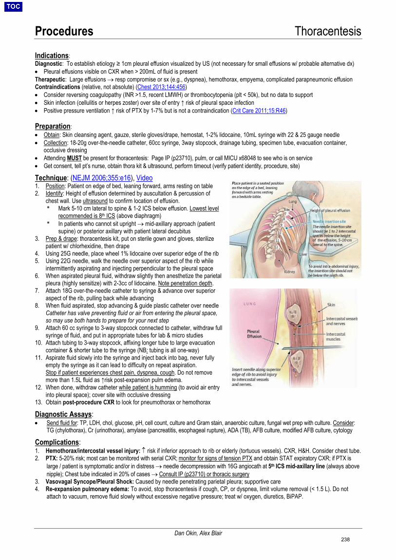
Procedures Thoracentesis
Dan Okin, Alex Blair
Indications: Diagnostic: To establish etiology ≥ 1cm pleural effusion visualized by US (not necessary for small effusions w/ probable alternative dx) • Pleural effusions visible on CXR when > 200mL of fluid is presentTherapeutic: Large effusions → resp compromise or sx (e.g., dyspnea), hemothorax, empyema, complicated parapneumonic effusionContraindications (relative, not absolute) (Chest 2013;144:456)• Consider reversing coagulopathy (INR >1.5, recent LMWH) or thrombocytopenia (plt < 50k), but no data to support• Skin infection (cellulitis or herpes zoster) over site of entry ↑ risk of pleural space infection• Positive pressure ventilation ↑ risk of PTX by 1-7% but is not a contraindication (Crit Care 2011;15:R46)
Preparation: • Obtain: Skin cleansing agent, gauze, sterile gloves/drape, hemostat, 1-2% lidocaine, 10mL syringe with 22 & 25 gauge needle• Collection: 18-20g over-the-needle catheter, 60cc syringe, 3way stopcock, drainage tubing, specimen tube, evacuation container,
occlusive dressing• Attending MUST be present for thoracentesis: Page IP (p23710), pulm, or call MICU x68048 to see who is on service• Get consent, tell pt’s nurse, obtain thora kit & ultrasound, perform timeout (verify patient identity, procedure, site)
Technique: (NEJM 2006;355:e16), Video 1. Position: Patient on edge of bed, leaning forward, arms resting on table2. Identify: Height of effusion determined by auscultation & percussion of
chest wall. Use ultrasound to confirm location of effusion.* Mark 5-10 cm lateral to spine & 1-2 ICS below effusion. Lowest level
recommended is 8th ICS (above diaphragm)* In patients who cannot sit upright → mid-axillary approach (patient
supine) or posterior axillary with patient lateral decubitus3. Prep & drape: thoracentesis kit, put on sterile gown and gloves, sterilize
patient w/ chlorhexidine, then drape4. Using 25G needle, place wheel 1% lidocaine over superior edge of the rib5. Using 22G needle, walk the needle over superior aspect of the rib while
intermittently aspirating and injecting perpendicular to the pleural space6. When aspirated pleural fluid, withdraw slightly then anesthetize the parietal
pleura (highly sensitize) with 2-3cc of lidocaine. Note penetration depth.7. Attach 18G over-the-needle catheter to syringe & advance over superior
aspect of the rib, pulling back while advancing8. When fluid aspirated, stop advancing & guide plastic catheter over needle
Catheter has valve preventing fluid or air from entering the pleural space,so may use both hands to prepare for your next step
9. Attach 60 cc syringe to 3-way stopcock connected to catheter, withdraw fullsyringe of fluid, and put in appropriate tubes for lab & micro studies
10. Attach tubing to 3-way stopcock, affixing longer tube to large evacuationcontainer & shorter tube to the syringe (NB: tubing is all one-way)
11. Aspirate fluid slowly into the syringe and inject back into bag, never fullyempty the syringe as it can lead to difficulty on repeat aspiration.Stop if patient experiences chest pain, dyspnea, cough. Do not removemore than 1.5L fluid as ↑risk post-expansion pulm edema.
12. When done, withdraw catheter while patient is humming (to avoid air entryinto pleural space); cover site with occlusive dressing
13. Obtain post-procedure CXR to look for pneumothorax or hemothorax
Diagnostic Assays: • Send fluid for: TP, LDH, chol, glucose, pH, cell count, culture and Gram stain, anaerobic culture, fungal wet prep with culture. Consider:
TG (chylothorax), Cr (urinothorax), amylase (pancreatitis, esophageal rupture), ADA (TB), AFB culture, modified AFB culture, cytology
Complications: 1. Hemothorax/intercostal vessel injury: ↑ risk if inferior approach to rib or elderly (tortuous vessels). CXR, H&H. Consider chest tube.2. PTX: 5-20% risk; most can be monitored with serial CXR; monitor for signs of tension PTX and obtain STAT expiratory CXR; if PTX is
large / patient is symptomatic and/or in distress → needle decompression with 16G angiocath at 5th ICS mid-axillary line (always abovenipple); Chest tube indicated in 20% of cases → Consult IP (p23710) or thoracic surgery
3. Vasovagal Syncope/Pleural Shock: Caused by needle penetrating parietal pleura; supportive care4. Re-expansion pulmonary edema: To avoid, stop thoracentesis if cough, CP, or dyspnea, limit volume removal (< 1.5 L). Do not
attach to vacuum, remove fluid slowly without excessive negative pressure; treat w/ oxygen, diuretics, BiPAP.
238
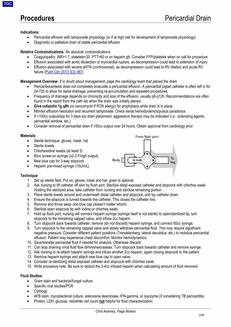
Procedures Pericardial Drain
Chris Kearney, Paige Mclean
Indications: • Pericardial effusion with tamponade physiology (or if at high risk for development of tamponade physiology)• Diagnostic or palliative drain of stable pericardial effusion
Relative Contraindications: No absolute contraindications • Coagulopathy: INR>1.7, platelets<20, PTT>60 or on heparin gtt. Consider FFP/platelets when on call for procedure• Effusion associated with aortic dissection or myocardial rupture, as decompression could lead to extension of injury• Effusion associated with severe pHTN (controversial), as decompression could lead to RV dilation and acute RV
failure (Pulm Circ 2013 3(3) 467)
Management Overview: If in doubt about management, page the cardiology team that placed the drain • Pericardiocentesis does not completely evacuate a pericardial effusion. A pericardial pigtail catheter is often left in for
24-72h to allow for serial drainage, preventing re-accumulation and repeated procedures.• Frequency of drainage depends on chronicity and size of the effusion, usually q6-q12h. Recommendations are often
found in the report from the cath lab when the drain was initially placed.• Give cefazolin 1g q8h (or vancomycin if PCN allergy) for prophylaxis while drain is in place.• Monitor effusion resolution and recurrent tamponade. Check serial hemodynamics/pulsus paradoxus.• If >100cc output/day for 3 days s/p drain placement, aggressive therapy may be indicated (i.e., sclerosing agents,
pericardial window, etc.)• Consider removal of pericardial drain if <50cc output over 24 hours. Obtain approval from cardiology prior.
Materials: • Sterile technique: gloves, mask, hat• Sterile towels• Chlorhexidine swabs (at least 3)• 60cc screw-on syringe (x2-3 if high output)• New blue cap for 3-way stopcock• Heparin pre-mixed syringe (10U/mL)
Technique: 1. Set up sterile field. Put on, gloves, mask and hat, gown is optional2. Ask nursing to lift catheter off skin by flush port. Sterilize distal exposed catheter and stopcock with chlorhex swab.
Holding the sterilized area, take catheter from nursing and sterilize remaining portion3. Place sterile towels around and underneath distal catheter and stopcock, and lay catheter down4. Ensure the stopcock is turned towards the catheter. This closes the catheter line.5. Remove and throw away one blue cap (doesn’t matter which).6. Sterilize open stopcock tip with iodine or chlorhex swab.7. Hold up flush port; nursing will connect heparin syringe (syringe itself is not sterile) to open/sterilized tip, turn
stopcock to the remaining capped valve, and infuse 2cc heparin.8. Turn stopcock back towards catheter, remove (do not discard) heparin syringe, and connect 60cc syringe.9. Turn stopcock to the remaining capped valve and slowly withdraw pericardial fluid. This may require significant
negative pressure. Consider different patient positions (Trendelenberg, lateral decubitus, etc.) to mobilize pericardialeffusion. Patient may experience chest discomfort. Monitor hemodynamics
10. Save/transfer pericardial fluid if needed for analysis. Otherwise discard.11. Can stop draining once fluid flow diminishes/ceases. Turn stopcock back towards catheter and remove syringe.12. Ask nursing to re-attach heparin syringe and infuse another 2cc heparin, again closing stopcock to the patient13. Remove heparin syringe and attach new blue cap to open valve.14. Consider re-sterilizing distal exposed catheter and stopcock with chlorhex swab.15. Write procedure note. Be sure to deduct the 2-4cc infused heparin when calculating amount of fluid removed.
Fluid Studies: • Gram stain and bacterial/fungal culture• Specific viral studies/PCR• Cytology• AFB stain, mycobacterial culture, adenosine deaminase, IFN-gamma, or lysozyme (if considering TB pericarditis)• Protein, LDH, glucose, red/white cell count not helpful for fluid characterization
239
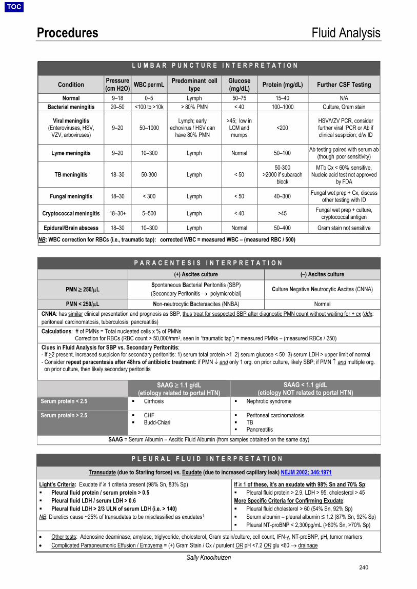
Procedures Fluid Analysis
Sally Knooihuizen
L U M B A R P U N C T U R E I N T E R P R E T A T I O N
Condition Pressure (cm H2O) WBC per mL Predominant cell
type Glucose (mg/dL) Protein (mg/dL) Further CSF Testing
Normal 9–18 0–5 Lymph 50–75 15–40 N/A Bacterial meningitis 20–50 <100 to >10k > 80% PMN < 40 100–1000 Culture, Gram stain
Viral meningitis (Enteroviruses, HSV,
VZV, arboviruses) 9–20 50–1000
Lymph; early echovirus / HSV can
have 80% PMN
>45; low inLCM andmumps
<200 HSV/VZV PCR, consider further viral PCR or Ab if clinical suspicion; d/w ID
Lyme meningitis 9–20 10–300 Lymph Normal 50–100 Ab testing paired with serum ab (though poor sensitivity)
TB meningitis 18–30 50-300 Lymph < 50 50-300
>2000 if subarach block
MTb Cx < 60% sensitive, Nucleic acid test not approved
by FDA
Fungal meningitis 18–30 < 300 Lymph < 50 40–300 Fungal wet prep + Cx, discuss other testing with ID
Cryptococcal meningitis 18–30+ 5–500 Lymph < 40 >45 Fungal wet prep + culture, cryptococcal antigen
Epidural/Brain abscess 18–30 10–300 Lymph Normal 50–400 Gram stain not sensitive
NB: WBC correction for RBCs (i.e., traumatic tap): corrected WBC = measured WBC – (measured RBC / 500)
P A R A C E N T E S I S I N T E R P R E T A T I O N(+) Ascites culture (–) Ascites culture
PMN ≥ 250/µL Spontaneous Bacterial Peritonitis (SBP) (Secondary Peritonitis → polymicrobial)
Culture Negative Neutrocytic Ascites (CNNA)
PMN < 250/µL Non-neutrocytic Bacterascites (NNBA) Normal CNNA: has similar clinical presentation and prognosis as SBP, thus treat for suspected SBP after diagnostic PMN count without waiting for + cx (ddx: peritoneal carcinomatosis, tuberculosis, pancreatitis) Calculations: # of PMNs = Total nucleated cells x % of PMNs
Correction for RBCs (RBC count > 50,000/mm3, seen in “traumatic tap”) = measured PMNs – (measured RBCs / 250) Clues in Fluid Analysis for SBP vs. Secondary Peritonitis: - If >2 present, increased suspicion for secondary peritonitis: 1) serum total protein >1 2) serum glucose < 50 3) serum LDH > upper limit of normal- Consider repeat paracentesis after 48hrs of antibiotic treatment: if PMN ↓ and only 1 org. on prior culture, likely SBP; if PMN ↑ and multiple org.on prior culture, then likely secondary peritonitis
SAAG ≥ 1.1 g/dL (etiology related to portal HTN)
SAAG < 1.1 g/dL (etiology NOT related to portal HTN)
Serum protein < 2.5 Cirrhosis Nephrotic syndrome
Serum protein > 2.5 CHF Budd-Chiari
Peritoneal carcinomatosis TB Pancreatitis
SAAG = Serum Albumin – Ascitic Fluid Albumin (from samples obtained on the same day)
P L E U R A L F L U I D I N T E R P R E T A T I O N
Transudate (due to Starling forces) vs. Exudate (due to increased capillary leak) NEJM 2002; 346:1971 Light’s Criteria: Exudate if ≥ 1 criteria present (98% Sn, 83% Sp) Pleural fluid protein / serum protein > 0.5 Pleural fluid LDH / serum LDH > 0.6 Pleural fluid LDH > 2/3 ULN of serum LDH (i.e. > 140)NB: Diuretics cause ~25% of transudates to be misclassified as exudates1
If ≥ 1 of these, it’s an exudate with 98% Sn and 70% Sp: Pleural fluid protein > 2.9, LDH > 95, cholesterol > 45More Specific Criteria for Confirming Exudate: Pleural fluid cholesterol > 60 (54% Sn, 92% Sp) Serum albumin – pleural albumin ≤ 1.2 (87% Sn, 92% Sp) Pleural NT-proBNP < 2,300pg/mL (>80% Sn, >70% Sp)
• Other tests: Adenosine deaminase, amylase, triglyceride, cholesterol, Gram stain/culture, cell count, IFN-γ, NT-proBNP, pH, tumor markers• Complicated Parapneumonic Effusion / Empyema = (+) Gram Stain / Cx / purulent OR pH <7.2 OR glu <60 → drainage
240
Procedures Tube Management
Sally Knooihuizen
N A S O G A S T R I C T U B E SIndications: • Decompression of SBO or minimize vomiting in ileus• Enteral feeding / med administration; charcoal admin (ODs),
oral contrast or colonoscopy prep• Lactulose (hepatic encephalopathy)
Contraindications • Head / maxillofacial trauma, basilar skull fracture, or recent
neurosurgical intervention• Esophageal stricture or ≥ grade 2 varices / recent banding
(discuss w/ GI if uncertainty regarding varices / banding)
Supplies:• NGT, lubricant/viscous lidocaine (“UroJet”), Chux, emesis basin, cup of water with ice and straw, 60mL syringe, tape• If NGT needed for decompression: use 14 to 16 Fr Salem sump NGT (larger diameter, ↓ clogging)
NGT Placement:• Assess patency and symmetry of nares by direct visualization• Consider topical anesthetic (e.g., 4% lidocaine) pre-treatment• Position patient in upright “sniffing” position with neck flexed and chin to chest• Estimate distance of NGT insertion by measuring from xiphoid processearlobenose tip• Apply lubricant / ice to tip of NGT and/or apply viscous lidocaine directly into the nares• Insert NGT into nares along floor & apply pressure posterior & slightly inferiomedial, not upward• After passage of NGT into oropharynx (will feel curve & ↓resistance), have patient swallow water via straw while advancing rapidly
o If patient excessively coughs, gags, has change in voice or dyspnea, or increased resistance, STOP (never force) andsuspect improper location (in airway or coiled) and immediately withdraw. Look in posterior oropharynx for coiling.
• Advance to predetermined depth and insufflate air w/ 60cc syringe while auscultating over stomach for rush of air. May also seereturn of gastric contents. Inspect oropharynx to ensure no coiling before securing tube w/ tape or bridle if ↑risk removal (AMS)
• Confirming position: MUST confirm placement with KUB prior to feeding/meds given risk of placement in trachea/lungs.KUB will show NGT tip below the diaphragm. Optional for KUB if bilious return when NGT for decompression (bile = stomach).
TYPES OF NGTs & USES
• Dobhoff: PO formula, meds• 14, 16 Fr: Decompression
Dobhoff tube / Enteroflex: Thinner, more flexible; more comfortable but ↑ risk of placement into lung Requires 2-step 2-CXR placement method
1. Measure from nose to earlobe to mid-sternum → insert tube this distance → secure → Obtain CXR2. If CXR shows tip (1) past carina & (2) midline → advance into stomach → Repeat CXR → remove stylette
General Troubleshooting: • If tube coiling repeatedly in oropharynx on insertion, soak tip in ice water to make tube more rigid prior to insertion.• NGT to suction should “sump” – air should audibly enter blue port and exit main port; if not: (1) flush blue port with air (never fluids)
(2) flush main port with water (not NS, does not need to be sterile) (3) aspirate from main port → if not able to withdraw flush, NGTneeds to be advanced vs. withdrawn (KUB can guide)
• To prevent clogging or adherence to gastric wall, NGTs should be flushed with 30cc water & air Q8hr. If clogged, can try methodsto unclog tube as below in “Gastrostomy Tubes”
Complications (↑ with longer duration): • GI: malposition, coiling, knotting anywhere along course of tube, nasal/GI tract perforation. ↑ risk acid/stomach content reflux and
aspiration → consider PPI. Chronic suction → gastritis/pressure necrosis: consider removal if grossly bloody• Pulm: intubation of lung → inadvertent med, contrast, TF administration → PNA, pulm abscess, tracheal perforation, PTX, death• HEENT: nasal irritation, epistaxis, intracranial placement, skin erosion, sinusitis, alar necrosis, tracheoesophageal fistula/perfRemoval:• If for ileus/SBO, consider removal when passing flatus or resolved N/V. Alternatively, may remove when NGT output <1L over 24
hours. Consider clamp trial before removing (clamp 4 hours, then check residual. Remove if <150 cc)• Remove tape. Flush tube w/ 10mL air or NS. Turn OFF suction & clamp. Fold Chux around tube insertion site. Gently remove tube
241
Procedures Tube Management
Sally Knooihuizen
G A S T R O S T O M Y T U B E STroubleshooting • Clogging:
• Only tube feeds and elixir meds should be given through J tube• Attach 3cc syringe w/ warm H2O to female Leur adaptor. Push or pulse
plunger to force through debris. Flush w/ 30 cc warm H2O to ensure notclogged.
• Can also try Seltzer, ginger ale, Coca-Cola. If persistent, can trypancrealipase (Viokase) with sodium bicarb
• Leaking: retract balloon or mushroom back to skin level; do NOT insert largersize tube (can cause stoma to enlarge), call service who placed G tube ifpersistent
• Migration: can cause N/V (w/ or w/o feeds), dumping syndrome. Confirmplacement w/ tube injection study (30-60 mL gastrograffin f/b KUB)
• Falling out: replace w/ similar-sized Foey or feeding tube. Obtain tube study asabove.
• Local site infection: try topical abx +/- antifungal before PO (cephalexin, clinda)• Granulation tissue: check tube size (not too long or short); treat w/ warm
compresses and silver nitrate (w/ barrier cream on surrounding nl skin to protect)
• Clear, soft, graduated tubing held inplace w/ plastic mushroom-shapedring/balloon in stomach (~3 cm deeper inobese pts)
• May be replaced at bedside afterepithelialized track forms (~2-4 wks;delayed by malnutrition, steroids,immunosuppression)
• Gastrojejunostomy (GJ) tubes have 3access ports: G tube port, J tube port andballoon port
• Secured with vertical Hollister device• Venting means access port is attached to
a foley bag so contents/gas can flow outas needed
F O L E Y C A T H E T E R Special Circumstances: • Artificial Urinary Sphincter (AUS) - men s/p
prostatectomy c/b sig. urinary incontinence.DEACTIVATE device prior to placing foley. Placesmallest catheter possible (12F) and removeASAP.
Troubleshooting: • Difficulty in female patient: likely poor
positioning. Place sheets under hips & place pt inTrendelenburg
• Urethral trauma: blood at meatus. Leavecatheter ≥3-5d
• Foley is leaking:o Bladder spasms 2/2 infection, mucosal
irritation, overactive bladder. Startanticholinergic (oxybutynin 5mg TID PRN)
o Foley obstructed 2/2 sediment, kinked, domeof bladder, clot. Flush catheter & bladder US
o Urethra patulous (women w/ chronic indwellingcatheters)
Suprapubic Tubes: • Many types, usually standard Foley catheter• Know type & size catheter, who exchanges, how
exchanged, how frequently• Is this a new tract (<7d, ask urology to replace)
or established (years, you can try and replace)?• If need to reinsert, decompress balloon and
remove indwelling SPT tube. Use foley kit, preparea, apply lubricant to new tube, insert throughtract (may have to use some force) until urinereturn, inflate balloon and ensure tube is mobile,attach to foley bag
Choosing catheter (order from Central Supply, ED or Ellison 6 if not on floor) o Many contain Latex, use silicone if allergy; silicone also ↓risk CAUTIo 2-way Foleys (drainage & balloon ports):
16F (stock), 12F if stricture or device, 18F/20F Coudé if BPH or highbladder neck → insert curve up / nub on hub pointed toward umbilicus
o 3-way Foleys (drainage, balloon, irrigation ports): 20F/22F used forcontinuous bladder irrigation (CBI) in gross hematuria
Placement: 1. Lay patient flat, prep, hold penis upright (keep on stretch while advancing)2. Instill 10cc 2% viscous lidocaine (“UroJet”) into urethra3. Insert Foley catheter to the hub4. As catheter reaches bladder neck, keeping penis on stretch, point phallus
down towards toes (to mimic natural curve urethra).5. After urine return AND catheter hubbed, fill balloon w/ 10cc sterile H2O6. Gently withdraw catheter to bladder neck7. Verify position by flushing with 60cc fluid (catheter in bladder) and withdraw.
Inability to withdraw suggests:a. Bladder empty and sucking against bladder mucosa (instill 60 cc)b. Catheter in urethra or false passagec. Catheter outside bladder (undermined bladder neck in pt s/p
prostatectomy/TURP) 8. Don’t forget to reduce foreskin (if not, may cause paraphimosis = urologic
emergency)
Continuous bladder irrigation (CBI): consult urology to initiate • Indications: Gross hematuria (when you cannot see your hand through the
foley due to presence of blood) +/- with clots• Titrate flow to “fruit punch” colored urine (should be see through)• When discontinuing, usually start with clamp trial to ensure resolution before
removal
242
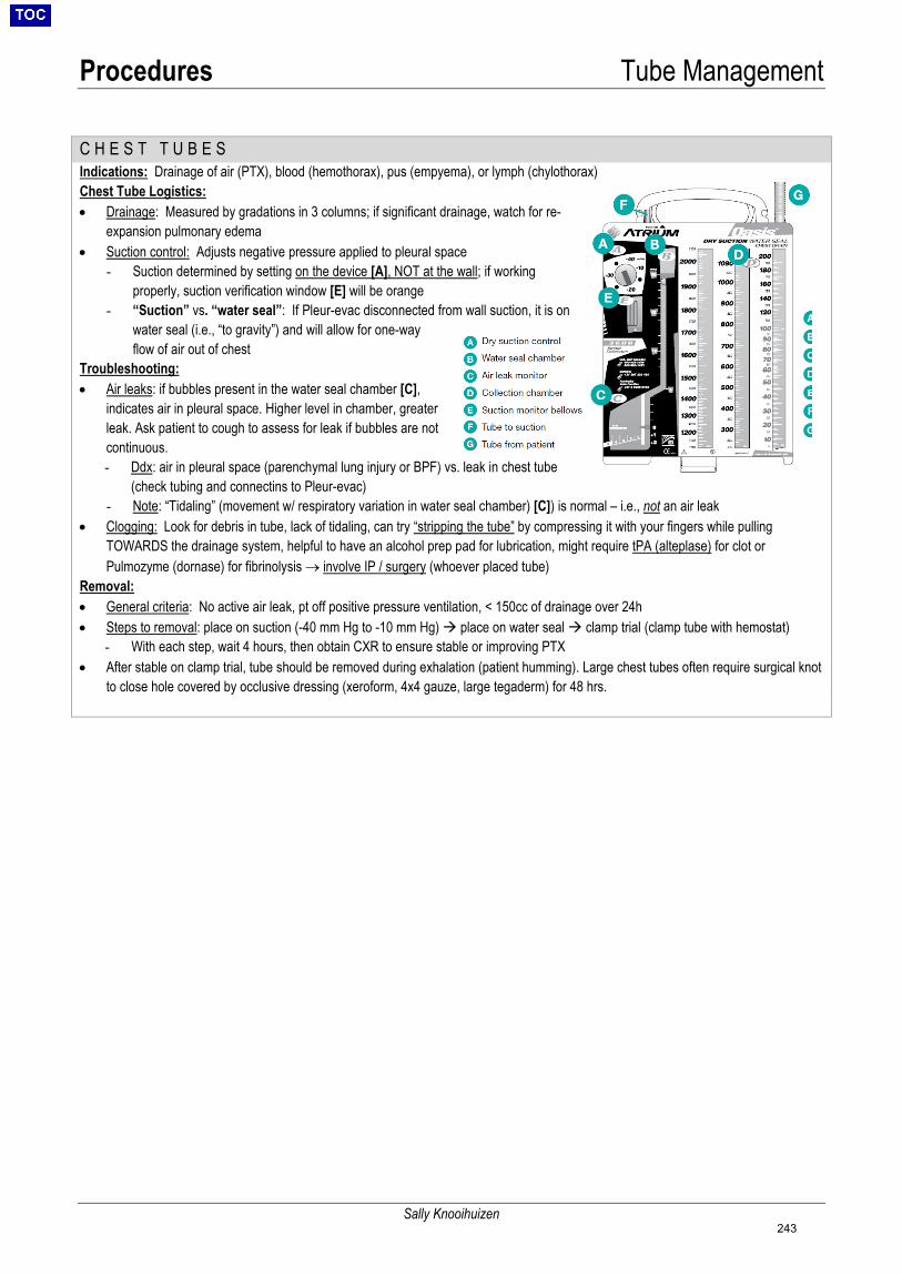
Procedures Tube Management
Sally Knooihuizen
C H E S T T U B E SIndications: Drainage of air (PTX), blood (hemothorax), pus (empyema), or lymph (chylothorax) Chest Tube Logistics: • Drainage: Measured by gradations in 3 columns; if significant drainage, watch for re-
expansion pulmonary edema• Suction control: Adjusts negative pressure applied to pleural space
- Suction determined by setting on the device [A], NOT at the wall; if workingproperly, suction verification window [E] will be orange
- “Suction” vs. “water seal”: If Pleur-evac disconnected from wall suction, it is onwater seal (i.e., “to gravity”) and will allow for one-wayflow of air out of chest
Troubleshooting: • Air leaks: if bubbles present in the water seal chamber [C],
indicates air in pleural space. Higher level in chamber, greaterleak. Ask patient to cough to assess for leak if bubbles are notcontinuous.- Ddx: air in pleural space (parenchymal lung injury or BPF) vs. leak in chest tube
(check tubing and connectins to Pleur-evac)- Note: “Tidaling” (movement w/ respiratory variation in water seal chamber) [C]) is normal – i.e., not an air leak
• Clogging: Look for debris in tube, lack of tidaling, can try “stripping the tube” by compressing it with your fingers while pullingTOWARDS the drainage system, helpful to have an alcohol prep pad for lubrication, might require tPA (alteplase) for clot orPulmozyme (dornase) for fibrinolysis → involve IP / surgery (whoever placed tube)
Removal: • General criteria: No active air leak, pt off positive pressure ventilation, < 150cc of drainage over 24h• Steps to removal: place on suction (-40 mm Hg to -10 mm Hg) place on water seal clamp trial (clamp tube with hemostat)
- With each step, wait 4 hours, then obtain CXR to ensure stable or improving PTX• After stable on clamp trial, tube should be removed during exhalation (patient humming). Large chest tubes often require surgical knot
to close hole covered by occlusive dressing (xeroform, 4x4 gauze, large tegaderm) for 48 hrs.
243

Procedures Exposures & Needle Sticks
Isaac Smith
***Post-exposure prophylaxis is most effective if started within 1-2 hours of exposure***
Please follow the steps below IMMEDIATELY in the event of an exposure to bodily fluids while on duty
1. Stop the procedure you were doing2. Immediately clean the affected area
o Sharp stick: Wash site immediately with soap/water. Alcohol-based agents are also virucidal to HBV, HCV, HIV.o Splash to open wound: Wash site immediately with soap/watero Splash to eye(s): Irrigate liberally for up to 5 minuteso Notify your department supervisor as needed; the charge nurse is often a very helpful resource
3. Call occupational health (OHS)• Monday-Friday 7am-5pm call 617-726-2217, located at 165 Charles River Plaza (CRP) Suite 404 (4th floor)• Outside of normal business hours, page the on-call occupational health provider at p21272• Have the following information available for the OHS staff member at the time of your call:
o Source patient’s: Name, MRN, DOB, location, MD, diagnosis, known Hx, exposure to HBV/HCV/HIV medso Needle: Brand, size, gauge, specific device, device manufacturer, safety design type, part of a kit?
4. Test the patient for HBV, HCV, and HIVo HBV/HCV: One gold top tube
Order HBsAg and HCV qualitative Ab; if patient known HCV+, also send HCV RNA If using paper form (available from OA), mark with BILLING NUMBER CL00009 so pt not charged
o HIV: Another gold top tube By law in Massachusetts [MGL Part I Title XVI Chapter 111 Section 70F (M.G.L. c. 111, §70F)] Written consent is required to release HIV results to a third party. In the event of an exposure, since
HIV status is being released to the exposed individual, written consent is assumed to be required.o Send HIV tube to STAT lab (results ~60 min once received), send HBV/HCV tube to standard core lab
IF the patient is CONSENTABLE IF the patient is NOT CONSENTABLE 1. Obtain a special HIV occupational exposure
consent form/lab requisition from the OA2. Write STAT result in the comment section3. Have the patient sign and then sign it yourself4. Ensure the form is marked with BILLING
NUMBER CL00009 so the patient is notcharged
1. A valid and invoked health care proxy (you need paperwork!) cansign the occupational exposure consent form, OR
2. Facility legal staff can assume temporary guardianship
NB: If the exposure occurs to a member of the primary team, the implication of the law is unclear, as that person is not technically a third party. Be conservative and obtain written consent anyway. If this is not possible, consider contacting Kimon Zachary (infectious disease), the Chiefs, the program director, or the chief medical officer.
5. Decide if you will initiate post-exposure prophylaxis (PEP) • Transmission factors increasing risk: hollow-bore needle, lack of barrier protection/direct skin penetration, depth of
needle penetration, increased amount of blood on the needle• Starting PEP is recommended if: patient has known HIV or testing is expected to take >2 hours
o M-F 7a-5p, PEP can be obtained at the OHS office. At all other times, you must go to the EmergencyDepartment (page the on-call OHS provider at p21272 to be fast-tracked in the ED for treatment)
PATHOGEN EXPOSURE RISK (IF PATIENT IS POSITIVE) POST-EXPOSURE PROPHYLAXIS (PEP)
HIV
Percutaenous (blood): 0.3% Mucocutaneous (blood): 0.09%
There has only been 1 confirmed case of occupational transmission since 1999 (CDC)
PEP can vary but usually includes 3 anti-retroviral drugs: • 2 NRTI: tenofovir PLUS emtricitabine (or lamivudine) AND• INSTI: dolutegravir (or raltegravir)
o INSTI can be substituted with a PI (darunavir) boosted byritonavir
• 28 days of treatment recommended but optimal length unknown• Regimen usually well-tolerated, side effects include:
o Common but mild: N/V, diarrhea, fatigue, HAo Rare: hepatitis, hyperglycemia, fevers, rash, pancytopenia
• Serial testing at 6wk, 12wk, and 6mo if patient positiveHCV Percutaneous: 1-2% No PEP; serial testing at 4wk, 12wk, and 6mo if patient positive
HBV Percutaneous: 30% Positive immune titers usually are an employment requirement Vaccine non-responders should be seen in occupational health
6. File a safety report!
244

Logistics Intern Swing Tasks
MacLean Sellars
Cardiac Monitoring (MGH Clinical Guidelines for Cardiac Monitoring) • Assess patient risk for life-threatening arrhythmia, ischemia, or hemodynamic instability in the next 24 hours
Low Risk Moderate Risk High Risk Monitoring - Cardiac monitoring for
diagnostic purposes only- Continuous cardiac monitoring- May be off monitor ONLY in presence oflicensed clinical personnel
- Continuous cardiac monitoring
Pt Location General care unit General care unit Step-down; ICU Travel - No cardiac monitor
- Unaccompanied- With cardiac monitor- Accompanied by MD, PA, NP, or RN
- With cardiac monitor- Accompanied by MD, PA, NP, or RN
Example indications*
- Indicated to make dx orguide treatment- Post-op Afib- Post-stroke Afib
- Typical chest pain- Acute decompensated HF- Uncontrolled Afib- 24 hrs s/p PPM/ICD placement- Suspected cardiogenic syncope- Actual or risk of QTc prolongation- Complicated ETOH withdrawal
- Early ACS- S/p cardiac arrest- Sepsis- Acute respiratory failure
* Refer to 2017 AHA guidelines on ECG monitoring for more detailed indications and monitoring duration (Circulation 2017;136:e273)• How to run telemetry: click on “Patient Data”
o Events: events sorted in reverse chronological order (eg: runs of NSVT, bradycardia)o FD Strip: telemetry strip for a specific moment in timeo FD Page: global view useful in identifying abrupt changes that can be zoomed in on using the FD Strip viewo Graphic Trends: graphic view of HR trends over timeo Calipers: interactive calipers used to calculate intervals on telemetry strip
O2 Saturation Monitoring (MGH Clinical Guidelines for O2 Saturation Monitoring) • Assess patient risk for hypoxemia or respiratory distress in the next 24 hours
Low Risk Moderate Risk High Risk Monitoring - Spot check O2 sats as
frequently as clinicallyindicated
- Continuous O2 sat monitoring- May be off monitor ONLY in presence oflicensed clinical personnel
- Continuous O2 sat monitoring
Pt location General care unit General care unit Step-down; ICU Travel - No O2 sat monitor
- Unaccompanied- With O2 sat monitor- Accomp. by MD, PA, NP, RN, or RRT
- With O2 sat monitor- Accomp. by MD, PA, NP, RN, or RRT
Example indications
- Stable chronic respiratorydisease- Post-procedure- Opioid naïve patientsreceiving PO narcotics
- COPD exacerbation- OSA not on CPAP- PCA use
- Acute respiratory distress- High-risk airway- NIPPV- Intubation- Continuous narcotic infusion
DVT Prophylaxis (MGH Anticoagulation Management Stewardship Committee VTE Prophylaxis Guidelines) Low Risk Moderate Risk High Risk
Risk factors - Ambulatory- Estimated LOS <48 hr- Not meeting moderate- or high-risk criteria
- Major surgery (>45 min, not craniotomy,ortho, spine, or for cancer)- Acute illness; immobility w/ est LOS >48h- H/o VTE, thrombophilia (incl. hormone tx)- Active malignancy
- Major surgery (craniotomy, ortho,spine, or for cancer)- Critical illness in ICU- 2+ moderate risk factors
Prophylaxis Ambulation Pharmacologic OR mechanical Pharmacologic AND mechanical • 30 / 30 / 30 Rule
o Pharmacologic prophylaxis: can be administered if platelets >30Ko Mechanical prophylaxis: SCD boots should not be off the pt for >30% of the dayo Ambulation: pts should ambulate 30 min/shift (60 min/day)
• Pharmacologic prophylaxis options:o Enoxaparin (lovenox): 40 mg SC daily, default in patients with CrCl >30 and BMI <40o Heparin (UFH): 5,000 units SC Q8H-Q12H, preferred in patients with CrCl <30 or BMI >40
Q8H dosing preferred in hospitalized cancer patients, as Q12H dosing is less effectiveo Fondaparinux: 2.5 mg SC daily (can be used if concern for HIT)
GI Prophylaxis • Indications (Crit Care Med 2016;44:1395):
o Admitted to ICU AND one of the following: 1) Mechanically ventilated >48 hr, 2) Coagulopathy (plt <50, INR >1.5, PTT >2xULN), 3) GI bleed in the last year, 4) TBI, spinal cord injury, or burns, 5) 2+ of the following: sepsis, occult GIB >6 days,steroids >60 mg prednisone daily, ICU LOS >7 days
• Prophylaxis options (PO unless contraindicated): PPI (omeprazole, esomeprazole, pantoprazole) or H2 blocker (famotidine)
245

Logistics Senior On Encounters
Jonathan Kochav, Jonathan Salik
Code: A – Access B – Backboard C – Code Status D – Defibrillator/Drips E – Epinephrine/Electr (150-200J, run tele) F – Family/Fluids G – Glucose ---------------------------------- Hypovolemia Hypoxia Hydrogen ion (acid) Hyper/Hypokalemia Hypothermia Hypoglycemia
Tamponade Tension pneumothorax Thrombus – MI Thrombus – PE Toxin Trauma
Hypotension Cardiogenic: MI, BB/CCB toxicity, acute myocarditis, valvular dz (AS)
Distributive S – Sepsis A – Anaphylaxis L – Liver disease T – Toxin A – Adrenal insufficiency S – Spinal shock S – Small person
Hypovolemic: blood loss, diuresis Obstructive: PE, tamponade
Acute Hypoxemia: Junk in the lung: Aspiration/Mucous Plug/Pneumonia Fluid in the lung: Pulmonary edema/ARDS MI, HTN, Tachycardia, Volume Pulmonary embolism Pneumothorax Pleural effusion
Tachycardia: Narrow: AVRT/AVNRT Afib/Aflutter with RVR MAT/Atrial Tachycardia Wide: VT PMVT SVT with aberrancy PM mediated/tracked Tx: Synchronized Cardioversion Narrow regular – 50J Narrow irregular – 120-200J Wide regular – 100J Adenosine: 6-12mg; use if narrow + regular consider if wide + regular unless WPW *If post-heart transplant, avoid unless advisedby heart failure/transplant team, will needreduced dose if used
Procainamide: 20-50mg/min until hypotension or QRS increases by 50% 1-4mg/min Amiodarone: 150mg IV as many times as needed until response 1mg/min gtt Lidocaine: 100mg 50mg q5m x31-2mg/min Magnesium: 2g IV
Numbers:
STEMI: 6-8282 PERT: 4-7378 SHOCK: 6-2241 STROKE: p34282 ICU Resource: p25213, 6-6718Floor Resource: p25101General Triage: p25205SICU Fellow : 4-49041,p22256MOD: p28482Intensivist: p26955,857-31-0741
Bradycardia: Conduction disease Vagal Right-sided MI Hypothyroid Medication effect Hypoxemia Increased ICP
Tx: Atropine: 0.5-1mg q3-5min; maximum 3mg Dopamine: 2-20mcg/kg/min Epinephrine: 2-10 mcg/min
Transcutaneous pacing Transvenous pacing
Seizure: Check FSBG 2-4mg IV Ativan x2, 20mg PR diazepam,or levetiracetam (Keppra) 20mg/kg
AMS CNS: CVA, ICH, seizure, infection, PRES Metabolic toxins: NH4, CO2, BUN, hypoglycemia Exogenous toxins: Med/drug, withdrawal Vitals: Hypoperfusion Miscellaneous severe: TTP, Addison, Thyroid
tPA Pulseless 50mg over 2 min, 50mg in 30 min Pulse 100mg over 2 hours Contraindications: prior ICH, ischemic CVA/head trauma, major surgery past 3 months, suspected aortic dissection, active bleeding Follow with heparin (bolus)
246
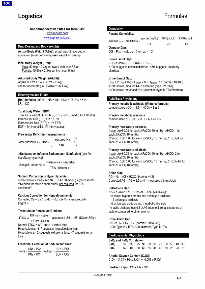
Logistics Formulas
Jonathan Salik
Recommended websites for formulas: www.mdcalc.com
www.nephromatic.com
Drug Dosing and Body Weights Actual Body Weight (ABW): actual weight recorded on admission (most commonly used weight for dosing)
Ideal Body Weight (IBW): Male: 50.0kg + 2.3kg for every inch over 5 feet Female: 45.5kg + 2.3kg per inch over 5 feet
Adjusted Body Weight (AdjBW): AdjBW = IBW + 0.4 x (ABW – IBW); use for obese pts (i.e., if ABW>1.3x IBW)
Electrolytes and Fluids [Na+] in fluids (mEq/L): NS = 154, ½NS = 77, 3% = 514, LR = 130
Total Body Water (TBW): TBW = F x weight; F = 0.6 ♂, 0.5 ♀ (or 0.5 and 0.45 if elderly) Intracellular fluid (ICF) = 2/3 TBW Extracellular fluid (ECF) = 1/3 TBW ECF = 3/4 interstitial, 1/4 intravascular
Free Water Deficit in Hypernatremia:
−×= 1140
Na measuredTBW (L)deficit water
∆Na based on Infusate Sodium (per 1L infusion) [use for hypoNa or hyperNa]:
1liters) (inTBW Na serumNa infusate
Na serum in change+
−=
Sodium Correction in Hyperglycemia: corrected Na = measured Na + (2.4/100 mg/dL) x (glucose–100) **Needed for routine chemistries; not required for ABG specimen**
Calcium Correction for Hypoalbuminemia: Corrected Ca = Ca (mg/dL) + 0.8 x (4.0 − measured alb (mg/dL))
Transtubular Potassium Gradient:
SOsmUOsmKserumKUrine
TTKG/
/= accurate if UNa > 25, UOsm>SOsm
Normal TTKG = 8-9, but >11 with K load Hyperkalemia: <6-7 suggests hypoaldosteronism Hypokalemia: <2 suggests extrarenal loss; >7 suggests renal loss
Fractional Excretion of Sodium and Urea:
FeNa =UNa × PCrPNa × UCr
UCrBUNPCrUUN
FeUrea×
×=
Osmolality Plasma Osmolality:
4.6
(mg/dL) EtOH
2.8
(mg/dL) BUN
18
(mg/dL) glucose(mEq/L) Na2osmcalc +++×=
Osmolar Gap: OG = Posm – calc osm (normal: < 10)
Stool Osmol Gap: SOG = Osmstool – 2 × (Nastool +Kstool) >125: suggests osmotic diarrhea; <50: suggests secretorydiarrhea
Urine Osmol Gap: Uosm = 2(UNa + UK) + Uurea / 2.8 + Uglucose / 18 (normal: 10-100) <150: shows impaired NH4+ excretion (type I/IV RTA) >400: shows increased NH4+ excretion (type II RTA/diarrhea)
Acid/Base Physiology Primary metabolic acidosis (Winter’s formula): compensated pCO2 = 1.5 × HCO3 + 8 ± 2
Primary metabolic alkalosis: compensated pCO2 = 0.7 × HCO3 + 20 ± 5
Primary respiratory acidosis: Acute: ∆pH 0.08 for each ∆PaCO2 10 mmHg; ∆HCO3 1 for each ∆PaCO2 10 mmHg Chronic: ∆pH 0.03 for each ∆PaCO2 10 mmHg; ∆HCO3 4 for each ∆PaCO2 10 mmHg
Primary respiratory alkalosis: Acute: ∆pH 0.08 for each ∆PaCO2 10 mmHg; ∆HCO3 2 for each ∆PaCO2 10 mmHg Chronic: ∆pH 0.03 for each ∆PaCO2 10 mmHg; ∆HCO3 4-5 for each ∆PaCO2 10 mmHg
Anion Gap: AG = Na – (Cl + HCO3) [normal ~12] Corrected AG = AG + 2.5 x (4 – measured alb (mg/dL))
Delta-Delta Gap: ∆∆G = ∆AG* / ∆HCO3 = (AG - 12) / (24-HCO3): <1 mixed hyperchloremic and anion gap acidosis; 1-2 anion gap acidosis>2 anion gap acidosis and metabolic alkalosis
*In lactic acidosis, use 0.6*∆AG (due to ↓ renal clearance oflactate compared to other anions)
Urine Anion Gap: UAG = UNa + UK – UCl (normal: -20 to +20)
>20: Type I/IV RTA; <20: diarrhea/Type II RTA
Cardiovascular Physiology SaO2 and PaO2 Correlation: SaO2 99 98 95 90 88 80 73 60 50 40 30 PaO2 149 100 80 60 55 48 40 30 26 23 18
Arterial Oxygen Content (CaO2): CaO2 = (1.34 x Hb x SaO2) + (0.003 x PaO2)
Cardiac Output: CO = HR x SV
247
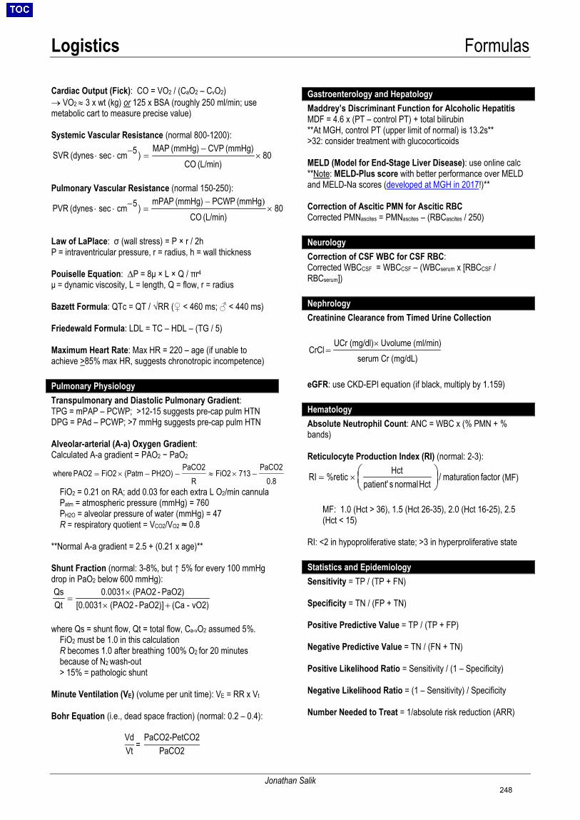
Logistics Formulas
Jonathan Salik
Cardiac Output (Fick): CO = VO2 / (CaO2 – CvO2) → VO2 ≈ 3 x wt (kg) or 125 x BSA (roughly 250 ml/min; usemetabolic cart to measure precise value)
Systemic Vascular Resistance (normal 800-1200):
80(L/min) CO
(mmHg) CVP(mmHg) MAP)5cmsec(dynes SVR ×
−=−⋅⋅
Pulmonary Vascular Resistance (normal 150-250):
80(L/min) CO
(mmHg PCWP(mmHg) mPAP)5cmsec(dynes PVR ×
−=−⋅⋅
)
Law of LaPlace: σ (wall stress) = P × r / 2h P = intraventricular pressure, r = radius, h = wall thickness
Pouiselle Equation: ∆P = 8μ × L × Q / πr4 μ = dynamic viscosity, L = length, Q = flow, r = radius
Bazett Formula: QTc = QT / √RR (♀ < 460 ms; ♂ < 440 ms)
Friedewald Formula: LDL = TC – HDL – (TG / 5)
Maximum Heart Rate: Max HR = 220 – age (if unable to achieve >85% max HR, suggests chronotropic incompetence)
Pulmonary Physiology Transpulmonary and Diastolic Pulmonary Gradient: TPG = mPAP – PCWP; >12-15 suggests pre-cap pulm HTN DPG = PAd – PCWP; >7 mmHg suggests pre-cap pulm HTN
Alveolar-arterial (A-a) Oxygen Gradient: Calculated A-a gradient = PAO2 − PaO2
0.8PaCO2
713FiO2R
PaCO2PH2O)(PatmFiO2PAO2 where −×≈−−×=
FiO2 = 0.21 on RA; add 0.03 for each extra L O2/min cannula Patm = atmospheric pressure (mmHg) = 760 PH2O = alveolar pressure of water (mmHg) = 47 R = respiratory quotient = VCO2/VO2 ≈ 0.8
**Normal A-a gradient = 2.5 + (0.21 x age)**
Shunt Fraction (normal: 3-8%, but ↑ 5% for every 100 mmHg drop in PaO2 below 600 mmHg):
vO2)-(Ca PaO2)] - (PAO2[0.0031PaO2) - (PAO20.0031
QtQs
+××
=
where Qs = shunt flow, Qt = total flow, Ca-vO2 assumed 5%. FiO2 must be 1.0 in this calculation R becomes 1.0 after breathing 100% O2 for 20 minutes because of N2 wash-out > 15% = pathologic shunt
Minute Ventilation (VE) (volume per unit time): VE = RR x Vt
Bohr Equation (i.e., dead space fraction) (normal: 0.2 – 0.4):
VdVt =
PaCO2-PetCO2PaCO2
Gastroenterology and Hepatology Maddrey’s Discriminant Function for Alcoholic Hepatitis MDF = 4.6 x (PT – control PT) + total bilirubin **At MGH, control PT (upper limit of normal) is 13.2s** >32: consider treatment with glucocorticoids
MELD (Model for End-Stage Liver Disease): use online calc **Note: MELD-Plus score with better performance over MELD and MELD-Na scores (developed at MGH in 2017!)**
Correction of Ascitic PMN for Ascitic RBC Corrected PMNascites = PMNascites – (RBCascites / 250)
Neurology Correction of CSF WBC for CSF RBC: Corrected WBCCSF = WBCCSF – (WBCserum x [RBCCSF / RBCserum])
Nephrology Creatinine Clearance from Timed Urine Collection
CrCl =UCr (mg/dl)× Uvolume (ml/min)
serum Cr (mg/dL)
eGFR: use CKD-EPI equation (if black, multiply by 1.159)
Hematology Absolute Neutrophil Count: ANC = WBC x (% PMN + % bands)
Reticulocyte Production Index (RI) (normal: 2-3):
factor maturation / Hct normal spatient'
Hct%reticRI
×= (MF)
MF: 1.0 (Hct > 36), 1.5 (Hct 26-35), 2.0 (Hct 16-25), 2.5 (Hct < 15)
RI: <2 in hypoproliferative state; >3 in hyperproliferative state
Statistics and Epidemiology Sensitivity = TP / (TP + FN)
Specificity = TN / (FP + TN)
Positive Predictive Value = TP / (TP + FP)
Negative Predictive Value = TN / (FN + TN)
Positive Likelihood Ratio = Sensitivity / (1 – Specificity)
Negative Likelihood Ratio = (1 – Sensitivity) / Specificity
Number Needed to Treat = 1/absolute risk reduction (ARR)
248
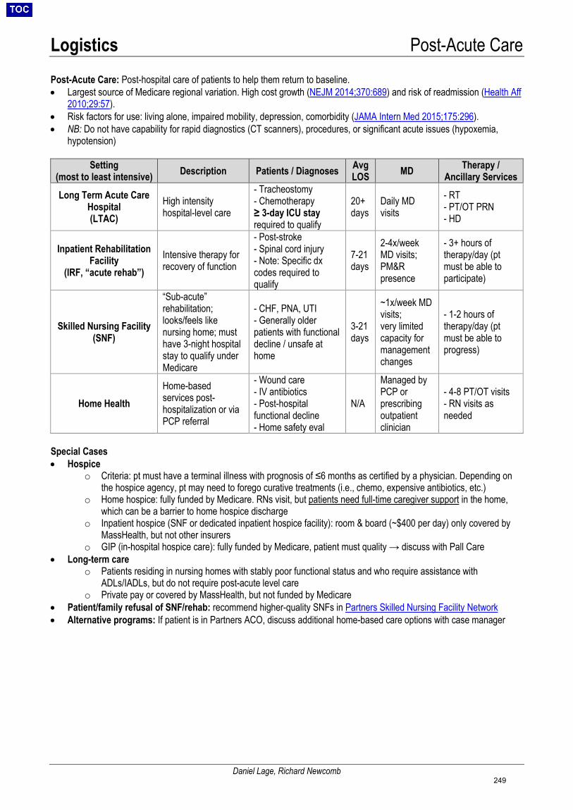
Logistics Post-Acute Care
Daniel Lage, Richard Newcomb
Post-Acute Care: Post-hospital care of patients to help them return to baseline. • Largest source of Medicare regional variation. High cost growth (NEJM 2014;370:689) and risk of readmission (Health Aff
2010;29:57).• Risk factors for use: living alone, impaired mobility, depression, comorbidity (JAMA Intern Med 2015;175:296).• NB: Do not have capability for rapid diagnostics (CT scanners), procedures, or significant acute issues (hypoxemia,
hypotension)
Setting (most to least intensive) Description Patients / Diagnoses Avg
LOS MD Therapy / Ancillary Services
Long Term Acute Care Hospital (LTAC)
High intensity hospital-level care
- Tracheostomy- Chemotherapy≥ 3-day ICU stayrequired to qualify
20+ days
Daily MD visits
- RT- PT/OT PRN- HD
Inpatient Rehabilitation Facility
(IRF, “acute rehab”) Intensive therapy for recovery of function
- Post-stroke- Spinal cord injury- Note: Specific dxcodes required toqualify
7-21days
2-4x/weekMD visits;PM&Rpresence
- 3+ hours oftherapy/day (ptmust be able toparticipate)
Skilled Nursing Facility (SNF)
“Sub-acute” rehabilitation; looks/feels like nursing home; must have 3-night hospital stay to qualify under Medicare
- CHF, PNA, UTI- Generally olderpatients with functionaldecline / unsafe athome
3-21days
~1x/week MD visits; very limited capacity for management changes
- 1-2 hours oftherapy/day (ptmust be able toprogress)
Home Health Home-based services post-hospitalization or via PCP referral
- Wound care- IV antibiotics- Post-hospitalfunctional decline- Home safety eval
N/A
Managed by PCP or prescribing outpatient clinician
- 4-8 PT/OT visits- RN visits asneeded
Special Cases • Hospice
o Criteria: pt must have a terminal illness with prognosis of ≤6 months as certified by a physician. Depending onthe hospice agency, pt may need to forego curative treatments (i.e., chemo, expensive antibiotics, etc.)
o Home hospice: fully funded by Medicare. RNs visit, but patients need full-time caregiver support in the home,which can be a barrier to home hospice discharge
o Inpatient hospice (SNF or dedicated inpatient hospice facility): room & board (~$400 per day) only covered byMassHealth, but not other insurers
o GIP (in-hospital hospice care): fully funded by Medicare, patient must quality → discuss with Pall Care• Long-term care
o Patients residing in nursing homes with stably poor functional status and who require assistance withADLs/IADLs, but do not require post-acute level care
o Private pay or covered by MassHealth, but not funded by Medicare• Patient/family refusal of SNF/rehab: recommend higher-quality SNFs in Partners Skilled Nursing Facility Network• Alternative programs: If patient is in Partners ACO, discuss additional home-based care options with case manager
249

Logistics Discharge Summaries
Ryan Flanagan
General Considerations • Must be completed at the time of discharge for all patients being discharged to a facility or home with VNA services• Can be completed within 24 hours of discharge for patients being discharged home without services• CC copies to attending of record, PCP, and outpatient subspecialists
Discharge Summary Components • D/C Doc Checklist: Checklist of all items required for patient discharge. As you go through the Discharge Summary tab,
click on the “Refresh” button to move completed items from the “Not Completed” to “Completed” column. All componentsmust be completed prior to a discharge to a facility. Only the “D/C Order Rec” needs to be completed for a patient to bedischarged home.
• D/C Order Rec:o Review Prior to Admission Medications: Update home medication list. If they were not accurately verified at the
time of admission, the discharge medication list will be inaccurate and may be confusing to a patient withregards to which medications to modify, continue, or discontinue.
o Discharge Problem List: Add all relevant hospital problems and diagnoses – this is important for a patient’stransition to outpatient providers. You can change the “Principal” diagnosis for billing purposes. “Resolved”problems will not remain on a patient’s problem list after they are discharged.
o Reconcile Meds for Discharge: Choose whether to modify, resume, or stop taking each pre-admissionmedication. You can also prescribe new medications that are being given inpatient. Select patient’s preferredpharmacy for e-prescriptions.
o Place New Orders: Select appropriate discharge disposition. Under “Order Sets and Pathways”, select “GeneralAdult Discharge Order Set” and complete. Do NOT complete “Referral to Home Health VNA” or the “face to face”– this will be done by case management.
• AVS Pt Instructions:o Reason for admission: Briefly state the patient's medical diagnosis/reason for admission in 1-3 words using
patient-friendly language (eg: pneumonia, low sodium level, urinary tract infection, fainting, etc.).o Important events, results, medication changes, and instructions: This is meant FOR THE PATIENT to clarify the
reason for his/her hospitalization, highlight important testing/interventions, and briefly explain dischargeinstructions/follow-up plans. Important to consider health literacy, native language, and language fluency foreach individual patient.
• Additional Patient Instructions: You can leave this section blank. Sometimes residents add wound care or post-procedureinstructions here; specialists may leave specific instructions here as well.
• Scheduled Partners Appts: This will automatically populate with upcoming scheduled Partners appointments.• Follow-Up: All relevant non-Partners appointments should be manually entered here. You can also enter appointments
that have yet to be made with a phone number for the patient to call.• Brief Summary: Update the one-liner one last time. It is helpful to make this a brief but comprehensive summary of the
entire hospitalization to preface the details in the hospital course.• Hospital Course: Use the admission H&P and most recent progress note to identify which problems should be included in
the Hospital Course. Lump rather than split your hash-tagged problems into paragraph form. For each problem, include:1)relevant presenting symptoms and exam findings, 2) labs, imaging, and studies used to diagnose the problem, 3)consultant recommendations, 4) treatment course and discharge plan, 5) post-hospital follow-up items (including repeatlabs and f/u incidentalomas). Less is more – focus on the big-picture overview and the clinical reasoning thatguided your decision-making and management of each problem. Try to avoid MGH-specific abbreviations (eg:RUQUS, LENIs). Some find it helpful to copy and paste the admission H&P below the hospital course, especially if thepatient is followed by a provider outside of Partners.
• Discharge Exam: Copy and paste exam from last progress note, but make sure this is up to date.• Provider Follow Ups: Identify key lab, imaging, and other items that a provider needs to follow up after discharge. Please
note that any pending pathology and send out tests should be included here as they do not automatically pull into thepending results section. Be sure to review all imaging results and consultant recommendations for follow-up. Someresidents comment on medication titration suggestions and key medication changes.
• Finalize DC Summary: Once the discharge orders are signed, a new button appears under this section that allows you tocreate the actual discharge summary that will be filed in the Epic. Click the button and then click into the note writingspace that opens on the right. Press F2 and select the general medicine discharge summary template from the drop-down menu. This will create a discharge summary containing the contents of the other free text fields already completed.You can edit this once signed as long as your attending has not already signed the addendum.
250
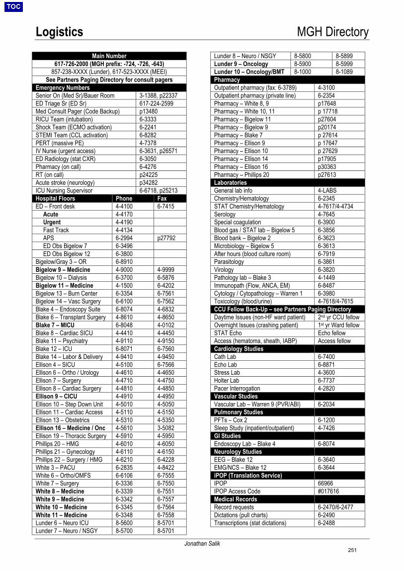
Logistics MGH Directory
Jonathan Salik
Main Number 617-726-2000 (MGH prefix: -724, -726, -643)
857-238-XXXX (Lunder), 617-523-XXXX (MEEI)See Partners Paging Directory for consult pagers
Emergency Numbers Senior On (Med Sr)/Bauer Room 3-1388, p22337ED Triage Sr (ED Sr) 617-224-2599Med Consult Pager (Code Backup) p13480 RICU Team (intubation) 6-3333Shock Team (ECMO activation) 6-2241STEMI Team (CCL activation) 6-8282PERT (massive PE) 4-7378IV Nurse (urgent access) 6-3631, p26571ED Radiology (stat CXR) 6-3050Pharmacy (on call) 6-4276RT (on call) p24225 Acute stroke (neurology) p34282 ICU Nursing Supervisor 6-6718, p25213Hospital Floors Phone Fax ED – Front desk 4-4100 6-7415 Acute 4-4170 Urgent 4-4190 Fast Track 4-4134 APS 6-2994 p27792 ED Obs Bigelow 7 6-3496 ED Obs Bigelow 12 6-3800Bigelow/Gray 3 – OR 6-8910Bigelow 9 – Medicine 4-9000 4-9999Bigelow 10 – Dialysis 6-3700 6-5876Bigelow 11 – Medicine 4-1500 6-4202Bigelow 13 – Burn Center 6-3354 6-7561Bigelow 14 – Vasc Surgery 6-6100 6-7562Blake 4 – Endoscopy Suite 6-8074 4-6832Blake 6 – Transplant Surgery 4-8610 4-8650Blake 7 – MICU 6-8048 4-0102Blake 8 – Cardiac SICU 4-4410 4-4450Blake 11 – Psychiatry 4-9110 4-9150Blake 12 – ICU 6-8071 6-7560Blake 14 – Labor & Delivery 4-9410 4-9450Ellison 4 – SICU 4-5100 6-7566Ellison 6 – Ortho / Urology 4-4610 4-4650Ellison 7 – Surgery 4-4710 4-4750Ellison 8 – Cardiac Surgery 4-4810 4-4850Ellison 9 – CICU 4-4910 4-4950Ellison 10 – Step Down Unit 4-5010 4-5050Ellison 11 – Cardiac Access 4-5110 4-5150Ellison 13 – Obstetrics 4-5310 4-5350Ellison 16 – Medicine / Onc 4-5610 3-5082Ellison 19 – Thoracic Surgery 4-5910 4-5950Phillips 20 – HMG 4-6010 4-6050Phillips 21 – Gynecology 4-6110 4-6150Phillips 22 – Surgery / HMG 4-6210 6-4228White 3 – PACU 6-2835 4-8422White 6 – Ortho/OMFS 6-6106 6-7555White 7 – Surgery 6-3336 6-7550White 8 – Medicine 6-3339 6-7551White 9 – Medicine 6-3342 6-7557White 10 – Medicine 6-3345 6-7564White 11 – Medicine 6-3348 6-7558Lunder 6 – Neuro ICU 8-5600 8-5701Lunder 7 – Neuro / NSGY 8-5700 8-5701
Lunder 8 – Neuro / NSGY 8-5800 8-5899Lunder 9 – Oncology 8-5900 8-5999Lunder 10 – Oncology/BMT 8-1000 8-1089Pharmacy Outpatient pharmacy (fax: 6-3789) 4-3100Outpatient pharmacy (private line) 6-2354Pharmacy – White 8, 9 p17648 Pharmacy – White 10, 11 p 17718 Pharmacy – Bigelow 11 p27604 Pharmacy – Bigelow 9 p20174 Pharmacy – Blake 7 p 27614 Pharmacy – Ellison 9 p 17647 Pharmacy – Ellison 10 p 27629 Pharmacy – Ellison 14 p17905 Pharmacy – Ellison 16 p30363 Pharmacy – Phillips 20 p27613 Laboratories General lab info 4-LABSChemistry/Hematology 6-2345STAT Chemistry/Hematology 4-7617/4-4734Serology 4-7645Special coagulation 6-3900Blood gas / STAT lab – Bigelow 5 6-3856Blood bank – Bigelow 2 6-3623Microbiology – Bigelow 5 6-3613After hours (blood culture room) 6-7919Parasitology 6-3861Virology 6-3820Pathology lab – Blake 3 4-1449Immunopath (Flow, ANCA, EM) 6-8487Cytology / Cytopathology – Warren 1 6-3980Toxicology (blood/urine) 4-7618/4-7615CCU Fellow Back-Up – see Partners Paging Directory Daytime Issues (non-HF ward patient) 2nd yr CCU fellow Overnight Issues (crashing patient) 1st yr Ward fellow STAT Echo Echo fellow Access (hematoma, sheath, IABP) Access fellow Cardiology Studies Cath Lab 6-7400Echo Lab 6-8871Stress Lab 4-3600Holter Lab 6-7737Pacer Interrogation 4-2820Vascular Studies Vascular Lab – Warren 9 (PVR/ABI) 6-2034Pulmonary Studies PFTs – Cox 2 6-1200Sleep Study (inpatient/outpatient) 4-7426GI Studies Endoscopy Lab – Blake 4 6-8074Neurology Studies EEG – Blake 12 6-3640EMG/NCS – Blake 12 6-3644iPOP (Translation Service) IPOP 66966 IPOP Access Code #017616 Medical Records Record requests 6-2470/6-2477Dictations (pull charts) 6-2490Transcriptions (stat dictations) 6-2488
251
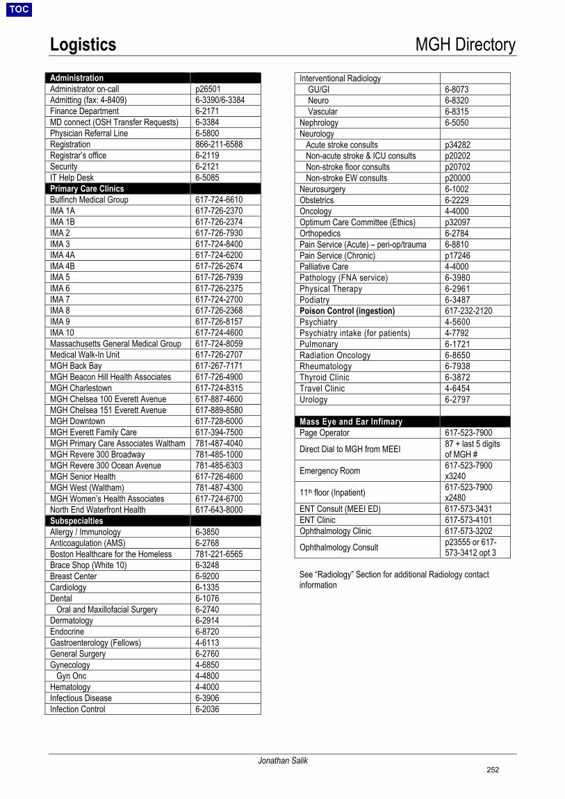
Logistics MGH Directory
Jonathan Salik
Administration Administrator on-call p26501 Admitting (fax: 4-8409) 6-3390/6-3384Finance Department 6-2171MD connect (OSH Transfer Requests) 6-3384Physician Referral Line 6-5800Registration 866-211-6588Registrar’s office 6-2119Security 6-2121IT Help Desk 6-5085Primary Care Clinics Bulfinch Medical Group 617-724-6610IMA 1A 617-726-2370IMA 1B 617-726-2374IMA 2 617-726-7930IMA 3 617-724-8400IMA 4A 617-724-6200IMA 4B 617-726-2674IMA 5 617-726-7939IMA 6 617-726-2375IMA 7 617-724-2700IMA 8 617-726-2368IMA 9 617-726-8157IMA 10 617-724-4600Massachusetts General Medical Group 617-724-8059Medical Walk-In Unit 617-726-2707MGH Back Bay 617-267-7171MGH Beacon Hill Health Associates 617-726-4900MGH Charlestown 617-724-8315MGH Chelsea 100 Everett Avenue 617-887-4600MGH Chelsea 151 Everett Avenue 617-889-8580MGH Downtown 617-728-6000MGH Everett Family Care 617-394-7500MGH Primary Care Associates Waltham 781-487-4040MGH Revere 300 Broadway 781-485-1000MGH Revere 300 Ocean Avenue 781-485-6303MGH Senior Health 617-726-4600MGH West (Waltham) 781-487-4300MGH Women’s Health Associates 617-724-6700North End Waterfront Health 617-643-8000Subspecialties Allergy / Immunology 6-3850Anticoagulation (AMS) 6-2768Boston Healthcare for the Homeless 781-221-6565Brace Shop (White 10) 6-3248Breast Center 6-9200Cardiology 6-1335Dental 6-1076 Oral and Maxillofacial Surgery 6-2740Dermatology 6-2914Endocrine 6-8720Gastroenterology (Fellows) 4-6113General Surgery 6-2760Gynecology 4-6850 Gyn Onc 4-4800Hematology 4-4000Infectious Disease 6-3906Infection Control 6-2036
Interventional Radiology GU/GI 6-8073 Neuro 6-8320 Vascular 6-8315Nephrology 6-5050Neurology Acute stroke consults p34282 Non-acute stroke & ICU consults p20202 Non-stroke floor consults p20702 Non-stroke EW consults p20000 Neurosurgery 6-1002Obstetrics 6-2229Oncology 4-4000Optimum Care Committee (Ethics) p32097 Orthopedics 6-2784Pain Service (Acute) – peri-op/trauma 6-8810Pain Service (Chronic) p17246 Palliative Care 4-4000Pathology (FNA service) 6-3980Physical Therapy 6-2961Podiatry 6-3487Poison Control (ingestion) 617-232-2120Psychiatry 4-5600Psychiatry intake (for patients) 4-7792Pulmonary 6-1721Radiation Oncology 6-8650Rheumatology 6-7938Thyroid Clinic 6-3872Travel Clinic 4-6454Urology 6-2797
Mass Eye and Ear Infimary Page Operator 617-523-7900
Direct Dial to MGH from MEEI 87 + last 5 digits of MGH #
Emergency Room 617-523-7900x3240
11th floor (Inpatient) 617-523-7900x2480
ENT Consult (MEEI ED) 617-573-3431ENT Clinic 617-573-4101Ophthalmology Clinic 617-573-3202
Ophthalmology Consult p23555 or 617-573-3412 opt 3
See “Radiology” Section for additional Radiology contact information
252

Logistics NWH Directory
Kelsey Lau-Min
Main Number 617-243-6000
Key Pagers 4 Usen Admitting SAR/NF p56789 4 West Admitting SAR/NF p56788 Locum Tenens/Covering Intensivist p57651 Hospitalist p51253 Hospital Floors ED 6193 / 6194 ICU (2nd floor) 6587 3 West 6363 4 Usen 6459 4 West 6400 6 Usen 6307 6 East 1670 Laboratories Main Lab (for add-ons) 6300 Hematology 6095 Blood Bank 6091 Chemistry 8389 Urine Studies 6090 Microbiology 6096 Pathology 6140 Radiology Main Number 6600 / 6076 Radiology Reading Room 6162 ED Radiology 6185 Ultrasound 6581 CT (main) 6725 CT (ED and after hours) 6505 MRI 6217 PET 6334 Nuclear Medicine 6087 Interventional Radiology 6800 / 3761 Night Watch 617-732-5657 Cardiac Studies ECG 6229 Echo 6231 / 2665 ETT or Nuclear Stress 5375 / 6229 /
6087 Ancillary Staff Nursing Supervisor p57711 Pharmacy 6012 Respiratory Therapy 6213 Phlebotomy 5903 Speech Language Pathology 6548 Infection Control 6282 Case Management/Social Work 6695 Chaplain 6634 Interpreter Services 6698
Miscellaneous OR 6289 PACU 6295 GI Unit 6151 Dialysis 6203 Pulmonary Lab 6127 EEG/EMG 6624 Anticoagulation Clinic 6147 Cancer Center 1230 Cardiovascular Health Center 7100 Infusion Clinic 6350 Occupational Health 6168 Admissions 5500 CareFinder (new NWH PCP) 6566 MDConnect (transfer to MGH) 877-637-3337 DOM Office (6 South) 6467 Chief Medical Resident 6470 Outside Calls 617-243-6841 Getting to NWH (2014 Washington St, Newton, MA) • You will receive transportation information prior to
your NWH rotation • Note that all transportation stipends are taxed • Driving
o Stipend covers gas and tolls o On day 1, park in patient garage for ~$10 o Pay $15 cash/check at parking/security
office for pass to park in employee garage behind West Entrance
• Ride-sharing o If using a ride-sharing service, consider
carpooling to stay within stipend • Public transportation
o Take MBTA Green Line D outbound train to Riverside -> Woodland stop -> NWH is 2 blocks to the left
• Single day/night coverage (eg: NF coverage, HIT) o Use Uber for Business → MGH DOM
Internal Medicine Residency Program account
253