hormone predictors of abnormal bone microarchitecture in women with anorexia nervosa
TRANSCRIPT

Hormone Predictors of Abnormal Bone Microarchitecture InWomen with Anorexia Nervosa
Elizabeth A. Lawson1, Karen K. Miller1, Miriam A. Bredella2, Catherine Phan2, MadhusmitaMisra1, Erinne Meenaghan1, Lauren Rosenblum1, Daniel Donoho1, Rajiv Gupta2, and AnneKlibanski11 Neuroendocrine Unit, Massachusetts General Hospital and Harvard Medical School, Boston, MA021142 Department of Radiology, Massachusetts General Hospital and Harvard Medical School, Boston,MA 02114
AbstractOsteopenia is a complication of anorexia nervosa (AN) associated with a two- to three-fold increasein fractures. Nutritional deficits and hormonal abnormalities are thought to mediate AN-induced boneloss. Alterations in bone microarchitecture may explain fracture risk independent of bone mineraldensity (BMD). Advances in CT imaging now allow for noninvasive evaluation of trabecularmicrostructure at peripheral sites in vivo. Few data are available regarding bone microarchitecturein AN. We therefore performed a cross-sectional study of 23 women (12 with AN and 11 healthycontrols) to determine hormonal predictors of trabecular bone microarchitecture. Outcome measuresincluded bone microarchitectural parameters at the ultradistal radius by flat panel Volume CT(fpVCT); BMD at the PA and lateral spine, total hip, femoral neck and ultradistal radius by dualenergy X-ray absorptiometry (DXA); and IGF-I, leptin, estradiol, testosterone and free testosteronelevels. Bone microarchitectural measures, including apparent (app.) bone volume fraction, app.trabecular thickness, and app. trabecular number, were reduced (p<0.03) and app. trabecular spacingwas increased (p=0.02) in AN vs. controls. Decreased structural integrity at the ultradistal radius wasassociated with decreased BMD at all sites (p=0.05) except for total hip. IGF-I, leptin, testosteroneand free testosterone levels predicted bone microarchitecture. All associations between both IGF-Iand leptin levels and bone microarchitectural parameters, and most associations between androgenlevels and microarchitecture remained significant after controlling for body mass index. Weconcluded that bone microarchitecture is abnormal in women with AN. Endogenous IGF-I, leptinand androgen levels predict bone microarchitecture independent of BMI.
KeywordsAnorexia nervosa; IGF-I; leptin; androgens; flat-panel volume CT
Corresponding Author: Elizabeth A. Lawson, M.D., Neuroendocrine Unit, Bulfinch 457B, Massachusetts General Hospital, Boston, MA02114, [email protected], Tel: 617-726-3870, Fax: 617-726-5072.The authors have no conflicts to declare.Publisher's Disclaimer: This is a PDF file of an unedited manuscript that has been accepted for publication. As a service to our customerswe are providing this early version of the manuscript. The manuscript will undergo copyediting, typesetting, and review of the resultingproof before it is published in its final citable form. Please note that during the production process errors may be discovered which couldaffect the content, and all legal disclaimers that apply to the journal pertain.
NIH Public AccessAuthor ManuscriptBone. Author manuscript; available in PMC 2011 February 1.
Published in final edited form as:Bone. 2010 February ; 46(2): 458. doi:10.1016/j.bone.2009.09.005.
NIH
-PA Author Manuscript
NIH
-PA Author Manuscript
NIH
-PA Author Manuscript

IntroductionAnorexia nervosa (AN) is a psychiatric disease affecting 0.3% of the female population, andis characterized by self-induced chronic starvation and associated with severe bone loss andincreased risk of fractures [1]. In a study of outpatient women with AN, mean age 24 years,92% had osteopenia and 38% had osteoporosis by dual energy X-ray absorptiometry (DXA)[2]. A two- to threefold increase in fracture risk has been reported in this population [3,4]. Themechanism for AN-induced bone loss is multifactorial, and a low formation, high resorptionstate has been identified [5]. Low endogenous estrogen and testosterone secretion anddecreased levels of nutritionally-dependent hormones, including IGF-I and leptin, have beenimplicated [6]. IGF-I, a nutritionally regulated anabolic hormone that is low in states of chronicstarvation, is thought to be a particularly important mediator of AN-induced bone loss. Lowlevels of IGF-I in women with AN are associated with decreased levels of bone formation[5]. Administration of rhIGF-I increases markers of bone formation and, in estrogen-treatedpatients, is the only therapy that has been shown in a randomized, placebo-controlled study toimprove bone mineral density (BMD) in women with AN [7]. In contrast, studies of estrogen-progestin therapy, effective in treating postmenopausal osteoporosis, have failed to showefficacy in preventing or improving AN-associated bone loss [7,8]. Serum androgen levelshave been demonstrated to be low in women with AN [9] and short-term administration oflow-dose testosterone increases procollagen type I C-terminal peptide (PICP), a marker of boneformation [10].
As in other populations at risk for osteoporosis, BMD has been the primary method used toevaluate skeletal integrity in women with AN in both clinical and research settings. However,it is increasingly clear that other measures of bone quality, particularly parameters ofmicroarchitecture, are significant independent predictors of fracture risk in other populations[11–14] and therefore may be important in AN. Newer imaging technologies, including ultra-high resolution flat panel based volume CT (fpVCT), allow for noninvasive visualization ofbone microstructure [15,16]. There is limited information about bone microarchitecture, andto our knowledge, there are no studies investigating the role of hormones in modulatingmicroarchitectural parameters in AN. This study investigates the relationship betweenstructural integrity of bone utilizing fpVCT and endocrine abnormalities in AN. Specifically,we hypothesized that IGF-I and other nutritionally-dependent hormones would predictparameters of bone microstructure.
Subjects and MethodsProtocol
23 women aged 18–45 years, including 12 women with AN and 11 HC of similar age, werestudied. All women with AN were ambulatory and fulfilled all Diagnostic and StatisticalManual IV (DSM-IV) criteria for AN, including weight <85% of ideal body weight (7).Exclusion criteria for AN and HC included pregnancy, diabetes mellitus, thyroid, cardiac, liveror renal disease, or medications known to affect bone metabolism, including estrogens,progestins, androgens, bisphosphonates or recombinant parathyroid hormone. In addition,exclusion criteria for HC included history of disordered eating or amenorrhea. Subjects werereferred to the study by eating disorder providers in the New England area or recruited throughadvertisements. The study was approved by the Partners Health Care Institutional ReviewBoard, and written, informed consent was obtained from all subjects. Study visits took placeat the Clinical Research Center at Massachusetts General Hospital. History, physical exam,and serum samples were obtained. Research dieticians measured metabolic weight and heightand calculated body mass index (kg/m2). Body composition and BMD at the AP spine (L1–L4), lateral spine (L2–L4), total hip, femoral neck and ultradistal radius were determined byDXA (Hologic QDR 4500, Waltham, MA).
Lawson et al. Page 2
Bone. Author manuscript; available in PMC 2011 February 1.
NIH
-PA Author Manuscript
NIH
-PA Author Manuscript
NIH
-PA Author Manuscript
Measurement of bone microarchitectureBone microarchitecture at the nondominant ultradistal radius, a site of predominantlytrabecular bone previously shown to predict vertebral fractures [17], was assessed using ultra-high resolution flat panel based volume CT (fpVCT) (Siemens, Forchheim, Germany). ThefpVCT prototype consists of a CT gantry with a bore diameter of 40 cm integrated with amodified X-ray tube and a 2-D digital flat-panel detector system. The imaging was conductedat 100kV, 30mA, with pulsed X-ray source in the 2x2 binning mode (voxel size 0.2 × 0.2 ×0.2 mm3). A 50% X-ray duty cycle was used. Scan time was 20 seconds. The radiation doseof a distal radius fpVCT scan was 0.027 mSv. Trabecular structure parameters obtained byfpVCT were calculated using MicroView software (GE Healthcare, Waukesha, WI). For eachdistal radius, a 3-dimensional region of interest (ROI) was defined within the ultradistal radiusto cover a maximum area of trabecular bone without including any cortical bone within theROI. To improve reproducibility, all ROIs were placed by one observer. The observer wasblinded to the patient status (AN vs. HC). Trabecular bone was then segmented from marrowwith the individual threshold defined by the auto-threshold function of MicroView.
Apparent (app.) measures of trabecular structure were calculated, including app. trabecularbone volume (BV) fraction [app. BV/app. trabecular volume (TV),%], app. trabecular number(app. TbN, mm−3) defined as the inverse of the mean spacing, app. trabecular thickness (app.TbTh, mm) and separation (app. TbSp, mm), derived from app. BV/TV and app. TbN usingstandard methods from histomorphometry (i.e. TbTh=(BV/TV)/TbN and TbSp=(1-BV/TV)/TbN). These parameters are defined as “apparent” because the spatial resolution is lower thanthat required for standard bone histomorphometry [18].
Biochemical AnalysisSerum IGF-I levels were measured using an Immulite 2000 automated immunoanalyzer(Diagnostic Products Corporation, Inc. [DPC], Los Angeles, CA), by a solid-phase enzyme-labeled chemiluminescent immunometric assay, with an intra-assay coefficient of variation(CV) of 2.3 – 3.9 and an analytical sensitivity of 20 ng/mL. Leptin levels were measured usinga radioimmunoassay (RIA) kit from LINCO Research, a division of Millipore Inc. (St. Charles,MO). The intra-assay CV was 8.3 – 3.4%, and the sensitivity was 0.5 ng/mL. Serumtestosterone was measured by RIA kit (DPC, Inc.) with a minimum detection limit of 2 ng/dLand an intra-assay CV of 4.1–10.5%. Sex hormone binding globulin (SHBG) was measuredby immunoradiometric assay (DPC, Inc.), with a minimum detection limit of 0.5 nmol/L andan intra-assay CV of 2.8 – 5.3%. Free testosterone was calculated from total testosterone andSHBG by an equation based on the laws of mass action, which has been validated in comparisonto free testosterone by equilibrium dialysis in women [19]. Estradiol levels were measuredusing a Chemiluminescent Microparticle Immunoassay kit from Architect (AbbottLaboratories, Abbott Park, IL), with a within-run CV of 1.5–6.4% for concentrations of 45–192 pg/mL and a functional sensitivity = 14 pg/mL.
Data AnalysisThe means and standard error of the means (SEM) of clinical characteristics, hormone levels,BMD, and bone microstructure parameters were calculated for the AN and HC groups. Thesignificance of differences between groups was calculated using the Student’s t test at the 5%significance level. Linear regression analyses between trabecular structure parameters andBMD measurements at the different sites, and between trabecular structure, DXA and hormonelevels were performed, and Pearson correlation coefficients are reported. Multivariate least-square analyses were constructed to control for BMI. Variables were log-transformed beforebeing entered into the regression analyses. Statistical analysis was performed using JMPsoftware (SAS Institute, Cary, NC).
Lawson et al. Page 3
Bone. Author manuscript; available in PMC 2011 February 1.
NIH
-PA Author Manuscript
NIH
-PA Author Manuscript
NIH
-PA Author Manuscript

ResultsPatient Characteristics
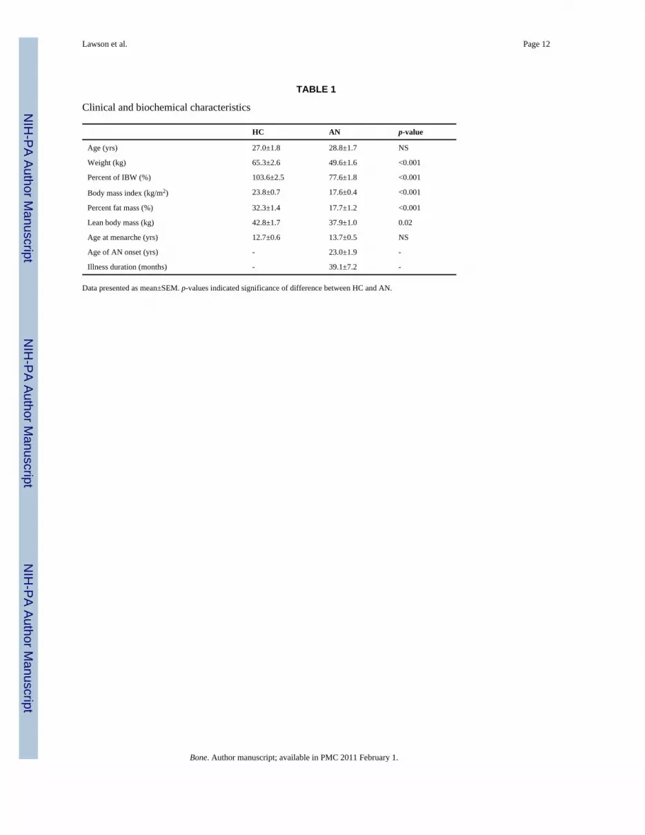
Baseline clinical characteristics are described in Table 1. Women with AN and HC were ofsimilar age. As expected, women with AN had lower weight, percent of ideal body weight(IBW), body mass index (BMI), percent fat mass, and lean body mass.
Bone Mineral Density and Trabecular MicroarchitectureDifferences in BMD and trabecular microarchitecture between the groups are shown in Figures1 and 2. Women with AN had lower mean BMD Z-scores than HC at all sites. Mean app. BV/TV, app. TbTh, and TbN were significantly lower and TbSp was higher in women with ANcompared to HC. Table 2 summarizes the relationship between trabecular microarchitectureat the ultradistal radius as measured by fpVCT and bone mineral density by DXA. Decreasedstructural integrity at the radius was significantly associated with lower BMD at all sites exceptfor total hip.
Relationship Between Bone Parameters and HormonesAssociations between bone parameters and hormones are shown in Table 3. IGF-I and leptinlevels predicted greater structural integrity by all microarchitectural parameters measured.After controlling for BMI, the associations between both IGF-I and leptin and all of thesemeasures were statistically significant. Testosterone levels were positively associated withbone volume fraction and trabecular thickness and inversely associated with trabecular spacing.The association with bone volume fraction remained significant after controlling for BMI. Freetestosterone was positively associated with bone volume fraction and trabecular thickness.These associations remained significant after controlling for BMI. Estradiol levels were notsignificantly associated with microarchitectural parameters, though they were associated withBMD at several sites before, but not after, controlling for BMI.
DiscussionHormonal abnormalities associated with severe undernutrition are important in thepathogenesis of bone loss in AN. However, they have not previously been examined in relationto trabecular microarchitecture, an important determinant of fracture risk. In contrast topostmenopausal osteoporosis where estrogen is the key factor, AN-mediated bone loss is drivenby chronic starvation. We show that nutritionally-mediated hormones, including IGF-I andleptin, and androgen levels predict bone microstructure in women with AN.
Novel noninvasive imaging technology allowing for visualization of trabecularmicroarchitecture in humans introduces a new paradigm for evaluating bone quality. Emergingdata indicate that bone microarchitecture offers important information about bone fragility[11–14]. Several studies have demonstrated increased fracture risk associated with abnormalbone microarchitecture and normal BMD, suggesting that microstructure may be a moresensitive measure than BMD in assessing such risk [20]. Parameters of bone microarchitectureare also abnormal in postmenopausal and corticosteroid-induced osteoporosis [21,22] and areassociated with increased fracture risk independent of BMD [23]. A recent study of 82postmenopausal women found that trabecular microstructure was a better predictor of recentspinal fracture than BMD [24]. Current treatments for osteoporosis, including bisphosphonatesand parathyroid hormone [25–27], have been shown to preserve or improve bonemicroarchitecture, a mechanism that may contribute to the reduced fracture risk in patientsprescribed these medications.
Lawson et al. Page 4
Bone. Author manuscript; available in PMC 2011 February 1.
NIH
-PA Author Manuscript
NIH
-PA Author Manuscript
NIH
-PA Author Manuscript

IGF-I is exquisitely sensitive to nutrient intake and has potent autocrine and paracrine effectson bone growth and remodeling. Experiments in cultured rat calvariae have shown that IGF-Istimulates DNA and collagen synthesis while reducing collagen degradation [28,29]. In vitroand in vivo research in animal models indicates that IGF-I is a critical factor in promotingnormal longitudinal bone growth [30–32]. IGF-I is anabolic to bone, associated with boneformation and increased BMD, and is predictive of fracture risk [33–35]. AN is characterizedby low levels of IGF-I and correspondingly reduced markers of bone formation [5,36,37].Recombinant hIGF-I increases bone formation and is the only medical therapy to date shownin a randomized, placebo-controlled study to improve BMD in estrogen-treated women withAN [5,7]. A population-based study of 205 women and 269 men found that IGF-I levels werepositively associated with trabecular thickness and negatively associated with trabecularnumber in young men, but not in older men or in women [38]. In contrast, in our study ofwomen with AN, we demonstrate a clear relationship between IGF-I levels and favorabletrabecular microarchitecture at the ultradistal radius, independent of BMI. This may be due tothe importance of IGF-I deficiency in the pathogenesis of bone loss in our population. Aprospective study comparing bone microarchitecture in women with AN receiving rhIGF-I tothose on placebo will be important to establish causality.
Serum levels of leptin, an adipocyte-derived anorexigenic hormone, are low in women withAN, likely representing a physiologic adaptation to starvation as levels normalize with weightrecovery [39,40]. Although leptin is known to modulate bone turnover through complex centraland peripheral effects, the overall influence of leptin on bone is unclear and may differ basedon skeletal site [41]. Increased BMD in obesity [42], low BMD in underweight populations[2,43], and an association between BMD and BMI [44] argue for a protective effect of leptinon bone in humans. However, mouse models of leptin deficiency demonstrate increasedmarkers of bone formation and bone mass despite hypogonadism, and central administrationof leptin reduces bone mass in leptin-deficient and wild type mice [45]. In contrast, in vitroand in vivo animal data suggest that peripheral leptin is anabolic to bone by promotingosteoblast proliferation and inhibiting osteoclastogenesis [46–48]. In our study, leptinremained a significant positive predictor of bone microarchitecture after controlling for BMI.
Gonadal steroids, specifically testosterone and estradiol are both important determinants ofBMD. Testosterone and estradiol levels were associated with bone microarchitecture in healthyolder men and women, but not in young men and women aged 20–39 years [38]. Hypogonadalmen have been shown to have decreased trabecular networks and fraction of bone volume atthe distal tibia compared to eugonadal men as assessed by μMRI [49]. A study of tenhypogonadal men showed that with testosterone replacement, parameters of bonemicroarchitecture using this same technique improved [50]. A prospective observational studyof early postmenopausal women reported that those receiving estrogen maintained or improvedtheir bone microarchitecture at the distal tibia by μMRI over 12 to 24 months, while those whowere not on estrogen therapy demonstrated deterioration in microarchitecture [51]. In AN, bothtotal and free testosterone [9] are low, and low-dose transdermal testosterone administrationincreases surrogate markers of bone metabolism in this population [10], although effects onbone mass or microarchitecture are not known. Two randomized, placebo-controlled studieshave shown that estrogen administration is not effective at increasing BMD in women withAN [7,52]. In the current study, we report that testosterone predicts bone volume fraction andfree testosterone predicts bone volume fraction and trabecular thickness, independent of BMI.In contrast, we did not find significant associations between estradiol levels and parameters ofbone microstructure.
There is limited research on trabecular microarchitecture in women with AN-induced boneloss, with only two published studies, neither of which examined endocrine predictors. Miloset al. found decreased BV/TV, decreased TbN, and increased TbSp but no difference in TbTh
Lawson et al. Page 5
Bone. Author manuscript; available in PMC 2011 February 1.
NIH
-PA Author Manuscript
NIH
-PA Author Manuscript
NIH
-PA Author Manuscript

at the distal radius by three-dimensional peripheral quantitative computer tomography in agroup of 36 women with AN compared to 30 healthy women [53]. Galusca et al. reporteddecreased TbN and increased TbSp but no difference in BV/TV or TbTh at the distal radiusand tibia in women with a greater than two year history of AN; there were no differences intrabecular microarchitecture between those with a more recent diagnosis of AN and healthycontrols; these parameters were associated with disease duration [43]. In children with ANwho should be accruing bone mass, we recently reported altered trabecular microarchitecturedespite normal BMD, indicating that changes in microarchitecture may precede BMDabnormalities in this population [54].
Importantly, we found that trabecular microstructure at the ultradistal radius predicts BMD atmultiple skeletal sites, including the spine, the region most severely affected in AN. Using thisperipheral site rich in trabecular bone and previously shown to predict vertebral fractures[17] as the focus of microarchitecture assessment and measure of response to therapy is animportant area of future investigation.
In conclusion, we demonstrate that nutritionally-regulated IGF-I, leptin and androgens,anabolic hormones deficient in women with AN, are correlates of bone microarchitecture inthis population. In addition, we report abnormal trabecular microstructure in women with AN.Microarchitecture at the ultradistal radius assessed using noninvasive high-resolution CTtechnology correlates with BMD at multiple sites including the spine, the most severelyaffected skeletal site in women with AN. This technique may provide additional informationabout bone fragility and risk of fracture. Because this is a cross-sectional study, causality cannotbe established. Prospective randomized controlled trials investigating the effect of hormoneadministration on trabecular bone microarchitecture will be important.
AcknowledgmentsThis work was supported in part by the following grants from the National Institutes of Health: RO1 DK052625, MO1RR01066, UL1 RR02575801. The Clinical Investigator Training Program: Harvard/MIT Health Sciences andTechnology – Beth Israel Deaconess Medical Center, in collaboration with Pfizer Inc. and Merck & Co.
We thank the nurses and bionutritionists in the Massachusetts General Hospital Clinical Research Center and thepatients who participated in the study.
References1. Hoek HW, van Hoeken D. Review of the prevalence and incidence of eating disorders. International
Journal of Eating Disorders 2003;34:383–396. [PubMed: 14566926]2. Grinspoon S, Thomas E, Pitts S, Gross E, Mickley D, Miller K, Herzog D, Klibanski A. Prevalence
and Predictive Factors for Regional Osteopenia in Women with Anorexia Nervosa. Ann Intern Med2000;133:790–794. [PubMed: 11085841]
3. Peter Vestergaard CE, Støving René K, Hagen Claus, Mosekilde Leif, Brixen Kim. Fractures in patientswith anorexia nervosa, bulimia nervosa, and other eating disorders - A nationwide register study.International Journal of Eating Disorders 2002;32:301–308. [PubMed: 12210644]
4. Lucas AR, Melton LJ 3rd, Crowson CS, O’Fallon WM. Long-term fracture risk among women withanorexia nervosa: a population-based cohort study. Mayo Clin Proc 1999;74:972–7. [PubMed:10918862]
5. Grinspoon S, Baum H, Lee K, Anderson E, Herzog D, Klibanski A. Effects of short-term recombinanthuman insulin-like growth factor I administration on bone turnover in osteopenic women with anorexianervosa. J Clin Endocrinol Metab 1996;81:3864–3870. [PubMed: 8923830]
6. Lawson EA, Klibanski A. Endocrine abnormalities in anorexia nervosa. Nat Clin Pract EndocrinolMetab 2008;4:407–14. [PubMed: 18542109]
Lawson et al. Page 6
Bone. Author manuscript; available in PMC 2011 February 1.
NIH
-PA Author Manuscript
NIH
-PA Author Manuscript
NIH
-PA Author Manuscript

7. Grinspoon S, Thomas L, Miller K, Herzog D, Klibanski A. Effects of Recombinant Human IGF-I andOral Contraceptive Administration on Bone Density in Anorexia Nervosa. J Clin Endocrinol Metab2002;87:2883–2891. [PubMed: 12050268]
8. Klibanski A, Biller BMK, Schoenfeld DA, Herzog DB, Saxe VC. The Effects of EstrogenAdministration on Trabecular Bone Loss in Young Women with Anorexia Nervosa. Journal of ClinicalEndocrinology and Metabolism 1995;80:898–904. [PubMed: 7883849]
9. Miller KK, Lawson EA, Mathur V, Wexler TL, Meenaghan E, Misra M, Herzog DB, Klibanski A.Androgens in women with anorexia nervosa and normal-weight women with hypothalamicamenorrhea. J Clin Endocrinol Metab 2007;92:1334–9. [PubMed: 17284620]
10. Miller KK, Grieco KA, Klibanski A. Testosterone administration in women with anorexia nervosa.J Clin Endocrinol Metab 2005;90:1428–33. [PubMed: 15613421]
11. Kleerekoper M, Villanueva AR, Stanciu J, Rao DS, Parfitt AM. The role of three-dimensionaltrabecular microstructure in the pathogenesis of vertebral compression fractures. Calcif Tissue Int1985;37:594–7. [PubMed: 3937580]
12. Link TM, Vieth V, Matheis J, Newitt D, Lu Y, Rummeny EJ, Majumdar S. Bone structure of thedistal radius and the calcaneus vs BMD of the spine and proximal femur in the prediction ofosteoporotic spine fractures. Eur Radiol 2002;12:401–8. [PubMed: 11870442]
13. Patel PV, Prevrhal S, Bauer JS, Phan C, Eckstein F, Lochmuller EM, Majumdar S, Link TM.Trabecular bone structure obtained from multislice spiral computed tomography of the calcaneuspredicts osteoporotic vertebral deformities. J Comput Assist Tomogr 2005;29:246–53. [PubMed:15772547]
14. Sornay-Rendu E, Boutroy S, Munoz F, Delmas PD. Alterations of Cortical and TrabecularArchitecture Are Associated With Fractures in Postmenopausal Women, Partially Independent ofDecreased BMD Measured by DXA: The OFELY Study. Journal of Bone and Mineral Research2007;22:425–433. [PubMed: 17181395]
15. Gupta R, Grasruck M, Suess C, Bartling SH, Schmidt B, Stierstorfer K, Popescu S, Brady T, FlohrT. Ultra-high resolution flat-panel volume CT: fundamental principles, design architecture, andsystem characterization. Eur Radiol 2006;16:1191–205. [PubMed: 16528556]
16. Reichardt B, Sarwar A, Bartling SH, Cheung A, Grasruck M, Leidecker C, Bredella MA, Brady TJ,Gupta R. Musculoskeletal applications of flat-panel volume CT. Skeletal Radiology 2008;37:1069–76. [PubMed: 18443787]
17. Link TM, Bauer J, Kollstedt A, Stumpf I, Hudelmaier M, Settles M, Majumdar S, Lochmuller E-M,Eckstein F. Trabecular bone structure of the distal radius, the calcaneus, and the spine. Which sitepredicts fracture status of the spine best? Investigative Radiology 2004;39:487–97. [PubMed:15257210]
18. Parfitt A. Bone histomorphometry: proposed system for standardization of nomenclature, symboles,and units. Calcif Tissue Int 1988;42:284–6. [PubMed: 3135094]
19. Miller KK, Rosner W, Lee H, Hier J, Sesmilo G, Schoenfeld D, Neubauer G, Klibanski A.Measurement of free testosterone in normal women and women with androgen deficiency:comparison of methods. J Clin Endocrinol Metab 2004;89:525–33. [PubMed: 14764757]
20. Link TM, Lotter A, Beyer F, Christiansen S, Newitt D, Lu Y, Schmid C, Majumdar S. Changes inCalcaneal Trabecular Bone Structure after Heart Transplantation: An MR Imaging Study. Radiology2000;217:855–862. [PubMed: 11110954]
21. Boutroy S, Bouxsein M, Munoz F, Delmas P. In Vivo Assessment of Trabecular BoneMicroarchitecture by High-Resolution Peripheral Quantitative Computed Tomography. J ClinEndocrinol Metab 2005;90:6508–15. [PubMed: 16189253]
22. Chappard D, Legrand E, Basle M, Fromont P, Racineux J, Rebel A, Audran M. Altered trabeculararchitecture induced by corticosteroids: a bone histomorphometric study. J Bone Miner Res1996;11:676–85. [PubMed: 9157783]
23. Legrand E, Chappard D, Pascaretti C, Duquenne M, Krebs S, Rohmer V, Basle M-F, Audran M.Trabecular bone microarchitecture, bone mineral density, and vertebral fractures in maleosteoporosis. Journal of Bone and Mineral Research 2000;15:13–19. [PubMed: 10646109]
Lawson et al. Page 7
Bone. Author manuscript; available in PMC 2011 February 1.
NIH
-PA Author Manuscript
NIH
-PA Author Manuscript
NIH
-PA Author Manuscript

24. Ito M, Ikeda K, Nishiguchi M, Shindo H, Uetani M, Hosoi T, Orimo H. Multi-Detector Row CTImaging of Vertebral Microstructure for Evaluation of Fracture Risk. Journal of Bone and MineralResearch 2005;20:1828–36. [PubMed: 16160740]
25. Dufresne T, Chmielewski P, Manhart M, Johnson T, Borah B. Risedronate preserves bone architecturein early postmenopausal women in 1 year as measured by three-dimensional microcomputedtomography. Calcif Tissue Int 2003;73:423–32. [PubMed: 12964065]
26. Borah B, Dufresne T, Chmielewski P, Johnson T, Chines A, Manhart M. Risedronate preserves bonearchitecture in postmenopausal women with osteoporosis as measured by three-dimensionalmicrocomputed tomography. Bone 2004;34:736–46. [PubMed: 15050906]
27. Black DM, Bouxsein ML, Palermo L, McGowan JA, Newitt D, Rosen E, Majumdar S, Rosen CJ. forthe PTHO-WRg. Randomized Trial of Once-Weekly PTH(1–84) on Bone Mineral Density andRemodeling. J Clin Endocrinol Metab 2008:2007–2781.
28. Hock J, Centrella M, Canalis E. Insulin-like growth factor I has independent effects on bone matrixformation and cell replication. Endocrinology 1988;122:254–60. [PubMed: 3335207]
29. McCarthy T, Centrella M, Canalis E. Regulatory effects of insulin-like growth factors-I and -II onbone collagen synthesis in rat calvarial cultures. Endocrinology 1989;124:301–9. [PubMed:2909370]
30. Scheven B, Hamilton N. Longitudinal bone growth in vitro: effects of insulin-like growth factor Iand growth hormone. Acta Endocrinol 1991;124:602–7. [PubMed: 2028719]
31. Yakar S, Rosen C, Beamer W, Ackert-Bicknell C, Wu Y, Liu J, Ooi G, Setser J, Frystyk J, BoisclairY, LeRoith D. Circulating levels of IGF-1 directly regulate bone growth and density. J Clin Invest2002;110:771–81. [PubMed: 12235108]
32. Isgaard J, Nilsson A, Lindahl A, Jansson JO, Isaksson OG. Effects of local administration of GH andIGF-1 on longitudinal bone growth in rats. Am J Physiol Endocrinol Metab 1986;250:E367–372.
33. Sugimoto T, Nishiyama K, Kuribayashi F, Chihara K. Serum levels of insulin-like growth factor(IGF) I, IGF-binding protein (IGFBP)-2, and IGFBP-3 in osteoporotic patients with and withoutspinal fractures. J Bone Miner Res 1997;12:1272–9. [PubMed: 9258758]
34. Langlois JA, Rosen CJ, Visser M, Hannan MT, Harris T, Wilson PWF, Kiel DP. Association BetweenInsulin-Like Growth Factor I and Bone Mineral Density in Older Women and Men: The FraminghamHeart Study. J Clin Endocrinol Metab 1998;83:4257–4262. [PubMed: 9851760]
35. Garnero P, Sornay-Rendu E, Delmas PD. Low serum IGF-1 and occurrence of osteoporotic fracturesin postmenopausal women. The Lancet 2000;355:898–899.
36. Stoving RK, Veldhuis JD, Flyvbjerg A, Vinten J, Hangaard J, Koldkjar OG, Kristiansen J, Hagen C.Jointly Amplified Basal and Pulsatile Growth Hormone (GH) Secretion and Increased ProcessIrregularity in Women with Anorexia Nervosa: Indirect Evidence for Disruption of FeedbackRegulation within the GH-Insulin-Like Growth Factor I Axis. J Clin Endocrinol Metab1999;84:2056–2063. [PubMed: 10372710]
37. Misra M, Miller KK, Bjornson J, Hackman A, Aggarwal A, Chung J, Ott M, Herzog DB, JohnsonML, Klibanski A. Alterations in Growth Hormone Secretory Dynamics in Adolescent Girls withAnorexia Nervosa and Effects on Bone Metabolism. J Clin Endocrinol Metab 2003;88:5615–5623.[PubMed: 14671143]
38. Khosla S, Melton LJ III, Achenbach SJ, Oberg AL, Riggs BL. Hormonal and BiochemicalDeterminants of Trabecular Microstructure at the Ultradistal Radius in Women and Men. J ClinEndocrinol Metab 2006;91:885–891. [PubMed: 16368747]
39. Grinspoon S, Gulick T, Askari H, Landt M, Lee K, Anderson E, Ma Z, Vignati L, Bowsher R, HerzogD, Klibanski A. Serum leptin levels in women with anorexia nervosa. J Clin Endocrinol Metab1996;81:3861–3863. [PubMed: 8923829]
40. Gendall KA, Kaye WH, Altemus M, McConaha CW, La Via MC. Leptin, neuropeptide Y, and peptideYY in long-term recovered eating disorder patients. Biol Psychiatry 1999;46:292–9. [PubMed:10418705]
41. Hamrick MW, Pennington C, Newton D, Xie D, Isales C. Leptin deficiency produces contrastingphenotypes in bones of the limb and spine. Bone 2004;34:376–383. [PubMed: 15003785]
42. Albala C, Yanez M, Devoto E, Sostin C, Zeballos L, Santos JL. Obesity as a protective factor forpostmenopausal osteoporosis. Int J Obes Relat Metab Disord 1996;20:1027–32. [PubMed: 8923160]
Lawson et al. Page 8
Bone. Author manuscript; available in PMC 2011 February 1.
NIH
-PA Author Manuscript
NIH
-PA Author Manuscript
NIH
-PA Author Manuscript

43. Galusca B, Zouch M, Germain N, Bossu C, Frere D, Lang F, Lafage-Proust M-H, Thomas T, VicoL, Estour B. Constitutional Thinness: Unusual Human Phenotype of Low Bone Quality. J ClinEndocrinol Metab 2008;93:110–117. [PubMed: 17956951]
44. Rubin LA, Hawker GA, Peltekova VD, Fielding LJ, Ridout R, Cole DE. Determinants of peak bonemass: clinical and genetic analyses in a young female Canadian cohort. J Bone Miner Res1999;14:633–43. [PubMed: 10234586]
45. Ducy P, Amling M, Takeda S, Priemel M, Schilling AF, Beil FT, Shen J, Vinson C, Rueger JM,Karsenty G. Leptin Inhibits Bone Formation through a Hypothalamic Relay: A Central Control ofBone Mass. Cell 2000;100:197–207. [PubMed: 10660043]
46. Cornish J, Callon KE, Bava U, Lin C, Naot D, Hill BL, Grey AB, Broom N, Myers DE, NicholsonGC, Reid IR. Leptin directly regulates bone cell function in vitro and reduces bone fragility in vivo.J Endocrinol 2002;175:405–415. [PubMed: 12429038]
47. Hamrick MW, Della-Fera MA, Choi Y-H, Pennington C, Hartzell D, Baile CA. Leptin TreatmentInduces Loss of Bone Marrow Adipocytes and Increases Bone Formation in Leptin-Deficient ob/obMice. Journal of Bone and Mineral Research 2005;20:994–1001. [PubMed: 15883640]
48. Martin A, de Vittoris R, David V, Moraes R, Begeot M, Lafage-Proust M-H, Alexandre C, Vico L,Thomas T. Leptin Modulates both Resorption and Formation while Preventing Disuse-Induced BoneLoss in Tail-Suspended Female Rats. Endocrinology 2005;146:3652–3659. [PubMed: 15845621]
49. Benito M, Gomberg B, Wehrli FW, Weening RH, Zemel B, Wright AC, Song HK, Cucchiara A,Snyder PJ. Deterioration of Trabecular Architecture in Hypogonadal Men. J Clin Endocrinol Metab2003;88:1497–1502. [PubMed: 12679429]
50. Benito M, Vasilic B, Wehrli FW, Bunker B, Wald M, Gomberg B, Wright AC, Zemel B, CucchiaraA, Snyder PJ. Effect of Testosterone Replacement on Trabecular Architecture in Hypogonadal Men.Journal of Bone and Mineral Research 2005;20:1785–1791. [PubMed: 16160736]
51. Wehrli FW, Ladinsky GA, Jones C, Benito M, Magland J, Vasilic B, Popescu AM, Zemel B,Cucchiara AJ, Wright AC, Song HK, Saha PK, Peachey H, Snyder PJ. In vivo magnetic resonancedetects rapid remodeling changes in the topology of the trabecular bone network after menopauseand the protective effect of estradiol. J Bone Miner Res 2008;23:730–40. [PubMed: 18251704]
52. Klibanski A, Biller BM, Schoenfeld DA, Herzog DB, Saxe VC. The effects of estrogen administrationon trabecular bone loss in young women with anorexia nervosa. J Clin Endocrinol Metab1995;80:898–904. [PubMed: 7883849]
53. Milos G, Spindler A, Ruegsegger P, Seifert B, Muhlebach S, Uebelhart D, Hauselmann HJ. Corticaland trabecular bone density and structure in anorexia nervosa. Osteoporosis Int 2005;16:783–90.
54. Bredella MA, Misra M, Miller KK, Madisch I, Sarwar A, Cheung A, Klibanski A, Gupta R. Distalradius in adolescent girls with anorexia nervosa: trabecular structure analysis with high-resolutionflat-panel volume CT. Radiology 2008;249:938–46. [PubMed: 19011190]
Lawson et al. Page 9
Bone. Author manuscript; available in PMC 2011 February 1.
NIH
-PA Author Manuscript
NIH
-PA Author Manuscript
NIH
-PA Author Manuscript

Figure 1.Bone mineral density. Women with AN (black) had significantly lower Z-scores at the PAspine (−1.68±0.28 vs. −0.08±0.33), lateral spine (−1.64±0.26 vs. 0.50±0.38), ultradistal radius(−0.72±0.18 vs. 0.77±0.33), total hip (−1.14±0.29 vs. 0.32±0.36), and femoral neck (−1.18±0.28 vs. 0.28±0.39), compared to controls (grey) (*p=0.006).
Lawson et al. Page 10
Bone. Author manuscript; available in PMC 2011 February 1.
NIH
-PA Author Manuscript
NIH
-PA Author Manuscript
NIH
-PA Author Manuscript

Figure 2.Trabecular structure. Women with AN (black) had lower A) BV/TV (0.34±0.02 vs. 0.43±0.01%), B)TbTh (0.28±0.01 vs. 0.31±0.01 mm), and C) TbN (1.19±0.05 vs. 1.38±0.05mm−3), and higher D) TbSp (0.58±0.05 vs. 0.43±0.02 mm), compared to controls (grey)(p=0.02). BV/TV, apparent bone volume fraction; TbTh, apparent trabecular thickness; TbN,apparent trabecular number; TbSp, apparent trabecular spacing.
Lawson et al. Page 11
Bone. Author manuscript; available in PMC 2011 February 1.
NIH
-PA Author Manuscript
NIH
-PA Author Manuscript
NIH
-PA Author Manuscript
NIH
-PA Author Manuscript
NIH
-PA Author Manuscript
NIH
-PA Author Manuscript
Lawson et al. Page 12
TABLE 1
Clinical and biochemical characteristics
HC AN p-value
Age (yrs) 27.0±1.8 28.8±1.7 NS
Weight (kg) 65.3±2.6 49.6±1.6 <0.001
Percent of IBW (%) 103.6±2.5 77.6±1.8 <0.001
Body mass index (kg/m2) 23.8±0.7 17.6±0.4 <0.001
Percent fat mass (%) 32.3±1.4 17.7±1.2 <0.001
Lean body mass (kg) 42.8±1.7 37.9±1.0 0.02
Age at menarche (yrs) 12.7±0.6 13.7±0.5 NS
Age of AN onset (yrs) - 23.0±1.9 -
Illness duration (months) - 39.1±7.2 -
Data presented as mean±SEM. p-values indicated significance of difference between HC and AN.
Bone. Author manuscript; available in PMC 2011 February 1.

NIH
-PA Author Manuscript
NIH
-PA Author Manuscript
NIH
-PA Author Manuscript
Lawson et al. Page 13
TABLE 2
Bivariate correlations between trabecular microarchitecture at the ultradistal radius and BMD at all sites
app. BV/TV (%) app. TbTh (mm) app. TbN (mm−3) TbSp (mm)
AP spine BMD (g/cm2) 0.59** 0.47* 0.55** −0.45**
Lateral spine BMD (g/cm2) 0.57** 0.46* 0.53** −0.53**
Ultradistal radius BMD (g/cm2) 0.54** 0.45* 0.50* −0.50*
Total hip BMD (g/cm2) 0.36† 0.33 0.29 −0.31
Femoral neck BMD (g/cm2) 0.61** 0.44* 0.58** −0.57**
†p<0.10,
*p=0.05,
**p<0.01
Bone. Author manuscript; available in PMC 2011 February 1.

NIH
-PA Author Manuscript
NIH
-PA Author Manuscript
NIH
-PA Author Manuscript
Lawson et al. Page 14
TAB
LE 3
Rel
atio
nshi
p be
twee
n bo
ne p
aram
eter
s and
hor
mon
es
IGF-
1L
eptin
Est
radi
olT
esto
ster
one
Free
Tes
tost
eron
e
Bon
e vo
lum
e fr
actio
n (a
pp. B
V/T
V, %
)0.
55**
a0.
72**
a0.
250.
55*a
0.43
*a
Trab
ecul
ar th
ickn
ess (
app.
TbT
h, m
m)
0.42
*a0.
56**
a0.
190.
67**
0.52
*a
Trab
ecul
ar n
umbe
r (ap
p. T
bN, m
m−3
)0.
50*a
0.61
**a
0.26
0.32
0.23
Trab
ecul
ar sp
acin
g (T
bSp,
mm
)−0
.56*
*a−0
.65*
*a−0
.21
−0.4
4*−0
.37
AP
spin
e B
MD
(g/c
m2 )
0.23
0.69
**0.
57*
0.27
0.13
Late
ral s
pine
BM
D (g
/cm
2 )0.
020.
72**
a0.
66**
0.29
0.11
Ultr
adis
tal r
adiu
s BM
D (g
/cm
2 )0.
170.
65**
0.48
*0.
40†
0.07
Tota
l hip
BM
D (g
/cm
2 )0.
10.
360.
43†
0.33
0.15
Fem
oral
nec
k B
MD
(g/c
m2 )
0.36
a0.
70**
a0.
49*
0.44
*0.
13
† p<0.
10,
* p=0.
05,
**p<
0.01
,
a p<0.
05 a
fter c
ontro
lling
for B
MI
Bone. Author manuscript; available in PMC 2011 February 1.