highly efficient phosphors in cancer sensing and pdt
TRANSCRIPT

Highly efficient phosphors in cancer sensing and PDT
Brian G. Yust*a, Lawrence C. Mimun a, Dhiraj K. Sardar a, Gangadharan Ajith Kumar a, Peter J. Hornsbyb, Jason Rochab
aDept. of Physics & Astronomy, Univ. of Texas at San Antonio, One UTSA Circle, San Antonio, Tx, USA 78249-1644;
bThe Univ. of Texas Health Science Ctr. at San Antonio
ABSTRACT
Highly efficient upconverting phosphors (NaYF4) doped with erbium ions are bio-conjugated and used for cancer imaging and photodynamic therapy. Once they are conjugated, the particles are injected into mice to demonstrate that cancer imaging with a near-infrared excitation source is possible. Finally, the particles are also conjugated with a photosensitive molecule with strong absorption near the upconversion emission peak (~ 550nm). The upconversion energy causes the photosensitive molecule to create highly reactive oxidative species, which puncture and kill the cell to which it is attached. These particles are then used in a mouse model, and the size of the tumors is modeled as a function of the dosage and duration of the photodynamic therapy.
Keywords: upconverting nanoparticles, upconversion imaging, near-infrared to visible, NaYF4, NaMnF3, infrared photodynamic therapy
1. INTRODUCTION Upconverting nanomaterials have been gaining much attention recently in the research community due to their various possible applications in photonics, imaging, and medicine. Specifically, upconversion from the near-infrared to visible has many applications for biomedical imaging and therapy as the near infrared can penetrate easily through biological tissues. Of critical importance for these types of applications is the upconversion efficiency of the nanoparticles since the lowest possible power for the excitation source is desired to minimize thermal damage to the biological specimen or patient. For near-infrared to visible upconversion, the erbium-ytterbium and thulium-ytterbium codoped systems are very attractive because of the relatively large absorption cross-section of ytterbium and the efficiency of upconversion to the other rare earth ions. It has been known for quite some time that NaYF4 is one of the most efficient hosts for such upconverting systems at the nanoscale, and indeed this has been one of the most studied and implemented systems for biomedical purposes. We have synthesized by various routes NaYF4 and NaMnF3 particles doped with rare earth ions, studied and characterized their optical and morphological properties, and functionalized their surfaces to make them fully water-soluble and easily bioconjugated.
Our ultimate goal is to conjugate photosensitive molecules, such as protoporphyrin IX, to these particles to create a near-infrared photodynamic therapy route. First, however, it is vital to understand how the surface functionaliztion may alter the absorption and emission properties. We have coated the particles with silica, polyvinylpyrrolidone (PVP), and polyethylene glycol (PEG) to increase their water-solubility and to facilitate conjugating with photosensitizers and targeting biomolecules. One reason why we have begun considering other host structures, such as NaMnF3, is that there is a particular interest in being able to engineer a particle to have a single band of upconversion emission. This would allow the particles to be used for multiplexed assays and would increase the efficiency of energy transfer to photosensitizers. Ideally, the final nanoparticle system will be excited at one wavelength for imaging and tracking and then excited with a different wavelength to initiate the energy transfer to the photosensitizer. Here, we report on our current work towards designing and implementing such a dual-mode nanoparticle system for imaging and photodynamic therapy.
2. EXPERIMENTAL METHODS & MATERIALS NaYF4 and NaMnF3 nanoparticles doped with either erbium and ytterbium or thulium and ytterbium were synthesized using the thermal decomposition, solvothermal, and hydrothermal synthesis methods. Those nanoparticles synthesized
*[email protected]; phone 1 210 458-6905; utsa.edu/laserlab
Reporters, Markers, Dyes, Nanoparticles, and Molecular Probes for Biomedical Applications IV, edited by Samuel Achilefu, Ramesh Raghavachari, Proc. of SPIE Vol. 8233, 823312
© 2012 SPIE · CCC code: 1605-7422/12/$18 · doi: 10.1117/12.908986
Proc. of SPIE Vol. 8233 823312-1
Downloaded From: http://proceedings.spiedigitallibrary.org/ on 04/07/2015 Terms of Use: http://spiedl.org/terms
via the thermal decomposition and solvothermal routes were then surface functionalized with a coating of silica or PEG. This coating changes the particles from being hydrophobic due to the remaining oleic acid ligands to being hydrophyllic and allows for much easier bioconjugation. The hydrothermal route utilized eschewed the use of oleic acid for this reason, and instead uses polymers to mediate nucleation during the growth phase and capping of the particles during cool-down in a one-pot type of synthesis.
2.1 Thermal Decomposition Synthesis
Following the well known thermal decomposition (TD) synthesis, sodium, yttrium, and rare earth trifluoroacetates are added to equal parts oleic acid and octadecene, degassed under nitrogen atmosphere, and then heated to 250 - 350ºC for 15 - 45 minutes.1,2 The molar ratios are such that the resultant particles have 20% ytterbium and 2% erbium doping. These particles are precipitated out using ethanol and cyclohexane, washed thoroughly, and dried. This synthesis method provided the most uniform size distribution, however, the oleic acid ligand cap makes them hydrophobic and, thus, difficult to disperse in biologically useful media.
2.2 Solvothermal Synthesis
In a typical solvothermal synthesis following well documented protocols, a solution of ethanol, oleic acid, sodium hydroxide, and rare earth chlorides or nitrates was prepared and vigorously stirred to form a homogenous solution.3 Then, sodium fluoride was added dropwise with the final solution left to stir for another ten minutes. This was then transferred to a 20mL Teflon container, sealed in an autoclave, and treated at 160 - 200ºC for 2 – 24 hours. These were precipitated out with ethanol and washed with ethanol and water multiple times. This method also yields hydrophobic nanoparticles due to the use of oleic acid, and the resultant nanoparticles need to be further surface functionalized before biomedical imaging use.
2.3 Hydrothermal Synthesis
A hydrothermal synthesis was adapted for this work which uses water, ethanol, and various polymers as the mediating solvent for nucleation and capping of these particles, wherein the only major change from the reference was using rare earth chlorides or nitrates as the rare earth ion sources.4 The polymers used for this study were polyvinylpyrrolidone (PVP) and polyethylene glycol (PEG), as they are known to have have low toxicity and are easy to conjugate with biomolecules.4,6 The resultant nanoparticles were precipitated out with ethanol and washed with ethanol and water multiple times. This synthesis yielded particles with fairly uniform size and good water-solubility, due to the PVP or PEG coating present at the surface. Dispersed in water, the particles would stay suspended for at least 24 hours.
2.4. Silica coating of Thermal Decomposition Particles
In order to increase water solubility and ease of bioconjugation of the thermal decomposition synthesized particles, a silica coating was added following a well known route.5 This was selected because, up to this point, the trifluoroacetate route tends to yield the most uniform and highest crystallinity NaYF4 nanoparticles and provides a very high yield. The final coating on the particles was ~5nm thick, the optimum size for enhancing the emission and for resonant energy transfer to conjugated molecules.
3. RESULTS & DISCUSSION 3.1 Optical and Morphological Properties
Primarily, the NaYF4 nanoparticles involved in this work were doped with 20% ytterbium and 2% erbium by molar ratio, as they provide very bright and efficient upconversion emission for the imaging work. Some samples were also prepared with 20% ytterbium and 2% thulium by molar ratio, as the thulium emission may overlap better with the absorption of certain photosensitizers and, therefore, provide a more efficient energy transfer for the photodynamic therapy. With the ytterbium co-doped systems, the excitation source was a 980nm diode laser since this wavelength coincides with one of ytterbiums strongest absorption peaks (Fig. 1). For the upconversion system including erbium doped into NaYF4, the upconversion results in a strong emission at 539nm with a slightly weaker emission at 653nm (Fig. 2). This is a well known upconversion mechanism, and is a key component to our near-infrared photodynamic therapy scheme (Fig. 3), where the energy level diagram is further detailed.
Proc. of SPIE Vol. 8233 823312-2
Downloaded From: http://proceedings.spiedigitallibrary.org/ on 04/07/2015 Terms of Use: http://spiedl.org/terms

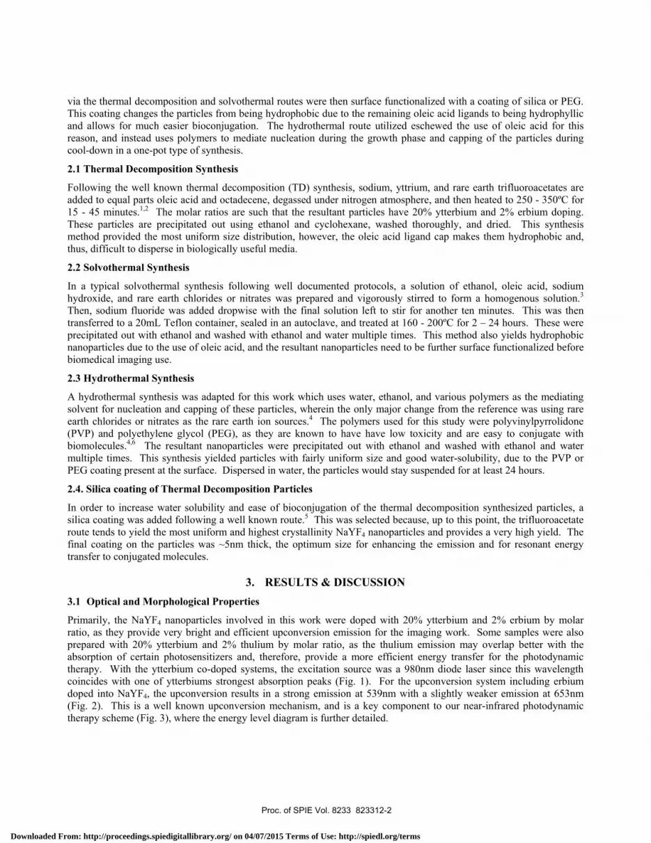
Fig. 1: Absorbance of NaYF4: ErYb Fig. 2: Emission of NaYF4: ErYb by (TD) Upon adding a functional coat of silica to the NaYF4 nanoparticles, the overall emission of the nanoparticles was noticed to have increased in intensity with a higher ratio of green emission (Fig. 4). This is a due to the surface being cleared of quenching bonds and surface defects being passivated, such that the amount of nonradiative processes is decreased for the system. As mentioned before, the thermal decomposition synthesis provided the highest uniformity of size and shape, as can be seen by comparing Figs. 5 & 6.
Fig. 3: Energy diagram for IRPDT scheme Fig. 4: Change in emission with silica coating
Proc. of SPIE Vol. 8233 823312-3
Downloaded From: http://proceedings.spiedigitallibrary.org/ on 04/07/2015 Terms of Use: http://spiedl.org/terms
4UU UU UU (OU
Wavelength (nm)LWU UU UU IUU
Wavelength (nm)
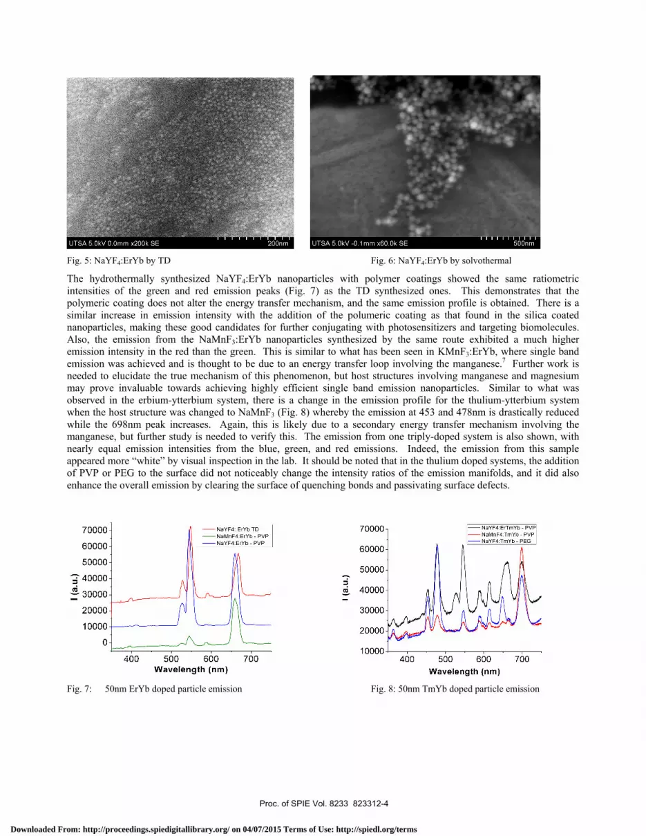
Fig. 5: NaYF4:ErYb by TD Fig. 6: NaYF4:ErYb by solvothermal The hydrothermally synthesized NaYF4:ErYb nanoparticles with polymer coatings showed the same ratiometric intensities of the green and red emission peaks (Fig. 7) as the TD synthesized ones. This demonstrates that the polymeric coating does not alter the energy transfer mechanism, and the same emission profile is obtained. There is a similar increase in emission intensity with the addition of the polumeric coating as that found in the silica coated nanoparticles, making these good candidates for further conjugating with photosensitizers and targeting biomolecules. Also, the emission from the NaMnF3:ErYb nanoparticles synthesized by the same route exhibited a much higher emission intensity in the red than the green. This is similar to what has been seen in KMnF3:ErYb, where single band emission was achieved and is thought to be due to an energy transfer loop involving the manganese.7 Further work is needed to elucidate the true mechanism of this phenomenon, but host structures involving manganese and magnesium may prove invaluable towards achieving highly efficient single band emission nanoparticles. Similar to what was observed in the erbium-ytterbium system, there is a change in the emission profile for the thulium-ytterbium system when the host structure was changed to NaMnF3 (Fig. 8) whereby the emission at 453 and 478nm is drastically reduced while the 698nm peak increases. Again, this is likely due to a secondary energy transfer mechanism involving the manganese, but further study is needed to verify this. The emission from one triply-doped system is also shown, with nearly equal emission intensities from the blue, green, and red emissions. Indeed, the emission from this sample appeared more “white” by visual inspection in the lab. It should be noted that in the thulium doped systems, the addition of PVP or PEG to the surface did not noticeably change the intensity ratios of the emission manifolds, and it did also enhance the overall emission by clearing the surface of quenching bonds and passivating surface defects.
Fig. 7: 50nm ErYb doped particle emission Fig. 8: 50nm TmYb doped particle emission
Proc. of SPIE Vol. 8233 823312-4
Downloaded From: http://proceedings.spiedigitallibrary.org/ on 04/07/2015 Terms of Use: http://spiedl.org/terms

3.2 Imaging in Mice
To ensure that these particles could in fact be excited by an infrared laser at low power and imaged through biological tissues, some bare particles made by the thermal decomposition route were injected underneath the skin of lab mice and imaged under a microscope (Figs. 9 – 12) . It is clear that these particles can be imaged clearly through thin layers of tissue and should be ideal for deeper imaging and therapy modalities. After injection of the particles, the mice were monitored for one week with no inflammation or other adverse side effects noted. The particles were still clearly visible upon illumination with the infrared laser after one week’s time, and there was no appearance of the particles migrating under the skin.
Fig. 9: Mouse injected with NaYF4 particles Fig. 10: Microscope image of mouse skin without 980nm
excitation
Fig. 11: 1μm particles with 980nm excitation Fig. 12: 300nm particles with 980nm excitation
4. SUMMARY & FUTURE IMPLEMENTATION While our preliminary results are very promising, both for synthesizing highly efficient upconverting nanoparticles and exciting and visualizing them through biological tissues, there is still much work to be done towards realizing a dual-
Proc. of SPIE Vol. 8233 823312-5
Downloaded From: http://proceedings.spiedigitallibrary.org/ on 04/07/2015 Terms of Use: http://spiedl.org/terms

mode nanoparticle for imaging and photodynamic therapy. Although we were unable to complete the photodynamic and tumor reduction portion of this work to date, significant progress towards implementing these nanoparticles has been completed, and we will extend this work towards the proposed cancer therapy experiments in the near future. Thus far, NaYF4 nanoparticles have been synthesized and surface coated by various means and were fully characterized. It was found that while thermal decomposition provides a high uniformity of size and crystallinity, nanoparticles fabricated by this method are hydrophobic and require surface modification, which can be expensive and requires more time per batch of nanoparticles. We are currently investigating the use of alternative solvents in the thermal decomposition method which do not leave a ligand cap on the nanoparticles after the synthesis process or which leave hydrophyllic polymer coatings on the surface, such as PVP or PEG. In the meantime, equally efficient upconverting particles which are water-soluble were easily fabricated by the hydrothermal route, although the size and shape distributions are less uniform. These polymeric coatings also improve biocompatibility of the nanoparticles, as PEG and PVP are known to be less toxic than many other coating materials.
The next phase of this work will involve conjugating photosensitive molecules to the polymer-coated NaYF4 nanoparticles, co-doped with either erbium-ytterbium or thulium-ytterbium such that the emission profile of the particle overlaps well with the absorption profile of the photosensitizer. The overall energy transfer efficiency between the upconverting nanoparticle and the photosensitizer will be quantified by measuring singlet oxygen production, and in vitro studies will be carried out where the therapeutic effects of this nanosystem will be verified. Concurrently, new host structures with more singular emission, such as NaMnF3, will be further studied and optimized. Finally, the best nanosystem for dual-mode imaging and photodynamic therapy, likely a triply doped nanoparticle with one upconversion route for imaging and another for therapy, will be fabricated and implemented in in vitro and in vivo studies.
This research was supported by the National Science Foundation Partnerships for Research and Education in Materials (NSF-PREM) Grant No. DMR - 0934218.
REFERENCES
[1] Ye, X., Collins, J.E., et. al., “Morphologically controlled synthesis of colloidal upconversion nanophosphors and their shape-directed self-assembly”, Proc. of the National Academy of Sciences, vol. 107, no. 52, pp. 22430–22435 (2010).
[2] Mai, H.X., Zhang, Y.W., et. al., “High-Quality Sodium Rare-Earth Fluoride Nanocrystals:Controlled Synthesis and Optical Properties”, Journal of the American Chemical Society, vol. 128, no. 19, pp 6427-6436 (2006).
[3] Chen, Z., Chen, H., et al., “Versatile Synthesis Strategy for Carboxylic Acid-functionalized Upconverting Nanophosphors as Biological Labels”, Journal of the American Chemical Society, vol. 130, no. 10, pp. 3023-3029 (2008).
[4] Wang, M., Mi, C.C., et. al., “One-step synthesis and characterization of water-soluble NaYF4:Yb,Er/Polymer nanoparticles with efficient up-conversion fluorescence”, Journal of Alloys and Compounds, vol. 485, pp L24-L27 (2009).
[5] Deng, M., Ma, Y., et. al., “Monodisperse Upconversion NaYF4 Nanocrystals: Syntheses and Bioapplications”, Nano Res., vol. 4, no. 7, pp 685-694 (2011)
[6] Xiong, L., Yang, T., et al., “Long-term in vivo biodistribution imaging and toxicity of polyacrylic acid-coated upconversion nanophosphors”, Biomaterials, vol. 31, pp 7078-7085 (2010).
[7] Wang, J., Wang, F., et. al., “Single-Band Upconversion Emission in Lanthanide-Doped KMnF3 Nanocrystals”, Angew. Chem. Int. Ed., vol. 50, pp 10369-10372 (2011).
Proc. of SPIE Vol. 8233 823312-6
Downloaded From: http://proceedings.spiedigitallibrary.org/ on 04/07/2015 Terms of Use: http://spiedl.org/terms