high electron mobility of phosphorous-doped homoepitaxial zno thin films grown by pulsed-laser...
TRANSCRIPT

High electron mobility of phosphorous-doped homoepitaxial ZnO thin filmsgrown by pulsed-laser depositionMatthias Brandt, Holger von Wenckstern, Heidemarie Schmidt, Andreas Rahm, Gisela Biehne et al. Citation: J. Appl. Phys. 104, 013708 (2008); doi: 10.1063/1.2953066 View online: http://dx.doi.org/10.1063/1.2953066 View Table of Contents: http://jap.aip.org/resource/1/JAPIAU/v104/i1 Published by the AIP Publishing LLC. Additional information on J. Appl. Phys.Journal Homepage: http://jap.aip.org/ Journal Information: http://jap.aip.org/about/about_the_journal Top downloads: http://jap.aip.org/features/most_downloaded Information for Authors: http://jap.aip.org/authors
Downloaded 06 Oct 2013 to 202.116.1.148. This article is copyrighted as indicated in the abstract. Reuse of AIP content is subject to the terms at: http://jap.aip.org/about/rights_and_permissions
High electron mobility of phosphorous-doped homoepitaxial ZnO thin filmsgrown by pulsed-laser deposition
Matthias Brandt,a� Holger von Wenckstern, Heidemarie Schmidt,b� Andreas Rahm,c�
Gisela Biehne, Gabriele Benndorf, Holger Hochmuth, Michael Lorenz,Christoph Meinecke, Tilman Butz, and Marius GrundmannInstitut für Experimentelle Physik II, Universität Leipzig, P.O. Box 10 09 20, 04009 Leipzig, Germany
�Received 3 January 2008; accepted 7 May 2008; published online 10 July 2008�
The transport properties of phosphorous-doped ZnO thin films, grown by pulsed-laser deposition onthermally pretreated hydrothermally grown ZnO single-crystal substrates, are reported. The ZnO:Pthin films show very good morphological and structural properties as confirmed by atomic forcemicroscopy �AFM�, high resolution x-ray diffraction, and Rutherford backscattering �RBS�channeling. Steps of height c /2 are visible in AFM investigations for all samples. For an oxygenpartial pressure of 0.1 mbar, two-dimensional growth was found. RBS channeling of a ZnO:P filmshows a minimum yield of 0.034 which is comparable to that of an annealed substrate �0.033�. Halleffect measurements revealed that all films are n-type for the present growth conditions. Peakmobilities of 800 cm2 /Vs have been observed around 70 K, in line with the high structural qualityof the samples. Room-temperature mobility in ZnO:P is up to 170 cm2 /Vs. © 2008 AmericanInstitute of Physics. �DOI: 10.1063/1.2953066�
I. INTRODUCTION
The II-IV semiconductor ZnO has attracted considerableinterest in the past years due to its outstanding material pa-rameters, e.g., its wide and direct band gap of 3.37 eV and itshigh exciton binding energy of about 60 meV, making it astrong candidate for blue and ultraviolet light-emittingdevices.1 Control of the transport properties is required, inparticular fabrication of p-type ZnO with high mobility. Thishas been the subject of extensive studies in the past years,and several results have been published reporting the fabri-cation of p-type ZnO.2,3 As-grown phosphorous-doped ZnOthin films usually show n-type conductivity.2–4 Samplesshowing p-type conductivity and good structural propertiessimultaneously have not been reported so far. Indeed, forZnO samples doped with antimony it was concluded that theapparent defect structure leads to the formation of a highnumber of lattice distortions and a low structural quality.5
For phosphorous-doped ZnO a similar defect structure hasbeen proposed,6 which could likewise be accompanied by adegradation of the structural properties. Even regardingn-type material, the structural properties of phosphorous-doped thin films are clearly inferior compared to nominallyundoped ZnO. With values smaller than 30 cm2 /Vs, the mo-bility of n-type phosphorus-doped ZnO thin films is signifi-cantly lower than that in undoped ZnO.2,3 High ionized im-purity concentrations in the films can only partially accountfor the low mobility. One particular reason for low mobilitiesis the likely formation of low-angle grain boundaries insamples grown heteroepitaxially.7–10 These grain boundaries
cause an additional scattering channel.11 Whereas the mobil-ity in grain-free bulk ZnO reaches values of up to2000 cm2 /Vs �Refs. 12–14� at about 50 K, lower Hall mo-bilities are reported for epitaxial ZnO layers.15,16 Homoepi-taxial growth of ZnO thin films has long been considered away to avoid grain formation and therefore to improve theelectron transport properties considerably. The literature,however, does not yield a large number of reports on ho-moepitaxial growth of ZnO. Recent investigations indicatepoor surface properties of the commercially available hydro-thermal crystals, which hinder the growth of high-qualityepitaxial layers.17,18 The substrates could, however, be trans-formed into a state suitable for homoepitaxy by a thermalannealing process.17
The successful growth of high-quality ZnO thin films bypulsed-laser deposition �PLD� on thermally pretreated ZnOsubstrates has been reported in a previous publication.18
However, transport data were not available yet due to the lowconductivity of undoped films making them indistinguish-able from the substrate. Transport properties of homoepi-taxial ZnO:P thin films have not been reported so far. We findhigh peak mobilities of 800 cm2 /Vs at 70 K and170 cm2 /Vs at room temperature.
II. EXPERIMENTAL
In this work, phosphorous-doped ZnO thin films weregrown by PLD at a substrate temperature of 650 °C andoxygen partial pressures ranging from 3�10−4 to 0.1 mbar.The target, containing 0.01 wt % P2O5, has been ablated us-ing the 248 nm line of a KrF laser at a pulse energy of 600mJ and a repetition rate of 10 Hz. A total of 30 000 pulseswas used, starting with a 300 pulse nucleation layer depos-ited at a reduced ablation rate of 1 Hz. The structural prop-erties of the samples have been characterized by high-resolution x-ray diffraction �HR-XRD� measurements using
a�Author to whom correspondence should be addressed. Electronic mail:[email protected].
b�Present address: Forschungszentrum Dresden-Rossendorf, Dresden, Ger-many.
c�Present address: Solarion AG, Leipzig, Germany.
JOURNAL OF APPLIED PHYSICS 104, 013708 �2008�
0021-8979/2008/104�1�/013708/6/$23.00 © 2008 American Institute of Physics104, 013708-1
Downloaded 06 Oct 2013 to 202.116.1.148. This article is copyrighted as indicated in the abstract. Reuse of AIP content is subject to the terms at: http://jap.aip.org/about/rights_and_permissions
a Philips X’Pert materials research diffractometer with apoint-shaped Cu K�1 primary beam from a Bartels mono-chromator with four Ge�220� reflections. The detector waseither kept open �for rocking curve measurements� or aBonse–Hart analyzer crystal with an acceptance angle of 12arcsec �triple-axis configuration, for reciprocal space maps�was used. The film thickness was calculated from the periodof the 2�–� intensity fringes. The morphological propertieswere studied by atomic force microscopy �AFM� utilizing aVeeco Dimensions 3100 with a Nanoscope IVa controller.Proton induced x-ray emission �PIXE� and Rutherford back-scattering �RBS� channeling experiments have been carriedout by irradiation with 1.1 and 1.25 MeV protons at theLIPSION �Ref. 19� ion beamline. The chemical compositionof the samples was deduced from the PIXE spectra.Temperature-dependent Hall effect measurements were per-formed between 20 and 325 K using the van der Pauwmethod. The photoluminescence �PL� exited by a HeCd laser��=325 nm� was measured at 2 K.
III. RESULTS AND DISCUSSION
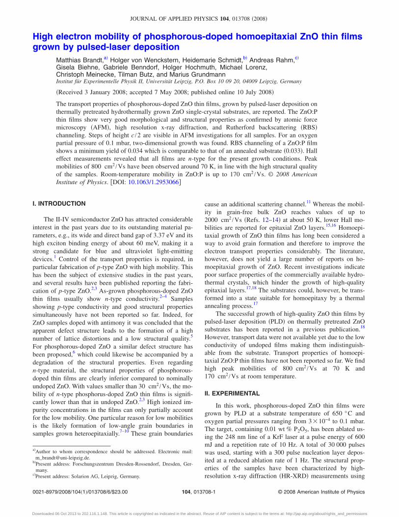
The results of the AFM measurements are depicted inFig. 1. A strong dependence of the surface morphology onthe growth conditions was observed. The root-mean-squaresurface roughness rrms on 2�1 �m2 scan area decreaseswith increasing oxygen partial pressure from rrms=1.06 nmat 2�10−3 mbar �Fig. 1�c�� to rrms=0.175 nm at 0.1 mbar�Fig. 1�a��. Likewise the surface morphology is changing.Samples deposited at an oxygen partial pressure below 0.1mbar show a three-dimensional growth mode, leading to theformation of grains with clearly visible boundaries. Withinthe grains, steps of height c /2 were observed. The averagesize of the grains is 400 nm for p�O2�=3�10−4 andp�O2�=2�10−3 mbar �Fig. 1�c�� and about 300 nm forp�O2�=0.016 mbar oxygen partial pressure �Fig. 1�b��, re-spectively. In contrast, the sample grown at 0.1 mbar oxygenpartial pressure shows a vicinal surface without grain struc-ture; the growth mode is two-dimensional �2D� �Fig. 1�a��.
A. Structural and morphological properties
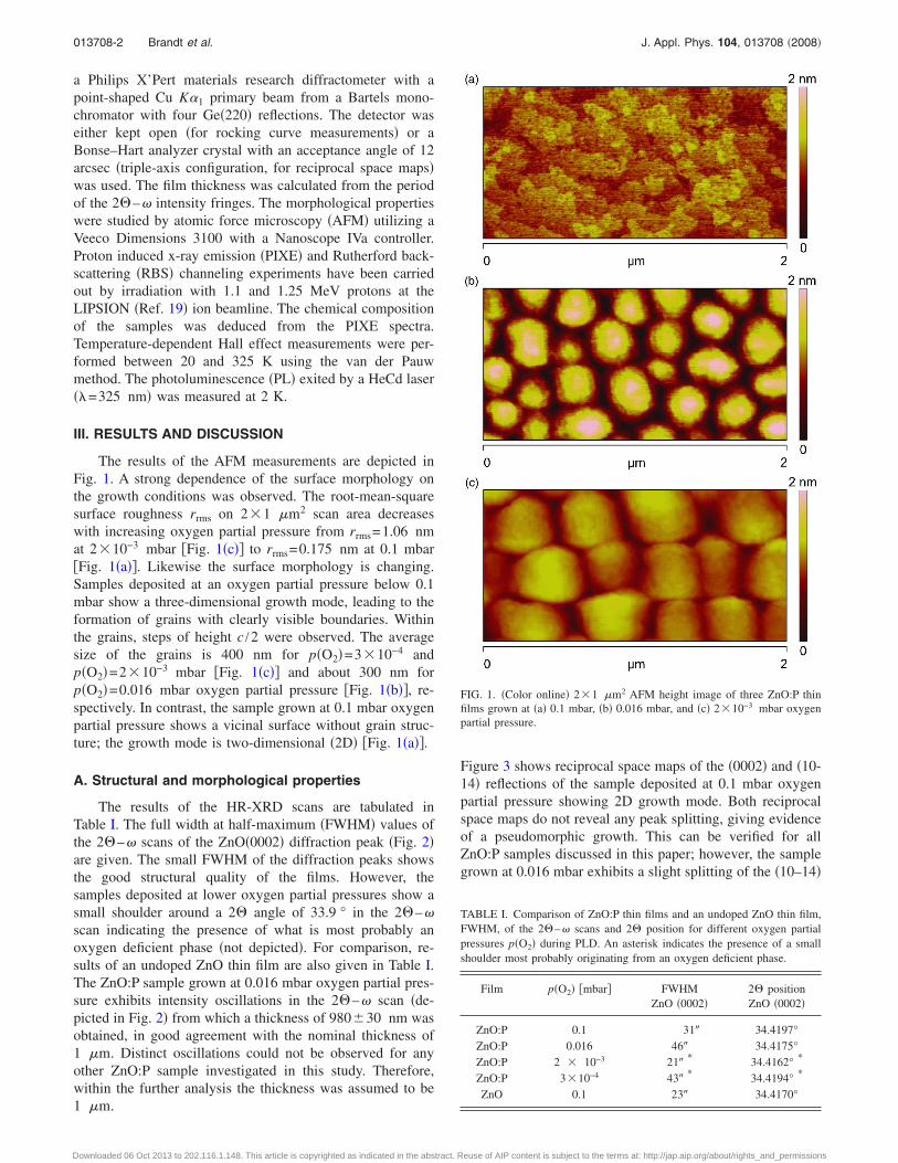
The results of the HR-XRD scans are tabulated inTable I. The full width at half-maximum �FWHM� values ofthe 2�–� scans of the ZnO�0002� diffraction peak �Fig. 2�are given. The small FWHM of the diffraction peaks showsthe good structural quality of the films. However, thesamples deposited at lower oxygen partial pressures show asmall shoulder around a 2� angle of 33.9 ° in the 2�–�scan indicating the presence of what is most probably anoxygen deficient phase �not depicted�. For comparison, re-sults of an undoped ZnO thin film are also given in Table I.The ZnO:P sample grown at 0.016 mbar oxygen partial pres-sure exhibits intensity oscillations in the 2�–� scan �de-picted in Fig. 2� from which a thickness of 980�30 nm wasobtained, in good agreement with the nominal thickness of1 �m. Distinct oscillations could not be observed for anyother ZnO:P sample investigated in this study. Therefore,within the further analysis the thickness was assumed to be1 �m.
Figure 3 shows reciprocal space maps of the �0002� and �10-14� reflections of the sample deposited at 0.1 mbar oxygenpartial pressure showing 2D growth mode. Both reciprocalspace maps do not reveal any peak splitting, giving evidenceof a pseudomorphic growth. This can be verified for allZnO:P samples discussed in this paper; however, the samplegrown at 0.016 mbar exhibits a slight splitting of the �10–14�
TABLE I. Comparison of ZnO:P thin films and an undoped ZnO thin film,FWHM, of the 2�–� scans and 2� position for different oxygen partialpressures p�O2� during PLD. An asterisk indicates the presence of a smallshoulder most probably originating from an oxygen deficient phase.
Film p�O2� �mbar� FWHMZnO �0002�
2� positionZnO �0002�
ZnO:P 0.1 31� 34.4197°ZnO:P 0.016 46� 34.4175°ZnO:P 2 � 10−3 21�
* 34.4162° *
ZnO:P 3�10−4 43�* 34.4194° *
ZnO 0.1 23� 34.4170°
FIG. 1. �Color online� 2�1 �m2 AFM height image of three ZnO:P thinfilms grown at �a� 0.1 mbar, �b� 0.016 mbar, and �c� 2�10−3 mbar oxygenpartial pressure.
013708-2 Brandt et al. J. Appl. Phys. 104, 013708 �2008�
Downloaded 06 Oct 2013 to 202.116.1.148. This article is copyrighted as indicated in the abstract. Reuse of AIP content is subject to the terms at: http://jap.aip.org/about/rights_and_permissions
and �0002� reflex, indicating pseudomorphic growth with aperpendicular mismatch of 270�15 ppm. Very small asym-metric peak broadening due to minor mosaicity and the finitecrystallite size is visible in the reciprocal space maps. Pleasenote the logarithmic intensity scale in these reciprocal spacemaps.
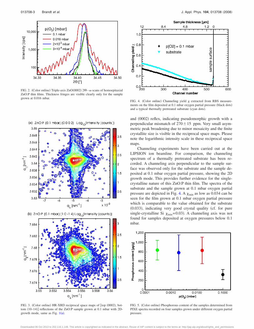
Channeling experiments have been carried out at theLIPSION ion beamline. For comparison, the channelingspectrum of a thermally pretreated substrate has been re-corded. A channeling axis perpendicular to the sample sur-face was observed only for the substrate and the sample de-posited at 0.1 mbar oxygen partial pressure, showing the 2Dgrowth mode. This provides further evidence for the single-crystalline nature of this ZnO:P thin film. The spectra of thesubstrate and the sample grown at 0.1 mbar oxygen partialpressure are depicted in Fig. 4. A �min as low as 0.034 can beseen for the film grown at 0.1 mbar oxygen partial pressurewhich is comparable to the value obtained for the substrate�0.033�, indicating very good crystal quality �cf. for puresingle-crystalline Si �min=0.03�. A channeling axis was notfound for samples deposited at oxygen pressures below 0.1mbar.
FIG. 2. �Color online� Triple-axis ZnO�0002� 2�–� scans of homoepitaxialZnO:P thin films. Thickness fringes are visible clearly only for the samplegrown at 0.016 mbar.
FIG. 3. �Color online� HR-XRD reciprocal space maps of �top �0002�, bot-tom �10–14�� reflections of the ZnO:P sample grown at 0.1 mbar with 2D-growth mode, same as Fig. 1�a�.
FIG. 4. �Color online� Channeling yield � extracted from RBS measure-ments on the film deposited at 0.1 mbar oxygen partial pressure �black dots�and a typical thermally pretreated substrate �cyan dots�.
FIG. 5. �Color online� Phosphorous content of the samples determined fromPIXE spectra recorded on four samples grown under different oxygen partialpressure.
013708-3 Brandt et al. J. Appl. Phys. 104, 013708 �2008�
Downloaded 06 Oct 2013 to 202.116.1.148. This article is copyrighted as indicated in the abstract. Reuse of AIP content is subject to the terms at: http://jap.aip.org/about/rights_and_permissions
B. Chemical composition
The phosphorous content of the ZnO:P thin films wasdetermined by PIXE experiments and is depicted versus theoxygen partial pressure in Fig. 5. The phosphorous content inthe samples differs significantly from the nominal concentra-tion in the target �nominally 0.01 wt % P2O5, thus�43 �g /g P in ZnO�. PIXE investigations of the target re-vealed that the concentration in the ablated region is45�11 �g /g P in ZnO. The concentration of phosphorousin the films is significantly higher, and further depends on theoxygen partial pressure. The actual concentrations are onlyslightly above the detection limit of the PIXE analysis andtherefore subject to a relatively large error, which is depictedin Fig. 5 as well. As can be seen, the apparent concentrationin the film exceeds that of the target by a factor of 5 to 20.
C. Electrical properties
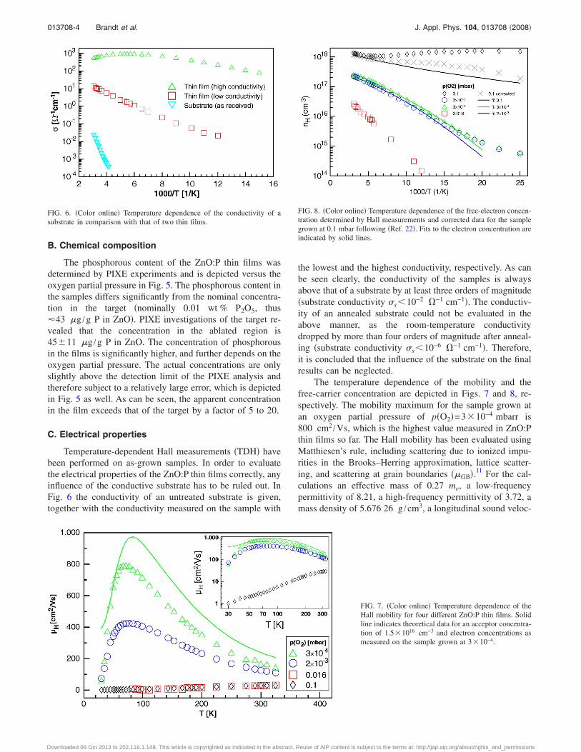
Temperature-dependent Hall measurements �TDH� havebeen performed on as-grown samples. In order to evaluatethe electrical properties of the ZnO:P thin films correctly, anyinfluence of the conductive substrate has to be ruled out. InFig. 6 the conductivity of an untreated substrate is given,together with the conductivity measured on the sample with
the lowest and the highest conductivity, respectively. As canbe seen clearly, the conductivity of the samples is alwaysabove that of a substrate by at least three orders of magnitude�substrate conductivity s10−2 �−1 cm−1�. The conductiv-ity of an annealed substrate could not be evaluated in theabove manner, as the room-temperature conductivitydropped by more than four orders of magnitude after anneal-ing �substrate conductivity s10−6 �−1 cm−1�. Therefore,it is concluded that the influence of the substrate on the finalresults can be neglected.
The temperature dependence of the mobility and thefree-carrier concentration are depicted in Figs. 7 and 8, re-spectively. The mobility maximum for the sample grown atan oxygen partial pressure of p�O2�=3�10−4 mbarr is800 cm2 /Vs, which is the highest value measured in ZnO:Pthin films so far. The Hall mobility has been evaluated usingMatthiesen’s rule, including scattering due to ionized impu-rities in the Brooks–Herring approximation, lattice scatter-ing, and scattering at grain boundaries ��GB�.11 For the cal-culations an effective mass of 0.27 me, a low-frequencypermittivity of 8.21, a high-frequency permittivity of 3.72, amass density of 5.676 26 g /cm3, a longitudinal sound veloc-
FIG. 6. �Color online� Temperature dependence of the conductivity of asubstrate in comparison with that of two thin films.
FIG. 7. �Color online� Temperature dependence of theHall mobility for four different ZnO:P thin films. Solidline indicates theoretical data for an acceptor concentra-tion of 1.5�1016 cm−3 and electron concentrations asmeasured on the sample grown at 3�10−4.
FIG. 8. �Color online� Temperature dependence of the free-electron concen-tration determined by Hall measurements and corrected data for the samplegrown at 0.1 mbar following �Ref. 22�. Fits to the electron concentration areindicated by solid lines.
013708-4 Brandt et al. J. Appl. Phys. 104, 013708 �2008�
Downloaded 06 Oct 2013 to 202.116.1.148. This article is copyrighted as indicated in the abstract. Reuse of AIP content is subject to the terms at: http://jap.aip.org/about/rights_and_permissions
ity of 6077.6 m /s, an acoustic deformation potential of 18.9eV, a piezoelectric coefficient of 0.21, and a Debye tempera-ture of 837 K have been used. For the two samples grownunder the lowest oxygen partial pressures, exhibiting thehighest mobility, a reasonable fit was achieved without in-cluding scattering at grain boundaries �GB. If this process isincluded, a fit is only possible for neglectable intergrain bar-rier heights. However, grainlike structures are visible in theAFM scans of the films in question �Fig. 1�c��, which leadsto the conclusion that their boundaries do not incorporatecarrier traps in a concentration such that the depletion layersforming have a significant impeding effect. However, eitheran additional scattering channel or a parallel conductionthrough a region with lower mobility must be present for thesamples grown at the lowest oxygen partial pressures, ac-counting for the deviation between the experimental and thecalculated data at high temperature. The free-electron con-centration at low temperatures is higher than it would beexpected, which could be due to conduction in a parallellayer. A definite answer cannot be given from the experimen-tal data available. For the film grown at the highest oxygenpartial pressure, a very low carrier mobility is observed to-gether with a high, and almost temperature-independent car-rier concentration. Certainly the transport in that film isdominated by a degenerate layer located either at thesurface,20 or the film-substrate interface. A similar behaviorhas been observed in undoped ZnO thin films grown onsapphire.21 A correction scheme22 has been applied, leadingto a more pronounced temperature dependence in n. How-ever, the concentration of free carriers is still close to theMott transition point, resulting in a low mobility. The donoractivation energy determined from the fit is reduced due tothe high donor concentration. This behavior can be explainedby a nonabrupt boundary of the highly conductive layer,which cannot be treated within the framework of the appliedcorrection scheme. Under these circumstances the electricalproperties of the film cannot be separated, as the highly con-ductive layer is clearly dominating the electronic transport atall temperatures. For the sample grown at 0.016 mbar a verylow mobility is observed, together with a rather low carrierconcentration, indicating the presence of grain boundaryscattering as an additional scattering channel. The acceptorconcentration cannot be obtained unambiguously from the fitof the mobility. However, the relatively low mobility pointsto the presence of both grain boundary and ionized impurityscattering, which in conjunction with the low free-electronconcentration points toward a high degree of compensation.We therefore give a lower limit for the donor and acceptorconcentration, as well as the donor activation energy, point-ing out, that actual values might be higher.
In order to model the free-electron concentration, thesolution of the charge balance equation considering a singledonor in the presence of compensating acceptors was suffi-cient. The acceptor concentrations for the samples grown atthe lowest oxygen partial pressures could be extracted fromfits of the mobility, and have been fixed modelling the free-carrier concentrations. The results of these fits are tabulatedin Table II.
Donor activation energies of around 35 meV have beenobserved for the samples grown under the lowest oxygenpressure. For the sample grown at 0.016 mbar the dominantdonor cannot be determined unambiguously. An effectivemass like donor could be dominant, as well as a level withhigher activation energy. The level observed in the samplegrown at 0.1 mbar oxygen partial pressure is subject to someuncertainty because of the high dopant concentration in thesample, and could correlate to the levels between 10 and 15meV reported in the literature.12,23
D. Optical properties
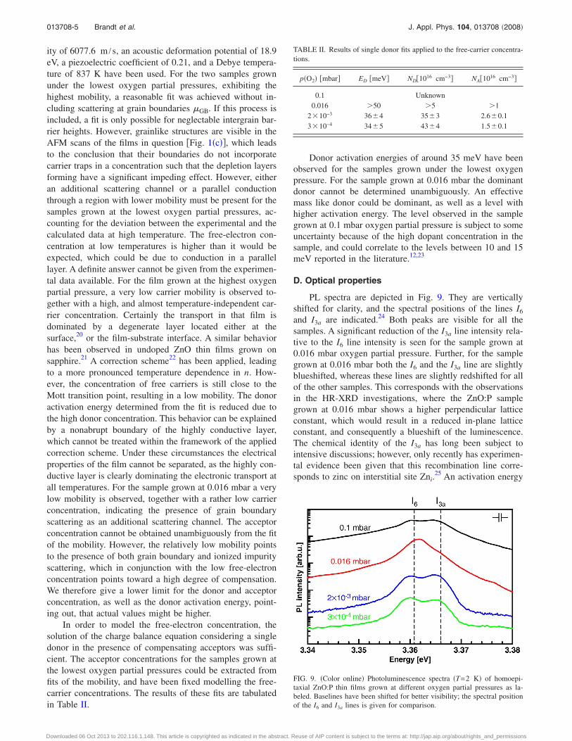
PL spectra are depicted in Fig. 9. They are verticallyshifted for clarity, and the spectral positions of the lines I6
and I3a are indicated.24 Both peaks are visible for all thesamples. A significant reduction of the I3a line intensity rela-tive to the I6 line intensity is seen for the sample grown at0.016 mbar oxygen partial pressure. Further, for the samplegrown at 0.016 mbar both the I6 and the I3a line are slightlyblueshifted, whereas these lines are slightly redshifted for allof the other samples. This corresponds with the observationsin the HR-XRD investigations, where the ZnO:P samplegrown at 0.016 mbar shows a higher perpendicular latticeconstant, which would result in a reduced in-plane latticeconstant, and consequently a blueshift of the luminescence.The chemical identity of the I3a has long been subject tointensive discussions; however, only recently has experimen-tal evidence been given that this recombination line corre-sponds to zinc on interstitial site Zni.
25 An activation energy
TABLE II. Results of single donor fits applied to the free-carrier concentra-tions.
p�O2� �mbar� ED �meV� ND�1016 cm−3� NA�1016 cm−3�
0.1 Unknown0.016 �50 �5 �1
2�10−3 36�4 35�3 2.6�0.13�10−4 34�5 43�4 1.5�0.1
FIG. 9. �Color online� Photoluminescence spectra �T=2 K� of homoepi-taxial ZnO:P thin films grown at different oxygen partial pressures as la-beled. Baselines have been shifted for better visibility; the spectral positionof the I6 and I3a lines is given for comparison.
013708-5 Brandt et al. J. Appl. Phys. 104, 013708 �2008�
Downloaded 06 Oct 2013 to 202.116.1.148. This article is copyrighted as indicated in the abstract. Reuse of AIP content is subject to the terms at: http://jap.aip.org/about/rights_and_permissions
of the dominant donor of ED�35 meV has been identifiedfor two of the samples. Following the Haynes rule, and usingthe parameters given in Ref. 24, the donor binding energycorrelated with the PL peak labeled I3a is about 37.5 meV,giving the indication that the donor level ED�35 meV ob-served in TDH most likely has the same origin as the I3a
recombination line. We therefore assign this level to zinc onthe interstitial site, as has already been proposed in Ref. 26.However, a more detailed investigation is necessary to sub-stantiate these findings. For the sample grown at 0.016 mbarthe recombination line I6 dominates the luminescence spec-trum. This is known to be the AlZn donor with an activationenergy of around 51.5 meV.24 This finding coincides with theminimal activating energy found for the sample grown at0.016 mbar oxygen partial pressure as tabulated in Table II.
IV. CONCLUSION
In summary, high-quality phosphorous-doped ZnO thinfilms have been grown homoepitaxially by PLD. AFM mea-surements have shown that the surfaces exhibit steps ofheight c /2. Grains of up to 400 nm have been observed forthe samples grown at oxygen partial pressures 0.1 mbar.2D growth was found for an oxygen partial pressure of 0.1mbar. It has been shown that the conductivity of the substrateis always negligible compared to that of the ZnO:P filmsinvestigated. Hall effect measurements revealed that all filmsare n-type for the present growth conditions. A peak mobilityof 800 cm2 /Vs was observed for the film deposited at thelowest oxygen partial pressure of 3�10−4 mbar. With in-creasing oxygen partial pressure a decrease in the electronmobility can be observed, and for the sample grown at thehighest partial pressure the existence of a degenerate layer isevident from the transport measurements. A connection be-tween the I3a line observed in PL measurements for all thesamples investigated and the donor level at �35 meV waspointed out. Following Sann et al. �Ref. 25�, this level hasbeen assigned to the zinc interstitial Zni. A reduction of therelative density of this defect has been found for the filmexhibiting the perpendicular mismatch in comparison to allthe other films, indicating a higher formation probability ofthis defect in the latter. It could be speculated that the for-mation of the Zni defect leads to a slight reduction of the unitcell volume.
ACKNOWLEDGMENTS
This work has been supported by Deutsche Forschungs-gemeinschaft in the framework of Schwerpunktprogramm
SPP1136 �H.v.W.�, and by the Bundesministerium für Bil-dung und Forschung in the framework of FKZ 03N8708�H.S., H.H.�.
1D. M. Bagnall, Y. F. Chen, Z. Zhu, T. Yao, S. Koyama, M. Y. Shen, and T.Goto, Appl. Phys. Lett. 70, 2230 �1997�.
2K.-K. Kim, H.-S. Kim, D.-K. Hwang, J.-H. Lim, and S.-J. Park, Appl.Phys. Lett. 83, 63 �2003�.
3Y. W. Heo, S. J. Park, K. Ip, S. J. Pearton, and D. P. Norton, Appl. Phys.Lett. 83, 1128 �2003�.
4S. J. Pearton, D. P. Norton, K. Ip, Y. W. Heo, and T. Steiner, Prog. Mater.Sci. 50, 293 �2005�.
5W. Guo, A. Allenic, Y. B. Chen, X. Q. Pan, Y. Che, Z. D. Hu, and B. Liu,Appl. Phys. Lett. 90, 242108 �2007�.
6W.-J. Lee, J. Kang, and K. J. Chang, Phys. Rev. B 73, 024117 �2006�.7E. M. Kaidashev, M. Lorenz, H. von Wenckstern, A. Rahm, H.-C. Sem-melhack, K.-H. Han, G. Benndorf, C. Bundesmann, H. Hochmuth, and M.Grundmann, Appl. Phys. Lett. 82, 3901 �2003�.
8T. Tsurumi, S. Nishizawa, N. Ohashi, and T. Ohgaki, Jpn. J. Appl. Phys.,Part 1 38, 3682 �1999�.
9J. W. Sun, Y. M. Lu, Y. C. Liu, D. Z. Shen, Z. Z. Zhang, B. H. Li, J. Y.Zhang, B. Yao, D. X. Zhao, and X. W. Fan, Appl. Phys. Lett. 89, 232101�2006�.
10J. Steinhauser, S. Fay, N. Oliveira, E. Vallat-Sauvain, and C. Ballif, Appl.Phys. Lett. 90, 142107 �2007�.
11J. W. Orton, B. J. Goldsmith, M. J. Powell, and J. A. Chapman, Appl.Phys. Lett. 37, 557 �1980�.
12P. Wagner and R. Helbig, J. Phys. Chem. Solids 35, 327 �1974�.13D. C. Look, J. W. Hemsky, and J. R. Sizelove, Phys. Rev. Lett. 82, 2552
�1999a�.14D. C. Look, G. C. Farlow, P. Reunchan, S. Limpijumnong, S. B. Zhang,
and K. Nordlund, Phys. Rev. Lett. 95, 225502 �2005�.15H. von Wenckstern, M. Brandt, H. Schmidt, G. Biehne, R. Pickenhain, H.
Hochmuth, M. Lorenz, and M. Grundmann, Appl. Phys. A: Mater. Sci.Process. 88, 135 �2007�.
16H. Tampo, A. Yamada, P. Fons, H. Shibata, K. Matsubara, K. Iwata, S.Niki, K. Nakahara, and H. Takasu, Appl. Phys. Lett. 84, 4412 �2004�.
17S. Graubner, C. Neumann, N. Volbers, B. K. Meyer, J. Blasing, and A.Krost, Appl. Phys. Lett. 90, 042103 �2007�.
18H. von Wenckstern, H. Schmidt, C. Hanisch, M. Brandt, C. Czekalla, G.Benndorf, G. Biehne, A. Rahm, H. Hochmuth, M. Lorenz, and M Grund-mann, Phys. Status Solidi �RRL� 1, 129 �2007b�.
19T. Reinert, Ph.D. thesis, Universität Leipzig �2001�.20O. Schmidt, P. Kiesel, C. G. V. De Walle, N. M. Johnson, J. Nause, and G.
H. Dohler, Jpn. J. Appl. Phys., Part 1 44, 7271 �2005�.21H. von Wenckstern, M. Brandt, J. Lenzner, G. Zimmermann, H. Hoch-
muth, M. Lorenz, and M. Grundmann, in Proceedings of MRS, Vol. 957�2007c�.
22D. C. Look and R. J. Molnar, Appl. Phys. Lett. 70, 3377 �1997�.23E. Ziegler, A. Heinrich, H. Oppermann, and G. Stöver, Phys. Status Solidi
A 66, 635 �1981�.24B. K. Meyer, H. Alves, D. M. Hofmann, W. Kriegseis, D. Forster, F.
Bertram, J. Christen, A. Hoffmann, M. Straßburg, M. Dworzak, U. Habo-eck, and A. V. Rodina, Phys. Status Solidi B 241, 231 �2004�.
25J. Sann, J. Stehr, A. Hofstaetter, D. M. Hofmann, A. Neumann, M. Lerch,U. Haboeck, A. Hoffmann, and C. Thomsen, Phys. Rev. B 76, 195203�2007�.
26D. C. Look, J. W. Hemsky, and J. R. Sizelove, Phys. Rev. Lett. 82, 2552�1999b�.
013708-6 Brandt et al. J. Appl. Phys. 104, 013708 �2008�
Downloaded 06 Oct 2013 to 202.116.1.148. This article is copyrighted as indicated in the abstract. Reuse of AIP content is subject to the terms at: http://jap.aip.org/about/rights_and_permissions