helicobacter pylori infection induces oxidative stress and programmed cell death in human gastric...
TRANSCRIPT

Published Ahead of Print 11 June 2007. 2007, 75(8):4030. DOI: 10.1128/IAI.00172-07. Infect. Immun.
Istvan Boldogh, Peter B. Ernst and Sheila E. CroweVictor E. Reyes, Janak Patel, Bernadette Dirden-Kramer, Song-Ze Ding, Yutaka Minohara, Xue Jun Fan, Jide Wang, Death in Human Gastric Epithelial Cells Oxidative Stress and Programmed Cell
Infection InducesHelicobacter pylori
http://iai.asm.org/content/75/8/4030Updated information and services can be found at:
These include:
REFERENCEShttp://iai.asm.org/content/75/8/4030#ref-list-1at:
This article cites 79 articles, 38 of which can be accessed free
CONTENT ALERTS more»articles cite this article),
Receive: RSS Feeds, eTOCs, free email alerts (when new
http://journals.asm.org/site/misc/reprints.xhtmlInformation about commercial reprint orders: http://journals.asm.org/site/subscriptions/To subscribe to to another ASM Journal go to:
on Novem
ber 5, 2013 by guesthttp://iai.asm
.org/D
ownloaded from
on N
ovember 5, 2013 by guest
http://iai.asm.org/
Dow
nloaded from
on Novem
ber 5, 2013 by guesthttp://iai.asm
.org/D
ownloaded from
on N
ovember 5, 2013 by guest
http://iai.asm.org/
Dow
nloaded from
on Novem
ber 5, 2013 by guesthttp://iai.asm
.org/D
ownloaded from
on N
ovember 5, 2013 by guest
http://iai.asm.org/
Dow
nloaded from
on Novem
ber 5, 2013 by guesthttp://iai.asm
.org/D
ownloaded from
on N
ovember 5, 2013 by guest
http://iai.asm.org/
Dow
nloaded from
on Novem
ber 5, 2013 by guesthttp://iai.asm
.org/D
ownloaded from
on N
ovember 5, 2013 by guest
http://iai.asm.org/
Dow
nloaded from
on Novem
ber 5, 2013 by guesthttp://iai.asm
.org/D
ownloaded from

INFECTION AND IMMUNITY, Aug. 2007, p. 4030–4039 Vol. 75, No. 80019-9567/07/$08.00�0 doi:10.1128/IAI.00172-07Copyright © 2007, American Society for Microbiology. All Rights Reserved.
Helicobacter pylori Infection Induces Oxidative Stress and ProgrammedCell Death in Human Gastric Epithelial Cells�
Song-Ze Ding,1 Yutaka Minohara,2 Xue Jun Fan,2 Jide Wang,2 Victor E. Reyes,2 Janak Patel,2Bernadette Dirden-Kramer,2 Istvan Boldogh,3 Peter B. Ernst,4 and Sheila E. Crowe4*
Departments of Microbiology1 and Medicine,4 University of Virginia, Charlottesville, Virginia 22908, and Departments ofPediatrics2 and Microbiology and Immunology,3 University of Texas Medical Branch, Galveston, Texas 77555
Received 1 February 2007/Returned for modification 10 May 2007/Accepted 29 May 2007
Helicobacter pylori infection is associated with altered gastric epithelial cell turnover. To evaluate the role ofoxidative stress in cell death, gastric epithelial cells were exposed to various strains of H. pylori, inflammatorycytokines, and hydrogen peroxide in the absence or presence of antioxidant agents. Increased intracellularreactive oxygen species (ROS) were detected using a redox-sensitive fluorescent dye, a cytochrome c reductionassay, and measurements of glutathione. Apoptosis was evaluated by detecting DNA fragmentation andcaspase activation. Infection with H. pylori or exposure of epithelial cells to hydrogen peroxide resulted inapoptosis and a dose-dependent increase in ROS generation that was enhanced by pretreatment with inflam-matory cytokines. Basal levels of ROS were greater in epithelial cells isolated from gastric mucosal biopsyspecimens from H. pylori-infected subjects than in cells from uninfected individuals. H. pylori strains bearingthe cag pathogenicity island (PAI) induced higher levels of intracellular oxygen metabolites than isogenic cagPAI-deficient mutants. H. pylori infection and hydrogen peroxide exposure resulted in similar patterns ofcaspase 3 and 8 activation. Antioxidants inhibited both ROS generation and DNA fragmentation by H. pylori.These results indicate that bacterial factors and the host inflammatory response confer oxidative stress to thegastric epithelium during H. pylori infection that may lead to apoptosis.
Helicobacter pylori infection has been implicated in thepathogenesis of gastritis, peptic ulcer disease, gastric carci-noma, and gastric lymphoma (10, 18), but the mechanismsleading from chronic active gastritis to other disease manifes-tations remain unclear. Various bacterial factors as well as thehost response are believed to contribute to the outcome ofinfection with H. pylori (25). Strains bearing the cag pathoge-nicity island (PAI) (15), which includes the cagA gene, havebeen shown to be associated with increased gastric inflamma-tion (57), increased bacterial load, and both peptic ulcer dis-ease and gastric cancer (11). Increased induction of gastricepithelial cytokines that recruit and activate immune/inflam-matory cells is also observed with these strains (17, 41, 55, 67).Although the full functions of genes included in the cag PAIremain unclear, it is known that cag PAI-positive strains acti-vate specific transcription factors and cell signaling pathways(37, 41, 52, 55, 66).
Bacterial and host factors can damage the gastric mucosalbarrier and lead to alterations of epithelial cell growth anddifferentiation. H. pylori infection is associated with increasedcellular proliferation in vivo (13, 43) although most in vitrostudies demonstrate bacterial inhibition of cell growth (76),suggesting that factors other than H. pylori regulate cell growthin the complex milieu of the infected gastric mucosa. Increasednumbers of apoptotic cells are found in the gastric epitheliumof infected patients (36, 45, 50, 58), suggesting that inductionof apoptosis may be a common method of cell growth regula-
tion for H. pylori (24). In agreement with these findings, we andothers have demonstrated that H. pylori induces programmedcell death in cultured gastric epithelial cells, as do proinflam-matory cytokines that are released during infection (3, 4, 26,44, 68, 76). Proliferative (13, 43) and apoptotic rates (24, 36,50) have both been shown to return to control levels aftereradication of infection.
There is increasing evidence that microbial pathogens induceoxidative stress in infected host cells (29, 65, 69), and this mayrepresent an important mechanism leading to epithelial injury inH. pylori infection (70). It is known from other cell systems thatoxidative stress regulates cell cycle events via multiple pathways,with net responses that include aberrant proliferation, adaptationcytotoxicity, and cell death (35). Oxidative stress could well play arole in the altered epithelial proliferation, increased apoptosis(16, 34), and increased oxidative DNA damage (7, 16, 27) asso-ciated with H. pylori infection. Evidence for this includes in-creased levels of reactive oxygen species (ROS) measured in themucosae of infected patients (16, 21, 23). While activated, ROS-releasing phagocytic leukocytes recruited to the gastric mucosaduring infection represent one obvious source of oxidative stress(21, 79), other studies demonstrate that H. pylori itself also gen-erates ROS (51) and that ROS accumulate in gastric epithelialcells (5, 6, 73). In addition, proinflammatory cytokines induceROS in various cell types (47, 48, 61), and the decreased levels ofascorbic acid that are associated with H. pylori infection (64, 71)also contribute to a pro-oxidative environment. H. pylori infectionhas been shown to increase expression and activity of spermineoxidase, which oxidizes polyamines that are abundant in epithelialcells to release hydrogen peroxide (77), suggesting another mech-anism by which H. pylori induces oxidative stress.
To examine oxidative stress that may occur during H. pylori
* Corresponding author. Mailing address: Division of Gastroenter-ology and Hepatology, Department of Medicine, P.O. Box 800708,Charlottesville, VA 22908-0708. Phone: (434) 243-9309. Fax: (434)982-0044. E-mail: [email protected].
� Published ahead of print on 11 June 2007.
4030

infection, measurements of intracellular ROS were made incultured and native gastric epithelial cells after exposure to H.pylori or hydrogen peroxide (H2O2), either alone or in combi-nation with cytokines that are increased in infection. Antioxi-dants were used to evaluate the role of oxidative stress in theinduction of apoptosis by these stimuli. To determine the roleof the cag PAI in the induction of oxidative stress, cag PAI-bearing H. pylori strains and their isogenic mutants deficient inthe cag PAI were compared for their abilities to generate ROSin gastric epithelial cells.
MATERIALS AND METHODS
Cell lines. Gastric epithelial cell lines Kato III, NCI-N87, and AGS (AmericanType Culture Collection, Manassas, VA) were grown in standard conditionsaccording to previously published methods (19, 26, 78). Briefly, cells were cul-tured in flasks containing RPMI 1640 medium supplemented with 10% fetal calfserum at 37°C in a humidified 5% CO2 incubator. To ensure the cells were incomparable stages of growth at the time of stimulation, cultured gastric epithelialcells were seeded at an average density of 1.2 � 106 cells/cm2 24 h beforestimulation. Cell viability was assessed by trypan blue exclusion.
Isolation of native epithelial cells. Using a protocol approved by our institu-tional review boards, four to six pinch biopsy specimens were collected from theantral gastric mucosae of consenting adult subjects undergoing medically indi-cated espohagogastroduodenoscopy. Subjects were considered infected with H.pylori if one or more of the following tissue-based diagnostic tests were positive:rapid urease testing, routine histopathology, and immunostaining (42). Biopsysamples were transported to the laboratory in cold, sterile collection medium(calcium- and magnesium-free buffered Hanks’ salt solution with 5% fetal calfserum and penicillin-streptomycin). The tissues were rinsed, gently teased apart,and added to media containing 1 mM dithiothreitol (Sigma Chemical Co., St.Louis, MO) and 1 mM EDTA (Sigma Chemical Co.) (26, 78). After gentleagitation at 37°C for 1 h, the resulting cell suspension was washed and stainedwith trypan blue to assess cell viability. Only preparations with greater than80% viability were used for subsequent experiments. Purity was assessed bylabeling the cells with fluorescein isothiocyanate-conjugated monoclonal an-tibodies to an epithelial cell-specific antigen (clone Ber-EP4; Dakopatts A/S,Glostrup, Denmark) and measuring the staining by flow cytometry as previ-ously reported (26, 78).
Bacteria. H. pylori strains were maintained on blood agar plates under mi-croaerophilic conditions as reported previously (19). Bacteria were culturedovernight in brucella broth supplemented with 10% fetal calf serum beforecentrifugation at 2,500 � g for 15 min and resuspension in phosphate-bufferedsaline (PBS). The strains used included CagA� LC-11, originally isolated from achild with duodenal ulcer disease (19), and two strains bearing the cag PAI,26695 and 84-183, and their isogenic cag PAI-negative mutants, 8-1 and 2-1,respectively (kindly provided by Doug Berg, Washington University, St. Louis,MO) (1, 41, 75). Formalin-killed bacteria were prepared as previously reported(19, 26) by centrifugation of an overnight culture, suspended in 0.5% formalin,and stored at 4°C. On the day of the experiment, the formalin-killed bacteriawere washed three times with PBS and resuspended in PBS to give a concen-tration comparable to the that of the suspension containing viable bacteria.
Stimulation of epithelial cells. Various strains of H. pylori were added toepithelial cells at ratios of bacteria to epithelial cells ranging from 1:1 to 1,000:1to assess strain specificity and dose relationships of epithelial cell responses.Most experiments were conducted using ratios of 300:1. Bacterial numbers weredetermined as previously reported (26) by measuring absorbance at 530 nm usinga spectrophotometer (DU-65; Beckman Instruments, Inc., Fullerton, CA) andcomparing the value to a standard curve generated by quantifying viable organ-isms from aliquots of bacteria at various concentrations that were also assessedfor absorbance. Bacterial motility was confirmed by phase-contrast microscopybefore experimental use. In some experiments, formalin-killed bacteria, cell-freebacterial culture supernatants, or control culture media were tested. H2O2
(Sigma Chemical Co.) was added at concentrations varying from 50 to 1,000 �M,doses similar to those used in other studies examining the effects of ROS ongastrointestinal epithelial cells (30, 32). Human recombinant tumor necrosisfactor alpha (TNF-�), gamma interferon (IFN-�), and interleukin-1� (IL-1�)(R&D Systems, Minneapolis, MN) were used at doses of 10 ng/ml, 100 U/ml, and10 ng/ml, respectively. We have previously shown that these doses of cytokinestimulate apoptosis (26) and induce IL-8 (19) in gastric epithelial cells. Assays to
detect ROS and DNA degradation were performed at various times after stim-ulation as described below.
Detection of intracellular oxygen metabolites. (i) Redox-sensitive dye method.Tubes containing 106 cultured or freshly isolated gastric epithelial cells wereloaded with 5 �M dichlorofluorescin diacetate (DCFH2-DA) (Molecular Probes,Eugene, OR) according to standard methods (14, 63, 80). After incubation at37°C for 15 min, the cells were washed and resuspended before being examinedby flow cytometry (FACScan; Becton Dickinson, San Jose, CA). Measurementswere made at baseline and at 5-minute intervals after adding the stimulantsdescribed above. For experiments to evaluate dose responses and different bac-terial strains, this technique was adapted for use in a 96-well plate (2, 62). Briefly,105 epithelial cells/well were cultured overnight in media with or without cyto-kines and loaded with 10 �M DCFH2-DA before washing and adding the stim-ulants described above. Measurements of fluorescence were made using a Fluo-roCounter (Packard, Downer Grove, IL). DCFH2-DA freely enters the cell andis cleaved by cellular esterases to the nonfluorescent DCFH2, which remainsintracellular. Oxidants including hydroxyl radical, hydrogen peroxide, and per-oxynitrite, but not superoxide anion, have been shown to oxidize DCFH2 tofluorescent dichlorofluorescein (DCF) (39, 46, 80). Production of these oxidativespecies has been shown to be proportional to the fluorescence detected in manycell systems using this redox-sensitive dye (14, 63, 80).
(ii) Cytochrome c assay. Using previously reported methods (5, 73) 105 gastricepithelial cells/well were cultured overnight in 96-well culture plates and washedwith PBS before addition of 1 ml of Hanks’ balanced salt solution containing 80�M cytochrome c (Sigma) to each well. Superoxide dismutase (SOD) (Sigma)was added to the reference wells at a concentration of 40 �g/ml. At various timesafter stimulation spectrophotometric determinations were carried out at 550 nm.The amount of superoxide anion released was measured by the SOD-inhibitablereduction of cytochrome c and expressed as nmol/1 � 105 cells/unit of time.
(iii) Measurement of GSH levels. Glutathione (gamma-glutamylcysteinylgly-cine [GSH]) levels were measured using a commercially available colorimetricassay (OXIS International, Inc., Portland, OR) according to the manufacturer’sinstructions. Briefly, at various times after stimulation, cells were homogenizedand centrifuged at 3,000 � g and supernatants treated with reagents to generatechromophore thiones before measuring absorbance at 400 nm. Diethylmaleate(100 �M; Sigma), which irreversibly depletes intracellular GSH (22), was used asa positive control.
Antioxidants. Inhibition of the effects of oxidative stress were performed bythe addition of the GSH precursor N-acetylcysteine (NAC) to cultures of epi-thelial cells grown in media alone or stimulated with H2O2 or H. pylori. Tenmillimolar NAC was added 1 h before stimulation in all experiments since similarconcentrations have been used in other studies of oxidative injury (60), and ourinitial dose-response experiments demonstrated this to be an optimum dose toinhibit DCF fluorescence after stimulation with 400 �M H2O2.
Other antioxidants used to inhibit ROS generation included sodium azide,diphenylene iodonium (DPI) chloride, and allopurinol at doses previously shownto inhibit oxidant-induced effects in guinea pig gastric epithelial cells (73). GSH,SOD, catalase, hydroxyl radical scavengers, dimethylthiourea (DMTU), andmannitol, as well as iron-chelating agents desferrioxamine (DESF), and diethyl-triaminepentaacetic acid (DTPA), were also examined for their ability to inhibitROS generation. The doses employed were based on those used in previouslypublished studies of ROS generation in gastric epithelial cells (5, 73) and othercell types (49, 60). All antioxidants were obtained from the Sigma Chemical Co.
Assays of apoptosis. (i) Apoptosis ELISA. Apoptosis was assessed using asensitive enzyme-linked immunosorbent assay (ELISA) (Boehringer-MannheimBiochemicals, Indianapolis, IN) that detects endonucleosomes exposed duringthe DNA fragmentation that occurs in apoptosis but not necrosis. Previousstudies demonstrated that this assay yielded results similar to those of otherassays of apoptosis but with increased sensitivity since apoptosis could be de-tected in as few as 102 cells (26). Briefly, absorbance was measured at 405 nm byMultiskan (model MCC/340; Titertek Instruments, Inc., Irvine, CA) and com-pared with that of a substrate solution as a blank. The apoptotic index wascalculated by dividing the absorbance of stimulated cells by the absorbance ofcontrol cells. Cells treated with 800 U/ml IFN-� for 6 h followed by 100 �g/mlanti-Fas antibody (CH 11; Kamiya Biomedical Company, Thousand Oaks, CA)were used as a positive control in some experiments since this treatment waspreviously shown to induce maximal levels of apoptosis in gastric epithelial cells.
(ii) Caspase activation assays. Caspase activity was determined using modifi-cations of previously published methods (28, 31) using specific synthetic fluoro-genic substrates. Kato III cells (0.5 � 106) stimulated with H. pylori or 400 �MH2O2 were harvested and centrifuged at 1,500 rpm for 5 min. The cell pelletswere lysed in 0.1 ml buffer (50 mM HEPES buffer, pH 7.5, 10% sucrose, and0.1% Triton X-100) for 20 min on ice. After centrifugation at 10,000 � g for 10
VOL. 75, 2007 H. PYLORI, OXIDATIVE STRESS, AND APOPTOSIS 4031
min at 4°C, 100 �l of supernatant was transferred to a fresh tube containing 1 �lof 1 M dithiothreitol. After tubes had been placed on ice for 15 min, specificcaspase substrates were added to a final concentration of 50 mM, incubated atroom temperature for 1 h, and then diluted to 1 ml with PBS, with fluorescencemeasured using a spectrofluorophotometer (excitation, 400 nm; emission, 505nm). The substrates used for various caspase activity determinations were asfollows: Z-VDVAD-AFC (benzyloxycarbonyl [CBZ]–Val-Asp-Val-Ala-Asp–7-
amino-4-trifluoromethyl coumarin [AFC]) for caspase 2, also known as Nedd2;Z-DEVD-AFC (CBZ–Asp-Glu-Val-Asp–AFC) for caspase 3, also known asCPP32; and Z-IETD-AFC (CBZ–Ile-Glu-Thr-Asp–AFC) for caspase 8, alsoknown as Flice (Enzyme Systems Products, Livermore, CA). Each sample wasanalyzed in duplicate. The AFC fluorescence units versus concentration of AFCwere graphed, and the slope was used to convert fluorescence units generated bythe enzyme to activity.
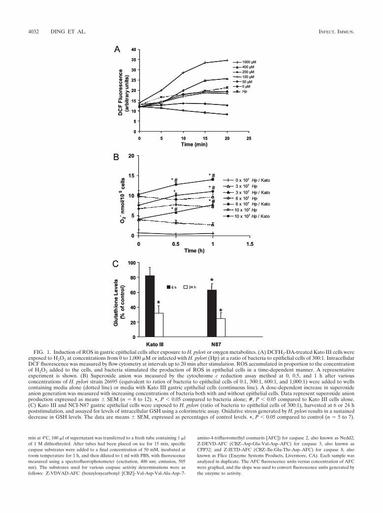
FIG. 1. Induction of ROS in gastric epithelial cells after exposure to H. pylori or oxygen metabolites. (A) DCFH2-DA-treated Kato III cells wereexposed to H2O2 at concentrations from 0 to 1,000 �M or infected with H. pylori (Hp) at a ratio of bacteria to epithelial cells of 300:1. IntracellularDCF fluorescence was measured by flow cytometry at intervals up to 20 min after stimulation. ROS accumulated in proportion to the concentrationof H2O2 added to the cells, and bacteria stimulated the production of ROS in epithelial cells in a time-dependent manner. A representativeexperiment is shown. (B) Superoxide anion was measured by the cytochrome c reduction assay method at 0, 0.5, and 1 h after variousconcentrations of H. pylori strain 26695 (equivalent to ratios of bacteria to epithelial cells of 0:1, 300:1, 600:1, and 1,000:1) were added to wellscontaining media alone (dotted line) or media with Kato III gastric epithelial cells (continuous line). A dose-dependent increase in superoxideanion generation was measured with increasing concentrations of bacteria both with and without epithelial cells. Data represent superoxide anionproduction expressed as means � SEM (n � 8 to 12). *, P 0.05 compared to bacteria alone; #, P 0.05 compared to Kato III cells alone.(C) Kato III and NCI-N87 gastric epithelial cells were exposed to H. pylori (ratio of bacteria to epithelial cells of 300:1), harvested at 6 or 24 hpoststimulation, and assayed for levels of intracellular GSH using a colorimetric assay. Oxidative stress generated by H. pylori results in a sustaineddecrease in GSH levels. The data are means � SEM, expressed as percentages of control levels. *, P 0.05 compared to control (n � 5 to 7).
4032 DING ET AL. INFECT. IMMUN.
To determine the specificity of the responses, the general caspase inhibitorZ-VAD (Bachem, Torrance, CA) as well as specific caspase inhibitors Z-VD-VAD-CH2F, Z-DVED-CH2F, and Z-IETD-CH2F (Enzyme Systems Products,Livermore, CA) for caspases 2, 3, and 8, respectively, were used at 20 and 100�M before stimulation with H. pylori (28, 31).
Statistical analysis. Results are expressed as the means � standard errors ofthe means (SEM). Data were compared by Student’s t test (unpaired unlessotherwise noted) or analysis of variance, and results were considered significantif P values were less than 0.05.
RESULTS
Induction of intracellular ROS in response to H. pylori.Infection of DCFH2-DA-treated gastric epithelial cell lineswith H. pylori was associated with a rapid increase in fluores-cence compared to levels of fluorescence measured in unin-fected control cells, indicating increased accumulation of in-tracellular ROS in infected cell lines (Fig. 1A). No increase inDCF fluorescence was detected when DCFH2-DA-treatedbacteria were assayed by flow cytometry in the absence ofepithelial cells. Since superoxide anion production is notthought to directly induce DCF fluorescence (80), the cyto-chrome c reduction assay was used to demonstrate a dose-dependent increase in superoxide anion in H. pylori-infectedKato III cells (Fig. 1B). The results of our cytochrome c re-duction assay confirm the findings of Nagata et al. (51) andshow evidence of superoxide anion generation by bacteriaalone, although additional superoxide anion is measured overtime when epithelial cells are present with the bacteria (Fig.1B). As shown in Fig. 1C, GSH levels were decreased in bothKato III cells at 24 h after infection and NCI-N87 cells at 6 and24 h after infection, providing evidence of a more sustainedeffect of infection through increased intracellular ROS. Takentogether, these data indicate that H. pylori organisms releasesuperoxide anion and demonstrate that additional ROS aregenerated in host cells through bacterial interaction with epi-thelial cells.
Bacteria prepared directly from blood agar plates did notconsistently induce fluorescence in DCFH2-DA-treated gastricepithelial cells in contrast to preparations made from overnightbrucella broth cultures. There was no early effect of cell-freebacterial culture supernatants or formalin-killed H. pylori, butformalin-killed bacteria increased levels of fluorescence inDCFH2-DA-treated Kato III cells at later time points (116% �2% of control [mean � SEM]; n � 3; P 0.05 compared tocontrol at 30 min). Formalin-killed bacteria also decreasedlevels of GSH (54.6% � 9.4% of control [mean � SEM]; n �3; P 0.05 compared to control at 24 h). Together, theseresults suggest that viable motile bacteria are necessary forearly generation of oxidative stress seen in cultured humangastric epithelial cells whereas oxidative stress induced bykilled bacteria is more delayed.
ROS induction in native human gastric epithelial cells. Inorder to validate the use of gastric epithelial cell lines in studiesof human disease pathogenesis, it is important that findingsobserved in cultured cells be demonstrated in native cells ei-ther in situ or in isolated cell preparations. Our data, obtainedusing an approach that we have employed to demonstrate theexpression of various immune adhesion or accessory mole-cules, including the class II major histocompatibility complex(26) and B7 (78), in native gastric epithelial cells, indicate that
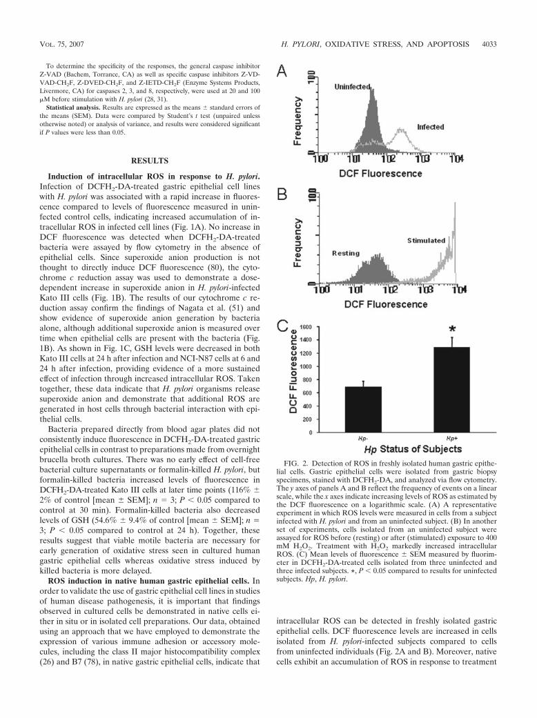
intracellular ROS can be detected in freshly isolated gastricepithelial cells. DCF fluorescence levels are increased in cellsisolated from H. pylori-infected subjects compared to cellsfrom uninfected individuals (Fig. 2A and B). Moreover, nativecells exhibit an accumulation of ROS in response to treatment
FIG. 2. Detection of ROS in freshly isolated human gastric epithe-lial cells. Gastric epithelial cells were isolated from gastric biopsyspecimens, stained with DCFH2-DA, and analyzed via flow cytometry.The y axes of panels A and B reflect the frequency of events on a linearscale, while the x axes indicate increasing levels of ROS as estimated bythe DCF fluorescence on a logarithmic scale. (A) A representativeexperiment in which ROS levels were measured in cells from a subjectinfected with H. pylori and from an uninfected subject. (B) In anotherset of experiments, cells isolated from an uninfected subject wereassayed for ROS before (resting) or after (stimulated) exposure to 400mM H2O2. Treatment with H2O2 markedly increased intracellularROS. (C) Mean levels of fluorescence � SEM measured by fluorim-eter in DCFH2-DA-treated cells isolated from three uninfected andthree infected subjects. *, P 0.05 compared to results for uninfectedsubjects. Hp, H. pylori.
VOL. 75, 2007 H. PYLORI, OXIDATIVE STRESS, AND APOPTOSIS 4033

with exogenous oxidative metabolites similar to that observedin cell lines (Fig. 2C).
Induction of intracellular ROS in response to exogenousoxidative metabolites or inflammatory cytokines. Treatment ofKato III, AGS, and NCI-N87 cells with increasing concentra-tions of H2O2 resulted in a time- and dose-dependent increasein levels of fluorescence in DCFH2-DA-treated epithelial cells,indicating accumulation of intracellular ROS (Fig. 1A). Thepatterns of the response were similar in the three cell linestested. These results confirm that gastric epithelial cells re-spond to exogenous oxidative metabolites with an increase inintracellular ROS analogous to many other cell types (2, 14, 62,63, 80). As certain cytokines that are increased during H. pyloriinfection, including IFN-�, TNF-�, and IL-1� (53), can induceapoptosis (26, 76) and are reported to induce oxidative stress(47, 48, 61), we examined these cytokines for their ability togenerate ROS in gastric epithelial cells. Although TNF-� hasbeen shown to induce a transient surge of ROS in some cellsystems (48, 61), no increase in fluorescence was detected byflow cytometry up to 20 min after stimulating DCFH2-DA-treated Kato III or NCI-N87 cells with 10 ng/ml TNF-� or 100U/ml IFN-�. However, longer-term (overnight) exposure tothese cytokines, as well as 10 ng/ml IL-1�, resulted in increasedbasal levels of fluorescence and enhanced DCF fluorescenceresponses to bacteria and H2O2 (Fig. 3). The results demon-strating increased basal levels of ROS after overnight cytokinetreatment are consistent with the reduced levels of GSH mea-sured 24 h after treatment with IFN-� or TNF-� (data notshown). These experiments indicate that factors generatedthrough the host response to infection can also contribute tooxidative stress in the gastric mucosa.
Effect of cag PAI on ROS accumulation in gastric epithelialcells. Since H. pylori strains bearing the cag PAI are known toinduce more inflammation and are associated with the moresignificant disease manifestations of chronic H. pylori infection(25), we examined bacteria with and without the cag PAI fortheir effect on ROS accumulation in Kato III and NCI-N87cells. As shown in Fig. 4A, both the 26695 and 84-183 strains,
which contain the cag PAI, induced intracellular fluorescencewhile the corresponding cag PAI-deficient isogenic mutants,8-1 and 2-1, had a more limited effect. A similar difference insuperoxide generation was noted at 30 and 60 min after stim-ulation with strain 26695 and its isogenic mutant, 8-1 (Fig. 4B).These data suggest that bacterial genetic factors may play arole in the generation of oxidative stress. It is not clear why cagPAI-negative strains did not induce DCF fluorescence at levelsover those in uninfected control cells while cag PAI-negativestrains were capable of inducing superoxide at greater levelsthan in control cells. This may reflect the different rates atwhich superoxide is generated compared to other ROS.
Inhibition of oxidative stress in gastric epithelial cellstreated with antioxidants. As shown in Fig. 5A, 10 mM NACsignificantly reduced DCF fluorescence in Kato III cells afterstimulation with H2O2 or H. pylori. Similar inhibitory effects ofNAC were seen in epithelial cells isolated from gastric biopsy
FIG. 3. Effect of proinflammatory cytokines on ROS generation.Kato III cells were treated overnight with media alone (no cytokine) ormedia containing 10 ng/ml TNF-�, 100 U/ml IFN-�, or 10 ng/ml IL-1�before stimulation with media alone (control), 400 �M H2O2, or H.pylori at a ratio of bacteria to epithelial cells of 300:1. Peak increasesin ROS levels (measured as increases in DCF fluorescence) are de-picted as means � SEM (n � 3 to 6). All three cytokines increasedbasal levels of fluorescence and ROS responses to H. pylori, whileIFN-� and IL-1� also increased ROS generation after H2O2 stimula-tion. *, P 0.05 compared to cells without cytokine treatment.
FIG. 4. Induction of ROS by cag PAI-bearing strains in gastricepithelial cells. (A) Kato III cells were infected with cag PAI-positivestrains 26695 and 84-183 (solid bars) or their isogenic cag PAI-negativemutants, 8-1 and 2-1, respectively (open bars), at comparable concen-trations. Peak increases in DCF fluorescence occurring within 40 minof infection are expressed as percentages of levels in uninfected controlcells and depicted as means � SEM (n � 5 to 7). *, P 0.05 for PAI�
strains compared to their PAI counterparts and to control levels.(B) Kato III cells were infected with the cag PAI-positive strain 26695or its isogenic mutant, 8-1, at comparable concentrations. The amountof superoxide anion released was measured by the cytochrome c assay.Values at 30 and 60 min after infection are depicted as means � SEM(n � 6, three replicates each). **, P 0.0001; *, P 0.05 (comparedto control levels for both the PAI� strain and its PAI counterpart); #,P 0.01 for cells infected with the PAI� strain compared to the PAI
strain at both time points.
4034 DING ET AL. INFECT. IMMUN.

specimens obtained from uninfected subjects (Fig. 5B) and inNCI-N87 cells (data not shown). Other antioxidants weretested for their ability to inhibit ROS generation after H. pyloriinfection (Fig. 6 and 7). As shown in Fig. 6, GSH, the hydroxyl
scavenger DMTU, and the iron chelators DESF and DTPAeach significantly inhibited H. pylori-induced DCF fluores-cence. Other antioxidants including catalase, mannitol, andSOD had no inhibitory effect. In order to determine the cel-lular source of ROS, sodium azide to inhibit mitochondrialelectron transport, allopurinol to inhibit xanthine oxidase, andDPI, which inhibits NADPH oxidase, were added to cells be-fore H. pylori stimulation. Only allopurinol had a significantinhibitory effect on superoxide anion generation after infection(Fig. 7). Together, the results of these inhibitor studies suggestthat H. pylori infection leads to the formation of several speciesof ROS within gastric epithelial cells including superoxideanion, hydrogen peroxide, hydroxyl radical, and peroxynitrite.
Role of ROS in apoptosis induced by H. pylori. As we havepreviously shown that H. pylori induces apoptosis in gastricepithelial cells (26), we examined a role for oxidative stress inmediating this response (i) by mimicking the effect of H. pyloriwith exogenous oxidative metabolites and (ii) by inhibiting theresponse to bacterial infection with antioxidants. As shown inFig. 8A, a dose-dependent decrease in cell viability and adose-dependent increase in apoptosis were observed whenNCI-N87 cells were treated with various concentrations ofH2O2. The degree of apoptosis induced by 400 �M H2O2
approached that induced by a CD95-activating anti-Fas anti-body in IFN-�-treated cells. This treatment was used as apositive control as it was previously shown to induce maximalapoptosis in gastric epithelial cells. H. pylori infection was alsoshown to induce apoptosis (Fig. 8B) but to a lesser degree thanthat induced by 400 �M H2O2. Although we demonstratedgreater ROS accumulation in gastric epithelial cells very earlyduring infection with cag PAI-positive strains, this did notcorrelate with an increased rate of apoptosis measured at 48 hafter infection. This discrepancy likely reflects the observationthat longer-term effects on ROS, as determined by measuringGSH levels, did not differ significantly at 6 or 24 h of infectionbetween cag PAI-positive and -negative strains (data notshown). Both H. pylori and H2O2 stimulated the activation ofcaspases 3 and 8, but only H. pylori had a significant effect oncaspase 2 activation (Table 1). Inhibition of H. pylori-inducedcaspase activation by the general caspase inhibitor Z-VAD, aswell as by specific caspase inhibitors, demonstrates the speci-
FIG. 5. Inhibition of ROS induction by NAC. (A) DCFH2-DA-loaded Kato III cells were treated with 10 mM NAC or media alone 1 hbefore stimulation with media, 400 �M H2O2, or H. pylori at a ratio ofbacteria to epithelial cells of 300:1. Data are depicted as mean levels ofmaximal DCF fluorescence within 20 min of stimulation in NAC-treated cells expressed as percentages of fluorescence in cells withoutNAC (means � SEM; n � 4 to 6 experiments). *, P 0.05 comparedto cells without NAC pretreatment. (B) Identical experiments per-formed with epithelial cells isolated from subjects without H. pyloriinfection (n � 3). *, P 0.05 compared to cells without NAC pre-treatment.
FIG. 7. Effects of the xanthine oxidase inhibitor allopurinol on H.pylori-induced superoxide anion in Kato III cells. Cells were incubatedwith various concentrations of allopurinol (10 to 100 �M) for 1 hbefore stimulation with H. pylori strain 26695 at a ratio of bacteria toepithelial cells of 300:1. Superoxide anion was assessed by cytochromec assay 30 min after stimulation. Each experiment was performed intriplicate. Values are means � SEM of three separate experiments. *,P 0.05 compared to control cells without allopurinol using a pairedStudent t test.
FIG. 6. Effects of antioxidants on H. pylori-induced ROS produc-tion in Kato III cells. DCFH2-DA-loaded Kato III cells were treatedwith various antioxidants or media alone 1 h before stimulation with H.pylori strain 26695 at a ratio of bacteria to epithelial cells of 300:1.Antioxidants tested in these experiments were 10 mM GSH, 50 mMDMTU, 5 mM DESF, and DTPA. Data are depicted as mean levels ofDCF fluorescence 30 min after stimulation with H. pylori in untreatedcells (H. pylori) or in antioxidant-treated cells (H. pylori � drug), aspercentages of values for uninfected, untreated control cells, � SEM(n � 3 or 4 experiments). The effects of drugs alone are also shown. *,P 0.05; **, P 0.01 (compared to H. pylori alone using a pairedStudent t test).
VOL. 75, 2007 H. PYLORI, OXIDATIVE STRESS, AND APOPTOSIS 4035
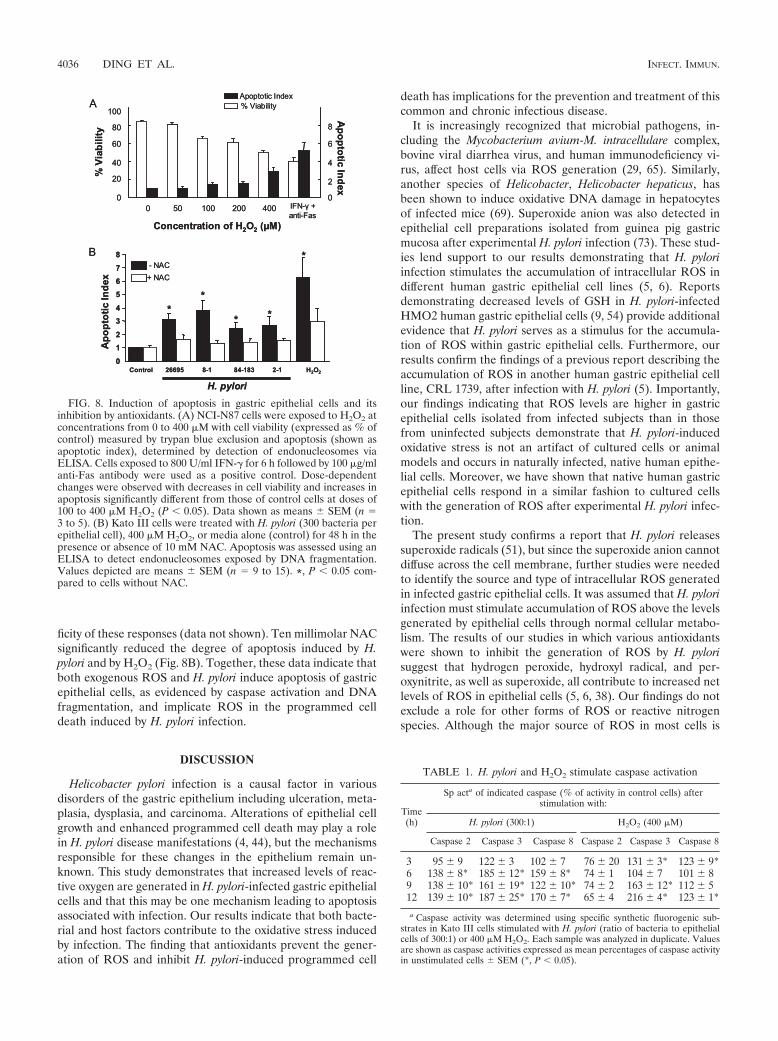
ficity of these responses (data not shown). Ten millimolar NACsignificantly reduced the degree of apoptosis induced by H.pylori and by H2O2 (Fig. 8B). Together, these data indicate thatboth exogenous ROS and H. pylori induce apoptosis of gastricepithelial cells, as evidenced by caspase activation and DNAfragmentation, and implicate ROS in the programmed celldeath induced by H. pylori infection.
DISCUSSION
Helicobacter pylori infection is a causal factor in variousdisorders of the gastric epithelium including ulceration, meta-plasia, dysplasia, and carcinoma. Alterations of epithelial cellgrowth and enhanced programmed cell death may play a rolein H. pylori disease manifestations (4, 44), but the mechanismsresponsible for these changes in the epithelium remain un-known. This study demonstrates that increased levels of reac-tive oxygen are generated in H. pylori-infected gastric epithelialcells and that this may be one mechanism leading to apoptosisassociated with infection. Our results indicate that both bacte-rial and host factors contribute to the oxidative stress inducedby infection. The finding that antioxidants prevent the gener-ation of ROS and inhibit H. pylori-induced programmed cell
death has implications for the prevention and treatment of thiscommon and chronic infectious disease.
It is increasingly recognized that microbial pathogens, in-cluding the Mycobacterium avium-M. intracellulare complex,bovine viral diarrhea virus, and human immunodeficiency vi-rus, affect host cells via ROS generation (29, 65). Similarly,another species of Helicobacter, Helicobacter hepaticus, hasbeen shown to induce oxidative DNA damage in hepatocytesof infected mice (69). Superoxide anion was also detected inepithelial cell preparations isolated from guinea pig gastricmucosa after experimental H. pylori infection (73). These stud-ies lend support to our results demonstrating that H. pyloriinfection stimulates the accumulation of intracellular ROS indifferent human gastric epithelial cell lines (5, 6). Reportsdemonstrating decreased levels of GSH in H. pylori-infectedHMO2 human gastric epithelial cells (9, 54) provide additionalevidence that H. pylori serves as a stimulus for the accumula-tion of ROS within gastric epithelial cells. Furthermore, ourresults confirm the findings of a previous report describing theaccumulation of ROS in another human gastric epithelial cellline, CRL 1739, after infection with H. pylori (5). Importantly,our findings indicating that ROS levels are higher in gastricepithelial cells isolated from infected subjects than in thosefrom uninfected subjects demonstrate that H. pylori-inducedoxidative stress is not an artifact of cultured cells or animalmodels and occurs in naturally infected, native human epithe-lial cells. Moreover, we have shown that native human gastricepithelial cells respond in a similar fashion to cultured cellswith the generation of ROS after experimental H. pylori infec-tion.
The present study confirms a report that H. pylori releasessuperoxide radicals (51), but since the superoxide anion cannotdiffuse across the cell membrane, further studies were neededto identify the source and type of intracellular ROS generatedin infected gastric epithelial cells. It was assumed that H. pyloriinfection must stimulate accumulation of ROS above the levelsgenerated by epithelial cells through normal cellular metabo-lism. The results of our studies in which various antioxidantswere shown to inhibit the generation of ROS by H. pylorisuggest that hydrogen peroxide, hydroxyl radical, and per-oxynitrite, as well as superoxide, all contribute to increased netlevels of ROS in epithelial cells (5, 6, 38). Our findings do notexclude a role for other forms of ROS or reactive nitrogenspecies. Although the major source of ROS in most cells is
FIG. 8. Induction of apoptosis in gastric epithelial cells and itsinhibition by antioxidants. (A) NCI-N87 cells were exposed to H2O2 atconcentrations from 0 to 400 �M with cell viability (expressed as % ofcontrol) measured by trypan blue exclusion and apoptosis (shown asapoptotic index), determined by detection of endonucleosomes viaELISA. Cells exposed to 800 U/ml IFN-� for 6 h followed by 100 �g/mlanti-Fas antibody were used as a positive control. Dose-dependentchanges were observed with decreases in cell viability and increases inapoptosis significantly different from those of control cells at doses of100 to 400 �M H2O2 (P 0.05). Data shown as means � SEM (n �3 to 5). (B) Kato III cells were treated with H. pylori (300 bacteria perepithelial cell), 400 �M H2O2, or media alone (control) for 48 h in thepresence or absence of 10 mM NAC. Apoptosis was assessed using anELISA to detect endonucleosomes exposed by DNA fragmentation.Values depicted are means � SEM (n � 9 to 15). *, P 0.05 com-pared to cells without NAC.
TABLE 1. H. pylori and H2O2 stimulate caspase activation
Time(h)
Sp acta of indicated caspase (% of activity in control cells) afterstimulation with:
H. pylori (300:1) H2O2 (400 �M)
Caspase 2 Caspase 3 Caspase 8 Caspase 2 Caspase 3 Caspase 8
3 95 � 9 122 � 3 102 � 7 76 � 20 131 � 3* 123 � 9*6 138 � 8* 185 � 12* 159 � 8* 74 � 1 104 � 7 101 � 89 138 � 10* 161 � 19* 122 � 10* 74 � 2 163 � 12* 112 � 512 139 � 10* 187 � 25* 170 � 7* 65 � 4 216 � 4* 123 � 1*
a Caspase activity was determined using specific synthetic fluorogenic sub-strates in Kato III cells stimulated with H. pylori (ratio of bacteria to epithelialcells of 300:1) or 400 �M H2O2. Each sample was analyzed in duplicate. Valuesare shown as caspase activities expressed as mean percentages of caspase activityin unstimulated cells � SEM (*, P 0.05).
4036 DING ET AL. INFECT. IMMUN.

mitochondrial electron transport, sodium azide did not inhibitROS generation by H. pylori in our studies. Similarly, theNADPH oxidase inhibitor DPI was without effect while allo-purinol, a xanthine oxidase inhibitor, reduced ROS accumula-tion by bacterial infection. These findings contrast with thestudy of guinea pig mucosal cells in which it was shown that H.pylori induced superoxide anion in epithelial cells through anNADPH-oxidase-like system (73). Since there are limitationsto using pharmacological inhibitors to determine the type andsource of ROS, it is not surprising that such differences exist.Substantial variation in the ability of such agents to inhibitROS and ROS-mediated events in various cell types is re-ported in the literature (5, 48, 61, 65, 73), and differences inmechanisms of ROS generation may vary according to the celltype and species of origin (73).
The current study indicates that host factors also contributeto oxidative stress during H. pylori infection. Since activatedneutrophils or macrophages are potent sources of ROS, in-cluding H2O2 (74), we studied exogenous H2O2 for its effect ongastric epithelial cells. A dose-dependent accumulation of in-tracellular ROS was observed in both cultured and freshlyisolated human gastric epithelial cells. Cytokines that are in-creased in the gastric mucosae of infected subjects, includingTNF-�, IFN-�, and IL-1� (53), also induced oxidative stressand augmented oxidative responses to both H. pylori and H2O2
in our studies. Although cytokines have been reported to in-duce ROS in other cell types (47, 48, 61), our results indicatethat cytokine-mediated oxidative signaling occurs in gastricepithelial cells.
The role of bacterial genotype has been a focus of recentinvestigations into H. pylori pathogenesis. H. pylori strains havebeen classified based on their expression of the cagA gene andthe cag PAI (15) as well as the vacA genotype. Strains that arecagA� have been shown to be associated with increased gastricinflammation, increased bacterial load, and both peptic ulcerdisease and gastric cancer (4, 11, 57). Strains bearing the PAIinduce higher levels of IL-8 (41) and activate transcriptionfactors NF-�B (37, 66) and AP-1 (activator protein 1) (52, 55).cag PAI status also affects gastric epithelial apoptosis (40) andoxidative DNA damage (20, 56). Our results, which indicatethat cag PAI status influences the ability of H. pylori to induceintracellular ROS in gastric epithelial cells, provide furtherinsight into how bacterial genetic factors may play a role indisease pathogenesis. Moreover, since cag PAI-positive strainsare associated with greater inflammation, the host responsemay also contribute to enhanced oxidative stress associatedwith these strains. The differential induction of ROS shown inthe present study may be relevant to the reported associationsof the cag PAI and the activation of epithelial cell signalingpathways (41, 52, 55). As the genome sequence of one of thestrains used in this study, strain 26695, has been determined(41), the opportunity exists to identify more specific bacterialgenes that regulate ROS generation.
We have shown that both H. pylori infection and exogenousROS treatment induce caspase activation and DNA fragmen-tation while antioxidant treatment inhibits the induction ofapoptosis due to H. pylori infection. Further evidence thatoxidative stress may be involved in the alterations of epithelialcell growth in H. pylori infection is found in a study in whichdecreased epithelial cell apoptosis was observed in gastric tis-
sues from H. pylori-infected patients treated with antioxidanttherapy only (45). Although ROS have not been previouslyshown to play a role in programmed cell death of gastricepithelial cells, ROS have been implicated in apoptosis result-ing from various stimuli (12, 34) in other cell types. Of partic-ular relevance to our findings is a recent report of ROS in-volvement in apoptosis induced in host cells by bovine viraldiarrhea virus (65). The present study does not address themechanisms whereby oxidative stress leads to apoptosis, al-though ROS have been shown to contribute to p53 (59)-,Fas-Fas ligand (8, 33)-, ceramide (60)-, and TNF-mediatedkilling (72). It is known that mammalian cells respond to oxi-dative stress with the initial generation of ROS and the sub-sequent activation of redox-sensitive signaling pathways whichcontrol the transcription of genes that may regulate cellgrowth, repair, and death processes. Studies to examine redox-dependent pathways leading to epithelial cell death during H.pylori infection are in progress.
In summary, we have demonstrated that H. pylori infection,exogenous oxidative metabolites, and inflammatory cytokinesinduce the generation of intracellular reactive oxygen speciesin gastric epithelial cells. These in vitro results are corrobo-rated by the higher levels of ROS measured in native epithelialcells from individuals infected with H. pylori. Our findingssuggest that bacterial genotype may be an important determi-nant of the level of oxidative stress generated by infection. Weconclude that oxidative stress may play a role in the increasedprogrammed cell death that occurs during infection, since an-tioxidant treatment inhibited H. pylori-induced apoptosis. Fur-ther studies are necessary to explore how oxidative stress reg-ulates epithelial responses to H. pylori infection, as this willprovide new insight into the pathogenesis of H. pylori-associ-ated conditions.
ACKNOWLEDGMENTS
We acknowledge the excellent technical assistance of Thuyang N.Nguyen. We are grateful to Doug Berg (Washington University, St.Louis, MO) for providing us with the bacterial strains used in many ofthese studies.
Support from the National Institutes of Health (RO1 DK51677,RO1 DK50669, R21 AI48173, and R01 DK61769), the John SealyMemorial Endowment Fund (Development Grant), and a UTMBPresident’s Cabinet Award is also acknowledged.
REFERENCES
1. Akopyants, N. S., S. W. Clifton, D. Kersulyte, J. E. Crabtree, B. E. Youree,C. A. Reece, N. O. Burkanov, E. S. Drazek, B. A. Roe, and D. E. Berg. 1998.Analyses of the cag pathogenicity island of Helicobacter pylori. Mol. Micro-biol. 28:37–53.
2. Anasagasti, M. J., A. Alvarez, C. Avivi, and F. Vidal-Vanaclocha. 1996.Interleukin-1-mediated H2O2 production by hepatic sinusoidal endotheliumin response to B16 melanoma cell adhesion. J. Cell. Physiol. 167:314–323.
3. Ashktorab, H., M. Neapolitano, C. Bomma, C. Allen, A. Ahmed, A. Dubois,T. Naab, and D. T. Smoot. 2002. In vivo and in vitro activation of caspase-8and -3 associated with Helicobacter pylori infection. Microbes Infect. 4:713–722.
4. Backert, S., T. Schwarz, S. Miehlke, C. Kirsch, C. Sommer, T. Kwok, M.Gerhard, U. B. Goebel, N. Lehn, W. Koenig, and T. F. Meyer. 2004. Func-tional analysis of the cag pathogenicity island in Helicobacter pylori isolatesfrom patients with gastritis, peptic ulcer, and gastric cancer. Infect. Immun.72:1043–1056.
5. Bagchi, D., G. Bhattacharya, and S. J. Stohs. 1996. Production of reactiveoxygen species by gastric cells in association with Helicobacter pylori. FreeRadic. Res. 24:439–450.
6. Bagchi, D., T. R. McGinn, X. Ye, M. Bagchi, R. L. Krohn, A. Chatterjee, andS. J. Stohs. 2002. Helicobacter pylori-induced oxidative stress and DNAdamage in a primary culture of human gastric mucosal cells. Dig. Dis. Sci.47:1405–1412.
VOL. 75, 2007 H. PYLORI, OXIDATIVE STRESS, AND APOPTOSIS 4037
7. Baik, S.-C., H.-S. Youn, M.-H. Chung, W.-K. Lee, M.-J. Cho, G.-H. Ko, C.-K.Park, H. Kasai, and K.-H. Rhee. 1996. Increased oxidative DNA damage inHelicobacter pylori-infected human gastric mucosa. Cancer Res. 56:1279–1282.
8. Bauer, M. K. A., M. Vogt, M. Los, J. Siegel, S. Weselborg, and K. Schulze-Osthoff. 1998. Role of reactive oxygen intermediates in activation-inducedCD95 (APO-1/Fas) ligand expression. J. Biol. Chem. 273:8048–8055.
9. Beil, W., B. Obst, K.-F. Sewing, and S. Wagner. 2000. Helicobacter pylorireduces intracellular glutathione in gastric epithelial cells. Dig. Dis. Sci.45:1769–1773.
10. Blaser, M. J., and J. Parsonnet. 1994. Parasitism by the “slow” bacteriumHelicobacter pylori leads to altered gastric homeostasis and neoplasia. J. Clin.Investig. 94:4–8.
11. Blaser, M. J., G. I. Perez-Perez, H. Kleanthous, T. L. Cover, R. M. Peek,P. H. Chyou, G. N. Stemmermann, and A. Nomura. 1995. Infection withHelicobacter pylori strains possessing cagA is associated with an increasedrisk of developing adenocarcinoma of the stomach. Cancer Res. 55:2111–2115.
12. Buttke, T. M., and P. A. Sandstrom. 1994. Oxidative stress as a mediator ofapoptosis. Immunol. Today 15:7–10.
13. Cahill, R. J., H. Xia, C. Kilgallen, S. Beattie, H. Hamilton, and C. O’Morain.1995. Effect of eradication of Helicobacter pylori infection on gastric epithe-lial cell proliferation. Dig. Dis. Sci. 40:1627–1631.
14. Carter, W. O., P. K. Narayanan, and J. P. Robinson. 1994. Intracellularhydrogen peroxide and superoxide anion detection in endothelial cells.J. Leukoc. Biol. 55:253–258.
15. Censini, S., C. Lange, Z. Xiang, J. E. Crabtree, P. Ghiara, M. Borodovsky,R. Rappuoli, and A. Covacci. 1996. Cag, a pathogenicity island of Helico-bacter pylori, encodes type I-specific and disease-associated virulence factors.Proc. Natl. Acad. Sci. USA 93:14648–14653.
16. Clement, M. V., and S. Pervaiz. 1999. Reactive oxygen intermediates regu-late cellular response to apoptotic stimuli: an hypothesis. Free Radic. Res.30:247–252.
17. Crabtree, J. E., S. M. Farmery, I. J. D. Lindley, N. Figura, P. Peichl, andD. S. Tompkins. 1994. CagA/cytotoxic strains of Helicobacter pylori andinterleukin-8 in gastric epithelial cell lines. J. Clin. Pathol. 47:945–950.
18. Crowe, S. E. 2005. Helicobacter infection, chronic inflammation, and thedevelopment of malignancy. Curr. Opin. Gastroenterol. 21:32–38.
19. Crowe, S. E., L. Alvarez, P. M. Sherman, Y. Jin, M. Dytoc, R. H. Hunt, J.Patel, M. J. Muller, and P. B. Ernst. 1995. Expression of interleukin-8 andCD54 by human gastric epithelium after H. pylori infection in vitro. Gastro-enterology 108:65–74.
20. Danese, S., F. Cremonini, A. Armuzzi, M. Candelli, A. Papa, V. Ojetti, A.Pastorelli, S. Di Caro, G. Zannoni, P. De Sole, G. Gasbarrini, and A.Gasbarrini. 2001. Helicobacter pylori CagA-positive strains affect oxygen freeradicals generation by gastric mucosa. Scand. J. Gastroenterol. 36:247–250.
21. Davies, G. R., N. J. Simmonds, T. R. J. Stevens, M. T. Sheaff, N. Banatvala,I. F. Laurenson, D. R. Blake, and D. S. Rampton. 1994. Helicobacter pyloristimulates antral mucosal reactive oxygen metabolite production in vivo. Gut35:179–185.
22. Deneke, S. M., and B. L. Fanburg. 1997. Regulation of cellular glutathione.Am. J. Physiol. 257:L163–L173.
23. Drake, I. M., N. P. Mapstone, C. J. Schorah, K. L. M. White, D. M. Chalmers,M. F. Dixon, and A. T. R. Axon. 1998. Reactive oxygen species activity and lipidperoxidation in Helicobacter pylori associated gastritis: relation to gastricmucosal ascorbic acid concentrations and effect of H. pylori eradication. Gut42:768–771.
24. Ernst, P. B., Y. Jin, J. Navarro, V. E. Reyes, and S. E. Crowe. 1994. Overviewof the immune response to H. pylori, p. 221–232. In R. H. Hunt (ed.),Helicobacter pylori: basic mechanisms to clinical cure. Kluwer AcademicPublishers, Lancaster, United Kingdom.
25. Ernst, P. B., D. A. Peura, and S. E. Crowe. 2006. The translation of Helico-bacter pylori basic research to patient care. Gastroenterology 130:188–206.
26. Fan, X. J., S. E. Crowe, S. Behar, H. Gunasena, G. Ye, H. Haeberle, N. VanHouten, W. K. Gourley, P. B. Ernst, and V. E. Reyes. 1998. The effect of classII MHC expression on adherence of Helicobacter pylori and induction ofapoptosis in gastric epithelial cells: a mechanism for Th1 cell-mediateddamage. J. Exp. Med. 187:1659–1669.
27. Farinati, F., R. Cardin, P. Degan, M. Rugge, F. D. Mario, P. Bonvicini, andR. Naccarato. 1998. Oxidative DNA damage accumulation in gastric carci-nogenesis. Gut 42:351–356.
28. Furlong, I. J., R. Ascaso, R. A. Lopez, and M. K. Collins. 1997. Intracellularacidification induces apoptosis by stimulating ICE-like protease activity.J. Cell Sci. 110(Pt. 5):653–661.
29. Giri, D. K., R. T. Mehta, R. G. Kansal, and B. B. Aggarwal. 1998. Mycobac-terium avium-intracellulare complex activates nuclear transcription factor-�Bin different cell types through reactive oxygen intermediates. J. Immunol.161:4834–4841.
30. Grisham, M. B., T. S. Gaginella, C. von Ritter, H. Tamai, R. M. Be, and D. N.Granger. 1990. Effects of neutrophil-derived oxidants on intestinal perme-ability, electrolyte transport, and epithelial cell viability. Inflammation 14:531–542.
31. Grossmann, J., S. Mohr, E. G. Lapentina, C. Fiocchi, and A. D. Levine. 1998.Sequential and rapid activation of select caspases during apoptosis of normalintestinal epithelial cells. Am. J. Physiol. 274:G1117–G1124.
32. Hocker, M., I. Rosenberg, R. Xavier, R. J. Henihan, B. Wiedenmann, S.Rosewicz, D. K. Podolsky, and T. C. Wang. 1998. Oxidative stress activatesthe human histidine decarboxylase promoter in AGS gastric cancer cells.J. Biol. Chem. 273:23046–23054.
33. Hug, H., S. Strand, A. Grambihler, J. Galle, V. Hack, W. Stremmel, P. H.Krammer, and P. R. Galle. 1997. Reactive oxygen intermediates are involvedin the induction of CD95 ligand mRNA expression by cytostatic drugs inhepatoma cells. J. Biol. Chem. 272:28191–28193.
34. Jacobson, M. D. 1996. Reactive oxygen species and programmed cell death.Trends Biochem. Sci. 21:83–86.
35. Janssen, Y. M. W., B. Van Houten, P. J. A. Borm, and B. T. Mossman. 1993.Cell and tissue responses to oxidative damage. Lab. Investig. 69:261–274.
36. Jones, N. L., P. T. Shannon, E. Cutz, H. Yeger, and P. M. Sherman. 1997.Increase in proliferation and apoptosis of gastric epithelial cells early in thenatural history of Helicobacter pylori infection. Am. J. Pathol. 151:1695–1703.
37. Keates, S., Y. S. Hitti, M. Upton, and C. P. Kelly. 1997. Helicobacter pyloriinfection activates NF-�B in gastric epithelial cells. Gastroenterology 113:1099–1109.
38. Kim, J. M., J. S. Kim, H. C. Jung, Y. K. Oh, H. Y. Chung, C. H. Lee, and I. S.Song. 2003. Helicobacter pylori infection activates NF-�B signaling pathwayto induce iNOS and protect human gastric epithelial cells from apoptosis.Am. J. Physiol. Gastrointest. Liver Physiol. 285:G1171–G1180.
39. Kooy, N. W., J. A. Royall, and H. Ischiropoulos. 1997. Oxidation of2�,7�-dichlorofluorescin by peroxynitrite. Free Radic. Res. 27:245–254.
40. Le’Negrate, G., V. Ricci, V. Hofman, B. Mograbi, P. Hofman, and B. Rossi.2001. Epithelial intestinal cell apoptosis induced by Helicobacter pylori de-pends on expression of the cag pathogenicity island phenotype. Infect. Im-mun. 69:5001–5009.
41. Li, S. D., D. Kersulyte, I. J. Lindley, B. Neelam, D. E. Berg, and J. E.Crabtree. 1999. Multiple genes in the left half of the cag pathogenicity islandof Helicobacter pylori are required for tyrosine kinase-dependent transcrip-tion of interleukin-8 in gastric epithelial cells. Infect. Immun. 67:3893–3899.
42. Luthra, G. K., A. R. DiNuzzo, W. K. Gourley, and S. E. Crowe. 1998.Comparison of biopsy and serological methods of diagnosis of Helicobacterpylori infection and the potential role of antibiotics. Am. J. Gastroenterol.93:1291–1296.
43. Lynch, D. A. F., N. P. Mapstone, A. M. T. Clarke, G. M. Sobala, P. Jackson,L. Morrison, M. F. Dixon, P. Quirke, and A. T. R. Axon. 1995. Cell prolif-eration in Helicobacter pylori associated gastritis and the effect of eradicationtherapy. Gut 36:346–350.
44. Maeda, S., H. Yoshida, Y. Mitsuno, Y. Hirata, K. Ogura, Y. Shiratori, andM. Omata. 2002. Analysis of apoptotic and antiapoptotic signalling pathwaysinduced by Helicobacter pylori. Mol. Pathol. 55:286–293.
45. Mannick, E. E., L. E. Bravo, G. Zarama, J. L. Realpe, X.-J. Zhang, B. Ruiz,E. T. H. Fontham, R. Mera, M. J. S. Miller, and P. Correa. 1996. Induciblenitric oxide synthase, nitrotyrosine and apoptosis in Helicobacter pylorigastritis: effect of antibiotics and antioxidants. Cancer Res. 56:3238–3243.
46. Marchesi, E., C. Rota, Y. C. Fann, C. F. Chignell, and R. P. Mason. 1999.Photoreduction of the fluorescent dye 2�-7�-dichlorofluorescein: a spin trap-ping and direct electron spin resonance study with implications for oxidativestress measurements. Free Radic. Biol. Med. 26:148–161.
47. Matsubara, T., and M. Ziff. 1986. Increased superoxide anion release fromhuman endothelial cells in response to cytokines. J. Immunol. 137:3295–3298.
48. Meier, B., H. H. Radeke, S. Selle, M. Younes, H. Sies, K. Resch, and G. G.Habermehl. 1989. Human fibroblasts release reactive oxygen species in re-sponse to interleukin-1 or tumour necrosis factor-�. Biochem. J. 263:539–545.
49. Menconi, M. J., N. Unno, M. Smith, D. E. Aguirre, and M. P. Fink. 1998.Nitric oxide donor-induced hyperpermeability of cultured intestinal epithe-lial monolayers: role of superoxide radical, hydroxyl radical, and peroxyni-trite. Biochim. Biophys. Acta 1425:189–203.
50. Moss, S. F., J. Calam, B. Agarwal, S. Wang, and P. G. Holt. 1996. Inductionof gastric epithelial apoptosis by Helicobacter pylori. Gut 38:498–501.
51. Nagata, K., H. Yu, M. Nishikawa, M. Kashiba, A. Nakamura, E. F. Sato,T. Tamura, and M. Inoue. 1998. Helicobacter pylori generates superoxideradicals and modulates nitric oxide metabolism. J. Biol. Chem. 273:14071–14073.
52. Naumann, M., S. Wessler, C. Bartsch, B. Wieland, A. Covacci, R. Haas, andT. F. Meyer. 1999. Activation of activator protein 1 and stress responsekinases in epithelial cells colonized by Helicobacter pylori encoding the cagpathogenicity island. J. Biol. Chem. 274:31655–31662.
53. Noach, L. A., N. B. Bosma, J. Jansen, F. J. Hoek, S. J. van-Deventer, andG. N. Tytgat. 1994. Mucosal tumor necrosis factor-alpha, interleukin-1 betaand interleukin-8 production in patients with Helicobacter pylori. Scand. J.Gastroenterol. 29:425–429.
54. Obst, B., S. Wagner, K. F. Sewing, and W. Beil. 2000. Helicobacter pyloricauses DNA damage in gastric epithelial cells. Carcinogenesis 21:1111–1115.
55. O’Hara, A. M., A. Bhattacharyya, R. C. Mifflin, M. F. Smith, K. A. Ryan,
4038 DING ET AL. INFECT. IMMUN.

K. G. Scott, M. Naganuma, A. Casola, T. Izumi, S. Mitra, P. B. Ernst, andS. E. Crowe. 2006. Interleukin-8 induction by Helicobacter pylori in gastricepithelial cells is dependent on apurinic/apyrimidinic endonuclease-1/redoxfactor-1. J. Immunol. 177:7990–7999.
56. Papa, A., S. Danese, A. Sgambato, R. Ardito, G. Zannoni, A. Rinelli, F. M.Vecchio, N. Gentiloni-Silveri, A. Cittadini, G. Gasbarrini, and A. Gasbarrini.2002. Role of Helicobacter pylori CagA� infection in determining oxidative DNAdamage in gastric mucosa. Scand. J. Gastroenterol. 37:409–413.
57. Peek, R. M., Jr., G. G. Miller, K. T. Tham, G. I. Perez-Perez, X. Zhao, J. C.Atherton, and M. J. Blaser. 1995. Heightened inflammatory response andcytokine expression in vivo to cagA� Helicobacter pylori strains. Lab. Investig.71:760–770.
58. Peek, R. M., Jr., S. F. Moss, K. Y. Tham, G. I. Perez-Perez, S. Wang, G. G.Miller, J. C. Atherton, P. R. Holt, and M. J. Blaser. 1997. Helicobacter pyloricagA� strains and dissociation of gastric epithelial cell proliferation fromapoptosis. J. Natl. Cancer Inst. 89:863–868.
59. Polyak, K., Y. Xia, J. L. Zweier, K. W. Kinzler, and B. Vogelstein. 1997. Amodel of p53-induced apoptosis. Nature 389:300–305.
60. Quillet-Mary, A., J. P. Jaffrezou, V. Mansat, C. Bordier, J. Naval, and G.Laurent. 1997. Implication of mitochondrial hydrogen peroxide generationin ceramide-induced apoptosis. J. Biol. Chem. 272:21388–21395.
61. Radeke, H. H., B. Meier, N. Topley, J. Floge, G. G. Habermehl, and K.Resch. 1990. Interleukin 1-� and tumor necrosis factor-� induce oxygenradical production in mesangial cells. Kidney Int. 37:767–775.
62. Rosenkranz, A. R., S. Schmaldienst, K. M. Stuhlmeier, W. Chen, W. Knapp,and G. J. Zlabinger. 1992. A microplate assay for the detection of oxidativeproducts using 2�,7�-dichlorofluorescin-diacetate. J. Immunol. Methods 156:39–45.
63. Royall, J. A., and H. Ischiropoulos. 1993. Evaluation of 2�,7�-dichlorofluo-rescin and dihydrorhodamine 123 as fluorescent probes for intracellularH2O2 in cultured endothelial cells. Arch. Biochem. Biophys. 302:348–355.
64. Ruiz, B., J. C. Rood, E. T. H. Fontham, G. T. Malcom, F. M. Hunter, M.Sobhan, W. D. Johnson, and P. Correa. 1994. Vitamin C concentration ingastric juice before and after anti-Helicobacter pylori treatment. Am. J. Gas-troenterol. 89:533–539.
65. Schweizer, M., and E. Peterhans. 1999. Oxidative stress in cells infected withbovine viral diarrhoea virus: a crucial step in the induction of apoptosis.J. Gen. Virol. 80:1147–1155.
66. Sharma, S. A., M. K. Tummuru, M. J. Blaser, and L. D. Kerr. 1998. Acti-vation of IL-8 gene expression by Helicobacter pylori is regulated by tran-scription factor nuclear factor-�B in gastric epithelial cells. J. Immunol.160:2401–2407.
67. Sharma, S. A., M. K. R. Tummuru, G. G. Miller, and M. J. Blaser. 1995.Interleukin-8 response of gastric epithelial cell lines to Helicobacter pyloristimulation in vitro. Infect. Immun. 63:1681–1687.
68. Shibayama, K., Y. Doi, N. Shibata, T. Yagi, T. Nada, Y. Iinuma, and Y.Arakawa. 2001. Apoptotic signaling pathway activated by Helicobacter pyloriinfection and increase of apoptosis-inducing activity under serum-starvedconditions. Infect. Immun. 69:3181–3189.
69. Sipowicz, M. A., P. Chomarat, B. A. Diwan, M. A. Anver, Y. C. Awasthi, J. M.
Ward, J. M. Rice, K. S. Kasprzak, C. P. Wild, and L. M. Anderson. 1997.Increased oxidative DNA damage and hepatocyte overexpression of specificcytochrome p450 isoforms in hepatitis of mice infected with Helicobacterhepaticus. Am. J. Pathol. 151:933–941.
70. Smoot, D. T., T. B. Elliott, H. W. Verspaget, D. Jones, C. R. Allen, K. G.Vernon, T. Bremner, L. C. Kidd, K. S. Kim, J. D. Groupman, and H.Ashktorab. 2000. Influence of Helicobacter pylori on reactive oxygen-inducedgastric epithelial cell injury. Carcinogenesis 21:2091–2095.
71. Sobalo, G. M., C. J. Schorah, S. Shires, D. A. F. Lynch, B. Gallacher, M. F.Dixon, and A. T. R. Axon. 1993. Effect of eradication of Helicobacter pylori ongastric juice ascorbic acid concentrations. Gut 34:1038–1041.
72. Suematsu, N., H. Tsutsui, J. Wen, D. Kang, M. Ikeuchi, T. Ide, S. Hayashidani,T. Shiomi, T. Kubota, N. Hamasaki, and A. Takeshita. 2003. Oxidative stressmediates tumor necrosis factor-alpha-induced mitochondrial DNA damage anddysfunction in cardiac myocytes. Circulation 107:1418–1423.
73. Teshima, S., K. Rokutan, T. Nikawa, and K. Kishi. 1998. Guinea pig gastricmucosal cells produce abundant superoxide anion through an NADPHoxidase-like system. Gastroenterology 115:1186–1196.
74. Thelen, M., B. Dewald, and M. Baggiolini. 1993. Neutrophil signal transduc-tion and activation of the respiratory burst. Physiol. Rev. 73:797–821.
75. Tomb, J.-F., O. White, A. R. Kerlavage, R. A. Clayton, G. G. Sutton, R. D.Fleischmann, K. A. Ketchum, H. P. Klenk, S. Gill, B. A. Dougherty, K.Nelson, J. Quackenbush, L. Zhou, E. F. Kirkness, S. Peterson, B. Loftus, D.Richardson, R. Dodson, H. G. Khalak, A. Glodek, K. McKenney, L. M.Fitzgerald, N. Lee, M. D. Adams, E. K. Hickey, D. E. Berg, J. D. Gocayne,T. R. Utterback, J. D. Peterson, J. M. Kelley, M. D. Cotton, J. M. Weidman,C. Fujii, C. Bowman, L. Watthey, E. Wallin, W. S. Hayes, M. Borodovsky,P. D. Karp, H. O. Smith, C. M. Fraser, and J. C. Venter. 1997. The completegenome sequence of the gastric pathogen Helicobacter pylori. Nature 388:539–547.
76. Wagner, S., W. Beil, J. Westermann, R. P. Logan, C. T. Bock, C. Trautwein,J. S. Bleck, and M. P. Manns. 1997. Regulation of gastric epithelial cellgrowth by Helicobacter pylori: evidence for a major role of apoptosis. Gas-troenterology 113:1836–1847.
77. Xu, H., R. Chaturvedi, Y. Cheng, F. I. Bussiere, M. Asim, M. D. Yao, D.Potosky, S. J. Meltzer, J. G. Rhee, S. S. Kim, S. F. Moss, A. Hacker, Y. Wang,R. A. Casero, Jr., and K. T. Wilson. 2004. Spermine oxidation induced byHelicobacter pylori results in apoptosis and DNA damage: implications forgastric carcinogenesis. Cancer Res. 64:8521–8525.
78. Ye, G., C. Barrera, X. J. Fan, W. K. Gourley, S. E. Crowe, P. B. Ernst, andV. E. Reyes. 1997. Expression of B7-1 and B7-2 costimulatory molecules byhuman gastric epithelial cells. Potential role in CD4� T cell activation duringHelicobacter pylori infection. J. Clin. Investig. 99:1628–1636.
79. Zhang, Q. B., J. B. Dawodu, A. Husain, G. Etolhi, C. G. Gemmell, and R. I.Russell. 1997. Association of antral mucosal levels of interleukin 8 andreactive oxygen radicals in patients infected with Helicobacter pylori. Clin.Sci. 92:69–73.
80. Zhu, H., G. L. Bannenberg, P. Moldeus, and H. G. Shertzer. 1994. Oxidationpathways for the intracellular probe 2�,7�-dichlorofluorescin. Arch. Toxicol.68:582–587.
Editor: A. D. O’Brien
VOL. 75, 2007 H. PYLORI, OXIDATIVE STRESS, AND APOPTOSIS 4039