application of maldi-tof mass spectrometry for helicobacter pylori study
TRANSCRIPT
STRUCTURE AND FUNCTION OF BIOPOLYMERS
UDC 577.336 + 667.287.4 + 543.51
Application of MALDI-TOF mass spectrometry for study on fibrillar and oligomeric aggregates ofalpha-synuclein
O. V. Severinovskaya1, V. B. Kovalska2, M. Yu. Losytskyy2, V. V. Cherepanov3,
V. Subramaniam4, S. M. Yarmoluk2
1O. O. Chuiko Institute of Surface Chemistry, NAS of Ukraine17, Generala Naumova Str., Kyiv, Ukraine, 03164 2Institute of Molecular Biology and Genetics, NAS of Ukraine150, Akademika Zabolotnoho Str., Kyiv, Ukraine, 031433Institute of Physics, NAS of Ukraine46, Nauky Ave., Kyiv, Ukraine, 030284Nanobiophysics Group, MESA + Institute for Nanotechnology and MIRA Institute for Biomedical Technology, University of TwentePO Box 217, 7500 AE Enschede, The Netherlands
Aim. To study the α-synuclein (ASN) aggregates of different structural origin, namely amyloid fibrils and sphe-rical oligomers, in comparison with a native protein. Methods. MALDI TOF mass spectrometry and atomic for-ce microscopy (AFM). Results. The mass spectra of native and fibrillar ASN have similar character, i. e. they are characterized by the well pronounced peak of protein molecular ion, the low molecular weight associates, andrather low contain of fragmentation products. The spectrum of oligomeric aggregate is characterized by the high contain of fragmentation products, low intensity of protein molecular ion and the absence of peaks of associates.Conclusions. The difference between the spectra of fibrillar and oligomeric ASN could be explained, first, by thedifferent content of the «residual» monomeric ASN and the protein degradation products in the studied samples,and, second, by the different structure-depended mechanisms of the protein degradation induced by the laser ioni-zation. We suggested that the MALDI-TOF mass spectroscopy is a method useful for the investigation of ASN ag-gregation and characterization of its high order self-associates; besides, there is an interest in estimating the po- tency of the MALDI-TOF for the analysis of aggregation of various amyloidogenic proteins.
Keywords: alpha-synuclein, MALDI-TOF, amyloid fibril, oligomeric aggregate, AFM.
Introduction. Pathogenesis of some harmful disorders among them neurodegenerative disorders (Parkinson,Alzheimer’s diseases), prion diseases, type II diabetesis associated with the spontaneous uncontrolled protein aggregation, particularly with the formation of amyloid fibrils. Besides, a wide range of proteins, not involved ina certain disease, are able to form amyloid aggregates[1]. Thefore, the study on the protein aggregation is anactual biomedical task.
Among the methods of protein analysis the MALDI-TOF mass spectrometry is known as a popular and ver-satile tool [2]. In the proteomics, particularly in the stu-dies on protein non-controlled aggregation, MALDI-TOF is mostly applied for the identification of proteinorigin by a proteolysis-based mass mapping method[3, 4].
The method of direct (excluding proteolysis diges-tion) MALDI-TOF has a low descriptiveness for thecharacterization of the protein high-molecular aggre-gates and was applied to analyze the fibrillization in-
190
ISSN 0233–7657. Biopolymers and Cell. 2014. Vol. 30. N 3. P. 190–196 doi: http://biopolymers.org.ua/doi/bc.000895
Institute of Molecular Biology and Genetics, NAS of Ukraine, 2014

termediates and side products, e. g. the misfolded be-ta-lactoglobuline dimers and protein degradation pro-ducts [5].
Alpha-synuclein (ASN) is a small natively unfoldedprotein that plays a central role in the etiology of Parkin- son’s disease. It forms amyloid fibrils that are found inLewy bodies, i. e. cell depositions in the brain that arecharacteristic of this disease [6, 7]. Besides, during theaggregation process ASN forms small oligomeric inter- mediates, which are considered as presumably toxicspecies [8–10].
Here we report the examination of the ASN aggre-gates populations of different structural origin, namelyamyloid fibrils and spherical oligomers, by the methodof MALDI-TOF mass spectrometry. We suggest that the filamentous amyloid fibrils and spherical oligomers dueto the distinct folding of the protein molecules and asso-ciation/degradation on the aggregation pathway couldpossess the mass spectra of different character. Besides,we intend to evaluate the applicability of MALDI-TOFas a method for the analysis of protein aggregates andstudy on the aggregation process.
Materials and methods. Reagents. Recombinanthuman wild-type ASN was expressed and purified in10 mM Tris-HCl, 50 mM NaCl (pH 7.4) as described ear- lier [11]. ASN amyloid fibrils and oligomers were obtai- ned according to [11] and [12] correspondingly.
MALDI-TOF studies. The samples of native, fibril-lar and oligomeric ASN were prepared for the MALDI-TOF analysis as follows: matrix material (12 mg of 3,5-dimethoxy-4-hydroxycinnamic (sinapinic) acid) was dissolved in 1 ml of water/acetonytyl solution 1:1 (v/v)with addition of 0.1 % (v/v) of trifluoroacetic acid. Theobtained solution was incubated during 10 min at 30 °C in ultrasonic bath up to the full dissolving of the acid.[13]. The 50 µmol protein samples in 0,05 M Tris-HClbuffer (pH 7.9) with concentration of 1mg/ml werethen added to the matrix solution in 1:1 ratio. Small ali- quots of the mixture were applied to the steel probe tips and dried. Mass analysis was performed on the «Auto-flex II» («Bruker Daltonics», Germany) MALDI-TOFmass spectrometer with nitrogen laser (λ = 337 nm).Spectra were obtained for the mass range 7 000 to 100000 m/z. in positive ion reflectron registration mode.The spectra were obtained by summing the data of 100laser shots.
Atomic force microscopy studies. The structure ofASN aggregates was studied by means of AFM («Sol-ver Pro M» system, NT-MDT, RF). The scanning ratewas 30 µm/s. For the formation of subnanolayer consis-ting of separate nanoobjects, the reaction solutions we-re diluted 30 times with bidistilled water. Then a dropof the solution was applied on the freshly cleaved surfa- ce of mica. The AFM measurements were carried out in a tapping mode at ambient conditions after the full eva-poration of the solvent. The AFM probes of type NSG01(NT-MDT) were used. The average diameter values ofASN aggregates were determined based on their heightsin the AFM images.
Results and discussion. Structural peculiarities ofnative, fibrillar and oligomeric ASN. ASN is a small(140 amino acids) protein that is known to be nativelyunfolded in solutions. The protein's amino acid sequen- ce consists of a basic N-terminal region (residues 1–95) containing repeats of highly conserved KTKEGV hexa-meric motif, and an acidic C-terminal region (residues96–140) [14], the first 100 residues are predicted to ha-ve α-helical propensity [15].
Amyloid fibrils are stable protein aggregates, withcross-β filament structure where the protein molecules,making up the β-sheet, are arranged perpendicular tothe fibril axis [16]. The core of the ASN fibril containsmainly the protein residues of the basic region (appro-ximately residues 38–95 [17]). We have estimated thecharacteristics of the ASN fibrils population by AFM.These fibrils are both linear and branched long filamentswith the diameter of single filament about 3–8 nm (Fig.1). The bundles of fibrils formed by overlapping of sing-le filaments were observed in the studied sample as well.The strong fluorescent sensitivity of the amyloid-specific cyanine dye 7519 to the ASN fibrils was pre-viously reported [18].
The ASN oligomers are known as spherical proteinaggregates of the size in the range 2–20 nm [19–21], be-sides, the structures with annular and tubular morpho-logy have also been reported [21–23].
Our AFM study demonstrated the ASN oligomerpopulation to be mostly the species of the height from 3till 6 nm (Fig. 2). Besides, the structures formed due tothe coalescence of these aggregates are observed; thediameter of such «super-oligomeric» structures beingup to 10 nm.
191
MALDI-TOF MASS SPECTROMETRY FOR STUDY ON AGGREGATES OF α-SYNUCLEIN

192
SEVERINOVSKAYA O. V. ET AL.
Despite the oligomer aggregates are mainly consi-dered to possess beta-sheet structure [24, 25], the pre-sence of α-helical content has been shown as well [19].The difference between the secondary structure motifsof amyloid fibrils and oligomeric aggregates is indi-cated by the different sensitivity of amyloid-specificcyanine dyes to these ASN formations [26]. The noti-ceably lower fluorescent response of the dyes on the
presence of oligomers comparing to fibrils is explainedby the lower content of beta-pleated regions accessiblefor the dye molecules.
MALDI-TOF studies. The mass spectra of native,fibrillar and oligomeric ASN are presented in Fig. 3.All spectra contain the peaks corresponding to the pro-tein molecular ion about 14460 m/z and a wide range of protein fragmentation peaks, but only two of these peaks
2
4
0
6
2 1040 6 8 µm
a b
µm0 62 4 8 100
6
4
2
8
108
Fig. 1. AFM image of amyloid fibril of ASN (a) and Z-profile along the line marked on the image (b)
µm0 62 4 80
6
4
2
8
a b
2 840 6 µm
2
4
0
6
8
10
Fig. 2. AFM image of oligomer aggregates of ASN (a) and Z-profile along the line marked on the image (b)
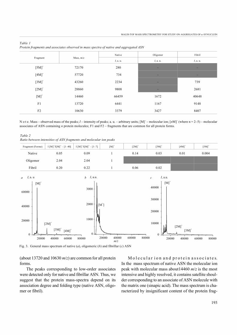
(about 13720 and 10630 m/z) are common for all protein forms.
The peaks corresponding to low-order associateswere detected only for native and fibrillar ASN. Thus, wesuggest that the protein mass-spectra depend on itsassociation degree and folding type (native ASN, oligo-mer or fibril).
M o l e c u l a r i o n a n d p r o t e i n a s s o c i a t e s.In the mass spectrum of native ASN the molecular ionpeak with molecular mass about14460 m/z is the mostintensive and highly resolved, it contains satellite shoul-der corresponding to an associate of ASN molecule withthe matrix one (sinapic acid). The mass spectrum is cha-racterized by insignificant content of the protein frag-
193
MALDI-TOF MASS SPECTROMETRY FOR STUDY ON AGGREGATES OF α-SYNUCLEIN
Fragment Mass, m/zNative Oligomer Fibril
I, a. u. I, a. u. I, a. u.
[5M]+ 72170 280 – –
[4M]+ 57720 734 – –
[3M]+ 43260 2234 – 739
[2M]+ 28860 9808 – 2681
[M]+ 14460 66459 1672 40648
F1 13720 6441 1167 9148
F2 10630 3379 3427 8407
N o t e. Mass – observed mass of the peaks; I – intensity of peaks; a. u. – arbitrary units; [M]+ – molecular ion; [nM]+ (where n = 2–5) – molecularassociates of ASN containing n protein molecules; F1 and F2 – fragments that are common for all protein forms.
Table 1Protein fragments and associates observed in mass spectra of native and aggregated ASN
Fragment (Forme) I [M]+/I[M]+ – [1–40] I [M]+/I[M]+ – [1–7] [M]+ [2M]+ [3M]+ [4M]+ [5M]+
Native 0.05 0.09 1 0.14 0.03 0.01 0.004
Oligomer 2.04 2.04 1
Fibril 0.20 0.22 1 0.06 0.02
Table 2Ratio between intensities of ASN fragments and molecular ion peaks
m/z
a b c
20000 40000 60000 800000
20000
40000
60000
[4M]+[3M]+
[2M]+
[M]+
I, a. u
20000 40000 60000 800000
1000
2000
3000
[M+]
I, a.u.
20000 40000 60000 800000
10000
20000
30000
40000
[3M]+[2M]+
[M]+
I, a.u.
Fig. 3. General mass spectrum of native (a), oligomeric (b) and fibrillar (c) ASN
mentation products. The series of peaks with the inten-sities decreasing upon the mass increase are detected inthe high mass region; they correspond to the protein mo- lecular associates from dimer to pentamer with molecu- lar masses about 28860, 43260, 57720 and 72170 m/z,correspondingly (Table 1, 2). The association is causedby intermolecular interactions and is typically obser-ved in the MALDI-TOF mass spectra of different pro-teins (albumin, insulin, lysozime etc.) [4, 13], the forma- tion of ASN dimer, trimer and tetramer was also descri- bed [15].
In the fibrillar protein spectrum, similarly to that of the native ASN, the peak of molecular ion is the most in-tensive. The low order associates are also presented inthe spectrum, but the number of aggregated molecules(only dimers and trimers were observed) and intensity of peaks are lower than for the native protein. On the otherhand, the number and intensity of the peaks of fragmen-tation products for the fibrillar ASN is enhanced as com- pared to the native protein.
The mass spectrum of oligomer significantly differsfrom that of native and fibrillar protein. It is characteri-
zed by a very high fragmentation degree (the peak ofmolecular ion is less intensive than the main peaks offragmentation products) and by the absence of associa-tes peaks.
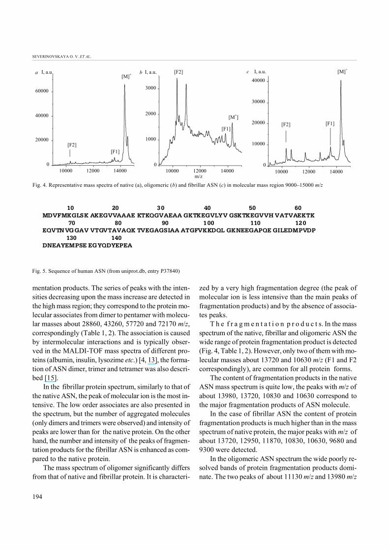
T h e f r a g m e n t a t i o n p r o d u c t s. In the massspectrum of the native, fibrillar and oligomeric ASN thewide range of protein fragmentation product is detected (Fig. 4, Table 1, 2). However, only two of them with mo- lecular masses about 13720 and 10630 m/z (F1 and F2correspondingly), are common for all protein forms.
The content of fragmentation products in the nativeASN mass spectrum is quite low, the peaks with m/z of about 13980, 13720, 10830 and 10630 correspond tothe major fragmentation products of ASN molecule.
In the case of fibrillar ASN the content of proteinfragmentation products is much higher than in the mass spectrum of native protein, the major peaks with m/z of about 13720, 12950, 11870, 10830, 10630, 9680 and9300 were detected.
In the oligomeric ASN spectrum the wide poorly re-solved bands of protein fragmentation products domi-nate. The two peaks of about 11130 m/z and 13980 m/z
194
SEVERINOVSKAYA O. V. ET AL.
a b c
10000 12000 140000
20000
40000
60000
[F2][F1]
[M]+I, a.u
10000 12000 140000
1000
2000
3000
[F2]
[F1]
[M+]
I, a.u.
m/z10000 12000 14000
0
10000
20000
30000
40000
[F2] [F1]
[M]+I, a.u.
Fig. 4. Representative mass spectra of native (a), oligomeric (b) and fibrillar ASN (c) in molecular mass region 9000–15000 m/z
10 20 30 40 50 60MDVFMKGLSK AKEGVVAAAE KTKQGVAEAA GKTKEGVLYV GSKTKEGVVH VATVAEKTK
70 80 90 100 110 120EQVTNVGGAV VTGVTAVAQK TVEGAGSIAA ATGFVKKDQL GKNEEGAPQE GILEDMPVDP
130 140DNEAYEMPSE EGYQDYEPEA
Fig. 5. Sequence of human ASN (from uniprot.db, entry P37840)

are twice more intensive than this of the molecular ionof ASN. Another intensive peak of about 10630 m/z isalso present in the spectra of native and fibrillar ASN.The fragment with m/z about 13720 that is common forall protein forms has low intensity in spectra of oligome- ric aggregate.
Explanations of a distinct character of the fibrillarand oligomeric ASN mass-spectra. We could proposetwo explanations of the distinctions in the mass-spectra of fibrillar and oligomeric ASN.
T h e f i r s t e x p l a n a t i o n is that the similarityof the native and fibrillar ASN mass specta is caused by the presence of an excess of non-aggregated protein inthe fibrillar ASN sample. Since the ASN monomericmolecules are easily ionized they give a high intensivepeak of the molecular ion and associates.
The distinction between the fragmentation of fibrils and oligomers could be caused by the presence of spe-cific degradation products formed during the aggrega-tion reaction or later storage of the samples.
A n o t h e r p o s s i b l e e x p l a n a t i o n is dis-similarity in the folding of protein molecules in the amy- loid fibrils and the oligomeric species and different struc-ture of these aggregates.
It may be supposed that due to their regular ladder-like structure the degradation of the amyloid fibrils upon ionization in a large extent occurs through the tearingoff the intact protein molecules from the end (one by one)of the filament. These protein molecules are responsib-le for an intensive peak of the molecular ion and peaks ofthe low-ordered associates.
Besides, the laser-induced degradation could occurthrough a break in the protein chain, that leads to the ap-pearance of new intensive bands of the protein frag-mentation products. This mechanism is proved by high-er number, content and lower resolution of the proteinfragment peaks in the spectrum of fibrils comparingwith the native ASN.
The large content of fragmentation products in thespectrum of the oligomeric aggregates and their lowresolution may be explained by the poor ionization andpoor stability of these structures. We assume that due to their spherical shape the oligomeric aggregates degra-ded through the lost of protein fragments from the ope-ned surface regions. The suggestion about the hindered ability of degradation of the oligomeric aggregate through
the tearing off a whole protein molecule is supported by the very low intensity of molecular ion.
О. В. Се ве ри но вська, В. Б. Ко в альська, М. Ю. Ло сиць кий, В. В. Че ре па нов, В. Суб ра маніам, С. М. Ярмо люк
Ви ко рис тан ня ме то ду MALDI-TOF масс-спек тро метрії для вив чен ня фібри ляр них та оліго мер них аг ре гатів аль фа-си нук леї ну
Ре зю ме
Мета. Вив чен ня аг ре гатів аль фа-си нук леї ну (ASN) різно го струк -ту ро во го по ход жен ня, а саме – амілої дних фібрил і сфе рич нихоліго мерів порівня но з на тив ним білком. Ме то ди. MALDI-TOFмас-спек тро метрія та атом но-си ло ва мікрос копія (АFМ). Ре -зуль та ти. Мас-спек три на тив но го і фібри ляр но го ASN ма ютьподібний ха рак тер – для них ха рак терні інтен сив ний пік мо ле ку -ляр но го іона білка, піки низ ь ко мо ле ку ляр них асоціатов та до сить не знач ний вміст про дуктів фраг мен тації білка. У той же час успектрі оліго мер них аг ре гатів спос теріга ють ся ви со ка кон цент-рація про дуктів фраг мен тації білка, низ ь ка інтен сивність мо ле ку -ляр но го іона та відсутність піків са мо а соціатів. Вис нов ки. Різ-ницю між спек тра ми фиб ри ляр но го та оліго мер но го ASN мож напо яс ни ти як на явністю у зраз ках «за лиш ко во го» ASN і про дуктівдег ра дації білка, так і різни ми струк ту ро во за леж ни ми ме ханіз-мами руй ну ван ня цих двох видів аг ре гатів при ла зерній де сорб-ції/іонізації. MALDI-TOF мас-спек тро метрію мож на за про по ну -ва ти як ме тод вив чен ня аг ре гації та аналізу ви со ко мо ле ку ляр нихаг ре гатів ASN. Та кож пред став ляє інте рес виз на чен ня ефек тив -ності цьо го ме то ду для досліджен ня аг ре гатів різних амілої до -генних білків.
Ключові сло ва: аль фа-си нук леїн, MALDI-TOF, амілої дна фібри-ла, оліго мерні аг ре га ти, АFМ.
О. В. Се ве ри нов ская, В. Б. Ко ва льская, М. Ю. Ло сиц кий, В. В. Че ре па нов, В. Суб ра ма ни ам, С. Н. Ярмо люк
Исполь зо ва ние ме то да MALDI-TOF масс-спек тро мет рии для из уче ния фиб рил ляр ных и оли го мер ных аг ре га тов аль фа-си нук ле и на
Ре зю ме
Цель. Изу че ние аг ре га тов аль фа-си нук ле и на (ASN) раз лич нойструк ту ры, а имен но – ами ло ид ных фиб рилл и сфе ри чес ких оли -гомеров в срав не нии с на тив ным бел ком. Ме то ды. MALDI-TOFмасс-спек тро мет рия и атом но-си ло вая мик рос ко пия (АFМ). Ре -зуль та ты. Масс-спек тры на тив но го и фиб рил ляр но го ASN име -ют по до бный ха рак тер – для них ха рак тер ны ин тен сив ный пикмо ле ку ляр но го иона бел ка, пики низ ко мо ле ку ляр ных ас со ци а тов,а так же дос та точ но не зна чи тель ное со дер жа ние про дук товфраг мен та ции бел ка. В то же вре мя в спек тре оли го мер ных агре-га тов на блю да ют ся вы со кая кон цен тра ция про дук тов фраг мен -та ции бел ка, мо ле ку ляр ный ион низ кой ин тен сив нос ти и от сут-ствие пи ков ас со ци а тов бел ка. Вы во ды. Раз ли чие меж ду спект-рами фиб рил ляр но го и оли го мер но го ASN мож но об ъ яс нить каксо дер жа ни ем «из быт ка» ASN и про дук тов дег ра да ции бел ка,так и раз лич ны ми струк тур но-за ви си мы ми ме ха низ ма ми раз ру -ше ни я ми этих двух ви дов аг ре га тов при ла зер ной де сор бции/ио -ни за ции. MALDI-TOF масс-спек тро мет рию мож но пред ло житьв ка чес тве ме то да из уче ния аг ре га ции и ана ли за вы со ко мо ле ку -ляр ных аг ре га тов ASN. Так же пред став ля ет ин те рес опре де ле-
195
MALDI-TOF MASS SPECTROMETRY FOR STUDY ON AGGREGATES OF α-SYNUCLEIN

196
SEVERINOVSKAYA O. V. ET AL.
ние эф фек тив нос ти MALDI-TOF для ис сле до ва ния аг ре га тов раз-лич ных ами ло и до ген ных бел ков.
Клю че вые сло ва: аль фа-си нук ле ин, MALDI-TOF, ами ло ид наяфибрил ла, оли го мер ные аг ре га ты, АFМ.
REFERENCES
1. Green J, Goldsbury C, Mini T, Sunderji S, Frey P, Kistler J, Co-oper G, Aebi U. Full-length rat amylin forms fibrils followingsubstitution of single residues from human amylin. J Mol Biol.2003;326(4):1147–56.
2. Marvin LF, Roberts MA, Fay LB. Matrix-assisted laserdesorption/ionization time-of-flight mass spectrometry inclinical chemistry. Clin Chim Acta. 2003;337(1–2):11–21.
3. Nazabal A, Weber J. Characterization and quantitation ofantibody aggregates by high mass MALDI mass spectrometry. J Biomol Tech. 2010; 21(3 Suppl): S36.
4. Metods in enzymology. Amyloid, prions and other protein aggre-gates, part C. Eds I. Kheterpal, R. Wetzel. Amsterdam, ElsevierInc., 2006; Vol. 413. 375 p.
5. Hamada D, Dobson CM. A kinetic study of beta-lactoglobulinamyloid fibril formation promoted by urea. Protein Sci. 2002;11 (10):2417–26.
6. Uversky VN. Neuropathology, biochemistry, and biophysics ofalpha-synuclein aggregation. J Neurochem. 2007;103(1):17–37.
7. Bartels AL, Leenders KL. Parkinson's disease: the syndrome, the pathogenesis and pathophysiology. Cortex. 2009;45(8):915–21.
8. Lashuel HA, Petre BM, Wall J, Simon M, Nowak RJ, Walz T,Lansbury PT Jr. Alpha-synuclein, especially the Parkinson's di-sease-associated mutants, forms pore-like annular and tubularprotofibrils. J Mol Biol. 2002;322(5):1089–102.
9. Volles MJ, Lansbury PT Jr. Zeroing in on the pathogenic form of alpha-synuclein and its mechanism of neurotoxicity in Parkin-son's disease. Biochemistry. 2003;42(26):7871–8.
10. Volles MJ, Lee SJ, Rochet JC, Shtilerman MD, Ding TT, Kessler JC, Lansbury PT Jr. Vesicle permeabilization by protofibrillaralpha-synuclein: implications for the pathogenesis andtreatment of Parkinson's disease. Biochemistry. 2001;40(26):7812–9.
11. van Raaij ME, Segers-Nolten IM, Subramaniam V. Quantitativemorphological analysis reveals ultrastructural diversity of amy-loid fibrils from alpha-synuclein mutants. Biophys J. 2006;91(11):L96–8.
12. van Rooijen BD, Claessens MM, Subramaniam V. Lipid bilayerdisruption by oligomeric alpha-synuclein depends on bilayercharge and accessibility of the hydrophobic core. Biochim Bio-phys Acta. 2009;1788(6):1271–8.
13. Dekina SS, Romanovska II, Gromovoy TYu. Influence of poly-mers on lysozyme molecules association. Biopolym Cell. 2011;27(6):442–445.
14. Sode K, Ochiai S, Kobayashi N, Usuzaka E. Effect of reparationof repeat sequences in the human alpha-synuclein on fibrillationability. Int J Biol Sci. 2006;3(1):1–7.
15. Wang W, Perovic I, Chittuluru J, Kaganovich A, Nguyen LT, Li-ao J, Auclair JR, Johnson D, Landeru A, Simorellis AK, Ju S, Co-okson MR, Asturias FJ, Agar JN, Webb BN, Kang C, Ringe D,Petsko GA, Pochapsky TC, Hoang QQ. A soluble α-synucleinconstruct forms a dynamic tetramer. Proc Natl Acad Sci USA.2011;108(43):17797–802.
16. Rambaran RN, Serpell LC. Amyloid fibrils: abnormal protein as-sembly. Prion. 2008;2(3):112-7.
17. Cho MK, Kim HY, Fernandez CO, Becker S, Zweckstetter M.Conserved core of amyloid fibrils of wild type and A30P mutantα-synuclein. Protein Sci. 2011;20(2):387–95.
18. Volkova KD, Kovalska VB, Yu Losytskyy M, Veldhuis G, Se-gers-Nolten GM, Tolmachev OI, Subramaniam V, Yarmoluk SM.Studies of interaction between cyanine dye T-284 and fibrillar al-pha-synuclein. J Fluoresc. 2010;20(6):1267–74.
19. Apetri MM, Maiti NC, Zagorski MG, Carey PR, Anderson VE.Secondary structure of alpha-synuclein oligomers: characteri-zation by raman and atomic force microscopy. J Mol Biol. 2006;355(1):63–71.
20. Conway KA, Lee SJ, Rochet JC, Ding TT, Williamson RE, Lans-bury PT Jr. Acceleration of oligomerization, not fibrillization, is a shared property of both alpha-synuclein mutations linked toearly-onset Parkinson's disease: implications for pathogenesisand therapy. Proc Natl Acad Sci U S A. 2000;97(2):571–6.
21. Lashuel HA, Petre BM, Wall J, Simon M, Nowak RJ, Walz T,Lansbury PT Jr. Alpha-synuclein, especially the Parkinson's di-sease-associated mutants, forms pore-like annular and tubularprotofibrils. J Mol Biol. 2002;322(5):1089–102.
22. Ding TT, Lee SJ, Rochet JC, Lansbury PT Jr. Annular alpha-synuclein protofibrils are produced when spherical protofibrilsare incubated in solution or bound to brain-derived membranes.Biochemistry. 2002;41(32):10209–17.
23. Quist A, Doudevski I, Lin H, Azimova R, Ng D, Frangione B,Kagan B, Ghiso J, Lal R. Amyloid ion channels: a commonstructural link for protein-misfolding disease. Proc Natl AcadSci USA. 2005;102(30):10427–32.
24. Volles MJ, Lee SJ, Rochet JC, Shtilerman MD, Ding TT, Kessler JC, Lansbury PT Jr. Vesicle permeabilization by protofibrillaralpha-synuclein: implications for the pathogenesis and treatmentof Parkinson's disease. Biochemistry. 2001;40(26):7812–9.
25. Kaylor J, Bodner N, Edridge S, Yamin G, Hong DP, Fink AL.Characterization of oligomeric intermediates in alpha-synuclein fibrillation: FRET studies of Y125W/Y133F/Y136F alpha-sy-nuclein. J Mol Biol. 2005;353(2):357–72.
26. Kovalska VB, Losytskyy MY, Tolmachev OI, Slominskii YL, Se-gers-Nolten GM, Subramaniam V, Yarmoluk SM. Tri- and penta-methine cyanine dyes for fluorescent detection of α-synucleinoligomeric aggregates. J Fluoresc. 2012;22(6):1441–8.
Received 31.12.13