heavy metal toxicity to bacteria – are the existing growth media accurate enough to determine...
TRANSCRIPT
Chemosphere 90 (2013) 1195–1200
Contents lists available at SciVerse ScienceDirect
Chemosphere
journal homepage: www.elsevier .com/locate /chemosphere
Heavy metal toxicity to bacteria – Are the existing growth media accurate enoughto determine heavy metal toxicity?
I.V.N. Rathnayake a,b,c, Mallavarapu Megharaj a,b,⇑, G.S.R. Krishnamurti a,b, Nanthi S. Bolan a,b, Ravi Naidu a,b
a Centre for Environmental Risk Assessment and Remediation (CERAR), University of South Australia, Mawson Lakes Boulevard, Mawson Lakes, South Australia 5095, Australiab CRC for the Contaminant Assessment and Remediation of the Environment (CRC CARE), Mawson Lakes Boulevard, Mawson Lakes, South Australia 5095, Australiac Department of Microbiology, Faculty of Science, University of Kelaniya, Kelaniya GQ 11600, Sri Lanka
h i g h l i g h t s
" A new bacterial growth medium containing low levels of metal-chelates was formulated." This medium has high free metal ion activity and more suitable to determine metal toxicity." This medium provides a viable option for the study of metals–bacteria interactions.
a r t i c l e i n f o
Article history:Received 3 May 2012Received in revised form 21 July 2012Accepted 8 September 2012Available online 4 October 2012
Keywords:Heavy metal toxicitySpeciationBioavailabilityCadmiumCopperBacteria
0045-6535/$ - see front matter � 2012 Elsevier Ltd. Ahttp://dx.doi.org/10.1016/j.chemosphere.2012.09.036
⇑ Corresponding author at: Centre for EnvironmRemediation (CERAR), University of South AustraliaMawson Lakes, South Australia 5095, Australia. Tel.:83023057.
E-mail address: [email protected].
a b s t r a c t
A new minimal medium was formulated considering the limitations of the existing media for testingheavy metal sensitivity to bacteria. Toxicity of cadmium and copper to three bacteria was investigatedin the new medium and compared with three other media commonly used to study the effect of the toxicmetals. Based on speciation data arrived at using ion-selective electrodes, the available free-metal con-centration in solution was highest in the MES-buffered medium. This finding was strongly supportedby the estimated EC50 values for the metals tested based on the toxicity bioassays. The free-ionic cad-mium and copper concentrations in the medium provide more accurate determination of metal concen-trations that affects the bacteria, than with most of other existing media. This will avoid doubts on othermedia and misleading conclusions relevant to the toxicity of heavy metals to bacteria and provides a bet-ter option for the study of metal–bacteria interactions.
� 2012 Elsevier Ltd. All rights reserved.
1. Introduction
Toxicity of various heavy metals to bacteria isolated fromnumerous habitats in the terrestrial environment has been exten-sively studied for many years. Many different approaches havebeen used to study the metal toxicity to bacteria either as singleisolates (Babich and Stotzky, 1977; Keeling and Cater, 1998;Tangaromsuk et al., 2002; Madhaiyan et al., 2007), or mixed cultures(Saeki et al., 2002; Zhang et al., 2003) in solution, or in communitiesin soil, water, etc. (Diaz-Raviña and Bååth, 1996; Rajapaksha et al.,2004; Oliveira and Pampulha, 2006), to mention a few.
Using widely different bacteriological culture media, a broadrange of metal concentrations have been reported on the tolerance
ll rights reserved.
ental Risk Assessment and, Mawson Lakes Boulevard,+61 8 83025044; fax: +61 8
au (M. Megharaj).
of heavy metals by bacteria from various environments (Duxburyand Bicknell, 1983; El-Aziz et al., 1991; Martensson and Torstens-son, 1996; Malik and Jaiswal, 2000; Roane and Pepper, 2000;Amoroso et al., 2002; Renella et al., 2003; Yilmaz, 2003;Piotrowska-Seget et al., 2005; Chen et al., 2006; Sprocati et al.,2006; Abou-Shanab et al., 2007; Kim et al., 2007). The main basisfor the choice of a specific medium was its ability to encouragegrowth of the specified bacteria or group of bacteria in that med-ium (Angle and Chaney, 1989; Richards et al., 2002; Chen et al.,2006; Miranda and Rojas, 2006; Congeevaram et al., 2007). Themedia were supplemented with different concentrations of nutri-ent metal salts and were inoculated with relevant bacterial isolateor group of isolates. The growth was then measured by variousparameters such as, turbidity, biomass, and enzyme activities todetermine the sensitivity by different criteria, viz., EC50, MIC (min-imum inhibitory concentration), LD50, etc., after specified time ofincubation at a specified temperature either in solid (Hassenet al., 1998; Saeki et al., 2002) or liquid media (Hassen et al.,1998; Clausen, 2000).
1196 I.V.N. Rathnayake et al. / Chemosphere 90 (2013) 1195–1200
In most studies, bacteriological culture media that were usedcontained undefined organic components such as nutrient broth(Hassen et al., 1998), peptone, tryptone, and yeast extract (Babichand Stotzky, 1977; Konopka et al., 1999; Chen et al., 2006; Kimet al., 2007), and beef extract (Malik and Jaiswal, 2000). Media withdefined chemical composition (Martensson and Torstensson, 1996;Amoroso et al., 2002; Legatzki et al., 2003) but with very highamount (2–10 g L�1) of carbon source, e.g. glucose, gluconate, man-nitol, etc. have also been used. Use of the above mentioned mediaresulted in very high levels of metal tolerance by the bacteriagrown in those media (Megharaj et al., 2003). The complexationor chelation of metals to the unspecified organic constituents inthe medium and the lack of stability constants of metal–organiccomplexes might have resulted in an overestimate of the metal tol-erance levels of bacteria under investigation.
Another important component of a bacteriological culture med-ium is the pH buffer. In culture media pH buffers are present at high-er concentrations than other medium components. Phosphate is themost common buffer used in microbiological media (Brynhildsenet al., 1988; Farrell et al., 1993; Martensson and Torstensson,1996). Addition of phosphate to the media can cause precipitationof heavy metal ions as insoluble phosphates reducing their bioavail-ability and toxicity. The phosphate buffers were also substitutedwith Zwitterionic biological buffers, so called ‘Good’s buffers’ (Goodand Izawa, 1972; Good et al., 1996), such as MES [2-(n-morpholino)ethane sulfonic acid, pKa = 6.15, Angle and Chaney, 1989], PIPES[1,4-piperazinediethanesulfonic acid, pKa = 6.8, Hoffman et al.,2005], HEPES [4-2-hydroxyethyl-l-piperazine-ethanesulfonic acid,pKa = 7.55, Knotek-Smith et al., 2003; Teitzel and Parsek, 2003),MOPS [3-N-morpholino-propansulfonic acid, pKa = 7.2, Rietheret al., 2001] and TRIS [trishydroxy methyl amino methane,pKa = 8.06, Mergeay et al., 1985; Rubikas et al., 1997; Legatzkiet al., 2003; Abou-Shanab et al., 2007; Madhaiyan et al., 2007] tooptimize the metal bioavailability in the culture media (Roane andPepper, 1999).
Besides the presence of phosphate as buffer and undefined or-ganic compounds, the main drawback in the use of most of thesemedia is the small amount of the free metal ion activity in themedium. The free metal ion activity determines the microbial re-sponse to the metal toxicity and the free ion activity model (FIAM)is utilized uniformly in estimating metal uptake and, thus, metaltoxicity (Campbell, 1995). Metal in aqueous phase can be presentin the medium in many different forms (Hughes and Poole, 1991;Traina and Laperche, 1999), depending on chemical compositionand the pH of the medium. However, limited attention has beenpaid to the nature of metal species present in the medium inunderstanding metal uptake and toxicity (Hughes and Poole,1991). Thus, it is essential to determine the free metal ion activity,which is the most critical factor that determines the metal toxicityto bacteria, before making the choice of a bacteriological medium(Mergeay, 1995).
Computer speciation modeling of the bioassay media conductedin parallel with the toxicity experiments might be useful in inter-preting the biological effects of the heavy metals (Farrell et al.,1993). The geochemical modeling software like MINTEQL,GEOCHEM-PC (Hughes and Poole, 1991; Farrell et al., 1993; Twisset al., 2001; Teitzel and Parsek, 2003; Hoffman et al., 2005) is a verypowerful tool in predicting metal speciation which can be related tobioavailability at equilibrium; however, the computer simulationcalculations apply only to systems in thermodynamic equilibriumand do not reflect the slow reactions that take place in the mediumor microbial transformations of metallic species during the micro-bial growth (Hughes and Poole, 1991). Indeed, many of the micro-biological media are rarely at equilibrium (Hoffman et al., 2005).The ion selective electrodes (ISE) offer an excellent alternative forestimating the free metal ion concentration (>10�7 M).
The main objectives of this study were (1) to formulate a newminimal medium which provides optimum free heavy metal ionconcentration in solution resulting in maximum toxicity to bacte-ria and (2) to compare the suitability of existing media to deter-mine heavy metal toxicity to bacteria isolated from 3 differentpristine soils which have cadmium and copper levels below theAustralian National Environment Protection (Assessment of sitecontamination) Measure (NEPM) ecological investigation levels(EILs) for the metals in soil (NEPM, 1999).
2. Materials and methods
2.1. Comparison of existing bacteriological culture media
Three bacteriological media, which are commonly in use in thetoxicological studies, were used in this study to compare the effectof the media components on the availability of the heavy metalsadded to the media. All the media were prepared using MilliQwater.
TRIS minimal medium (pH 7.0) (Mergeay et al., 1985) consists ofthe following components (g L�1): TRIS HCl, 6.06; NaCl, 4.68; KCl,1.49; NH4Cl, 1.07; Na2SO4, 0.43; MgCl2�6H2O, 0.20; CaCl2�2H2O,0.03; Na2HPO4, 0.04, Fe(III)NH4 citrate, 0.005, glucose, 2.00; and1 mL of the SL7 oligoelement solution (Biebl and Pfenning, 1981).PTY80 broth (pH 6.5) (Konopka et al., 1999) consists of the followingcomponents (g L�1): peptone, 0.08; tryptone, 0.08; yeast extract,0.08; and 2-(N-morpholino) ethanesulfonic acid (MES), 1.95. Trypti-case soy broth (pH 7.3) consists of the following components (g L�1):Trypticase peptone 17; peptone 3; NaCl 5; K2HPO4 2.5; and glucose2.5. The media were prepared according to the manufacturer’sinstructions (Oxoid). The pH of the media was adjusted and the med-ia were sterilized by autoclaving.
2.2. Formulation of a new minimal bacteriological medium
The MES buffered minimal medium (MBMM) was formulatedby considering the nutritional requirements of bacteria; the nutri-ents were added in minimum concentrations to obtain good inoc-ulum and to result in fair amounts of free metal ion concentration.The constituents (g L�1) of the medium (pH 6.4) are as follows: 2-(N-morpholino) ethanesulfonic acid (MES), 1.95; Na2HPO4, 0.01;NH4Cl, 0.05; CaSO4, 0.14; MgSO4�7H2O, 0.24; KCl, 0.02; FeSO4�7H2-
O; 0.004, and 1 mL of SL7 trace element solution (Biebl andPfenning, 1981) consisting of the following constituents (mg L�1):ZnCl2, 70; MnCl2�4H2O, 100; H3BO3, 60; CoCl2�6H2O 200;CuCl2�2H2O, 20; NiCl2�6H2O, 20; NaMoO4�2H2O, 40; and 1 mL ofHCl (25%). Glucose (0.2%) as the carbon source was also added tothe medium.
2.3. Speciation of Cd and Cu
The speciation of Cd and Cu in the media used was arrived atusing a software geochemical equilibrium speciation model (VisualMINTEQ, Gustafsson, 2005) and with Ion Selective Electrodes (ISEs)(Phoenix Electrode Co.). The Cd2+ and Cu2+ standards were pre-pared in MilliQ water using the nitrate salts (Sigma–Aldrich) to ob-tain concentrations between 0.05 and 10 mg L�1 and filtered with0.45 lm Millipore sterilized filters. 2 mL of 5 M NaNO3 was addedto each standard to keep the ionic strength constant.
2.4. Dose response experiments using bacterial isolates
Three bacteria belong to the genus Bacillus (Bacillus megaterium,Bacillus thuringiensis, and Bacillus simplex) were selected based onthe identification of the bacteria in prestine soils. Inocula were
I.V.N. Rathnayake et al. / Chemosphere 90 (2013) 1195–1200 1197
prepared by inoculating the medium with each of the bacterial iso-lates and incubating them for 24–48 h at 25 �C on an orbital shakerin order to obtain the inoculum in the logarithmic phase. Afterincubation, the cultures were centrifuged at 12000g for 10 min,washed and re-suspended in the same medium to result in an opti-cal density of around 0.09 at 600 nm using the Synergy™ micro-plate reader with the aid of KC4 software.
Media were amended with different concentrations(0–8.0 mg L�1) of heavy metals (individual metal), and inoculatedseparately with the 24–48 h old inocula prepared as explainedabove. Controls were made with only the metal solution, mediawith inocula but without metal, and media amended with metalsbut without inocula. Cultures were incubated and the optical den-sity of the culture medium was measured at 600 nm at 24 h inter-vals from the day of initial inoculation to the metal amendedmedia for 4 d.
2.5. Statistical analysis
Non-linear regression analysis was performed to fit the data ob-tained for the heavy metal toxicity experiments to the logisticmodel Y ¼ Y0=ð1þ ebðx�EC50ÞÞ, where Y = % growth with metal x(mg L�1), Y0 = % growth in control, and b and EC50 are fitting param-eters (Van Ewijk and Hoekstra, 1993; Diaz-Raviña and Bååth,1996). The EC50 (statistically derived estimate of a concentrationof a substance resulting in 50% effective reduction in growth withina specified time) values were estimated based on the dose re-sponse curves established as described above, and presented asmean value. The 95% confidence intervals were also calculated,presented as ±of the mean EC50 value.
3. Results
3.1. Speciation of Cd and Cu
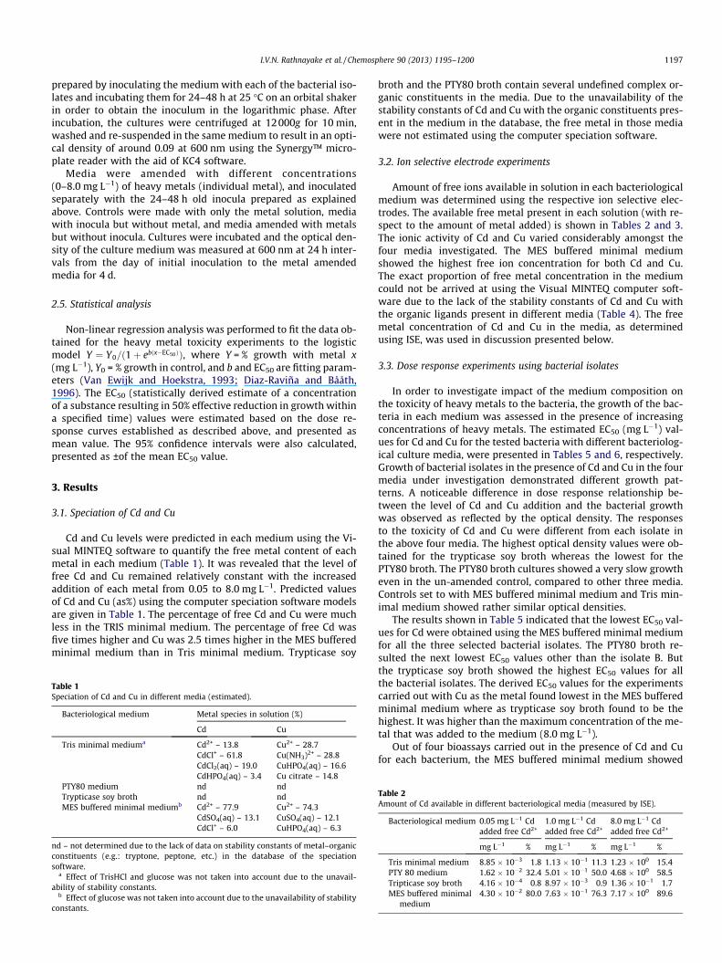
Cd and Cu levels were predicted in each medium using the Vi-sual MINTEQ software to quantify the free metal content of eachmetal in each medium (Table 1). It was revealed that the level offree Cd and Cu remained relatively constant with the increasedaddition of each metal from 0.05 to 8.0 mg L�1. Predicted valuesof Cd and Cu (as%) using the computer speciation software modelsare given in Table 1. The percentage of free Cd and Cu were muchless in the TRIS minimal medium. The percentage of free Cd wasfive times higher and Cu was 2.5 times higher in the MES bufferedminimal medium than in Tris minimal medium. Trypticase soy
Table 1Speciation of Cd and Cu in different media (estimated).
Bacteriological medium Metal species in solution (%)
Cd Cu
Tris minimal mediuma Cd2+ – 13.8 Cu2+ – 28.7CdCl+ – 61.8 Cu(NH3)2+ – 28.8CdCl2(aq) – 19.0 CuHPO4(aq) – 16.6CdHPO4(aq) – 3.4 Cu citrate – 14.8
PTY80 medium nd ndTrypticase soy broth nd ndMES buffered minimal mediumb Cd2+ – 77.9 Cu2+ – 74.3
CdSO4(aq) – 13.1 CuSO4(aq) – 12.1CdCl+ – 6.0 CuHPO4(aq) – 6.3
nd – not determined due to the lack of data on stability constants of metal–organicconstituents (e.g.: tryptone, peptone, etc.) in the database of the speciationsoftware.
a Effect of TrisHCl and glucose was not taken into account due to the unavail-ability of stability constants.
b Effect of glucose was not taken into account due to the unavailability of stabilityconstants.
broth and the PTY80 broth contain several undefined complex or-ganic constituents in the media. Due to the unavailability of thestability constants of Cd and Cu with the organic constituents pres-ent in the medium in the database, the free metal in those mediawere not estimated using the computer speciation software.
3.2. Ion selective electrode experiments
Amount of free ions available in solution in each bacteriologicalmedium was determined using the respective ion selective elec-trodes. The available free metal present in each solution (with re-spect to the amount of metal added) is shown in Tables 2 and 3.The ionic activity of Cd and Cu varied considerably amongst thefour media investigated. The MES buffered minimal mediumshowed the highest free ion concentration for both Cd and Cu.The exact proportion of free metal concentration in the mediumcould not be arrived at using the Visual MINTEQ computer soft-ware due to the lack of the stability constants of Cd and Cu withthe organic ligands present in different media (Table 4). The freemetal concentration of Cd and Cu in the media, as determinedusing ISE, was used in discussion presented below.
3.3. Dose response experiments using bacterial isolates
In order to investigate impact of the medium composition onthe toxicity of heavy metals to the bacteria, the growth of the bac-teria in each medium was assessed in the presence of increasingconcentrations of heavy metals. The estimated EC50 (mg L�1) val-ues for Cd and Cu for the tested bacteria with different bacteriolog-ical culture media, were presented in Tables 5 and 6, respectively.Growth of bacterial isolates in the presence of Cd and Cu in the fourmedia under investigation demonstrated different growth pat-terns. A noticeable difference in dose response relationship be-tween the level of Cd and Cu addition and the bacterial growthwas observed as reflected by the optical density. The responsesto the toxicity of Cd and Cu were different from each isolate inthe above four media. The highest optical density values were ob-tained for the trypticase soy broth whereas the lowest for thePTY80 broth. The PTY80 broth cultures showed a very slow growtheven in the un-amended control, compared to other three media.Controls set to with MES buffered minimal medium and Tris min-imal medium showed rather similar optical densities.
The results shown in Table 5 indicated that the lowest EC50 val-ues for Cd were obtained using the MES buffered minimal mediumfor all the three selected bacterial isolates. The PTY80 broth re-sulted the next lowest EC50 values other than the isolate B. Butthe trypticase soy broth showed the highest EC50 values for allthe bacterial isolates. The derived EC50 values for the experimentscarried out with Cu as the metal found lowest in the MES bufferedminimal medium where as trypticase soy broth found to be thehighest. It was higher than the maximum concentration of the me-tal that was added to the medium (8.0 mg L�1).
Out of four bioassays carried out in the presence of Cd and Cufor each bacterium, the MES buffered minimal medium showed
Table 2Amount of Cd available in different bacteriological media (measured by ISE).
Bacteriological medium 0.05 mg L�1 Cdadded free Cd2+
1.0 mg L�1 Cdadded free Cd2+
8.0 mg L�1 Cdadded free Cd2+
mg L�1 % mg L�1 % mg L�1 %
Tris minimal medium 8.85 � 10�3 1.8 1.13 � 10�1 11.3 1.23 � 100 15.4PTY 80 medium 1.62 � 10�2 32.4 5.01 � 10�1 50.0 4.68 � 100 58.5Tripticase soy broth 4.16 � 10�4 0.8 8.97 � 10�3 0.9 1.36 � 10�1 1.7MES buffered minimal
medium4.30 � 10�2 80.0 7.63 � 10�1 76.3 7.17 � 100 89.6
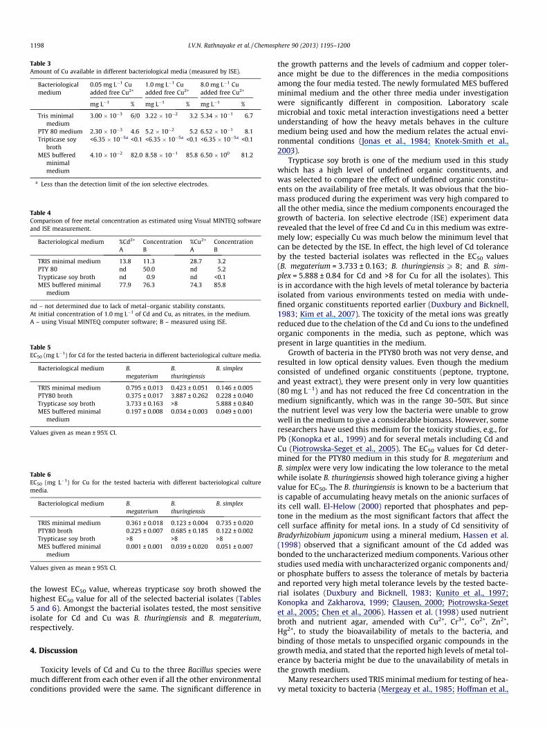
Table 3Amount of Cu available in different bacteriological media (measured by ISE).
Bacteriologicalmedium
0.05 mg L�1 Cuadded free Cu2+
1.0 mg L�1 Cuadded free Cu2+
8.0 mg L�1 Cuadded free Cu2+
mg L�1 % mg L�1 % mg L�1 %
Tris minimalmedium
3.00 � 10�3 6/0 3.22 � 10�2 3.2 5.34 � 10�1 6.7
PTY 80 medium 2.30 � 10�3 4.6 5.2 � 10�2 5.2 6.52 � 10�1 8.1Tripticase soy
broth<6.35 � 10�5a <0.1 <6.35 � 10�5a <0.1 <6.35 � 10�5a <0.1
MES bufferedminimalmedium
4.10 � 10�2 82.0 8.58 � 10�1 85.8 6.50 � 100 81.2
a Less than the detection limit of the ion selective electrodes.
Table 4Comparison of free metal concentration as estimated using Visual MINTEQ softwareand ISE measurement.
Bacteriological medium %Cd2+ Concentration %Cu2+ ConcentrationA B A B
TRIS minimal medium 13.8 11.3 28.7 3.2PTY 80 nd 50.0 nd 5.2Trypticase soy broth nd 0.9 nd <0.1MES buffered minimal
medium77.9 76.3 74.3 85.8
nd – not determined due to lack of metal–organic stability constants.At initial concentration of 1.0 mg L�1 of Cd and Cu, as nitrates, in the medium.A – using Visual MINTEQ computer software; B – measured using ISE.
Table 5EC50 (mg L�1) for Cd for the tested bacteria in different bacteriological culture media.
Bacteriological medium B.megaterium
B.thuringiensis
B. simplex
TRIS minimal medium 0.795 ± 0.013 0.423 ± 0.051 0.146 ± 0.005PTY80 broth 0.375 ± 0.017 3.887 ± 0.262 0.228 ± 0.040Trypticase soy broth 3.733 ± 0.163 >8 5.888 ± 0.840MES buffered minimal
medium0.197 ± 0.008 0.034 ± 0.003 0.049 ± 0.001
Values given as mean ± 95% CI.
Table 6EC50 (mg L�1) for Cu for the tested bacteria with different bacteriological culturemedia.
Bacteriological medium B.megaterium
B.thuringiensis
B. simplex
TRIS minimal medium 0.361 ± 0.018 0.123 ± 0.004 0.735 ± 0.020PTY80 broth 0.225 ± 0.007 0.685 ± 0.185 0.122 ± 0.002Trypticase soy broth >8 >8 >8MES buffered minimal
medium0.001 ± 0.001 0.039 ± 0.020 0.051 ± 0.007
Values given as mean ± 95% CI.
1198 I.V.N. Rathnayake et al. / Chemosphere 90 (2013) 1195–1200
the lowest EC50 value, whereas trypticase soy broth showed thehighest EC50 value for all of the selected bacterial isolates (Tables5 and 6). Amongst the bacterial isolates tested, the most sensitiveisolate for Cd and Cu was B. thuringiensis and B. megaterium,respectively.
4. Discussion
Toxicity levels of Cd and Cu to the three Bacillus species weremuch different from each other even if all the other environmentalconditions provided were the same. The significant difference in
the growth patterns and the levels of cadmium and copper toler-ance might be due to the differences in the media compositionsamong the four media tested. The newly formulated MES bufferedminimal medium and the other three media under investigationwere significantly different in composition. Laboratory scalemicrobial and toxic metal interaction investigations need a betterunderstanding of how the heavy metals behaves in the culturemedium being used and how the medium relates the actual envi-ronmental conditions (Jonas et al., 1984; Knotek-Smith et al.,2003).
Trypticase soy broth is one of the medium used in this studywhich has a high level of undefined organic constituents, andwas selected to compare the effect of undefined organic constitu-ents on the availability of free metals. It was obvious that the bio-mass produced during the experiment was very high compared toall the other media, since the medium components encouraged thegrowth of bacteria. Ion selective electrode (ISE) experiment datarevealed that the level of free Cd and Cu in this medium was extre-mely low; especially Cu was much below the minimum level thatcan be detected by the ISE. In effect, the high level of Cd toleranceby the tested bacterial isolates was reflected in the EC50 values(B. megaterium = 3.733 ± 0.163; B. thuringiensis P 8; and B. sim-plex = 5.888 ± 0.84 for Cd and >8 for Cu for all the isolates). Thisis in accordance with the high levels of metal tolerance by bacteriaisolated from various environments tested on media with unde-fined organic constituents reported earlier (Duxbury and Bicknell,1983; Kim et al., 2007). The toxicity of the metal ions was greatlyreduced due to the chelation of the Cd and Cu ions to the undefinedorganic components in the media, such as peptone, which waspresent in large quantities in the medium.
Growth of bacteria in the PTY80 broth was not very dense, andresulted in low optical density values. Even though the mediumconsisted of undefined organic constituents (peptone, tryptone,and yeast extract), they were present only in very low quantities(80 mg L�1) and has not reduced the free Cd concentration in themedium significantly, which was in the range 30–50%. But sincethe nutrient level was very low the bacteria were unable to growwell in the medium to give a considerable biomass. However, someresearchers have used this medium for the toxicity studies, e.g., forPb (Konopka et al., 1999) and for several metals including Cd andCu (Piotrowska-Seget et al., 2005). The EC50 values for Cd deter-mined for the PTY80 medium in this study for B. megaterium andB. simplex were very low indicating the low tolerance to the metalwhile isolate B. thuringiensis showed high tolerance giving a highervalue for EC50. The B. thuringiensis is known to be a bacterium thatis capable of accumulating heavy metals on the anionic surfaces ofits cell wall. El-Helow (2000) reported that phosphates and pep-tone in the medium as the most significant factors that affect thecell surface affinity for metal ions. In a study of Cd sensitivity ofBradyrhizobium japonicum using a mineral medium, Hassen et al.(1998) observed that a significant amount of the Cd added wasbonded to the uncharacterized medium components. Various otherstudies used media with uncharacterized organic components and/or phosphate buffers to assess the tolerance of metals by bacteriaand reported very high metal tolerance levels by the tested bacte-rial isolates (Duxbury and Bicknell, 1983; Kunito et al., 1997;Konopka and Zakharova, 1999; Clausen, 2000; Piotrowska-Segetet al., 2005; Chen et al., 2006). Hassen et al. (1998) used nutrientbroth and nutrient agar, amended with Cu2+, Cr3+, Co2+, Zn2+,Hg2+, to study the bioavailability of metals to the bacteria, andbinding of those metals to unspecified organic compounds in thegrowth media, and stated that the reported high levels of metal tol-erance by bacteria might be due to the unavailability of metals inthe growth medium.
Many researchers used TRIS minimal medium for testing of hea-vy metal toxicity to bacteria (Mergeay et al., 1985; Hoffman et al.,
I.V.N. Rathnayake et al. / Chemosphere 90 (2013) 1195–1200 1199
2005; Madhaiyan et al., 2007; Abou-Shanab et al., 2007). Accordingto the EC50 data obtained in this study (Tables 5 and 6), it is evidentthat toxicity of Cd and Cu in this medium was high compared toMES buffered medium. It can be due to many reasons. TRIS is thebuffer used in this medium. Even though TRIS is a non-phosphatebuffer, it acts as a non-selective metal binder in the medium. Also,this medium contains a very high level of anions such as chloride(Hughes and Poole, 1991), which can act as interfering ions result-ing in a decrease in the toxicity of cadmium and copper.
MES buffered minimal medium is free from phosphate contain-ing pH buffers, and those have been replaced with MES which is azwitterionic buffer. These zwitterionic buffers have very low inter-ferences with biological processes due to the presence of anionicand cationic sites as non-interacting sulfonate and cationic ammo-nium groups (Sigma–Aldrich, 2008). In their study on complexa-tion of Cu by zwitterionic aminosulfonic buffers, Mash et al.(2003) reported that MES did not bind Cu within their operationalpH range (5.5–6.7). Also, the zwitterionic nature of this buffermakes it more water soluble (Sigma–Aldrich, 2008). The formu-lated medium contained a very low phosphate content(0.01 g L�1) to meet the phosphorus nutrient requirement of thebacteria. Therefore as evident in thermodynamic speciation modelcalculations and ion selective electrode experiment data, highestlevel of free metal ion concentration (both Cd and Cu) is observed(Table 4) in this medium. The presence of high levels of free Cd andCu in this medium, posed the highest toxicity level out of all fourmedia tested. The estimated EC50 values, which were the lowestin the whole experiment (Tables 5 and 6), therefore, proved thatthe high percentage of free Cd and Cu in the medium (Tables 2and 3) reflected in the severe toxicity to the bacterial isolates.
In most of the culture media, sugars (e.g. glucose) have beenused as the carbon source for the growth of bacteria. However, inTRIS minimal medium and MES buffered minimal medium, verylow amounts (0.2%) of glucose were used. Addition of higheramounts of glucose in the media may decrease the toxicity towardsbacteria growing in the culture medium, because of its ability tobind with metals. Brynhildsen et al. (1988) studied the effects ofglucose concentrations on Cd, Cu, Hg, Zn toxicity to Klebsiella spe-cies using 14C-labeled glucose at pH 6.0. In their study they haveinvestigated several carbon concentrations ranging from 0.01 to40 mg L�1, which are equivalent to soluble C concentrations in nat-ural environments. They have found that the toxicity of Cd, Cu andZn to the Klebsiella species was affected considerably by the carbonconcentration.
The ISEs measure only the free metal ion concentration in solu-tion which reflects the truly bioavailable fraction of the solublemetal. The free Cd ion concentration as obtained from ISE experi-ment during this study has a significant relationship with theEC50 values determined based on the metal sensitivity bioassaydata. The log(free Cd conc) and log(free Cu conc) has significantrelationship with the corresponding EC50 values. Log(Cu) = 1.133–0.271 EC50 (r2 = 0.761, P = 0.0002) and log(Cd) = 1.594–0.226 EC50
(r2 = 0.611, P = 0.003).
5. Conclusion
The findings of the current study suggest that the formulatedminimal bacteriological medium (MES buffered minimal medium)is a better alternative for the testing of heavy metal toxicity to bac-teria. Most of the other media used previously by other investiga-tors have significant limitations in determining the heavy metaltoxicity, even though they are being used for the studies. Ionic con-centrations, if measured accurately, can help in assessing toxicityto bacteria and plant roots. Junction potential was not taken intoconsideration in the measurement of ionic concentrations by ISE
in the present study. These results would be more meaningful ifmeasurements are carried out using other techniques such as,polarograph, and using other toxic metals. Amongst the bacterialisolates tested, the most sensitive isolate for Cd and Cu wasB. thuringiensis and B. megaterium, respectively. This new formula-tion with high level of free metal concentration provides a betteroption for the study of interaction between metals and bacteria.
Novelty statement
Most of the growth media used for heavy-metal toxicity to bac-teria contains undefined organic components/high phosphates thatcan chelate test metals thereby overestimate the metal toxicity.Considering these limitations, a new minimal medium with highfree-metal ion-activity was formulated and tested Cu and Cd toxic-ity to bacteria. The free-ionic cadmium and copper concentrationsin the medium gave more accurate determination of metal concen-trations that affects the bacteria, than with most of other existingmedia. This will avoid misleading conclusions using other media,relevant to heavy-metal toxicity to bacteria and provides a betteroption for the study metals–bacteria interactions.
Acknowledgements
This research was supported by the University of SouthAustralia Presidents Scholarship (UPS) in collaboration withCooperative Research Centre for Contamination Assessment andRemediation of the Environment (CRC CARE) Australia PhDfellowship.
References
Abou-Shanab, A.I., van Berkum, P., Angle, J.S., 2007. Heavy metal resistance andgenotypic analysis of metal resistance genes in Gram-positive and Gram-negative bacteria present in Ni-rich serpentine soil and in the rhizosphere ofAlyssum murale. Chemosphere 68, 360–367.
Amoroso, M.J., Oliver, G., Castro, G.R., 2002. Estimation of growth inhibition bycopper and cadmium in heavy metal tolerant actinomycetes. J. Basic Microbiol.42, 231–237.
Angle, J.S., Chaney, R.L., 1989. Cadmium resistance screening in nitrilotriacetic-buffered minimal media. Appl. Environ. Microbiol. 55, 2101–2104.
Babich, H., Stotzky, G., 1977. Sensitivity of various bacteria, includingactinomycetes, and fungi to cadmium and the influence of pH on sensitivity.Appl. Environ. Microbiol. 33, 681–695.
Biebl, H., Pfenning, N., 1981. Isolation of members of the family Rhodospirillaceae.In: Starr, M.P., Stolp, J., Truper, H.G., Balows, A., Schegel, H.G. (Eds.), TheProkaryotes, Handbook on Habitats, Isolation and Identification of Bacteria, vol.1. Springer-Verlag, Berlin, pp. 267–273.
Brynhildsen, L., Lundgren, B.V., Allard, B., Rosswall, T., 1988. Effects of glucoseconcentrations on cadmium, copper, mercury, and zinc toxicity to Klebsiella sp.Appl. Environ. Microbiol. 54, 1689–1693.
Campbell, P.G.C., 1995. Interactions between trace metals and organisms: critiqueof the free-ion activity model. In: Tessier, A., Turner, D.R. (Eds.), MetalSpeciation and Bioavailability in Aquatic Systems. John Wiley & Sons, NewYork, pp. 45–102.
Chen, B.Y., Wu, C.H., Chang, J.S., 2006. An assessment of the toxicity of metals toPseudomonas aeruginosa PU21 (Rip64). Biores. Technol. 97, 1880–1886.
Clausen, C.A., 2000. Isolating metal-tolerant bacteria capable of removing copper,chromium, and arsenic from treated wood. Waste Manage. Res. 18, 264–268.
Congeevaram, S., Dhanarani, J., Park, M., Dexilin, K., Thamaraiselvi, K., 2007.Biosorption of chromium and nickel by heavy metal resistant fungal andbacterial isolates. J. Hazard. Mater. 146, 270–277.
Diaz-Raviña, M., Bååth, E., 1996. Development of metal tolerance in soil bacterialcommunities exposed to experimentally increased metal levels. Appl. Environ.Microbiol. 62, 2970–2977.
Duxbury, T., Bicknell, B., 1983. Metal tolerant bacterial populations from naturaland polluted soils. Soil Biol. Biochem. 15, 243–250.
El-Aziz, R., Angle, J.S., Chaney, R.L., 1991. Metal tolerance of Rhizobium melilotiisolated from heavy-metal contaminated soil. Soil Biol. Biochem. 23, 795–798.
El-Helow, E.R., 2000. Cadmium biosorption by a cadmium resistant strain of Bacillusthuringiensis: regulation and optimization of cell surface affinity for metalcations. Biometals 13, 273–280.
Farrell, R.E., Germida, J.J., Huang, P.M., 1993. Effects of chemical speciation ingrowth media on the toxicity of mercury(II). Appl. Environ. Microbiol. 59, 1507–1514.
Good, N.E., Izawa, S., 1972. Hydrogen ion buffers. Methods Enzymol. 24, 53–68.
1200 I.V.N. Rathnayake et al. / Chemosphere 90 (2013) 1195–1200
Good, N.E., Winget, G.D., Winter, W., Connolly, T.N., Izawa, S., Singh, R.M.M., 1996.Hydrogen ion buffers for biological research. Biochemistry 5, 467–477.
Gustafsson, J.P., 2005. Visual MINTEQ 2.32. <http://www.lwr.kth.se/English/OurSoftware/vminteq/index.htm>.
Hassen, A., Saidi, N., Cherif, M., Boudabous, A., 1998. Resistance of environmentalbacteria to heavy metals. Biores. Technol. 64, 7–15.
Hoffman, D.R., Okon, J.L., Sandrin, T.R., 2005. Medium composition affects thedegree and pattern of cadmium inhibition of naphthalene biodegradation.Chemosphere 59, 919–927.
Hughes, M.N., Poole, P.K., 1991. Metal speciation and microbial growth-the hard(and soft) facts. J. Gen. Microbiol. 137, 725–734.
Jonas, R.B., Gilmour, C.C., Stoner, D.I., Weir, M.M., Tuttle, J.H., 1984. Comparison ofmethods to measure acute metal and organometal toxicity to natural aquaticmicrobial communities. Appl. Environ. Microbiol. 47, 1005–1011.
Keeling, A.A., Cater, G.I.F., 1998. Toxicity of copper, lead, nickel and zinc in agarculture to aerobic, diazotrophic bacteria extracted from waste-derived compost.Chemosphere 37, 1073–1077.
Kim, S.U., Cheong, Y.H., Seo, D.C., Hur, J.S., Heo, J.S., Cho, J.S., 2007. Characterisationof heavy metal tolerance and biosorption capacity of bacterium strain CPB4(Bacillus spp.). Water Sci. Technol. 55, 105–111.
Knotek-Smith, H.M., Deobald, L.A., Ederer, M., Crawford, D.L., 2003. Cadmium stressstudies; media development, enrichment, consortia analysis, andenvironmental relevance. BioMetals 16, 251–261.
Konopka, A., Zakharova, T., 1999. Quantification of bacterial lead resistance viaactivity assays. J. Microbiol. Methods 37, 17–22.
Konopka, A., Zakharova, T., Bischoff, M., Oliver, L., Nakatsu, C., Turco, R.F., 1999.Microbial biomass and activity in lead-contaminated soil. Appl. Environ.Microbiol. 65, 2256–2259.
Kunito, T., Nagaoka, K., Tada, N., Saeki, K., Senoo, K., Oyaizu, H., Matsumoto, S., 1997.Characterization of Cu-resistant bacterial communities in Cu-contaminatedsoils. Soil Sci. Plant Nutr. 43, 709–717.
Legatzki, A., Franke, S., Lucke, S., Hoffmann, T., Anton, A., Neumann, D., Nies, D.H.,2003. First step towards a quantitative model describing Czc-mediated heavymetal resistance in Ralstonia metallidurans. Biodegradation 14, 153–168.
Madhaiyan, M., Poonguzhali, S., Sa, T., 2007. Metal tolerating methylotrophicbacteria reduces nickel and cadmium toxicity and promotes plant growth oftomato (Lycopersicon esculentum L.). Chemosphere 69, 220–228.
Malik, A., Jaiswal, R., 2000. Metal resistance in Pseudomonas strains isolated fromsoil treated with industrial wastewater. World J. Microbiol. Biotechnol. 16, 177–182.
Martensson, A.M., Torstensson, I., 1996. Monitoring sewage sludge usingheterotrophic nitrogen fixing microorganisms. Soil Biol. Biochem. 28, 1621–1630.
Mash, H.E., Chin, Y., Sigg, L., Hari, R., Xue, H., 2003. Complexation of copper byzwitterionic aminosulfonic (good) buffers. FEMS Microbiol. Ecol. 75, 671–677.
Megharaj, M., Avudainayagam, S., Naidu, R., 2003. Toxicity of hexavalent chromiumand its reduction by bacteria isolated from soil contaminated with tannerywaste. Curr. Microbiol. 47, 51–54.
Mergeay, M., 1995. Heavy metal resistances in microbial ecosystems. In: AkkermansA.D.L., van Elsas, J.D., de Bruij, F.J. (Eds.), Molecular Microbial Ecology Manual.Kluwer Academic, Dordrecht, Boston, pp. 1–17.
Mergeay, M., Nies, D., Schlegel, H.G., Gerits, J., Charles, P., Van Gijsegem, F., 1985.Alcaligenes eutrophus CH34 is a facultative chemolithotroph with plasmid-bound resistance to heavy metals. J. Bacteriol. 162, 328–334.
Miranda, C.D., Rojas, R., 2006. Copper accumulation by bacteria and transfer toscallop larvae. Mar. Pollut. Bull. 52, 293–300.
NEPM (National Environmental Protection) Assessment of Site ContaminationMeasure, 1999. <http://www.ephc.gov.au/pdf/cs/csmeasure.pdf>.
Oliveira, A., Pampulha, M.E., 2006. Effects of long-term heavy metal contaminationon soil microbial characteristics. J. Biosci. Bioeng. 102, 157–161.
Piotrowska-Seget, Z., Cycon, M., Kozdroj, J., 2005. Metal-tolerant bacteria occurringin heavily polluted soil and mine spoil. Appl. Soil Ecol. 28, 237–246.
Rajapaksha, R.M.C.P., Tobor-Kaplon, M.A., Bååth, E., 2004. Metal toxicity affectsfungal and bacterial activities in soil differently. Appl. Environ. Microbiol. 70,2966–2973.
Renella, G., Ortigoza, A.L.R., Landi, L., Nannipieri, P., 2003. Additive effects of copperand zinc on cadmium toxicity on phosphate activities and ATP content of soil asestimated by the ecological dose (ED50). Soil Biol. Biochem. 35, 1203–1210.
Richards, J.W., Krumholz, G.D., Chval, M.S., Tisa, L.S., 2002. Heavy metal resistancepatterns of Frankia strains. Appl. Environ. Microbiol. 68, 923–927.
Riether, K.B., Dollard, M.A., Billard, P., 2001. Assessment of heavy metalbioavailability using Escherichia coli zntAp::lux and copAp::lux-basedbiosensors. Appl. Microbiol. Biochem. 57, 712–716.
Roane, T.M., Pepper, I.L., 2000. Microorganisms and metal pollutants. In: Roane,T.M., Gerba, C.P., Pepper, I.L. (Eds.), Environmental Microbiology. AcademicPress, San Diego, California, London, pp. 403–423.
Roane, T.M., Pepper, I.L., 1999. Microbial responses to environmentally toxiccadmium. Microbial. Ecol. 38, 358–364.
Rubikas, J., Matulis, D., Leipus, A., Urbaitiene, D., 1997. Nickel resistance inEscherichia coli V38 is dependent on the concentration used for induction.FEMS Microbiol. Lett. 155, 193–198.
Saeki, K., Kunito, T., Oyaizu, H., Matsumoto, M., 2002. Relationships betweenbacterial tolerance and forms of copper and zinc in soils. J. Environ. Qual. 31,1570–1575.
Sigma–Aldrich, 2008. Preparation of culture media, Microbiology. <http://www.sigmaaldrich.com/Area_of_Interest/Biochemicals/BioUltra/Biological_Buffers_html>.
Sprocati, A.R., Alisi, C., Segre, L., Tasso, F., Galletti, M., Cremisini, C., 2006.Investigating heavy metal resistance, bioaccumulation and metabolic profileof a metallophile microbial consortium native to an abandoned mine. Sci. TotalEnviron. 366, 649–658.
Tangaromsuk, J., Pokethitiyook, P., Kruatrachue, M., Upatham, E.S., 2002. Cadmiumbiosorption by Sphingomonas paucimobilis biomass. Biores. Technol. 85, 103–105.
Teitzel, G.M., Parsek, M.R., 2003. Heavy metal resistance of biofilm and planktonicPseudomonas aeruginosa. Appl. Environ. Microbiol. 69, 2313–2320.
Traina, S.J., Laperche, V., 1999. Contaminant bioavailability in soil, sediments, andaquatic environments. Proc. Natl. Acad. Sci. 96, 3365–3371.
Twiss, M.R., Errecalde, O., Fortin, C., Campbell, P.G.C., Jumarie, C., Denizeau, F.,Berkelaar, E., Hale, B., van Rees, K., 2001. Coupling the use of computer chemicalspeciation models and culture techniques in laboratory investigations of tracemetal toxicity. Chem. Spec. Bioavail. 13, 9–24.
Van Ewijk, P.H., Hoekstra, J.A., 1993. Calculation of the EC50 and its confidenceinterval when subtoxic stimulus is present. Ecotoxicol. Environ. Saf. 25, 25–32.
Yilmaz, E.I., 2003. Metal tolerance and biosorption capacity of Bacillus circulansstrain EB1. Res. Microbiol. 154, 409–415.
Zhang, Y., Zhang, Z., Suzuki, K., Maekawa, T., 2003. Uptake and mass balance of tracemetals for methane producing bacteria. Biomass Bioenergy 25, 427–443.